subdivision of lectures - gmch.gov.in lectures/ophthalmology/1 conjunctiva.pdf · bulbar...
TRANSCRIPT
Dr Parul IchhpujaniAssistant ProfessorDeptt. Of Ophthalmology,Government Medical College and Hospital, Sector 32, Chandigarh

Subdivision of LecturesAPPLIED ANATOMY Parts Structure GlandsSYMPTOMATIC CONDITIONS Hyperaemia Chemosis Ecchymosis Xerosis DiscolorationDEGENERATIVE CONDITIONS Pinguecula Pterygium Concretions
INFLAMMATIONS OF CONJUNCTIVA
Infective conjunctivitis – Bacterial – Chlamydial – Viral Allergic conjunctivitis Granulomatous conjunctivitis
CYSTS AND TUMOURS
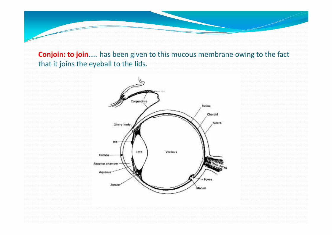
Conjoin: to join….. has been given to this mucous membrane owing to the fact that it joins the eyeball to the lids.
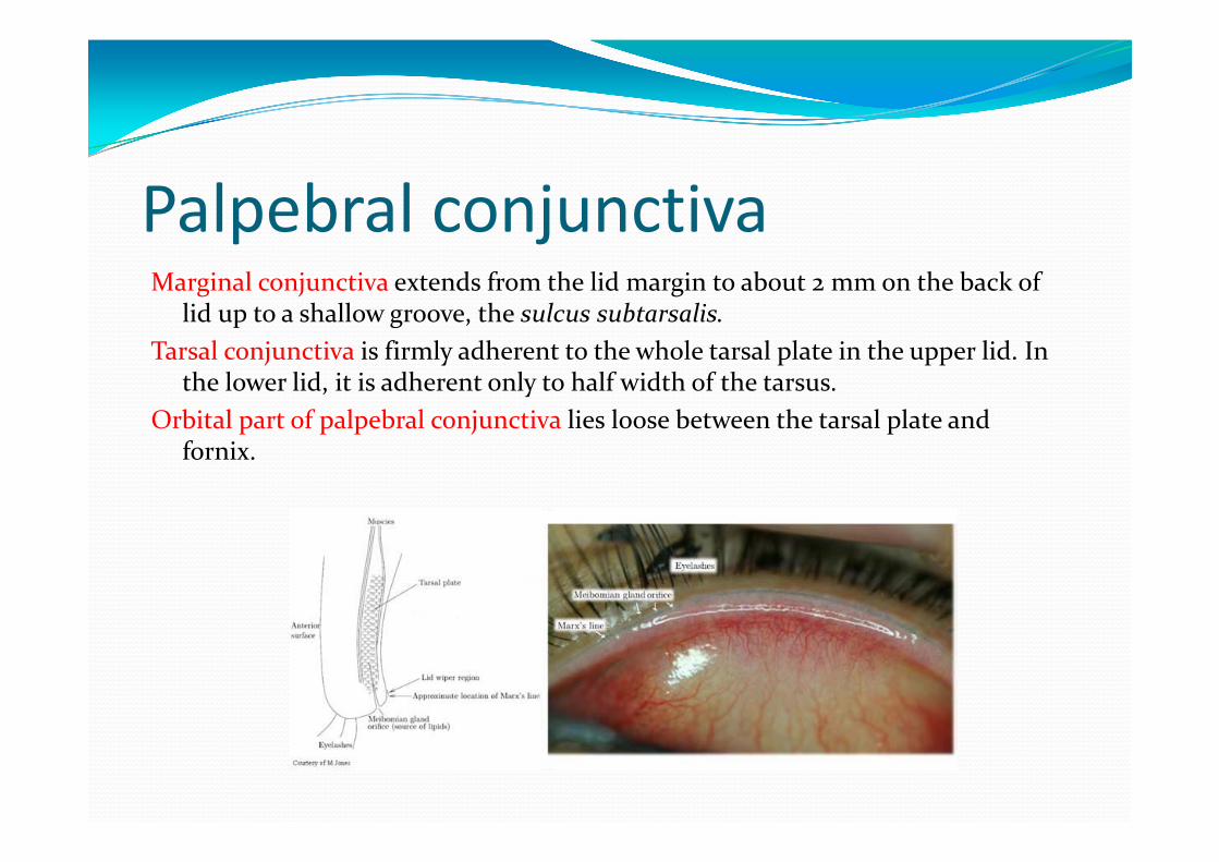
Palpebral conjunctivaMarginal conjunctiva extends from the lid margin to about 2 mm on the back of
lid up to a shallow groove, the sulcus subtarsalis.Tarsal conjunctiva is firmly adherent to the whole tarsal plate in the upper lid. In
the lower lid, it is adherent only to half width of the tarsus.Orbital part of palpebral conjunctiva lies loose between the tarsal plate and
fornix.
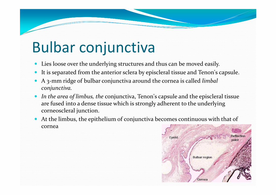
Bulbar conjunctiva Lies loose over the underlying structures and thus can be moved easily. It is separated from the anterior sclera by episcleral tissue and Tenon's capsule. A 3‐mm ridge of bulbar conjunctiva around the cornea is called limbal
conjunctiva. In the area of limbus, the conjunctiva, Tenon's capsule and the episcleral tissue
are fused into a dense tissue which is strongly adherent to the underlying corneoscleral junction.
At the limbus, the epithelium of conjunctiva becomes continuous with that of cornea
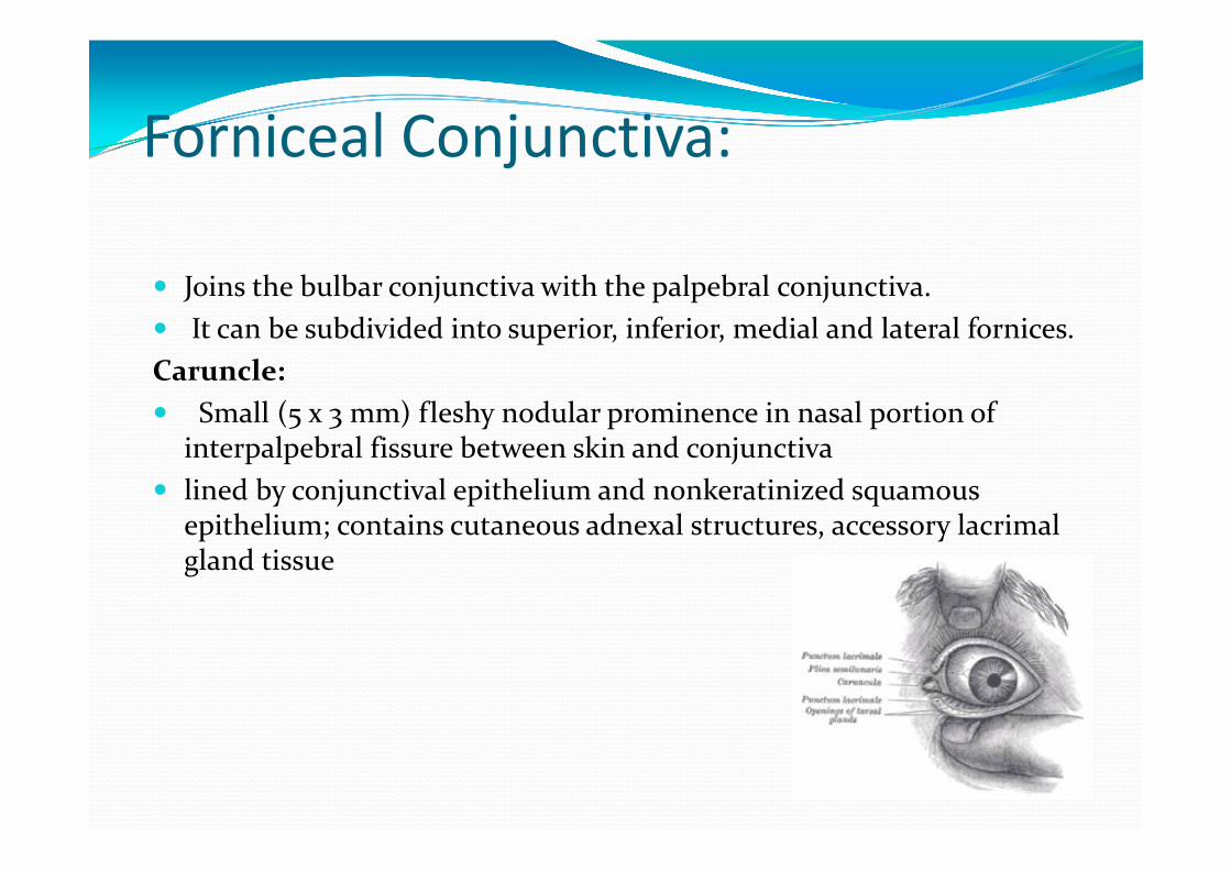
Forniceal Conjunctiva:
Joins the bulbar conjunctiva with the palpebral conjunctiva. It can be subdivided into superior, inferior, medial and lateral fornices.Caruncle: Small (5 x 3 mm) fleshy nodular prominence in nasal portion of
interpalpebral fissure between skin and conjunctiva lined by conjunctival epithelium and nonkeratinized squamous
epithelium; contains cutaneous adnexal structures, accessory lacrimalgland tissue
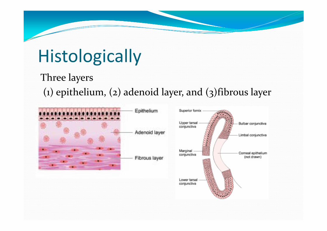
HistologicallyThree layers(1) epithelium, (2) adenoid layer, and (3)fibrous layer
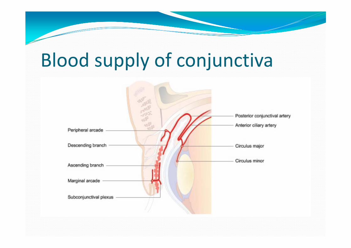
Blood supply of conjunctiva
Venous drainage:Drain into the venous plexus of eyelids and some around the cornea into the anterior ciliary veins.
Nerve supply of conjunctiva
A circumcorneal zone of conjunctiva is supplied bythe branches from long ciliary nerves which supplythe cornea. Rest of the conjunctiva is supplied by the branches from lacrimal, infratrochlear, supratrochlear, supraorbital and frontal nerves.
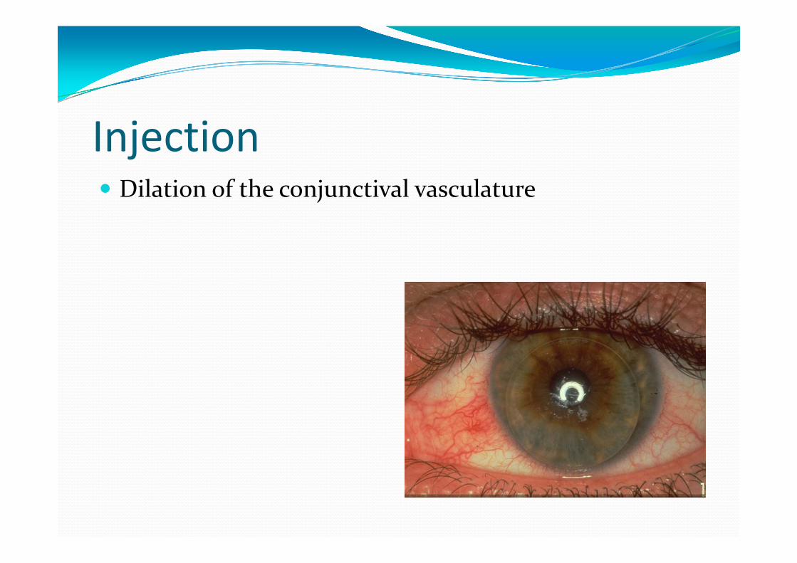
Injection Dilation of the conjunctival vasculature
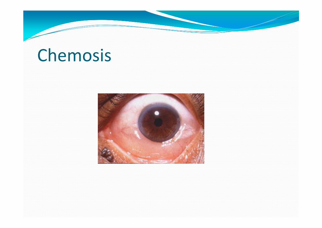
Chemosis
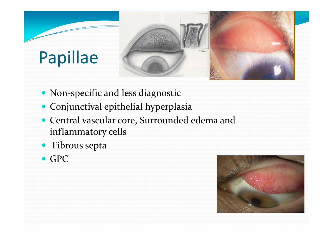
Papillae
Non‐specific and less diagnostic Conjunctival epithelial hyperplasia Central vascular core, Surrounded edema and inflammatory cells
Fibrous septa GPC
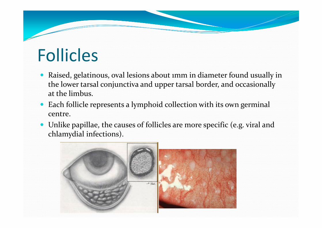
Follicles Raised, gelatinous, oval lesions about 1mm in diameter found usually in
the lower tarsal conjunctiva and upper tarsal border, and occasionally at the limbus.
Each follicle represents a lymphoid collection with its own germinal centre.
Unlike papillae, the causes of follicles are more specific (e.g. viral and chlamydial infections).
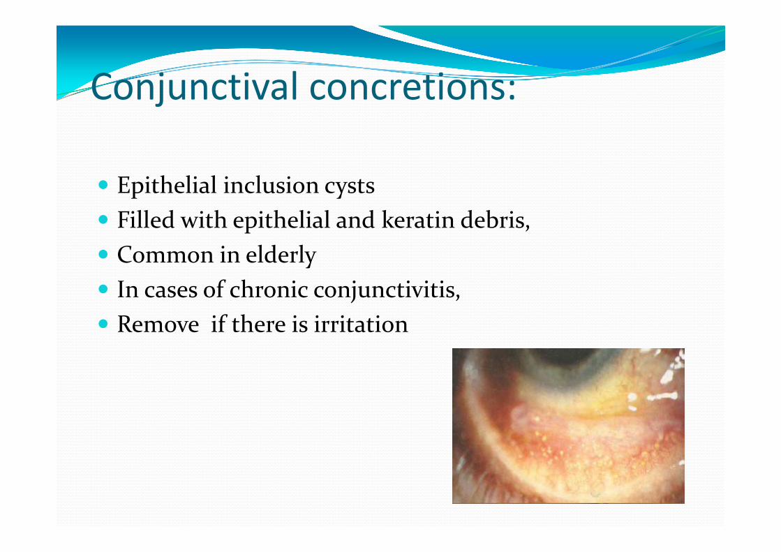
Conjunctival concretions:
Epithelial inclusion cysts Filled with epithelial and keratin debris, Common in elderly In cases of chronic conjunctivitis, Remove if there is irritation
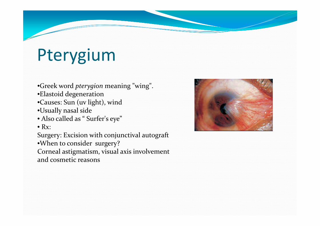
Pterygium•Greek word pterygion meaning "wing".•Elastoid degeneration•Causes: Sun (uv light), wind•Usually nasal side• Also called as “ Surfer's eye”• Rx:Surgery: Excision with conjunctival autograft•When to consider surgery?Corneal astigmatism, visual axis involvement and cosmetic reasons
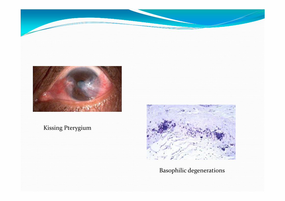
Basophilic degenerations
Kissing Pterygium
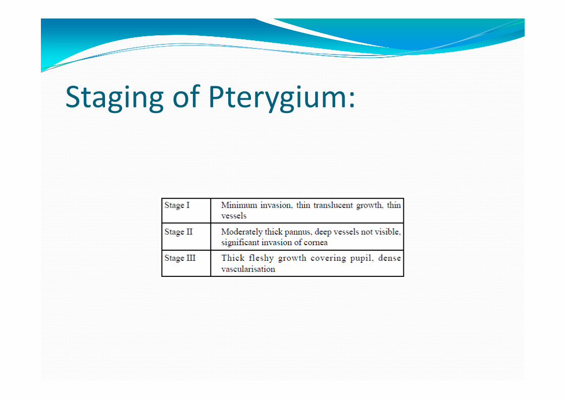
Staging of Pterygium:

Pinguecula
Benign Composed of collagen and elastin
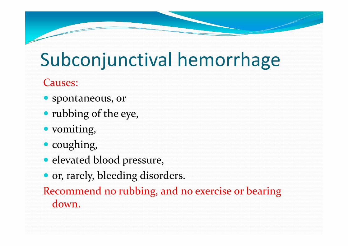
Subconjunctival hemorrhage Causes: spontaneous, or rubbing of the eye, vomiting, coughing, elevated blood pressure, or, rarely, bleeding disorders. Recommend no rubbing, and no exercise or bearing down.
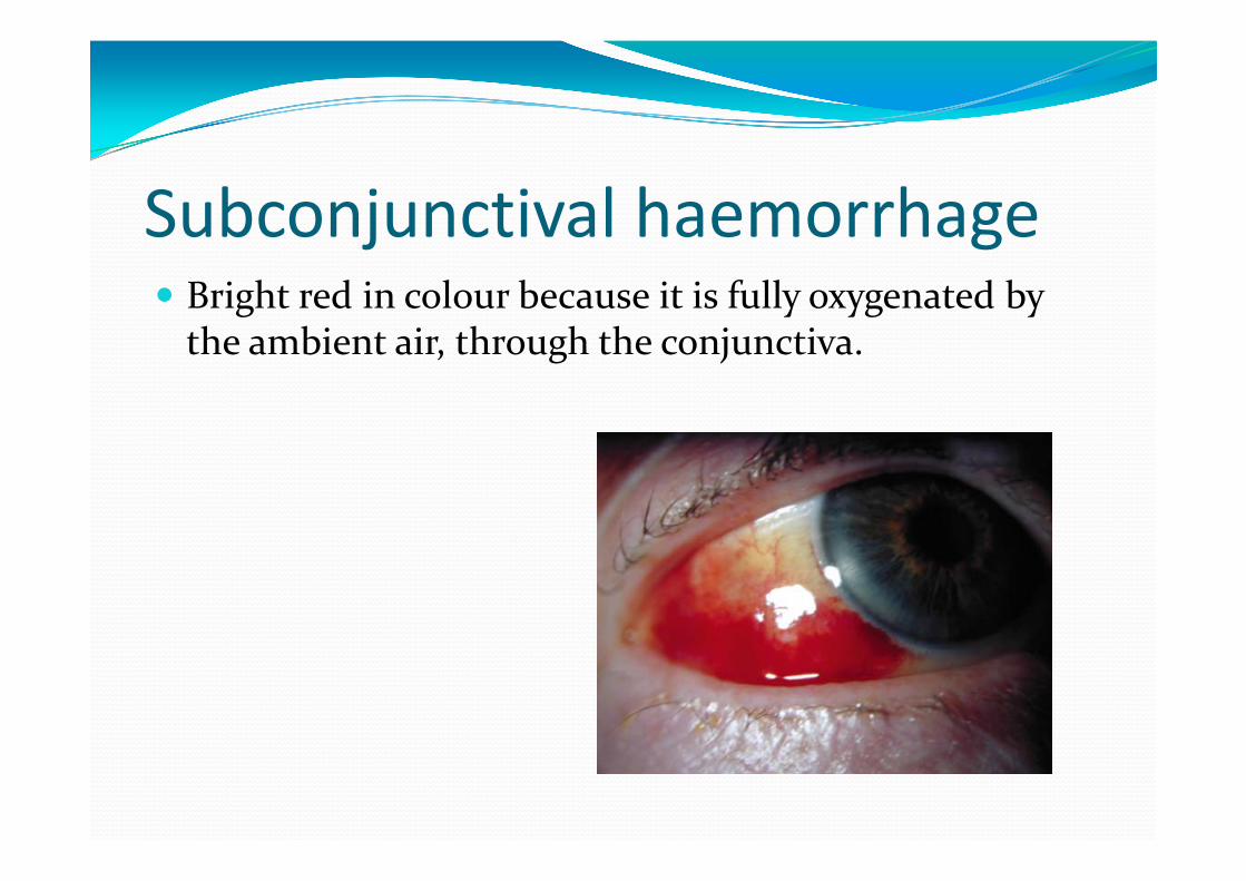
Subconjunctival haemorrhage Bright red in colour because it is fully oxygenated by the ambient air, through the conjunctiva.
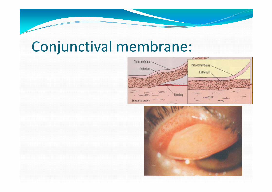
Conjunctival membrane:
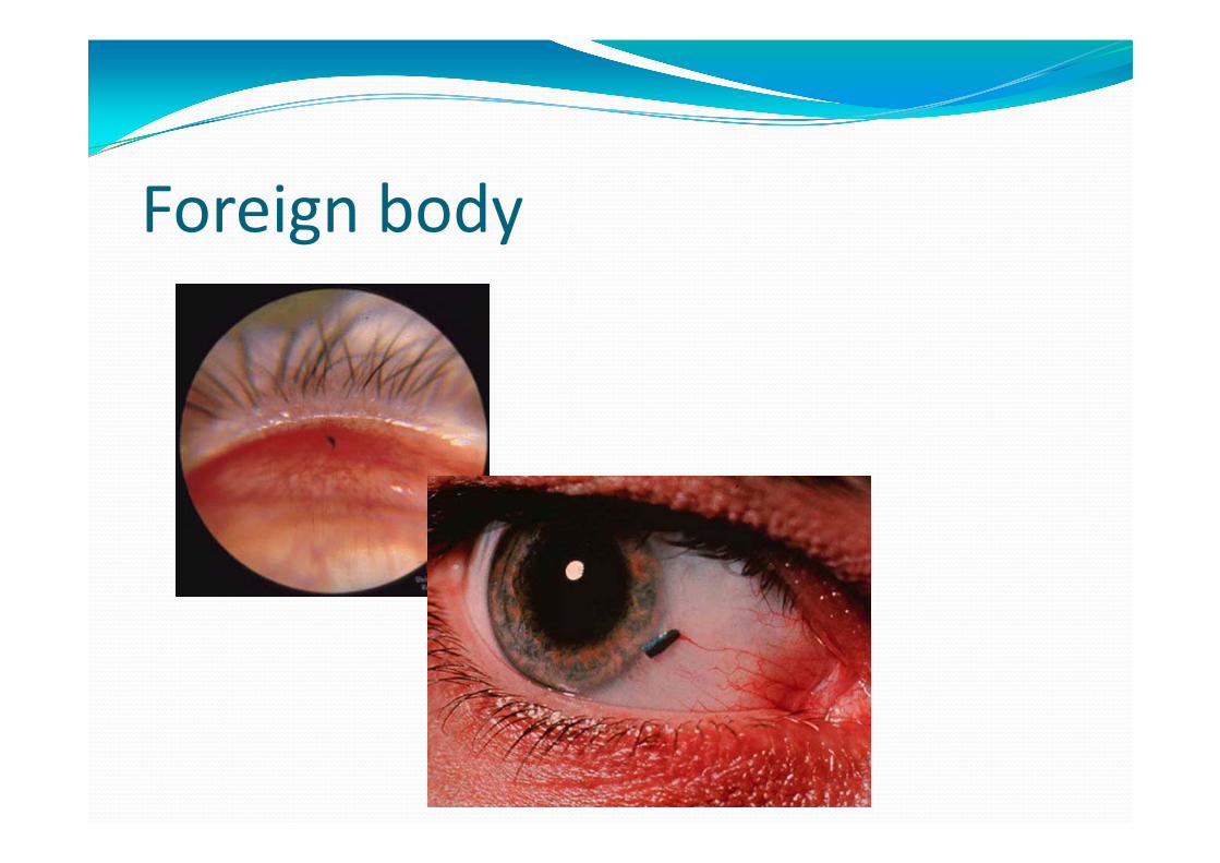
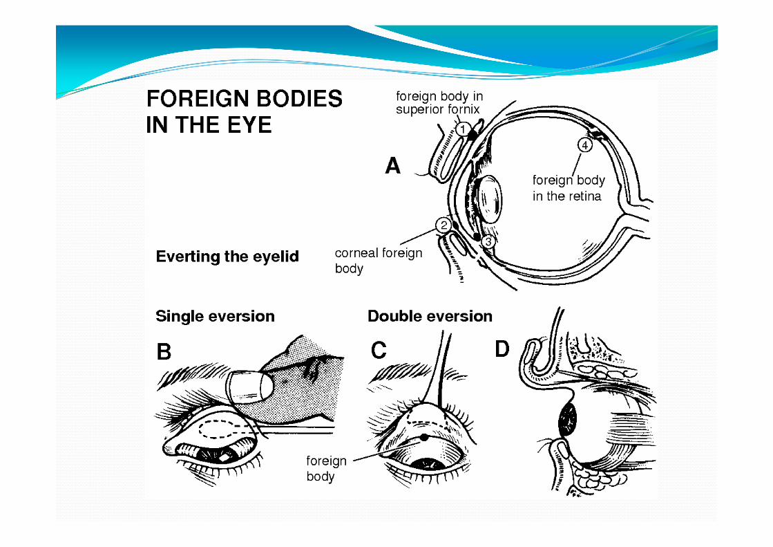
Foreign body

Conjunctival Xerosis:
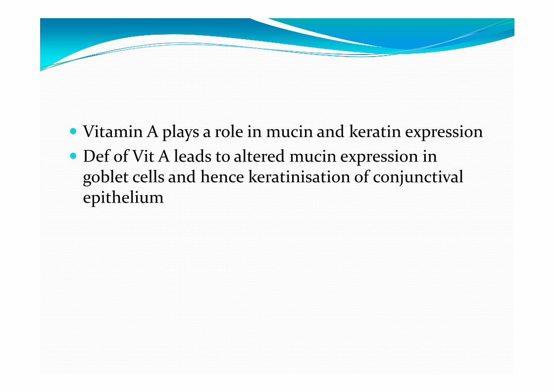
Vitamin A plays a role in mucin and keratin expression Def of Vit A leads to altered mucin expression in goblet cells and hence keratinisation of conjunctivalepithelium