suberoylanilide hydroxamic acid, a histone deacetylase ...hydroxamic acid, a histone deacetylase...
TRANSCRIPT
ORIGINAL RESEARCHpublished: 23 September 2015
doi: 10.3389/fnmol.2015.00052
Suberoylanilide hydroxamic acid, ahistone deacetylase inhibitor,attenuates postoperative cognitivedysfunction in aging miceMin Jia 1, Wen-Xue Liu 1, He-Liang Sun 1, Yan-Qing Chang 2, Jiao-Jiao Yang 1,3,4,Mu-Huo Ji 1, Jian-Jun Yang 1,3,4* and Chen-Zhuo Feng 2*
1 Department of Anesthesiology, Jinling Hospital, School of Medicine, Nanjing University, Nanjing, China, 2 Institute of AgingResearch, School of Medicine, Hangzhou Normal University, Hangzhou, China, 3 Jiangsu Province Key Laboratoryof Anesthesiology, Xuzhou Medical College, Xuzhou, China, 4 Jiangsu Province Key Laboratory of Anesthesia and AnalgesiaApplication Technology, Xuzhou, China
Edited by:Benedict C. Albensi,
University of Manitoba, Canada
Reviewed by:Mary M. Torregrossa,
University of Pittsburgh, USAMauro Costa-Mattioli,
Baylor College of Medicine, USA
*Correspondence:Jian-Jun Yang,
Department of Anesthesiology,Jinling Hospital, School of Medicine,
Nanjing University,305 East Zhongshan Road,
Nanjing 210002, [email protected];Chen-Zhuo Feng,
Institute of Aging Research,School of Medicine,
Hangzhou Normal University,58 Haishu Road, Yuhang District,
Hangzhou 311121, [email protected]
Received: 25 June 2015Accepted: 28 August 2015
Published: 23 September 2015
Citation:Jia M, Liu W-X, Sun H-L, Chang Y-Q,
Yang J-J, Ji M-H, Yang J-J andFeng C-Z (2015) Suberoylanilide
hydroxamic acid, a histonedeacetylase inhibitor, attenuates
postoperative cognitive dysfunction inaging mice.
Front. Mol. Neurosci. 8:52.doi: 10.3389/fnmol.2015.00052
Postoperative cognitive dysfunction (POCD) is a recognized clinical entity characterizedwith cognitive deficits after anesthesia and surgery, especially in aged patients.Previous studies have shown that histone acetylation plays a key role in hippocampalsynaptic plasticity and memory formation. However, its role in POCD remains tobe determined. Here, we show that suberoylanilide hydroxamic acid (SAHA), ahistone deacetylase inhibitor, attenuates POCD in aging Mice. After exposed tothe laparotomy, a surgical procedure involving an incision into abdominal walls toexamine the abdominal organs, 16- but not 3-month old male C57BL/6 micedeveloped obvious cognitive impairments in the test of long-term contextual fearconditioning. Intracerebroventricular (i.c.v.) injection of SAHA at the dose of (20 µg/2µl) 3 h before and daily after the laparotomy restored the laparotomy-inducedreduction of hippocampal acetyl-H3 and acetyl-H4 levels and significantly attenuatedthe hippocampus-dependent long-term memory (LTM) impairments in 16-month oldmice. SAHA also reduced the expression of cleaved caspase-3, inducible nitricoxide synthase (iNOS) and N-methyl-D-aspartate (NMDA) receptor-calcium/calmodulindependent kinase II (CaMKII) pathway, and increased the expression of brain-derivedneurotrophic factor (BDNF), synapsin 1, and postsynaptic density 95 (PSD95). Takentogether, our data suggest that the decrease of histone acetylation contributesto POCD and may serve as a target to improve the neurological outcomeof POCD.
Keywords: postoperative cognitive dysfunction, aging, histone acetylation, neuroapoptosis, synaptic plasticity
Abbreviations: POCD, postoperative cognitive dysfunction; HATs, histone acetyltransferases; HDACs, histone deacetylases;SAHA, suberoylanilide hydroxamic acid; DMSO, dimethylsulfoxide; iNOS, inducible nitric oxide synthase; BDNF, brain-derived neurotrophic factor; PSD95, postsynaptic density 95; NMDA, N-methyl-D-aspartate; CaMKII, calcium/calmodulindependent kinase II; HE staining, Hematoxylin-eosin staining; PBS, phosphate-buffered saline; PFA, phosphate-bufferedparaformaldehyde; STM, short-term memory; LTM, long-term memory; TBST, Tris-Buffered Saline Tween; Ac-H3K9,histone H3(acetyl K9); Ac-H3K14, histone H3(acetyl K14); Ac-H4K5, histone H4 (acetyl K5); Ac-H4K12, histone H4 (acetylK12); Ac-H3, acetyl histone H3; Ac-H4, acetyl histone H4 (Lys5/8/12/16); GAPDH, genes glyceraldehyde-3-phosphatedehydrogenase; real-time PCR, real-time polymerase chain reaction; AD, Alzheimer’s disease; LTD, long-term depression;LTP, long-term potentiation.
Frontiers in Molecular Neuroscience | www.frontiersin.org 1 September 2015 | Volume 8 | Article 52
Jia et al. SAHA attenuates POCD
Introduction
Postoperative cognitive dysfunction (POCD) is a cognitiveprogressive deterioration in memory and concentrationfollowing exposure to anesthesia and surgery (Amar et al., 1998;Terrando et al., 2011; Hovens et al., 2012). These cognitivedeficits result in prolonged hospitalization and decreasedquality of life (Moller et al., 1998). Tissue damage inducedneuro-inflammation and altered reactivity of the immunesystem after operation are considered to play a major rolein the development of POCD, which elicits neuron damages,affects synaptic function, and thereby induces cognitiveimpairments (Wan et al., 2007; Fidalgo et al., 2011). However,the molecular mechanisms underlying POCD remain largely tobe determined.
Epigenetic dysregulation on the expression of key genesis widely involved in the etiology of brain disorders,including Alzheimer’s disease (AD), Huntington’s disease,Parkinson’s disease, and Rubinstein-Taybi syndrome (Petrijet al., 1995; Kazantsev and Thompson, 2008; Chuang et al.,2009; Francis et al., 2009; Peleg et al., 2010; Gräff and Tsai,2013). Histone acetylation is one of the most commonforms of epigenetic modification, which is controlled bythe balance between histone acetyltransferases (HATs) andhistone deacetylases (HDACs; Fischer et al., 2010; Haggartyand Tsai, 2011; McQuown et al., 2011). In general, histoneacetylation facilitates gene transcription, whereas histonedeacetylation results in gene silencing (Fischer et al., 2010;Haggarty and Tsai, 2011; McQuown et al., 2011). A substantialbody of evidence suggests that the dysregulation of histoneacetylation contributes to the pathogenesis of neurodegenerativediseases, and targeted restoration of histone acetylationby HDAC inhibitors shows neuroprotective effects onneurodegenerative diseases (Petrij et al., 1995; Dash et al.,2010; Kilgore et al., 2010; Haettig et al., 2011; Ji et al.,2014).
The similar clinical symptoms has been revealed betweenPOCD and neurodegenerative disorders (Wang et al., 2013;Luo et al., 2014; Xu et al., 2014). However, comparing withthe studies of neurodegenerative diseases, the potential functionof histone acetylation in POCD remains primarily unknown.Therefore, based on the pre-clinical animal mode of thelaparotomy-induced cognitive deficits (Rosczyk et al., 2008;Barrientos et al., 2012; Hovens et al., 2014), which surgicalprocedure involving an incision into the abdominal wall toexamine the abdominal organs, we investigated the role ofhistone acetylation and potential therapeutic effect of an HDACinhibitor, suberoylanilide hydroxamic acid (SAHA), on POCD.
Materials and Methods
AnimalsAll animal experiments were carried out in accordance withthe National Institutes of Health Guide for the Care and Useof Laboratory Animals, USA. The study protocol was approvedby the Institutional Animal Care and Use Ethics Committee,Jinling Hospital, Nanjing University, Nanjing, China. The
mice were purchased from The Animal Center of JinlingHospital, Nanjing, China and efforts were made to minimizethe number of animals used and their suffering. The micewere housed under specific pathogen-free conditions in atemperature-controlled room of 23 ± 1◦C on a 12-h light-darkcycle, with ad libitum access to food and water. Mice wereallowed 7 days to acclimate to the laboratory conditions beforeexperiments.
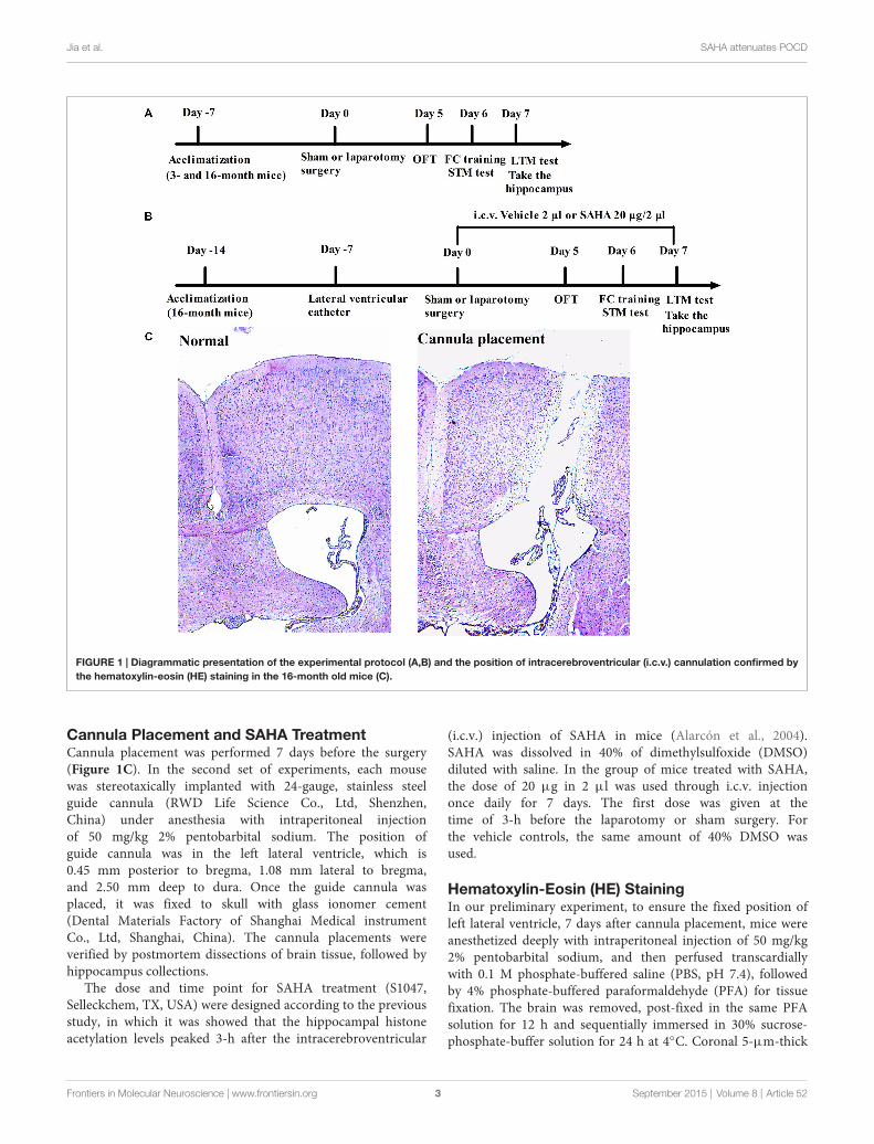
Study Groups of AnimalsIn the first set of experiments, the 3- and 16-month old maleC57BL/6 mice were used. Thirty-two 3-month old mice weighing25–32 g and thirty-two 16-month old mice weighing 33–40 gwere randomly assigned to receiving laparotomy or sham surgery(n = 16 for each group). The experimental protocol was presentedin Figure 1A.
In the second set of experiments with SAHA (a histonedeacetylase inhibitor) treatment, sixty-four 16-month old malemice weighing 33–40 g were randomly assigned to the followingfour groups: Sham + Vehicle group, mice received vehicletreatment and sham surgery; Sham + SAHA group, mice receivedSAHA treatment and sham surgery; Laparotomy + Vehiclegroup, mice received vehicle treatment and laparotomy; andLaparotomy + SAHA group, mice received SAHA treatment andlaparotomy (n = 16 for each group). The experimental protocolwas presented in Figure 1B.
SurgeryThe Laparotomy or sham surgery was performed as previouslydescribed (Rosczyk et al., 2008; Barrientos et al., 2012; Hovenset al., 2014). Anesthesia was induced with 1.5% isofluranein 100% oxygen in mice and was still maintained with 1.5%isoflurane bymice anesthesiamask during the surgery procedure.After the abdominal region of mice was shaved and cleaned withiodophor disinfectant, a 1.5 cm vertical incision, approximately0.5 cm below the lower right rib, was created. The viscera andmusculature were vigorously manipulated by inserting a sterileprobe into the body cavity and stretching the musculature.Intestine was then exteriorized and manipulated between thesurgeon’s thumb and forefinger. The intestines were then placedback into the peritoneal cavity. The surgeries were lasted forapproximately 15 min. After that, the peritoneal lining, musclewall and the skin were closed with three dissolvable suturesand four silk thread sutures, respectively. The exterior woundswere dressed with polysporin to prevent potential infection. Toeliminate the effect of hypoxia and acidosis to the experiment,both hypoxia and acidosis were analyzed by using arterial bloodgas as we described before Li et al. (2014). Isoflurne anesthesiawas stopped immediately for all groups of mice once the suturewas in place. After recovery from anesthesia, the mice wereplaced back into their home cages with ad libitum access tofood and water. For the sham surgery, mice were anesthetized,shaved, cleaned and the incision was sutured under isofluraneanesthesia for the same duration as those that the laparotomysurgical mice spent. Without manipulation of the viscera ormusculature, the incision was closed and treated as describedabove.
Frontiers in Molecular Neuroscience | www.frontiersin.org 2 September 2015 | Volume 8 | Article 52
Jia et al. SAHA attenuates POCD
FIGURE 1 | Diagrammatic presentation of the experimental protocol (A,B) and the position of intracerebroventricular (i.c.v.) cannulation confirmed bythe hematoxylin-eosin (HE) staining in the 16-month old mice (C).
Cannula Placement and SAHA TreatmentCannula placement was performed 7 days before the surgery(Figure 1C). In the second set of experiments, each mousewas stereotaxically implanted with 24-gauge, stainless steelguide cannula (RWD Life Science Co., Ltd, Shenzhen,China) under anesthesia with intraperitoneal injectionof 50 mg/kg 2% pentobarbital sodium. The position ofguide cannula was in the left lateral ventricle, which is0.45 mm posterior to bregma, 1.08 mm lateral to bregma,and 2.50 mm deep to dura. Once the guide cannula wasplaced, it was fixed to skull with glass ionomer cement(Dental Materials Factory of Shanghai Medical instrumentCo., Ltd, Shanghai, China). The cannula placements wereverified by postmortem dissections of brain tissue, followed byhippocampus collections.
The dose and time point for SAHA treatment (S1047,Selleckchem, TX, USA) were designed according to the previousstudy, in which it was showed that the hippocampal histoneacetylation levels peaked 3-h after the intracerebroventricular
(i.c.v.) injection of SAHA in mice (Alarcón et al., 2004).SAHA was dissolved in 40% of dimethylsulfoxide (DMSO)diluted with saline. In the group of mice treated with SAHA,the dose of 20 µg in 2 µl was used through i.c.v. injectiononce daily for 7 days. The first dose was given at thetime of 3-h before the laparotomy or sham surgery. Forthe vehicle controls, the same amount of 40% DMSO wasused.
Hematoxylin-Eosin (HE) StainingIn our preliminary experiment, to ensure the fixed position ofleft lateral ventricle, 7 days after cannula placement, mice wereanesthetized deeply with intraperitoneal injection of 50 mg/kg2% pentobarbital sodium, and then perfused transcardiallywith 0.1 M phosphate-buffered saline (PBS, pH 7.4), followedby 4% phosphate-buffered paraformaldehyde (PFA) for tissuefixation. The brain was removed, post-fixed in the same PFAsolution for 12 h and sequentially immersed in 30% sucrose-phosphate-buffer solution for 24 h at 4◦C. Coronal 5-µm-thick
Frontiers in Molecular Neuroscience | www.frontiersin.org 3 September 2015 | Volume 8 | Article 52
Jia et al. SAHA attenuates POCD
cryostat sections were cut for the routine hematoxylin-eosin (HE)staining.
Each section was stained in Harris’s hematoxylin solution for8 min, differentiated in 1% acid alcohol for 30 s. After rinsingin 95% alcohol, the slides were counterstained in eosin-phloxineB solution for 45 s. After being dehydrated in a graded seriesof ethanol and cleared in xylene solutions, the sections weremounted for observation under a light microscope (OlympusBX53F, Tokyo, Japan).
Open Field TestAll behavioral procedures were performed during the lightphase of the cycle between 10:00 A.M. and 4:00 P.M. ina sound-isolated room. Five days after the laparotomy, themice were subjected to the open field test. The open fieldapparatus was positioned in a dimly lit room and consistedof a white Plexiglas chamber (40 cm × 40 cm with walls40 cm high). Each mouse was placed at the center of the arenaand left to explore the whole field for 5 min of recording byusing the video tracking system (XR-XZ301, Shanghai SoftmazeInformation Technology Co. Ltd, Shanghai, China). The totaldistance traveled and the time spent in the center was measuredas the parameter of anxiolytic behavior. Between each test,the surface of the arena was thoroughly cleaned with 75%alcohol to avoid the presence of olfactory cues. Tests wererecorded by a person who was blinded to the grouping ofmice.
Fear Conditioning TestOn the sixth day after the laparotomy, mice were subjected tofear conditioning test by using the fear conditioning paradigm(XR-XC404, Shanghai Softmaze Information Technology Co.Ltd, Shanghai, China). A mouse was placed in a conditioningtraining chamber (30 cm × 30 cm with walls 45 cm high)enclosed by a soundproof box with a camera fixed on top.After a 3 min baseline exploratory period in the chamber,mice received one tone (30 s, 70 dB, 3 kHz)-foot-shock (2 s,0.75 mA) pairing. The foot-shock was carried out at the last2 s of tone stimulation. Afterward, the mice were left in theconditioning box for additional 30 s before being returnedto their home cage. Two hours after the training session,one batch of mice was place again in the training chamberand subjected to the short-term memory (STM) test. Duringa period of 5 min in the absence of tone and foot shockto test contextual fear conditioning to evaluate hippocampus-dependent memory, the freezing behavior of each mousewas scored every 5 s. Two hours after the contextual fearconditioning test, the mice were placed to a novel chamber forthe cued (tone) fear conditioning test to evaluate amygdala-dependent memory. After a 3 min exploratory period in thenew chamber, a training tone (30 s, 70 dB, 3 kHz) wasapplied for another 3 min and freezing behavior was scoredduring this tone period. The long-term memory (LTM) wasperformed at the time 24-h after training session and anotherbatch of mice were used. Between each test, the chamber wasthoroughly cleaned with 75% alcohol to avoid the presenceof olfactory cues. The fear conditioning was administered and
evaluated by a person blinded to the group assignment ofmice.
Preparation of Protein ExtractsTwo hours after the LTM test, mice were sacrificed and thehippocampus was harvested. The samples for measuring histoneacetylation were prepared as described before Kilgore et al.(2010). Briefly, each sample was homogenized in the buffercontaining 50 mM Tris-HCl, pH 7.5, 25 mM KCl, 250 mMsucrose, 2 mM sodium butyrate, 1 mM sodium orthovanadate,0.5 mM PMSF and 1× protease inhibitor cocktail (sigma, MO,USA). After centrifuge at 7700 × g for 1 min at 4◦C to pelletnuclei, 0.4 N H2SO4 was added to the pellet used for separatingthe histones. Then trichloroacetic acid with 10 mM sodiumdeoxycholate was added to supernatant to precipitate histoneand incubate on ice for 30 min. After centrifuge at 14000 × gfor 30 min at 4◦C, the pellet of histone was washed onceby acidified acetone and then resuspended in 10 mM Tris-HCl, pH 8.0.
For measuring the proteins of inducible nitric oxide synthase(iNOS), brain-derived neurotrophic factor (BNDF), synapsin 1,PSD-95, NR2A, NR2B, calcium/calmodulin dependent kinase II(CaMKIIα), and CaMKIIβ, Radio-Immunoprecipitation Assay(RIPA) buffer containing 1 × protease inhibitor cocktail wasused. Homogenates were centrifuged at 13000 × g at 4◦C for10 min and the supernatants were collected for western blot.
Western BlotApproximately 1 µg of histone protein or 50 µg of totalprotein per lane was separately by polyacrylamide gels andthen transferred to a polyvinylidene difluoride membrane.After being incubated in blocking buffer of 5% non-fatmilk in Tris-Buffered Saline Tween (TBST), membranes wereincubated overnight in each primary antibody at 4◦C. Theprimary antibodies used were anti-histone H3 (1:900; CellSignaling, MA, USA), anti-histone H4 (1:900; Cell Signaling,MA, USA), anti-acetyl histone H3 (1:800; Merck Millipore,Darmstadt, Germany), anti-acetyl histone H4 (Lys5/8/12/16;1:800; Merck Millipore, Darmstadt, Germany), anti-histoneH3 (acetyl K9; 1:800; Abcam, MA, UK), anti-histone H3(acetyl K14; 1:800; Merck Millipore, Darmstadt, Germany),anti-histone H4 (acetyl K5; 1:800; Abcam, MA, UK), anti-histone H4 (acetyl K12; 1:900; Abcam, MA, UK), anti-Cleaved Caspase-3 (1:900; Cell Signaling, MA, USA), anti-iNOS(1:2000; ANBO, CA, USA), anti-BDNF (1:1500; Santa Cru, CA,USA), anti-Synapsin 1 (1:2500; Merck Millipore, Darmstadt,Germany), anti-postsynaptic density 95 (PSD95) (1:1500;Abcam, MA, UK), anti-NMDAR2A (1:1000; Abcam, MA, UK),anti-NMDAR2B (1:1000; Abcam, MA, UK), anti-CaMKIIα(1:1000; Abcam, MA, UK), anti-CaMKIIβ (1:1000; Abcam,MA, UK). Membranes were washed with TBST and incubatedwith appropriate secondary antibodies (goat anti-rabbit orgoat anti-mouse; Santa Cru, CA, USA). Protein bands werevisualized by using enhanced chemiluminescence method andquantitatively analyzed with Image J Quant Software (NIH,Bethesda, MD, USA). The densities of histone acetylationbands were normalized to those of histone from the same
Frontiers in Molecular Neuroscience | www.frontiersin.org 4 September 2015 | Volume 8 | Article 52
Jia et al. SAHA attenuates POCD
sample. The results from various experimental conditions werenormalized to the data of mice in the Sham + Vehiclegroup.
Real-Time PCRReal-time polymerase chain reaction (Real-time PCR) wasperformed as described previously (Feng et al., 2011). TotalRNA was extracted from hippocampus of mouse usingRNeasy micro kit (Qiagen, Valencia, CA, USA). Primersfor real-time PCR were designed based on the reportedsequence of mouse gene iNOS, BDNF, synapsin 1, PSD95,NR2A, NR2B, CaMKIIα, and CaMKIIβ and designed byOligoPerfect Designer. The primers in conserved codingregion were preferred, if the gene has various transcripts.The sequences of the primers were detailed in Table 1.Quantitative PCRs were carried out in triplicate using eachcDNA sample that was equivalent to 50 ng of stating totalRNA. SYBR Green Quantitative PCR protocol was performedby using iQ SYBR Green Supermix (Bio-rad, CA, USA)in the Bio-Rad CFX96 real-time detection system (Bio-rad, CA, USA). To account for the possible differencesin staring cDNA, quantitative PCR of the housekeepinggenes glyceraldehyde-3-phosphate dehydrogenase (GAPDH)was also carried out for each sample. After PCR reaction,samples were subjected to a temperature ramp (from70–95◦C, 2◦C/s) with continuous fluorescence monitoringfor melting curve analysis. For each PCR product, a singlenarrow peak was obtained by melting curve analysis at thespecific temperature. The relative amount of mRNA in eachsample was determined using the comparative threshold cyclemethod and then normalized those of housekeeping geneGAPDH.
Statistical AnalysisData are presented as the mean ± S.E.M. and analyzed by theStatistical Product for Social Sciences (SPSS; version 17.0, IL,USA). The difference among groups was determined by two-way analysis of variance followed by Bonferroni’s post hoc test.Age and surgery type, or surgery type and drug treatment, wereconsidered as two independent factors. The P values of age,surgery type, drug and interaction of factors were presented byPage, Psurg, Pdrug and Pint respectively. A P value < 0.05 wasregarded as statistical significance.
Results
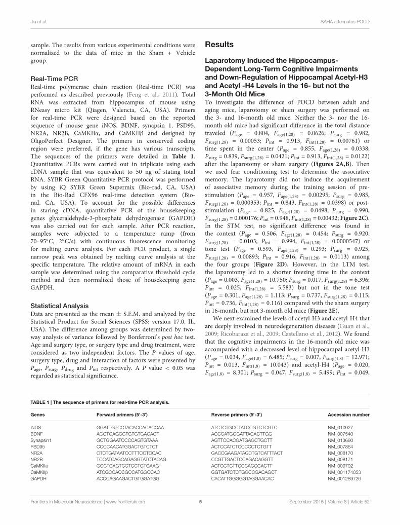
Laparotomy Induced the Hippocampus-Dependent Long-Term Cognitive Impairmentsand Down-Regulation of Hippocampal Acetyl-H3and Acetyl -H4 Levels in the 16- but not the3-Month Old MiceTo investigate the difference of POCD between adult andaging mice, laparotomy or sham surgery was performed onthe 3- and 16-month old mice. Neither the 3- nor the 16-month old mice had significant difference in the total distancetraveled (Page = 0.804, Fage(1,28) = 0.0626; Psurg = 0.982,Fsurg(1,28) = 0.00053; Pint = 0.913, Fint(1,28) = 0.00761) ortime spent in the center (Page = 0.855, Fage(1,28) = 0.0338;Psurg = 0.839, Fsurg(1,28) = 0.0421; Pint = 0.913, Fint(1,28) = 0.0122)after the laparotomy or sham surgery (Figures 2A,B). Thenwe used fear conditioning test to determine the associativememory. The laparotomy did not induce the acquirementof associative memory during the training session of pre-stimulation (Page = 0.957, Fage(1,28) = 0.00295; Psurg = 0.985,Fsurg(1,28) = 0.000353; Pint = 0.843, Fint(1,28) = 0.0398) or post-stimulation (Page = 0.825, Fage(1,28) = 0.0498; Psurg = 0.990,Fsurg(1,28) = 0.000176; Pint = 0.948, Fint(1,28) = 0.00432; Figure 2C).In the STM test, no significant difference was found inthe context (Page = 0.506, Fage(1,28) = 0.454; Psurg = 0.920,Fsurg(1,28) = 0.0103; Pint = 0.994, Fint(1,28) = 0.0000547) ortone test (Page = 0.593, Fage(1,28) = 0.293; Psurg = 0.925,Fsurg(1,28) = 0.00893; Pint = 0.916, Fint(1,28) = 0.0113) amongthe four groups (Figure 2D). However, in the LTM test,the laparotomy led to a shorter freezing time in the context(Page = 0.003, Fage(1,28) = 10.750; Psurg = 0.017, Fsurg(1,28) = 6.396;Pint = 0.025, Fint(1,28) = 5.583) but not in the tone test(Page = 0.301, Fage(1,28) = 1.113; Psurg = 0.737, Fsurg(1,28) = 0.115;Pint = 0.736, Fint(1,28) = 0.116) compared with the sham surgeryin 16-month, but not 3-month old mice (Figure 2E).
We next examined the levels of acetyl-H3 and acetyl-H4 thatare deeply involved in neurodegeneration diseases (Guan et al.,2009; Ricobaraza et al., 2009; Castellano et al., 2012). We foundthat the cognitive impairments in the 16-month old mice wasaccompanied with a decreased level of hippocampal acetyl-H3(Page = 0.034, Fage(1,8) = 6.485; Psurg = 0.007, Fsurg(1,8) = 12.971;Pint = 0.013, Fint(1,8) = 10.043) and acetyl-H4 (Page = 0.020,Fage(1,8) = 8.301; Psurg = 0.047, Fsurg(1,8) = 5.499; Pint = 0.049,
TABLE 1 | The sequence of primers for real-time PCR analysis.
Genes Forward primers (5′-3′) Reverse primers (5′-3′) Accession number
iNOS GGATTGTCCTACACCACACCAA ATCTCTGCCTATCCGTCTCGTC NM_010927BDNF AGCTGAGCGTGTGTGACAGT ACCCATGGGATTACACTTGG NM_007540Synapsin1 GCTGGAATCCCCAGTGTAAA AGTTCCACGATGAGCTGCTT NM_013680PSD95 CCCCAACATGGACTGTCTCT ACTCCATCTCCCCCTCTGTT NM_007864NR2A CTCTGATAATCCTTTCCTCCAC GACCGAAGATAGCTGTCATTTACT NM_008170NR2B TCCATCAGCAGAGGTATCTACAG CCGTTGACTCCAGACAGGTT NM_008171CaMKIIα GCCTCAGTCCTCCTGTGAAG ACTCCTCTTCCCACCCACTT NM_009792CaMKIIβ ATCGCCACCGCCATGGCCAC GGTGATCTCTGGCCGACAGCT NM_001174053GAPDH ACCCAGAAGACTGTGGATGG CACATTGGGGGTAGGAACAC NM_001289726
Frontiers in Molecular Neuroscience | www.frontiersin.org 5 September 2015 | Volume 8 | Article 52
Jia et al. SAHA attenuates POCD
FIGURE 2 | Impact of the cognition and histone acetylation in the 3- and 16-month old mice after surgery. (A,B) Performance of total distance traveledand time spent in the center during the open field test. Data are presented as the mean ± S.E.M. (n = 16). (C) Performance of freezing time during the fearconditioning training session. Data are presented as the mean ± S.E.M. (n = 16). (D,E) Performance during fear conditioning tests 2- or 24-h after laparotomy. Dataare presented as the mean ± S.E.M. (n = 8). (F) The acetylation level of histone H3 and H4 in the 3- or 16-month-old mice after laparotomy. Results are mean ±
S.E.M. (n = 3). ∗p < 0.05 compared with the 16-month old mice subjected to sham surgery.
Fint(1,8) = 5.359), whereas the 3-month old mice whose memorywere not damaged did not show such a reduction (Figure 2F).
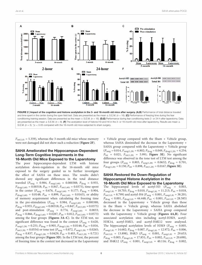
SAHA Ameliorated the Hippocampus-DependentLong-Term Cognitive Impairments in the16-Month Old Mice Exposed to the LaparotomyThe poor hippocampus-dependent LTM with histoneacetylation down-regulation in the 16-month old miceexposed to the surgery guided us to further investigatethe effect of SAHA on these mice. The results didn’tshowed any significant differences in the total distancetraveled (Psurg = 0.984, Fsurg(1,60) = 0.000388; Pdrug = 0.955,Fdrug(1,60) = 0.00318; Pint = 0.847, Fint(1,60) = 0.0375), time spentin the center (Psurg = 0.676, Fsurg(1,60) = 0.177; Pdrug = 0.904,Fdrug(1,60) = 0.0148; Pint = 0.899, Fint(1,60) = 0.0163), or abilityof memory acquirement when calculating the freezing timein the pre-stimulation (Psurg = 0.984, Fsurg(1,60) = 0.000388;Pdrug = 0.955, Fdrug(1,60) = 0.00318; Pint = 0.847, Fint(1,60) = 0.0375)and post-stimulation (Psurg = 0.774, Fsurg(1,60) = 0.0833;Pdrug = 0.866, Fdrug(1,60) = 0.0287; Pint = 0.812, Fint(1,60) = 0.0573)among the four groups (Figures 3A–C). In the STM test, nosignificant difference was found in the context (Psurg = 0.620,Fsurg(1,28) = 0.251; Pdrug = 0.905, Fdrug(1,28) = 0.0146; Pint = 0.816,Fint(1,28) = 0.0554) or tone test (Psurg = 0.872, Fsurg(1,28) = 0.0265;Pdrug = 0.837, Fdrug(1,28) = 0.0429; Pint = 0.403, Fint(1,28) = 0.721)among the four groups (Figure 3D). In the LTM test, the percentof freezing time in the context test decreased in the Laparotomy
+ Vehicle group compared with the Sham + Vehicle group,whereas SAHA diminished the decrease in the Laparotomy +SAHA group compared with the Laparotomy + Vehicle group(Psurg = 0.014, Fsurg(1,28) = 6.802; Pdrug = 0.049, Fdrug(1,28) = 4.254;Pint = 0.021, Fint(1,28) = 5.991; Figure 3E). No significantdifference was observed in the tone test of LTM test among thefour groups (Psurg = 0.803, Fsurg(1,28) = 0.0632; Pdrug = 0.701,Fdrug(1,28) = 0.150; Pint = 0.898, Fint(1,28) = 0.0167; Figure 3E).
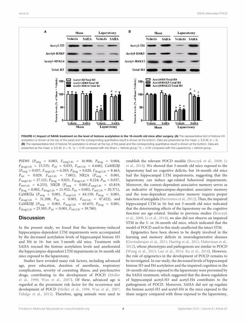
SAHA Restored the Down-Regulation ofHippocampal Histone Acetylation in the16-Month Old Mice Exposed to the LaparotomyThe hippocampal levels of acetyl-H3 (Psurg = 0.003,Fsurg(1,8) = 18.703; Pdrug = 0.010, Fdrug(1,8) = 11.213; Pint = 0.018,Fint(1,8) = 8.799) and acetyl-H4 (Psurg < 0.001, Fsurg(1,8) = 38.201;Pdrug < 0.001, Fdrug(1,8) = 44.448; Pint < 0.001, Fint(1,8) = 28.585)decreased in the Laparotomy + Vehicle group than thosein the Sham + Vehicle group, whereas SAHA abolishedthe decrease in the Laparotomy + SAHA group comparedwith the Laparotomy + Vehicle group (Figures 4A,B). Fourassociated acetylation sites including acetyl-H3K9, acetyl-H3K14, acetyl-H4K5, and acetyl-H4K12 were analyzed.The hippocampal acetylation levels of H3K9 (Psurg = 0.005,Fsurg(1,8) = 14.682; Pdrug = 0.007, Fdrug(1,8) = 12.972; Pint = 0.006,Fint(1,8) = 13.898), H4K5 (Psurg < 0.001, Fsurg(1,8) = 29.653;Pdrug = 0.003, Fdrug(1,8) = 17.851; Pint = 0.002, Fint(1,8) = 20.698),and H4K12 (Psurg < 0.001, Fsurg(1,8) = 40.116; Pdrug = 0.002,
Frontiers in Molecular Neuroscience | www.frontiersin.org 6 September 2015 | Volume 8 | Article 52
Jia et al. SAHA attenuates POCD
FIGURE 3 | Impact of suberoylanilide hydroxamic acid (SAHA) treatment on the cognitive performance in the 16-month old mice after surgery.(A,B) Performance of total distance traveled and time spent in the center during the open field test. Data are presented as the mean ± S.E.M. (n = 16).(C) Performance of freezing time during the fear conditioning training session. Data are presented as the mean ± S.E.M. (n = 16). (D,E) Performance during the fearconditioning tests 2- or 24-h after laparotomy. Data are presented as the mean ± S.E.M. (n = 8). ∗p < 0.05 compared with the Sham + Vehicle group; #p < 0.05compared with the Laparotomy + Vehicle group.
Fdrug(1,8) = 19.008; Pint < 0.001, Fint(1,8) = 26.543) decreasedin the Laparotomy + Vehicle group compared with the Sham+ Vehicle group, whereas SAHA blocked the decreases in theLaparotomy + SAHA group compared with the Laparotomy+ Vehicle group (Figures 4A,B). No significant difference wasobserved in the level of acetyl-H3K14 among the four groups(Psurg = 0.169, Fsurg(1,8) = 2.288; Pdrug = 0.588, Fdrug(1,8) = 0.319;Pint = 0.829, Fint(1,8) = 0.0501; Figures 4A,B).
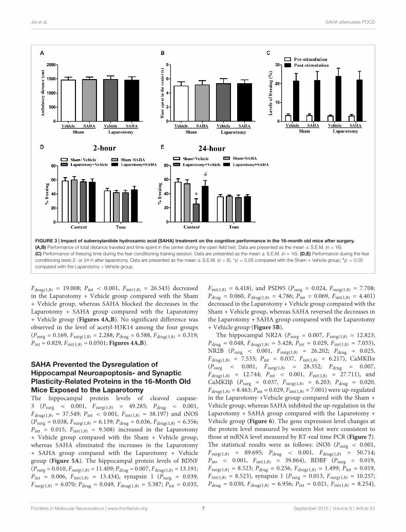
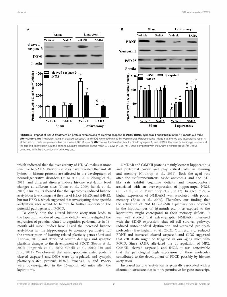
SAHA Prevented the Dysregulation ofHippocampal Neuroapoptosis- and SynapticPlasticity-Related Proteins in the 16-Month OldMice Exposed to the LaparotomyThe hippocampal protein levels of cleaved caspase-3 (Psurg < 0.001, Fsurg(1,8) = 49.285; Pdrug < 0.001,Fdrug(1,8) = 37.549; Pint < 0.001, Fint(1,8) = 38.197) and iNOS(Psurg = 0.038, Fsurg(1,8) = 6.139; Pdrug = 0.036, Fdrug(1,8) = 6.356;Pint = 0.015, Fint(1,8) = 9.508) increased in the Laparotomy+ Vehicle group compared with the Sham + Vehicle group,whereas SAHA eliminated the increases in the Laparotomy+ SAHA group compared with the Laparotomy + Vehiclegroup (Figure 5A). The hippocampal protein levels of BDNF(Psurg = 0.010, Fsurg(1,8) = 11.409; Pdrug = 0.007, Fdrug(1,8) = 13.191;Pint = 0.006, Fint(1,8) = 13.434), synapsin 1 (Psurg = 0.039,Fsurg(1,8) = 6.070; Pdrug = 0.049, Fdrug(1,8) = 5.387; Pint = 0.035,
Fint(1,8) = 6.418), and PSD95 (Psurg = 0.024, Fsurg(1,8) = 7.708;Pdrug = 0.060, Fdrug(1,8) = 4.786; Pint = 0.069, Fint(1,8) = 4.401)decreased in the Laparotomy + Vehicle group compared with theSham + Vehicle group, whereas SAHA reversed the decreases inthe Laparotomy + SAHA group compared with the Laparotomy+ Vehicle group (Figure 5B).
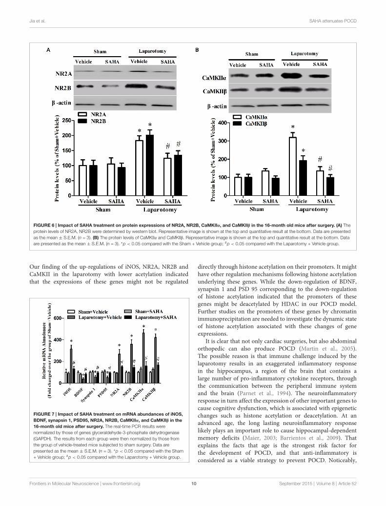
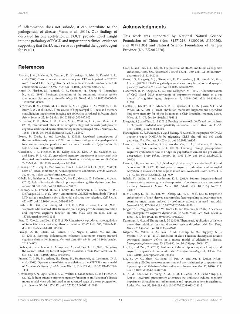
The hippocampal NR2A (Psurg = 0.007, Fsurg(1,8) = 12.823;Pdrug = 0.048, Fdrug(1,8) = 5.428; Pint = 0.029, Fint(1,8) = 7.055),NR2B (Psurg < 0.001, Fsurg(1,8) = 26.202; Pdrug = 0.025,Fdrug(1,8) = 7.533; Pint = 0.037, Fint(1,8) = 6.217), CaMKIIα(Psurg < 0.001, Fsurg(1,8) = 28.352; Pdrug = 0.007,Fdrug(1,8) = 12.744; Pint < 0.001, Fint(1,8) = 27.711), andCaMKIIβ (Psurg = 0.037, Fsurg(1,8) = 6.203; Pdrug = 0.020,Fdrug(1,8) = 8.463; Pint = 0.029, Fint(1,8) = 7.001) were up-regulatedin the Laparotomy +Vehicle group compared with the Sham +Vehicle group, whereas SAHA inhibited the up-regulation in theLaparotomy + SAHA group compared with the Laparotomy +Vehicle group (Figure 6). The gene expression level changes atthe protein level measured by western blot were consistent tothose at mRNA level measured by RT-real time PCR (Figure 7).The statistical results were as follows: iNOS (Psurg < 0.001,Fsurg(1,8) = 89.695; Pdrug < 0.001, Fdrug(1,8) = 50.714;Pint < 0.001, Fint(1,8) = 39.864), BDBF (Psurg = 0.019,Fsurg(1,8) = 8.523; Pdrug = 0.256, Fdrug(1,8) = 1.499; Pint = 0.019,Fint(1,8) = 8.523), synapsin 1 (Psurg = 0.013, Fsurg(1,8) = 10.257;Pdrug = 0.030, Fdrug(1,8) = 6.956; Pint = 0.021, Fint(1,8) = 8.254),
Frontiers in Molecular Neuroscience | www.frontiersin.org 7 September 2015 | Volume 8 | Article 52
Jia et al. SAHA attenuates POCD
FIGURE 4 | Impact of SAHA treatment on the level of histone acetylation in the 16-month old mice after surgery. (A) The representative blot of histone H3acetylation is shown at the top of the panel and the corresponding quantitative result is shown at the bottom. Data are presented as the mean ± S.E.M. (n = 3).(B) The representative blot of histone H4 acetylation is shown at the top of the panel and the corresponding quantitative result is shown at the bottom. Data arepresented as the mean ± S.E.M. (n = 3). ∗p < 0.05 compared with the Sham + Vehicle group; #p < 0.05 compared with the Laparotomy + Vehicle group.
PSD95 (Psurg = 0.003, Fsurg(1,8) = 16.908; Pdrug = 0.004,Fdrug(1,8) = 15.535; Pint = 0.033, Fint(1,8) = 6.646), CaMKIIβ(Psurg = 0.037, Fsurg(1,8) = 6.203; Pdrug = 0.020, Fdrug(1,8) = 8.463;Pint = 0.029, Fint(1,8) = 7.001), NR2A (Psurg < 0.001,Fsurg(1,8) = 27.121; Pdrug = 0.021, Fdrug(1,8) = 8.224; Pint = 0.037,Fint(1,8) = 6.253), NR2B (Psurg < 0.001,Fsurg(1,8) = 43.819;Pdrug = 0.002, Fdrug(1,8) = 21.952; Pint = 0.002, Fint(1,8) = 20.371),CaMKIIα (Psurg < 0.001, Fsurg(1,8) = 64.159; Pdrug < 0.001,Fdrug(1,8) = 31.208; Pint < 0.001, Fint(1,8) = 47.622), andCaMKIIβ (Psurg < 0.001, Fsurg(1,8) = 65.655; Pdrug < 0.001,Fdrug(1,8) = 25.505; Pint < 0.001, Fint(1,8) = 39.780).
Discussion
In the present study, we found that the laparotomy-inducedhippocampus-dependent LTM impairments were accompaniedby the decreased acetylation levels of hippocampal histone H3and H4 in 16- but not 3-month old mice. Treatment withSAHA rescued the histone acetylation levels and amelioratedthe hippocampus-dependent LTM impairments in 16-month oldmice exposed to the laparotomy.
Studies have revealed many risk factors, including advancedage, poor education, duration of anesthesia, respiratorycomplications, severity of coexisting illness, and psychoactivedrugs, contributing to the development of POCD (Molleret al., 1998; Wan et al., 2007). Of these, advanced age isregarded as the prominent risk factor for the occurrence anddevelopment of POCD (Moller et al., 1998; Wan et al., 2007;Fidalgo et al., 2011). Therefore, aging animals were used to
establish the relevant POCD models (Rosczyk et al., 2008; Liet al., 2014). We showed that 3-month old mice exposed to thelaparotomy had no cognitive deficits, but 16-month old micehad the hippocampal LTM impairments, suggesting that thelaparotomy can induce age-related behavioral impairments.Moreover, the context-dependent associative memory serves asan indicative of hippocampus-dependent associative memoryand the tone-dependent associative memory requires properfunction of amygdala (Barrientos et al., 2012). Thus, the impairedhippocampal LTM in 16- but not 3-month old mice indicatedthat the deteriorating effects of the laparotomy on the cognitivefunction are age-related. Similar to previous studies (Rosczyket al., 2008; Li et al., 2014), we also did not observe an impairedSTM in the 3- or 16-month old mice, which indicated that themodel of POCD used in this study unaffected the intact STM.
Epigenetics have been shown to be deeply involved in thelearning and memory deficits in neurodegenerative diseases(Govindarajan et al., 2011; Haettig et al., 2011; Haberman et al.,2012), whose phenotypes and pathogenesis are similar to POCD(Wang et al., 2013; Luo et al., 2014; Xu et al., 2014). However,the role of epigenetics in the development of POCD remains tobe investigated. In our study, the decreased levels of hippocampalhistone H3 and H4 acetylation and the impaired cognition in the16-month old mice exposed to the laparotomy were prevented bythe SAHA treatment, which suggested that the down-regulationof hippocampal acetyl-H3 and acetyl-H4 contributes to thepathogenesis of POCD. Moreover, SAHA did not up-regulatethe histone acetyl-H3 and acetyl-H4 in the mice exposed to thesham surgery compared with those exposed to the laparotomy,
Frontiers in Molecular Neuroscience | www.frontiersin.org 8 September 2015 | Volume 8 | Article 52
Jia et al. SAHA attenuates POCD
FIGURE 5 | Impact of SAHA treatment on protein expressions of cleaved caspase-3, iNOS, BDNF, synapsin 1 and PSD95 in the 16-month old miceafter surgery. (A) The protein levels of cleaved caspase-3 and iNOS were determined by western blot. Representative image is at the top and quantitative result isat the bottom. Data are presented as the mean ± S.E.M. (n = 3). (B) The result of western blot for BDNF, synapsin 1, and PSD95. Representative image is shown atthe top and quantitation is at the bottom. Data are presented as the mean ± S.E.M. (n = 3). ∗p < 0.05 compared with the Sham + Vehicle group; #p < 0.05compared with the Laparotomy + Vehicle group.
which indicated that the over activity of HDAC makes it moresensitive to SAHA. Previous studies have revealed that not alllysines in histone proteins are affected in the development ofneurodegenerative disorders (Miao et al., 2014; Zhong et al.,2014) and different diseases induce histone acetylation levelchanges at different sites (Guan et al., 2009; Itzhak et al.,2013). Our results showed that the laparotomy induced histoneacetylation level changes at the sites of H3K9, H4K5, and H4K12,but not H3K14, which suggested that investigating these specificacetylation sites would be helpful to further understand thepotential pathogenesis of POCD.
To clarify how the altered histone acetylation leads tothe laparotomy-induced cognitive deficits, we investigated theexpression of proteins related to cognition performance in 16-month old mice. Studies have linked the increased histoneacetylation in the hippocampus to memory permissive forthe transcription of learning-related plasticity genes (Ravi andKannan, 2013) and attributed neuron damages and synapticplasticity changes to the development of POCD (Bozon et al.,2002; Jungwirth et al., 2009; Cibelli et al., 2010; Lin andZuo, 2011). We observed that neuroapoptosis-related proteinscleaved caspase-3 and iNOS were up-regulated, and synapticplasticity-related proteins BDNF, synapsin 1, and PSD95were down-regulated in the 16-month old mice after thelaparotomy.
NMDAR and CaMKII proteins mainly locate at hippocampusand prefrontal cortex and play critical roles in learningand memory (Coultrap et al., 2014). Both the aged ratsafter the isoflurane/nitrous oxide anesthesia and the AD-like rats exhibit cognitive deficits and neuroapoptosisassociated with an over-expression of hippocampal NR2B(Liu et al., 2012; Mawhinney et al., 2012). In aged mice, ahigher expression of NMDAR2 was associated with poorermemory (Zhao et al., 2009). Therefore, our finding thatthe activation of NMDAR2-CaMKII pathway was observedin the hippocampus of 16-month old mice exposed to thelaparotomy might correspond to their memory deficits. Itwas well studied that extra-synaptic NMDARs interferedwith the BDNF expression, shut off cell survival pathway,induced mitochondrial dysfunction and activated pro-deathmolecules (Hardingham et al., 2002). Our results of reducedBDNF and increased cleaved caspase-3 and iNOS suggestedthat cell death might be triggered in our aging mice withPOCD. Since SAHA alleviated the up-regulation of NR2,CaMKII, cleaved caspase-3 and iNOS, it was conceivablethat the pathological high expression of these moleculescontributed to the development of POCD possibly by histoneacetylation.
Increased histone acetylation is generally associated with achromatin structure that is more permissive for gene transcript.
Frontiers in Molecular Neuroscience | www.frontiersin.org 9 September 2015 | Volume 8 | Article 52
Jia et al. SAHA attenuates POCD
FIGURE 6 | Impact of SAHA treatment on protein expressions of NR2A, NR2B, CaMKIIα, and CaMKIIβ in the 16-month old mice after surgery. (A) Theprotein levels of NR2A, NR2B were determined by western blot. Representative image is shown at the top and quantitative result at the bottom. Data are presentedas the mean ± S.E.M. (n = 3). (B) The protein levels of CaMKIIα and CaMKIIβ. Representative image is shown at the top and quantitative result at the bottom. Dataare presented as the mean ± S.E.M. (n = 3). ∗p < 0.05 compared with the Sham + Vehicle group; #p < 0.05 compared with the Laparotomy + Vehicle group.
Our finding of the up-regulations of iNOS, NR2A, NR2B andCaMKII in the laparotomy with lower acetylation indicatedthat the expressions of these genes might not be regulated
FIGURE 7 | Impact of SAHA treatment on mRNA abundances of iNOS,BDNF, synapsin 1, PSD95, NR2A, NR2B, CaMKIIα, and CaMKIIβ in the16-month old mice after surgery. The real-time PCR results werenormalized by those of genes glyceraldehyde-3-phosphate dehydrogenase(GAPDH). The results from each group were then normalized by those fromthe group of vehicle-treated mice subjected to sham surgery. Data arepresented as the mean ± S.E.M. (n = 3). ∗p < 0.05 compared with the Sham+ Vehicle group; #p < 0.05 compared with the Laparotomy + Vehicle group.
directly through histone acetylation on their promoters. It mighthave other regulation mechanisms following histone acetylationunderlying these genes. While the down-regulation of BDNF,synapsin 1 and PSD 95 corresponding to the down-regulationof histone acetylation indicated that the promoters of thesegenes might be deacetylated by HDAC in our POCD model.Further studies on the promoters of these genes by chromatinimmunoprecipitation are needed to investigate the dynamic stateof histone acetylation associated with these changes of geneexpressions.
It is clear that not only cardiac surgeries, but also abdominalorthopedic can also produce POCD (Martin et al., 2005).The possible reason is that immune challenge induced by thelaparotomy results in an exaggerated inflammatory responsein the hippocampus, a region of the brain that contains alarge number of pro-inflammatory cytokine receptors, throughthe communication between the peripheral immune systemand the brain (Parnet et al., 1994). The neuroinflammatoryresponse in turn affect the expression of other important genes tocause cognitive dysfunction, which is associated with epigeneticchanges such as histone acetylation or deacetylation. At anadvanced age, the long lasting neuroinflammatory responselikely plays an important role to cause hippocampal-dependentmemory deficits (Maier, 2003; Barrientos et al., 2009). Thatexplains the facts that age is the strongest risk factor forthe development of POCD, and that anti-inflammatory isconsidered as a viable strategy to prevent POCD. Noticeably,
Frontiers in Molecular Neuroscience | www.frontiersin.org 10 September 2015 | Volume 8 | Article 52
Jia et al. SAHA attenuates POCD
if inflammation does not subside, it can contribute to thepathogenesis of disease (Vacas et al., 2013). Our findings ofdecreased histone acetylation in POCD provide novel insightinto the pathology of POCD and important preclinical evidencessupporting that SAHAmay serve as a potential therapeutic agentfor POCD.
Acknowledgments
This work was supported by National Natural ScienceFoundation of China (Nos. 81271216, 81300946, 81300262,and 81471105) and Natural Science Foundation of JiangsuProvince (No. BK2012778).
References
Alarcón, J. M., Malleret, G., Touzani, K., Vronskaya, S., Ishii, S., Kandel, E. R.,et al. (2004). Chromatin acetylation, memory and LTP are impaired in CBP+/−
mice: a model for the cognitive deficit in rubinstein-taybi syndrome and itsamelioration. Neuron 42, 947–959. doi: 10.1016/j.neuron.2004.05.021
Amar, D., Fleisher, M., Pantuck, C. B., Shamoon, H., Zhang, H., Roistacher,N., et al. (1998). Persistent alterations of the autonomic nervous systemafter noncardiac surgery. Anesthesiology 89, 30–42. doi: 10.1097/00000542-199807000-00008
Barrientos, R. M., Frank, M. G., Hein, A. M., Higgins, E. A., Watkins, L. R.,Rudy, J. W., et al. (2009). Time course of hippocampal IL-1 beta and memoryconsolidation impairments in aging rats following peripheral infection. BrainBehav. Immun. 23, 46–54. doi: 10.1016/j.bbi.2008.07.002
Barrientos, R. M., Hein, A. M., Frank, M. G., Watkins, L. R., and Maier, S. F.(2012). Intracisternal interleukin-1 receptor antagonist prevents postoperativecognitive decline and neuroinflammatory response in aged rats. J. Neurosci. 32,14641–14648. doi: 10.1523/jneurosci.2173-12.2012
Bozon, B., Davis, S., and Laroche, S. (2002). Regulated transcription ofthe immediate-early gene Zif268: mechanisms and gene dosage-dependentfunction in synaptic plasticity and memory formation. Hippocampus 12,570–577. doi: 10.1002/hipo.10100
Castellano, J. F., Fletcher, B. R., Kelley-Bell, B., Kim, D. H., Gallagher, M.,and Rapp, P. R. (2012). Age-related memory impairment is associated withdisrupted multivariate epigenetic coordination in the hippocampus. PLoS One7:e33249. doi: 10.1371/journal.pone.0033249
Chuang, D. M., Leng, Y., Marinova, Z., Kim, H. J., and Chiu, C. T. (2009). Multipleroles of HDAC inhibition in neurodegenerative conditions. Trends Neurosci.32, 591–601. doi: 10.1016/j.tins.2009.06.002
Cibelli, M., Fidalgo, A. R., Terrando, N., Ma, D., Monaco, C., Feldmann, M., et al.(2010). Role of interleukin-1beta in postoperative cognitive dysfunction. Ann.Neurol. 68, 360–368. doi: 10.1002/ana.22082
Coultrap, S. J., Freund, R. K., O’Leary, H., Sanderson, J. L., Roche, K. W.,Dell’Acqua, M. L., et al. (2014). Autonomous CaMKII mediates both LTP andLTD using a mechanism for differential substrate site selection. Cell Rep. 6,431–437. doi: 10.1016/j.celrep.2014.01.005
Dash, P. K., Orsi, S. A., Zhang, M., Grill, R. J., Pati, S., Zhao, J., et al. (2010).Valproate administered after traumatic brain injury provides neuroprotectionand improves cognitive function in rats. PLoS One 5:e11383. doi: 10.1371/journal.pone.0011383
Feng, C., Cao, L., and Zuo, Z. (2011). RNA interference-produced autoregulationof inducible nitric oxide synthase expression. FEBS Lett. 585, 2488–2492.doi: 10.1016/j.febslet.2011.06.032
Fidalgo, A. R., Cibelli, M., White, J. P., Nagy, I., Maze, M., and Ma,D. (2011). Systemic inflammation enhances laparotomy surgery-inducedcognitive dysfunction in mice. Neurosci. Lett. 498, 63–66. doi: 10.1016/j.neulet.2011.04.063
Fischer, A., Sananbenesi, F., Mungenast, A., and Tsai, L. H. (2010). Targetingthe correct HDAC (s) to treat cognitive disorders. Trends Pharmacol. Sci. 31,605–617. doi: 10.1016/j.tips.2010.09.003
Francis, Y. I., Fà, M., Ashraf, H., Zhang, H., Staniszewski, A., Latchman, D. S.,et al. (2009). Dysregulation of histone acetylation in the APP/PS1 mouse modelof Alzheimer’s disease. J. Alzheimers Dis. 18, 131–139. doi: 10.3233/JAD-2009-1134
Govindarajan, N., Agis-Balboa, R. C., Walter, J., Sananbenesi, F., and Fischer, A.(2011). Sodium butyrate improves memory function in an Alzheimer’s diseasemouse model when administered at an advanced stage of disease progression.J. Alzheimers Dis. 26, 187–197. doi: 10.3233/JAD-2011-110080
Gräff, J., and Tsai, L. H. (2013). The potential of HDAC inhibitors as cognitiveenhancers. Annu. Rev. Pharmacol. Toxicol. 53, 311–330. doi: 10.1146/annurev-pharmtox-011112-140216
Guan, J. S., Haggarty, S. J., Giacometti, E., Dannenberg, J. H., Joseph, N., Gao,J., et al. (2009). HDAC2 negatively regulates memory formation and synapticplasticity. Nature 459, 55–60. doi: 10.1038/nature07925
Haberman, R. P., Quigley, C. K., and Gallagher, M. (2012). Characterizationof CpG island DNA methylation of impairment-related genes in a ratmodel of cognitive aging. Epigenetics 7, 1008–1009. doi: 10.4161/epi.21291
Haettig, J., Stefanko, D. P., Multani, M. L., Figueroa, D. X., McQuown, S. C., andWood, M. A. (2011). HDAC inhibition modulates hippocampus-dependentlong-term memory for object location in a CBP-dependent manner. Learn.Mem. 18, 71–79. doi: 10.1101/lm.1986911
Haggarty, S. J., and Tsai, L. H. (2011). Probing the role of HDACs andmechanismsof chromatin-mediated neuroplasticity. Neurobiol. Learn. Mem. 96, 41–52.doi: 10.1016/j.nlm.2011.04.009
Hardingham, G. E., Fukunaga, Y., and Bading, H. (2002). Extrasynaptic NMDARsoppose synaptic NMDARs by triggering CREB shut-off and cell deathpathways. Nat. Neurosci. 5, 405–414. doi: 10.1038/nn835
Hovens, I. B., Schoemaker, R. G., van der Zee, E. A., Heineman, E., Izaks,G. J., and van Leeuwen, B. L. (2012). Thinking through postoperativecognitive dysfunction: how to bridge the gap between clinical and pre-clinicalperspectives. Brain Behav. Immun. 26, 1169–1179. doi: 10.1016/j.bbi.2012.06.004
Hovens, I. B., van Leeuwen, B. L., Nyakas, C., Heineman, E., van der Zee, E. A., andSchoemaker, R. G. (2014). Postoperative cognitive dysfunction and microglialactivation in associated brain regions in old rats. Neurobiol. Learn. Mem. 118,74–79. doi: 10.1016/j.nlm.2014.11.009
Itzhak, Y., Liddie, S., and Anderson, K. L. (2013). Sodium butyrate-inducedhistone acetylation strengthens the expression of cocaine-associated contextualmemory. Neurobiol. Learn. Mem. 102, 34–42. doi: 10.1016/j.nlm.2013.03.007
Ji, M., Dong, L., Jia, M., Liu, W., Zhang, M., Ju, L., et al. (2014). Epigeneticenhancement of brain-derived neurotrophic factor signaling pathway improvescognitive impairments induced by isoflurane exposure in aged rats. Mol.Neurobiol. 50, 937–944. doi: 10.1007/s12035-014-8659-z
Jungwirth, B., Zieglgänsberger, W., Kochs, E., and Rammes, G. (2009). Anesthesiaand postoperative cognitive dysfunction (POCD). Mini. Rev. Med. Chem. 9,1568–1579. doi: 10.2174/138955709791012229
Kazantsev, A. G., and Thompson, L. M. (2008). Therapeutic application of histonedeacetylase inhibitors for central nervous system disorders. Nat. Rev. Drug.Discov. 7, 854–868. doi: 10.1038/nrd2681
Kilgore, M., Miller, C. A., Fass, D. M., Hennig, K. M., Haggarty, S. J.,Sweatt, J. D., et al. (2010). Inhibitors of class 1 histone deacetylases reversecontextual memory deficits in a mouse model of Alzheimer’s disease.Neuropsychopharmacology 35, 870–880. doi: 10.1038/npp.2009.197
Lin, D., and Zuo, Z. (2011). Isoflurane induces hippocampal cell injury andcognitive impairments in adult rats. Neuropharmacology 61, 1354–1359.doi: 10.1016/j.neuropharm.2011.08.011
Liu, Z., Lv, C., Zhao, W., Song, Y., Pei, D., and Xu, T. (2012). NR2B-containing NMDA receptors expression and their relationship to apoptosis inhippocampus of Alzheimer’s disease-like rats. Neurochem. Res. 37, 1420–1427.doi: 10.1007/s11064-012-0726-0
Li, X. M., Zhou, M. T., Wang, X. M., Ji, M. H., Zhou, Z. Q., and Yang, J. J.(2014). Resveratrol pretreatment attenuates the isoflurane-induced cognitiveimpairment through its anti-inflammation and -apoptosis actions in agedmice.J. Mol. Neurosci. 52, 286–293. doi: 10.1007/s12031-013-0141-2
Frontiers in Molecular Neuroscience | www.frontiersin.org 11 September 2015 | Volume 8 | Article 52
Jia et al. SAHA attenuates POCD
Luo, X., Yang, L., Chen, X., and Li, S. (2014). Tau hyperphosphorylation:a downstream effector of isoflurane-induced neuroinflammation in agedrodents.Med. Hypotheses 82, 94–96. doi: 10.1016/j.mehy.2013.11.015
Maier, S. F. (2003). Bi-directional immune-brain communication: implicationsfor understanding stress, pain, and cognition. Brain Behav. Immun. 17, 69–85.doi: 10.1016/s0889-1591(03)00032-1
Martin, T. J., Kahn, W. R., and Eisenach, J. C. (2005). Abdominal surgerydecreases food-reinforced operant responding in rats: relevance of incisionalpain. Anesthesiology 103, 629–637. doi: 10.1097/00000542-200509000-00028
Mawhinney, L. J., de Rivero Vaccari, J. P., Alonso, O. F., Jimenez, C. A., Furones,C., Moreno, W. J., et al. (2012). Isoflurane/nitrous oxide anesthesia inducesincreases in NMDA receptor subunit NR2B protein expression in the aged ratbrain. Brain Res. 1431, 23–34. doi: 10.1016/j.brainres.2011.11.004
McQuown, S. C., Barrett, R. M., Matheos, D. P., Post, R. J., Rogge, G. A., Alenghat,T., et al. (2011). HDAC3 is a critical negative regulator of long-term memoryformation. J. Neurosci. 31, 764–774. doi: 10.1523/JNEUROSCI.5052-10.2011
Miao, F., Chen, Z., Genuth, S., Paterson, A., Zhang, L., Wu, X., et al. (2014).Evaluating the role of epigenetic histone modifications in the metabolicmemory of type 1 diabetes. Diabetes 63, 1748–1762. doi: 10.2337/db13-1251
Moller, J. T., Cluitmans, P., Rasmussen, L. S., Houx, P., Rasmussen, H., Canet,J., et al. (1998). Long-term postoperative cognitive dysfunction in the elderly:ISPOCD1 study. Lancet 351, 857–861. doi: 10.1016/s0140-6736(97)07382-0
Parnet, P., Amindari, S., Wu, C., Brunke-Reese, D., Goujon, E., Weyhenmeyer,J. A., et al. (1994). Expression of type I and type II interleukin-1 receptorsin mouse brain. Brain Res. Mol. Brain Res. 27, 63–70. doi: 10.1016/0169-328x(94)90185-6
Peleg, S., Sananbenesi, F., Zovoilis, A., Burkhardt, S., Bahari-Javan, S., Agis-Balboa, R. C., et al. (2010). Altered histone acetylation is associated withage-dependent memory impairment in mice. Science 328, 753–756. doi: 10.1126/science.1186088
Petrij, F., Giles, R. H., Dauwerse, H. G., Saris, J. J., Hennekam, R. C., Masuno,M., et al. (1995). Rubinstein-Taybi syndrome caused by mutations in thetranscriptional co-activator CBP. Nature 376, 348–351. doi: 10.1038/376348a0
Ravi, B., and Kannan, M. (2013). Epigenetics in the nervous system: an overviewof its essential role. Indian J. Hum. Genet. 19, 383–391. doi: 10.4103/0971-6866.124357
Ricobaraza, A., Cuadrado-Tejedor, M., Pérez-Mediavilla, A., Frechilla, D., DelRío, J., and García-Osta, A. (2009). Phenylbutyrate ameliorates cognitivedeficit and reduces tau pathology in an Alzheimer’s disease mouse model.Neuropsychopharmacology 34, 1721–1732. doi: 10.1038/npp.2008.229
Rosczyk, H. A., Sparkman, N. L., and Johnson, R. W. (2008). Neuroinflammationand cognitive function in aged mice following minor surgery. Exp. Gerontol.43, 840–846. doi: 10.1016/j.exger.2008.06.004
Terrando, N., Brzezinski, M., Degos, V., Eriksson, L. I., Kramer, J. H., Leung, J. M.,et al. (2011). Perioperative cognitive decline in the aging population.Mayo Clin.Proc. 86, 885–893. doi: 10.4065/mcp.2011.0332
Vacas, S., Degos, V., Feng, X., and Maze, M. (2013). The neuroinflammatoryresponse of postoperative cognitive decline. Br. Med. Bull. 106, 161–178.doi: 10.1093/bmb/ldt006
Wan, Y., Xu, J., Ma, D., Zeng, Y., Cibelli, M., and Maze, M. (2007). Postoperativeimpairment of cognitive function in rats: a possible role for cytokine-mediatedinflammation in the hippocampus. Anesthesiology 106, 436–443. doi: 10.1097/00000542-200703000-00007
Wang, Y., Chen, Z., Zhao, Y., Shi, R., Wang, Y., Xu, J., et al. (2013). Epigeneticsas a new therapeutic target for postoperative cognitive dysfunction. Med.Hypotheses 80, 249–251. doi: 10.1016/j.mehy.2012.11.041
Xu, Z., Dong, Y., Wang, H., Culley, D. J., Marcantonio, E. R., Crosby, G., et al.(2014). Age-dependent postoperative cognitive impairment and Alzheimer-related neuropathology in mice. Sci. Rep. 4:3766. doi: 10.1038/srep03766
Zhao, X., Rosenke, R., Kronemann, D., Brim, B., Das, S. R., Dunah,A. W., et al. (2009). The effects of aging on N-methyl-D-aspartatereceptor subunits in the synaptic membrane and relationships to long-termspatial memory. Neuroscience 162, 933–945. doi: 10.1016/j.neuroscience.2009.05.018
Zhong, T., Qing, Q. J., Yang, Y., Zou, W. Y., Ye, Z., Yan, J. Q., et al. (2014).Repression of contexual fear memory induced by isoflurane is accompanied byreduction in histone acetylation and rescued by sodium butyrate. Br. J. Anaesth.113, 634–643. doi: 10.1093/bja/aeu184
Conflict of Interest Statement: The authors declare that the research wasconducted in the absence of any commercial or financial relationships that couldbe construed as a potential conflict of interest.
Copyright © 2015 Jia, Liu, Sun, Chang, Yang, Ji, Yang and Feng. This is an open-access article distributed under the terms of the Creative Commons AttributionLicense (CC BY). The use, distribution and reproduction in other forums ispermitted, provided the original author(s) or licensor are credited and that theoriginal publication in this journal is cited, in accordance with accepted academicpractice. No use, distribution or reproduction is permitted which does not complywith these terms.
Frontiers in Molecular Neuroscience | www.frontiersin.org 12 September 2015 | Volume 8 | Article 52