submitted: accepted: cardiomyopathy: a - jscimed … q yaméogo et al. (2017) email: j cardiol clin...
TRANSCRIPT

CentralBringing Excellence in Open Access
Journal of Cardiology & Clinical Research
Cite this article: Yaméogo NV, Kagambèga LJ, Seghda A, Owona A, Kaboré O, et al. (2017) Bromocriptine in Management of Peripartum Cardiomyopathy: A Randomized Study on 96 Women in Burkina Faso. J Cardiol Clin Res 5(2): 1098.
*Corresponding author
Nobila Valentin Yameogo, Department of Cardiology, Yalgado Ouédraogo University Hospital 03 BP 7022 Ouagadougou 03, Burkina Faso, Africa, Tel: 226-78 80 55 48; Email:
Submitted: 12 December 2016
Accepted: 16 February 2017
Published: 17 February 2017
Copyright© 2017 Yaméogo et al.
OPEN ACCESS
Keywords•Peripartum cardiomyopathy•Bromocriptine•Prognosis•Burkina Faso
Research Article
Bromocriptine in Management of Peripartum Cardiomyopathy: A Randomized Study on 96 Women in Burkina FasoNobila Valentin Yaméogo1,2*, Larissa Justine Kagambèga1, Arthur Seghda1, Amalia Owona1, Olivia Kaboré1, Elisée Kaboré1, Georges Rc Millogo1,2, KJonas Kologo1, BJean Yves Toguyéni1, André K Samadoulougou1,2, Jean L Ankoandé3, and Patrice Zabsonré1,2
1Department of Cardiology, Yalgado OuédraogoUniversity Hospital of Ouagadougou, Africa2Department of Medical Sciences, University Of Ouagadougou, Africa3Department of Gynaecology and Obstetrics, Yalgado OuédraogoUniversity Hospital of Ouagadougou, Africa4Aristide Le Dantec University Hospital, Dakar plateau, Sénégal
Abstract
In order to assess the role of bromocriptine in management of peripartum cardiomyopathy (PPCM), we conducted a randomized study from April 1, 2010 to June 30, 2012. Patients randomized for standard heart failure therapy (Br-) received heart failure conventional therapy and those randomized for bromocriptine (Br+) received standard heart failure therapy plus bromocriptine 2.5 mg twice daily for 4 weeks. Clinical assessment, electrocardiogram, and echocardiography were performed at baseline, 2 weeks, 1, 3, 6 and 12 months. Endpoints criteria were mortality, changes in dyspnea and evolution of echocardiographic parameters: End diastole Left Ventricular Diameter, End Systole Left Ventricular Diameter, Left Ventricular Ejection Fraction and Tricuspid Annular Plan Systolic Excursion (TAPSE).
Results: Ninety six (96) women were included for the study. There were no significant differences in baseline characteristics. The mean value of EDLVD was 58.7 mm in Br+ and 57.6 mm in Br- (p = 0.091) while the ESLVD was 48.4 mm in Br+ and 48.8 mm in Br- (p = 0.074). The mean LVEF was 37.2% in Br+ and 37.5% in Br- (p = 0.129) and the mean TAPSE was 19.9 mm in Br+ and 18.9 mm in Br- (p = 0.718). At Six months, cumulative death were stayed 8 (16.6%) in Br+ but 14 (29.1 %) in Br- (p = 0.0001). Echocardiographic findings demonstrated better improvement in ventricles function in Br+. Mean EDLVD was 53.4 mm in Br+ and 55.1 mm in Br- (p = 0.002). The mean LVEF was 49.9% in Br+ and 40.9% in Br- (p = 0.001) and the mean TAPSE was 22.0 mm in Br+ and 20.7 mm in Br- (p = 0.001). At 12 months, mean LVEF was 53.9 % in Br+ and 45.9 % in Br- (p = 0.001) and mean TAPSE was 22.7 mm in Br+ and 20.9 mm in Br- (p = 0.001). LVEF increased from 37.2% on admission to 49.9% at six months and to 53.9% at 12 months in Br+ while it increased from 37.5% on admission to 40.9% at six months and to 45.9% at 12 months in Br-.
INTRODUCTIONPeripartum cardiomyopathy (PPCM) is a life-threatening
heart disease of unknown etiology characterized by a sudden onset of heart failure in the last month of pregnancy and/or in the first months postpartum [1]. PPCM is very rare in Europe [2], but common in West Africa [3].
In general, treatment of PPCM was considered to be similar
to other types of congestive heart failure [4,5].But recently, the anti angiogenic 16-kDa N-terminal prolactin fragment (16K PRL), produced by the full-length nursing hormone prolactin (PRL) cleavage by cathepsin D, has been discovered with a potential factor initiating and driving PPCM [6]. Bromocriptine that reduces the prolactin production by its dopamine agonist actions may improve outcomes in patients with peripartum cardiomyopathy by eliminating the cleaved form of prolactin despite the activation of the cleaving enzyme.

CentralBringing Excellence in Open Access
Yaméogo et al. (2017)Email:
2/8J Cardiol Clin Res 5(2): 1098 (2017)
The aim of this study was to assess the effectiveness of bromocriptine in the treatment of peripartum cardiomyopathy
METHODSStudy Design and Patient Recruitment
Our study was approved by the human research ethics committee of the Yalgado OUEDRAOGO Teaching Hospital, Ouagadougou, Burkina Faso, and complies with the declaration of Helsinki. All patients and control subjects were given a consent form duly signed before being recruited. Ninety six consenting consecutive patients diagnosed with PPCM and fulfilling the inclusion criteria were enrolled in the study. All patients were included and randomized; the decision for the first patient to receive bromocriptine depends on the outcome of a coin of 10 Francs CFA played “heads or tails”. Once drawn “heads”, the patient was put under bromocriptine (subgroup Br+) and the next patient did not received the bromocriptine but only standard therapy of heart failure (Subgroup Br-) and so on.
The study was conducted at the Yalgado OUEDRAOGO Teaching Hospital. Patients were referred from local clinics, secondary hospitals, emergencies unit, and the Obstetrics department of the Yalgado OUEDRAOGO Teaching Hospital. Past medical history of preexisting cardiac symptoms and signs, occurrence of preeclampsia, and mode of delivery were obtained from the patient and confirmed by examination of the obstetric file carried by each patient.
Symptoms and signs were recorded during the first consultation at the cardiology unit at the Yalgado Ouedraogo Teaching Hospital (baseline) and during a follow-up period of 12months. Clinical assessment, electrocardiogram, chest x-ray, echocardiography, and blood analysis were performed at baseline. Clinical assessment and echocardiography were then performed at 2 weeks, 1, 3, 6 and 12 months.
Inclusion criteria were symptoms of congestive heart failure developed in the last month of pregnancy or during the five months postpartum, with no other identifiable cause for heart failure, and left ventricle ejection fraction (LVEF) < 45% by transthoracic echocardiography. Exclusion criteria were composed by systolic blood pressure >150 or <95 mm Hg or diastolic >105 mm Hg, sepsis, autoimmune disease, HIV serology positivity, impaired renal function (defined as urea and/or creatinine >1.5 times the normal upper limit) and any clinical condition that precluded inclusion in the study such as ischemic heart disease, anemic heart or malignancy.
All patients received standard heart failure treatment with the diuretic furosemide and the angiotensin-converting enzyme (ACE) inhibitor captopril. Patients with an LVEF < 35% or ventricular thrombus received anticoagulation therapy with fluindione for 6 months. Captopril doses were titrated upward as tolerated during the first 4 weeks after diagnosis and then maintained throughout the 12-month study period remaining. Furosemide dose was decreased as indicated according to clinical assessment during the 12-month study period.
The 48 patients randomized to standard therapy (Br-) were treated as outlined above. The 48 patients randomized to standard therapy plus bromocriptine (Br+) received bromocriptine 2.5 mg
twice a day for 4 weeks. After the initial screening and baseline visits, monthly outpatient visits were scheduled for clinical assessment and evaluation of medication compliance. Clinical status of hospitalized patients was evaluated each day until its improvement before discharge. The study has been conducted from April 1, 2010 to June 30, 2012.
Endpoints criteria were mortality, changes in dyspnea (NYHA class) and evolution of echocardiographic parameters: End diastole Left Ventricular Diameter (EDLVD), End Systole Left Ventricular Diameter (ESLVD), Left Ventricular Ejection Fraction (LVEF) and Tricuspid Annular Plan Systolic Excursion (TAPSE).
All patients were informed from impossibility to breastfeed their babies and then all babies benefit from artificial lactation Data analysis was realized from SAS V9.0 statistical software. Comparison of means and proportions between subgroups was performed by Chi-square statistics and independent t test. Significance was assumed at a value of p ˂ 0.05.
RESULTSWe enrolled 99 black-african women but 3 were excluded
because of their HIV serology positivity. Then, 96 women were included for the study. On admission, all of them had at least stage III NYHA dyspnea.
Sociodemographic characteristics
The mean age of the sample was 29.4 ± 3.4 years (16 and 36 years). The age group of 20-25 years was the most represented (29.16%). Patients’ distribution by age group is summarized in Figure (1). The mean age was 29.1 ± 3.1 years in Br+ and 29.5 ± 3.8 years in Br-. The difference was not statistically significant (p= 0.449). More than two thirds of the patients had a low socioeconomic level (77.1% or 74 patients), while a high socioeconomic level was found in nine (09) patients (9.4%).
Patients’ past medical history
No past medical history of hypertension or heart disease was found. There was no concept of smoking. The average of pregnancies per woman was 3.6 ± 1.5 pregnancies (1 and 9). More than the half had at least four pregnancies. The average gravidity was 3.6 ± 1.4 in Br+ and 3.2 ± 1.1 pregnancies in Br-. There was no significant difference between the two subgroups (p = 0.615). Twin pregnancies was noted in 10 cases, including
0
5
10
15
20
25
30
15-20 20-25 25-30 30-35 35-40
23
28
22
14
9Number
Age group (years)
Figure 1 Patients’ distribution by age range.
CentralBringing Excellence in Open Access
Yaméogo et al. (2017)Email:
3/8J Cardiol Clin Res 5(2): 1098 (2017)
four (04) cases in the Br+ and six (06) in the Br-. The first heart failure symptoms on set after delivery were observed in 83.33% (80 women) and before delivery in 16 patients. Table (1) presents the period of symptoms onset by subgroup.
Follow up of Clinical signs and Para clinical findings at admission
Clinical signs: All women suffered from dyspnea. Thus, 69.7% presented a stage IV NYHA dyspnea and stage III was found in 30.3%. Women distribution by NYHA functional stage is presented on Table (2). Heart failure, cough and chest pain were present in 51 (53.12%) and 44 (45.84%) cases respectively. Table (3) summarizes physical examination results.
Paraclinical fingings:
Biology: The hemoglobin was 11.9 ± 2.4 g/dl (9.1 and 13.6 g/dl) in Br+ and 11.7 ± 2.1 g/dl (9.3 and 14.4 g/dl) in Br- without statistically significant difference between the two groups (p = 0.899). Creatinin value was normal in every subgroup.
Chest x-ray: The mean cardiothoracic rate was 57.4 ± 2.2% (49.7 and 61.1%) in Br+ and 57.2 ± 1.8% (49.5 and 62.1%) in Br-
(p = 0.684). Pleural effusion was present in 6 cases that is 4 in Br+ and 2 in Br-. Infectious lung was found in 8 cases in Br+and 6 cases in Br-.
Electrocardiogram: The sinus rhythm was found in majority of women. A trial fibrillation and a trial tachysystoly was found in 1 case respectively. Electrocardiogram signs are summarized in Table (4).
Echocardiography data: Cardiac cavities were enlarged in all cases. The mean value of EDLVD was 58.7 ± 3.3 mm(55.2 and 63.2 mm) in Br+and 57.6 ± 3.8 mm (52.3 and 63.0 mm) in Br- (p =0.091) while the ESLVD was 48.4 ± 4.1 mm (42.0 and 53.1mm) in Br+ and 48.8 ± 3.9 mm (44.1 and 53 mm) in Br- (p = 0.074). The mean LVEF was 37.2 ± 6.6% (22.3and 44.4 %) in Br+ and 37.5 ± 4.8% (22.3 and 44.0%) in Br- (p = 0.129) and the mean TAPSE was 19.9 ± 3.1 mm (10.3 and 28.6 mm) in Br+ and 18.9 ± 0.9 mm(10.1 and 26.2 mm) in Br- (p = 0.718).
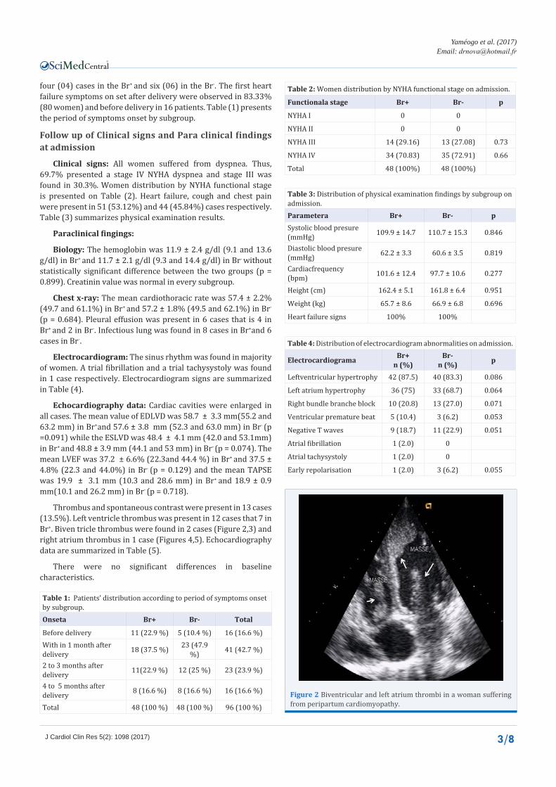
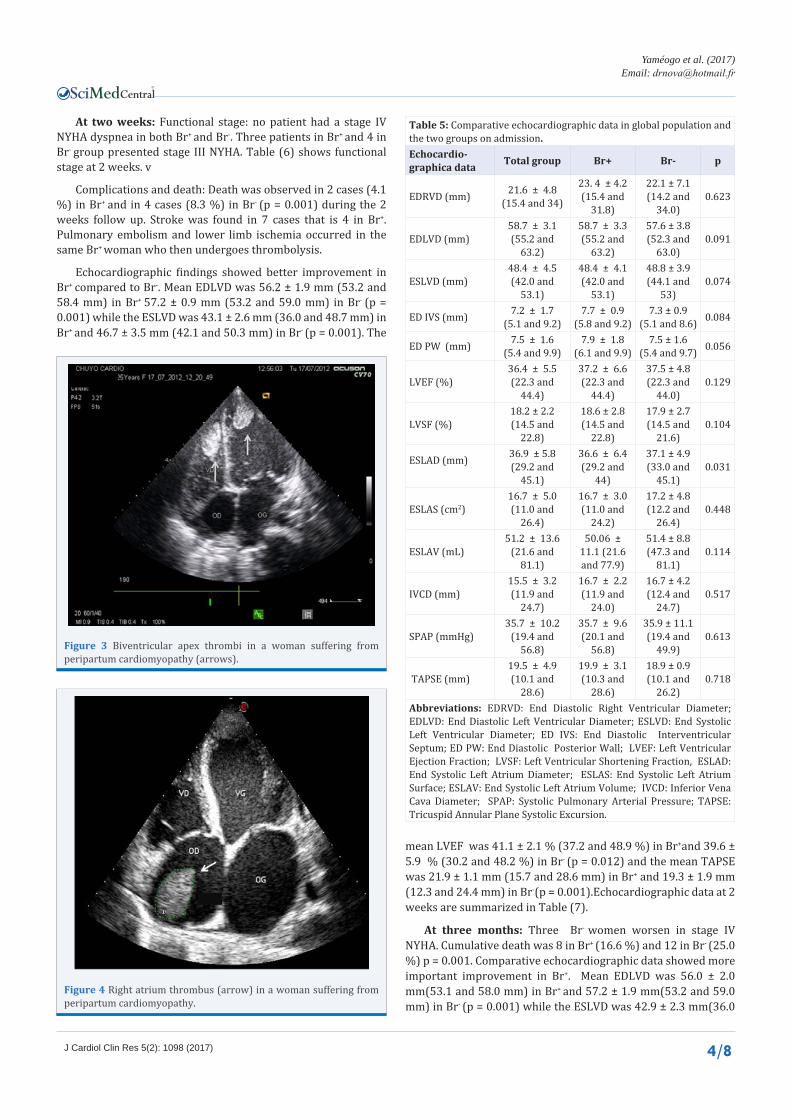
Thrombus and spontaneous contrast were present in 13 cases (13.5%). Left ventricle thrombus was present in 12 cases that 7 in Br+. Biven tricle thrombus were found in 2 cases (Figure 2,3) and right atrium thrombus in 1 case (Figures 4,5). Echocardiography data are summarized in Table (5).
There were no significant differences in baseline characteristics.
Table 1: Patients’ distribution according to period of symptoms onset by subgroup.Onseta Br+ Br- Total
Before delivery 11 (22.9 %) 5 (10.4 %) 16 (16.6 %)With in 1 month after delivery 18 (37.5 %) 23 (47.9
%) 41 (42.7 %)
2 to 3 months after delivery 11(22.9 %) 12 (25 %) 23 (23.9 %)
4 to 5 months after delivery 8 (16.6 %) 8 (16.6 %) 16 (16.6 %)
Total 48 (100 %) 48 (100 %) 96 (100 %)
Table 2: Women distribution by NYHA functional stage on admission.
Functionala stage Br+ Br- p
NYHA I 0 0
NYHA II 0 0
NYHA III 14 (29.16) 13 (27.08) 0.73
NYHA IV 34 (70.83) 35 (72.91) 0.66
Total 48 (100%) 48 (100%)
Table 3: Distribution of physical examination findings by subgroup on admission.Parametera Br+ Br- pSystolic blood presure (mmHg) 109.9 ± 14.7 110.7 ± 15.3 0.846
Diastolic blood presure (mmHg) 62.2 ± 3.3 60.6 ± 3.5 0.819
Cardiacfrequency (bpm) 101.6 ± 12.4 97.7 ± 10.6 0.277
Height (cm) 162.4 ± 5.1 161.8 ± 6.4 0.951
Weight (kg) 65.7 ± 8.6 66.9 ± 6.8 0.696
Heart failure signs 100% 100%
Table 4: Distribution of electrocardiogram abnormalities on admission.
Electrocardiograma Br+n (%)
Br-n (%) p
Leftventricular hypertrophy 42 (87.5) 40 (83.3) 0.086
Left atrium hypertrophy 36 (75) 33 (68.7) 0.064
Right bundle branche block 10 (20.8) 13 (27.0) 0.071
Ventricular premature beat 5 (10.4) 3 (6.2) 0.053
Negative T waves 9 (18.7) 11 (22.9) 0.051
Atrial fibrillation 1 (2.0) 0
Atrial tachysystoly 1 (2.0) 0
Early repolarisation 1 (2.0) 3 (6.2) 0.055
Figure 2 Biventricular and left atrium thrombi in a woman suffering from peripartum cardiomyopathy.
CentralBringing Excellence in Open Access
Yaméogo et al. (2017)Email:
4/8J Cardiol Clin Res 5(2): 1098 (2017)
At two weeks: Functional stage: no patient had a stage IV NYHA dyspnea in both Br+ and Br-. Three patients in Br+ and 4 in Br- group presented stage III NYHA. Table (6) shows functional stage at 2 weeks. v
Complications and death: Death was observed in 2 cases (4.1 %) in Br+ and in 4 cases (8.3 %) in Br- (p = 0.001) during the 2 weeks follow up. Stroke was found in 7 cases that is 4 in Br+. Pulmonary embolism and lower limb ischemia occurred in the same Br+ woman who then undergoes thrombolysis.
Echocardiographic findings showed better improvement in Br+ compared to Br-. Mean EDLVD was 56.2 ± 1.9 mm (53.2 and 58.4 mm) in Br+ 57.2 ± 0.9 mm (53.2 and 59.0 mm) in Br- (p = 0.001) while the ESLVD was 43.1 ± 2.6 mm (36.0 and 48.7 mm) in Br+ and 46.7 ± 3.5 mm (42.1 and 50.3 mm) in Br- (p = 0.001). The
mean LVEF was 41.1 ± 2.1 % (37.2 and 48.9 %) in Br+and 39.6 ± 5.9 % (30.2 and 48.2 %) in Br- (p = 0.012) and the mean TAPSE was 21.9 ± 1.1 mm (15.7 and 28.6 mm) in Br+ and 19.3 ± 1.9 mm (12.3 and 24.4 mm) in Br-(p = 0.001).Echocardiographic data at 2 weeks are summarized in Table (7).
At three months: Three Br- women worsen in stage IV NYHA. Cumulative death was 8 in Br+ (16.6 %) and 12 in Br- (25.0 %) p = 0.001. Comparative echocardiographic data showed more important improvement in Br+. Mean EDLVD was 56.0 ± 2.0 mm(53.1 and 58.0 mm) in Br+ and 57.2 ± 1.9 mm(53.2 and 59.0 mm) in Br- (p = 0.001) while the ESLVD was 42.9 ± 2.3 mm(36.0
Figure 3 Biventricular apex thrombi in a woman suffering from peripartum cardiomyopathy (arrows).
Figure 4 Right atrium thrombus (arrow) in a woman suffering from peripartum cardiomyopathy.
Table 5: Comparative echocardiographic data in global population and the two groups on admission.Echocardio-graphica data Total group Br+ Br- p
EDRVD (mm) 21.6 ± 4.8 (15.4 and 34)
23. 4 ± 4.2(15.4 and
31.8)
22.1 ± 7.1(14.2 and
34.0)0.623
EDLVD (mm)58.7 ± 3.1 (55.2 and
63.2)
58.7 ± 3.3(55.2 and
63.2)
57.6 ± 3.8(52.3 and
63.0)0.091
ESLVD (mm)48.4 ± 4.5(42.0 and
53.1)
48.4 ± 4.1(42.0 and
53.1)
48.8 ± 3.9(44.1 and
53)0.074
ED IVS (mm) 7.2 ± 1.7(5.1 and 9.2)
7.7 ± 0.9(5.8 and 9.2)
7.3 ± 0.9(5.1 and 8.6) 0.084
ED PW (mm) 7.5 ± 1.6(5.4 and 9.9)
7.9 ± 1.8(6.1 and 9.9)
7.5 ± 1.6(5.4 and 9.7) 0.056
LVEF (%)36.4 ± 5.5 (22.3 and
44.4)
37.2 ± 6.6(22.3 and
44.4)
37.5 ± 4.8(22.3 and
44.0)0.129
LVSF (%)18.2 ± 2.2(14.5 and
22.8)
18.6 ± 2.8(14.5 and
22.8)
17.9 ± 2.7(14.5 and
21.6)0.104
ESLAD (mm) 36.9 ± 5.8 (29.2 and
45.1)
36.6 ± 6.4(29.2 and
44)
37.1 ± 4.9(33.0 and
45.1)0.031
ESLAS (cm2)16.7 ± 5.0(11.0 and
26.4)
16.7 ± 3.0(11.0 and
24.2)
17.2 ± 4.8(12.2 and
26.4)0.448
ESLAV (mL)51.2 ± 13.6
(21.6 and 81.1)
50.06 ± 11.1 (21.6 and 77.9)
51.4 ± 8.8(47.3 and
81.1)0.114
IVCD (mm)15.5 ± 3.2 (11.9 and
24.7)
16.7 ± 2.2(11.9 and
24.0)
16.7 ± 4.2(12.4 and
24.7)0.517
SPAP (mmHg)35.7 ± 10.2
(19.4 and 56.8)
35.7 ± 9.6(20.1 and
56.8)
35.9 ± 11.1 (19.4 and
49.9)0.613
TAPSE (mm)19.5 ± 4.9 (10.1 and
28.6)
19.9 ± 3.1(10.3 and
28.6)
18.9 ± 0.9(10.1 and
26.2)0.718
Abbreviations: EDRVD: End Diastolic Right Ventricular Diameter; EDLVD: End Diastolic Left Ventricular Diameter; ESLVD: End Systolic Left Ventricular Diameter; ED IVS: End Diastolic Interventricular Septum; ED PW: End Diastolic Posterior Wall; LVEF: Left Ventricular Ejection Fraction; LVSF: Left Ventricular Shortening Fraction, ESLAD: End Systolic Left Atrium Diameter; ESLAS: End Systolic Left Atrium Surface; ESLAV: End Systolic Left Atrium Volume; IVCD: Inferior Vena Cava Diameter; SPAP: Systolic Pulmonary Arterial Pressure; TAPSE: Tricuspid Annular Plane Systolic Excursion.

CentralBringing Excellence in Open Access
Yaméogo et al. (2017)Email:
5/8J Cardiol Clin Res 5(2): 1098 (2017)
Table 6: Distribution of patients according to functional stage at 2 weeks.NYHA Br+ Br- pNYHA I 0 0 NYHA II 13 (27.0) 21 (43.7) 0.0001NYHA III 3 (6.2) 4 (8.3) 0.0872NYHA IV 0 0
Table 7: Comparative echocardiographic data in global population and the two groups on 2 weeks.Echocardiographica data Total group Br+ Br- P
EDRVD (mm) 20.7 ± 4.9 (14.5 and 29.0)
20.1 ± 4.2 (15.4 and 28.8)
21.9 ± 2.4 (14.5 and 29.0) 0.027
EDLVD (mm) 57.1 ± 1.1 (53.2 and 59.0)
56.2 ± 1.9 (53.2 and 58.4)
57.2 ± 0.9 (53.2 and 59.0) 0.001
ESLVD (mm) 48.1 ± 1.9 (36 and 50.3)
43.1 ± 2.6 (36.0 and 48.7)
46.7 ± 3.5 (42.1 and 50.3) 0.001
EDIVS (mm) 7.3 ± 1.7(5.2 and 9.2)
7.7 ± 0.9(5.8 and 9.2)
7.5 ± 0.9(5.2 et 8.9) 0.061
EDPW (mm) 7.6 ± 1.2(6.1 and 9.9)
7.9 ± 1.3(6.4 and 9.9)
7.5 ± 1.0(6.1 and 8.9) 0.055
LVEF (%) 39.2 ± 3.2 (30.2 and 48.9)
41.1 ± 2.1 (37.2 and 48.9)
39.6 ± 5.9 (30.2 and 48.2) 0.012
LVSF (%) 23.0 ± 2.3 (19.0 and 28.0)
24.7 ± 2.4 (19.9 and 28.0)
20.1 ± 1.0 (19.0 and 28.0) 0.021
ESLAD (mm) 36.9 ± 5.8(29.2 and 43.1)
36.6 ± 2.4(29.2 and 42.4)
37.1 ± 2.9(33.0 and 43.1) 0.031
ESLAS (cm2) 16.6 ± 5.0(11.0 and 24.4)
15.7 ± 3.0(11.0 and 22.2)
17.0 ± 4.1(12.2 and 24.4) 0.001
ESLAV (mL) 50.2 ± 7.6 (21.6 and 60.3)
45.6 ± 4.9(21.6 and 50.9)
47.6 ± 2.8(21.6 and 60.3) 0.001
IVCD (mm) 15.1 ± 3.2(10.9 and 20.7)
12.9 ± 1.2(10.9 and 20.1)
15.9 ± 3.0(11.4 and 20.7) 0.001
SPAP (mmHg) 26.4 ± 6.2 (18.4 and 36.9)
26.4 ± 6.6 (18,4 and 36.9)
27.9 ± 9.1 (18.4 and 36.9) 0.003
TAPSE (mmHg) 20.2 ± 5.1 (12.3 and 28.6)
21.9 ± 1.1 (15.7 and 28.6)
19.3 ± 1.9 (12.3 and 24.4) 0.001
and 48.2 mm) in Br+ and 46.3 ± 2.0 mm(36.0 and 49.7 mm) in Br- (p = 0.001). The mean LVEF was47.1 ± 4.0 %(39.1 and 52.0 %) in Br+ and 39.9 ± 5.9 % (30.2 and 48.9 %) in Br- (p = 0.001) and the mean TAPSE was 21.9 ± 1.1 mm (15.9 and 28.6 mm) in Br+ and 20.3 ± 1.9 mm (14.8 and 24.4 mm) in Br- (p = 0.001). Echocardiographic data at 3 months are presented on Table (8).
At Six months: Two women in Br+(4.1%) and 6 in Br-
(12.5%) were re-hospitalized (p = 0.0001). Cumulative death were stayed 8 (16.6%) in Br+but14 (29.1 %) in Br- (p = 0.0001). Echocardiographic findings demonstrated better improvement in ventricles function in Br+. Mean EDLVD was 53.4 ± 2.0 mm (50.2 and 58.0 mm) in Br+ and 55.1 ± 2.0 mm (51.2 and58.2 mm) in Br- (p = 0.002) while the ESLVD was 40.0 ± 2.4 mm (36.0 and 48.3 mm) in Br+ and 43.3 ± 2.0 mm (44.1 and 49.7 mm)in Br- (p = 0.001). The mean LVEF was 49.9 ± 2.1 % (43.1 and 52.7 %) in Br+ and 40.9 ± 5.9 % (32.2 and 48.9 %) in Br- (p = 0.001) and the mean TAPSE was 22.0 ± 1.2 mm (16.9 and 28.6 mm) in Br+ and 20.7 ± 2.0 mm (14.9 and 24.4 mm) in Br- (p = 0.001).Table (9) shows comparative echocardiographic data at 6 months. Given symptoms onset, ventricles systolic function improvement was better when onset occurred before delivery and more preferably within 1 month after delivery although there was difference between subgroups (Table 10).
At 12 months: Neither other death nor re-hospitalization was noted. Mean LVEF was 53.9 ± 4.1 % (39.1 and 64.6 %) in Br+
and 45.9 ± 5.9 % (32.6 and 55.4 %) in Br- (p = 0.001) and mean TAPSE was 22.7 ± 1.1 mm (15.7 and 28.9 mm) in Br+and 20.9 ± 1.7 mm (15.7 and 24.7 mm) in Br- (p = 0.001). Echocardiographic data at 12 months are summarized in Table (11) and Figure (5) shows LVEF evolution during follow up. LVEF increased from 37.2% on admission to 49.9% at six months and to 53.9% at 12 months in Br+ while it increased from 37.5% on admission to 40.9% at six months and to 45.9% at 12 months in Br-.
DISCUSSIONPeripartum cardiomyopathy (PPCM) remains a disease
with misunderstood etiopathogenesis. It is the major cause of pregnancy-induced heart failure and is associated with high morbidity and mortality [8-11]. The true incidence of PPCM is unknown, as clinical presentation varies. Current estimates ranging from 1:299 (Haiti), 1:1000 (South Africa) to 1:3186 in the USA are primarily based on case series from single center or retrospective questionnaires [12, 13]. No data exists on the frequency of the disease in West Africa but primary studies shown that the disease seemed to be frequent. The extensive research undertaken since these last year’s helped in identifying etiologic

CentralBringing Excellence in Open Access
Yaméogo et al. (2017)Email:
6/8J Cardiol Clin Res 5(2): 1098 (2017)
Table 8: Comparative echocardiographic data in global population and the two groups on 3 months.Echocardiographic data Total group Br+ Br- P
EDRVD (mm) 20.6 ± 4.7 (14.5 and 29.0)
20.1 ± 3.2 (15.4 and 28.8)
21.2 ± 2.4 (14.5 and 29.0) 0.021
EDLVD (mm) 56.6 ± 2.3 (53.2 and 59.0)
56.0 ± 2.0 (53.1 and 58.0)
57.2 ± 1.9 (53.2 and 59.0) 0.001
ESLVD (mm) 48.1 ± 1.3 (36.0 and 49.7)
42.9 ± 2.3 (36.0 and 48.2)
46.3 ± 2.0 (36.0 and 49.7) 0.001
EDIVS (mm) 7.3 ± 1.7 (5.2 and 9.2)
7.7 ± 0.9 (5.8 and 9.2)
7.5 ± 0.9 (5.2 and 8.9) 0.061
EDPW (mm) 7.6 ± 1.0 (6.4 and 9.9)
7.9 ± 1.2 (6.4 and 9.9)
7.5 ± 1.0 (6.4 and 8.9) 0.055
LVEF (%) 44.2 ± 3.2 (30.2 and 52.0)
47.1 ± 4.0 (39.1 and 52.0)
39.9 ± 5.9 (30.2 and 48.9) 0.001
LVSF (%) 23.0 ± 2.3 (20.1 and 31.3)
26.4 ± 3.4 (20.1 and 31.3)
23.1 ± 2.0 (20.3 and 28.0) 0.001
ESLAD (mm) 36.9 ± 5.8 (29.2 and 43.0)
36.6 ± 4.2 (29.2 and 42.0)
37.1 ± 4.9 (31.0 and 43.0) 0.021
ESLAS (cm2) 16.6 ± 5.0 (11.0 and 24.1)
15.7 ± 2.1 (11.0 and 22.1)
17.0 ± 4.1 (12.2 and 24.1) 0.001
ESLAV (mL) 49.2 ± 1.6 (20.6 and 50.9)
45.3 ± 4.2 (20.6 and 50.3)
48.2 ± 2.4 (21.1 and 50.9) 0.001
IVCD (mm) 15.1 ± 3.2 (10.9 and 19.0)
12.7 ± 1.3 (10.9 and 17.0)
13.9 ± 3.0 (10.4 and 19.0) 0.051
SPAP (mmHg) 26.4 ± 6.2 (18.4 and 36.9)
26.4 ± 6.6 (18.4 and 36.9)
27.9 ± 7.1 (18.4 and 36.9) 0.003
TAPSE (mm) 21.2 ± 5.0 (14.8 and 28.6)
21.9 ± 1.1 (15.9 and 28.6)
20.3 ± 1.9 (14.8 and 24.4) 0.001
Table 9: Echocardiographic data on six months.Echocardiographic data Total group Br+ Br- P
EDRVD (mm) 20.1 ± 4.7 (14.5 and 28.4)
20.0 ± 3.0 (14.5 and 28.1)
21.0 ± 1.0 (16.5 and 28.4) 0.027
EDLVD (mm) 56.1 ± 1.9 (50.2 and 58.2)
53.4 ± 2.0 (50.2 and 58.0)
55.1 ± 2.0 (51.2 and 58.2) 0.002
ESLVD (mm) 44.2 ± 2.8 (36.0 and 49.7)
40.0 ± 2.4 (36.0 and 48.3)
43.3 ± 2.0 (44.1 and 49.7) 0.001
ETD IVS (mm) 7.3 ± 1.7(5.2 and 9.2)
7.7 ± 0.9(5.8 and 9.2)
7.5 ± 0.9(5.2 and 8.9) 0.069
ETD PW (mm) 7.6 ± 1.0(6.4 and 9.9)
7.9 ± 1.1(6.4 and 9.9)
7.5 ± 0.9(6.4 and 8.6) 0.054
LVEF (%) 45.7 ± 3.2 (32.2 and 52.7)
49.9 ± 2.1 (43.1 and 52.7)
40.9 ± 5.9 (32.2 and 48.9) 0.001
LVSF (%) 24.0 ±2.3 (20.1 and 35.1)
28.9 ± 3.4 (23.1 and 35.1)
24.1 ± 2.0 (20.3 and 28.0) 0.001
ESLAD (mm) 36.9 ± 5.8(29.2 and 43.0)
36.2 ± 6.2(29.2 and 43.0)
36.9 ± 4.0(32.0 and 43.0) 0.021
ESLAS (cm2) 16.6 ± 5.0(11.0 and 24.1)
15.7 ± 2.1(11.0 and 22.1)
17.0 ± 4.1(12.2 and 24.1) 0.001
ESLAV (mL) 49.2 ± 1.6 (21.1 and 51.9)
45.3 ± 3.2(20.6 and 50.3)
48.2 ± 2.4(21.1 and 51.9) 0.001
IVCD (mm) 14.1 ± 3.2(10.4 and 19.0)
12.7 ± 1.3(10.4 and 17.0)
13.9 ± 3.0(10.4 and 19.0) 0.051
SPAP (mmHg) 26.4 ± 6.0 (17.0 and 36.9)
21.4 ± 3.1 (17.4 and 29.9)
26.9 ± 9.1 (17.0 and 36.9) 0.003
TAPSE (mm) 21.2 ± 5.0 (14.9 and 28.6)
22.0 ± 1.2 (16.9 and 28.6)
20.7 ± 2.0 (14.9 and 24.4) 0.001
factors. Enhanced oxidative stress and the subsequent cleavage of the nursing hormone prolactin into an anti-angiogenic 16 kDa sub fragment emerged as a potential causal factor of the disease [7]. Therefore, Bromocriptine that block Prolactin production has been identified as a therapy way. From there, a small pilot study suggested that prolactin blockade by Bromocriptine in addition to standard therapy prevents repeated episodes of peripartum cardiomyopathy in patients presenting with a subsequent pregnancy [15]. A survey of more than 1400 pregnant women
who took Bromocriptine primarily during the first few weeks of pregnancy found no evidence of increased rates of spontaneous abortion or congenital malformations [16]. However, safety issues were raised for patients taking Bromocriptine in the early postpartum phase: a few case reports describe an increased risk of thrombotic events, such as myocardial infarction and retinal vein occlusion, in these patients [17- 19]. But in fact, never thrombotic events in peripartum cardiomyopathy patients treated with Bromocriptine and heparin have been observed [20]. In a proof of

CentralBringing Excellence in Open Access
Yaméogo et al. (2017)Email:
7/8J Cardiol Clin Res 5(2): 1098 (2017)
Table 10: Improvement of LVEF at 6 months in the two groups according to dyspnea onset period.
Dyspneaa on set periodLVEF (%)
pBr+ Br-
Before delivery 55.5 ± 5.7 51.8 ± 5.7 0.004
With in 1 month after delivery 62.3 ± 3.9 55.5 ± 5.8 0.0001
2 to 3 months after delivery 54.8 ± 3.4 48.1 ± 4.9 0.002
4 to 5 months after delivery 51.3 ± 2.9 48.0 ± 3.3 0.003
Table 11: Comparative echocardiographic data at 12 months.Echocardio-graphic data Total group Br+ Br- P
EDRVD (mm)20.0 ± 4.2(14.0 and
28.0)
19.3 ± 3.0 (14.0 and
23.1)
19.8 ± 2.6 (14.5 and
28.0)0.028
EDLVD (mm)54.0 ± 2.1(49.7 and
57.2)
52.0 ± 2.0 (49.7 and
55.3)
54.3 ± 2.7 (50.2 and
57.2)0.001
ESLVD (mm)44.1 ± 2.3(36.0 and
49.4)
39.6 ± 2.1 (36.0 and
46.0)
43.0 ± 1.7 (40.1 and
49.4)0.001
EDIVS (mm) 7.3 ± 1.7(5.2 and 9.2)
7.7 ± 0.9 (5.8 and 9.2)
7.5 ± 0.9 (5.2 and 7.9) 0.067
EDPW (mm) 7.6 ± 1.0(6.5 and 9.9)
8.2 ± 0.8 (6.5 and 9.9)
7.5 ± 0.8 (6.5 and 8.9) 0.053
LVEF (%)49.4 ± 3.2(32.6 and
64.6)
53.9 ± 4.1 (39.1 and
64.6)
45.9 ± 5.9 (32.6 and
55.4)0.001
LVSF (%)26.0 ± 2.3(21.4 and
36.4)
29.7 ± 3.4 (21.4 and
36.4)
25.4 ± 2.0 (21.4 and
29.7)0.001
ESLAD (mm)36.9 ± 5.2(29.2 and
43.0)
35.3 ± 4.8 (29.2 and
42.0)
36.2 ± 4.0 (30.0 and
43.0)0.021
ESLAS (cm2)16.1 ± 2.3(11.0 and
20.1)
15.1 ± 2.0 (11.0 and
20.1)
16.9 ± 2.9 (12.2 and
20.1)0.051
ESLAV (mL)44.2 ± 2.6(20.6 and
50.4)
42.3 ± 3.4 (20.6 and
49.6)
45.2 ± 4.4 (21.1 and
50.4)0.001
IVCD (mm) 14.0 ± 2.9(10.9 and
18.3)
12.7 ± 1.3 (10.9 and
17.0)
13.9 ± 3.0 (10.9 and
18.3)0.051
SPAP (mmHg)26.1 ± 6.0(15.4 and
36.0)
20.2 ± 3.7 (15.4 and
29.9)
26.3 ± 4.1 (17.0 and
36.0)0.003
TAPSE (mm)21.3 ± 4.0(15.7 and
28.9)
22.7 ± 1.1 (15.7 and
28.9)
20.9 ± 1.7 (15.7 and
24.7)0.001
concept study, Sliwa (South Africa) included 20 patients suffering from PPCM. The 10 patients randomized to standard therapy (PPCM-Std group) were treated by conventional heart failure therapy. The 10 patients randomized to standard therapy plus Bromocriptine (PPCM-Br) received Bromocriptine 2.5 mg twice a day for 2 weeks followed by 2.5 mg daily for 6 weeks in addition to standard heart failure therapy. After the initial screening and baseline visits, monthly outpatient visits were scheduled for clinical assessment and evaluation of medication compliance. Treatment with Bromocriptine resulted in a significantly larger
rate of LV recovery at 6 months compared with a comparable group of 10 women with PPCM treated with standard heart failure therapy alone. PPCM-Br patients displayed greater recovery of left ventricular ejection fraction (27% to 58%) compared with PPCM-Std patients (27% to 36%) (P = 0.012) at 6 months. In addition, there was a lower rate of mortality in PPCM-Br (1 versus 4 patients) [14]. German registry analysis demonstrated that pregnancy-associated hypertension was associated with a better outcome while a baseline LVEF ˂ 25 % was associated with a worse outcome [20]. In our study, clinical and echocardiographic improvement was already marked the second week follow up. Clinical and echocardiographic parameters improvement was constant throughout the follow up. But echocardiographic parameters, including left ventricular systolic function were significantly improved at 6 months in the subgroup Br+ with a statistically significant difference. At 6 months indeed, cumulative death were 8 (16.6%) in Br+ and 14 (29.1 %) in Br- (p = 0.0001). Echocardiographic findings demonstrated better improvement in ventricles function in Br+. Mean EDLVD was 53.4 mm in Br+
and 55.1 mm in Br- (p = 0.002) while the ESLVD was 40.0 mm in Br+ and 43.3 mm in Br- (p = 0.001). The mean LVEF was 49.9 % in Br+ and 40.9 % in Br- (p = 0.001) and the mean TAPSE was 22.0 mm in Br+ and 20.7 mm in Br- (p = 0.001). Recovering LVEF was greater in patients whose symptoms appeared before the birth or within one month after delivery. Right ventricle function assessed by TAPSE was also more improved in subgroup Br+. No thromboembolic phenomenon due to Bromocriptine was observed. Mortality was significantly higher in the subgroup Br-. These results corroborate those of Sliwa. At 12 months, the recovery of ventricular systolic function was almost complete in subgroup Br+. Deaths were recorded up to 6th month. After this date, there has been no death up to 12 months. Deaths were most important in Br- (p = 0.0001) as shown Sliwa [14]. This suggests that the critical phase of disease surveillance is in the range of diagnosis to 6th month.
CONCLUSIONThis study shows that peripartum cardiomyopathy is common
in cardiology practice in our context. It affects middle-aged women of 29.4 years, with low socioeconomic level and multi gravide. Bromocriptine associated with standard treatment of
Figure 5 Evolution of left ventricular ejection fraction during follow up.

CentralBringing Excellence in Open Access
Yaméogo et al. (2017)Email:
8/8J Cardiol Clin Res 5(2): 1098 (2017)
Yaméogo NV, Kagambèga LJ, Seghda A, Owona A, Kaboré O, et al. (2017) Bromocriptine in Management of Peripartum Cardiomyopathy: A Randomized Study on 96 Women in Burkina Faso. J Cardiol Clin Res 5(2): 1098.
Cite this article
heart failure leads to a rapid and almost complete recovery of left and right ventricular function and reduces mortality associated with this disease. The effectiveness of this drug is greater in patients whose symptoms on set occurred before delivery or in the first month following this event. No major adverse events associated with its use have been highlighted. In view of these results, bromocriptine should be the central molecule in the treatment of peripartum cardiomyopathy, in addition of course to conventional treatment of heart failure.
REFERENCES1. Hilfiker-Kleiner D, Sliwa K, Drexler H. Peripartum cardiomyopathy:
recent insights in its pathophysiology. Trends Cardiovasc Med. 2008; 18: 173-179.
2. Ferrière M, Sacrez A, Bouhour JB, Cassagnes J, Geslin P, Dubourg O, et al. Cardiomyopathy in the peripartum period: current aspects. A multicenter study. 11 cases. Arch Mal Coeur Vaiss. 1990; 83: 1563-1569.
3. Cenac A, Djibo A. Postpartum cardiac failure in Sudanese-Sahelian Africa: clinical prevalence in western Niger. Am J Trop Med Hyg. 1998; 58: 319-323.
4. Pearson GD, Veille JC, Rahimtoola S, Hsia J, Oakley CM, Hosenpud JD, et al. Peripartum cardiomyopathy: National Heart, Lung, and Blood Institute and Office of Rare Diseases (National Institutes of Health) workshop recommend.JAMA. 2000; 283: 1183-1188.
5. Rahimtoola SH, Tak T. The use of digitalis in heart failure. Curr Probl Cardiol. 1996; 21: 781-853.
6. Hilfiker-Kleiner D, Kaminski K, Podewski E, Bonda T, Schaefer A, Sliwa K , et al. A cathepsin D-cleaved 16 kDa form of prolactin mediates postpartum cardiomyopathy. Cell. 2007; 128: 589-600.
7. Hilfiker-Kleiner D, Meyer GP, Schieffer E, Goldmann B, Podewski E, Struman I, et al. Recovery from postpartum cardiomyopathy in 2 patients by blocking prolactin release with bromocriptine. J Am Coll Cardiol. 2007; 50: 2354-2355.
8. Pearson GD, Veille JC, Rahimtoola S, Hsia J, Oakley CM, Hosenpud JD, et al. Peripartum cardiomyopathy: National Heart, Lung, and Blood Institute and Office of Rare Diseases (National Institutes of Health) workshop recommendations and review. JAMA, 2000; 283: 1183-1188.
9. Selle T, Renger I, Labidi S,Bultmann I, Hilfiker-Kleiner D. Reviewing peripartum cardiomyopathy: current state of knowledge. Future Cardiol. 2009; 5: 175-189.
10. Sliwa K, Fett J, Elkayam U. Peripartum cardiomyopathy. Lancet. 2006; 368: 687-693.
11. Sliwa K, Hilfiker-Kleiner D, Petrie MC, Mebazaa A, Pieske B, Buchmann E, et al. Current state of knowledge on aetiology, diagnosis, management, and therapy of peripartum cardiomyopathy: a position statement from the Heart Failure Association of the European Society of Cardiology Working Group on peripartum cardiomyopathy. Eur J Heart Fail. 2010; 12:767-778.
12. Blauwet LA, Cooper LT. Diagnosis and management of peripartum cardiomyopathy. Heart. 2011; 97: 1970-1981.
13. Elkayam U. Clinical characteristics of peripartum cardiomyopathy in the United States: diagnosis, prognosis, and management. J Am Coll Cardiol. 2011; 58: 659-670.
14. Karen S, Lori, Kemi T, Elena L, Jan-Peter S, Anthony Br, John M, et al. Evaluation of Bromocriptine in the Treatment of Acute Severe Peripartum Cardiomyopathy A Proof-of-Concept Pilot Study. Circulation 2010; 121: 1465-1473.
15. Hilfiker-Kleiner D, Kaminski K, Podewski E, Bonda T, Schaefer A, Sliwa K , et al. A cathepsin D-cleaved 16 kDa form of prolactin mediates postpartum cardiomyopathy. Cell. 2007; 128: 589-600.
16. Turkalj I, Braun P, Krupp P. Surveillance of bromocriptine in pregnancy. JAMA. 1982; 247: 1589-1591.
17. Hopp L, Haider B, Iffy L. Myocardial infarction postpartum in patients taking bromocriptine for the prevention of breast engorgement. Int J Cardiol. 1996; 57: 227-232.
18. Loewe C, Dragovic LJ. Acute coronary artery thrombosis in a postpartum woman receiving Bromocriptine. Am J Forensic Med Pathol.1998; 19:258-260.
19. Nagaki Y, Hayasaka S, Hiraki S, Yamada Y. Central retinal vein occlusion in a woman receiving bromocriptine. Ophthalmologica. 1997; 211: 397-398.
20. Haghikia A, Podewski E, Libhaber E, Labidi S, Fischer D, Roentgen P, et al. Phenotyping and outcome on contemporary management in a German cohort of patients with peripartum cardiomyopathy. Basic Res Cardiol. 2013; 108: 366.