successful removal of endobronchial lipoma by flexible broncho
TRANSCRIPT
82
http://dx.doi.org/10.4046/trd.2013.74.2.82 ISSN: 1738-3536(Print)/2005-6184(Online)Tuberc Respir Dis 2013;74:82-85CopyrightⒸ2013. The Korean Academy of Tuberculosis and Respiratory Diseases. All rights reserved.
Successful Removal of Endobronchial Lipoma by Flexible Broncho-scopy Using Electrosurgical SnareSeong Cheol Yun, M.D.1, Moon Jun Na, M.D.1, Eugene Choi, M.D.1, Sun Jung Kwon, M.D.1, Seong Ju Lee, M.D.1, Sun Hee Oh, M.D.1, Eun Jung Cha, M.D.2, Ji Woong Son, M.D.1
Departments of 1Internal Medicine and 2Pathology, Konyang University Hospital, Konyang University College of Medicine, Daejeon, Korea
A 62-year-old man with a chronic cough presented with atelectasis of the left upper lobe on chest X-ray. Chest computed tomography showed an atelectasis in the left upper lobe with bronchial wall thickening, stenosis, dilatation, and mucoid impaction. We performed bronchoscopy and found a well-circumscribed mass on the left upper lobe bronchus. The mass was removed by flexible bronchoscopy using an electrosurgical snare and diagnosed with lipoma. An endobronchial lipoma is a rare benign tumor that can be treated by a surgical or endoscopic approach. We report the successful removal of endobronchial lipoma via flexible bronchoscopic electrosurgical snare.
Key Words: Bronchoscopy; Electrocoagulation; Lipoma
Address for correspondence: Ji Woong Son, M.D.Department of Internal Medicine, Konyang University Hospital, Konyang University College of Medicine, 158, Gwanjeodong-ro, Seo-gu, Daejeon 302-718, KoreaPhone: 82-42-600-8817, Fax: 82-42-600-9090E-mail: [email protected]
Received: Jun. 23, 2012Revised: Jul. 3, 2012Accepted: Jul. 20, 2012
CC It is identical to the Creative Commons Attribution Non-Commercial License (http://creativecommons.org/licenses/by-nc/3.0/).
Introduction
Benign tumors originating from the tracheobronchial
tree are very uncommon, especially incidence of endo-
bronchial lipoma in all lung tumors ranges from only
0.1% to 0.5% in all lung tumors1. Lipomas are
slow-growing tumors that can remain clinically silent for
a prolonged period. When symptoms develop, they are
usually manifest as recurrent infections, which, if re-
maining undiagnosed over time, can lead to bron-
chiectasis secondary to endobronchial obstruction2.
Clinical manifestations of endobronchial lipoma are pre-
sented variably, cough, wheezing, hemoptysis, chest
pain, atelectasis in chest X-ray, recurrent pneumonia,
and, rarely, empyema3-5
. The removal of endobronchial
lipoma can be achieved by surgical or bronchoscopic
methods; generally through rigid bronchoscopy2,6. We
present a case of endobronchial lipoma that was suc-
cessfully removed via flexible bronchoscopy using an
electrosurgical snare.
Case Report
A 62-year-old man visited the respiratory outpatient
department with symptoms of a chronic cough and ate-
lectasis of the left upper lobe on chest X-ray. The pa-
tient had no specific past history and family history.
Physical examination revealed clear breath sound with-
out rales and wheezing sound. Laboratory tests were
within normal range. Pulmonary function tests revealed
a mild obstructive pattern (forced expiratory volume 1
second [FEV1], 96%; FEV1/forced vital capacity, 69%).
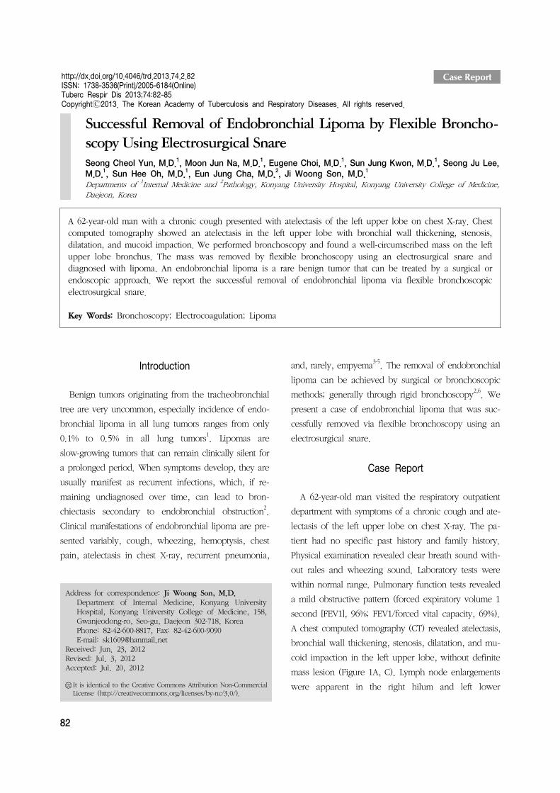
A chest computed tomography (CT) revealed atelectasis,
bronchial wall thickening, stenosis, dilatation, and mu-
coid impaction in the left upper lobe, without definite
mass lesion (Figure 1A, C). Lymph node enlargements
were apparent in the right hilum and left lower
Case Report
Tuberculosis and Respiratory Diseases Vol. 74. No. 2, Feb. 2013
83
Figure 1. Chest X-ray and chest computed tomog-raphy (CT) image. (A) At admission, chest X-ray re-vealed atelectasis. (B) After1.5 months, chest X-ray showed improvement of atelectasis. (C) At admis-sion, chest CT revealed mucoid impaction in left upper lobe bronchus. (D) After 1.5 months, chest CTshowed no definite mass lesion in the left upper lobebronchus.
paratrachea. Two focal calcified granuloma were seen
in the right lung.
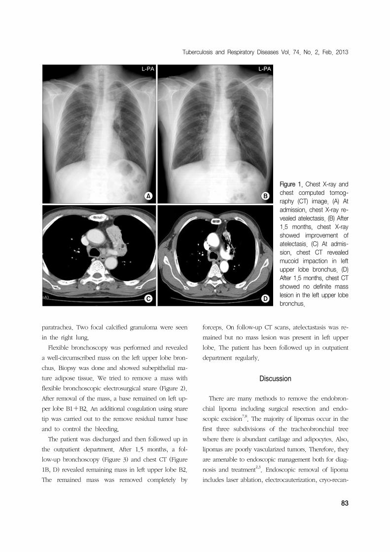
Flexible bronchoscopy was performed and revealed
a well-circumscribed mass on the left upper lobe bron-
chus. Biopsy was done and showed subepithelial ma-
ture adipose tissue. We tried to remove a mass with
flexible bronchoscopic electrosurgical snare (Figure 2).
After removal of the mass, a base remained on left up-
per lobe B1+B2. An additional coagulation using snare
tip was carried out to the remove residual tumor base
and to control the bleeding.
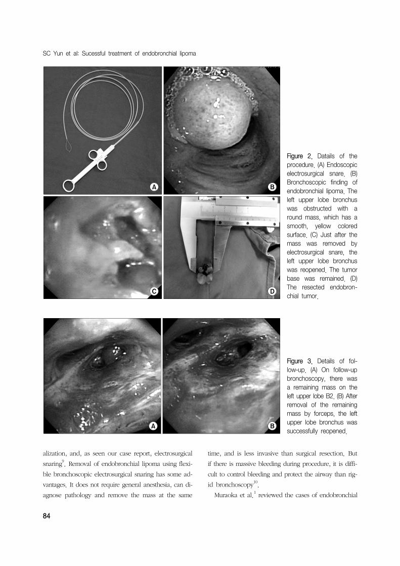
The patient was discharged and then followed up in
the outpatient department. After 1.5 months, a fol-
low-up bronchoscopy (Figure 3) and chest CT (Figure
1B, D) revealed remaining mass in left upper lobe B2.
The remained mass was removed completely by
forceps. On follow-up CT scans, atelectastasis was re-
mained but no mass lesion was present in left upper
lobe. The patient has been followed up in outpatient
department regularly.
Discussion
There are many methods to remove the endobron-
chial lipoma including surgical resection and endo-
scopic excision7,8. The majority of lipomas occur in the
first three subdivisions of the tracheobronchial tree
where there is abundant cartilage and adipocytes. Also,
lipomas are poorly vascularized tumors. Therefore, they
are amenable to endoscopic management both for diag-
nosis and treatment2,3
. Endoscopic removal of lipoma
includes laser ablation, electrocauterization, cryo-recan-
SC Yun et al: Sucessful treatment of endobronchial lipoma
84
Figure 2. Datails of the procedure. (A) Endoscopic electrosurgical snare. (B) Bronchoscopic finding of endobronchial lipoma. The left upper lobe bronchus was obstructed with a round mass, which has a smooth, yellow colored surface. (C) Just after the mass was removed by electrosurgical snare, the left upper lobe bronchus was reopened. The tumorbase was remained. (D) The resected endobron-chial tumor.
Figure 3. Details of fol-low-up. (A) On follow-up bronchoscopy, there was a remaining mass on the left upper lobe B2. (B) After removal of the remaining mass by forceps, the left upper lobe bronchus was successfully reopened.
alization, and, as seen our case report, electrosurgical
snaring9. Removal of endobronchial lipoma using flexi-
ble bronchoscopic electrosurgical snaring has some ad-
vantages. It does not require general anesthesia, can di-
agnose pathology and remove the mass at the same
time, and is less invasive than surgical resection. But
if there is massive bleeding during procedure, it is diffi-
cult to control bleeding and protect the airway than rig-
id bronchoscopy10.
Muraoka et al.1 reviewed the cases of endobronchial
Tuberculosis and Respiratory Diseases Vol. 74. No. 2, Feb. 2013
85
lipoma reported in Japan. Forty patients underwent sur-
gical resection: four pneumonectomies, 24 lobectomies,
eight bilobectomies, and four resections by broncho-
tomy. Seventeen patients have undergone broncho-
scopic treatment: seven patients received Nd-YAG laser
therapy, five tumors were removed by electrosurgical
snaring forceps via flexible bronchoscope and five pa-
tients received a combined therapy using both laser
techniques and polypectomy snares. No tumor re-
currence was reported for these 17 patients after the
bronchoscopic procedure. Bronchoscopic electrocaut-
ery, compared with Nd-YAG laser therapy, is the more
simple and cost-effective, produces less significant bron-
chial stenosis and causes limited damage to the bron-
chial mucosa, although the outcome of both treatments
are virtually the same11-14
. But, in spite of several bene-
fits of bronchoscopic resection, surgical resection is in-
dicated for some patients. The conditions under which
surgical resection is preferred are difficulty of definite
diagnosis and possible complicated malignant tumor,
peripheral destructive lung disease due to long-term ate-
lectasis or pneumonia, extrabronchial growth or sub-
pleural lipomatous disease, or expected technical diffi-
culties during the bronchoscopic procedure due to mul-
tidirectional development of the tumor1.
In our opinion, there are differences between bron-
choscopic and gastrointestinal endoscopic tumor re-
moval by snare. On gastrointestinal endoscopy, it is
possible that the tumor base can be separated by in-
jection of dye fluid, so complete removal of a tumor
including the base can be accomplished. But in the case
of bronchoscopic tumor removal, it is difficult to apply
of the method used in gastrointestinal endoscopy. Thus,
we consider that a tumor should be initially removed
by the snare as much as possible, with the remaining
tumor base being coagulated by the snare tip or by ar-
gon-plasma coagulation.
In this report, we removed successfully endobron-
chial lipoma by electrosurgical snaring via flexible bron-
choscopy. Based on this case and literature review, flex-
ible bronchoscopic resection could be considered as the
first option for the treatment of bronchial lipoma.
References
1. Muraoka M, Oka T, Akamine S, Nagayasu T, Iseki M,
Suyama N, et al. Endobronchial lipoma: review of 64
cases reported in Japan. Chest 2003;123:293-6.
2. Nassiri AH, Dutau H, Breen D, Colchen A, Quiot JJ,
Nguyen B, et al. A multicenter retrospective study in-
vestigating the role of interventional bronchoscopic
techniques in the management of endobronchial
lipomas. Respiration 2008;75:79-84.
3. Iwabuchi H, Kamura T, Tanaka M, Kato H. A case of
endobronchial lipoma. Diagn Ther Endosc 1999;5:
263-7.
4. Cao D, Sun Y, Yang S. Endobronchial lipoma: an un-
usual cause of bronchial obstruction. Case Rep Med
2011;2011:939808.
5. Ouadnouni Y, Bouchikh M, Bekarsabein S, Achir A,
Smahi M, Msougar Y, et al. Endobronchial lipoma a
rare cause of pleural empyema: a case report. Cases
J 2009;2:6377.
6. Choi JC, Yu CM, Ryu YJ, Jeon K, Choi KA, Kwon OJ,
et al. The role of endoscopic surgery for completely
obstructive endobronchial benign tumor. Korean J
Intern Med 2006;21:15-9.
7. Hurt R. Benign tumours of the bronchus and trachea,
1951-1981. Ann R Coll Surg Engl 1984;66:22-6.
8. MacArthur CG, Cheung DL, Spiro SG. Endobronchial
lipoma: a review with four cases. Br J Dis Chest 1977;
71:93-100.
9. Lamprecht B, Hutarew G, Porsch P, Wegleitner B,
Studnicka M. Successful bronchoscopic cryorecanaliza-
tion in a case of endobronchial lipoma. Diagn Ther
Endosc 2011;2011:845686.
10. Landa JF. Indications for bronchoscopy. Chest 1978;
73(5 Suppl):686-90.
11. Boxem T, Muller M, Venmans B, Postmus P, Sutedja
T. Nd-YAG laser vs bronchoscopic electrocautery for
palliation of symptomatic airway obstruction: a cost-ef-
fectiveness study. Chest 1999;116:1108-12.
12. van Boxem AJ, Westerga J, Venmans BJ, Postmus PE,
Sutedja G. Photodynamic therapy, Nd-YAG laser and
electrocautery for treating early-stage intraluminal can-
cer: which to choose? Lung Cancer 2001;31:31-6.
13. Bolliger CT, Sutedja TG, Strausz J, Freitag L. Therapeu-
tic bronchoscopy with immediate effect: laser, electro-
cautery, argon plasma coagulation and stents. Eur
Respir J 2006;27:1258-71.
14. Choi SH, Kang JY, Joo YB, Kim SK, Mo EY, Lee SH,
et al. An endobronchial lipoma treated by broncho-
scopic excision. Korean J Med 2011;80:337-42.