sudden cardiac death in dogs, cats and humans: a
TRANSCRIPT

Sudden cardiac death in dogs, cats and
humans: a comparative review
Word count: 10 934
Céline Brugada Terradellas Student number: 01306714
Promotor: Prof. dr. Pascale Smets
Promotor: Prof. dr. Pedro Brugada Terradellas
A dissertation submitted to Ghent University in partial fulfilment of the requirements for the
degree of Master of Veterinary Medicine
Academic year: 2018 - 2019

Ghent University, its employees and/or students, give no warranty that the information provided in this
thesis is accurate or exhaustive, nor that the content of this thesis will not constitute or result in any
infringement of third-party rights.
Ghent University, its employees and/or students do not accept any liability or responsibility for any use
which may be made of the content or information given in the thesis, nor for any reliance which may
be placed on any advice or information provided in this thesis.

3
Voorwoord
Bij deze wil ik graag enkele personen uitdrukkelijk bedanken. In de eerste plaats Prof. Pascale Smets
(DVM, PhD, EBVS European Veterinary Specialist in Small Animal Cardiology) voor de uitstekende
begeleiding bij het hele proces dat tot dit uiteindelijke werk heeft geleid. De grenzeloze passie voor
het uitbreiden en verder op de kaart zetten van de veterinaire cardiologie was hetgene waar ik me
meteen in herkende en zorgde voor een uiterst aangename en productieve samenwerking.
Verder wens ik ook Prof. Em. Dr. Pedro Brugada (Scientific Director, Cardiovascular Division, Free
University of Brussels (UZ Brussel) VUB., CEO Medical Centre Prof. Brugada, Aalst, Belgium., Director,
Arrhythmia Unit, Hospiten Estepona, Spain) en tevens mijn vader, van harte te bedanken voor alle
steun en hulp bij het schrijven van deze Masterproef. Zijn oeverloze kennis en jarenlange ervaring zijn
van onschatbare waarde gebleken tijdens het tot stand komen van dit werk. Het was een fantastische
ervaring om als vader en dochter hieraan te kunnen samenwerken.

4
1. List of abbreviations
2. Summary
3. Introduction
4. Literature review
4.1. Cardiomyopathies and associated arrhythmias
4.1.1. Dilated cardiomyopathy
4.1.1.1. DCM in dogs
4.1.1.2. DCM in humans
4.1.1.3. DCM in cats
4.1.2. Hypertrophic cardiomyopathy
4.1.2.1. HCM in cats
4.1.2.2. HCM in humans
4.1.2.3. HCM in dogs
4.1.3. Arrhythmogenic right ventricular cardiomyopathy
4.2. Primary electrical cardiac dysfunctions
4.2.1. Channelopathies
4.2.1.1. Channelopathies in humans
4.2.1.2. Channelopathies in dogs and cats
4.2.2. German Shepherd genetic arrhythmias
4.2.3. Catecholamine dependent arrhythmias
4.2.3.1. Catecholaminergic polymorphic ventricular tachycardia in humans
4.2.3.2. CDA in dogs and cats
4.3. Ischemic myocardial events
4.3.1. Ischemic myocardial infarctions in humans
4.3.2. Possible role in SCD in dogs and cats
5. Discussion and conclusions
6. References

5
1. List of abbreviations
• AF: atrial fibrillation
• AP: action potential
• AMI: acute myocardial infarction
• ARVC: arrhythmogenic right ventricular cardiomyopathy
• ARVD: arrhythmogenic right ventricular dysplasia
• ATE: arterial thromboembolism
• BrS: Brugada syndrome
• BSA: body surface area
• CAD: coronary artery disease
• CASQ2: cardiac -specific isoform of calsequestrin gene
• CDA: catecholamine dependent arrhythmias
• CHF: congestive heart failure
• CPVT: catecholaminergic polymorphic ventricular tachycardia
• CTE: coronary thrombo-embolism
• cTnI: cardiac troponin I
• DCM: dilated cardiomyopathy
• DLVOTO: dynamic left ventricular outflow obstruction
• DRVOTO: dynamic right ventricular outflow obstruction
• ECG: electrocardiogram
• EF: ejection fraction
• HCM: hypertrophic cardiomyopathy
• ICD: implantable cardioverter-defibrillator
• IDC: idiopathic cardiomyopathy
• IVF: idiopathic ventricular fibrillation
• LA: left atrium
• LV: left ventricle
• LVEDV: left ventricle end diastolic volume
• LQTS: long QT syndrome
• NT-proBNP: N-terminal prohormone of brain-natriuretic peptide
• PTE: pulmonary throbmo-embolism
• RA: right atrium
• RV: right ventricle
• RyR2: cardiac ryanodine receptor
• SAM: systolic anterior motion
• SCD: sudden cardiac death
• SVPC: supraventricular premature complex
• SQTS: short QT syndrome
• TdP: torsades de pointes
• VF: ventricular fibrillation
• VPC: ventricular premature complex

6
2. Summary
Plotse dood is een van de meest voorkomende doodsoorzaken bij de mens in Westerse landen. Naar
schatting 85% van al deze gevallen zijn van cardiale aard (Magi et al., 2017). Ventriculaire
tachyaritmieën zijn de meest voorkomende oorzaak van plotse cardiale dood (Kirchof et al., 2006). Ook
bij dieren komt dit vaak voor. In vergelijking met de humane geneeskunde is er heel wat minder
geweten over de pathogenese en onderliggende genetica van plotse cardiale dood bij dieren.
In deze review zullen we de meest voorkomende oorzaken van plotse cardiale dood bespreken. Dit
zullen we doen voor zowel de mens als verschillende diersoorten zijnde hond en kat. De focus zal
vooral op de pathogenese liggen, en meer specifiek op de gekende gelijkenissen en verschillen tussen
de mens en dieren, en zelfs tussen verschillende rassen. We hopen uiteindelijk nieuwe inzichten te
verwerven wat betreft de pathogenese en diagnose van plotse cardiale dood bij dieren.
Deze vergelijking zal twee kanten opgaan. Langs de ene kant zullen we informatie uit klinische studies
uit de humane geneeskunde halen, maar ook uit meer experimentele studies gebaseerd op
dierproeven. Deze vinden plaats met behulp van onder andere knock-out muizenstammen en
ingeteelde kolonies (Camacho et al., 2016), maar ook van grote zoogdieren zoals katten en honden
(Duncker et al., 2015; Hamlin, 2007). Langs de andere kant zullen we ook data uit humane studies
gebruiken in een poging tot het beter begrijpen van de pathogenetische mechanismes van plotse
cardiale dood bij dieren.
Sudden cardiac death is one of the most common causes of death in humans in Western countries.
Approximately 85% of all these cases are of cardiac origin (Magi et al., 2017). Ventricular
tachyarrhythmias are the most common cause of sudden cardiac death (Kirchof et al., 2006). In animal
SCD also commonly occurs. When compared to human medicine, a lot less information is currently
available concerning pathogenesis and underlying genetics of SCD in animals.
In this review the most common causes of sudden cardiac death will be discussed. This will be the case
for human but also several other species being cats and dogs. The focus will be put on pathogenesis
and more specifically on the known differences and similarities between humans and animal, and even
between several breeds. We hope to create new insights on pathogenesis and diagnostics concerning
sudden cardiac death in animals.
This comparison will be two folded. On one side information will be gathered from clinical studies in
human medicine, but also from more experimental studies based on animal testing. These usually rely
on for example knock-out strains of mice and in-bred colonies (Camacho et al., 2016), but also on larger
animal models such as dogs and cats (Duncker et al., 2015; Hamlin, 2007). On the other hand, data
from human studies will be used in order to try and better understand pathogenetic mechanisms of
sudden cardiac death in animals.

7
3. Introduction
The following paragraphs represent a summary of the current information regarding sudden cardiac
death (SCD) in humans, dogs and cats. The main goal of the literature review will then be to further
analyze every condition and make a comparison between humans and animals considering main
components of the origin of the diseases such as genetics and pathophysiology.
Humans
First of all, a few well-known diseases in humans will be discussed. There is an overwhelming body of
scientific data emphasizing the importance of coronary artery disease (CAD) as the most important
cause of SCD in humans. However, other causes of SCD not related to CAD are for example other
structural heart diseases such as dilated cardiomyopathy (DCM), hypertrophic cardiomyopathy (HCM)
or arrhythmogenic right ventricular cardiomyopathy (ARVC). On the other side, human SCD can occur
in individuals with a pure cardiac electrical disease without structural abnormalities. The most common
of these diseases are Brugada syndrome (BrS), long QT syndrome (LQTS), short QT syndrome (SQTS)
and catecholaminergic polymorphic ventricular tachycardia (CPVT) (Papadakis et al., 2018).
Apart from pure electrical diseases, several forms of structural heart disease can predispose to
ventricular tachycardia or ventricular fibrillation and thus possibly cause SCD. Many of these structural
diseases are inherited disorders and are transmitted according to Mendelian patterns (autosomal
dominant or recessive). They can be responsible for up to 26% of SCDs (Sweeting, 2018). In humans,
CAD is the most common cause of SCD at advanced age, but other structural heart diseases play also
a role.
The LQTS (Romano-Ward syndrome) was the first syndrome described in humans as a pure electrical
disorder without structural abnormalities. The first description dates back more than 50 years. For
years, it was widely accepted that the long QT syndrome was the number one cause of sudden cardiac
death in humans with a normal structural heart on autopsy. More than 15 different forms are currently
known (Perez-Riera, 2018). One form of this syndrome (type 3) is caused by a gain-of-function
mutation in the SCN5A gene. This gene encodes for the alpha subunit of the cardiac sodium channel.
The syndrome is characterized by abnormal sodium influx in the cardiomyocytes with a prolonged QT
interval as a result (Wilde, 2018). Prolongation (as well as shortening) of the QT interval is strongly
linked to ventricular tachyarrhythmias. These are a direct cause for sudden cardiac death (Tse, 2017).
In the long QT syndrome, the typical polymorphic tachycardia is known as ‘torsades de pointes’ (TdP)
(Shimizu, 2012; Bezzina, 2001).
Besides the long QT syndrome, there is also the short QT syndrome. Just like in long QT syndrome,
several mutations have been linked to the disease. In general, there is either hyperfunction of
potassium channels, or hypofunction of the calcium channels. This causes shortened atrial and

8
ventricular refractory periods, shortening of the action potential and short QT intervals on ECG. These
then typically predispose for atrial or ventricular fibrillation and thus SCD1.
Catecholaminergic polymorphic ventricular tachycardia (CPVT) leads to SCD also because of ventricular
arrhythmias. These occur because of mutations in the ryanodine receptor encoded by the gene RYR2.
The calcium transport and handling are disrupted by these mutations. Typically, the arrhythmias occur
during exercise because of the increased sensitivity of the calcium handling to catecholamines.
The idea that long QT syndrome is the leading cause of arrhythmic death caused by a channelopathy,
has recently been debunked. The results of a prospective study concluded that it was not long QT
syndrome, but Brugada syndrome that is the leading cause of these sudden unexpected deaths.
Approximately 28% of the studied families and 15% of the studied relatives where diagnosed with
Brugada syndrome (Papadakis, 2018). This syndrome is also a primary electrical heart disease. When
considering the pathophysiology behind these two channelopathies, Brugada syndrome is in some
ways the mirror-image of long QT syndrome type 3. It is caused by a loss-of- function mutation in the
SCN5A gene which causes the encoding of defective sodium channels. The decrease in sodium channel
current leads to ventricular fibrillation and possible sudden death (Sweeting, 2018).
Dogs
The first species apart from humans that will be studied are dogs. Structural heart diseases have
commonly been linked to SCD in dogs. The most important ones will be discussed.
After mitral valve disease (MVD), dilated cardiomyopathy (DCM) is the second most common heart
disease in dogs. This structural heart disease frequently has a familial basis, just like in humans. Mostly
large and giant breeds are affected (Simpson et al., 2015). Several breeds are strongly predisposed,
such as the Doberman Pinscher with a 58% cumulative incidence for the disease (Wess et al., 2010).
Also, the Irish Wolfhound, Newfoundlands, Portuguese Waterdog and German Shepherd are known
to be often affected (Freeman et al., 2017). Inheritability was investigated in several breeds. In the
Doberman Pinschers for example, an autosomal dominant transmission with incomplete penetrance
is suggested, and in juvenile Portuguese Waterdogs an autosomal recessive transmission (Tidholm and
Jonsson, 2005).
In dogs with DCM, the primary problem is a decreased systolic function (Simpson et al., 2015). Both
left and right heart are usually affected. As a result of the myocardial damage, arrhythmias can occur.
Also, the renin- angiotensin-aldosteron-system is activated because of the lacking ‘pump’. This then
leads to sodium retention, ventricular remodeling left ventricular dysfunction and dilation (Tripathi et
al., 2017). This dilation can also affect the annulus of the valve. As a result, some dogs also obtain
secondary valvular insufficiencies. Nevertheless, this has minor importance in the context of SCD.
1 From: https://www.escardio.org/Journals/E-Journal-of-Cardiology-Practice/Volume-9/Short-QT-
Syndrome. Consulted lastly May 8th 2018.

9
Early diagnosis of structural heart disease and especially DCM is key in the prevention of its
complications. It has been shown that Dobermans with systolic dysfunction treated with pimobendan
(Vetmedin, Boehringer Ingelheim Vetmedica GmbH) stayed in the pre-clinical phase of disease 9
months longer than the placebo-treated dogs, stressing the need for early diagnosis and treatment
(Summerfield et al., 2012). The problem is that there is often a very long occult phase with mild to
absent clinical signs. This makes diagnosis a true challenge (Simpson et al., 2015). Apart from DCM,
several other structural heart diseases associated with ventricular arrhythmias and SCD are known in
dogs. For example, ARVC which was mostly studied in Bulldogs and Boxers (Boujon and Amberger,
2003; Hamlin, 2007; Simpson et al., 2015; Cunningham et al., 2018).
Dogs with DCM often display malignant ventricular arrhythmias, which is a very important
complication because the arrhythmias can cause SCD. For example, about one-third of Doberman
Pinschers in the occult stage of DCM can die from SCD (Klüser et al., 2016). Some of these arrhythmias
can be detected during a 24-hour Holter monitoring (Simpson et al., 2015).
Electrical diseases can also cause SCD in dogs. For example, ventricular arrhythmias without a known
underlying structural cause are described in young German shepherds (Moïse et al., 1994; Jesty et al.,
2003; Hamlin, 2007; Pariaut et al., 2011; Ribas et al., 2018) and potentially Rhodesian ridgebacks
(Meurs et al., 2016). There are suggestions that channelopathies could play a great role in sudden
cardiac death in animals, like they do in humans, but data is scarce at the moment (Schwartz et al.,
2017).
R on T phenomenon was described as a possible electrical dysfunction causing SCD in dogs. A Golden
Retriever previously diagnosed with severe subaortic stenosis and LA dilation died suddenly while
wearing a Holter monitor. The dog portrayed premature ventricular complexes occurring onto the T
wave (R on T phenomenon). These further evolved into typical polymorphic ventricular tachycadia also
known as torsades de pointes (Gunasekaran et al., 2017).
Also, it is generally believed that myocardial infarctions are very uncommon in cats and dogs, but
recently a role in patients with symptoms of acute and severe heart failure is becoming more and more
present (Wilson and Scheel, 1982; Vanoli et al., 1991; Driehuys et al., 1998). However, the cause of
these myocardial infarctions may not be CAD, but rather systemic disease with enhanced thrombotic
risk. A link with Duchenne muscular dystrophy is possible, but more research is required to confirm
this suspicion (Schneider et al., 2016).
Cats
Cats will also be included in this study, because they are also a possible victim of SCD. Just like in dogs,
several conditions are known to cause arrhythmias and sudden death. Here we will specifically discuss
the best known and most common structural heart disease: hypertrophic cardiomyopathy. More
research is still needed concerning primary electrical diseases causing SCD in cats.
10
Approximately 10-15% of pet cats have some form or degree of hypertrophic cardiomyopathy (Payne
et al., 2015; Freeman et al., 2017). This primary structural heart disease affects cats of all ages and
breeds, but mostly Domestic Shorthairs and males (Freeman et al., 2017). In some breeds such as the
Maine Coon, Ragdoll and British Shorthair, HCM is often seen more in younger individuals (Lefbom et
al., 2001). Other breeds are also predisposed such as the Sphynx, Scottish Fold, Persian and Burmese.
In the Ragdoll and Maine Coone, an autosomal dominant transmission with incomplete penetrance
was found. The mutation is seen in a gene responsible for the myosin-binding protein C in the
sarcomeres of the myocardium. Recently, a new causative mutation was identified by Schipper et al.
(2019) being the MYH7 c.5647G>A variant in a Domestic Shorthair. Most likely multiple other not yet
identified mutations can cause the disease (Freeman et al., 2017).
Briefly, the pathogenesis of HCM is as follows. Because of the mutation, there is a defect in the
sarcomeres. Compensatory hypertrophy of the myocardial walls and interventricular septum will take
place as a result. Echocardiography shows that both the left as well as the right ventricle are usually
involved in the hypertrophic process (Schober et al., 2016). Also, fibrosis and disarray of the myocardial
fibres are typical findings, as is disturbed calcium transport in and out of myocardial cells (Ferasin,
2009).
What is important with regard to SCD, is that cats with asymptomatic HCM have significantly more
frequent and complex ventricular and supraventricular arrhythmias than normal cats. This was proven
based on echocardiography, ECG, Doppler blood pressure measurements and 24-h Holter monitoring
(Jackson et al., 2014; Hanas et al., 2017). As we already stated, these could possibly lead to SCD like
they do in dogs. More studies are needed to prove that these findings are directly linked to increased
SCD in asymptomatic HCM cats (Jackson et al., 2014).
11
4. Literature review
4.1. Cardiomyopathies and associated arrhythmias
4.1.1. Dilated cardiomyopathy
4.1.1.1. Dogs
The pathogenesis of DCM in dogs is very similar to that in humans. It can be defined as a classic
structural cardiomyopathy, which implies there is an aberrant structure of the heart. The disease can
be primary and thus genetically inherited or can be secondary attained. The primary form is caused by
genetic defects that encode for defective myocardial cells, who typically portray decreased inotropy
and thus cause systolic dysfunction of the myocardium. Phenotypically, it is characterized by
progressive dilation and thinning of the myocardial wall and possibly interventricular septum.
In the Doberman Pinscher a cumulative incidence of 58% was noted (Wess et al., 2010). Also, the Irish
Wolfhound, Newfoundland, Boxers, Great Dane, St. Bernard, Portuguese Waterdog and German
Shepherd are high risk breeds (Freeman et al., 2017; Simpson et al., 2015). In humans several
responsible mutations were already identified, but in dogs no exact causative gene defect were found
yet. An autosomal recessive pattern is likely in juvenile Portuguese Waterdogs, whereas autosomal
dominant transmission was suspected in the Doberman Pinscher (Tidholm and Jönsson, 2005). In Great
Danes transmission is probably linked to the X-chromosome (Wess et al., 2010).
Secondary DCM is often seen a result of nutritional deficiency, such as taurine-deficiency. Taurine is a
free amino acid and is not considered an essential amino acid in dogs. It is highly important for
numerous physiological processes such as proper retinal function (which will be discussed later in cats),
reduction in platelet aggregation, anti-oxidant in all cells including cardial cells and most importantly
for us, is responsible for adequate myocardial function. The exact ways in which taurine operates as
an aid in myocardial cell function are still unknown, but it is suspected it helps modulate tissue calcium
concentrations and availability. Also, taurine is a direct antagonist of angiotensin II and thus interacts
with the RAAS activation which plays a huge role in DCM progression (Sanderson, 2006). Knowing this,
it is no surprise taurine deficiency can lead to (severe) myocardial dysfunction. The link between
taurine and DCM was first noticed in foxes (Moise N.S., 1989; Sanderson, 2006). Rapidly, the
pathogenesis of DCM in dogs was reconsidered, and experts found that 17% of 75 dogs with DCM in a
study had low taurine plasma levels. These dogs with low taurine levels were mostly ‘unusual DCM
breeds’ such as American Cocker Spaniels and Golden Retrievers, which led to the idea of a non-
primary DCM (Kramer et al., 1995). Lately, newer and unconventional diets such as grain-free and
‘boutique’ diets are getting more and more attention. These diets typically contain very different
nutrient sources compared to more classical balanced diets. The exact link is not yet identified but
causes including dietary deficiency or reduced bioavailability of taurine or its precursors are suggested.
Also, a different processing of the dietary taurine or increased faecal excretion are possible (Tidholm
and Jönsson, 2005; Kaplan et al., 2018).
Secondary DCM can also be tachycardia-induced. This form of DCM is usually reversible and is also
seen in humans. As in other forms of DCM, myocardial contractility is also decreased. Apart from this,
other consequences are increased afterload/peripheral vascular resistance. Diastolic function is also
affected. Very typical is the incomplete relaxation of the myocardium because of abnormal increase in
calcium levels in the sarcoplasmatic reticulum during tachycardia resulting in diastolic contracture. The

12
left ventricle usually displays ‘normal’ DCM characteristics with the most prominent one being dilation
of the lumen, whereas the right ventricle also develops eccentric hypertrophy (Gupta and Figueredo,
2014).
Infectious and toxic agents such as doxorubicin where also related to secondary dilatory changes of
the heart in dogs. Doxorubicin is an antitumor antibiotic used to treat for example lymphoma.
Especially in already at-risk breeds such as Boxers, the incidence of DCM during chemotherapy with
doxorubicin was quite remarkable with 15.4%, whereas in other breeds a rather low incidence of 3%
was noticed (Hallman et al., 2018). Not all dogs showed clinical symptoms, but when they did, they
usually emerged 6 months after doxorubicin use. Duration of infusion with doxorubicin had no
influence on the incidence of cardiotoxicity. Mostly premature contractions and decreased fractional
shortening were seen, but also decreased systolic function and arrhythmias (Hallman et al., 2018). This
last finding is important when considering sudden death.
Since ventricular contractions are responsible for adequate circulation of the blood and along with it
sufficient cardiac output, the renin-angiotensin-aldosteron-system will be activated in order to
maintain correct arterial blood pressure. This is a result of the decreased systolic dysfunction. The
activation of this cascade results in expression of renin by the kidney granular cells, which hydrolyses
angiotensinogen to angiotensin I. After, angiotensin converting enzyme (ACE) converts angiotensin I
to angiotensin II which has smooth muscle contracting properties promoting vasoconstriction. Another
effect of renin is the activation of the sympathetic nervous system. Also, angiotensin II will stimulate
aldosterone secretion, which is responsible for sodium-potassium homeostasis in the body (Patel et
al., 2017). Retention of sodium results in fluid retention, which eventually causes volume overload of
the heart. The main issue here is the resulting dilation of both left and right ventricles which then
predisposes to arrhythmias. From human studies, we know that myocardial scarring is the main trigger
for arrhythmias (Horowitz et al., 1980). This is a direct cause for SCD.
Whether a patient is affected by primary or secondary DCM, the consequences often remain the same
and are usually quite serious. The dilation of the left and often also the right ventricle has several
effects. First of all, systolic function will further decrease, and also the annulus of the valves can be
affected by the dilation, resulting in a secondary valve insufficiency. As already stated, this is less
important concerning SCD, although it will contribute to the volume overload. What is important, is
the fact that most dogs with DCM display malignant arrhythmias and thus have a direct link to SCD.
This can easily be explained: when the myocardium is damaged it is more likely that arrhythmias occur
because of the disrupted cell function. Often ectopic foci will start firing signals resulting into
(supra)ventricular premature complexes. These can then easily evolve to for example ventricular
tachycardia and/or fibrillation and death (Simpson et al., 2015).
What is absolutely essential in this story, is to recognize DCM usually has three stages concerning the
clinical progression of the disease. During the first stage no morphological or electrical abnormalities
can be detected. This stage can last several years. In the second stage, also called the occult or
preclinical phase, there are still no or very subtle clinical signs but there may be morphological or
electrical abnormalities such as an enlarged left ventricle or arrhythmias on ECG such as VPCs. For the
owner, the dog will appear practically normal during this phase which makes diagnosis very
challenging. Eventually when the dog reaches the third stage, clinical signs of heart failure such as

13
ascites, coughing, exercise intolerance, lethargy and pulmonary edema will be noticeable (Wess et al.,
2010; Simpson et al., 2015).
In high risk breeds, such as Doberman Pinschers and Great Danes, diagnostic tools such as
echocardiography, ECG and 24-hour Holter monitoring are essential in the battle against congestive
heart failure. Especially these types of dogs should be screened from a young age on to make sure that
structural or electrical changes are noticed as early as possible. This suspicion was confirmed in the
Protect study showing that pimobendan (Vetmedin, Boehringer Ingelheim Vetmedica GmbH)
treatment in Dobermans with systolic dysfunction prolonged their time in the occult phase with 9
months as opposed to placebo-treated dogs, thus stressing the importance of an early diagnosis
(Summerfield et al., 2012).
Most dogs from at risk breeds such as Doberman Pinschers are diagnosed with occult DCM are
between 5 and 7 years old, but dogs as young as 2 years old can already be affected. Screening should
start at the age of 3. Ideally, according to Wess et al. (2017) these screenings using echocardiography
and Holter monitoring should be repeated yearly to make timely diagnosis possible. This way we can
also try and eliminate affected male dogs as soon as possible from breeding programs so less affected
puppies will be born in the future. When performing Holter monitoring, 24h monitoring is advised in
order to get a complete and thorough idea of the ECG. In Doberman Pinschers a maximum of 50 VPC’s
per 24h is considered normal and more than 300 VPC’s per 24h is diagnostic for occult DCM (O’Sullivan
et al., 2007; Wess et al., 2017). Because risk factors for SCD were poorly defined Klüser et al. (2016)
investigated predictive parameters for SCD in Doberman Pinschers diagnosed with DCM. They found
that the ejection fraction (EF) alone, as is used in human medicine as a predictive factor, is insufficient.
This is mainly due to the fact that not all dogs with DCM suffer from decreased systolic dysfunction.
They evaluated the prognostic value of echocardiographic and Holter-ECG variables, but also cardiac
Troponin I (cTnI) and N-terminal prohormone of brain-natriuretic peptide (NT-proBNP) and tried to
provide conditional inference trees based on these variables. Volume overload of the left ventricle
(which was expressed as a Left Ventricle End Diastolic Volume (LVEDV) on Body Surface Area (BSA))
was found to be the best predictor for SCD. EF and left ventricular end-systolic volume where strongly
linked to the LVEDV/BSA.
4.1.1.2. Humans
Many of the structural diseases are inherited disorders and are transmitted according to Mendelian
patterns (for example autosomal dominant or recessive). They can be responsible for up to 26% of
SCDs (Sweeting, 2018). Also in humans DCM is a well-known cardiomyopathy. Just like in dogs, the
condition is characterized by progressive thinning of the myocardial wall and decreasing systolic
function.
It is usually seen as an inherited primary structural disease but can also occur as a secondary
phenomenon due to for example coronary artery disease. It is considered a mixed (genetic and non-
genetic) condition according to Wexler et al. (2009) but the disease is familial in approximately 25% of
the cases (Collis and Elliott, 2017). Mutations are usually seen in genes that encode for cytoskeletal
proteins, sarcomeres, intercalated discs and nuclear membranes (Collis and Elliott, 2017). DCM is the
most common cardiomyopathy seen in humans (Sweet et al., 2015). The incidence is approximately 5
in 100 000 adult and 0,57 in 100 000 children, which is probably an underestimation since many cases
remain unrecognized (Wexler et al., 2009; Collis and Elliott, 2017). Blacks and males have a 2,5 times
higher risk compared to whites and women (Dec and Fuster, 1994). Symptoms may present as

14
shortness of breath, fatigue, cough, orthopnea, paroxysmal nocturnal dyspnea and edema (Wexler et
al., 2009).
When considering primary inherited DCM, in 30-40% of the cases a causative genetic mutation is
found. Mutations were already detected in more than 50 different genes such as for example SCNA5A
or DSP. For several of these genes genetic screening is possible. Other primary cases are considered of
idiopathic origin. A confirmed family history of DCM is the only clinical phenotype available to
distinguish between familial and idiopathic primary DCM (Sweet et al., 2015). In most of the cases, the
pattern seems to be autosomal dominant, but also autosomal recessive, X-linked recessive and
mitochondrial inheritance were reported (Dec and Furster, 1994).
DCM can also occur as a secondary result of viral myocarditis or other cytotoxic insults, for example
Coxsackie virus B3 antibodies were often found in large amounts in patients with both Idiopathic
Cardiomyopathy (IDC) and myocarditis. The exact link between the two still remains unknown, but an
autoimmune response triggered by induction difficulties in the MHC gene could possibly play a role
(Dec and Fuster, 1994). Also, immune abnormalities, and metabolic, energetic and contractile
abnormalities such as abnormalities of cardiac high-energy phosphate substrate of abnormal actin-
myosin interaction could lead to secondary myocardial dilatory changes (Dec and Fuster, 1994).
Diagnostic criteria for DCM on echocardiography are fractional shortening <25% and/or ejection
fraction <45%, and also left ventricular end diastolic diameter >117% after excluding any known cause
of myocardial disease. (Sweet et al., 2015). Also, screening is very important in humans, just like in
dogs, since the occult phase exists here too. In human medicine multiple techniques are used to do
this, such as echocardiography, ECG and genetic testing.
Exactly like in dogs, pathogenetic pathways in humans lead to RAAS-activation, but most importantly
in our study predispose for possible (malignant) arrhythmias that can cause SCD. The mechanism
behind this revolves around myocardial ischemia. This ischemia can be explained by defective
myocardial perfusion, decreased wall motion and abnormal cell metabolism that are often present in
patient with DCM. Ischemia can be a direct cause of arrhythmias: we could state it is a structural defect
turning into an electrical defect (Fang et al., 2010).
4.1.1.3. Cats
DCM in cats is very rare. It is most frequently caused by taurine deficiency, but as Pion et al. (1987)
stated, the incidence drastically reduced when the importance of taurine supplementation in cat food
was recognized. It this case DCM is usually reversible but irreversible retinal degeneration can be seen.
Nowadays, cats with taurine related DCM are usually fed a non-traditional diet (Pion et al., 1987).
Hambrook and Bennett (2012) studied the effect of pimobendan in thirty-two cats with non-taurine
responsive DCM and concluded that the mean survival time was four times higher in the pimobendan
treated cats than in the non-pimobendan treated cats. A gender or breed predisposition was not
confirmed (Pion et al., 1992).
Symptoms and consequences for cats with DCM are much similar to those in dogs and humans. It also
predisposes for malignant arrhythmias and thus SCD since it compromises normal myocardial
structure. An important side note is that since systolic function is impaired in DCM patients, symptoms

15
of left heart failure will emerge, but in some cats also the right heart is affected. In dogs and humans,
we would suspect clinical signs such as lung edema, coughing, exercise intolerance, but in cats also
pleural effusion is seen in cases of left-sided CHF. This is probably due to a different anatomy of the
pulmonary venous system and different porosity of subpleural capillary beds in the thorax of the cat
(Johns et al., 2012). Also gallop rhythm (third and fourth heart sounds), murmurs and (aortic)
thromboembolism can be a sign of DCM in cats (Pion et al., 1992).
Recently, DCM was diagnosed in a juvenile Pallas cat (a wild feline species) with dyspnea based on
echocardiography and was confirmed at necropsy. Both atria and ventricles were dilated and
histologically fibroelastosis of the endocardium was seen (Gudenschwager et al., 2019).
4.1.2. Hypertrophic cardiomyopathy
4.1.2.1. Cats
Hypertrophic cardiomyopathy (HCM) is also a primary structural disease of the heart. It is characterized
by diastolic dysfunction, caused by hypertrophy of the free walls and interventricular septum leading
to impaired filling of the ventricles and atrial dilation. This concentric hypertrophy is of compensatory
origin, since genetic mutations cause decreased function of the myocardial cells. Mostly disarray of the
myocardial sarcomeres is seen, along with fibrosis and impaired calcium transport in and out of the
myocardial cells (Ferasin, 2009). Also, enlargement of the left atrium (LA) is very often seen, which is a
direct hemodynamic result of the diastolic dysfunction that causes volume overload of the LA.
Echocardiography shows that also the right ventricle is involved in the hypertrophic process in about
50% of the cats with HCM. Also, severity of LV and RV hypertrophy are related (Schober et al., 2016).
A striking 10-15% of all cats is affected by HCM (Payne et al., 2015; Freeman et al., 2017). There is a
clear predisposition in Domestic Shorthairs and males, but also the Sphynx, Scottish Fold, Persian and
Burmese are often diagnosed with HCM, but all genders, ages and breeds can be affected (Freeman et
al., 2017). Important to note is that even though male cats are predisposed for HCM, they are at no
higher risk to develop CHF of SCD than female affected cats (Fuentes and Wilkie, 2017). In British
Shorthairs, Ragdolls and Maine Coons, the disease is also often seen in younger individuals (Lefbom et
al., 2001).
As we already stated, the disease is primary inherited and just like in DCM, a lot of research is still
required to identify exact causative mutations. In the Ragdoll and Maine Coon an autosomal dominant
transmission with incomplete penetrance in the gene encoding for myosin-binding protein C (MYPBC3)
is very likely to be one of the causes, just like in humans, but others still need to be identified (Maron
and Fox, 2015). Recently, a new causative mutation for HCM in cats was identified by Schipper et al.
(2019). It was a MYH7 c.5647G>A variant in a Domestic Shorthair of which the orthologous variant
already was reported in a human with HCM.
Often cats with HCM are first recognized because of a systolic heart murmur (20-60%) which may have
been noticed on routine examination, but many cats have innocent heart murmurs or murmurs caused
by other heart diseases (Fuentes and Wilkie, 2017). Gallop rhythm is actually a much more specific
finding for HCM than a murmur and is heard when diastolic filling is audible. It is usually typical for
diastolic dysfunction as it is heard when the left ventricle is less flexible and pressures in the LA rise.
Audible gallop rhythm or arrhythmia are markers for high-risk HCM (Fuentes and Wilkie, 2017). Due to

16
pulmonary oedema,pleural effusion and even ascites also dyspnea and/or tachycardia can be seen as
symptoms, but the cat can have no symptoms at all (Ferasin et al., 2003). Sometimes, arterial
thromboembolism or even sudden death can be the first manifestation of HCM in cats (Freeman et al.,
2017). ATE is a consequence of stasis of blood in the enlarged left atrium. The flow is slower and
coagulability increases. This way thrombi can be formed and can follow arterial circulation. Often the
thrombus then gets stuck at the level of aortic bifurcation, causing the classic symptoms such as
paresis/paralysis of the hind limbs, acute severe pain, decreased rectal temperature and pale and cold
hind limbs (Borgeat et al., 2013). Even though this is the classical presentation, the syndrome is not
solely caused by arterial occlusion only, since Imhoff (1961) found that experimental ligation of the
feline distal aorta does not reproduce this exact clinical syndrome. Prostaglandins and serotonin along
with vasoactive mediators probably also play a critical role in the onset of the phenomenon.
HCM is normally diagnosed on echocardiography by measuring the left ventricular free wall and
interventricular septum thickness. Commonly seen in HCM cats is systolic anterior motion of thre
mitral valve, often accompanied by dynamic left ventricular outflow obstruction. Around 30% of cats
with HCM are affected by this. In this case, the mitral valve moves towards the interventricular septum
during systole, which causes a dynamic obstruction of the left ventricular outflow area, i.e., of the
aorta. Normally this occurs due to deviant implantation of papillary muscles and abnormal chordae
tendineae (Fuentes and Wilkie, 2017). When the RV is involved, an obstruction of its outflow area can
also occur due to the interventricular septum and RV free wall moving towards each other. This
phenomenon increases in severity with increase of heart rate and/or contractility and of course when
the concentric hypertrophy is more pronounced. Important to note is that no increased risk for cardiac
death was seen in cats with accompanying outflow obstructions, but since these usually cause a
murmur and thus are more easily noticed and have a higher chance of being treated appropriately,
these cats will have a greater chance at positive outcome than HCM affected cats without a murmur
(Fox et al. 1995; Rush et al., 2002). Mean survival time after HCM diagnosis was 1276 days (Payne et
al., 2010).
When performing ECG on these cats, not all of them show arrhythmias, but when they do it is usually
episodes of VPCs. Côté and Jaeger (2008) found that 96% of all cats with ventricular tachyarrhythmias
in their study had underlying structural heart disease, indicating the importance of the search for an
underlying cause when arrhythmias are seen. On the other hand, they investigated the hypothesis that
ventricular tachyarrhythmias could serve as a possible predictor for underlying cardiomyopathy. In
addition to this hypothesis, it became clear that ventricular tachyarrhythmias were linked much more
frequently to myocardial disease than they were to congenital heart malformations. This may occur
due to the high arrhythmogenicity of cardiomyopathies or simply due to their higher prevalence.
Some causes of SCD cannot be classified strictly into the previously mentioned categories. For example,
other causes of arrhythmia in cats include hyperthyroidism, restrictive and/or unknown
cardiomyopathy, cardiac neoplasia and bacterial endocarditis. These usually cause secondary left
ventricle hypertrophy similar to HCM findings, but strictly cannot be put under the same primary
cardiomyopathic category (Côté and Jaeger, 2008).

17
4.1.2.2. Humans
The prevalence of HCM in humans is about 1 in 500 is affected when considering screening data and
the disease is usually inherited by an autosomal dominant pattern with age-related penetrance (Shah,
2017). Mutations in 11 genes are known but about 70% them occur in the genes encoding for the
myosin-binding protein C and the beta-myosin heavy chain of the sarcomeres (Maron and Maron,
2013). Unfortunately, in only about 50% of clinically affected probands the causative mutation is
found. The typical disarray of the muscle fibres and fibrosis which we see in cats, are also known in
humans (Maron and Maron, 2013; Maron and Fox, 2015; Shah, 2017).
The disease can possibly lead to CHF, stroke (embolic and usually associated with AF, syncope and of
course SCD, but in most cases is completely asymptomatic (Maron and Maron, 2013; Shah, 2017).
Spontaneous ventricular fibrillation is the most common direct cause for SCD in these patients (Collis
and Elliott, 2017). Even when no risk factors are present, a patient with HCM still has an annual 0,6%
mortality chance of SCD and the risk peaks in age group 8 to 16-year-old. Major risk factors include
previous cardiac arrest, sustained and non-sustained ventricular tachycardia, unexplained syncope,
family history of SCD and maximal left ventricular wall thickness > 3cm (Shah, 2017).
4.1.2.3. Dogs
HCM in dogs is extremely rare. When it does occur, is has a very similar pathogenesis to HCM in cats
and humans. LV free wall and interventricular septum hypertrophy is seen. Although most dogs remain
asymptomatic, signs of CHF and even syncope or SCD can be seen. Diagnosis still remains difficult and
requires echocardiography to reveal myocardial hypertrophy (Liu et al., 1979).
Most diagnosed cases were in male dogs under 3 years of age. Since reported cases are so sparse, it is
difficult to draw a conclusion about predisposition. Breeds that already have been diagnosed with HCM
include mostly Rottweilers and Dalmatians, but also the German Shepherd, Doberman Pinscher,
Pointer, Walker hound, Airedale terrier, Great Dane, Cocker Spaniel, Bull dog, Boston Terrier,
Pembroke Corgi, Rhodesian Ridgeback and a Shih Tzu were reported (Liu et al., 1979; Washizu et al.,
2003).
Also, Washizu et al. (2003) reported a 14-year-old Yorkshire Terrier with cardiac murmur and
convulsive seizures who was diagnosed with HCM and had typical myocardial disarray and fibrosis. The
suspected late onset of the disease in this dog is even more rare.
Liu et al. (1979) fully examined 10 dogs on necropsy with findings of heart disease resembling HCM.
What is striking is that 3 out of 10 dogs suffered from sudden death, with no symptoms of heart failure
prior. Also, 3 dogs showed signs of complete heart block. The main difference with cats and humans is
that no causative genetic mutation was found yet in dogs. There is a high suspicion these genetic
defects will also lay in sarcomere proteins, but there is no evidence today. What is remarkable is Liu et
al. (1979) also found that only 2 out of 10 dogs had typical findings on histology like in cats and humans,
being myocardial cell disarrange. Left ventricular free wall thickness varied between 10 and 19mm. We
have to keep in mind that the findings on necropsy were only resembling HCM and histological exam
did not often fit the usual findings. Since HCM is not a disease solely diagnosed on ECG, this means
these dogs could have possibly been affected by secondary HCM and not the primary inherited form.

18
4.1.3. Arrhythmogenic right ventricular cardiomyopathy
ARVC was first described in humans and is characterized by atrophy and fibrofatty infiltration of the
right ventricular myocardium. The right ventricular free wall is affected, mostly in the typical ‘triangle
of dysplasia’ being the subtricuspid outflow tract, infundibular inflow tract and apex (Boujon and
Amberger, 2003; Stadiotti et al., 2017). It is a primary genetically inherited structural heart disease,
which also eventually leads to electrical deficiencies of the heart in a similar way to HCM and DCM.
Usually it follows a Mendelian autosomal dominant pattern. Mutations in over 13 genes were already
found in humans. Most of these mutations occur in the genes encoding for desmosomes, a protein
containing structure specialized in cell-to-cell adherence, for example PKP2 (Stadiotti et al., 2017).
Besides these mutations it is also suspected that myocardial atrophy can be induced by patchy
myocarditis, in this case it is not known whether the inflammation is secondary to myocardial death or
if it is the cause (Basso et al., 1996). Patients with ARVC, also called ARD (arrhythmogenic right
ventricular dysplasia), are very susceptible to malignant arrhythmias and thus SCD. Especially the
young are often affected (Fox et al., 2000). Apart from SCD, ARVC can also lead to CHF. Symptoms of
right-sided CHF like exercise intolerance, syncope and ascites are frequently seen. Usually the right
heart is affected, but in 16-76% percent of cases also the LV is involved (Miles et al., 2019). The most
important thing to remember is that ARVC can cause arrhythmias even in the early stage of the disease,
which again stresses the need for screening and early diagnosis (Leren et al., 2016).
ARVC is also known in dogs. Several cases were reported, among which were a Boxer, Weimaraner and
a Husky. The findings are also very similar to the ones in humans and cats, suggesting that it probably
is the almost the same exact disease (Simpson et al., 1994; Boujon and Amberger, 2003; Eason et al.,
2015). Cunningham et al. (2018) studied 31 cases of English Bulldogs with presumed ARVC and found
that mostly male dogs around the age of 9 were being diagnosed with the disease. Exactly like in
humans, there was a wide spectrum of the disease in dogs, with 15 out of 31 dogs suffering from
clinical or subclinical arrhythmias and 15 out of 31 dogs displaying symptoms of congestive heart
failure. English Bulldogs usually are affected by quite a specific form of ARVC, with typical lesions found
in the area of the right outflow tract (Santilli et al., 2009; Cunningham et al., 2018)
Also, cats can be affected. Fox et al. (2000) identified 12 cats with symptoms of right CHF with great
suspicion of ARVC. Very similar findings to human ARVC were noted. For example, histopathologically,
fibrous, fibrofatty or fatty infiltration and loss of myocardial cells was found. Also, RV free wall thinning
and typical echocardiographic findings such as enlargement and hypokinesis of the RA and RV were
seen in all studied cats.

19
4.2. Primary electrical cardiac dysfunctions
4.2.1. Channelopathies
4.2.1.1. Humans
Data emphasize the importance of coronary artery disease (CAD) as the most important cause of SCD
in humans. However, there are other causes of SCD not related to CAD. As mentioned above, human
SCD can be caused by other structural heart diseases such as dilated cardiomyopathy (DCM),
hypertrophic cardiomyopathy (HCM) or arrhythmogenic right ventricular cardiomyopathy (ARVC). In
contrast, human SCD also occurs in individuals with a pure cardiac electrical disease without structural
abnormalities. The most common of these diseases are Brugada syndrome (BrS), long QT syndrome
(LQTS), short QT syndrome (SQTS) and catecholaminergic polymorphic ventricular tachycardia (CPVT)
(Papadakis et al., 2018).
The LQTS was the first syndrome described in humans as a pure electrical disorder without structural
abnormalities. The first description dates back more than 50 years (Ward O.C., 1964). There are several
forms of this syndrome, one being the autosomal recessive form which presents with prolonged QT
interval on ECG and also congenital deafness. This form is also known as Jervell and Lange-Nielsen
syndrome. The autosomal dominant form, which occurs much more often, typically presents without
deafness and is known as the Romano-Ward syndrome. It is diagnosed on ECG when QT intervals
exceed >470 ms in males and >480 ms in females (after correction for heart rate when needed)
(Bohnen et al., 2017).
For years, it was widely accepted that the long QT syndrome was the number one cause of sudden
cardiac death in humans with a normal structural heart on autopsy. More than 15 different forms are
currently known (Perez-Riera, 2018). One form of this syndrome (type 3) is caused by a gain-of-function
mutation in the SCN5A gene. This gene encodes for the alpha subunit of the cardiac sodium channel.
The syndrome is characterized by abnormal sodium influx in the cardiomyocytes with a prolonged QT
interval as a result (Wilde, 2018). Voltage-gated sodium channels which are activated by depolarization
initiate action potentials (AP) in cardiac myocytes. The rate of conduction of the AP through the
myocardium is strictly related to the speed at which the cardiac AP rises. This is therefore dependent
of the density of active sodium channels and their rate of activation. (Catterall, 2018). The mutations
in LQTS result in an increase of depolarizing currents over repolarizing ones and thus will cause
increased refractory periods from channels that remain depolarized longer. This will create pockets of
functional block (Martin et al., 2012). Prolongation (as well as shortening) of the QT interval is strongly
linked to ventricular tachyarrhythmias. These are a direct cause for sudden cardiac death (Tse, 2017).
In the long QT syndrome, the typical polymorphic tachycardia is known as ‘torsades de pointes’
(Shimizu, 2012; Bezzina, 2001). Torsades de pointes can also be drug-induced, for example after use
of dofetilide (Tkosyn, Pfizer) or tedisamil (Pulzium, Solvay Pharmaceuticals), which are both class III
anti-arrhythmic drugs used for chemical cardioconversion after atrial fibrillation Barrett et al., 2001).
Besides the long QT syndrome, there is also the short QT syndrome. Just like in long QT syndrome,
several mutations have been linked to the disease. In general, there is either hyperfunction of
potassium channels, or hypofunction of the calcium channels. Three different gain-of-function
mutations in genes encoding for cardiac potassium channels (KCNH2, KCNQ1, and KCNJ2) were already
found (Borggrefe etal., 2005). This causes shortened atrial and ventricular refractory periods,
shortening of the action potential and short (shorter than 300 to 320 ms) QT intervals on ECG. These

20
then typically predispose for atrial or ventricular fibrillation and thus SCD1. Increased transmural
repolarization gradients were shown to be the underlying cause in the arrhythmogenic mechanism
(Martin et al., 2012).
The idea that the long QT syndrome is the leading cause of arrhythmic death caused by a
channelopathy, has recently been debunked. The results of a prospective study concluded that it was
not the long QT syndrome, but the Brugada syndrome that is the leading cause of these sudden
unexpected deaths. Approximately 28% of the studied families and 15% of the studied relatives where
diagnosed with Brugada syndrome (Papadakis, 2018). This syndrome is also a primary electrical heart
disease. When considering the pathophysiology behind these two channelopathies, Brugada syndrome
is in some ways the mirror-image of the long QT syndrome type 3. It is caused by a loss-of- function
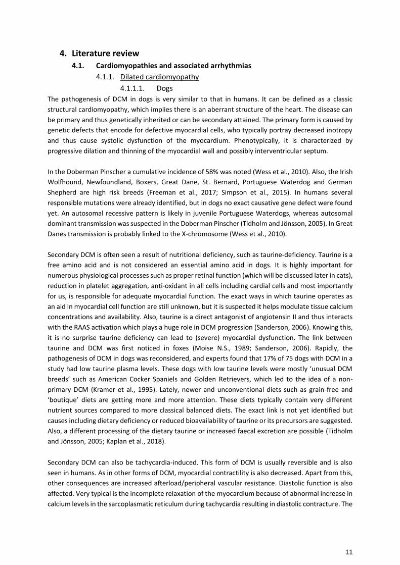
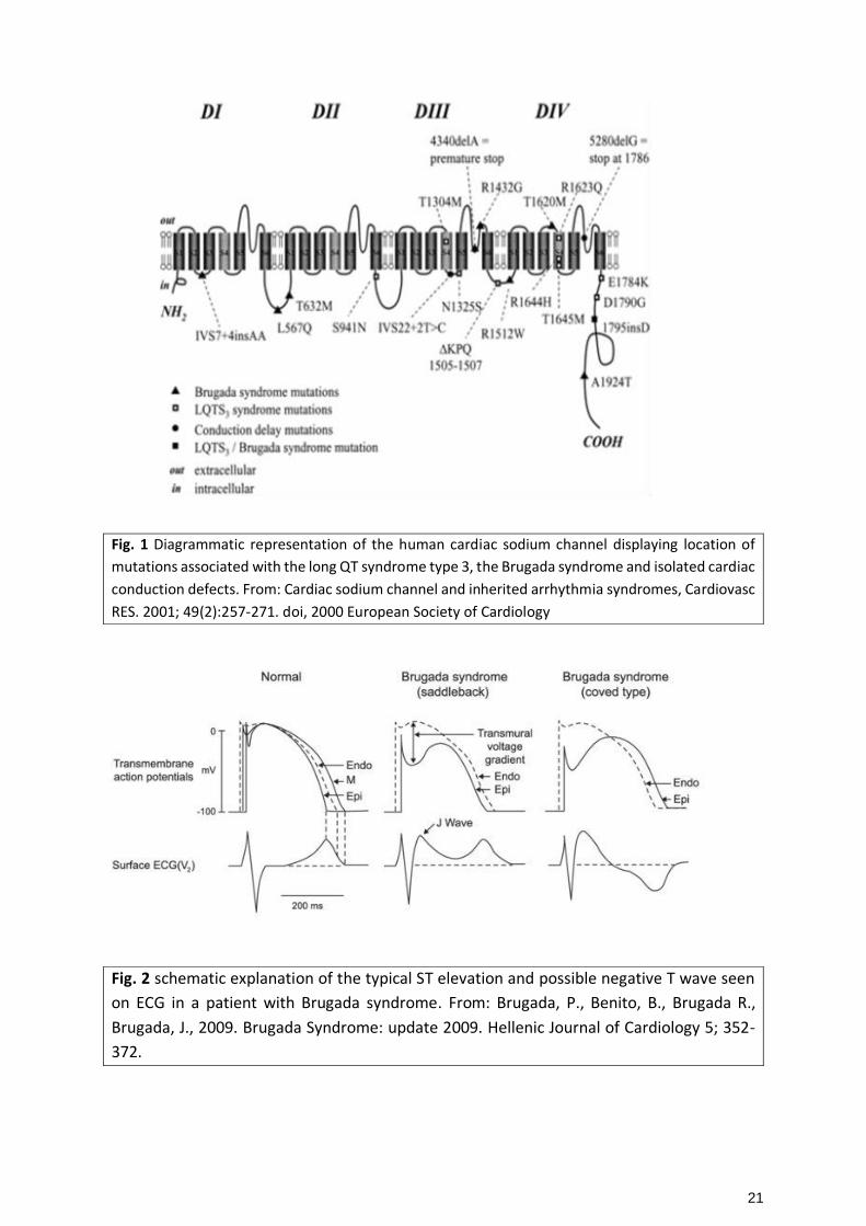
mutation in the SCN5A gene which causes the encoding of defective sodium channels (Fig. 1). This can
be seen as no expression of the sodium channel, disrupted current of the sodium channel due to a shift
in the voltage and time dependence of the channel during activation, inactivation of reactivation, or
even early inactivation of the sodium channel (Hyun and Fillipich, 2006). This way, there is a delay of
the action potential, causing a greater transmural voltage gradient between endocardium and
epicardium leading to the typical ST-elevation (Fig. 2). The decrease in sodium channel current leads
to ventricular fibrillation and possible sudden death (Brugada and Brugada, 1992; Brugada et al., 2009;
Sweeting, 2018).
Another cause of SCD in humans is idiopathic ventricular fibrillation (IVF) or tachycardia. Since the
pathology is still very poorly understood we cannot correctly categorize it. IVF can only be diagnosed
by exclusion which implies that no common diagnostic means were able to identify the underlying
cause of the arrhythmia. By definition no structural heart disease was unmasked, no abnormalities
were seen on ECG nor echocardiography nor MRI. Ajmaline tests are also negative, as is genetic testing.
The disease can also manifest as a non-lethal syncope, cardiac arrest or seizures and is characterized
by transient spontaneous polymorphic ventricular fibrillations/tachycardia (Visser et al., 2016).
21
Fig. 1 Diagrammatic representation of the human cardiac sodium channel displaying location of
mutations associated with the long QT syndrome type 3, the Brugada syndrome and isolated cardiac
conduction defects. From: Cardiac sodium channel and inherited arrhythmia syndromes, Cardiovasc
RES. 2001; 49(2):257-271. doi, 2000 European Society of Cardiology
Fig. 2 schematic explanation of the typical ST elevation and possible negative T wave seen
on ECG in a patient with Brugada syndrome. From: Brugada, P., Benito, B., Brugada R.,
Brugada, J., 2009. Brugada Syndrome: update 2009. Hellenic Journal of Cardiology 5; 352-
372.

22
4.2.2.2. Channelopathies in dogs and cats
Data on channelopathies causing primary electrical cardiac disease in cats and dogs is almost non-
existent, but recently their importance as a component of SCD has got more and more attention. In
small animals no genetic mutations were found yet despite several cases of sudden cardiac death being
reported seemingly caused by these conduction defects as will be discussed further on.
Channelopathies in animals were described in other medical fields concerning other pathologies such
as in neurology, e.g. canine myotonia and achromatopsia (Tanaka et al., 2015; Lowrie and Garosi,
2017).
Several cases of sudden death suspected to be caused by channelopathies in dogs were reported. For
Meurs et al. (2016) SCD of 4 young related Rhodesian Ridgebacks was motive for further investigation
since no structural heart disease nor previous symptoms were found. Holter, echocardiography and
necropsy were performed on as many related animals as possible, in order to try and analyze possible
inheritance patterns. Since males and females were almost equally affected and unaffected parents
did sometimes produce affected offspring, an autosomal recessive inheritance pattern was strongly
suspected. Yet, an autosomal dominant pattern with incomplete penetrance could not be completely
ruled out. Important to note is that Meurs et al. (2016) does not conclude on any ethiopathogenesis,
but a cardiac channelopathy is mentioned as one of the possible underlying diseases.
Also, in the English Springer Spaniel SCD with no evident underlying structural heart disease was
reported. This was the first case report were inherited LQTS in dogs was suspected (Ware et al., 2015).
LQTS in dogs is already known to be caused by toxins, certain drugs or electrolyte inbalances. An
otherwise healthy 5-year old Springer Spaniel who recently delivered her first litter died suddenly after
activity. Three of her puppies also suffered SCD and in several other offspring Holter and ECG
monitoring revealed abnormal elongation of the QT interval and biphasic T-waves. Gene sequencing
revealed that the puppies displaying these prolonged QT intervals had mutations in the KCNQ1 gene,
which was a defect that most likely appeared de novo in the dam. These findings are very similar to
LQTS known in humans, were mutations in the KCNQ1 gene are the number one cause for the disease.
In humans this syndrome was the first channelopathy to be described causing SCD and it is still a major
cause of SCD in humans without structural heart disease (Ware et al., 2015).
Even though not much has been described about cardiac channelopathies in dogs, in cats even less in
known. Feline fatal arrhythmias are mostly caused by underlying structural heart disease such as HCM.
Currently, there are no reports about primary electrical disorders caused by channelopathies in cats.
4.2.2. German Shepherd genetic arrhythmias
German Shepherds are known to suffer SCD (Ribas and Pariaut, 2018). An inherited disease that follows
a sex-linked or autosomal dominant pattern is suggested but is not confirmed to date. Typically, SCD
occurs during sleep or after exercise and is induced by ventricular tachycardia that evolves to
ventricular fibrillation and eventually asystole. The abnormal ventricular repolarization is actually very
similar as is seen in LQTS and Brugada syndrome in humans. Moise et al. (1994) stated that all dogs
studied died between the age of 4 and 30 months old and polymorphic non-sustained ventricular
tachycardia was present in all of them. Yet, no prolonged QT interval is seen nor are there usually any
previous echocardiographic abnormalities found. Normally no histopathological anomalies are present

23
when investigated post-mortem. Since there are no structural causes found, a purely electrical
disorder is very likely, but the pathology remains very poorly understood (Moise et al., 1994; Hyun and
Fillipich, 2006). Jesty et al. (2013) found that German Shepherd dogs with these ventricular
arrhythmias had lower ATP2A2/SERCA2a expression. The SERCA pump plays a big role in the calcium
homeostasis within the cardiomyocyte. It is known that abnormal calcium kinetics can lead to
abnormal Aps and thus can contribute to these arrhythmias. Of course, no provocative
pharmacological tests such as ajmaline or procainamide are available to exclude Brugada syndrome.
4.2.3. Catecholamine dependent arrhythmias
4.2.3.1. Catecholaminergic polymorphic ventricular tachycardia in
humans
Catecholaminergic polymorphic ventricular tachycardia (CPVT) is an inherited electrical dysfunction of
the heart leading to arrhythmias, syncopes and possible SCD. It was first described in 1960 by Berg.
The disease is characterized by bidirectional ventricular tachycardia or polymorphic ventricular
tachycardia seen on ECG. It typically occurs during exercise or periods of emotional stress (Liu et al.,
2007). The cause of this increased risk during these periods is release of catecholamines: these increase
the sensitivity of calcium handling. The incidence of the disease is estimated at 15% of all cases of SCD
without structural heart disease (Lieve et al., 2016).
The causative mutations behind this genetic deficiency are two folded. One form of the disease is
inherited with an autosomal dominant pattern and is caused by mutations in the gene encoding for
the cardiac ryanodine receptor (RyR2). Another form is inherited recessively and originates in defective
cardiac-specific calsequestrin genes (CASQ2). Both genes regulate efflux from the sarcoplasmatic
reticulum into the cardiomyocyte and thus play an important role in cardiac excitability and
contractility (Venetucci et al., 2012).
The disease usually has its onset during childhood or adolescence with most patients being around 10
years old. Without accurate treatment the mortality rate is around 30% before 40 years of age (Lieve
et al., 2016). Sometimes supraventricular arrhythmias can be seen in these young patients preceding
the actual ventricular tachycardia. This occurs probably due to the disrupted calcium handling in the
atrium (Di Pino et al., 2014).
4.2.3.2. Catecholamine dependent arrhythmias in dogs and cats
Currently no analogue disease as seen in humans is described in dogs or cats. Further research is
required to exclude whether it actually does not occur or is just not yet recognized.

24
4.3. Ischemic myocardial events
4.3.1. Ischemic myocardial infarctions in humans
Arteriosclerosis leading to coronary artery occlusion and causing ischemia of the myocardium is
currently the most common cause of death in humans (Scansen, 2017). These myocardial infarctions
are usually of the ischemic and not the hemorrhagic type. It is a very commonly occurring problem
affecting approximately 4% of the population in their lifetime (Antman and Loscalzo, 2018). It is a local
thrombus formed in the coronary arteries mostly as a result of atherosclerosis (Hackel and Jennings,
1988). Atherosclerosis is characterized by fatty degeneration of the extramural coronary arteries (Falk
and Jönsson, 2000). This atherosclerotic plaque then weakens and partially breaks off. When coming
into contact with the circulating blood, prothrombotic factors are activated and a local thrombus is
formed. This causes occlusion of the vessels and thus creates an ischemic zone in the part of the
myocardium being perfused by this vessel.
Classical risk factors in humans are well studied. The Framingham risk factors (the algorithm analyzing
the 10-year risk of cardiovascular disease for an individual) include: diabetes, smoking, hyperlipidemia,
hypertension and unhealthy diet but also hormonal factors such as polycystic ovary syndrome and
psychological impact for example due to depression (Mehta et al., 2016).
It is of great importance to recognize signs of an acute myocardial infarction (AMI) as soon as possible
in order to be able to intervene quickly for the best prognosis possible and minimize myocardial
damage. Typical symptoms are acute heavy chest pain, dyspnea and collapse. Coventry et al. (2011)
even found significant differences in symptoms between men and women by which AMI presented.
Women had much lower odds to present with chest pain than men, whereas men showed much less
signs such as neck pain, nausea and syncope.
4.3.2. Possible role in SCD in dogs and cats
Not much attention was given to typical myocardial insults as we know them in human medicine to be
a cause of possible SCD in animals. Recently, more information is being obtained.
Coronary thrombosis or thrombo-embolism is the classical phenomenon causing myocardial infarcts:
occlusion of the arterial blood supply by an embolus causing ischemia. As for the mechanisms causing
coronary occlusions in cats and dogs, still a lot of pathophysiological factors have to be clarified. What
we do know is that this disease is very seldomly diagnosed in veterinary practice. Dogs tend to have a
left dominant coronary circulation, which imposes more unique collaterals than in species with right
dominant coronary circulation like humans (Scansen, 2017). This imposes a question: do these
occlusions really occur much less in dogs and cats or is it just that they have much less consequences
when they do occur?
Atherosclerosis, the most common cause of ischemic heart disease is men, is very rare in dogs (Falk
and Jönsson, 2000). Also, Falk and Jönsson (2000) found no gender or breed predisposition but
concluded that intramural arterial arteriosclerosis is in fact a very common finding on necropsy in dogs.
They also saw that 87 percent of dogs in their study group that suffered SCD had hyaline
arterioscleriosis, of which half had concurrent myocardial infarctions. Although, Liu et al. (1968) stated
that dogs are naturally resistant to the development of atherosclerosis and rarely experience any
clinical issues from it. Predisposing factors for AMI in dogs are unknown (Driehuys et al., 1998).

25
Most of the myocardial ischemic events in cats and dogs are described as being secondary to structural
heart disease and/or its hemodynamic consequences. Induced hypercoagulability or hypofibrinolytic
state is usually the underlying trigger for thrombi to be formed. Common related conditions include
neoplasia, disseminated intravascular coagulation, sepsis, hyperadrenocorticism and glucocorticoid
use (Driehuys et al., 1998; Machen and Sleeper, 2015). Underlying structural heart disease such as
HCM and DCM can also cause myocardial infarction in the end-stage (Machen and Sleeper, 2015).
Symptoms usually include lethargy, weakness, anorexia, dyspnea, collapse and vomiting. Affected dogs
often have history of hypercholesterolemia and lipidemia like in humans, but also hypothyroidism was
commonly previously diagnosed (Liu et al., 1986).
Methods for diagnosis of possible myocardial ischemic events include echocardiography where typical
signs as local hypokinesis of the muscle, hyperechoic fibrosis and wall thinning can be noticed (Park et
al., 2012). As for detection of the often-underlying hypercoagulability, D-dimers are not quite sensitive
enough, with a sensitivity of D-dimer concentrations >2,000 ng/ml to predict thrombo-embolism only
being 36% (Nelson and Andreasen, 2003). Thromboelastography would make for a much better option
but is currently rarely available in veterinary medicine practice.
5. Discussion and conclusions
A lot of similarities and interesting possibilities for further research were found for cardiac disease in
humans and in animals.
Structural heart disease causing SCD
Duncker et al. (2015) described the exponential increase in knowledge about cardiomyopathies mostly
due to advanced research on genomic levels in both humans and animals. New technologies like
CRISPR/Cas9 allow us to investigate mutations in animal models like zebrafish but also larger animals
such as pigs. The goal is to use this information in order to better understand pathogenesis in a variety
of species. Sleeper et al. (2009) concluded that therapeutic gene transfer using adeno-associated viral
vector seems to be very interesting as a therapy for several cardiomyopathies such as HCM, DCM and
ARVC. This therapy is used to normalize metabolic state (e.g. calcium handling and apoptosis) of
aberrant myocardial cells.
As for DCM, it is certain that dogs can act as a spontaneous model for human DCM. With exact
mutations in dogs still remaining unknown, affected dogs can act as a naturally occurring model for
testing new therapies, but also as subject for genetic screening (Duncker et al., 2015). High-risk breeds
for DCM such as Doberman Pinschers and Great Danes serve as ideal candidates. Especially Portuguese
Waterdogs, known for their rapid progression of DCM, could serve as an excellent spontaneous model
in hopes to directly transfer results of suppression of phospholamban (a membrane protein of the
cardiac calcium pump) expression onto human cardiomyopathies (Sleeper et al., 2009).
The exact mechanism underlying familial arrhythmias in Boxers remains unknown (Spier et al., 2001),
but since there are a lot of similarities with findings in human ARVC, the Boxer has been proposed as
a model for ARVC in humans (Hamlin, 2007). Also, cats present a very interesting spontaneous model

26
for human ARVC just like they do for HCM. Especially since the pathogenesis of this disease is not yet
completely resolved, findings in cats could lead to new insights in the disease in humans. Several
papers confirmed the huge similarities in findings concerning the disease in cats, dogs and humans
such as the typical ‘triangle of dysplasia’ (Simpson et al., 1994; Fox et al., 2000; Boujon and Amberger,
2003; Eason et al., 2015). This term describes the three typical locations to find histological
abnormalities in patients suffering from ARVC, being that the right ventricular wall was thinner in
subtricuspidal, apical and infundibular locations (Boujon and Amberger, 2003).
As already stated, cats were already used as a model for human HCM for years. Pathogenesis is
strikingly similar to that in cats which implies cats can serve as a spontaneous model for human HCM
(Maron and Fox, 2015). Maine Coon cats are especially interesting given the fact that their identified
autosomal dominant MYBPC3 mutation very much resembles the human form. Both species have
lower cMyBP-C protein, whether affected homogenous or heterogenous (Duncker et al., 2015). Also,
Ragdolls were identified with mutations in this same gene. Cats can serve as models for exploring new
therapies in order to prevent heart failure of SCD. &We can conclude that as for now cats have better
potential for serving as spontaneous models for HCM in humans since there are much more affected
individuals and the basics of genetic defaults are already known in this species. Unfortunately, genetic
research in identifying causative genetic mutations causing HCM in animals is still on the low rise.
Electrical dysfunctions causing SCD
Naturally occurring models of ventricular arrhythmias include the non-ischemic arrhythmias in
sleeping or resting German Shepherd puppies. As already stated above, the disease usually starts to
manifest as occasional VPCs and then rapidly progresses towards ventricular tachycardia (Moïse et al.,
1994; Jesty et al., 2003; Hamlin, 2007; Pariaut et al., 2011; Ribas et al., 2018). This type of spontaneous
model could serve as an excellent base for further research of many diseases in man such as the LQTS
but also dysautonomic arrhythmias (Hamlin, 2007).
Since not a vast amount of information is currently available concerning channelopathies in cats and
dogs, it could be very interesting to check whether mutations as are already known in humans, also
exist in these animals. One was already found when Ware et al. (2015) described a family of English
Springer Spaniels who suffered SCD had a mutation in the KCNQ1 gene, just like is known in human
LQTS. Unfortunately, in cats no cases of SCD allegedly caused by channelopathies where described.
Regarding CVPT, currently no analogue disease as seen in humans is described in dogs or cats. Further
research is required to exclude whether it actually does not occur or is just not yet recognized.
Dogs with ventricular arrhythmias due to gastric dilation/volvulus are known to resemble those
secondary to dysautonomia in humans. By further researching the exact pathogenesis of these
arrhythmias more insights could be obtained concerning diagnosis and treatment in both humans and
animals (Hamlin, 2007).
Ischemic myocardial infarctions
Atherosclerosis, the most common cause of ischemic heart disease is men, is very rare in dogs.
Myocardial infarctions are very seldomly diagnosed in practice, but Falk and Jönsson (2000) found that
intramural arterial arteriosclerosis is in fact a very common finding on necropsy in dogs and 87 percent
of dogs that suffer SCD have hyaline arterioscleriosis, of which half have concurrent myocardial
27
infarctions. Although, Liu et al. (1968) stated that dogs rarely experience any clinical issues from
atherosclerosis. For now, we can conclude that most dogs or cats suffering myocardial infarctions, do
so secondary to systemic disease like neoplasia which cause hypercoagulability or hypofibrinolytic
state (Driehuys et al., 1998; Machen and Sleeper, 2015). More research is warranted to confirm or
deny this suspicion. With further development and higher availability of diagnostic tools like thrombo-
elastography in the future, hopefully better and faster diagnostics will be possible.
Conclusions
The most interesting benefits of using larger mammals like cats and dogs instead of more classical test
animals like mice and rats, is that cats and dogs are a spontaneous model of these diseases. This means
they develop the disease naturally, not by manipulated processes in laboratories. In mice, usually
knock-out strands are used to create lines artificially affected by certain pathologies (Camacho et al.,
2016). The main issue with these researches lays in extrapolating the results to humans which for a
number of reasons is not always accurate. For example, the MHC protein which plays an important
role in sarcomeric function completely differs in humans and mice (Duncker et al., 2015). Also, larger
animals have a more heterogenous genetic background and their size allows easier surgical
interventions and clinical evaluation compared to smaller species (Sleeper et al., 2009). Furthermore,
large animals live in the same environment as humans and thus are exposed to the same
environmental factors, making results more reliable for extrapolation.
There are still some important difficulties that are encountered when trying to analyse underlying
genetics. Genetic testing trough DNA sequencing is already available, yet sometimes new genetic
mutations are identified for which pathogenesis is not yet fully understood. This can cause problems
concerning interpretation and is of little use for familial screening. Also, for example in only about 50%
of clinically affected human HCM probands the causative mutation is found (Maron and Maron, 2013).
Since these are all problems that are encountered in human medicine, we can state that this will also
be the case in the veterinary field.
In humans often the preferred treatment for serious arrhythmias that can lead to SCD is an implantable
cardioverter-defibrillator or ICD (Borggrefe et al., 2006). Of course, this option is still very rare and
unusual in animals even though the first defibrillator to be implanted was in a dog in 1970. After that,
two more cases of ICD implantation were reported (Nelson et al., 2006; Coats et al., 2010; Pariaut et
al., 2011). The main issue with this treatment in animals is the price of the intervention, but also the
non-fitting algorithms of the device since they were originally designed for human use. For example,
rapid sinus tachycardia (>250 bpm) which occurs frequently in dogs with ventricular arrhythmias, can
be a reason for inappropriate therapy. This is because the decision for defibrillation is made based on
rate and duration of the tachycardia, but usually maximum programmable rate for detection of this
ventricular arrhythmia is 250 bpm (Pariaut et al., 2011). On the other hand, animals displaying
arrhythmias secondary to primary cardiomyopathies such as DCM can serve as a subject of
investigation concerning arrhythmias and anti-arrhythmic drugs (Hamlin, 2007). Considering what is
presently known, it is clear that further research on SCD in animals is more than warranted.

28
6. References
1. Abbott, J., 2015. Feline Cardiomyopathy. In: Silverstein, D.C., Hopper, K., Small Animal
Critical Care Medicine Second Edn. Elsevier Saunders, St. Louis, Missouri, USA, pp. 218-
224.
2. Abed, M.A., Abu Ali, R.M., Abu Ras, M.M., Hamdallah, F.O., Khalil, A.A., Moser, D.K., 2015.
Symptoms of acute myocardial infarction: A correlational study of the discrepancy
between patients’ expectations and experiences. International Journal of Nursing Studies
52, 1591-1599.
3. Antman, E.M., Loscalzo, J., 2018. Ischemic Heart Disease. In: Jameson, J.L., Faci, A.S.,
Kasper, D.L., Hauser, S.L., Longo, D.L., Harrison’s Principles of Internal Medicine, Twentieth
Edn. McGraw-Hill Education, Colombus, OH, USA.
4. Barrett, T.D., Hennan, J.K., Fischbach, P.S., O’Neill, B.P., Driscoll, E.M. Jr., Lucchesi, B.R.,
2001. Tedisamil and dofetilide-induced torsade de pointes, rate and potassium
dependence. British Journal of Pharmacology 132, 1493-1500.
5. Basso, C., Thiene, G., Corrado, D., Angelini, A., Nava, A., Valente, M., 1996. Arrhythmogenic
right ventricular cardiomyopathy: Dysplasia, dystrophy or myocarditis? Circulation 94,
983-991.
6. Bezzina, C.R., Rook, M.B., Wilde, A.A.M., 2001. Cardiac sodium channel and inherited
arrhythmia syndromes. Cardiovascular Research 49, 257-271.
7. Bohnen, M.S., Peng, G., Robey, S.H., Terrenoire, C., Iyer, V., Sampson, K.J., Kass, R.S., 2017.
Molecular Pathophysiology of Congenital Long QT Syndrome. Physiological Reviews 1, 89-
134.
8. Borgeat, K., Wright, J., Garrod, O., Payne, J.R., Fuentes, V.L., 2014. Arterial
Thromboembolism in 250 cats in General Practice 2004-2012. Journal of Veterinary
Internal Medicine 28, 102-108.
9. Borggrefe, M.,Wolpert, C., Antzelevitch, C., Veltmann, C., Giustetto, C., Gaita, F., Schimpf,
R., 2006. Short QT syndrome Genotype-phenotype correlations. Journal of
Electrocardiology 4, 75-80.
10. Boswood, A., Häggström, J., Gordon, S.G., Wess, G., Stepien, R.L., Oyama, M.A., Keene,
B.W., Bonagura, J., MacDonald, K.A., Patteson, M., et al., 2016. Effect of Pimobendan in
Dogs with Preclinical Myxomatous Mitral Valve Disease and Cardiomegaly: The EPIC study:
A Randomized Clinical Trial. Journal of Veterinary Internal Medicine 30; 1765-1779.

29
11. Boujon, C.E., Amberger, C.N., 2003. Arrhythmogenic Right Ventricular Cardiomyopathy in
a Boxer. Journal of Veterinary Cardiology 5, 35-41.
12. Brugada P., Brugada, J., 1992. Right bundle branch block, persistent ST segment elevation
and sudden cardiac death: a distinct clinical and electrocardiographic syndrome. A
multicenter report. Journal of the American College of Cardiology 6, 1391-1396.
13. Brugada, P., Benito, B., Brugada R., Brugada, J., 2009. Brugada Syndrome: update 2009.
Hellenic Journal of Cardiology 5; 352-372.
14. Camacho, P., Fan, H., Liu, Z., He, J., 2016. Small mammalian animal models of hearts
disease. American Journal of Cardiovascular Disease 6, 70-80.
15. Catterall, W.A., 2018. Voltage-gated Sodium Channels and Electrical Excitability of the
Heart. Zipes, D., Jalife, J., Stevenson, W., In: Cardiac Electrophysiology: From Cell to
Bedside, Seventh Edn, Saunders Elsevier, St. Louis, MO, USA, pp. 1-10.
16. Coats, W., Politte, L., Flaker, G.C., 2010. The Development and Application of the
Implantable Cardioverter Defibrillator. Missouri Medicine 107, 48-52.
17. Collis, R., Elliott, P.M., 2017. Sudden cardiac death in inherited cardiomyopathy.
International Journal of Cardiology 237, 56-59.
18. Côté, E., Jaeger, R., 2008. Ventricular Tachyarrhythmias in 106 Cats: Associated Structural
Cardiac Disorders. Journal of Veterinary Internal Medicine 22, 1444-1446.
19. Côté, E., 2010. Feline Arrhythmias: An Update. Veterinary Clinics of North America: Small
Animal Practice 40, 643-650.
20. Coventry, L.L., Finn, J., Bremner, A.P., 2011. Sex differences in symptom presentation in
acute myocardial infarction: A systematic review and meta-analysis. Heart & Lung: The
Journal of Acute and Critical Care 40, 477-491.
21. Cunnigham S.M., Sweeney, J.T., MacGregor, J., Barton, B.A., Rush, J.E., 2018. Clinical
Features of Bulldogs with Presumed Arrhythmogenic Right Ventricular Cardiomyopathy:
31 cases (2001-2013). Journal of the American Animal Hospital Association 54, 95-102.
22. De Madron, E., Chetboul, V., Bussadori, C., 2015. Dilated Cardiomyopathy and Other
Cardiomyopathies in Dogs. In: Clinical Echocardiography of the Dog and Cat. Elsevier,
Amsterdam, Netherlands, pp. 181-205.
23. Dec, G.W., Fuster, V., 1994. Idiopathic dilated cardiomyopathy. The New England Journal
of Medicine 331, 1564-1575.

30
24. Di Pino, A., Caruso, E., Costanzo, L., Guccione, P., 2014. A novel RyR2 mutation in a 2-year-
old baby presenting with atrial fibrillation, atrial flutter, and atrial ectopic tachycardia.
Heart Rhythm 11, 1480 – 1483.
25. Driehuys, S., Van Winkle, T.J., Sammarco, C.D., Drobatz, K.J., 1998. Myocardial infarction
in dogs and cats: 37 cases (1985-1994). Journal of the American Veterinary Medical
Association 213, 1444-1448.
26. Duncker, D.J., Bakkers, J., Brundel, B.J., Robbins, J., Tardiff, J.C., Carrier, L., 2015. Animal
and in silico models for the study of sarcomeric cardiomyopathies. Cardiovascular
Research 105, 439-448.
27. Eason, B.D., Leach, S.B., Kuroki, K., 2015. Arrhythmogenic right ventricular cardiomyopathy
in a Weimaraner. Canadian Veterinary Journal 10; 1035-1039.
28. Egenvall, A., Bonnett, B.N., Häggström, J., 2006. Heart Disease as a Cause of Death in
Insured Swedish Dogs Younger than 10 Years of Age. Journal of Veterinary Internal
Medicine 20, 894-903.
29. Falk, T., Jönson, L., 2008. Ischaemic heart disease in the dog: a review of 65 cases. Journal
of Small Animal Practice 41, 97-103.
30. Fang, W., Zhang, J., He, Z.X., 2010. Myocardial ischemia in patient with dilated
cardiomyopathy. Nuclear Medicine Communications 31, 981-984.
31. Ferasin, L., 2009. Feline Myocardial Disease. Journal of Feline Medicine and Surgery 11,
183-194.
32. Ferasin, L., Sturgess, C.P., Cannon, M.J., Caney, S.M.A., Gruffydd-Jones, T.J., Wotton, P.R.,
2003. Feline idiopathic cardiomyopathy: a retrospective study of 106 cats (1994-2001).
Journal of Feline Medicine and Surgery 5, 151-159.
33. Fox, P.R., Liu, S.K., Maron, B.J., 1995. Echocardiographic assessment of spontaneously
occurring feline hypertrophic cardiomyopathy. An animal model of human disease.
Circulation 92, 2645-2651.
34. Fox, P.R., Maron, B.J., Basso, C., Liu, S.K., Thiene, G., 2000. Spontaneously occurring
arrhythmogenic right ventricular cardiomyopathy in the domestic cat. A new animal model
similar to human disease. Circulation 2000, 1863-1870.

31
35. Freeman, L.M., Rush, J.E., Stern, J.A., Huggins, G.S., Maron, M.S., 2017. Feline Hypertrophic
Cardiomyopathy: A Spontaneous Large Animal Model of Human HCM. Cardiology Research
Journal 8, 139-142.
36. Fuentes, V.L., Wilkie, L.J., 2017. Asymptomatic Hypertrophic Cardiomyopathy: Diagnosis
and Therpay. Veterinary Clinics of North America, Small Animal Practice 5, 1041-1051.
37. Gudenschwager, E.K., Abbott, J.A., LeRoith, T., 2019. Dilated cardiomyopathy with
endocardial fibroelastosis in a juvenile Pallas cat. Journal of Veterinary Diagnostic
Investigation, DOI.
38. Gunasekaran, T., Sanders, R.A., 2017. Sudden cardiac death in a dog during Holter
recording- R on T phenomenon. Journal of Veterinary Cardiology 19, 455-461.
39. Gupta, S., Figueredo, V.M., 2014. Tachycardia mediated cardiomyopathy:
pathophysiology, mechanisms, clinical features and management. Internation Journal of
Cardiology, DOI.
40. Hackel, D.B., Jennings, R.B., 1988. Ischemic Heart Disease. In: Rubin, E., Farber, J.L.,
Pathology First Edn. J.B. Lippincott Co, Philadelphia, Philadelphia, USA, pp. 515-527.
41. Hallman, B.E., Hauck, M.L., Williams, L.E., Hess, P.R., Steven, E.S., 2018. Incidence and risk
factors associated with development of clinical cardiotoxicity in dogs receiving
doxorubicin. Journal of Internal Veterinary Medicine, DOI.
42. Hambrook, L.E., Bennett, P.F., 2012. Effect of pimobendan on the clinical outcome and
survival of cats with non-taurine responsive dilated cardiomyopathy. Journal of Feline
Medicine and Surgery 14, 233-239.
43. Hamlin, L., Billman, G.E., 2007. Animal models of ventricular arrhythmias. Pharmacology &
Therapeutics 113, 276-295.
44. Hanas, S., Tidholm, A., Holst, B.S., 2017. Ambulatory electrocardiogram recordings in cats
with primary asymptomatic hypertrophic cardiomyopathy. Journal of Feline Medicine and
Surgery 19, 158-164.
45. Horowitz, L., Josephson, M., Harken, A., 1980. Epicardial and endocardial activation during
sustained ventricular tachycardia in man. Circulation 61, 1227-1238.
46. Hyun, C., Fillipich, L.J., 2006. Molecular genetics of sudden cardiac death in small animals
– A review. The Veterinary Journal 171, 39-50.
47. Imhoff, R.K., 1961. Production of aortic occlusion resembling acute aortic embolism
syndrome in cats. Nature 192, 979-980.

32
48. Jackson, B.L., Lehmkuhl, L.B., Adin, D.B., 2014. Heart rate and arrhythmia frequency of
normal cats compared to cats with asymptomatic hypertrophic cardiomyopathy. Journal
of Veterinary Cardiology 16, 215-225.
49. Jesty, S.A., Jung, S.W., Cordeiro, J.M., Gunn, T.M., Di Diego, J.M., Hemsley, S., Kornreich,
B.G., Hooker, G., Antzelevitch, C., Moïse, N.S., 2013. Cardiomyocyte calcium cycling in a
naturally occurring German Shepherd dog model of inherited ventricular arrhythmia and
sudden cardiac death. Journal of Veterinary Cardiology 15, 5-14.
50. Johns, S.M., Nelson, O.L., Gray, J.M., 2012. Left Atrial Function in Cats with Left-Sided
Cardiac Disease and Pleural Effusion or Pulmonary Edema. Journal of Veterinary internal
Medicine 26, 1134-1139.
51. Kaplan, J.L., Stern, J.A., Fascetti, A.J., Larsen, J.A., Skolnik, H., Peddle, G.D., Kienle, R.D.,
Waxman, A., Cocchiaro, M., Gunther-Harrington, C.T., et al., 2018. Taurine deficiency and
dilated cardiomyopathy in golden retrievers fed commercial diets. PLoS One 13, DOI.
52. Kirchof, P., Breithardt, G., Eckardt, L., 2016. Primary prevention of sudden cardiac death.
Heart 92, 1873-1878.
53. Kittleson, M.D., Meurs, K.M., Munro, M.J., Kittleson, J.A., Liu, S-K., Pion, P.D., Towbin, J.A.,
1999. Familial hypertrophic cardiomyopathy in Maine Coone cats: an animal model for
human disease. Circulation 24, 3172-3180.
54. Klüser, L., Holler, P.J., Simak, J., Tater, G., Smets, P., Rügamer, D., Küchenhoff, H., Wess, G.,
2016. Predictors of sudden cardiac death in Doberman Pinschers with dilated
cardiomyopathy. Journal of Veterinary Internal Medicine 30, 722-732.
55. Kramer, G.A., Kittleson, M.D., Fox, P.R., Lewis, J., Pion, P.D., 1995. Plasma Taurine
Concentration in Normal Dogs and in Dogs with Heart Disease. Journal of Veterinary
Internal Medicine 9, 253-258.
56. Lane, R.D., Reis, H.T., Hsu, C-H., Kern, K.B., Couderc, J.P., Moss, A.J., Zareba, W., 2018.
Abnormal Repolarization Duration During Everyday Emotional Arousal in Long QT
Syndrome and Coronary Artery Disease. The American Journal of Medicine 131, 565-572.
57. Lefbom, B.K., Rosenthal, S., Tyrell, W.D.J., Saunders, T.G., Ferguson, M.J., Rush, J.E., Lesser,
M.B., 2001. Severe hypertrophic cardiomyopathy in 10 young Ragdoll cats. Journal of
Veterinary Internal Medicine 15, 308.
58. Leren, I.S., Saberniak, J., Haland, T.F., Edvardsen, T., Haugaa, K., 2016. Combination of ECG
and Echocardiography for Identification of Arrhythmic Events in Early ARVC. Journal of
American College of Cardiology, DOI.

33
59. Lieve, K.V., van der Werf, C., Wilde, A.A., 2016. Catecholaminergic Polymorphic Ventricular
Tachycardia. Circulation Journal 80; 1285-1291.
60. Liu, N., Colombi, B., Raytcheva-Buono, E.V., Bloise, R., Priori, S.G., 2007. Catecholaminergic
Polymorphic Ventricular Tachycardia. Herz 32, 212-217.
61. Liu, S-K., Maron, B.J., Tilley, L.P., 1979. Hypertrophic Cardiomyopathy in the Dog, American
Journal of Pathology 94, 497-508.
62. Liu, S-K., Tilley, L.P., Tappe, J.P., Fox, P.R., 1986. Clinical and pathological findings in dogs
with atherosclerosis: 21 cases (1970-1983). Journal of the American Veterinary Medical
Association 189, 227-232.
63. Lowrie, M., Garosi, L., 2017. Classification of involuntary Movements in Dogs: Myoclonus
and Myotonia. Journal of Veterinary Internal Medicine 31, 979-987.
64. Machen, M.C., Sleeper, M., 2015. Ventricular Failure and Myocardial Infarction. In:
Silverstein D.C., Hopper, K., Small Animal Critical Care Medicine Second Edn. Elsevier
Saunders, St. Louis, Missouri, USA, pp. 214-217.
65. Maron, B.J., Maron, M.S., 2013. Hypertrophic cardiomyopathy. The Lancet 38, 242-255.
66. Maron, B.J., Fox, P.R., 2015. Hypertrophic cardiomyopathy in man and cats. Journal of
Veterinary Cardiology 17, S6-S9.
67. Martin, C.A., Matthews, G.D.K., Huang, C. L-H., 2012. Sudden cardiac death and inherited
channelopathy: the basic electrophysiology of the myocyte and myocardium in ion channel
disease. Heart 98, 536-543.
68. Magi, S., Lariccia, V., Maiolino, M., Amoroso, S., Gratteri, S., 2017. Sudden cardiac death:
focus on the genetics of channelopathies and cardiomyopathies. Journal of Biomedical
Science 24, DOI.
69. Mehta, P.K., Wei, P., Wenger, N.K., 2016. Ischemic Heart Disease in Women: A Focus on
Risk Factors. Trends in Cardiovascular Medicine 25, 140-151.
70. Meurs, K.M., Weidman, J.A., Rosenthal, S.L., Lahmers, K.K., Friedenberg, S.G., 2016.
Ventricular arrhythmias in Rhodesian Ridgebacks with a family history of sudden death and
results of a pedigree analysis for potential inheritance patterns. Journal of the American
Veterinary Medical Association 248, 1135-1138.
71. Miles, C., Finocchiaro, G., Papadakis, M., Gray, B., Westaby, J., Ensam, B. Sheppard, M.N.,
2019. Sudden Death and Left Ventricular Involvement in Arrhythmogenic
Cardiomyopathy. Circulation DOI.

34
72. Moise N.S., 1989. Cardiomyopathy in the fox and association with low dietary taurine. In:
Proceedings of the Seventh American College of Veterinary Internal Medicine Forum, pp.
834-845.
73. Moise, N.S., Meyers-Wallen, V., Flahive, W.J., Valentine, B.A., Scarlett, J.M., Brown, C.A.,
Chavkin, M.J., Dugger, D.A., Renaud-Farrell, S., Kornreich, B., et al., 1994. Inherited
ventricular arrhythmias and sudden death in German shephard dogs. Journal of the
American College of Cardiology 1, 233-243.
74. Nelson, O.L., Andreasen, C., 2003. The utility of plasma D-dimer to identify thrombo-
embolic disease in dogs. Journal of Veterinary internal Medicine 17, 830-834.
75. Nelson, O.L., Lahmers, S., Schneider, T., Thompson, P., 2006. The Use of an Implantable
Cardioverter Defibrillator in a Boxer Dog to Control Clinical Sings of Arrhythmogenic Right
Ventricular Cardiomyopathy. Journal of Veterinary Internal Medicine 20, 1232-1237.
76. O’Sullivan, M.L., O’Grady, M.R., Minors, S.L., 2007. Assessment of Diastolic Function by
Doppler Echocardiography in Normal Doberman Pinschers and Doberman Pinschers with
Dilated Cardiomyopathy. Journal of Veterinary Internal Medicine 21, 81-91.
77. Pariaut, R., Saelinger, C., Queiroz-Williams, P., Strickland, K.N., Marshall, H.C., 2011.
Implantable cardioverter-defibrillator in a German shepherd dog with ventricular
arrhythmias. Journal of Veterinary Cardiology 13, 203-210.
78. Park, N., Kim, J., Lee, M., Lee, S., Song, S., Lee, S., Kim, S., Park, Y., Eom, K., 2012.
Echocardiographic assessment of coronary artery flow in normal canines and model dogs
with myocardial infarction. Journal of Veterinary Science 15, 149.
79. Patel, S., Rauf, A., Khan, H., Abu-Izneid, T., 2017. Renin-angiotensin-aldosteron (RAAS): The
ubiquitous system for homeostatis and pathologies. Biomedicine & Pharmacotherapy 94,
317-325.
80. Papadakis, M., Papatheodorou, E., Mellor, G., Raju, H., Bastiaenen, R., Wijeyeratne, Y.,
Wasim, S., Ensam, B., Finocchiaro, G., Gray, B., et al., 2018. The Diagnostic Yield of Brugada
Syndrome After Sudden Death with Normal Autopsy. Journal of American College of
Cardiology 71, DOI.
81. Payne, J.R., Brodbelt, D.C., Fuentes, L., 2015. Cardiomyopathy prevalence in 780
apparently healthy cats in rehoming centres (the CatScan study). Journal of Veterinary
Cardiology, DOI.
35
82. Payne, J.R., Fuentes, V.L., Boswood, A., Connolly, D., Koffas, H., Brodbet, D., 2010.
Population characteristics and survival in 127 referred cats with hypertrophic
cardiomyopathy (1997 to 2005). Journal of Small Animal Practice 51, 540-547.
83. Pion, P.D., Kittleson, M.D., Rogers, Q.R., Morris, J.G., 1987. Myocardial failure in cats
associated with low plasma taurine: a reversible cardiomyopathy. Science 237, 764-768.
84. Pion, P.D., Kittleson, M.D., Thomas, W.P., Delellis, W.A., Rogers, Q.R., 1992. Response of
cats with dilated cardiomyopathy to taurine supplementation. Journal of the American
Veterinary Medicine Association 201, 275-284.
85. Pion, P.D., Kittleson, M.D., Thomas, W.P., Skiles, M.L., Rogers, Q.R., 1992. Clinical findings
in cats with dilated cardiomyopathy and relationship of findings to taurine deficiency.
Journal of American Veterinary Medical Association 15, 267-285.
86. Pérez-Riera, A.R., Barbosa-Barros, R., Raimundo, R.D., Penachini da Costa de Rezende
Barbosa, M., Esposito Sorpreso, I.C., de Abreu, L.C., 2018. The congenital long QT
syndrome type 3: An update. Indian Pacing and Electrophysiology Journal 18, 25-35.
87. Ribas, T., Pariaut, R., 2018. Inherited Ventricular Arrhythmias in German Shepherd Dogs.
Willis, R., Oliveira, P., Mavropoulou, A., In: Guide to Canine and Feline Electrocardiography,
First Edn, Wiley-Blackwell, Hoboken, NJ, USA, pp. 315-318.
88. Rishniw, M., 2017. Effect of Pimobendan in Dogs with Preclinical Myxomatous Mitral Valve
Disease and Cardiomegaly: The EPIC Study – A Randomized Clinical Trial. Advances in Small
Animal Medicine and Surgery 30, 1-3.
89. Rush, J.E., Freeman, L.M., Fenollosa, N.K., Brown, D.J., 2002. Population and survival
characteristics of cats with hypertrophic cardiomyopathy: 260 cases (1990–1999). Journal
of the American Veterinary Medical Association 220, 202-207.
90. Sanderson, S.L., 2006. Taurine and Carnitine in Canine Cardiomyopathy. The Veterinary
Clinics of North America: Small Animal Practice 36, 1325-1343.
91. Santilli, R.A., Bontempi, L.V., Perego, M., Fornai, L., Basso, C., 2009. Outflow tract
segmental arrhythmogenic right ventricular cardiomyopathy in an English Bulldog. Journal
of Veterinary Cardiology 11, 47-51.
92. Scansen, B.A., 2017. Coronary Artery Anomalies in Animals. Veterinary Sciences 20, DOI.
93. Schipper, T., Van Poucke, M., Sonck, L., Smets, P., Ducatelle, R., Broeckx, B.J.G., Peelman,
L.J., 2019. A feline orthologue of the human MYH7 c.5647G>A (Glu&833Lys)) variant
causes hypertrophic cardiomyopathy in a Domestic Shorthair cat. European Journal of
Human Genetics, in press.

36
94. Schneider, S.M., Coleman, A.E., Guo, L-J., Tou, S., Keene, B.W., Kornegay, J.N., 2016.
Suspected acute myocardial infarction in a dystrophin-deficient dog. Neuromuscular
Disorders 26, 361-366.
95. Schober, K.E., Savino, S.I., Yildiz, V., 2016. Right ventricular involvement in feline
hypertrophic cardiomyopathy. Journal of Veterinary Cardiology 18, 297-309.
96. Shah, M., 2017. Hypertrophic Cardiomyopathy. Cardiology in the Young 27, 25-30.
97. Shimizu, W., 2012. Diagnostic Evaluation of Long QT Syndrome. Cardiac Electrophysiology
Clinics 4, 29-37.
98. Simpson, K.W., Bonagura, J.D., Eaton, K.A., 1994. Right ventricular cardiomyopathy in a
dog. Journal of Veterinary Internal Medicine 8, 306-309.
99. Simpson, S., Edwards, J., Ferguson-Mignan, T.F.N., Cobb, M., Mongan, N.P., Rutland, C.S.,
2015. Genetics of Humans and Canine Dilated Cardiomyopathy. International Journal of
Genomics, DOI.
100. Sleeper, M.M., Bish, L.T., Sweeney, H.L., 2009. Gene Therapy in Large Animal Models
of Human Cardiovascular Genetic Disease. Institute for Laboratory Animal Research
Journal 50, 199-205.
101. Spier, A.W., Meurs, K.M., Muir, W.W., Lehmkuhl, L.B., Hamlin, R.L., 2001. Correlation
of QT dispersion with indices used to evaluate the severity of familial ventricular
arrhythmias in Boxers. American Journal of Veterinary Research 62, 1481-1485.
102. Stadiotti, I., Catto, V., Casella, M., Tondo, C., Pompilio, G., Sommariva, E., 2017.
Arrhythmogenic Cardiomyopathy: the Guilty Party in Adipogenesis. Journal of
Cardiovascular Translational Research, DOI.
103. Summerfield, N.J., Boswood, A., O’Grady, M.R.,Gordon, S.G., Dukes-McEwan, J.,
Oyama, M.A., Smith, S., Patteson, M., French, A.T., Culshaw, G.J., et al. 2012. Efficacy of
Pimobendan in the Prevention of Congestive Heart Failure or Sudden Death in Doberman
Pinschers with Preclinical Dilated Cardiomyopathy (The PROTECT Study). Journal of
Internal Veterinary Medicine 26, 1337-1349.
104. Sweet, M.E., Taylor, M.R.G., Mestroni, L., 2015. Diagnosis, prevalence, and screening
of familial dilated cardiomyopathy. Expert Opinion on Orphan Drugs 8, 869-876.
105. Sweeting, J., Semsarian, C., 2018. Sudden Cardiac Death in Athletes. Heart, Lung and
Circulation, DOI.

37
106. Tanaka, N., Dutrow, E.V., Miyadera, K., Delemotte, L., MacDermaid, C.M., Reinstein,
S.L., Crumley, W.R., Dixon, C.J., Casal, M.L., Klein, M.L., 2015. Canine CNGA3
GeneMutations Provide New Insights into Human Achromatopsia – Associated
Channelopathies and Treatment. Public Library of Science One 10, DOI.
107. Tidholm, A., Jönsson, L., 2005. Histologic Characterization of Canine Dilated
Cardiomyopathy. Veterinary Pathology 42, 1-8.
108. Tripathi, R., Sullivan, R., Fan, T-H. M., Wang, D., Sun, Y., Reed, G.L., Gladysheva, I.P.,
2017. Enhanced heart failure, mortality and renin activation in female mice with
experimental dilated cardiomyopathy. Public Library of Science One, DOI.
109. Tse, G., Chan, Y.W.F., Keung, W., Yan, B.P., 2017. Electrophysiological mechanisms of
long and short QT syndromes. International Journal of Cardiology Heart & Vasculature 14,
8-13.
110. Vanoli, E., De Ferrari G.M., Schwartz, P.J., 1991. Vagal stimulation and prevention of
sudden death in conscious dogs with a healed myocardial infarction. Circulation, DOI.
111. Venetucci, L., Denegri, M., Napolitano, C., Priori, S.G., 2012. Inherited calcium
channelopathies in the pathophysiology of arrhythmias. Nature Reviews Cardiology 10,
561-575.
112. Visser, M., van der Heijden, J.F., Doevendans, P.A., Loh, P., Wilde, A.A., Hassink, R.J.,
2016. Idiopathic Ventricular Fibrillation the Struggle for Definition, Diagnosis and Follow-
Up. Circulation: Arrhythmia and Electrophysiology, DOI.
113. Ward, O.C., 1964. A new familial cardiac syndrome in children. Journal of the Irish
Medical Association 54, 103-106.
114. Ware, W.A., Reina-Doreste, Y., Stern, J.A., Meurs, K.M., 2015. Sudden Death
Associated with QT Interval Prolongation and KCNQ1 Gene Mutation in a Family of English
Springer Spaniers. Journal of Veterinary Internal Medicine 29, 561-568.
115. Washizu, M., Takemura, N., Machida, N., Nawa, H., Yamamoto, T., Mitake, H., Washizu,
T., 2003. Hypertrophic Cardiomyopathy in an Aged Dog. Journal of Veterinary Medical
Science 6, 753-756.
116. Wess, G., Domenech, O., Dukes-McEwan, J., Häggström, J., Gordon, S., 2017. European
Society of Veterinary Cardiology screening guidelines for dilated cardiomyopathy in
Doberman Pinschers. Journal of Veterinary Cardiology 19, 405-415.
117. Wess, G., Mäurer, J., Simak, J., Hartman, K., 2010. Use of Simpson’s method of Disc to
Detect Early Echocardiographic Changes in Doberman Pinschers with Dilated
Cardiomyopathy. Journal of Veterinary Internal Medicine 24, 1069-1076.

38
118. Wess, G., Schulze, A., Butz, V., Simak, J., Killich, M., Keller, L.J.M., Maeurer, J.,
Hartmann, K., 2010. Prevalence of Dilated Cardiomyopathy in Doberman Pinschers in
Various Age Groups. Journal of Veterinary Internal Medicine 24, 533-538.
119. Wexler, R., Elton, T., Pleister, A., Feldman, D., 2009. Cardiomyopathy: An Overview.
American Family Physician 9, 778-784.
120. Wilde, A.A.M., Amin, A.S., 2018. Clinical Spectrum of SCN5A Mutations: Long QT
Syndrome, Brugada Syndrome, and Cardiomyopathy. Journal of the American College of
Cardiology, DOI.
121. Wilson J.L., Scheel, K.W., 1982. Myocardial Infarction in Dogs With Acute and Gradual
Occlusion of the Circumflex or the Right Coronary Arteries. The Anatomical Record 204,
113-122.