sugar levels modulate differential expression of maize ... · sugar levels modulate differential...
TRANSCRIPT
The Plant Cell, Vol. 4, 59-69, January 1992 O 1992 American Society of Plant Physiologists
Sugar Levels Modulate Differential Expression of Maize Sucrose Synthase Genes
Karen E. Koch,”‘ Kurt D. Nolte,a Edwin R. Duke,a Donald R. McCarty,b and Wayne T. Avignea a Fruit Crops Department, University of Florida, Gainesville, Florida 3261 1
Vegetable Crops Department, University of Florida, Gainesville, Florida 3261 1
The two genes encoding sucrose synthase in maize (Shl and Susl) show markedly different responses to changes in tissue carbohydrate status. This enzyme is widely regarded as pivotal to sucrose partitioning, import, and/or metabolism by developing plant organs. Excised maize root tips were incubated for varying periods in different sugars and a range of concentrations. The Shl mRNA was maximally expressed under conditions of limited carbohydrate supply ( ~ 0 . 2 % glucose). In contrast, Susl transcript levels were low or nondetectable under sugar-depleted conditions and peaked at 10-fold greater glucose concentrations (2.0%). Responses to other metabolizable sugars were similar, but L-glucose and elevation of osmolarity with mannitol had little effect. Plentiful sugar supplies thus increased expression of Susl , whereas reduced sugar availability enhanced Shl. At the protein level, shifts in abundance of subunits encoded by Shl and Susl were much less pronounced but corresponded to changes in respective mRNA levels. Although total enzyme activity did not show net change, cellular localization of sucrose synthase protein was markedly altered. In intact roots, sucrose synthase was most prevalent i n the stele and apex. In contrast, sugar depletion favored accumulation in periph- era1 cells, whereas high sugar levels resulted in elevated expression in all cell types. The differential response of the two Sucrose synthase genes to sugars provides a potential mechanism for altering the pattern of enzyme distribution in response to changing carbohydrate status and also for adjusting the sucrose-metabolizing capacity of importing cells relative to levels of available photosynthetic products.
INTRODUCTION
The role of sugars in gene regulation is well established in microbial systems (e.g., control of the lactose operon and catabolite repression in yeasts), and glucose-regulated genes have been described in animal cells (Lin and Lee, 1984). Specific gene responses to sugars in higher plants have also been found but primarily in connection with storage processes in potato (Rocha-Sosa et al., 1989; Salanoubat and Belliard, 1989; Wenzler et al. 1989; Müller-Rober et al., 1990) and sweet potato (Hattori et al., 1990). Sugar effects have also been observed for identified mRNAs associated with cell wall regeneration by protoplasts (Maas et al., 1990) and down regulation of photosynthesis in maize mesophyll protoplasts (Sheen, 1990). For bacteria and yeasts, sugar- modulated gene expression is an essential mechanism for adjustment to changes in nutrient availability (Carlson, 1987; Vyas et al., 1988; Schuster, 1989). This may also be true for higher plants in which their multicellular structure places more complex demands on adaptation and resource alloca- tion. Sugar effects on gene expression have the potential to profoundly influence carbohydrate distribution and utilization within and among plant parts.
To whom correspondence should be addressed.
For this reason, genes encoding sucrose synthase (EC 2.4.1.13) may have a role in key whole plant adjustments to altered sugar supplies. This enzyme is considered pivotal to sugar distribution and metabolism in many plant species in which developing structures depend on imported sucrose (Claussen 1983; Claussen et al., 1985; Sung et al., 1988; Nguyen-Quoc et al., 1990). In these tissues, sucrose syn- thase is often the primary enzyme responsible for sucrose cleavage. The reversible conversion of sucrose+UDP to fruc- tose + UDP-glucose is one of only two known pathways for metabolic utilization of sucrose in plant cells (Hawker, 1985; Huber and Akazawa, 1986). The sucrose synthase route is apparently favored in many instances and requires half the net energy of the alternate pathway through invertase (Black et al., 1987). The extent of sugar import into many plant or- gans has been correlated with sucrose synthase activity (Claussen, 1983; Sung et al., 1988; Nguyen-Quoc et al., 1990). Elevated activity has also been found in association with starch storage (Chourey and Nelson, 1976; Claussen et al., 1985, 1986; Doehlert, 1990), rapid cell wall synthesis (Hendrix, 1990), and increased carbon flow through the respi- ratory path (Black et al., 1987; Farrar and Williams, 1990).
Two genes in maize (Shl and Susl) encode sucrose syn- thase isozymes with very similar characteristics (SS1 and
60 The Plant Cell
SS2, respectively; Echt and Chourey, 1985). These proteinsare expressed in different tissues and under different condi-tions, however (Roland et al., 1988; Nguyen-Quoc et al.,1990). The Sh1 gene product was initially considered an "en-dosperm form" of sucrose synthase, and its gene name arosefrom the shrunken phenotype of kernels lacking a functionalShrunken-1 (Sh1) gene (Chourey and Nelson, 1976). Expres-sion of this sucrose synthase-encoding gene (Sh1) has sincebeen demonstrated in other maize tissues (Chourey et al.,1986; Springer et al., 1986; Heinlein and Starlinger, 1989) inwhich stress conditions further enhance transcript abun-dance (McCarty et al., 1986; Springer et al., 1986) and alterprotein distribution (Roland et al., 1988). The Sus1 gene,named for its product sucrose synthase, encodes the SS2protein of Echt and Chourey (1985). This isozyme is morewidely distributed among plant parts than is the Sh1 form(Chen and Chourey, 1988) and is present at higher levels inyoung leaves (Nguyen-Quoc et al., 1990).
The possibility that higher plant genes could respond tosugar levels was initially indicated by apparent effects of car-bohydrate supplies on enzyme activities in a range of tissues.Particularly evident were changes in activities of enzymes in-volved in sugar metabolism. Rises in sugar concentrationswere implicated in the elevation of fructan-synthesizing en-zyme activity in Lolium (Housley and Pollock, 1985) and su-crose synthase activity in excised leaves (Claussen et al.,1985) and detached potato tubers (Ross and Davies, 1992).Increased supplies of sugars also were found to enhance ac-tivity of invertase in Avena stem segments (Kaufman et al.,1973) and in cell cultures of sugar cane (Maretzki et al., 1974)and sugar beet (Masuda et al., 1988). In contrast, exogenoussugars (glucose) partially repressed invertase activity in elon-gating sugar cane internodes (Glasziou, 1969) and inhibitedthe increase in phleinase (a fructan hydrolase) that otherwiseaccompanies a depletion of tissue hexoses in orchard grass(Yamomoto and Mino, 1987).
The present research was motivated by sugar starvationstudies in which unidentified proteins appeared in responseto sugar depletion. A starvation "stress protein" was re-ported in pea root tips (Webster, 1980; Webster and Henry,1987), and "carbohydrate responsive proteins" in pearl millet(Baysdorfer and VanDerWoude, 1988). Our initial work indi-cated that carbohydrate deprivation could up regulate theSh1 gene in maize root tips and that sucrose addition woulddecrease levels of its message (Koch and McCarty, 1988).Consistent with this observation was the recent report thatthe Sh1 promoter could be repressed by sucrose in transientexpression assays (Maas et al., 1990). In contrast, other workhas indicated that sugar is a positive regulator of genes en-coding sucrose synthase (Salanoubat and Belliard, 1989) aswell as storage proteins in potato (Rocha-Sosa et al., 1989;Wenzler et al., 1989) and sweet potato (Hattori et al., 1990).
In this article, we demonstrate that in maize root tips,sugar-modulated gene expression includes an unexpected,differential responsiveness of an isozyme gene system en-coding sucrose synthases. The identity of the genes involvedunderscores the importance of their sugar responsiveness
as a potential means of adjusting form, function, and sucrosepartitioning within a multicellular organism.
RESULTS
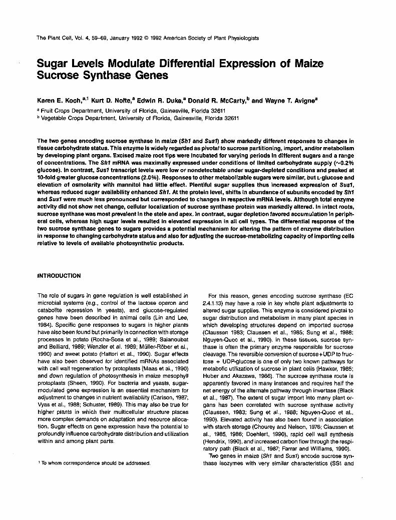
Figure 1 shows that the level of Sh1 mRNA increased relativeto total RNA in excised root tips incubated 24 hr without sug-ars. In contrast, the relative Sh1 transcript level was markedly
intact % glucose0 4.0
Sh1
Sus1 f
Figure 1. RNA Gel Blot Analysis of Sh1 and Sus1 Expression in RootTips Incubated with or without 4% Glucose.
RNA gel blots with equal aliquots (10 ng) of total RNA isolated frommaize root tips before (intact) or after a 24-hr incubation in solutionculture (with 0 or 4.0% glucose) were probed with 32P-labeled Sh1cDNA or Sus1 genomic clones for sucrose synthase. The film was ex-posed for 24 hr.
Sugar-Regulated Gene Expression 61
lower when 4% glucose was included in the medium. An op-posite response to sugars was observed for the Sus1 gene(Figure 1). Whereas Sus1 transcript levels were low or unde-tectable in excised root tips starved for 24 hr, substantiallymore message was present when 4% glucose was added tothe incubation medium. Relative amounts of Sus1 mRNA insugar-supplemented root tips were as great or greater thanthose of intact root tips. Both gene message levels in intactroot tips tended to vary with growing conditions (data notshown). Soluble sugars were depleted to minimal levels in ex-cised root tips within 24 hr unless exogenous carbohydratewas provided (data not shown; Saglio and Pradet, 1980).
The sugar-depletion response of the Sh1 gene was dis-tinct from its previously characterized anaerobic induction(Springer et al., 1986). Both alcohol dehydrogenase-1 (Adh 1)and Sh1 are up regulated under low oxygen; however, Sh1alone responded to low sugar supplies (data not shown). Toensure aerobic conditions, an airflow of 40% oxygen wasmaintained during root tip incubations. In addition, all experi-ments were routinely monitored for expression of Adh1 as afurther test for aerobic metabolism.
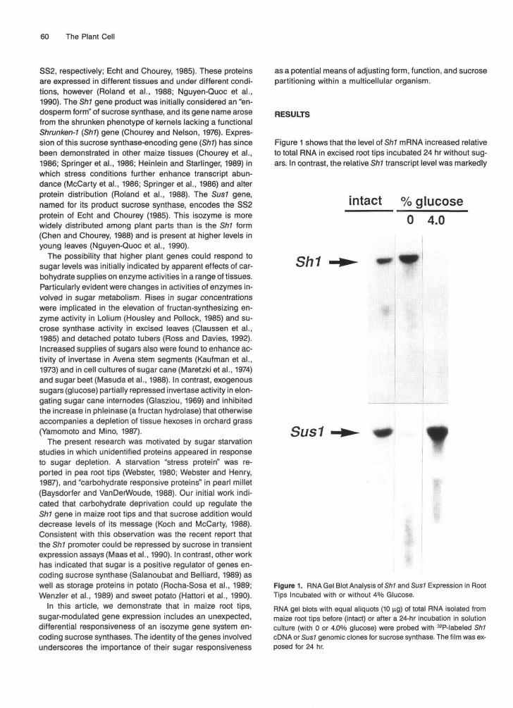
The differential responses of the Sh1 and Sus1 genes tosugars were further apparent when gradations of glucoseconcentrations were supplied to excised root tips. RNA gelblots in Figure 2A and quantifications of replicate experi-ments in Figure 2B show that Sh1 mRNA was most prevalentat low glucose concentrations, whereas relative abundanceof Sus7 mRNA was greatest when exogenous glucose levelswere high. Sh1 message typically peaked when ~0.2% glu-cose was added to media. Sus1 mRNA levels were maximalwhen 10-fold more glucose was present (2.0%). Overall,changes in the mRNA level showed an optimal rather thanan "on-off type of response.
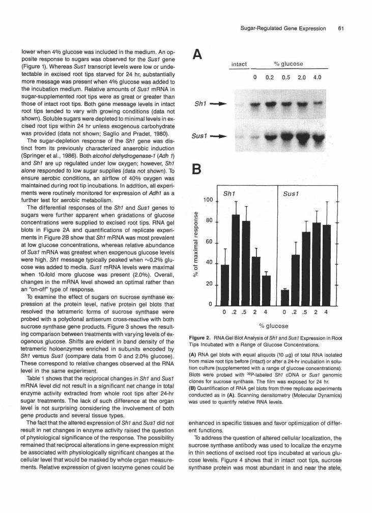
To examine the effect of sugars on sucrose synthase ex-pression at the protein level, native protein gel blots thatresolved the tetrameric forms of sucrose synthase wereprobed with a polyclonal antiserum cross-reactive with bothsucrose synthase gene products. Figure 3 shows the result-ing comparison between treatments with varying levels of ex-ogenous glucose. Shifts are evident in band density of thetetrameric holoenzymes enriched in subunits encoded bySh1 versus Sus1 (compare data from 0 and 2.0% glucose).These correspond to relative changes observed at the RNAlevel in the same experiment.
Table 1 shows that the reciprocal changes in Sh1 and Sus7mRNA level did not result in a significant net change in totalenzyme activity extracted from whole root tips after 24-hrsugar treatments. The lack of such difference at the organlevel is not surprising considering the involvement of bothgene products and several tissue types.
The fact that the altered expression of Sh1 and Sus7 did notresult in net changes in enzyme activity raised the questionOf physiological significance of the response. The possibilityremained that reciprocal alterations in gene expression mightbe associated with physiologically significant changes at thecellular level that would be masked by whole organ measure-ments. Relative expression of given isozyme genes could be
intact % glucose
0 0.2 0.5 2.0 4.0
Sh1
Sus1
B
0 .2 .5 2 4 0 .2 .5 2 4
% glucose
Figure 2. RNA Gel Blot Analysis of Sh1 and Sus1 Expression in RootTips Incubated with a Range of Glucose Concentrations.
(A) RNA gel blots with equal aliquots (10 ug) of total RNA isolatedfrom maize root tips before (intact) or after a 24-hr incubation in solu-tion culture (supplemented with a range of glucose concentrations).Blots were probed with 32P-labeled Sh1 cDNA or Sus1 genomicclones for sucrose synthase. The film was exposed for 24 hr.(B) Quantification of RNA gel blots from three replicate experimentsconducted as in (A). Scanning densitometry (Molecular Dynamics)was used to quantify relative RNA levels.
enhanced in specific tissues and favor optimization of differ-ent functions.
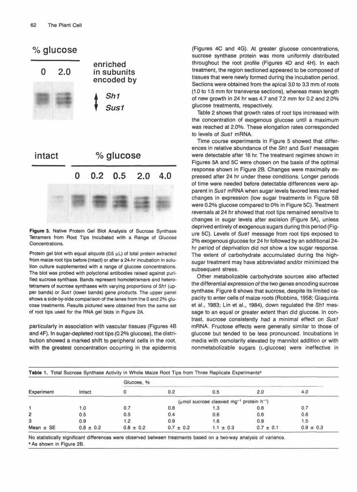
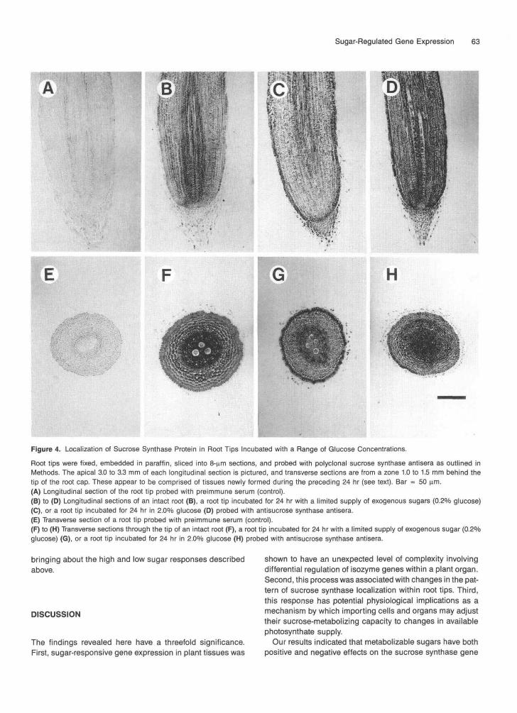
To address the question of altered cellular localization, thesucrose synthase antibody was used to localize the enzymein thin sections of excised root tips incubated at various glu-cose levels. Figure 4 shows that in intact root tips, sucrosesynthase protein was most abundant in and near the stele,
62 The Plant Cell
% glucose
0 2.0enrichedin subunitsencoded by
Sh1Sus1
intact % glucose
0 0.2 0.5 2.0 4.0
Figure 3. Native Protein Gel Blot Analysis of Sucrose SynthaseTetramers from Root Tips Incubated with a Range of GlucoseConcentrations.
Protein gel blot with equal aliquots (0.5 nL) of total protein extractedfrom maize root tips before (intact) or after a 24-hr incubation in solu-tion culture supplemented with a range of glucose concentrations.The blot was probed with polyclonal antibodies raised against puri-fied sucrose synthase. Bands represent homotetramers and hetero-tetramers of sucrose synthases with varying proportions of Sh1 (up-per bands) or Sus1 (lower bands) gene products. The upper panelshows a side-by-side comparison of the lanes from the 0 and 2% glu-cose treatments. Results pictured were obtained from the same setof root tips used for the RNA gel blots in Figure 2A.
particularly in association with vascular tissues (Figures 4Band 4F). In sugar-depleted root tips (0.2% glucose), the distri-bution showed a marked shift to peripheral cells in the root,with the greatest concentration occurring in the epidermis
(Figures 4C and 4G). At greater glucose concentrations,sucrose synthase protein was more uniformly distributedthroughout the root profile (Figures 4D and 4H). In eachtreatment, the region sectioned appeared to be composed oftissues that were newly formed during the incubation period.Sections were obtained from the apical 3.0 to 3.3 mm of roots(1.0 to 1.5 mm for transverse sections), whereas mean lengthof new growth in 24 hr was 4.7 and 7.2 mm for 0.2 and 2.0%glucose treatments, respectively.
Table 2 shows that growth rates of root tips increased withthe concentration of exogenous glucose until a maximumwas reached at 2.0%. These elongation rates correspondedto levels of Susl mRNA.
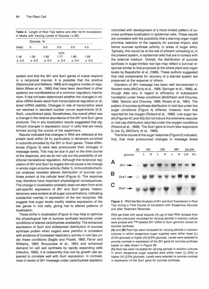
Time course experiments in Figure 5 showed that differ-ences in relative abundance of the Sh1 and Sus1 messageswere detectable after 16 hr. The treatment regimes shown inFigures 5A and 5C were chosen on the basis of the optimalresponse shown in Figure 2B. Changes were maximally ex-pressed after 24 hr under these conditions. Longer periodsof time were needed before detectable differences were ap-parent in Sus1 mRNA when sugar levels favored less markedchanges in expression (low sugar treatments in Figure 5Bwere 0.2% glucose compared to 0% in Figure 5C). Treatmentreversals at 24 hr showed that root tips remained sensitive tochanges in sugar levels after excision (Figure 5A), unlessdeprived entirely of exogenous sugars during this period (Fig-ure 5C). Levels of Sus1 message from root tips exposed to2% exogenous glucose for 24 hr followed by an additional 24-hr period of deprivation did not show a low sugar response.The extent of carbohydrate accumulated during the high-sugar treatment may have abbreviated and/or minimized thesubsequent stress.
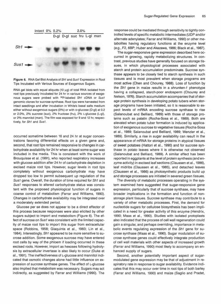
Other metabolizable carbohydrate sources also affectedthe differential expression of the two genes encoding sucrosesynthase. Figure 6 shows that sucrose, despite its limited ca-pacity to enter cells of maize roots (Robbins, 1958; Giaquintaet al., 1983; Lin et al., 1984), down regulated the Sh1 mes-sage to an equal or greater extent than did glucose. In con-trast, sucrose consistently had a minimal effect on SuslmRNA. Fructose effects were generally similar to those ofglucose but tended to be less pronounced. Incubations inmedia with osmolarity elevated by mannitol addition or withnonmetabolizable sugars (L-glucose) were ineffective in
Table 1. Total Sucrose Synthase Activity in Whole Maize Root Tips from Three Replicate Experiments3
Experiment IntactGlucose, %
0.2 0.5 2.0
No statistically significant differences were observed between treatments based on a two-way analysis of variance.a As shown in Figure 2B.
4.0
(nmol sucrose cleaved mg~1 protein h~')123Mean ± SE
1.00.50.90.8 ± 0.2
0.70.51.20.8 ± 0.2
0.80.40.90.7 ± 0.2
1.30.61.61.1 ± 0.3
0.60.60.90.7 ± 0.1
0.70.61.50.9 ± 0.3
Sugar-Regulated Gene Expression 63
H
Figure 4. Localization of Sucrose Synthase Protein in Root Tips Incubated with a Range of Glucose Concentrations.
Root tips were fixed, embedded in paraffin, sliced into 8-nm sections, and probed with polyclonal sucrose synthase antisera as outlined inMethods. The apical 3.0 to 3.3 mm of each longitudinal section is pictured, and transverse sections are from a zone 1.0 to 1.5 mm behind thetip of the root cap. These appear to be comprised of tissues newly formed during the preceding 24 hr (see text). Bar = 50 urn.(A) Longitudinal section of the root tip probed with preimmune serum (control).(B) to (D) Longitudinal sections of an intact root (B), a root tip incubated for 24 hr with a limited supply of exogenous sugars (0.2% glucose)(C), or a root tip incubated for 24 hr in 2.0% glucose (D) probed with antisucrose synthase antisera.(E) Transverse section of a root tip probed with preimmune serum (control).(F) to (H) Transverse sections through the tip of an intact root (F), a root tip incubated for 24 hr with a limited supply of exogenous sugar (0.2%glucose) (G), or a root tip incubated for 24 hr in 2.0% glucose (H) probed with antisucrose synthase antisera.
bringing about the high and low sugar responses describedabove.
DISCUSSION
The findings revealed here have a threefold significance.First, sugar-responsive gene expression in plant tissues was
shown to have an unexpected level of complexity involvingdifferential regulation of isozyme genes within a plant organ.Second, this process was associated with changes in the pat-tern of sucrose synthase localization within root tips. Third,this response has potential physiological implications as amechanism by which importing cells and organs may adjusttheir sucrose-metabolizing capacity to changes in availablephotosynthate supply.
Our results indicated that metabolizable sugars have bothpositive and negative effects on the sucrose synthase gene
64 The Plant Cell
Table 2. Length of Root Tips before and after 24-Hr Incubationsin Media with Varying Levels of Glucose (± SE)
Glucose, %
Intact
1.18± 0.2
0
1.30± 0.2
0.2
1.65± 0.4
0.5
(cm)1.77± 0.4
2.0
1.90± 0.4
4.0
1.59± 0.2
system and that the Sh1 and Sus1 genes of maize respondin a reciprocal manner. It is possible that the positive(Salanoubat and Belliard, 1989) and negative modes of regu-lation (Maas et al., 1990) that have been described in othersystems are manifestations of a common regulatory mecha-nism. It has not been determined whether the changes in rel-ative mRNA levels result from transcriptional regulation or al-tered mRNA stability. Changes in rate of transcription werenot resolved in standard nuclear run-on experiments (K.E.Koch, unpublished data). Nonetheless, the overall effect wasa change in the relative abundance of the SM and Sus1 geneproducts. The in situ localization results suggested that sig-nificant changes in expression occur in cells that are newlyformed during the course of the experiment.
Results indicated that changes in RNA are reflected at theprotein level within 24 hr, particularly in tetramers enrichedin subunits encoded by the Shl or Sus7 genes. These differ-ences (Figure 3) were less pronounced than changes inmessage levels. This may be due in part to the time courseof the response, and we do not rule out the possibility of ad-ditional translational regulation. Although the reciprocal reg-ulation of Sh1 and Sus1 by sugars did not cause a net changein whole organ enzyme activity (Table 1), immunohistochemi-cal analyses revealed altered distribution of sucrose syn-thase protein at the cellular level (Figure 4). The responsemay therefore have important physiological consequences.The change in localization probably does not stem from strictcell-specific expression of Sh1 and Susl genes. Hetero-tetramers were evident at all sugar concentrations, indicatingsubstantial overlap in expression of the two isozymes. Wesuggest that sugar levels modify relative expression of thetwo genes in root cells, giving rise to altered patterns oflocalization.
These shifts in localization (Figure 4) may help to optimizethe physiological role of sucrose synthase isozymes underconditions of altered carbohydrate availability. The enhancedexpression of Sus7 and widespread distribution of sucrosesynthase protein when sugars were plentiful is consistentwith evidence of increased respiratory activity in root tips un-der these conditions (Saglio and Pradet, 1980; Farrar andWilliams, 1990; Brouquisse et al., 1991) and enhanceddemand for cell wall synthesis by rapidly expanding cells(Hendrix, 1990). It is noteworthy that root tip growth rate ap-peared to correlate well with Sus7 expression. In contrast,rises in levels of Shl message under carbohydrate depletion
coincided with development of a more limited pattern of su-crose synthase localization in epidermal cells. These resultsare consistent with the possibility that a starving organ mightprioritize retention of the capacity for sucrose import, andhence sucrose synthase activity, in areas of sugar entry.Typically, this would be at the site of phloem unloading or, inthe present system, in epidermal cells that are in contact withthe external medium. Overall, the distribution of sucrosesynthase in sugar-limited root tips may reflect a survival re-sponse similar to that proposed at the whole plant and organlevels by Baysdorfer et al. (1988). These authors suggestedthat vital components for recovery of a starved system arepreserved at the expense of others.
Elevation of Sh1 message has been well documented inflooded roots (McCarty et al., 1986; Springer et al., 1986), al-though data vary in regard to efficiency of subsequenttranslation under these conditions (McElfresh and Chourey,1988; Talercio and Chourey, 1989; Ricard et al., 1991). Thepattern of sucrose synthase distribution in root tips under lowsugar conditions (Figure 4) differed, however, from thatreported for low oxygen (Roland et al., 1988). Low sugar lev-els (Figures 4C and 4G) did not induce the extensive vascularor root cap distribution reported under low oxygen conditions(Roland et al., 1988). The Sus7 gene is much less responsiveto low O2 (McCarty et al., 1986).
The time course of the sugar response (Figure 5) indicated,first, that most pronounced changes in message levels
Oh
glucose L
16h 24h 48h 24+24h
H L/H H/L
B
Sh1
Sus1
Sus1
Figure 5. RNA Gel Blot Analysis of Sh1 and Sus7 Expression in RootTips during a Time Course of Incubation with Exogenous Glucoseand after Treatment Reversals.
RNA gel blots with equal aliquots (10 ng) of total RNA isolated fromroot tips previously incubated for varying periods in solution culturewere probed with 32P-labeled SM cDNA or Sus7 genomic clones forsucrose synthase.(A) and (B) Root tips were incubated for varying periods in solutioncultures in which exogenous sugar supplies were either lower (L)(0.2% glucose) or higher (H) (2.0% glucose). Levels were selected toprovide contrast in expression of the SM gene for sucrose synthasebased on data shown in Figure 2B.(C) Root tips were incubated for varying periods in solution culturesin which exogenous sugar supplies were either lower (L) (0%) orhigher (H) (2.0% glucose). Levels were selected to provide contrastin expression of the Sus1 gene for sucrose synthase.
Sugar-Regulated Gene Expression 65
intact 0% 0.2% 2.0%D-gl D-gl sue fru L-gl man
Sh1
Sus1 W
Figure 6. RNA Gel Blot Analysis of SM and SusJ Expression in RootTips Incubated with Various Sources of Exogenous Sugars.
RNA gel blots with equal aliquots (10 ng) of total RNA isolated fromroot tips previously incubated for 24 hr in various sources of exoge-nous sugars were probed with 32P-labeled SM cDNA or Sus1genomic clones for sucrose synthase. Root tips were harvested fromintact seedlings and after incubation in White's basal salts mediumeither without exogenous sugars (0%) or with D-glucose (D-gl) at 0.2or 2.0%, 2% sucrose (sue), 2% fructose (fru), 2% L-glucose (L-gl),or 2% mannitol (man). The film was exposed for 9 and 12 hr, respec-tively, for Sh1 and Sus1.
occurred sometime between 16 and 24 hr at sugar concen-trations favoring differential effects on a given gene and,second, that root tips remained responsive to changes in car-bohydrate availability for 24 hr when at least some sugar wasincluded in the media. This is consistent with the work ofBrouquisse et al. (1991), who reported respiratory increaseswith glucose addition after 24 hr of carbohydrate depletion inexcised maize root tips. However, sugar levels in root tipscompletely without exogenous carbohydrate may havedropped too low to permit subsequent up regulation of theSus1 gene. Overall, the duration of time required for SM andSus1 responses to altered carbohydrate status was consis-tent with the proposed physiological function of sugars incoarse control of metabolism (Farrar and Williams, 1990).Changes in carbohydrate availability may be integrated overa moderately extended period.
Glucose per se does not appear to be a direct effector ofthis process because responses were also elicited by othersugars subject to import and metabolism (Figure 6). The ef-fect of sucrose on SusJ was consistent with the limited capac-ity of maize root tips to import this sugar from extracellularspace (Bobbins, 1958; Giaquinta et al., 1983; Lin et al.,1984). Interestingly, Sh1 appeared to be more sensitive to su-crose addition. Some exogenous sucrose may have enteredroot cells by way of the phloem if loading occurred in theseexcised roots. However, import as hexoses following hydroly-sis by extracellular invertase is also likely to occur (Duke etal., 1991). The ineffectiveness of L-glucose and mannitol indi-cated that osmotic changes alone had little influence on ex-pression of sucrose synthase genes. The effect of L-glucosealso implied that metabolism was necessary. Sugars may actindirectly, as suggested by Farrar and Williams (1990). The
response could be mediated through sensitivity to tightly con-trolled levels of specific metabolic intermediates (UDP and/oralternate adenylates; Farrar and Williams, 1990) or other me-tabolites having regulatory functions at the enzyme level(e.g., F2,6BP; Huber and Akazawa, 1986; Black et al., 1987).
The sugar-responsive gene expression described here oc-curred in growing, rapidly metabolizing structures. In con-trast, previous studies have generally focused on storage tis-sues, in which physiological processes associated withstarch and protein accumulation predominate. Sucrose syn-thase appears to be closely tied to starch synthesis in suchtissues and is most prevalent when storage programs aremost active (Chen and Chourey, 1988). Loss of function bythe Sh1 gene in maize results in a shrunken-1 phenotypehaving a collapsed, starch-poor endosperm (Chourey andNelson, 1976). Starch accumulation accompanies that of stor-age protein synthesis in developing potato tubers when stor-age programs have been initiated, so it is reasonable to ex-pect levels of mRNA encoding sucrose synthase to rise(Salanoubat and Belliard, 1989) with those of storage pro-teins such as patatin (Rocha-Sosa et al., 1989). Both areelevated when potato tuber formation is induced by applica-tion of exogenous sucrose to vegetative tissues (Rocha-Sosaet al., 1989; Salanoubat and Belliard, 1989; Wenzler et al.,1989). Similarly, a rise in sugar availability can result in theappearance of mRNA for storage proteins in atypical tissuesof sweet potatoes (Hattori et al., 1990) and for sucrose syn-thase in potato leaves where it is otherwise not observed(Salanoubat and Belliard, 1989). A similar effect has beenreported in eggplants at the level of protein synthesis (and en-zyme activity) in excised leaf sections (Claussen et al., 1986),leaf midribs (Claussen et al., 1985), and detached leaves(Claussen et al., 1986) as photosynthetic products build upand storage processes are initiated in severed green tissues.
The diversity of metabolic processes operating in the sys-tem examined here suggested that sugar-responsive geneexpression, particularly that of sucrose synthase, may havebroader implications in the formation and function of non-storage plant tissues. Sucrose synthase may contribute to avariety of other metabolic processes. First, the demand fornucleotide sugars for cellulose biosynthesis has been impli-cated in a need for greater activity of this enzyme (Hendrix,1990; Maas et al., 1990). Studies with isolated protoplastsalso indicated that the process of cell wall regeneration couldgain a singular, and perhaps overriding, importance in meta-bolic events regulating expression of the Shl gene for su-crose synthase (Maas et al., 1990). Sugar modulation of su-crose synthase genes could effectively integrate productionof cell wall materials with other aspects of increased growth(Farrar and Williams, 1990) most likely to accompany an en-hanced supply of sugars.
Second, another potentially important aspect of sugar-modulated gene expression may be that of adjustment in re-spiratory rate in response to sugar availability. Evidence indi-cates that this may occur over time in root tips of both barley(Farrar and Williams, 1990) and maize (Saglio and Pradet,
66 The Plant Cell
1980). Sucrose synthase activity could clearly be key to the rate of carbon entry into the respiratory path (Huber and Akazawa, 1986; Black et al., 1987); however, any coarse con- trol of respiratory activity by sugar effects on transcription probably includes a broad spectrum of genes (as in yeast; Schuster, 1989). Farrar and Williams (1990) note that (in a complex multicellular organism) sucrose would be an excel- lent messenger enabling sink metabolism to adjust to rates of supply from source leaves while minimizing cellular invest- ments in expensive metabolic machinery.
Third, another implication of differentially sugar-respon- sive sucrose synthase-encoding genes is the possible role of Sh7 in low sugar tolerancelrecovery. Its involvement in sur- viva1 may lie in its capacity to preserve (Baysdorfer et al., 1988) or possibly enhance (Huber and Akazawa, 1986) the capability for import or utilization of low levels of sugars.
Fourth, and finally, sucrose synthase may have an as yet undefined role in phloem transport. Claussen et al. (1985) proposed that sucrose synthase may in some way control su- crose levels in phloem. The suggestion is not surprising given the reversible nature of the sucrose synthase reaction (Hawker, 1985) and increasing evidence for vascular localiza- tion of sucrose synthase (Chen and Chourey, 1988; Lowell et al., 1989; Koch and Avigne, 1990; Yang and Russell, 1990). The contrasting expression of the two maize sucrose syn- thase genes and the different tissue localizations of their products indicate a complex and carefully controlled regula- tion of these isozymes.
METHODS
Plant Material
Hybrid Zea mays (NK508) was used for all experiments unless desig- nated otherwise (initial studies were conducted with a wild-type in- bred [W22]). Seeds were primed for 6 days at 10°C in polyethylene glycol 8,000 adjusted to -1.0 megapascals, with 2 glliter captan (Bodsworth and Bewley, 1981). Subsequent transfer to germination trays was preceded by 20 min in 1.05% (vlv) NaClO and 20 min of continuous rinsing with water. Germination took place in the dark at 18OC on two layers of moist 3MM paper (Whatman, Inc., Clifton, NJ) in 17 x 26 cm glass pans. A continuous airflow of 1 literlmin was provided for each pan throughout the 7-day period, with 40°/o 0 2
supplied during the final 48 hr before root tip excision. The moisture leve1 was adjusted daily by applying mist and draining excess water. Root tips (terminal 1 cm) were excised from adventitious roots under a sterile transfer hood.
Experimental Conditions
Approximately 175 root tips ( ~ 7 5 0 mg) were used for each experimen- tal treatment. These were incubated in the dark for 8 to 48 hr in Whites’ basal salts medium (mineral nutrients alone), either with or without an array of supplemental sugars. Each group of root tips was
agitated at 130 cycles per minute in a 125-mL side-arm Erlenmeyer flask with 50 mL of sterile media. Airflow (40% Od through airstones in each flask was maintained at 250 mL min-’ throughout the incubations.
RNA Extraction and Hybridization
Root tip samples were rinsed twice in sterile water, blotted dry, weighed, and frozen in liquid Na. Samples were ground into a fine powder in liquid N2, and RNA was extracted according to the method of McCarty (1986). RNA was quantified spectrophotometri- cally by absorbance at 260 nm.
Total RNA was separated by electrophoresis in 1Vo agarose gels containing formaldehyde (Thomas, 1980), blotted to a nylon mem- brane, and probed according to the method of Church and Gilbert (1984). Sh7 sucrose synthase cDNA (Sheldon et al., 1983) and genomic clones of Sus7 (McCarty et al., 1986), and Adh7 (Dennis et al., 1984) were radiolabeled by random primer. No cross-reactivity be- tween Sh7 and Sus7 probes was observed when hybridizations were conducted at high stringency. Blots were washed as described by Church and Gilbert (1984), followed by detection on x-ray film at -80 C. Scanning densitometry (Molecular Dynamics, Sunnyvale, CA) was used to quantify relative RNA levels.
Enzyme Assay
Activity of total sucrose synthase (encoded by both the Sh7 and Sus7 genes) was assayed using a rapid radiometric procedure developed to circumvent the instability of activity observed when this enzyme is extracted from vegetative tissues of maize (E.R. Duke, D.R. McCarty, L.C. Hannah, and K.E. Koch, unpublished data). The assay involved recovery of radiolabeled reaction product on DEAE ion exchange pa- per, an approach initially described by Delmer (1972) and Su and Preiss (1978). Samples were weighed (~0.25 g), frozen, and ground to a fine powder in liquid Na. A chilled mortar and pestle were used for subsequent grinding in 2.5 mL of extraction media (a bufferltissue ratio of 1O:l) containing 200 mM Hepes buffer (pH 7.5), 1 mM DTT, 5 mM MgCI, 1 mM phenylmethylsulfonyl fluoride, and 10% (wlv) poly- vinylpolypyrrolidone. Aliquots (200 pL) of each extract were passed through individual 1-mL Sephadex G 50-80 columns preequilibrated with extraction buffer and spun at 800 g for 1 min to remove sugars and other low molecular weight solutes. Eluate, obtained within 3.5 min after the start of extraction, was assayed for 5 min at 3OoC in a 50yL volume containing 20 pL of extract, 80 mM morpholinoeth- anesulfonic acid (pH 5.5), 5 mM NaF, 100 mM 14C-sucrose, and 5 mM UDP (omitted in controls). Reactions were terminated by the addition of 50 WL of Tris (pH 8.7), followed by boiling for 1 min. Radio- label in 14C-UDP-glucose was quantified after the entire reaction vol- ume was blotted onto disks of DEAE ion exchange paper, dried, and rinsed three times in deionized water (once for 2 hr in 40 mL of water shaking at 175 rpm, again for a second 2-hr period, then rinsed with a stream of deionized water for 30 sec). Accuracy depended on previ- ous removal of trace DEAE-binding contaminants (such as phos- phorylated sugars) from the 14C-sucrose substrate. Concentrated anion-free 14C-sucrose was obtained by collecting eluate (the first two to three drops) from the base of a V-tipped strip of DEAE paper (2.5 x 23 cm) after descending chromatography of commercially ob- tained 14C-sucrose (Du Pont-New England Nuclear, Boston, MA) in water.
Protein Gel Blots
Sugar-Regulated Gene Expression 67
ACKNOWLEDGMENTS
Proteins extracted as described above were resolved by native PAGE in a vertical gel apparatus (Hoeffer Scientific Instruments, San Fran- cisco, CA) using the discontinuous Tris-glycine buffer system of Laemmli (1970) without SDS. Polyacrylamide in the gels (1.0 mm x 14 cm x 16 cm) was 5% (whr) for the separating gel and 2.5% (w/v) for the stacking gel. Electrophoresis proceeded at 4% for 9 hr at 15 V and 11 hr at 125 V (constant current) with a bromphenol blue tracking dye.
Proteins were transferred to nitrocellulose and probed with anti- body essentially as described by Towbin et al. (1979). Following trans- fer, membranes were incubated in 3% (w/v) BSA in PBS plus 0.05% Tween 20. The BSA-coated membranes were carefully washed 3 x 20 min in PBS-Tween and reacted with rabbit antisucrose synthase antisera diluted 1:lOOO with PBS-Tween containing 0.2% (w/v) BSA. Polyclonal antibodies had been raised against a combination of Shl and Susl gene products extracted from whole maize kernels (W64A x 182E) 22 days after pollination. After probing, membranes were washed 3 x 20 min in PBSTween and incubated with secondary anti- bodies (alkaline phosphatase-conjugated goat anti-rabbit IgG [Bio- Rad, Richmond, CAI) diluted 1:2000 in PBS-Tween containing 0.2% (w/v) BSA. lncubation with antisera was carried out on a shaker for 1 hr at 24OC. Membranes were again rinsed in PBSTween 2 x 20 min, and then in PBS alone (20 min). Antibody hybridizations were subsequently visualized for 10 min in a reaction medium of 0.3 mg mL-' 5-bromo-4-chloro-3-indolyl phosphate, 0.6 mg mL-' nitro blue tetrazolium chloride(both from Sigma, St. Louis, MO), in 100 mM Tris- HCI (pH 9.5), containing 100 mM NaCl and 5 mM MgClp.
lmmunohistochemistry
Root tips excised from intact plants or harvested after experimental treatments were fixed in formalin acetic acid, dehydrated through a tertiary butyl alcohol series, and embedded in paraffin. Microscopic slides were acid washed and silinized according to Uhl (1986). Sec- tions (8 pm) were cut using a rotary microtome and floated on water- flooded slides. Paraffin ribbons were spread using an alcohol flame and dried onto slides by incubating at 45% overnight.
Antibody labeling of sucrose synthase protein was carried out at room temperature in a humid atmosphere using a procedure adapted from manufacturer's recommendations (Janssen Biotech N.V., Olen, Belgium). Briefly, sections were deparaffinized, rehydrated, and washed in PBS (pH 7.2). Nonspecific binding groups in the tissue were blocked by incubating in 5% heat-inactivated normal goat se- rum in PBS for 20 min. Sections were then treated with 500 pL of 1:250 diluted primary antiserum (polyclonal antisucrose synthase [described above]) for 60 min and washed in PBS (3 x 10 min). The 1:lOO diluted secondary antibody, goat anti-rabbit IgG linked to 5-nm- diameter colloidal gold particles (Zymed Laboratories, Inc., South San Francisco, CA), was allowed to react for 60 min, and the sections were washed in PBS (3 x 10 min), followed by deionized water (3 x 5 min). The sections were treated with freshly prepared silver en- hancement reagents (Janssen) for 3 min, washed with excess dis- tilled water, counterstained with Fast green, and permanently mounted for microscopic evaluation. The immunolabeled tissue was examined using a light microscope (Optiphot model; Nikon Inc., Mel- ville, NY), and bright-field photomicrographs were taken using color film (Ektachrome Professional, tungsten, 160 ASA; Kodak).
This research was supported by a grant from the National Science Foundation (Cellular Biochemistry) and by the University of Florida Agricultura1 Experiment Station (journal series No. R-02051).
Received September 16, 1991; accepted November 14, 1991.
REFERENCES
Baysdorfer, C., and VanDerWoude, W.J. (1988). Carbohydrate responsive proteins in the roots of Pennisetum americanum. Plant Physiol. 87, 566-570.
Baysdorfer, C., Warmbrodt, R.D., and VanDerWoude, W.J. (1988). Mechanisms of starvation tolerance in pearl millet. Plant Physiol.
Black, CC., Mustardy, L., Sung, S.S., Kormanik, P.P., h, D.-P., and Pas, N. (1987). Regulation and roles for alternative pathways of hexose metabolism in plants. Physiol. Plant. 69, 387-394.
Bodsworth, S., and Bewley, J.D. (1981). Osmotic priming of seeds of crop species with polyethylene glycol as a means of enhancing early and synchronous germination at cool temperatures. Can. J.
Brouquisse, R., James, F., Raymond, P., and Pradet, A. (1991). Study of glucose starvation in excised maize root tips. Plant Phys- iol. 96, 619-623.
Carlson, M. (1987). Regulation of sugar utilization in Saccharomyces species. J. Bacteriol. 169, 4873-4877.
Chen, Y.-C., and Chourey, P.S. (1988). Spatial and temporal expres- sion of the two sucrose synthase genes in maize: Immuno- histochemical evidence. Theor. Appl. Genet. 78, 553-559.
Chourey, P.S., and Nelson, O.E. (1976). The enzymatic deficiency conditioned by the shrunken-7 mutations in maize. Biochem. Ge- net. 14, 1041-1055.
Chourey, P.S., Latham, M.D., and Still, P.E. (1986). Expression of two sucrose synthetase genes in endosperm and seedling cells of maize: Evidence of tissue specific polymerization of protomers. MOI. Gen. Genet. 203, 251-255.
Church, G.M., and Gilbert, W. (1984). Genomic sequencing. Proc. Natl. Acad. Sci. USA 81, 1991-1995.
Claussen, W. (1983). lnvestigations on the relationship between the distribution of assimilates and sucrose synthetase activity in Sola- num melongena L. 2. Distribution of assimilates and sucrose syn- thetase activity. Z. Pflanzenphysiol. 110, 175-182.
Claussen, W., Hawker, J.S., and Loveys, B.R. (1985). Comparative investigations on the distribution of sucrose synthase activity and invertase activity within growing mature and old leaves of some 3-carbon photosynthetic pathway and 4-carbon photosynthetic pathway plant species. Physiol. Plant. 65, 275-280.
Claussen, W., Hawker, J.S., and Loveys, B.R. (1986). lnfluence of sucrose and hormones on the activity of sucrose synthase and in- vertase in detached leaves and leaf sections of eggplants Solanum melongena. J. Plant Physiol. 124, 345-358.
88, 1381-1387.
BOt. 59, 672-676.
68 The Plant Cell
Delmer, D.P. (1972). The regulatory properties of purified Phaseolus aureus sucrose synthetase. Plant Physiol. 50, 469-472.
Dennis, E., Gerlach, W., Proyor, A., Bennetson, J., Inglis, A., Llewellyn, D., Sachs, M., Ferl, R., and Peacock, W. (1984). Mo- lecular analysis of the alcohol dehydrogenase gene of maize. Nucl. Acids Res. 12, 3983-3990.
Doehlert, D. (1990). Distribution of enzyme activities within the de- veloping maize (Zea mays) kernel in relation to starch, oil, and pro- tein accumulation. Physiol. Plant. 78, 560-576.
Duke, E., McCarty, D., and Koch, K. (1991). Organ-specific invertase deficiency in the primary root of an inbred maize line. Plant Physiol.
Echt, C.S., and Chourey, P.S. (1985). A comparison of two sucrose synthase isozymes from normal and shrunken-7 maize. Plant Phys- iol. 79, 530-536.
Farrar, J.F., and Williams, J.H.H. (1990). Control of the rate of respi- ration in roots: Compartmentation, demand and the supply of sub- strate. In Compartmentation of Metabolism, M. Emes, ed (London: Butteworths), pp. 167-188.
Giaquinta, R.T., Lin, W., Sadler, N.L., and Franceschi, V.R. (1983). Pathway of phloem unloading of sucrose in corn roots. Plant Physiol. 72, 362-367.
Glasziou, K.T. (1969). Control of enzyme formation and inactivation in plants. Annu. Rev. Plant Physiol. 20, 62-88.
Hattori, T., Nakagawa, S., and Nakamura, K. (1990). High-leve1 ex- pression of tuberous root storage protein genes of sweet potato in stems of plantlets grown in vitro on sucrose medium. Plant MOI. Biol. 14, 595-604.
Hawker, J.S. (1985). Sucrose. In Biochemistry of Storage Carbohy- drates in Green Plants, P.M. Dey and R.A. Dixon, eds (London: Ac- ademic Press), pp. 1-51.
Heinlein, M., and Starlinger, P. (1989). Tissue- and cell-specific ex- pression of the two sucrose synthase isoenzymes in developing maize kernels. MOI. Gen. Genet. 215, 441-446.
Hendrix, D.L. (1990). Carbohydrates and carbohydrate enzymes in developing cotton ovules. Physiol Plant. 78, 85-92.
Housley, T.L., and Pollock, C.J. (1985). Photosynthesis and carbo- hydrate metabolism in detached leaves of Lolium temulentum L. New Phytol. 99, 499-507.
Huber, S.C., and Akazawa, T. (1986). A nove1 sucrose synthase path- way for sucrose degradation in cultured sycamore cells. Plant Physiol. 81, 1008-1013.
Kaufman, P.B., Ghosheh, N.S., LaCroix, J.D., Sarvjit, L.S., and Ikuma, H. (1973). Regulation of invertase levels in Avena stem seg- ments of gibberellic acid, sucrose, glucose, and fructose. Plant Physiol. 52, 221-228.
Koch, K.E., and Avigne, W. (1990). Postphloem, nonvascular trans- fer in citrus: Kinetics, metabolism, and sugar gradients. Plant Physiol. 93, 1405-1416.
Koch, K.E., and McCarty, D.R. (1988). lnduction of sucrose syn- thase by sucrose depletion in maize root tips. Plant Physiol. 86,35.
Laemmli, U.K. (1970). Cleavage of structural proteins during the as- sembly of the head of the bacteriophage T4. Nature 227,680-685.
Lin, A.Y., and Lee, A.S. (1984). lnduction of two genes by glucose starvation in hamster fibroblasts. Proc. Natl. Acad Sci. USA 81,
97, 523-527.
988-992.
Lin, W., Schmitt, M.R., Hitz, W.D., and Giaquinta, R.T. (1984). Sugar transport in isolated corn root protoplasts. Plant Physiol. 76,
Lowell, C.A., Tomlinson, P.T., and Koch, K.E. (1989). Sucrose- metabolizing enzymes in transport tissues and adjacent sink struc- tures in developing citrus fruit. Plant Physiol. 90, 1394-1402.
Maas, C., Schaal, S., and Werr, W. (1990). A feedback control ele- ment near the transcription start site of the maize Shrunken gene determines promoter activity. EMBO J. 9, 3447-3452.
Maretzki, A., Thom, M., and Nickell, L.G. (1974). Utilization and me- tabolism of carbohydrates in cell and callus cultures. In Tissue Cul- ture and Plant Science, H.E. Street, ed (London: Academic Press),
Masuda, H., Takahashi, T., and Sugawara, S. (1988). Acid and alka- line invertases in suspension cultures of sugar beet cells. Plant Physiol. 86, 312-317.
McCarty, D.R. (1986). A rapid and simple method for extracting RNA from maize tissues. Maize Gen. Coop. News Lett. 60, 61.
McCarty, D.R., Shaw, J.R., and Hannah, C.L. (1986). The cloning, genetic mapping, and expression of the constitutive sucrose syn- thase locus of maize. Proc. Natl. Acad. Sci. USA 83, 9099-9103.
McElfresh, K.C., and Chourey, P.S. (1988). Anaerobiosis induces transcription but not translation of sucrose synthase in maize. Plant Physiol. 87, 542-546.
Miiller-Rober, B.T., Kossmann, J., Hannah, L.C., Willmitzer, L., and Sonnewald, U. (1990). One of two different ADP-glucose pyrophosphorylase genes from potato responds strongly to elevated levels of sucrose. MOI. Gen. Genet. 224, 136-146.
Nguyen-Quoc, E., Krlvitrky, M., Huber, S.C., and Lecharny, A. (1990). Sucrose synthase in developing maize leaves: Regulation of activity by protein level during the import to export transition. Plant Physiol. 94, 516-523.
Ricard, B., Rivoal, J., Splteri, A., and Pradet, A. (1991). Anaerobic stress induces the transcription and translation of sucrose syn- thase in rice. Plant Physiol. 95, 669-674.
Robbins, W.J. (1958). Sucrose and growth of excised roots of an in- bred Zea mays. Proc. Natl. Acad. Sci. USA 44, 1210-1212.
Rocha-Sosa, M., Sonnewald, U., Frommer, W., Stratmann, M., Schell, J., and Willmitrer, L. (1989). Both developmental and metabolic signals activate the promoter of a class I patatin gene.
Roland, L.J., Chen, Y.-C., and Chourey, P.S. (1988). Anaerobic treatment alters the cell specific expression of Adh-7, Sh, and Sus genes in roots of maize seedlings. MOI. Gen. Genet. 218, 33-40.
Ross, H.A., and Davies, H.V. (1992). Sucrose metabolism in tubers of potato (Solanum tuberosum L.). Effects of sink removal and su- crose flux on sucrose-degrading enzymes. Plant Physiol. 98, in press.
Saglio, P.H., and Pradet, A. (1980). Soluble sugars, respiration, and energy charge during aging of excised maize root tips. Plant Phys- iol. 66, 516-519.
Salanoubat, M., and Belliard, G. (1989). The steady-state level of potato sucrose synthase mRNA is dependent on wounding, anaerobiosis, and sucrose concentration. Gene 84, 181-185.
Schuster, J.R. (1989). Regulated transcriptional systems for the production of proteins in yeast: Regulation by carbon source. In
894-897.
pp. 329-361.
EM00 J. 8, 23-29.
Sugar-Regulated Gene Expression 69
Yeast Genetic Engineering, P.J. Barr, A.J. Brike, and P. Valenzuela, eds (London: Butterworths), pp. 83-108.
Sheen, J. (1990). Metabolic repression of transcription in higher plants. Plant Cell 2, 1027-1038.
Sheldon, E., Ferl, R., Fedoroff, N., and Hannah, L.C. (1983). Isola- tion and analysis of a genomic clone encoding sucrose synthase in maize: Evidence for 2 introns in Sb. MOI. Gen. Genet. 190,
Springer, E., Werr, W., Starlinger, P., Bennett, D.C., Zokolica, M., and Freeling, M. (1986). The shrunken gene on chromosome 9 of Zea mays L. is expressed in various plant tissues and encodes an anaerobic protein. MOI. Gen. Genet. 205, 461-468.
Su, J.-C., and Prelss, J. (1978). Purification and properties of sucrose synthase from maize kernels. Plant Physiol. 61, 389-393.
Sung, S.J., Xu, D.-P., and Black, C.C. (1988). ldentification of ac- tively filling sucrose sinks. Plant Physiol. 89, 1117-1121.
Talercio, E.W., and Chourey, P.S. (1989). Post-transcriptional control of sucrose synthase expression in anaerobic seedlings of maize. Plant Physiol. 90, 1359-1364.
Thomas, P.S. (1980). Hybridization of denatured RNAand small DNA fragments transferred to nitrocellulose paper. Proc. Natl. Acad. Sci.
Towbin, H., Staehelin, T., and Gordon, J. (1979). Electrophoretic transfer of proteins from polyacrylamide gels to nitrocellulose
421-426.
USA 77, 5201-5205.
sheets: Procedure and some applications. Proc. Natl. Acad. Sci.
Uhl, G. (1986). Sectioning and slide treatment. In ln Situ Hybridization in Brain, G. Uhl, ed (New York: Plenum Press), pp. 266-267.
Vyas, N.K., Vyas, M.N., and Quiocho, F.A. (1988). Sugar and signal- transducer binding sites of the fscbericbia coli galactose chemo- receptor protein. Science 242, 1290-1295.
Webster, P.L. (1980). "Stress" protein synthesis in pea root meristem cells? Plant Sci. Lett. 20, 141-145.
Webster, P.L., and Henry, M. (1987). Sucrose regulation of protein synthesis in pea root meristem cells. Env. Exp. Bot. 27, 255-262.
Wenzler, H.C., Mignery, G.A., Fisher, L.M., and Park, W.D. (1989). Analysis of a chimeric class-l patatin-GUS gene in transgenic potato plants: High-leve1 expression in tubers and sucrose induc- ible expression in cultured leaf and stem explants. Plant MOI. Biol.
Yamamoto, S., and Mino, Y. (1987). Effect of sugar leve1 on phleinase induction in stem base of orchardgrass after defoliation. Physiol. Plant. 69, 456-460.
Yang, N.S., and Russell, D. (1990). Maize sucrose synthase-1 pro- rnoter directs phloem cell-specific expression of gus gene in trans- genic tobacco plants. Proc. Natl. Acad. Sci. USA 87, 4144-4148.
USA 76, 4350-4354.
12, 41-50.
DOI 10.1105/tpc.4.1.59 1992;4;59-69Plant Cell
K. E. Koch, K. D. Nolte, E. R. Duke, D. R. McCarty and W. T. AvigneSugar Levels Modulate Differential Expression of Maize Sucrose Synthase Genes.
This information is current as of June 21, 2018
Permissions X
https://www.copyright.com/ccc/openurl.do?sid=pd_hw1532298X&issn=1532298X&WT.mc_id=pd_hw1532298
eTOCs http://www.plantcell.org/cgi/alerts/ctmain
Sign up for eTOCs at:
CiteTrack Alerts http://www.plantcell.org/cgi/alerts/ctmain
Sign up for CiteTrack Alerts at:
Subscription Information http://www.aspb.org/publications/subscriptions.cfm
is available at:Plant Physiology and The Plant CellSubscription Information for
ADVANCING THE SCIENCE OF PLANT BIOLOGY © American Society of Plant Biologists