summer 2008 - neurosurgery at the university of pittsburgh
TRANSCRIPT

Adam S. Kanter, MDAssistant Professor of Neurological SurgeryDirector, Minimally Invasive Spine Program
Patients with back pain necessitating lumbar spinal fusion procedures have historically required extensive operations
resulting in significant blood loss, post-opera-tive pain, and a prolonged hospital stay with physical rehabilitation. Recent innovative advances in minimally invasive lateral access spinal techniques are now enabling surgeons to perform these same procedures in a safe and effective muscle sparing manner. This contemporary approach provides significant pain relief to patients who have lived with back and/or leg pain for years and have not responded favorably to conservative treat-ments such as physical therapy, steroid injec-tions, or pain medications. The lateral transpsoas retroperitoneal approach can be performed for a variety of conditions including degenerative disc disease, recurrent disc herniation, spinal in-stability, spondylolisthesis, pseudoarthrosis, osteomyelitis/discitis, and post-laminectomy syndrome. Further advances have more re-cently expanded patient candidacy to include anterior and lateral tumors of the thoraco-lumbar spine as well as those with debilitating spinal deformity (scoliosis). However, patient screening and selection is integral, as not all patients are suitable candidates for the lateral approach (e.g., patients with severe stenosis secondary to facet hypertrophy or other dorsal
Minimally invasive lateral access spine surgery gets patients back on their feet
compressive disease states may require posterior access for safe and effective treatment). Traditional posterior fusion tech-niques require the dissection and retraction of back muscles, bones, vessels, ligaments, and nerves; whereas traditional anterior ap-proaches through the abdominal musculature risk injury to major vascular structures such as the aorta and iliac vessels, as well as the
very delicate genitourinary structures. The lateral transpsoas approach enables the means to reproducibly address spinal pathology from the side of the patient, (utilizing novel dynamic real-time nerve localizing and monitoring techniques, thus minimizing surrounding tissue trauma and maximizing safety and efficacy. Lumbar interbody fusions attempt to eliminate the instability caused by degener-ated discs and facet joints that result in verte-bral slippage, loss of natural disc height, and pinching of the traversing spinal nerve roots. The direct lateral approach enables liberal access to these disc spaces, thus permitting the placement of extremely large interbody implants that indirectly restore natural disc height and decompress the spinal nerve roots via ligamentotaxy. The procedure is performed through a 3 cm incision located in the lateral flank of the patient. Surgical dissection is performed via the placement of serial dilators, each of which actively provides directional nerve
Artist rendering (left) and intra-operative image (right) of minimally invasive lateral access retractor system inserted through a 3 cm flank incision. Note the large width of the interbody implant in right image (arrow) thus enabling a greater surface area for fusion and improved long term stability.
Front (left) and lateral (right) intra-operative images reveal significant realignment of the lumbar spine in the coronal plane and restoration of disk height and lordosis in the sagittal plane. (See minimally invasive on page 6)

facultyneurosurgery
ofd e p a r t m e n t
C H A I R M A N ‘ S L E T T E R
P A G E 2
All University of Pittsburgh Neurosurgery News content is copyrighted and is meant solely for the educational purpose of the reader. Please consult your physician before taking any medical actions, or contact the University of Pittsburgh Department of Neurological Surgery at (412) 647-3685.
Editor: Douglas Kondziolka, MD • Production Editor: Paul Stanick Address: Department of Neurological Surgery, UPMC Presbyterian,
200 Lothrop Street, Pittsburgh, PA 15213 Phone: (412) 647-3685 • e-mail: [email protected]
Newsletter .pdf archive is available on our website at www.neurosurgery.pitt.edu/news/neuronews
If you would like to have your name removed from the University of Pittsburgh Neurosurgery News mailing list, please e-mail us at [email protected].
newsneurosurgeryP I T T S B U R G Ho fU N I V E R S I T Y
Minimally invasive spine techniques enhancing results
This newsletter highlights recent advances in minimally invasive procedures for the man-agement of a variety of spinal maladies. The
University of Pittsburgh’s Department of Neurosur-gery is academically and clinically vested in the belief that the future of spine care rests on the tenets of minimal tissue disruption combined with maximization of physiologic and mechani-cal function whether that be through pre-servative or restorative processes. The current focus on surgical access to the spine, spinal cord and nerve roots through ports placed through 10 mm skin incisions, points out that by gently dilating and spreading healthy tissue surgeons can uncompromisingly reach deep seated lesions while minimally disrupting overlying skin, fascia and muscle. This process not only reduces postoperative pain but at the same time preserves the anatomic integrity of the axial support elements. The ability to insert spinal instrumentation through these percutaneous routes drives home the point even more graphically when one real-
ProfessorsAmin Kassam, MD (Chairman) P. David Adelson, MD (Vice Chairman, Research)C. Edward Dixon, PhDMichael Horowitz, MD (Chief of Neurosurgery, UPMC Presbyterian)Larry W. Jenkins, PhDDouglas Kondziolka, MD, MSc (Vice Chairman, Education) L. Dade Lunsford, MDJohn J. Moossy, MD Ian F. Pollack, MD (Vice Chairman, Academic Affairs) Mingui Sun, PhD
Associate Professors Jeffrey Balzer, PhDPeter Gerszten, MD, MPH Ajay Niranjan, MDHideho Okada, MD, PhDHarold B. Weiss, MS, MPH, PhD
Assistant Professors Dave Atteberry, MDDavid Bissonette, PA-C, MBA (Executive Director)Boyle Cheng, PhD, ScDDonald J. Crammond, PhDJohnathan Engh, MD Anthony Fabio, PhD Paul Gardner, MDGlenn Gobbel, DVM, PhDPaola Grandi, PhDMiguel Habeych, MD, PhDAdam S. Kanter, MDArlan Mintz, MD, MScDavid O. Okonkwo, MD, PhD Daniel Prevedello, MDRichard Spiro, MD Elizabeth Tyler-Kabara, MD, PhD
Clinical Professors Adnan A. Abla, MDMatt El-Kadi, MD, PhDJoseph C. Maroon, MD (Vice Chairman, Community Network) Daniel Wecht, MD
Clinical Associate ProfessorMichael J. Rutigliano, MD, MBA
Clinical Assistant ProfessorsPedro Aguilar, MDEric M. Altschuler, MDJohn R. Baker, MD, PhDJohn Bookwalter, MDDaniel M. Bursick, MDDavid J. Engle, MDDavid L. Kaufmann, MDTheodore J. Spinks, MDParthasarathy Thirumala, MDMatthew Wetzel, MD
Research Assistant ProfessorsYue-Fang Chang, PhDWendy Fellows-Mayle, MAHong Qu Yan, MD, PhD
Clinical InstructorJeff Bost, PA-C
Research AssociatesEaster Jane, PhD Xiecheng Ma, MD
Visiting Research AssociateEaster P. Jane, PhD
Chief ResidentsKarl Lozanne, MDStephen Pirris, MDMartina Stippler, MD
U N I V E R S I T Y of P I T T S B U R G H N E U R O S U R G E R Y N E W S
for more information
www.neurosurgery.pitt.eduvisit our website at
on the University of Pittsburgh Department of Neurological Surgery,
CyberKnife (spine) ................. (412) 647-1700CyberKnife (cranial) .............. (412) 647-8312Endovascular ......................... (412) 647-7768Gamma Knife ........................ (412) 647-7744General Referral ..................... (412) 647-3685MINC ................................... (412) 647-6778
Movement Disorders ............. (412) 647-7744Neurotrauma ......................... (412) 647-1025Neurosurgical Oncology ........ (412) 647-8312Pediatric Neurosurgery .......... (412) 692-5090Spine ..................................... (412) 802-8199Synergy .................................. (412) 647-9786
Some Key Clinical Phone Numbers
izes that rigid devices can be placed without the previously utilized maximum exposures. Moving from minimally invasive to non-invasive surgery, the advantages provided by the Synergy S radiosurgery platform are profound. By using this new linear accelerator we are now
able to bring stereotactic radiosurgical precision to the subaxial spinal anatomy. Spinal tumors can now be managed with the same efficacy that we have come to ex-pect from cranial stereotactic radiosurgical systems. We feel that the combination of minimal access and non-invasive therapies will enhance patient comfort, function and survival. We look forward to the next
five years as we carefully chart our outcomes and compare them to previously charted results. •
Amin Kassam, MDProfessor and Chairman
Department of Neurological Surgery Director
Minimally Invasive endoNeurosurgery Center (MINC)

S U M M E R 2 0 0 8 • V O L U M E 9 , N U M B E R 3
P A G E 3
Minimally invasive cervical spine surgery: Posterior approach revisited Joseph C. Maroon, MDHeindl Scholar in Neuroscience Clinical Professor of Neurological Surgery
Jeff Bost, PACClinical Instructor
The persistent advance of minimally invasive surgery now influences all seg-ments of spine surgery. The advantages of
these approaches have in the past been out-lined in the pages of this newsletter in many different forms, including; microdiscectomy, endoscopic discectomy, and percutaneous spinal fusion using mini-plates, screws and cages to stabilize the spine. Many of these approaches benefit from the development of visualization devices, such as microscopic fiber optics and other equipment possessing advanced magnifica-tion that enables the surgeon to view tiny structures through a small portal. Addition-ally, advancement of the types of retractors used to dilate the skin allows for a smaller skin incision and enables the use of a muscle splitting rather than cutting approach. Thus the goal of minimally invasive surgery to provide less postoperative pain, often a faster hospital discharge and a quicker return to employment. Posterior cervical spine surgery has re-cently benefited from these advancements in minimally invasive approaches. Conventional approaches to the posterior cervical spine have notoriously resulted in severe post-op-erative pain and muscle spasm, often delay-ing hospital discharge and return to work, despite improvements in arm symptoms. The older approach resulted in a larger incision and more muscle trauma which resulted in severe neck pain and limited motion, often for several weeks. The advancement of the anterior cervical approach has been a direct result of this pain problem and has resulted in almost a complete shift of cervical spine surgery to use the anterior approach. In many cases, however, there is a natural advantage to ap-proach the cervical spine using the posterior approach. In those patients who have had prior anterior cervical spine fusion which would require re-operation through the same fusion and scar tissue, the posterior approach would avoid this area and allow decompression of the nerves from behind. Also, in patients with an isolated focal nerve root compression exiting the neural foramen
the posterior approach allows for a more focus decompression without disruption of the disc space and thereby preservation of spine mo-tion since no fusion is required. Over the last two years, recent ad-vancements in minimally invasive tissue retractors (see photos) and the operating room microscope have allowed us an oppor-tunity to allow professional athletes and now many others to recover more quickly from traditional posterior cervical spine surgery. As team neurosurgeon for the Pittsburgh Steelers, we evaluate players with injuries that may require cervical spine surgery. The typical anterior fusion approach can lead to the loss of play for several months or even end a career if the bone fusion is not adequate.
By using the minimally invasive posterior approach which uses an approximately 1 cm incision, we are able to remove the pressure from the exiting nerve root—caused by either a herniated disc or bone spur—and allow the player to return to play much sooner. In a recent series of three professional football players, using the minimally invasive posterior approach, they were able on average to return to play within three weeks of surgery. Although all recoveries are not the same, similar rapid improvements have been seen in non-athlete patients using this approach. As this approach that has been overlooked for the past two decades it is now time to be reconsidered as a viable alternative for cervical spine decompression. •
(top left) Posterior cervical incision; (top right) Dilating tubes used to dilate the incision; (bottom left) 1 cm port used for visualization; (bottom right) Joseph Maroon, MD, under microscope.
P A G E 4
U N I V E R S I T Y of P I T T S B U R G H N E U R O S U R G E R Y N E W S
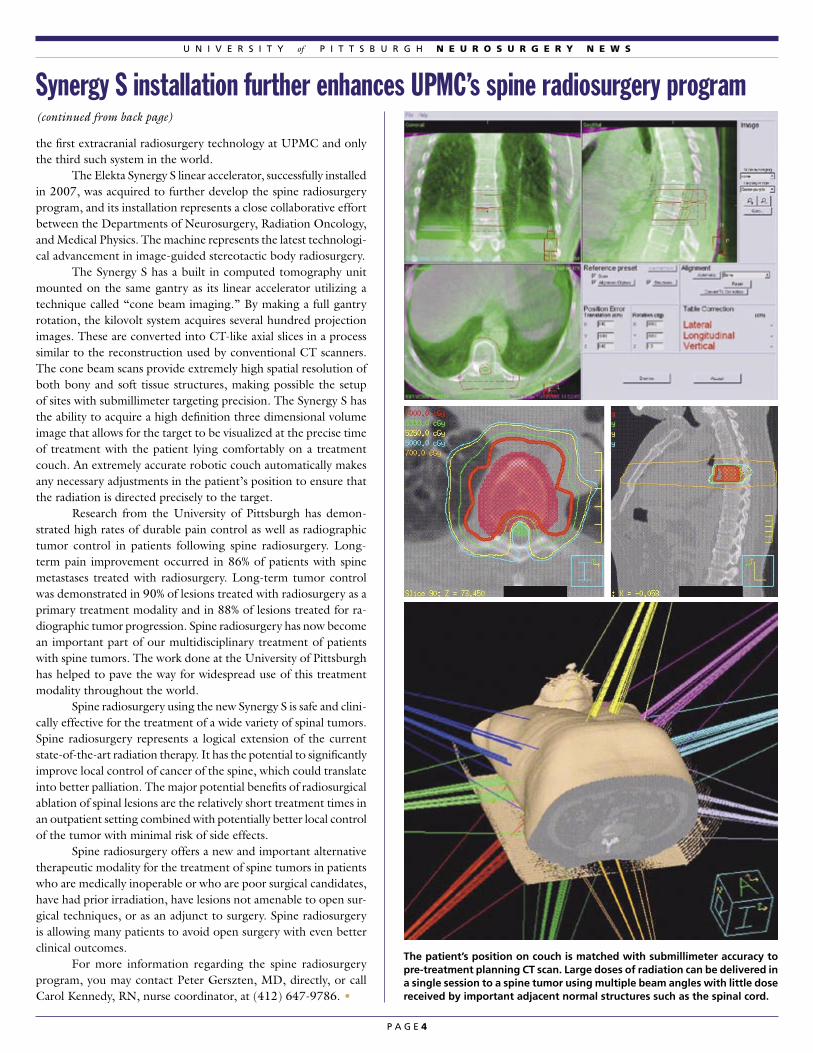
the first extracranial radiosurgery technology at UPMC and only the third such system in the world. The Elekta Synergy S linear accelerator, successfully installed in 2007, was acquired to further develop the spine radiosurgery program, and its installation represents a close collaborative effort between the Departments of Neurosurgery, Radiation Oncology, and Medical Physics. The machine represents the latest technologi-cal advancement in image-guided stereotactic body radiosurgery. The Synergy S has a built in computed tomography unit mounted on the same gantry as its linear accelerator utilizing a technique called “cone beam imaging.” By making a full gantry rotation, the kilovolt system acquires several hundred projection images. These are converted into CT-like axial slices in a process similar to the reconstruction used by conventional CT scanners. The cone beam scans provide extremely high spatial resolution of both bony and soft tissue structures, making possible the setup of sites with submillimeter targeting precision. The Synergy S has the ability to acquire a high definition three dimensional volume image that allows for the target to be visualized at the precise time of treatment with the patient lying comfortably on a treatment couch. An extremely accurate robotic couch automatically makes any necessary adjustments in the patient’s position to ensure that the radiation is directed precisely to the target. Research from the University of Pittsburgh has demon-strated high rates of durable pain control as well as radiographic tumor control in patients following spine radiosurgery. Long-term pain improvement occurred in 86% of patients with spine metastases treated with radiosurgery. Long-term tumor control was demonstrated in 90% of lesions treated with radiosurgery as a primary treatment modality and in 88% of lesions treated for ra-diographic tumor progression. Spine radiosurgery has now become an important part of our multidisciplinary treatment of patients with spine tumors. The work done at the University of Pittsburgh has helped to pave the way for widespread use of this treatment modality throughout the world. Spine radiosurgery using the new Synergy S is safe and clini-cally effective for the treatment of a wide variety of spinal tumors. Spine radiosurgery represents a logical extension of the current state-of-the-art radiation therapy. It has the potential to significantly improve local control of cancer of the spine, which could translate into better palliation. The major potential benefits of radiosurgical ablation of spinal lesions are the relatively short treatment times in an outpatient setting combined with potentially better local control of the tumor with minimal risk of side effects. Spine radiosurgery offers a new and important alternative therapeutic modality for the treatment of spine tumors in patients who are medically inoperable or who are poor surgical candidates, have had prior irradiation, have lesions not amenable to open sur-gical techniques, or as an adjunct to surgery. Spine radiosurgery is allowing many patients to avoid open surgery with even better clinical outcomes. For more information regarding the spine radiosurgery program, you may contact Peter Gerszten, MD, directly, or call Carol Kennedy, RN, nurse coordinator, at (412) 647-9786. •
Synergy S installation further enhances UPMC’s spine radiosurgery program (continued from back page)
The patient’s position on couch is matched with submillimeter accuracy to pre-treatment planning CT scan. Large doses of radiation can be delivered in a single session to a spine tumor using multiple beam angles with little dose received by important adjacent normal structures such as the spinal cord.
P A G E 5
S U M M E R 2 0 0 8 • V O L U M E 9 , N U M B E R 3
Adam S. Kanter, MDAssistant Professor of Neurological Surgery
David O. Okonkwo, MD, PhDAssistant Professor of Neurological Surgery
Richard M. Spiro, MDAssistant Professor of Neurological Surgery
Contemporary neurosurgical practice aims to provide patients with symptom relief and preservation of function via the least
invasive means. In regards to patients with degenerative cervical disc disease refractory to conservative measures, the anterior cervical decompression and fusion procedure has re-mained the surgical gold standard for decades on end. However, recent evidence regarding progressive adjacent level disc degeneration and alterations in spinal kinematics and global motion has prompted modern spine special-ists to seek a motion preserving alternative in hopes of lessening the long term impact of cervical fusion. The artificial cervical disc aims to do just that, and spine specialists at UPMC are so far pleased with the results. Cervical arthrodesis (fusion) may be beneficial to the diseased disc segment, but clinical and biomechanical studies have shown increased motion and intradiscal pressures in the adjacent levels; thus translating to an increased stress and strain on these non-operated discs, with a resultant acceleration in their degeneration. Cervical arthroplasty (artificial disc) has the potential to alleviate symptoms related to neural compression without effectively compromising segmental mobility. However, not all patients with cervi-cal disc disease are candidates for a motion preserving procedure. Ideal candidates include patients that have exhausted all forms of conservative mea-
Artificial disc technology preserves motion and function in the cervical spine
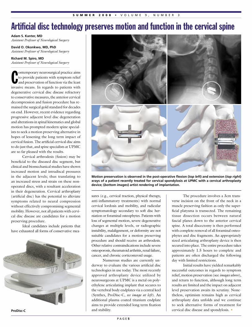
sures (e.g., cervical traction, physical therapy, anti-inflammatory treatments) with normal cervical lordosis and mobility, and radicular symptomatology secondary to soft disc her-niation or foraminal osteophytes. Patients with loss of segmental motion, severe degenerative changes at multiple levels, or radiographic instability, malalignment, or deformity are not suitable candidates for a motion preserving procedure and should receive an arthrodesis. Other relative contraindications include severe osteoporosis, rheumatoid arthritis, renal failure, cancer, and chronic corticosteroid usage. Numerous studies are currently un-derway to evaluate the various artificial disc technologies in use today. The most recently approved arthroplasty device utilized by neurosurgeons at UPMC is a metal on poly-ethylene articulating implant that secures to the vertebral body endplates via a central keel (Synthes, ProDisc-C, see image at left). An additional plasma coated titanium endplate aims to provide extended long term fixation and stability.
The procedure involves a 3cm trans-verse incision on the front of the neck in a muscle preserving fashion as only the super-ficial platysma is transected. The remaining tissue dissection occurs between natural fascial planes down to the anterior cervical spine. A total discectomy is then performed with complete removal of all foraminal osteo-phytes and disc fragments. An appropriately sized articulating arthroplasty device is then secured into place. The entire procedure takes approximately 1.5 hours to complete and patients are often discharged the following day with limited restrictions. Initial results have yielded remarkably successful outcomes in regards to symptom relief, motion preservation (see images above), and return to function, although long term results are limited and the impact on adjacent level preservation awaits its scrutiny. None-theless, optimism remains high as cervical arthroplasty data unfolds and we continue to seek alternative forms of treatment for cervical disc disease and spondylosis. •
Motion preservation is observed in the post-operative flexion (top left) and extension (top right) xrays of a patient recently treated for cervical spondylosis at UPMC with a cervical arthroplasty device; (bottom images) artist rendering of implantation.
ProDisc-C

P A G E 6
N E U R O S U R G E R Y N E W S
(continued from page 1)
localizing electromyographic (EMG) data to the surgeon for safe navigation near the lum-bar nerve plexus. Active neuromonitoring in addition to the use of real-time fluoroscopic guidance insures safety and accuracy as the expandable tubular retractor is carefully advanced through the psoas muscle to the desired disc space or vertebral body. Once the retractor system is secured, the disc material or pathologic tissue is removed and a large interbody spacer is placed (see images on front page). Dramatic widening of the collapsed disc space and stenotic foramen are clearly observed when comparing pre-operative to post-operative radiographs. In some cases, supplemental screws are required to increase the fusion construct’s stability. These can either be placed through the same lateral incision and/or in a posterior percutaneous minimally invasive fashion. Typically, healthy patients are able to walk again within hours of the lateral proce-dure’s completion, and many are discharged from the hospital after a short one- or two-day stay. Although the learning curve is steep, with experience and mastery, minimally invasive lat-eral access techniques can result in dramatically reduced operating times, blood loss, recovery period, and pain—getting patients back on their feet faster and safer than ever. •
Minimally invasive lateral access techniques show dramatic results
Adam Kanter, MD, assisted by nurse, Sara Nix, delicately places the interbody implant through the lateral access retractor system.
Matthew J. Tormenti, MDPGY-3 Resident
Peter C. Gerszten, MD, MPH, FACS Associate Professor of Neurological Surgery
Adam S. Kanter, MDAssistant Professor of Neurological Surgery
As improvements in the management of systemic cancers continue to advance, patients are surviving longer than ever.
Paradoxically, this prolonged life span has led to an increased incidence in clinically symp-tomatic metastatic disease. Spinal metastasis in particular remains one of the most com-mon sites, resulting in approximately 180,000 new diagnoses per year. The vast majority of these metastases can be treated without surgi-cal intervention. However, a subset of these patients with intractable pain, neurological dysfunction, or spinal instability require surgical intervention to preserve function and improve quality of life. Contemporary minimally invasive surgical techniques have contributed to this cause, allowing for the alleviation of pain and the improvement or preservation of neurological function. There are three reasons why cancer involving the spine causes pain: (1) the tumor infiltrates the bone marrow, (2) the tumor infiltrates and destroys the vertebra, resulting in a “pathologic fracture,” and (3) the tumor extends into the spinal canal, causing direct compression of neural structures resulting in pain, paralysis, sensory loss, and/or bowel or bladder dysfunction. Systemic chemotherapy and/or radiation therapy or radiosurgery is often all that is required to treat the major-ity of spine metastases. For patients with radiation insensitive tumors and those with severe destruction and/or destabilization of the vertebral column, with or without com-pression of the spinal cord, surgery is often required. The advent of minimally invasive techniques has added a new weapon in the spinal specialist’s arsenal. In patients with pathologic thoraco-lumbar compression fractures, the pain is often due to instability, thus stabilization is re-quired for relief. This can often be performed through a minimally invasive Kyphoplasty procedure. This technique involves placing a small hollow tube into the compressed vertebra and expanding a balloon tipped catheter to restore its height, followed by the injection of a liquid cement to fill the hollowed space. The cement hardens within minutes to provide immediate stabilization and pain relief. For those patients with compressive lesions unable to medically tolerate an open procedure, minimally invasive endoscopic
portal decompressions and percutaneous stabilization techniques may be employed. These procedures involve the use of specially designed tubular retractors placed through a 2cm incision allowing the surgeon just enough space to effectively decompress the neural structures and stabilize the spinal col-umn (see images below). Innovative real-time three-dimensional image guidance systems and fluoroscopic techniques allow the precise placement of instrumentation through these small incisions, thus minimizing muscle and tissue trauma, blood loss, procedure time, hospital stay, and healing time. This further enables patients to undergo adjuvant chemo and radiation therapy much sooner than if they had undergone a large open procedure. Growing concurrently with the tech-nological advancements in minimally invasive spinal techniques is our understanding of the indications for their implementation. Patients with spinal metastasis are now benefiting from these contemporary surgical strategies, with long lasting pain relief, spinal stability, and an overall improved quality of life. •
Expanding role for MIS surgery for cancer patients
Percutaneous screw placement provides a minimally invasive option to patients with spi-nal metastasis requiring spinal stabilization. (Top) Artist rendering depicting screw and rod placement and (bottom) the final stabilizing construct in a patient with intractable back pain from spinal metastasis

P A G E 7
▲ ▲ ▲ ▲ ▲ ▲ ▲ ▲ ▲ ▲ ▲ ▲▲▲▲▲▲▲▲▲▲▲▲▲
▲▲
▲▲
▲▲
▲▲
▲▲
▲▲
▲▲
▲▲
▲▲
▲▲
▲▲
▲▲
&
Ian Pollack Named Vice Chairman Walter Dandy Professor of Neurological Surgery and chief of pediatric neurosurgery at Children’s Hospital of Pittsburgh, Ian Pollack, MD, has been named vice chairman for academic affairs for the University of Pittsburgh Department of Neurological Surgery. Dr. Pollack is also co-director of the department’s neurosurgical oncology program. In making the announcement, department chairman Amin Kassam, MD, said, “Dr. Pollack has been the mainstay of our neuro-oncology program. His efforts are legendary nationally and interna-tionally and in my opinion represents one of the true triple threats.” Dr. Pollack has been with the University of Pittsburgh since 1992. He earned his medical degree from Johns Hopkins Univer-sity in Baltimore, and served a residency and research fellowship at the University of Pittsburgh School of Medicine. In addition, Dr. Pollack served a Van Wagenen Traveling Fellowship at the Hospital for Sick Children, Toronto, Canada; the University of Lausanne, Switzerland; and the University of Uppsala, Sweden. He was named chief of pediatric neurosurgery at Children’s Hospital in June of 2004, succeeding longtime chief A. Leland Albright, MD.
Center Treats 9000th Gamma Knife Patient The Center for Image-Guided Neu-rosurgery, led by Drs. L. Dade Lunsford and Douglas Kondziolka, treated its 9000th Gamma Knife patient on June 6. The center was estab-lished in 1987 when the first Gamma Knife in North America was installed at then-Presbyterian University Hospital. Subsequently, the center has helped introduce and pioneer every succeeding generation of this highly successful, non-invasive tool to manage tumors; arteriovenous malforma-tions and other pain or movement disorders.
Research • Elizabeth Tyler-Kabara, MD, PhD, and Wei Wang, MD, PhD, from the Department of Physical Medicine and Rehabilitation, were awarded $100,000 from the Clinical and Translational Science Institute (CTSI) Translational Tool Pilot Project (TTPP) program for their Micro-ECoG Brain Computer Interface project. • Dr. Lunsford and Devra Davis, PhD, of the UPCI Center for Environmental Oncology will serve as coinvestigators in a study funded by the Jennie Zoline Foundation to assess the pattern of cell phone usage in patients with acoustic neuromas.
Prominent Lectures • Amin Kassam, MD, was the keynote speaker at the Memo-rial Meeting of the Japanese Society for Skull Base Surgery in Tokyo, Japan, July 6-8 and a visiting professor at Dalhousie University in Halifax, Canada, June 25-26. Dr. Kassam was also an invited lecturer and course director at the Congreso Sociedad Espanola de Base de Craneo, in Santander, Spain, June 11-14; a course co-director for the International Hands-On Course on Endoscopic and Microscopic Microsurgery of the Skull Base in Nice, France, June 6-8; keynote speaker at the World Congress for Endoscopic Surgery of the Brain, Skull Base and Spine in Sao Paulo, Brazil, May 22-24; course director at the En-doscopic Approaches to Skull Base Lesions Course at the University of Ottawa/Ottawa Hospital in Ottawa, Canada, April 11-12; and
an invited lecturer at the NIH International Chordoma Research Workshop in North Bethesda, MD, April 4. • Peter Gerszten, MD, was the invited guest speaker at the University of Manitoba Health Sciences Centre in Winnipeg, Canada on June 24. Dr. Gerszten was also a visiting professor at the University of South Florida & Moffitt Cancer Center in Tampa, FL, June 19-20; and the invited guest speaker of the Bi-annual Meeting of the Neurosurgery Society of Venezuela in Maracaibo, Venezuela, May 28. • Dr. Lunsford served as visiting professor at Stanford Uni-versity, May 30.
Congratulations • A special black-tie reception and dinner was held June 21 at the Fox Chapel Golf Club honoring the department’s 2008 graduating chief residents, Dave Atteberry, MD, Johnathan Engh, MD, and Paul Gardner, MD. All three doctors announced plans to remain with the department as assistant professors as of July 1. During the evening, it was announced that Dr. Engh was selected as best resident teacher by the department faculty for 2007-08. Daniel Prevedello, MD, was chosen as best faculty teacher by the residents. • Joseph Maroon, MD, has qualified to
participate in the Hawaiian Ironman Champion-ship Triathlon scheduled for October 11 in Kona, Hawaii. Dr. Maroon qualified for the event—one of the most recognized endurance events in the world—by finishing fifth in the 65-70 age group at a qualifying half-marathon in Muncie, IN. • Dr. Maroon was named senior vice president of the American Academy of Anti-Aging Medicine. • Donald J. Crammond, PhD, was invited by the executive committee of the International Federation of Clinical Neurophysiology to serve on the editorial board of the journal Clinical
Neurophysiology. • Ava Puccio received a doctorate of philosophy degree from the University of Pittsburgh School of Nursing in April.
Media • A research study by Hank Weiss, PhD, showing the repeal of the Pennsylvania motorcycle helmet law has resulted in a major increase in head injuries received widespread regional and national media attention. • Dr. Maroon was featured in a July 22 Pittsburgh Tribune-Review article about his training for the Hawaiian Ironman. • Dr. Kassam was quoted in a June 3 abcnews.com article dealing with how doctors treat high-profile patients.
Welcome • Darcy Kavel, Hope George, Donna Flaherty, and Megan McShea, all physician assistants; Kelly Crow, patient information coordinator; Hannah Hetzel, research assistant for David Okonkwo, MD, PhD.
Personal Congratulations • Louisa Urgo, PA-C, and husband David had a baby boy (Joseph) on June 5; Daniel Prevedello, MD, and wife Priscilla had a baby daughter (Isabella) on April 30.
S U M M E R 2 0 0 8 • V O L U M E 9 , N U M B E R 3
Department of Neurological SurgeryUniversity of Pittsburgh Medical CenterUPMC Presbyterian/Suite B-400200 Lothrop StreetPittsburgh, PA 15213(412) [email protected]
www.neurosurgery.pitt.edu
Non-ProfitOrganizationU.S. Postage
PAIDPermit #4166Pittsburgh, PA
newsneurosurgeryP I T T S B U R G Ho fU N I V E R S I T Y
(412) 647-3685Patient Referrals
Recognized as an honor roll member of U.S.News & World Report’s ‘America’s Best Hospitals’ 2008University of Pittsburgh Medical Center
S U M M E R 2 0 0 8 • V O L U M E 9 , N U M B E R 3
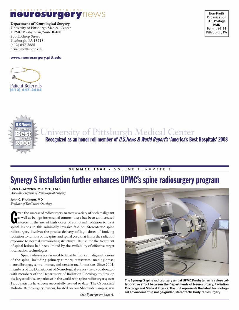
Synergy S installation further enhances UPMC’s spine radiosurgery programPeter C. Gerszten, MD, MPH, FACS Associate Professor of Neurological Surgery
John C. Flickinger, MDProfessor of Radiation Oncology
Given the success of radiosurgery to treat a variety of both malignant as well as benign intracranial tumors, there has been an increased interest in the use of high doses of conformal radiation to treat
spinal lesions in this minimally invasive fashion. Stereotactic spine radiosurgery involves the precise delivery of high doses of ionizing radiation to tumors of the spine and spinal cord that limits the radiation exposure to normal surrounding structures. Its use for the treatment of spinal lesions had been limited by the availability of effective target localization technologies. Spine radiosurgery is used to treat benign or malignant lesions of the spine, including primary tumors, metastases, meningiomas, neurofibromas, schwannomas, and vascular malformations. Since 2001, members of the Department of Neurological Surgery have collaborated with members of the Department of Radiation Oncology to develop the largest clinical experience in the world with spine radiosurgery; over 1,000 patients have been successfully treated to date. The CyberKnife Robotic Radiosurgery System, located on our Shadyside campus, was
(See Synergy on page 4)
The Synergy S spine radiosurgery unit at UPMC Presbyterian is a close col-laborative effort between the Departments of Neurosurgery, Radiation Oncology and Medical Physics. The unit represents the latest technologi-cal advancement in image-guided stereotactic body radiosurgery.