summer 2008 volume 16, number 1 registry the · summer 2008 volume 16, number 1 newsletter of the...
TRANSCRIPT

REGISTRYSummer 2008 Volume 16, Number 1
Newsletter of the NIDCD National Temporal Bone, Hearing and Balance Pathology Resource Registry
the
Conservation of residual hearing has become an important goal in cochlear implant surgery, particularly for patients who may be candidates for com-bined electrical and acoustic stimulation. In spite of advancements in
surgical technique and electrode design, residual hearing is lost in at least 10-20% of cochlear implant patients (1-3). It seems likely that a variety of factors contribute to hearing loss associated with implant surgery; however, mechanical trauma to various intracochlear structures probably plays an important role. Structures that are par-ticularly vulnerable to implant-related injury include the osseous lamina and basilar membrane, the modiolus, soft tissues of the lateral cochlear wall, and blood vessels associated with the scala tympani.
Although it has received relatively little attention in previous studies, vascular injury occurring during cochlear implant surgery may com-promise inner ear function and thereby contribute to loss of residual hearing. Vascular trauma is a concern because of the significant exposure of scala tympani vessels to the perilymphatic space where they are vulnerable to mechani-cal injury.Observations on the vessels of scala tympani are described below in relation to
ThE VaSCulaTuRE of SCala TYmpanI In RElaTIon To CoChlEaR ImplanTaTIonCharles G. Wright Ph.D., Karen S. Pawlowski Ph.D., Peter S. Roland M.D.Department of Otolaryngology – Head and Neck Surgery, Southwestern Medical Center, Dallas, Texas
See THE VASCULATURE OF THE SCALA TYMPANI..., page 3
Fig. 1Overview of veins in scala tympani in an osmium stained preparation. In this dissection, the osseous lamina and basilar membrane have been removed to provide a direct view into scala tympani (ST) of the lower apical and middle turns of the human cochlea. In the area enclosed by white brackets numerous venules draining lateral wall structures are seen converging to form the veins that cross the floor of scala tympani to join the posterior spiral vein coursing around the modiolus (M). The arrow indicates the point at which one of the veins on the floor of scala tympani joins the posterior spiral vein.
The REGISTRY is published semiannually by the NIDCD National Temporal Bone, Hearing and Balance Pathology Resource Registry. The Registry was established in 1992 by the National Institute on Deafness and Other Communication Disorders (NIDCD) of the National Institutes of Health to continue and expand upon the former National Temporal Bone Banks (NTBB) Program. The Registry promotes research on hearing and balance disorders and serves as a resource for the public and the scientific community about research on the pathology of the human auditory and vestibular systems.
CONTENT
SCIENTIFIC ARTICLE: The Vasculature of Scala Tympani in Relation to Cochlear Implantation ...................... 1
LAB SPOTLIGHTHuman Temporal Bone Laboratory Southwestern Texas MC .................. 6
REGISTRY AND OTHER NEWS: News and Announcements .............. 2 Otopathology Mini-Travel Fellowship ........................................ 2 Upcoming meetings ......................... 7 Order form for Temporal Bone Donation Brochures ......................... 8
aabb

The Registry • Vol. 16.1 Summer 2008 �
oTopaTholoGY mInI-TRaVEl fElloWShIp pRoGRam
The NIDCD National Temporal Bone Registry is pleased to announce the avail-ability of mini-travel fellowships. The fellowships provide travel funds for research technicians and young investigators to visit a temporal bone laboratory for a brief educational visit, lasting approximately one week. The emphasis is on the training of research assistants, technicians and junior faculty. The fellowships are available to:1) U.S. hospital departments who aspire to start a new temporal bone laboratory 2) Inactive U.S. temporal bone laboratories that wish to reactivate their collections or 3) Active U.S. temporal bone laboratories that wish to learn new research techniques
Up to two fellowship awards will be made each year ($1,000 per fellowship). The funds may be used to defray travel and lodging expenses. Applications will be de-cided on merit. Interested applicants should submit the following:1) A 1-2 page outline of the educational or training aspect of the proposed fellowship 2) Applicant’s curriculum vitae 3) Letter of support from temporal bone laboratory director or department chairman 4) Letter from the host temporal bone laboratory, indicating willingness to receive the traveling fellow
.
Applications should be sent to:
Saumil N. Merchant, M.D.NIDCD National Temporal Bone Registry
Massachusetts Eye and Ear Infirmary243 Charles StreetBoston, MA 02114
NIDCD to Celebrate 20th Anniversary with Scientific SymposiumThe National Institute on Deafness and Other Commu-nication Disorders (NIDCD) will be celebrating its 20th anniversary on Thursday, Oct. 23, 2008, with a sym-posium highlighting two decades of scientific research accomplishments supported by the NIDCD. The program begins at 8:30 a.m. at the Natcher Confer-ence Center on the campus of the National Institutes
of Health in Bethesda, MD. The symposium will consist of three sessions representing NIDCD’s primary areas of research: hearing and balance; smell and taste; and voice, speech, and language. Expected attendees include NIH staff members, scientists, advocacy organizations, congressional representatives and news media members. Check the NIDCD Web site, www.nidcd.nih.gov, for updates.
This year marks the 50th Anniversary of the Deafness Research Foundation. To recognize its achievements and to re-commit itself to the future, the Deafness Re-search Foundation hosted a special 50th Anniversary
Celebration on May 14, 2008 at The Hudson Theatre in New York City. The event was held in honor of their founder, Mrs. Collette Ramsey Baker. The Deafness Research Foundation is one of the largest and oldest hearing research organization in the US, and is dedicated to making a lifetime of hearing health possible for all people through quality research and education.
NEWS AND ANNOUNCEMENTSthe
REGISTRYDIRECTORS Joseph B. Nadol, Jr., M.D. Saumil N. Merchant, M.D. Steven D. Rauch, M.D. Michael J. McKenna, M.D. Joe C. Adams, Ph.D.
SCIENTIFIC ADVISORY COUNCIL Newton J. Coker, M.D. Howard W. Francis, M.D. Marlan R. Hansen, M.D. Raul Hinojosa, M.D. Akira Ishiyama, M.D. Herman A. Jenkins, M.D. Elizabeth M. Keithley, Ph.D. Robert I. Kohut, M.D. Fred H. LInthicum, Jr., M.D. Saumil N. Merchant, M.D. Joseph B. Nadol, Jr., M.D. Michael M. Paparella, M.D. Jai H. Ryu, Ph.D. Isamu Sando, M.D., D.M.Sc. P. Ashley Wackym, M.D. Charles G. Wright, Ph.D,
COORDINATOR Cindy Regan
ADMINISTRATIVE STAFF Richard A. Cortese Tammi N. King Carol Y. Ota
NEWSLETTER EDITORS General: Cindy Regan Medical: Saumil N. Merchant, M.D. Joseph B. Nadol, Jr., M.D.
NIDCD National Temporal Bone, Hearing and Balance Pathology Resource Registry Massachusetts Eye and Ear Infirmary 243 Charles Street Boston, MA 02114
(800) 822-1327 TOLL-FREE VOICE (617) 573-3711 VOICE (617) 573-3838 FAX EMAIL: [email protected] WEB: www.tbregistry.org
� The Registry • Vol. 16.1 Summer 2008
possible injury during cochlear implantation. The find-ings discussed here are based on examination of human temporal bone microdissections, conventional histological sections and scanning electron micrographs taken from recent anatomical studies in our laboratory. Additional illustrations and an expanded version of this brief review can be found at: http://www.utsouthwestern.edu/utsw/cda/dept28156/files/111715.html (see link to “Inner Ear Anatomy and Cochlear Implantation”).
The superficially located vessels of scala tympani are al-most entirely of the venous type. They include the venules found on the lateral wall and floor of scala tympani, the anterior and posterior spiral veins associated with the mo-diolus and the spiral vessel beneath the basilar membrane. The arrangement and morphological features of these ves-sels are illustrated in Figures 1-5. The venules of the lateral wall drain the stria vascularis and spiral ligament and they converge to form the small veins crossing the floor of scala
tympani which empty into the posterior spiral vein. The posterior spiral vein also collects blood from the spiral ganglion and receives connecting vessels from the ante-rior spiral vein which drains the osseous lamina and scala vestibuli. In the lower basal cochlear turn, the two spiral veins join to form the common modiolar vein, which after uniting with the vestibulocochlear vein, becomes the vein of the cochlear aqueduct (4), also known as the inferior cochlear vein.
As the micrographs included in this article illustrate, many of the venous vessels in scala tympani have little or no bony covering, leaving them essentially exposed to the perilymphatic space. They are therefore susceptible to compression or mechanical injury associated with insertion of electrode arrays. During the insertion procedure, implant arrays frequently tear or compress the delicate tissues of the spiral ligament immediately below the area of attachment of the basilar membrane (5,6). Scanning electron microscopic study has shown this portion of the spiral ligament to be composed of an open connective tissue meshwork which appears prone to mechanical disruption by electrode arrays (5). Such injuries will inevitably traumatize the venules cours-ing through the lower portion of the spiral ligament. These small vessels drain the stria vascularis and the part of the spiral ligament that lies adjacent to the lateral wall of scala media. Interruption of the venous outflow in the lateral wall would compromise oxygen delivery and thereby im-pair spiral ligament and strial function, which is essential for maintenance of the ionic composition of the cochlear fluids and the endolymphatic potential of the cochlear duct.
The bone of the modiolar wall is very fragile and shows numerous gaps and open spaces in its structure (7). The posterior spiral vein and vessels of the modiolar wall are therefore unprotected by bone in some areas (Fig. 4). Thus, perimodiolar electrode arrays positioned in close contact with the modiolus may injure these vessels. Such injury can occur if the tip of an electrode strikes the modiolar wall during insertion or if the body of the array is pushed into contact with the modiolus by a positioner device (8). Modiolar trauma may also occur if explantation becomes necessary because of the tendency for a perimodiolar array to be pulled into tighter contact with the modiolus as it is withdrawn from the cochlea.
In addition to the possibility of vascular trauma produced by electrode arrays, the veins in the lower basal turn of
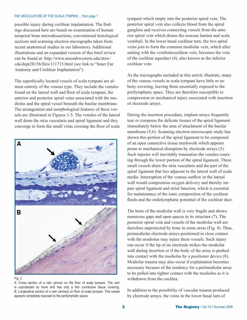
Fig. 2A: Cross section of a vein (arrow) on the floor of scala tympani. The vein is unprotected by bone and has only a thin connective tissue covering. B: Longitudinal section of a vein (arrows) on floor of scala tympani. This vessel appears completely exposed to the perilymphatic space.
THE VASCULATURE OF THE SCALA TYMPANI..., from page 1

The Registry • Vol. 16.1 Summer 2008 �
scala tympani are at risk of injury during surgical drilling associated with cochleostomy placement. Such injuries may occur during drilling of a standard promontory co-chleostomy, or in the course of a round window insertion, involving drilling of the anterior-inferior margin of the round window near the so-called crista fenestrae. Obser-vations from the authors’ laboratory have confirmed that the cochlear aqueduct may lie within one-half millimeter of the inferior margin of the round window membrane (9). Therefore, caution is necessary when drilling in this area to avoid injury to the common modiolar vein which enters the bony channel immediately adjacent to the aqueduct to become the vein of the cochlear aqueduct. Injury or occlu-sion of this vessel would be particularly significant since it is widely believed to provide virtually the entire venous drainage of the cochlea (4,10). As shown in Figure 3B, the common modiolar vein also receives the vestibulocochlear
vein which carries blood from the vestibular sensory organs and the basal end of the cochlea (4). There is, therefore, the potential for circulatory compromise of the vestibular apparatus as a result of vascular injury in the lower basal portion of scala tympani.
As noted above, the vein of the cochlear aqueduct is believed to provide almost 100% of the venous outflow of the cochlea in most individuals. The possibility of collateral venous circulation does, however, exist. In the older literature, a central auditory vein which follows the course of the auditory nerve is described (11,12). This vessel is now thought to be present inconsistently (4). In cases where such a vessel does exist, there may be connec-tions between it and the posterior spiral vessel and/or the common modiolar vein, providing the potential for collat-eral circulation (12). Another possible route for collateral drainage may be via connections between the cochlear veins and the vessels of the mucoperiosteum of the middle ear (13). It might, therefore, be expected that injury of the common modiolar vein or vein of the cochlear aqueduct during implant surgery could result in variable damage to inner ear tissues, depending on the degree to which col-lateral vessels are present in a particular patient.
Various types of basilar membrane damage have been reported in association with placement of electrode arrays in scala tympani. Electrodes may occasionally penetrate the basilar membrane and enter scala media or, if they remain within scala tympani, they may directly contact the underside of the basilar membrane and elevate the cochlear partition and/or fracture the osseous spiral lamina (5,6,14).
Fig. 3A: Dissection view of scala tympani and the modiolus in basal cochle-ar turn showing the posterior spiral vein (black arrows). A tributary vein (white arrow) on the modiolar wall joins the posterior spiral vein. ST, floor of scala tympani. Cross sections of similar vessels are shown in Fig. 4. B: Lower basal turn showing the common modiolar vein as it enters the cochlear aqueduct (black arrow). The white arrow indicates the vestibulocochlear vein which joins the common modiolar vein. ST, floor of scala tympani.
Fig. 4Cross sections of the modiolus showing veins coursing near the surface of the modiolar wall. In “A”, the vein indicated by the arrow has a very thin covering of bone facing scala tympani. In an adjacent section, shown in “B”, a similar vein (arrow) appears entirely exposed to the perilymphatic space of scala tympani. OL, osseous lamina; SG, spiral ganglion.

� The Registry • Vol. 16.1 Summer 2008
In either case, the spiral vessel and associated arterioles found on the lower surface of the basilar membrane and osseous lamina may be damaged or occluded. As shown in Figure 5, these vessels lie in an unprotected position where they could easily be subject to electrode trauma. Since the spiral vessel is believed to be important for oxygen deliv-ery to the organ of Corti (4,15), its injury could be a factor in loss of residual hearing in patients who have viable organ of Corti remaining on the basilar membrane.
In addition to circulatory compromise produced by vas-cular occlusion, bleeding due to implant-related injury of cochlear vessels may have detrimental effects on inner ear function. As has long been known, significant intracochlear bleeding due to hemorrhage can produce sudden hearing loss, a fact which has been confirmed by recent imaging studies (16). However, Radeloff et al. (17) have demon-strated that even a few microliters of blood introduced into
REfEREnCES1. Gstoettner W, Kiefer J, Baumgartner W-D, Pok S, Peters S, Adunka O. Hearing preservation in cochlear implantationfor electric acoustic stimulation. Acta Otolaryngol2004;124:348-352.2. Balkany TJ, Connell SS, Hodges AV, Payne SL, Telischi FF,Eshraghi AA, Angeli SI, Germanni R, Messiah S, Arheart KL.Conservation of residual acoustic hearing after cochlearImplantation. Otol Neurotol 2006;27:1083-1088.3. Berrettini S, Forli F, Passetti S. Preservation of residual hearing following cochlear implantation: comparison between three surgical techniques. J Laryngol Otol 2008 (Epub ahead of print).4. Schuknecht HF. Pathology of the Ear. 2nd edition.Lea & Febiger, Philadelphia. 1993.5. Roland PS, Wright C.G. Surgical aspects of cochlear implantation: Mechanisms of insertional trauma. Adv Otorhinolaryngol 2006;64:11-30.6. Nadol JB, Shiao JY, Burgess BJ, Ketten DR, Eddington DK, Gantz BJ, Kos I, Montandon P, Coker NJ, Roland JT,Shallop JK: Histopathology of cochlear implants in humans. Ann Otol Rhinol Laryngol 2001;110:883-891.7. Rask-Andersen H, Schrott-Fischer A, Pfaller K, GluekertR. Perilymph/modiolar communication routes in the human cochlea. Ear Hear 2006;27:457-465.8. Aschendorff A, Klenzner T, Richter B, Kubalek R,Nagursky H, Laszig R. Evaluation of the HiFocus Electrode array with positioner in human temporalbones. J Laryngol Otol 2003;117:527-531.9. Roland PS, Wright CG, Isaacson B. Cochlear implant electrode insertion: the round window revisited.Laryngoscope 2007;117:1397-1402.10. Axelsson A. The vascular anatomy of the cochlea in theguinea pig and in man. Acta Otolaryngol (Stockh)1968;Suppl 243:1-134.11. Siebenmann F. Die blutgefasse im labyrinthe desmenschlichen ohres. Bergmann, Wiesbaden. 1894.12. Portmann M, Sterkers JM, Charachon R, Chouard CH.The Internal Auditory Meatus. ChurchillLivingstone, Edinburgh, 197513. Watanabe Y, Nakashima T, Yanagita N. Venous com-munications of the cochlea after acute occlusion ofthe vein of the cochlear aqueduct. ArchOtorhinolarygol 1988;245:340-343.14. Wardrop P, Whinney D, Rebscher SJ, Roland TJ, Luxford W, Leake PA: A temporal bone study of insertion trauma and intracochlear position of cochlear implantelectrodes. I: Comparison of Nucleus banded andNucleus Contour electrodes. Hear Res 2005;203:54-67.15. Nomura Y. Vascular supply to the organ of Corti in man.Arch Otorhinolaryngol 1977;214:213-220.16. Vakkalanka S, Ey E, Goldenberg RA. Inner ear hemorrhageand sudden hearing sensorineural hearing loss. Amer J Otol 2000;21:764-765.17. Radeloff A, Unkelbach Mh, Tillein J, Braun S, Helbig S,Gstoettner W, Adunka OF. Impact of intrascalar blood onhearing. Laryngoscope 2007;117:58-62.
Fig. 5A: Cochlear cross section showing a vessel (arrow) emerging from the edge of the osseous lamina. OC, organ of Corti; ST, scala tympani. The vessel has a connective tissue covering, but is unprotected by bone. B: Scanning electron micrograph of the underside of the basilar membrane (BsM) and osseous lamina (OSL). The arrows indicate the spiral vessel beneath the basilar membrane, which is exposed to the perilymphatic space.
See THE VASCULATURE OF THE SCALA TYMPANI..., page 7

The Registry • Vol. 16.1 Summer 2008 �
LAB SPOTLIGHThuman Temporal Bone laboratory
Department of otolaryngology – head and neck Surgery, Southwestern medical Center
Dallas, Texas
Human temporal bone research at Southwestern Medical Center began in 1976 with work focused on development and pathology of the vestibular apparatus. At that time our interest centered on studies of the otolith organs in
undecalcified temporal bones. Most specimens were prepared by microdissection and studied by a combination of light and scanning electron microscopy.
After Dr. William Meyerhoff joined the Department as Chairman in 1982, the laboratory started to prepare temporal bones for celloidin embedding and sectioning. To date, over 300 temporal bones have been processed for either microdissection or celloidin sectioning. Many of the sectioned specimens in our collection were obtained from pediatric patients. They have provided a valuable resource for studies on temporal bone development and congenital abnormalities of the ear.
Microdissection and conventional sectioning methods are complementary approaches to human temporal bone research. Together. they permit a wide array of laboratory techniques to be applied in the study of temporal bone development, morphology, and pathology. As reflected in the list of references below, much of the work from our laboratory has been done using a combination of the two research methods.
In recent years, we have utilized the microdissection method for studies relating to cochlear implantation, including evaluation of new electrode designs and investigation of various factors contributing to cochlear injury associated with implant surgery. Dr. Peter Roland, who is currently Department Chairman, is an active member of our temporal bone research group and has fostered an integrated program of clinical and basic science research aimed at improving clinical outcomes in cochlear implant patients.
Publications From The Human Temporal Bone Laboratory,Southwestern Medical Center, Dallas1. Wright CG, Hubbard DG. Observations of otoconial membranes from human infants. Acta Otolaryngol 1978; 86:185-194.2. Weinberg AG, Wright CG. Stapedial Hypoplasia in partial DiGeorge Syndrome. Amer J Dis Child 1978;132:815-816.3. Wright CG, Hubbard DG, Clark GM. Observations of human fetal otoconial membranes. Ann Otol Rhinol Laryngol 1979;88:267-274.4. Wright CG, Habbard DG, Graham JW. Absence of otoconia in a human infant. Ann Otol Rhinol Laryngol 1979:88(pt 1):779-783.5. Wright CG, Hubbard DG. Morphological observations of human otoconial membranes. In: Gualtierotti. T. (ed.) The Vestibular System: Function and Morphology. Springer-Verlag, New York. 1981;88-100.6. Schaefer SD, Wright CG, Post JD, Frenkel, EP. Cis-Platinum vestibular toxicity. Cancer 1981;47:857-859.7. Wright CG, Weinberg AG, Hubbard DG, Rouse RC, Johnsson LG. Ear anomalies in an infant with Potter’s syndrome. Amer J Otolaryngol 1981;3:134-138.
8. Wright CG, Rouse RC, Johnsson LG, Weinberg AG, Hubbard DG. Vaterite otoconia in two cases of otoconial membrane dysplasia. Ann Otol Rhinol Laryngol 1982;91:193-199.9. Wright CG, Rouse RC, Zajic GH, Schaefer SD, Hubbard DG, Barnard LA. 1982. A calcareous concretion in the posterior semicircular duct of a human labyrinth. Amer J Otolaryngol 1982;3:196-201.10. Johnsson LG, Rouse RC, Wright CG, Henry PJ, Hawkins JE, Jr. Pathology of neuroepithelial suprastructures of the human inner ear. Amer J Otolaryngol 1982;3:77-90.11. Wright CG, Hubbard DG. SEM observations on development of human otoconia during the first trimester of gestation. Acta Otolaryngol 1982;94:7-18.12. Wright CG, Schaefer SD. Inner ear histopathology in patients treated with cis-platinum. Laryngoscope 1982; 92:1408-1413.13. Rutledge JC, Friedman JM, Harrod MJE, Currarino G, Wright CG, Pinckney L, Chen H. A “new” lethal multiple congenital anomaly syndrome: joint contractures, cerebellar hypoplasia, renal hypoplasia, urogenital anomalies, tongue cysts, shortness of limbs, eye abnormalities, defects of the heart, gallbladder agenesis, and ear malformations. Amer J Med Genetics 1984;19:255-264.
By Charles G. Wright Ph.D.

� The Registry • Vol. 16.1 Summer 2008
The Registry is planning to exhibit at these upcoming meetings
MEETINGS
AAO-HNS Annual Meeting and OTO Expo
Chicago, IL September 21-24, 2008
www.entnet.org
AARP Life@50+ National Event and Expo
Washington, DC September 4-6, 2008
www.aarp.org life@50+AARP’s National Event & Expo
scala tympani in laboratory animals cause significant permanent shifts in hearing thresholds. These findings raise the possibility that relatively small amounts of bleeding, such as might occur during cochlear implanta-tion, may negatively impact auditory function and thereby contribute to post-implantation hearing loss.
ACKNOWLEDGEMENT: The authors are grateful for publisher’s permission to use the illustrations shown in Figures 3A and 5, which are from Roland PS, Wright C.G. Surgical aspects of cochlear im-plantation: Mechanisms of insertional trauma. Adv Otorhinolaryngol 2006;64:11-30. Copyright S. Karger AG, Basel.
Address for correspondence:C.G. Wright, Ph.D.Department of Otolaryngology – Head and Neck SurgerySouthwestern Medical Center5323 Harry Hines BlvdDallas, TX 75390-9035Email: [email protected]
THE VASCULATURE OF THE SCALA TYMPANI..., from page 5, column 1
14. Wright CG, Brown OE, Meyerhoff WL, Rutledge JC. Inner ear anomalies in two cases of trisomy 18. Amer J Otolaryngol 1985;6:392-404.15. Wright CG, Brown OE, Meyerhoff WL, Rutledge JC. Auditory and temporal bone abnormalities in CHARGE association. Ann Oto Rhinol Laryngol 1986;95:480-486.16. Roland PS, Wright CG, Meyerhoff WL, Mickey, BE. Anatomic considerations in the posterior approach to the internal auditory canal. Ann Otol Rhinol Laryngol 1988;97:621-625.17. Wright CG, Meyerhoff WL. Microdissection in the study of human temporal bone morphology. Ann Otol Rhinol Laryngol 1989;98(Suppl 143):25-28.18. Wright CG, Meyerhoff WL. Pathology of otitis media. Ann Otol Rhinol Laryngol 1994; 103(Suppl 163):24-26.19. Levine JL, Wright CG, Pawlowski KS, Meyerhoff WL. Postnatal persistence of epidermoid rests in the human middle ear. Laryngoscope 1998;108:70-73.20. Pawlowski KS, Wright CG, Meyerhoff WL. 1998. Histologic demonstration of glycosaminoglycans in inner ear fluids. Acta Otolaryngol 1998;118:505-510.21. Bachor E, Wright CG, Karmody CS. The incidence and distribution of cupular deposits in the pediatric vestibular labyrinth. Laryngoscope 2002;112:147-151.22. Ulualp SO, Wright CG, Pawlowski KS, Roland PS. Histopathaological basis of hearing impairment in Wolf-Hirschhorn syndrome. Laryngoscope 2004;114:1426-1430.23. Ulualp SO, Wright CG, Pawlowski KS, Roland PS. Cochlear degeneration in Leigh disease: histopathologic features. Laryngoscope 2004;114:2239-2242.24. Ulualp SO, Wright CG, Roland PS. 2005. Temporal bone histopathologic findings in partial trisomy 13 and partial trisomy 14. Int J Pediatr Otorhinolaryngol 2005;69:781-789.25. Ulualp SO, Wright CG, Roland, PS. 2005. Spectrum of middle and inner ear abnormalities in infants with congenital heart defects. Otolaryngol Head Neck Surg. 2005; 133:260-268.26. Bachor E, Just T, Wright CG, Pau HW, Karmody C.S. Fixation of the stapes footplate in children: a clinical and temporal bone histopathologic study. Otol Neurotol 2005;26:866-873.27. Wright CG, Roland PS, Kuzma J. Advanced Bionics Thin Lateral and Helix II electrodes: a temporal bone study. Laryngoscope 2005;115:2041-2045.28. Wright CG, Roland PS. Temporal bone microdissection for anatomic study of cochlear implant electrodes. Cochlear Implants International 2005;6:159-168.29. Roland PS, Wright CG. 2006. Surgical aspects of cochlear implantation: Mechanisms of insertional trauma. Adv Otorhinolaryngol 2006;64:11-30.30. Roland PS, Wright CG, Isaacson B. 2007. Cochlear implant electrode insertion: the round window revisited. Laryngoscope 2007;117:1397-1402.

FREE BROCHURES FOR YOUR OFFICE OR CLINIC ABOUT TEMPORAL BONE RESEARCH AND DONATION
The Registry • Vol. 16.1 Summer 2008 �
NIDCD National Temporal Bone, Hearing & Balance Pathology Resource RegistryMassachusetts Eye and Ear Infirmary243 Charles StreetBoston, MA 02114-3096
NON-PROFIT ORGU.S. POSTAGE
PAIDBOSTON, MA
PERMIT NO. 53825
Address Service Requested
PLEASE! Notify us of your change of address before you move. Each undelivered newsletter is returned to the Registry office at a cost of $.70. Our loss is over $1.00 per unit.Thank you!
If you are willing to display either or both of these brochures, please complete the form below and return it to the Registry by mail or fax. The brochures will be sent to you free of charge. Please circle the amount requested for each brochure or write in amount not listed.
NAME:
ADDRESS:
ADDRESS:
CITY, STATE, ZIP:
TELEPHONE:
That Others May Hear _____ 25 50 100 The Gift of Hearing and Balance _____ 25 50 100
Mail or fax this form to the Registry at: NIDCD National Temporal Bone, Hearing and Balance Pathology Resource Registry, Massachusetts Eye and Ear Infirmary, 243 Charles Street, Boston, MA 02114
Toll-free phone: (800) 822-1327, Fax: (617) 573-3838, Email: [email protected]
That Others May Hear is a short brochure that briefly describes the functions of the Registry, and answers commonly asked questions regarding the temporal bone donation process. (Dimensions: 9” x 4”)
The Gift of Hearing and Balance: Learning about Temporal Bone Donation is a 16-page, full-color booklet which describes in more detail the benefits of temporal bone research. It also answers commonly asked questions regarding the temporal bone donation process. (Dimensions: 7” x 10”)