super-enhancer acquisition drives oncogene expression in
TRANSCRIPT

RESEARCH ARTICLE
Super-enhancer acquisition drives oncogene
expression in triple negative breast cancer
Ryan Raisner1, Russell BainerID2, Peter M. Haverty3, Kelli L. BenedettiID
4, Karen
E. GascoigneID1*
1 Department of Discovery Oncology, Genentech, Inc., South San Francisco, California, United States of
America, 2 Maze Therapeutics, South San Francisco, California, United States of America, 3 Department of
Bioinformatics, Genentech, Inc., South San Francisco, California, United States of America, 4 Department of
Cell and Tissue Biology, University of California, San Francisco, California, United States of America
Abstract
Triple Negative Breast Cancer (TNBC) is a heterogeneous disease lacking known molecular
drivers and effective targeted therapies. Cytotoxic chemotherapy remains the mainstay of
treatment for TNBCs, which have significantly poorer survival rates compared to other
breast cancer subtypes. In addition to changes within the coding genome, aberrant
enhancer activity is a well-established contributor to tumorigenesis. Here we use H3K27Ac
chromatin immunoprecipitation followed by sequencing (ChIP-Seq) to map the active cis-
regulatory landscape in TNBC. We identify distinct disease subtypes associated with spe-
cific enhancer activity, and over 2,500 unique superenhancers acquired by tumor cells but
absent from normal breast tissue. To identify potential actionable disease drivers, we
probed the dependency on genes that associate with tumor-specific enhancers by CRISPR
screening. In this way we identify a number of tumor-specific dependencies, including a pre-
viously uncharacterized dependency on the TGFβ pseudo-receptor BAMBI.
Introduction
Triple Negative Breast Cancer (TNBC) is a heterogenous disease lacking clear molecular driv-
ers. Transcriptional profiling has allowed stratification of the disease, primarily based on the
similarity of tumors to a basal or luminal cell state [1]. While this distinction can help predict
disease severity and outcome, it has yet to significantly impact treatment options. Previous
TNBC profiling efforts focused on transcribed genes, evaluating gene expression, DNA muta-
tions, and copy number changes. However, the non-coding regulatory elements that control
gene expression are also critical to defining the tumor cell state, and remain relatively poorly
explored in TNBC.
Gene-distal regulatory elements such as enhancers play an important role in the control of
gene expression. Such elements are identified by their chromatin state, and the combination of
histone modifications and chromatin binding proteins present. As such, technologies includ-
ing chromatin immunoprecipitation followed by sequencing (ChIP-Seq) are required to inter-
rogate these regions. In particular, acetylation of histone H3 at lysine 27 (H3K27Ac) is a well-
PLOS ONE
PLOS ONE | https://doi.org/10.1371/journal.pone.0235343 June 25, 2020 1 / 19
a1111111111
a1111111111
a1111111111
a1111111111
a1111111111
OPEN ACCESS
Citation: Raisner R, Bainer R, Haverty PM,
Benedetti KL, Gascoigne KE (2020) Super-
enhancer acquisition drives oncogene expression
in triple negative breast cancer. PLoS ONE 15(6):
e0235343. https://doi.org/10.1371/journal.
pone.0235343
Editor: Irina U. Agoulnik, Florida International
University, UNITED STATES
Received: March 6, 2020
Accepted: June 14, 2020
Published: June 25, 2020
Copyright: © 2020 Raisner et al. This is an open
access article distributed under the terms of the
Creative Commons Attribution License, which
permits unrestricted use, distribution, and
reproduction in any medium, provided the original
author and source are credited.
Data Availability Statement: RNA-Seq and ChIP-
Seq data are available from the European
Nucleotide Archive (ENA; accession number
PRJEB33558).
Funding: Funding for this study was provided by
Genentech. The funder provided support in the
form of salaries for authors [RR, PMH, KEG], but
did not have any additional role in the study design,
data collection and analysis, decision to publish, or
preparation of the manuscript. The specific roles of
these authors are articulated in the ‘author

established marker of active enhancers [2]. Genome-wide mapping and quantification of
active enhancers has identified an asymmetry in their distribution, with a small subset of
enhancers being significantly larger than the ‘typical’ [3]. These studies have defined a critical
role for these ‘Super Enhancers’ (SE) in the regulation of gene expression in tumors, and in
defining the transcriptional circuitry driving oncogenic growth [4]. In tumors, genes regulated
by SEs are enriched for oncogenic transcription factors and factors controlling lineage specifi-
cation and identify [5]. Such factors may not be commonly mutated or amplified in cancer,
and as such are not readily detectible as oncogenic drivers by traditional evaluation of the pro-
tein-coding genome.
To better understand the role of acquired SE activity in TNBC we used genome wide
H3K27Ac ChIP-Seq profiling to identify tumor-specific SEs, to which we assigned candidate
cis-regulatory relationships on the basis of local gene expression. We then used CRISPR
knockout screening to determine whether the genes associated with these tumor-specific SEs
constitute vulnerabilities not previously identified by traditional profiling efforts.
Results
Defining the superenhancer landscape in TNBC
To better understand the gene-regulatory landscape in TNBC, we performed RNA-seq and
ChIP-seq for histone H3K27Ac in a panel of 23 breast cancer cell lines and 10 primary tumors.
Samples were defined as TNBC by an absence of Estrogen Receptor (ER), Progesterone Recep-
tor (PR) or HER2 by Immunohistochemistry (IHC) or RNA expression. Three ER positive cell
lines were also included for comparison. To enable the identification of enhancer elements
specifically acquired by tumors but absent from normal breast tissue, we also profiled primary
human mammary epithelial cells (HMECs) from two different donors, as well as hTERT
immortalized HMECs, and vHMECS (variant HMECS which acquired immortalization spon-
taneously upon long term culture). To allow comparisons between tumors samples and dis-
tinct subtypes of normal breast cell, we included publicly available data sets from FACS sorted
luminal-progenitor, luminal-mature, basal and stroma breast cells in subsequent analysis steps
[6].
Enhancer and superenhancer (SE) elements were mapped and quantified by MACS and
ROSE software using previously established parameters (Fig 1A and methods) [5]. An average
of 23505 enhancers and 770 SEs were identified per tumor sample, and an average of 27764
enhancers and 980 SEs per non-tumor sample (S1 Table). These semi-redundant elements
were consolidated across all samples into consensus maps (see methods). This merging
resulted in a final composite map of 113,809 enhancers and 4,044 SEs for which each element
was observed minimally in one sample. To standardize across samples each SE was split into
20 kb bins for downstream analysis (Fig 1B and S2 Table). Collectively these analyses revealed
surprising variation in enhancers and SEs cross samples. Only 122 enhancers and no SEs were
conserved across all samples in our data set. We identified 1,646 enhancers and 206 SE
enhancers as present in normal but not tumor samples, and 23,946 enhancers and 2,643 SE
present in at least one tumor sample but absent from normal samples. We also identified 7,581
enhancers and 979 SEs as present uniquely in a single tumor sample, indicating extensive vari-
ation and a net gain in the enhancer and SE repertoire in tumors (Figs 1B and 1C and S1A).
To ask whether enhancer and SE patterns could be used to identify subgroups within our sam-
ple set, we used the top 10% most variable enhancers or SEs in our cell line samples to calculate
the correlation across all samples and perform unsupervised hierarchical clustering (Figs 1D
and S1B) [7,8]. In this way we identified seven prominent clusters of samples based on similar-
ity of SE profile. We observed that using individual enhancers (not size restricted) the same
PLOS ONE Enhancer mapping in TNBC
PLOS ONE | https://doi.org/10.1371/journal.pone.0235343 June 25, 2020 2 / 19
contributions’ section.’ The results published here
are in whole or part based upon data generated by
The Canadian Epigenetics, Epigenomics,
Environment and Health Research Consortium
(CEEHRC) initiative funded by the Canadian
Institutes of Health Research (CIHR), Genome BC,
Genome Canada and Genome Quebec. Information
about CEEHRC and the participating investigators
and institutions can be found at http://www.
epigenomes.ca/.
Competing interests: I have read the journal’s
policy and the authors of this manuscript have the
following competing interests: Ryan Raisner, Peter
M Haverty, & Karen E Gascoigne are employees of
Genentech and shareholders of Roche. This does
not alter our adherence to PLOS ONE policies on
sharing data and materials.
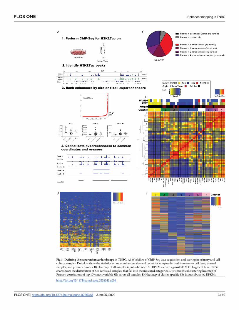
Fig 1. Defining the superenhancer landscape in TNBC. A) Workflow of ChIP-Seq data acquisition and scoring in primary and cell
culture samples. Dot plots show the statistics on superenhancers size and count for samples derived from tumor cell lines, normal
samples, and primary tumors. B) Heatmap of all samples input subtracted SE RPKMs scored against SE 20 kb fragment bins. C) Pie
chart shows the distribution of SEs across all samples, that fall into the indicated categories. D) Hierarchical clustering heatmap of
Pearson correlations of top 10% most variable SEs across all samples. E) Heatmap of cluster-specific SEs input subtracted RPKMs.
https://doi.org/10.1371/journal.pone.0235343.g001
PLOS ONE Enhancer mapping in TNBC
PLOS ONE | https://doi.org/10.1371/journal.pone.0235343 June 25, 2020 3 / 19

broad clustering pattern was identified, however clusters were less well defined (S1B Fig). We
therefore focused our subsequent analysis on SE data. We noted that Cluster 3 contained all
HMEC samples. Clusters 5 and 6 and 7 were comprised of primary TNBC samples, and Clus-
ters 1,2 and 4 contained a mix of cell line samples. Four of the samples showed no discernable
similarly in SE profile to any other sample, and therefore were not assigned a cluster. To assess
the impact of clustering breast cancer samples based on active cis-regulatory regions rather
than steady-state gene expression, we compared unbiased clustering of the same samples
based on SE profile and RNA-Seq profile (S1C Fig). While these analyses identified broadly
similar clusters, resolution was significantly greater with SE correlation. In particular, while
enhancer profiles of clusters 2 and 4 were anticorrelated (Pearson coefficient < 0), gene
expression profiles between these groups were much more similar.
TNBC SE profiles define unique tumor subsets
Analysis of the composition of the clusters defined by SE profile indicated unique characteris-
tics of each. Assessment of an EMT gene expression signature across the profiled samples indi-
cated that samples within Clusters 1 & 2 showed a more epithelial-like transcriptional profile,
while cluster 4 samples expressed a more mesenchymal-like transcriptome (Fig 1D) [9]. Fur-
thermore, Cluster 1 contained all hormone receptor (ER and AR) expressing cell lines samples.
This was consistent with PAM50 classifications. Although primary tumor samples clustered
separately from cell lines (likely due heterogeneity within primary sample cell composition as
well as cell line adaptation to long term culture) there was still significant correlation between
the sample types. Primary sample clusters 5 and 6 showed significant similarity to Cluster 4
(mesenchymal), while primary sample Cluster 7 showed high similarity to Clusters 1 and 2
(epithelial).
To further examine features driving clustering we identified the SEs whose activity best
stratified Clusters 1, 2, 3 and 4 (Fig 1E and S3 Table). We also identified a large group of SEs as
shared between Clusters 1 and 2. To better understand the gene networks controlled by these
‘cluster-defining’ enhancers, we sought to identify the genes most likely to be regulated by
these elements, using a combination of proximity to the SE, as well as correlation between
gene expression and enhancer size across the samples (Fig 2A and S4 Table) [7]. Briefly,
expression of each gene within 10 Mb of a given SE was correlated to the presence or absence
of the SE. Genes with a Pearson correlation co-efficient of 0.6 or great were then considered
putative target genes of that SE. Interestingly, while the majority of correlating gene lay proxi-
mal to the associated SE, a subset lay much further away (up to 5Mb), suggesting the presence
of long-range interactions (S4 Table and S1D Fig). Using this approach, we created a set of
putative SE-regulated genes unique to each cluster (S5 Table). Gene Set Enrichment Analysis
(GSEA) indicated that Cluster 1-defining enhancers predominately associate with genes
involved with hormone receptor signaling. Similarly, both Cluster 2 and shared Clusters
1&2-defining enhancers regulated genes associated with the luminal phenotype, and Cluster 4
enhancers were associated with genes defining a mesenchymal / basal cell state (Fig 2B). Clus-
ter 3 contained HMEC samples and consistent with this the SEs defining this cluster associated
with genes defining normal cells.
Tumor specific superenhancers drive oncogene expression
To define tumor-specific changes in the breast cis-regulatory landscape, we focused on SEs
present only in tumor samples and not normal breast. We identified 2,643 SEs present in at
least one tumor sample, but not found in normal samples. To remove potential artifacts pres-
ent only in highly passaged cancer cell lines, we required tumor-specific SEs to be present in at
PLOS ONE Enhancer mapping in TNBC
PLOS ONE | https://doi.org/10.1371/journal.pone.0235343 June 25, 2020 4 / 19

Fig 2. TNBC SE profiles define unique tumor subsets. A) Cartoon depiction of a fictional SE with overlapping and adjacent genes. Scatter plots show fictional
example gene expression RPKMs vs. SE sizes, B) Gene Set Enrichment Analysis (GSEA) results for each subgroup. Left panel shows GSEA results for putative
target genes from each Cluster-specific set of enhancers. Right panel shows example H3K27ac ChIP-Seq tracks and boxplots for superenhancers from each
Cluster. Each track shown is an overlay of the tracks for all samples present in each group. Boxplots for each track example shows the total distribution of SE
sizes for the samples in each Cluster. Mean, and interquartile range are shown; whiskers represent the minimum and maximum group values.
https://doi.org/10.1371/journal.pone.0235343.g002
PLOS ONE Enhancer mapping in TNBC
PLOS ONE | https://doi.org/10.1371/journal.pone.0235343 June 25, 2020 5 / 19

least 4 TNBC cell lines, at least one primary TNBC sample, and absent from all HMEC samples
(Fig 3A and 3B). We noted that in addition to tumor-specific enhancers identified in this way,
a number of SEs were present in normal breast samples, but significantly enlarged in tumor
samples. We included these regions in our analyses, defined as those enhancers where at least
2 times as many sequencing reads were counted in the region in tumor samples compared to
normal, and using the additional criteria described above. Using these criteria, we identified
781 tumors-specific SEs. We hypothesized that these tumor-specific enhancers may confer a
selective advantage during tumor development because they influence expression of genes crit-
ical for TNBC cell growth. In support of this notion we observed acquisition of tumor-specific
SEs in proximity to known oncogenes such as EGFR and MYC, as well as a large number of
genes whose role in TNBC has not yet been established (S6 Table).
To functionally validate the role of these SE-associated genes in tumor cell proliferation we
designed a custom gRNA library targeting this gene set (S8 Table). We performed a series of
CRISPR knockout screens in representative TNBC and normal breast cell lines, to determine
whether acquired SEs could drive transcriptional activity of genes that represent TNBC-spe-
cific vulnerabilities (Fig 3C). Results of the screen identified 263 tumor-specific SE-associated
genes were required for TNBC cell growth, as indicated by significant drop out from the
infected population (Fig 3D and S7 Table). Genes which conferred selective drop-out in tumor
lines compared to hTERT-HMEC were validated in arrayed format (S2 Fig). To assess the pre-
dictive power of SE acquisition for gene dependency, we evaluated the correlation between SE
acquisition and drop out in screen data. This analysis revealed a number of vulnerabilities
dependent on the presence of an associated tumor-specific enhancer, including known TNBC
oncogenes and novel candidates (Fig 3E).
BAMBI dependence is associated with superenhancer acquisition
We chose to further investigate one such candidate dependency, BAMBI, as a putative TNBC
oncogene whose dependence correlates with SE association. BAMBI (BMP and Activin Mem-
brane Bound Inhibitor) is a transmembrane glycoprotein most extensively studied for its role as
an inhibitor of the TGFβ signaling pathway [10]. CRISPR knock out of the BAMBI gene was asso-
ciated with significant growth inhibition in both long- and short-term growth assays in the TNBC
cell line HCC38, while having no impact on growth of HMEC cells (Fig 4A and 4B). Dependency
on BAMBI was confirmed by shRNA knockdown, and by suppression of the BAMBI TSS (Tran-
scriptional Start Site) by CAS9 fused to the Kruppel-associated box (KRAB) and gRNAs targeting
the BAMBI TSS (S3 Fig). Unexpectedly, the original SE associated with BAMBI based on BAMBI
gene expression correlations across the samples did not correlate well with dependency (Figs 4A
and S4A). However, additional enhancer and SE elements both proximal and distal to the BAMBI
gene were identified which correlated well with BAMBI dependency. These were identified in
HCC38 cells, as well as other BAMBI dependent cell lines identified in the Project Achilles cell
line screening data set (Figs 5A–5D and S4B and S9 Table). Dependency in these lines did not cor-
relate with BAMBI gene expression, copy number or mutation, suggesting an epigenetic predictor
and a role for SE acquisition in BAMBI dependence in breast cancer.
To address the role of putative BAMBI enhancers in control of BAMBI gene expression, we
used HCC38 cells expressing CAS9-KRAB and gRNAs targeted to individual enhancer peaks, as
well as the BAMBI TSS, to silence individual chromatin regions (Fig 6A). As expected, gRNAs
targeting the BAMBI TSS were able to repress BAMBI expression. Additionally, enhancer-tar-
geted gRNAs gave a range of repressive activity on BAMBI transcription, allowing identification
of enhancer peaks with the greatest influence on gene expression (Fig 6A). Due to adaptation to
long-term 2D culture, well established differences exist between cell lines and primary tumor
PLOS ONE Enhancer mapping in TNBC
PLOS ONE | https://doi.org/10.1371/journal.pone.0235343 June 25, 2020 6 / 19

samples. However, BAMBI-proximal enhancers that correlated with dependency in cell lines
were also identified in TNBC primary samples, suggesting such elements are active in primary
disease (Fig 6B and 6C). We also observed H3K4me3 peaks at a number of these variable
enhancer regions in primary TNBC samples, suggesting that expression of non-coding transcripts
in this active cis-regulatory region may play a role in BAMBI regulation (S5 Fig).
Fig 3. Tumor specific super-enhancers drive oncogene expression. A) Heatmap of tumor specific SEs across all samples as defined by
scoring as a SE in at least 4 tumor cell lines and 1 TNBC primary tumor, and by not a SE in all normal samples. B) Example ChIP-Seq
tracks for an acquired SE, putatively driving expression of GATA2. C) Diagram of screen workflow. D) CRISPR drop out screen results
for tumor-specific SE associated genes in the indicated cell lines. E) Representative example screen hits. Top panel plots compare gene
drop-out to SE size, bottom panel plots compare drop-out to RNA expression.
https://doi.org/10.1371/journal.pone.0235343.g003
PLOS ONE Enhancer mapping in TNBC
PLOS ONE | https://doi.org/10.1371/journal.pone.0235343 June 25, 2020 7 / 19

Discussion
The dysregulation of gene expression in cancer is well established, and cannot be adequately
explained by genomic alterations such as coding gene mutation and copy number changes.
Instead, such widespread changes are attributed to the extensive transcriptional rewiring that
occurs in cancer cells, including the utilization of different transcription factor repertoires,
Fig 4. BAMBI dependence is associated with superenhancer acquisition. A) CellTiter-Glo1measurement of cell
viability 4 days after BAMBI gRNA transfection. Each point corresponds to a different BAMBI targeted gRNAs, line
indicates the mean. BAMBI SE size is indicated below each cell line. B) Colony formation assays 10 days after BAMBI
gRNA transfection for the indicated cell lines.
https://doi.org/10.1371/journal.pone.0235343.g004
PLOS ONE Enhancer mapping in TNBC
PLOS ONE | https://doi.org/10.1371/journal.pone.0235343 June 25, 2020 8 / 19

and the activation of alternative gene-regulatory elements including enhancers [11–14]. A
deeper understanding of the epigenetic and transcriptional landscape of tumor and normal
cells has helped shed light on the mechanisms behind some of these changes, however, much
remains to be understood about how they occur and their role in tumorigenesis.
As a disease which lacks molecular drivers and targeted therapy options, TNBC is an area
where a deeper understanding of the epigenetic changes that occur during tumorigenesis may
be particularly impactful. With this in mind, here we describe the active cis-regulatory
Fig 5. Fine-resolution analysis of BAMBI superenhancer landscape. A) Project Achilles data representing BAMBI knock out in 342
cancer cell lines. Drop-out is represented by CERES score. Box denotes all cell lines that have significant growth dependencies on
BAMBI. B) Correlation between BAMBI dependency and the indicated genomic features in Project Achilles data. C) H3K27Ac
ChIP-Seq tracks for BAMBI-independent TNBC lines, HCC38, and BAMBI dependent non-TNBC cell lines identified in project
Achilles. High-variance enhancer regions are denoted below tracks. D) Left panel shows boxplot of summed BAMBI-proximal variant
enhancer peaks annotated in C). Middle panel shows boxplots of summed BAMBI-distal variant enhancer peaks. Right panel shows
boxplot of BAMBI RNA expression for BAMBI knock out sensitive and insensitive cell lines. Box indicates median, 25th and 75th
percentiles, whiskers indicate min and max. � indicates significant difference P<0.001 as determined by Mann-Whitney U test.
https://doi.org/10.1371/journal.pone.0235343.g005
PLOS ONE Enhancer mapping in TNBC
PLOS ONE | https://doi.org/10.1371/journal.pone.0235343 June 25, 2020 9 / 19

landscape of TNBC. Based on the epigenetic and transcriptional profiling of 10 primary
tumors, normal HMECs from multiple donors, and 23 TNBC cell lines, we document the
changes that occur in the enhancer landscape in tumor cells compared to their normal coun-
terparts. Unsurprisingly, significant differences were observed between primary and cell line
populations, and the use of cell lines in this analysis is caveated by their undoubted divergence
from primary tumors under the selection pressure of long-term 2D growth. Importantly how-
ever, many commonalities were also identified in the cis-regulatory landscapes, and cell line
inclusion in this study allowed follow up and functional validation not possible in primary
tissue.
Previous work has uncovered a surprising asymmetry in the distribution of enhancer size
genome wide, with a subset of enhancers in every cell being significantly larger than the aver-
age. While the exact size of these SE elements varies considerably between cell types, the
Fig 6. Enhancer regulation of BAMBI gene expression. A) Left panel shows H3K27Ac ChIP-Seq track of BAMBI-proximal region
denoting enhancer peaks targeted by dCAS9-KRAB gRNAs. Right panel shows BAMBI expression by qPCR after cells were infected
with the indicated gRNAs. B) Tracks show H3K27Ac ChIP-Seq signal of the BAMBI-proximal region in primary TNBC samples. C)
Boxplot summarizing variant H3K27Ac peaks for the indicated groups. Box indicates median, 25th and 75th percentiles, whiskers
indicate min and max.
https://doi.org/10.1371/journal.pone.0235343.g006
PLOS ONE Enhancer mapping in TNBC
PLOS ONE | https://doi.org/10.1371/journal.pone.0235343 June 25, 2020 10 / 19

asymmetric distribution is constant, and a set of SEs can be defined in every sample, as those
falling beyond the inflection point of a curve ranking all enhancers in a given sample by size
[15]. Biological characterization of the function of these elements and the genes they regulate
has uncovered a bias towards the regulation of lineage-defining transcription factor networks,
and in the context of cancer, in the regulation of oncogenic transcription factors [4,5]. Indeed,
there is precedent in a number of indications for the mapping and quantification of SE size to
be used to identify oncogenic transcriptional circuitry of importance for tumorigenesis
[3,7,8,16–18]. Here we apply this same approach to TNBC. While the cis-regulatory landscape
of any cell is undoubtedly vast and complex, in this study we chose to focus only on SE ele-
ments, and test the hypothesis that identification of the largest enhancers in primary and cell
line TNBC samples will help identify critical oncogenes and transcriptional networks in the
disease.
Using histone-H3K27Ac ChIP-Seq and analysis using MACS and ROSE software, we
mapped and quantified enhancers genome wide in TNBC samples. Using established ROSE
parameters consistent with other studies (see methods), we identified the subset of enhancers
in each sample considered superenhancers. Consistent with prior studies, we did not exclude
TSS elements from our ROSE derived SE calls, due to the significant overlap between SE
regions and TSS and gene bodies. We noted a large number of SE present in at least one tumor
sample but absent from normal breast samples, indicated a gain in SE activity in tumors.
Using unsupervised hierarchical clustering we compared SE landscapes across TNBC and nor-
mal breast samples, and were able to define subgroups of samples with more similar profiles.
Most prominently, clustering samples using the most variable SEs in the data set stratified
samples into epithelial and mesenchymal populations. Strikingly, although these broad group-
ings could also be observed in transcriptome data, the separation between the groups was sig-
nificantly greater in the SE data set. In particular, SE profiles for epithelial vs mesenchymal
samples were anti-correlated in the SE data, while positively correlated in the transcriptomic
data set. Similarly, while SE profiling was able to identify 3 different subgroups of primary
TNBC samples, these were indistinguishable based on their transcriptomes. Finally, several
cell line samples were un-clusterable using SE data, having enhancer profiles so unique they
did not significantly correlate with any other samples, suggesting a highly unique gene-regula-
tory landscape in these cells. In contrast, these cell lines did not stand out in transcriptome
data, having similar gene expression profiles to other more mesenchymal samples. Taken
together, these data suggest that information on SE activity in tumor cells provides additional
insight into the cancer cell state, beyond that of steady state gene expression. Furthermore, the
observation that cells with similar transcriptomes can have strikingly different enhancer pro-
files indicates a previously unappreciated diversity in the regulatory landscape of TNBC.
While outside the scope of this study, profiling the variations in transcription factor occupancy
at these sites may afford a deeper understanding of the factors dictating this diversity, and the
opportunity to target these unique states.
Comparison of TNBC and normal breast SE profiles identified a large number of SEs pres-
ent in tumor cells but absent from normal tissue, indicating that tumor-specific enhancer
acquisition is common. To understand the link between these alterations in the SE landscape
and gene expression changes in TNBC, we used an informatics approach to identify putative
SE-regulated genes, by correlating SE proximity and gene expression across samples to predict
genes most likely to be regulated by a given SE. This approach is caveated by the long-range
interactions possible between enhancers and promoters, and the likely possibility that within a
cell population an enhancer may regulate several genes, and vis versa, a gene may be regulated
by several enhancers. To accommodate this, gene expression correlations within 10 MB of
each SE were considered, as were multiple genes correlating with an individual SE. Previous
PLOS ONE Enhancer mapping in TNBC
PLOS ONE | https://doi.org/10.1371/journal.pone.0235343 June 25, 2020 11 / 19

studies have shown this approach to cross-validate well with Chromatin Conformation Cap-
ture assay data [7]. Analysis of gene sets associated with SE defined groups of TNBC samples
again indicated a stratification based on epithelial vs mesenchymal cell state, as well as hor-
mone receptor expression, and correlated broadly with PAM50 classification. Consistent with
previous observations, genes associated with tumor-specific SEs included known oncogenes
and lineage-defining transcription factors, as well as genes not previously associated with
TNBC.
Previous efforts to characterize the epigenetic landscape in cancer have been primary
descriptive. Here we expand to interrogate the functional role of these putative tumor-specific
SE regulated genes in TNBC growth and survival. Using CRISPR dropout screening we test
the hypothesis that acquisition of SE regulation during tumorigenesis may predict a depen-
dence on the associated gene. Consistent with this hypothesis, we identified 263 tumor-specific
SE regulated genes that were required for TNBC proliferation but not for growth of normal
breast cells. We chose to follow up on one such tumor-specific dependency which represents a
potential targetable vulnerability. We identified the TGFβ pseudo-receptor BAMBI as a SE
associated gene whose loss inhibited cell growth in HCC38 TNBC cells, but not in normal
HMEC cells. Analysis of Project Achilles cell line screening data identified a number of addi-
tional cell lines also dependent on BAMBI for growth. This dependency could not be predicted
by known features such as expression, mutational or copy number status. Upon detailed analy-
sis of the enhancer landscape we identified variable enhancer regions where enhancer presence
correlated well with BAMBI gene dependency. These enhancers were located in both BAMBI
proximal and distal regions. We hypothesize that while these enhancers may not directly con-
trol BAMBI expression, they may reflect or control a cell state that dictates a dependency on
BAMBI. Importantly, these variant enhancers were also present in primary tumor samples,
predicting BAMBI dependency in TNBC patients.
In conclusion, this study provides the first comprehensive, integrated map of the gene
expression and cis-regulatory landscape of TNBC. Analyses of these data indicate that when
combined with traditional cancer genomic and expression data, gene-regulatory information
can improve our understanding of the cancer cell state, in particular it’s diversity. It is interest-
ing to speculate that the great diversity of enhancer usage seen in this study may represent
unique transcription factor circuitries active in difference tumors. Although leading to com-
mon gene expression outputs, these unique circuitries may represent targetable vulnerabilities
in subsets of patients. The integrated approach taken here to uncover this diversity may pro-
vide additional information for both biomarker and drug target discovery efforts in TNBC,
and could be applied to other cancer types with a paucity of known molecular drivers.
Materials and methods
Cell culture and proliferation assays
All cancer cell lines in this study were obtained from ATCC, authenticated by STR analysis
and confirmed mycoplasma negative by PCR test. All cancer cell lines were cultured in RPMI
media supplemented with 10% fetal bovine serum and 2 mM glutamine. hTERT-HME cells
were obtained from ATCC, authenticated by STR analysis, confirmed mycoplasma negative by
PCR test and grow in MEBM media supplemented with growth factors (Lonza cat# 3151 and
#4136). Primary HMEC cells (ThermoFisher cat# A10565) were grown in HuMEC Ready
media (ThermoFisher cat# 12752–010). Puromycin concentration for all viral infected cultures
was maintained throughout growth at a concentration of 2 ug/mL. CAS9 stable lines were
selected for and maintained with Blasticidin at a final concentration of 2.5 ug/mL. Cell prolif-
eration was evaluated in a 384-well format using CellTiter-Glo1 reagent (Promega) according
PLOS ONE Enhancer mapping in TNBC
PLOS ONE | https://doi.org/10.1371/journal.pone.0235343 June 25, 2020 12 / 19

to manufacturer’s instructions, or by assessment of confluence using the Incucyte Zoom plat-
form in a 12 well plate format, with brightfield images acquired every 5 hours.
Primary samples
Frozen triple negative breast cancer tumor samples were acquired from Cureline (South San
Francisco). Samples were homogenized via mechanical disruption before processing for
RNA-Seq and ChiP-seq as described below.
Cell line generation
CAS9 expressing cell lines were generated by infection with pLenti 6.3 CAS9 virus, followed by
selection with Blasticidin. Pooled cultures were maintained in the presence of Blasticidin and
infected with sgRNA virus targeting PLK1, or non-targeting controls (NTC) to test for CAS9
activity. All cell lines had >80% killing after infection with PLK1 targeting gRNAs, as assessed
by CTG assay.
Clonogenic assays
Cells were plated at densities of 500, 1,000, or 2,000 cells per well and allowed to adhere over-
night in RPMI 10% FBS media. Wells were infected with normalized titers of guide virus with
a target MOI of 1. After 3 days post-infection, fresh media including puromycin 1 ug/mL was
added to each well. Media was changed every 3–4 days until control wells reached confluence.
Colonies were visualized by staining with 0.5% Crystal violet for 30 minutes at room
temperature.
Pooled CRISPR screening
Pooled CRISPR screening was performed in non-clonal CAS9 expressing cell lines as
described above and according to [19], using 8 sgRNAs per gene, with a total of 3875 sgRNAs
in the library (S8 Table). A MOI of 0.3 was targeted in each cell lines. The impact on growth of
each gene in the library was assessed by measuring the change in abundance of each gene’s 8
sgRNA guides from the reference timepoint (t = 0) to a late timepoint (t = 3). The top 3 guides
per gene were used to calculate the log fold-change (LFC) for each gene for each cell line. In
order to cross-compare data across different cell lines with varying CAS9 activity, LFC data
was normalized to positive (essential genes) and negative (NTC) control genes so that a value
of -1.0 corresponded to the average value of our positive controls, and 0.0 corresponded to the
average of our NTCs. All values at or below -0.5 were considered to be putative ‘hits’.
Gene expression analysis
RNA was purified from cells using the RNeasy kit (Qiagen) according to manufacturer’s
instructions. Quantitative RT-PCR was performed using Taqman assay (ThermoFisher Scien-
tific, Inc.) on ABI QuantStudio 7 Flex real-time PCR system. For whole transcriptome RNA-
sequencing RNA libraries were made using TruSeq RNA Sample Preparation Kit v2 (Illu-
mina). Size of the libraries was confirmed using Fragment Analyzer (Advanced Analytical
Technologies) and their concentration was determined by qPCR-based method using Library
quantification kit (KAPA). The libraries were multiplexed and then sequenced on Illumina
HiSeq2500 (Illumina) to generate 30M of single end 50 base pair reads. Gene set enrichment
analysis (GSEA) was performed using Broad Institute software (http://software.broadinstitute.
org/gsea/index.jsp).
PLOS ONE Enhancer mapping in TNBC
PLOS ONE | https://doi.org/10.1371/journal.pone.0235343 June 25, 2020 13 / 19

Chromatin immuno-precipitation (ChIP)
Following treatment 1 million cells or 500 mg of primary tumor were crosslinked in 1% form-
aldehyde for 15 minutes, then quenched with 125 mM glycine for 5 minutes. Primary tumors
were mechanically dissociated, then all cells were lysed and chromatin sheared by sonication
to an average length of 300–500 bp. Genomic DNA (Input) was prepared by treating aliquots
of chromatin with RNase, proteinase K and heat for de-crosslinking, followed by ethanol pre-
cipitation. Pellets were resuspended and the resulting DNA was quantified on a NanoDrop
spectrophotometer. Extrapolation to the original chromatin volume allowed quantitation of
the total chromatin yield. 30 μg of chromatin was precleared with protein A agarose beads
(Invitrogen). Immunoprecipitation was then carried out using the following antibodies: anti-
H3K27Ac (Active Motif catalog # 39133, lot #31814008, 4 ug per ChIP), anti-H3K4me3
(Active Motif catalog # 39159, lot # 15617005, 3 ul used per ChIP).
ChIP-qPCR, immunoprecipitated complexes were washed sequentially with low to high-
salt wash buffers, followed by a wash in TE, followed by elution from beads with TE + SDS 1%.
Eluates were treated with 10 μg of RNaseA for 30 minutes at 37˚C, followed by 20 μg of Pro-
teinase K for 30 minutes at 55˚C. Crosslinks were reversed by incubation overnight at 65˚C,
and ChIP DNA was purified using QIAQuick PCR purification columns (QIAGEN).
ChIP and Input DNAs were prepared for amplification by converting overhangs into phos-
phorylated blunt ends and adding an adenine to the 3’ ends. Illumina genomic adapters were
ligated and the sample was size-fractionated (200–300 bp) on an agarose gel. After a final PCR
amplification step (15 cycles), the resulting DNA libraries were quantified and sequenced on
Illumina NextSeq 500, producing 75 nt reads, single ended reads. (For sequencing QC metrics
see S1 Table).
CRISPRi cell line generation and assay
Repression of enhancer activity was achieved by stable expression of dCAS9-KRAB in HCC38
cells and then lentiviral infection of pLKO-based plasmids containing a gRNA targeting the
BAMBI transcriptional start site (TSS-1) (50-GCGTCCCTAGAGTCGAGCG-30), (TSS-2)
(50-AGCAACTTGTCGCGACCTG-30), extragenic regions of the BAMBI enhancer (enh_1)
(5’-CCTATATGTGAATCCACCT-3’), (enh_2) (5’-GTAATCCCAACTACTCCGG-3’),
(enh_3) (5’-AGTCAGTATACCAACACTG-3’), (enh_4) (5’-GAACCTGGACATCCTCC
AC-3’), (enh_5) (5’-AGACCGGGTTTCAGCACGT-3’), (enh_6) (5’-ATGTAACACATAC
CCACTG-3’), (enh_7) (5’-CCCCACGTAGCATCACCCA-3’), (enh_8) (5’-GTCTAATGTGT
GATAACTG-3’), or a negative control gene desert region (50-TCCCCCTCAGCCGTATT-30).
Cells were processed for RT-qPCR analysis after 5 days post-infection.
shRNA line construction and conditions
HCC38 cells were infected with virus containing doxocycline-inducible pZIP-TRE3G shRNA
constructs (Transomics) for 3 different BAMBI-targeting sequences (BAMBI sh1 5’-
TGCTGTTGACAGTGAGCGAAAGCAGACCTCAGCAACGATATAGTGAAGCCACA
GATGTATATCGTTGCTGAGGTCTGCTTGTGCCTACTGCCTCGGA-3’), (BAMBI sh2
5’-TGCTGTTGACAGTGAGCGACTGAGGATGCTTCGAAGTGAATAGTGAAGCCACA
GATGTATTCACTTCGAAGCATCCTCAGGTGCCTACTGCCTCGGA-3’), (BAMBI sh3
5’-TGCTGTTGACAGTGAGCGAGGCACGAGAACTGCTGTCTGATAGTGAAGCCACA
GATGTATCAGACAGCAGTTCTCGTGCCCTGCCTACTGCCTCGGA-3’), or an NTC
sequence. Inducible shRNA expression was confirmed by GFP expression. For clonogenic,
incucyte, and RNA samples, shRNA cell lines were induced for 2 days with 1 ug/mL
PLOS ONE Enhancer mapping in TNBC
PLOS ONE | https://doi.org/10.1371/journal.pone.0235343 June 25, 2020 14 / 19
doxycycline, following which cells were trypsinized, counted, and re-seeded for cell growth
assays and RT-qPCR in the presence of doxycycline.
Statistical analysis
ChIP-Seq data analysis. Reads were aligned to the human genome (GRCh38) using the
GSNAP algorithm, (http://research-pub.gene.com/gmap/ version 2013-10-10) with the follow-
ing settings: “-M 2 -n 10 -B 2 -i 1—pairmax-dna = 1000—terminal-threshold = 1000—gmap-
mode = none—clip-overlap”. Fragment length was determined by the strand cross-correlation
method. Reads were extended to this fragment length before coverage was calculated at a per-
nucleotide level using uniquely mapping reads.
Enhancer and superenhancers peak identification and scoring. H3K27ac ChIP peaks
were identified by the MACS version 2 software package [20] in conjunction with paired input
DNA samples with the callpeak function using default settings, genome set to ‘hs’, and peak
calling set to—broad. Enhancer and Superenhancer peaks were identified with the ROSE ver-
sion 1 software package [5,15] using the MACS output files. ROSE software was executed with
default parameters of 12.5 kb stitching distance, and TSS exclusion size set to 0, with the
genome set to hg19. MACs and ROSE output statistics are described in S1 Table. Individual
enhancers coordinates were derived from initial MACS output tables and filtered based on
maximum p-value = 0.05 and empirically determined peak sizes relative to paired input sam-
ples. Superenhancer consolidation across samples was accomplished using Bedtools version
2.2 software -merge function with a minimum overlap distance of 5000 bp between enhancers
[21]. Enhancer consolidation was done with Bedtools -merge function with a minimum over-
lap of 10 bp between enhancers. After merging enhancers into a consensus map, enhancers
that overlap TSS coordinates were removed using the Bedtools -subtract function. Enhancers
and superenhancer scoring for all samples against their respective consensus enhancer / SE
map was performed by scoring each sample’s IP and input alignment (.bam) files using the
bedtools -multicov function to determine the total reads for each element interval per sample.
Raw coverage values were then adjusted for read-depth, and the adjusted coverage values for
input were subtracted from their respective paired ChIP sample to give a final score for each
enhancer coordinate for each sample. The absolute value for a SE cutoff per sample is variable
due to the ROSE software and distribution of enhancer sizes across samples, therefore a value
of Log2(RPKM+1) => 6 was adopted as the SE cutoff threshold for sample scores according
to the consensus SE map. This score is inclusive to all of the originally identified SE from all
samples. All statistical comparisons of superenhancers between groups of cell lines were per-
formed with 2-tailed unpaired T-tests.
Superenhancer clustering. The top 10% most variable superenhancers were measured by
calculating the variance of SE sizes for all cell line samples. Hierarchical clustering and heat-
maps for SE data was done on the high variance SE with the ggplot (https://ggplot2.tidyverse.
org) and heatmap.3 (https://gist.github.com/nachocab/3853004) R packages, using the hclust
average linkage algorithm for calculating distances. Optimal cluster number was determined
empirically by testing various cluster sizes with the “Elbow Method”, “Sillouette Method”, and
“Gap statistic”. Based on the output of those tests and prior knowledge of our samples’ biologi-
cal state, a cluster number of 10 was chosen. Group assignments were done for clusters with 3
or more samples. SEs were assigned to groups 1–4 according to the criteria that the mean
group value minus the standard deviation did not overlap with the mean value of any of the
other groups. In the case of the SEs shared between groups 1 and 2, the group means minus
standard deviations populations were overlapping with one another, but were distinct from
groups 3 and 4.
PLOS ONE Enhancer mapping in TNBC
PLOS ONE | https://doi.org/10.1371/journal.pone.0235343 June 25, 2020 15 / 19
Gene to superenhancer mapping. To measure the correlation between genes and super-
enhancers, for each superenhancer the Pearson correlation between superenhancer size (log2)
of each cell line was calculated to the gene RPKM value (log2) for all genes within 10 Mb of the
enhancer coordinates. Genes which have a Pearson correlation of 0.6 or greater were consid-
ered to be associated with a given superenhancer. For each superenhancer, a minimum of two
genes with the highest correlations were assigned, regardless of a minimum value. In cases
where 3 or more genes were highly correlated to a SE, all genes above a correlation of 0.6 were
deemed to be associated with the SE.
Achilles data analysis. Dependency data was extracted from the Achilles cell line screen-
ing project using the 2018 Q4 public release of data (https://depmap.org/portal/achilles/). Cell
lines with a CERES drop-out score less than or equal to -0.5 were categorized a sensitive to
BAMBI knockout.
Defining BAMBI-distal SE signatures. Foe BAMBI dependent and independent cell
lines correlation matrices were created to identify superenhancers that were most positively
and negatively correlated with BAMBI dependency. We performed unsupervised hierarchical
clustering of the log2 ChIP data for both the top 600 positively- and top 600 anti-correlated
superenhancers. The signature size of 600 enhancers was determined empirically to give an
optimal p-value for distinguishing between BAMBI-sensitive and BAMBI-insensitive samples.
Accession numbers. The accession numbers for the RNA-Seq and ChIP-Seq data
reported here are ENA: PRJEB33558
Supporting information
S1 Fig. Enhancer and RNA correlations across samples. A) Pie chart shows the distribution
of enhancers that fall into the indicated categories. B) Hierarchical clustering heatmap of Pear-
son correlations of top 10% most variable enhancers across all samples. C) Hierarchical clus-
tering heatmap of Pearson correlations using RNA-seq values of top 10% most variable genes.
D) Scatterplot shows the distance of the best correlating genes to their associated SE.
(TIF)
S2 Fig. Primary validation of dropout screen results. Plots show arrayed validation of pri-
mary screen hits for all CAS9 lines. Whiskers denote max/min values. Middle bar indicates
mean value.
(TIF)
S3 Fig. Orthologous validation of the BAMBI phenotype. A) RT-qPCR quantification of
BAMBI TSS-targeting dCAS9-KRAB guides in HCC38 cells. B) Clonogenic assays for BAM-
BI-TSS and “gene desert” targeting gRNAs in dCAS9-KRAB HCC38 cells. C) Quantification
of clonogenic assay wells. D) RT-qPCR quantification of shRNA knockdown of BAMBI upon
doxycycline induction for 3 independent shRNA constructs, and a non-targeting control
shRNA at 7 days post-induction. E) Clonogenic assays of HCC38 cells after doxycycline induc-
tion of the indicated shRNAs. F) Incucyte growth assays of HCC38 cells after doxycycline
induction of the indicated shRNAs.
(TIF)
S4 Fig. SE acquisition predicts BAMBI dependence. A) H3K27ac ChIP-Seq tracks of the
BAMBI-adjacent region for the indicated cell lines. BAMBI associated superenhancers are
indicated below the tracks. B) Heatmap showing top correlating SEs with BAMBI dependence.
(TIF)
PLOS ONE Enhancer mapping in TNBC
PLOS ONE | https://doi.org/10.1371/journal.pone.0235343 June 25, 2020 16 / 19

S5 Fig. Dynamic H3K4me3 at the BAMBI in primary TNBC. Tracks show H3K4me3 signal
at the BAMBI locus in primary TNBC samples.
(TIF)
S1 Table. ChIP-Seq QC statistics and Rose output statistics.
(XLSX)
S2 Table. Consensus enhancer and superenhancer coordinates.
(XLSX)
S3 Table. Cluster specific superenhancers.
(XLSX)
S4 Table. Superenhancer to gene correlations and distances.
(XLSX)
S5 Table. Groups specific putative superenhancer associated genes.
(XLSX)
S6 Table. Putative tumor-specific SE driven genes for CRISPR screen.
(XLSX)
S7 Table. CRISPR dropout screen results.
(XLSX)
S8 Table. CRISPR gRNA library sequences.
(XLSX)
S9 Table. BAMBI-distal superenhancers correlating with dependency.
(XLSX)
Acknowledgments
We thank Robert Yauch and Andrea Cochran for helpful discussions and critical evaluation of
the research and manuscript. Project Achilles data was obtained from: Broad DepMap Achilles
18Q4 public. FigShare version 2 https://figshare.com/articles/DepMap_Achilles_18Q4_public/
7270880.
Author Contributions
Conceptualization: Ryan Raisner, Karen E. Gascoigne.
Data curation: Ryan Raisner.
Formal analysis: Ryan Raisner, Russell Bainer, Peter M. Haverty, Karen E. Gascoigne.
Investigation: Karen E. Gascoigne.
Methodology: Karen E. Gascoigne.
Resources: Kelli L. Benedetti.
Supervision: Karen E. Gascoigne.
Writing – original draft: Ryan Raisner.
PLOS ONE Enhancer mapping in TNBC
PLOS ONE | https://doi.org/10.1371/journal.pone.0235343 June 25, 2020 17 / 19

References1. Lehmann BD, Bauer JA, Chen X, Sanders ME, Chakravarthy AB, Shyr Y, et al. Identification of human
triple-negative breast cancer subtypes and preclinical models for selection of targeted therapies. J Clin
Invest. 2011; 121: 2750–2767. https://doi.org/10.1172/JCI45014 PMID: 21633166
2. Creyghton MP, Cheng AW, Welstead GG, Kooistra T, Carey BW, Steine EJ, et al. Histone H3K27ac
separates active from poised enhancers and predicts developmental state. Proc Natl Acad Sci U S A.
National Acad Sciences; 2010; 107: 21931–21936. https://doi.org/10.1073/pnas.1016071107 PMID:
21106759
3. Chapuy B, McKeown MR, Lin CY, Monti S, Roemer MGM, Qi J, et al. Discovery and Characterizationof
Super-Enhancer-Associated Dependencies in Diffuse Large B Cell Lymphoma. Cancer Cell. Elsevier
Inc; 2013; 24: 777–790. https://doi.org/10.1016/j.ccr.2013.11.003 PMID: 24332044
4. Hnisz D, Abraham BJ, Lee TI, Lau A, Saint-Andre V, Sigova AA, et al. Super-Enhancers in the Control
of Cell Identity and Disease. Cell. Elsevier Inc; 2013; 155: 934–947. https://doi.org/10.1016/j.cell.2013.
09.053 PMID: 24119843
5. Loven J, Hoke HA, Lin CY, Lau A, Orlando DA, Vakoc CR, et al. Selective Inhibition ofTumor Onco-
genes by Disruption of Super-Enhancers. Cell. Elsevier Inc; 2013; 153: 320–334. https://doi.org/10.
1016/j.cell.2013.03.036 PMID: 23582323
6. Pellacani D, Bilenky M, Kannan N, Heravi-Moussavi A, Knapp DJHF, Gakkhar S, et al. Analysis of Nor-
mal Human Mammary Epigenomes Reveals Cell-Specific Active Enhancer States and Associated
Transcription Factor Networks. CellReports. ElsevierCompany; 2016; 17: 2060–2074. https://doi.org/
10.1016/j.celrep.2016.10.058 PMID: 27851968
7. McKeown MR, Corces MR, Eaton ML, Fiore C, Lee E, Lopez JT, et al. Superenhancer Analysis
Defines Novel Epigenomic Subtypes of Non-APL AML, Including an RARα Dependency Targetable
by SY-1425, a Potent and Selective RARα Agonist. Cancer Discovery. American Association for
Cancer Research; 2017; 7: 1136–1153. https://doi.org/10.1158/2159-8290.CD-17-0399 PMID:
28729405
8. Lin CY, Erkek S, Tong Y, Yin L, Federation AJ, Zapatka M, et al. Active medulloblastoma enhancers
reveal subgroup-specific cellular origins. Nature. Nature Publishing Group; 2016;: 1–20. https://doi.org/
10.1038/nature16546 PMID: 26814967
9. Haverty PM, Lin E, Tan J, Yu Y, Lam B, Lianoglou S, et al. Reproducible pharmacogenomic profiling of
cancer cell line panels. Nature. Nature Publishing Group; 2016; 533: 333–337. https://doi.org/10.1038/
nature17987 PMID: 27193678
10. Onichtchouk D, Chen Y-G, Dosch R, Gawantka V, Delius H, Massague J, et al. Silencing of TGF-β sig-
nalling by the pseudoreceptor BAMBI. Nature. Nature Publishing Group; 1999; 401: 480–485. https://
doi.org/10.1038/46794 PMID: 10519551
11. Franco HL, Nagari A, Malladi VS, Li W, Xi Y, Richardson D, et al. Enhancer transcription reveals sub-
type-specific gene expression programs controlling breast cancer pathogenesis. Genome Res. Cold
Spring Harbor Lab; 2018; 28: 159–170. https://doi.org/10.1101/gr.226019.117 PMID: 29273624
12. Cohen AJ, Saiakhova A, Corradin O, Luppino JM, Lovrenert K, Bartels CF, et al. Hotspots of aberrant
enhancer activity punctuate the colorectal cancer epigenome. Nature Communications. Nature Publish-
ing Group; 2017; 8: 1–13. https://doi.org/10.1038/s41467-016-0009-6
13. The chromatin accessibility landscape of primary human cancers. 2018;: 1–15. https://doi.org/10.1126/
science.aav1898
14. Corces MR, Granja JM, Shams S, Louie BH, Seoane JA, Zhou W, et al. The chromatin accessibility
landscape of primary human cancers. Science. 2018; 362: eaav1898–15. https://doi.org/10.1126/
science.aav1898 PMID: 30361341
15. Whyte WA, Orlando DA, Hnisz D, Abraham BJ, Lin CY, Kagey MH, et al. Master Transcription Factors
and Mediator Establish Super-Enhancers at Key Cell Identity Genes. Cell. Elsevier Inc; 2013; 153:
307–319. https://doi.org/10.1016/j.cell.2013.03.035 PMID: 23582322
16. Roe J-S, Hwang C-I, Somerville TDD, Milazzo JP, Lee EJ, Da Silva B, et al. Enhancer Reprogramming
Promotes Pancreatic Cancer Metastasis. Cell. Elsevier Inc; 2017;: 1–35. https://doi.org/10.1016/j.cell.
2017.07.007 PMID: 28757253
17. Mack SC, Pajtler KW, Chavez L, Okonechnikov K, Bertrand KC, Wang X, et al. Therapeutic targeting of
ependymoma as informed by oncogenic enhancer profiling. Nature. Nature Publishing Group; 2017;: 1–
26. https://doi.org/10.1038/nature25169 PMID: 29258295
18. Ott CJ, Federation AJ, Schwartz LS, Kasar S, Klitgaard JL, Lenci R, et al. Enhancer Architecture and
Essential Core Regulatory Circuitry of Chronic Lymphocytic Leukemia. Cancer Cell. Elsevier; 2018; 34:
982–995.e7. https://doi.org/10.1016/j.ccell.2018.11.001 PMID: 30503705
PLOS ONE Enhancer mapping in TNBC
PLOS ONE | https://doi.org/10.1371/journal.pone.0235343 June 25, 2020 18 / 19

19. Callow MG, Watanabe C, Wickliffe KE, Bainer R, Kummerfield S, Weng J, et al. CRISPR whole-
genome screening identifies new necroptosis regulators and RIPK1 alternative splicing. Cell Death and
Disease. Springer US; 2018;: 1–13. https://doi.org/10.1038/s41419-018-0301-y PMID: 29449584
20. Zhang Y, Liu T, Genome CM, 2008. Model-based analysis of ChIP-Seq (MACS). genomebiologybio-
medcentralcom. https://doi.org/10.1186/gb-2008-9-9-r137)
21. Quinlan AR, Hall IM. BEDTools: a flexible suite of utilities for comparing genomic features. Bioinformat-
ics. 2010; 26: 841–842. https://doi.org/10.1093/bioinformatics/btq033 PMID: 20110278
PLOS ONE Enhancer mapping in TNBC
PLOS ONE | https://doi.org/10.1371/journal.pone.0235343 June 25, 2020 19 / 19