superficial muscles of back
TRANSCRIPT
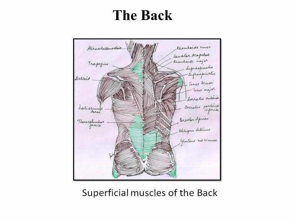
The Back

Scapula, anterior aspect
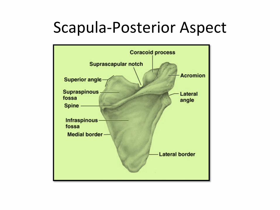
Scapula-Posterior Aspect
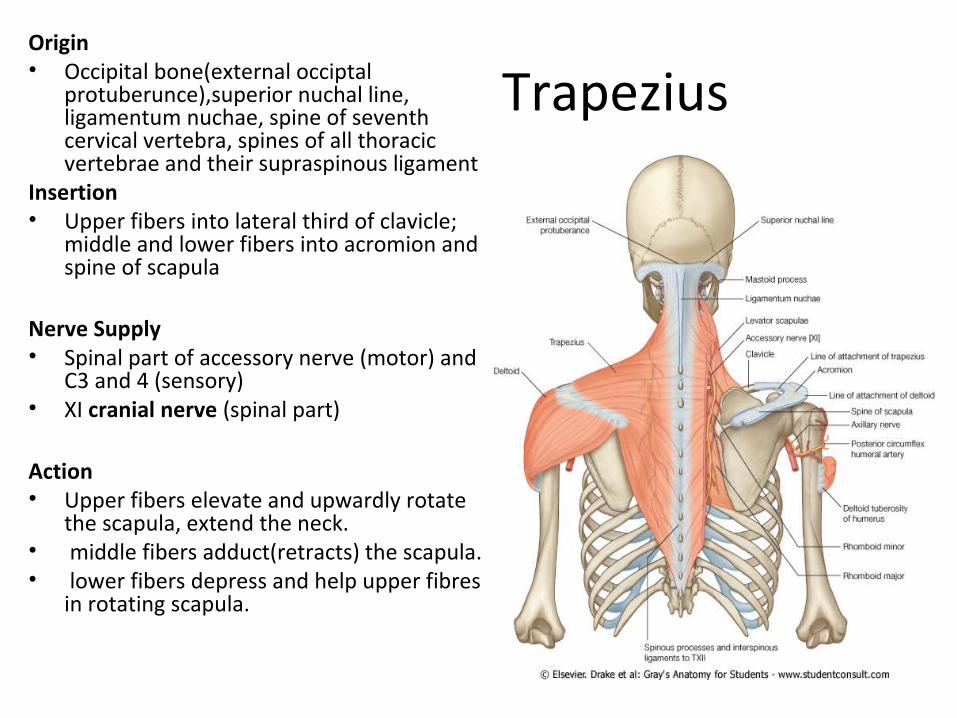
TrapeziusOrigin• Occipital bone(external occiptal
protuberunce),superior nuchal line, ligamentum nuchae, spine of seventh cervical vertebra, spines of all thoracic vertebrae and their supraspinous ligament
Insertion• Upper fibers into lateral third of clavicle;
middle and lower fibers into acromion and spine of scapula
Nerve Supply• Spinal part of accessory nerve (motor) and
C3 and 4 (sensory)• XI cranial nerve (spinal part)
Action • Upper fibers elevate and upwardly rotate
the scapula, extend the neck.• middle fibers adduct(retracts) the scapula.• lower fibers depress and help upper fibres
in rotating scapula.

Latissimus dorsiOrigin• Iliac crest, lumbar fascia, spines
of lower six thoracic vertebrae(T7-T12), lower three or four ribs, and inferior angle of scapula
Insertion• Floor of bicipital groove of
humerus Nerve Supply• Thoracodorsal nerve(C6, 7, 8)Action• Extends, adducts, and medially
rotates the arm • Its called the climbing muscle• Raising of the trunk above the
armImportant :As it winds around lower
border of teres major it forms posterior fold of axilla its lateral border forms a boundary of lumbar triangle.

Levator scapulae
OriginTransverse processes of
first fourth cervical vertebrae
InsertionMedial border of scapula Nerve supplyC3 and 4 and dorsal
scapular nerveActionRaises medial border of
scapulaImportant : Part of floor
of Posterior triangle
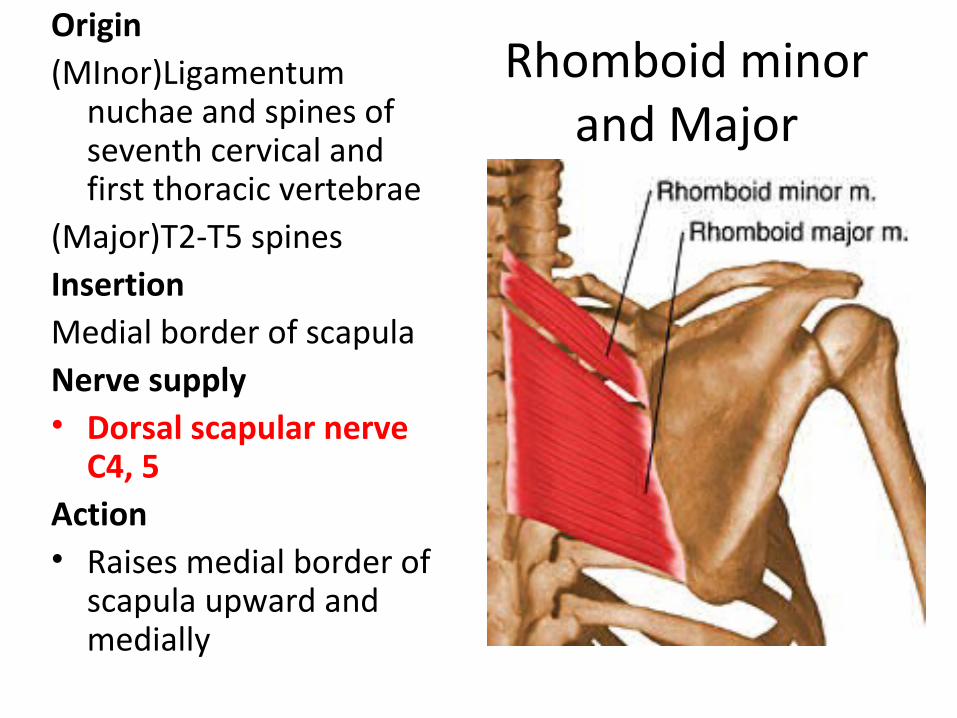
Rhomboid minor and Major
Origin(MInor)Ligamentum
nuchae and spines of seventh cervical and first thoracic vertebrae
(Major)T2-T5 spinesInsertionMedial border of scapula Nerve supply• Dorsal scapular nerve
C4, 5Action• Raises medial border of
scapula upward and medially
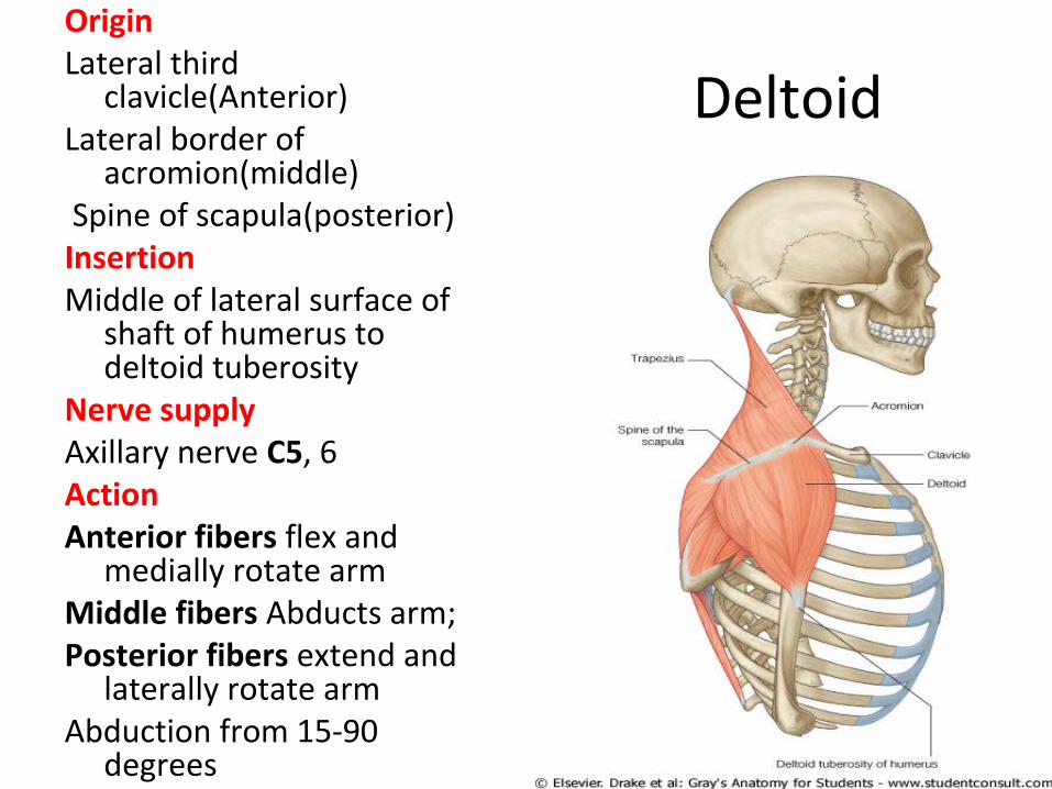
DeltoidOriginLateral third
clavicle(Anterior)Lateral border of
acromion(middle) Spine of scapula(posterior)InsertionMiddle of lateral surface of
shaft of humerus to deltoid tuberosity
Nerve supplyAxillary nerve C5, 6ActionAnterior fibers flex and
medially rotate armMiddle fibers Abducts arm; Posterior fibers extend and
laterally rotate armAbduction from 15-90
degrees

Applied anatomy Intramuscular injections are
given into the deltoid .The should be given in the lower
half of the muscle to avoid injury to axillary nerve.
In subacromial bursitis pressure over the deltoid below the acromion with the arm by the side causes pain.
However when the arm is abducted pressure over the same point causes no pain because the bursa disappears under the acromion. This is referred to as Dawbarn’s sign.

SupraspinatusOriginSupraspinous fossa of
scapulaInsertionGreater tuberosity of
humerus; capsule of shoulder joint
Nerve supplySuprascapular nerve C4, 5, 6ActionAbducts arm and stabilizes
shoulder jointInitiation of abduction 0-15
degrees

InfraspinatusOriginInfraspinous fossa of
scapulaInsertionGreater tuberosity of
humerus; capsule of shoulder joint
Nerve supplySuprascapular nerve C4, 5, 6ActionLaterally rotates arm and
stabilizes shoulder joint

Teres minor
OriginUpper two thirds of lateral
border of scapulaInsertionGreater tuberosity of
humerus; capsule of shoulder joint
Nerve supplyAxillary nerve (C4), C5, 6ActionLaterally rotates arm and
stabilizes shoulder joint

Subscapularis
OriginSubscapular fossaInsertionLesser tuberosity of humerusNerve supplyUpper and lower subscapular
nerves C5, 6, 7ActionMedially rotates arm and
stabilizes shoulder jointImportant: Multipennate

Teres major
OriginLower third of lateral border of
scapulaInsertionMedial lip of bicipital groove of
humerusNerve supplyLower subscapular nerve C6, 7ActionMedially rotates and adducts arm
and stabilizes shoulder jointImportant : Considered
continuation of subscapularis

Rotator Cuff• The rotator cuff is the
name given to the tendons of the subscapularis, supraspinatus, infraspinatus, and teres minor muscles
• Fused to the underlying capsule of the shoulder joint
• The cuff lies on the anterior, superior, and posterior aspects of the joint
• The cuff is deficient inferiorly, and this is a site of potential weakness.

Quadrangular Space Superiorly by the
subscapularis and capsule of the shoulder joint
Inferiorly by the teres major muscle
Medially by the long head of the triceps
laterally by the surgical neck of the humerus.
Contents The axillary nerve and
the posterior circumflex humeral vessels

Upper & Lower Triangular spaces
Upper Superiorly subscapularis
and teres minor Inferiorly teres major Laterally the long head of
the triceps • Contains circumflex
scapular vessels.Lower superiorly the teres
major medially long head of the
triceps brachii laterally Medial head of
triceps• Contains radial nerve and
profunda brachii artery.

The triangle of auscultation
• It is the site where breathing sounds can be heard most clearly, using a stethoscope.
Is formed by the vertebral or medial border of the scapula,
superior border of latissimus dorsi
the lateral border of the trapezius. It has a floor formed by
rhomboid major. It covers the intercostal space
between ribs 6 and 7 and rib 7. It lies superficial to the cardiac
orifice of the stomach on the left side, where splash of swallowed liquids was timed in cases of esophageal obstruction.
It is the site where breathing sounds can be heard most clearly, using a stethoscope.
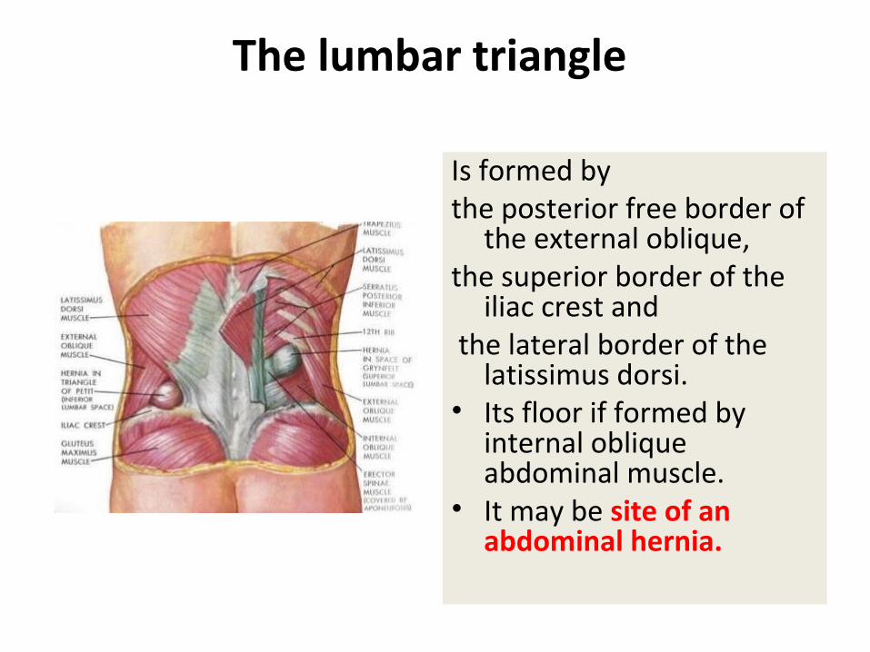
The lumbar triangle
Is formed by the posterior free border of
the external oblique, the superior border of the
iliac crest and the lateral border of the
latissimus dorsi.• Its floor if formed by
internal oblique abdominal muscle.
• It may be site of an abdominal hernia.
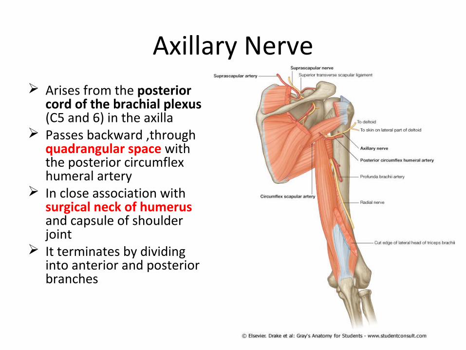
Axillary Nerve Arises from the posterior
cord of the brachial plexus (C5 and 6) in the axilla
Passes backward ,through quadrangular space with the posterior circumflex humeral artery
In close association with surgical neck of humerus and capsule of shoulder joint
It terminates by dividing into anterior and posterior branches

Branches
Articular branch to the shoulder joint
Anterior terminal branch supplies the deltoid and the skin that covers its lower part.
Posterior terminal branch supplies teres minor muscle and deltoid, then emerges as the upper lateral cutaneous nerve of the arm

Applied Aspect
• The axillary nerve may be damaged by
dislocation of shoulder or by fracture of surgical neck of humerus.
• The patient presents with loss of abduction of shoulder upto 90 degrees (as deltoid is paralysed), loss of rounded countour of shoulder and sensory loss over lower deltoid.
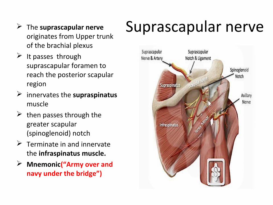
Suprascapular nerve The suprascapular nerve originates from Upper trunk of the brachial plexus
It passes through suprascapular foramen to reach the posterior scapular region
innervates the supraspinatus muscle
then passes through the greater scapular (spinoglenoid) notch
Terminate in and innervate the infraspinatus muscle.
Mnemonic(“Army over and navy under the bridge”)
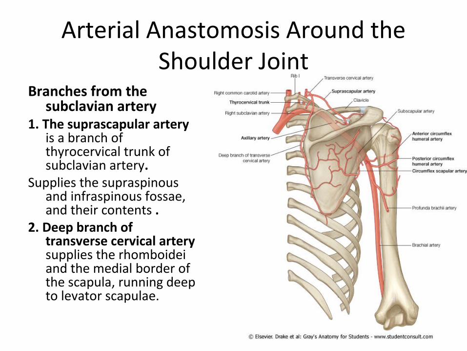
Arterial Anastomosis Around the Shoulder Joint
Branches from the subclavian artery
1. The suprascapular artery is a branch of thyrocervical trunk of subclavian artery.
Supplies the supraspinous and infraspinous fossae, and their contents .
2. Deep branch of transverse cervical artery supplies the rhomboidei and the medial border of the scapula, running deep to levator scapulae.

Branch from the axillary artery3. The circumflex scapular artery, a
branch of the subscapular artery which arises from the third part of axillary artery
Applied aspect • Scapular anastomoses is an
anastomoses between the first part of subclavian and third part of axillary artery. So it provides a collateral circulation through which blood can flow to the limb in case of blockage of distal part of subclavian artery or proximal part of axillary artery.