superficial roughness on composite procedures. part i ... composite enamel and ... clinical...
TRANSCRIPT
CLINICAL RESEARCH
70THE INTERNATIONAL JOURNAL OF ESTHETIC DENTISTRY
VOLUME 9 • NUMBER 1 • SPRING 2014
Correspondence to: Federico Ferraris
Spalto Borgoglio 81, 15121 Alessandria, Italy; E-mail: [email protected]
Superficial roughness on composite
surface, composite enamel and
composite dentin junctions after
different finishing and polishing
procedures. Part I: roughness after
treatments with tungsten carbide vs
diamond bursFederico Ferraris, DDS
Private practice, Alessandria, Italy
Alessandro Conti, DDS
Private practice, Casale Monferrato, Italy
FERRARIS & CONTI
71THE INTERNATIONAL JOURNAL OF ESTHETIC DENTISTRY
VOLUME 9 • NUMBER 1 • SPRING 2014
71THE INTERNATIONAL JOURNAL OF ESTHETIC DENTISTRY
VOLUME 9 • NUMBER 1 • SPRING 2014
Abstract
The aim of this study is to investigate
different instruments for finishing com-
posite restorations, as well as examining
different surfaces and interfaces of the
same restoration. The null hypothesis is
represented by the fact that there are
no significant differences on roughness
of composite restorations finishing be-
tween tungsten carbide and diamond
burs, furthermore the null hypothesis
is that there are no significant differ-
ences on roughness between finishing
on composite surfaces (C), composite-
enamel (CE) and composite-dentin (CD)
interfaces. The study was performed on
28 teeth, and class V cavities were pre-
pared on the extracted teeth. Restor-
ations were done in Filtek XTE nanofilled
composite (3M Espe) in a standardized
method, to then be finished. A compari-
son was made in the phase 1 between
tungsten carbide burs (16 blades), dia-
mond burs (46 μm), with a similar shape
by the same manufacturer (Komet).
Each surface received 5 bur applica-
tions. Consequently, an analysis with a
profilometer was performed. Phase 2
involved further confrontation of ulterior
finishing with ultrafine tungsten carbide
burs (30 blades) and with extra and ul-
trafine diamond burs (25 and 8 μm) (the
same shape as previously mentioned).
A second analysis was then performed
with a profilometer. All measurements
were taken on C surfaces, CE and CD
interfaces. Statistical analyses were car-
ried out with c2 test (a = 0.05).
Conclusions: The finishing procedures
with fine grit or toothing burs gave a
better smoothness with tungsten car-
bide burs compared to diamond burs.
While with the ultrafine grit no significant
differences were noted between tung-
sten carbide and diamond burs on the
CE and CD interfaces, the diamond bur
left less superficial roughness on the C
surfaces. With regards to the superficial
roughness of the different areas of res-
toration, it can be concluded that: mi-
nor roughness was detected on C sur-
faces, while the CD interface had the
most superficial roughness, regardless
of whether the diamond burs or tung-
sten carbide burs were used. This study
shows some statistical differences that
could not be clinically perceivable. The
clinical relevance could be resumed as
follows: the fine tungsten carbide burs
provided less roughness compared to
a fine diamond bur. There were no dif-
ferences between the ultrafine tungsten
carbide and diamond burs. The less fa-
vourable interface to be finished is CD,
compared to the CE interface and C sur-
faces.
(Int J Esthet Dent 2014;9:70–89)

CLINICAL RESEARCH
72THE INTERNATIONAL JOURNAL OF ESTHETIC DENTISTRY
VOLUME 9 • NUMBER 1 • SPRING 2014
Introduction
The prevention of secondary decay and
the esthetic optimization of composite
resin material on surfaces are two as-
pects of practical interest in restorative
dentistry. Both can have a close correla-
tion with the clinical phases of finishing
and polishing.
These are procedures that can be
considered secondary to the final prog-
nosis of composite restorations, but can
actually have a crucial clinical value.
From studies, it has been shown that
dentists spend 60 to 75% of their time
replacing restorations.1
The replacement of old restorations
is closely tied to the formation of sec-
ondary decay.2,3 Secondary decay
can be considered the primary lesions
around restorations;2 the main areas of
localization are the areas where there
is a greater stagnation of bacterial bio-
film, for example the cervical margins
of the restorations.4 The secondary de-
cay can manifest as deep or superficial
lesions in proximity to the restoration.5
The shrinkage of the polymerization im-
plicit in the composite can produce the
formation of interface gaps between
tooth and restoration6 and in vitro stud-
ies have associated the presence of
secondary caries lesions with microfis-
sures.7
Restorative materials with different
surface characteristics than teeth can
cause the formation of a biofilm that in-
creases the capacity of bacteria to colo-
nize in the oral cavity.8,9 Roughness and
hard to clean surfaces contribute to the
formation of pigmentation, accumulation
of plaque, inflammation of the gums, and
secondary cavities.10-13
The accumulation of this biofilm on
dental structures can cause secondary
cavities and the mechanical action of
brushing produces a disorganization on
this biofilm that can prevent or stop the
formation of cavities.14
According to some studies, it can be
concluded that microleakage and sur-
face roughness do not influence the
formation of white spot lesions around
composite resin restorations, while oth-
ers found that the presence of microle-
akage at the adhesive interface did not
significantly affect enamel demineraliza-
tion, reinforcing the lack of evidence that
there is an association between micro-
leakage and caries lesions adjacent to
the restoration.1-3,8 However, microleak-
age could still be considered an etio-
logic factor for secondary caries,7 fur-
thermore the bacterial adhesion on the
surface of composite resins has been
considered an important parameter in
the etiology of caries formation around
restorations.16
Another important aspect is repre-
sented by the esthetic appearance of
composite restorations, because this is
of great interest to both the dentist and
patient. Surface roughness influences
resistance to staining17,18 and the nat-
ural gloss of the restoration.19,20
An elevated quality in finishing and
polishing of restorations appears to be
highly important not only for the longev-
ity of the treatment, but also for esthetic
reasons. This can be obtained with ap-
propriate procedures.
Many finishing instruments are de-
signed to create smooth surfaces on
dental fillings. A great variety is com-
monly used for finishing and polishing,
such as the tungsten carbide burs of
72
FERRARIS & CONTI
73THE INTERNATIONAL JOURNAL OF ESTHETIC DENTISTRY
VOLUME 9 • NUMBER 1 • SPRING 2014
different toothing size, diamond burs of
different grit size, rubber tips, abrasive
discs, polishing strips and pastes.
The first decision for an operator,
who must finish resin composite light-
curable restorations, is which rotating
instruments to use: tungsten carbide
or diamond bur. These two types of in-
struments are different mainly because
tungsten carbide is a cutting instrument
and has geometrically defined blades,
whereas the diamond bur is an abra-
sive instrument, and has geometrically
undefined grains (diamond grains). In
literature, some authors consider tung-
sten carbide burs to be better,21 while
others may consider the diamond bur
to be better in obtaining minor super-
ficial roughness,22 while some authors
consider the two methods to be inter-
changeable.23-25
Another question that the clinician
should ask himself, is: with what cutting
capacity should the restoration be fin-
ished? In fact, it should not be taken for
granted that there are still significant dif-
ferences in different cutting capacity (for
example fine, extrafine or ultrafine) be-
tween the two materials, even for those
studies that have considered one instru-
ment to be more valid.
Another important consideration when
we speak of finishing is which is the most
important surface on a clinical level? In
fact, frequently superficial roughness is
scientifically investigated with different
finishing and polishing of the composite
surfaces, which from an esthetic point
of view, as well as the adhesion of bac-
terial biofilm, has validity. However, the
restoration-tooth interface is the most
important aspect to evaluate microleak-
age and eventual secondary decay that
can stem from it. Therefore, the under-
standing of the level of roughness of the
CE interface and CD interface can have
clinical importance. Finally it is already
known that with different polishing pro-
cedures the result obtained an enam-
el surface is similar to the unpolished
enamel26 and this factor once again
underlined the role of the finishing and
polishing phases.
The null hypothesis are represented
by:
No significant differences on rough-
ness of composite restorations finish-
ing between tungsten carbide and
diamond burs.
No significant differences on rough-
ness between finishing on C surfaces,
CE and CD interfaces.
The objective of this in vitro study is the
analysis of the different roughness ob-
tained on resin composite restorations
in class V cavities with different rotating
instrument materials, with different grit
and on different interfaces. The analysis
that is done can be summarized in three
fundamental aspects:
Comparing fine grit or toothing tung-
sten carbide and diamond burs
Comparing ultrafine grit or toothing
carbide and diamond burs.
Superficial roughness with all earlier
burs and grit/toothing on C surface,
CE interface, and CD interface.
The measurements taken with the pro-
filometer are the central surfaces of the
restoration in composite and the com-
posite-enamel interface (crown middle
third) and composite-dentin (cervical
third under the cementoenamel Junc-
tion [CEJ]).

CLINICAL RESEARCH
74THE INTERNATIONAL JOURNAL OF ESTHETIC DENTISTRY
VOLUME 9 • NUMBER 1 • SPRING 2014
Finishing and polishing protocol
The protocols considered in this study
include a sequence of burs (diamond vs
tungsten carbide) from fine to ultrafine
(analysed in this manuscript: part I) for
the finishing phases, then for the pol-
ishing phases, a sequence of polishers
(from the coarsest to the finest) and a
pre-impregnated polishing brush (ana-
lysed in part II of this manuscript, to be
published in a subsequent issue). All
details of manufacturing and codes of
burs, number of passages applications
on surfaces, number of restorations fin-
ished and polished with each single bur
are discussed in the materials and meth-
ods section.
Materials and methods
Preparation of cavities and
composite resin restorations
For this study 28 freshly extracted non-
carious human teeth were selected,
stored for 15 days in solution saturated
with thymol and then in water for the
duration of the study. The teeth were
mounted on a special positioning device
with transparent acrylic resin (Ortho-jet,
Lang Dental) embedding the root up to
4.0 mm below the CEJ. All teeth are mo-
lars (Figs 1 and 2). Two operators made
the cavities, and carried out the restor-
ations and finishing procedures. The
specimens were assigned in a random
fashion, and each operator then created
different cavities. For this study, the 14
buccal cavities will be considered, with
respective phases of adhesion, stratifi-
cation, finishing, and polishing. For each
single selected cavity to reduce variabil-
ity, specimen preparation, as well as fin-
ishing and polishing procedures, were
carried out by the same operator. All res-
torations and finishing procedures were
carried out with prismatic magnifying
loupes systems with 4 x magnification
and 300 mm focal distance (Zeiss).
Tooth preparation and restoration
A standardized tooth preparation was
applied to all specimens. Class V cavi-
Fig 1 An overview of all specimens prepared for
the in vitro study.
Fig 2 Specimen ready for the test.
FERRARIS & CONTI
75THE INTERNATIONAL JOURNAL OF ESTHETIC DENTISTRY
VOLUME 9 • NUMBER 1 • SPRING 2014
ties were made on each specimen. The
cavity dimensions followed the same
parameters and diamond burs mounted
on a red ring speed-increasing hand-
piece, transmission 1:5 with water spray
(INTRAcompact 25 LCS, Kavo). The bur
used was rounded with a 1.6 mm diam-
eter and drilled down to 107 μm (801
314 016, Komet) (Fig 4) than for the fin-
ishing phase of the cavity, a bur with the
same shape was used to drill down to
46 μm (8801 314 016, Komet) (Fig 5) the
final finishing was done with a polisher
(9608 314 030, Komet) (Fig 6). The cav-
ity dimensions are approximately the
following: depth 1.6 to 2.0 mm, width
5.0 to 6.0 mm, and 3.0 mm height in the
central part and 1.6 mm on the sides.
The cervical extension under the CEJ is
approximately 1.5 mm to have a com-
posite-dentin margin on which to carry
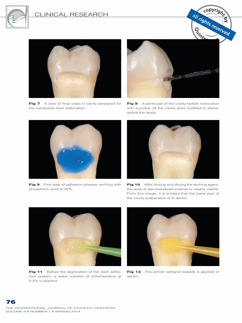
out the procedure (Figs 7 and 8).
Once the cavity preparation was com-
plete, adhesive procedures were carried
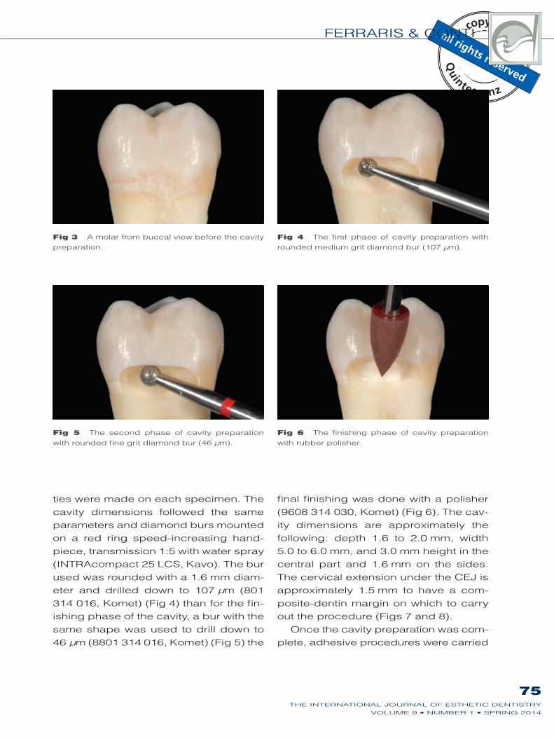
Fig 3 A molar from buccal view before the cavity
preparation.
Fig 4 The first phase of cavity preparation with
rounded medium grit diamond bur (107 μm).
Fig 5 The second phase of cavity preparation
with rounded fine grit diamond bur (46 μm).
Fig 6 The finishing phase of cavity preparation
with rubber polisher.
CLINICAL RESEARCH
76THE INTERNATIONAL JOURNAL OF ESTHETIC DENTISTRY
VOLUME 9 • NUMBER 1 • SPRING 2014
Fig 7 A view of final class V cavity prepared for
the composite resin restoration.
Fig 8 A particular of the cavity before restoration
with a probe: all the cavity were codified to stand-
ardize the study.
Fig 9 First step of adhesion phases: etching with
phosphoric acid at 35%.
Fig 10 After rinsing and drying the etching agent,
the area of demineralized enamel is clearly visible.
From this image, it is evident that the lower part of
the cavity preparation is in dentin.
Fig 11 Before the application of the resin adhe-
sive system, a water solution of chlorhexidine at
0.2% is applied.
Fig 12 The primer (ethanol based) is applied in
dentin.
FERRARIS & CONTI
77THE INTERNATIONAL JOURNAL OF ESTHETIC DENTISTRY
VOLUME 9 • NUMBER 1 • SPRING 2014
out in order to complete the composite
restoration by etching with phosphoric
acid 35% (Ultratech, Ultradent) for 30 s
in enamel and 20 s in dentin (Figs 9 and
10). The cavity was rinsed for 60 sec-
onds with a constant spray of water and
air, and a chlorexidine galenic digluco-
nate solution at 0.2% was applied on the
dentin for 20 s27 (Fig 11), the liquid was
then aspirated slightly without drying the
dentinal substratum, in order to avoid the
collapse of the collagen fibers. The ad-
hesive system that was used is an etch
and rinse in 3 steps (Optibond FL, Kerr),
the alcohol-based primer was applied
for 60 s (Fig 12) and after drying, a res-
inous bond was applied on the enamel
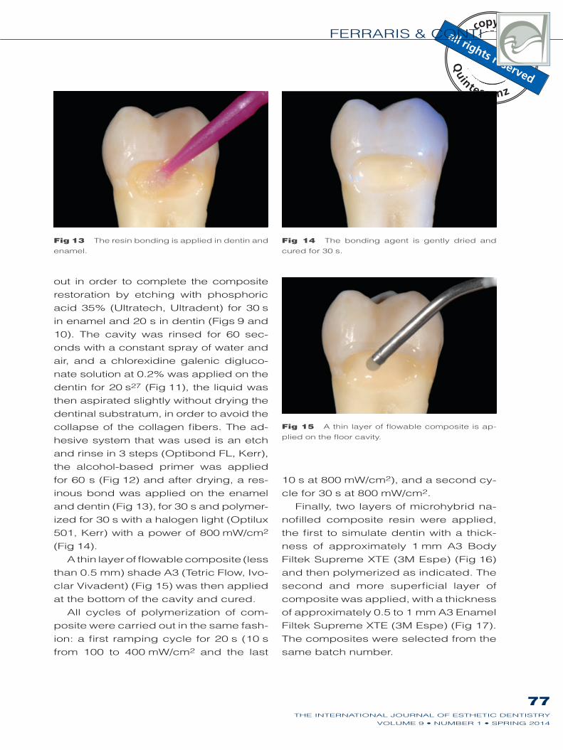
and dentin (Fig 13), for 30 s and polymer-
ized for 30 s with a halogen light (Optilux
501, Kerr) with a power of 800 mW/cm2
(Fig 14).
A thin layer of flowable composite (less
than 0.5 mm) shade A3 (Tetric Flow, Ivo-
clar Vivadent) (Fig 15) was then applied
at the bottom of the cavity and cured.
All cycles of polymerization of com-
posite were carried out in the same fash-
ion: a first ramping cycle for 20 s (10 s
from 100 to 400 mW/cm2 and the last
10 s at 800 mW/cm2), and a second cy-
cle for 30 s at 800 mW/cm2.
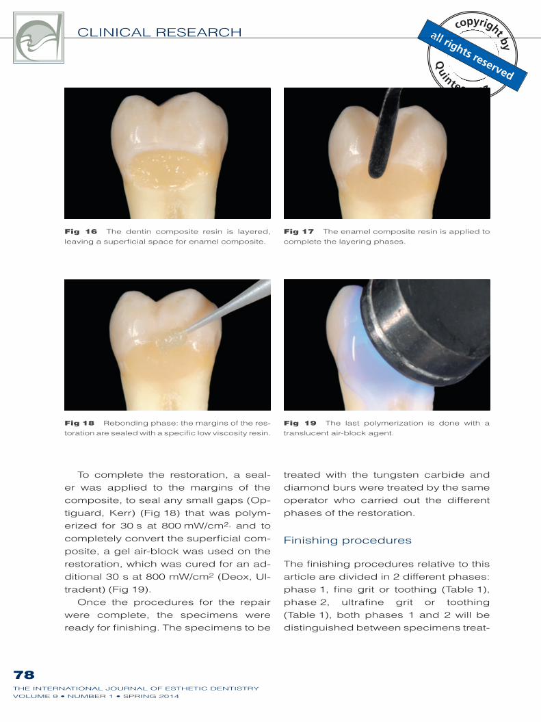
Finally, two layers of microhybrid na-
nofilled composite resin were applied,
the first to simulate dentin with a thick-
ness of approximately 1 mm A3 Body
Filtek Supreme XTE (3M Espe) (Fig 16)
and then polymerized as indicated. The
second and more superficial layer of
composite was applied, with a thickness
of approximately 0.5 to 1 mm A3 Enamel
Filtek Supreme XTE (3M Espe) (Fig 17).
The composites were selected from the
same batch number.
Fig 13 The resin bonding is applied in dentin and
enamel.
Fig 14 The bonding agent is gently dried and
cured for 30 s.
Fig 15 A thin layer of flowable composite is ap-
plied on the floor cavity.
CLINICAL RESEARCH
78THE INTERNATIONAL JOURNAL OF ESTHETIC DENTISTRY
VOLUME 9 • NUMBER 1 • SPRING 2014
To complete the restoration, a seal-
er was applied to the margins of the
composite, to seal any small gaps (Op-
tiguard, Kerr) (Fig 18) that was polym-
erized for 30 s at 800 mW/cm2, and to
completely convert the superficial com-
posite, a gel air-block was used on the
restoration, which was cured for an ad-
ditional 30 s at 800 mW/cm2 (Deox, Ul-
tradent) (Fig 19).
Once the procedures for the repair
were complete, the specimens were
ready for finishing. The specimens to be
treated with the tungsten carbide and
diamond burs were treated by the same
operator who carried out the different
phases of the restoration.
Finishing procedures
The finishing procedures relative to this
article are divided in 2 different phases:
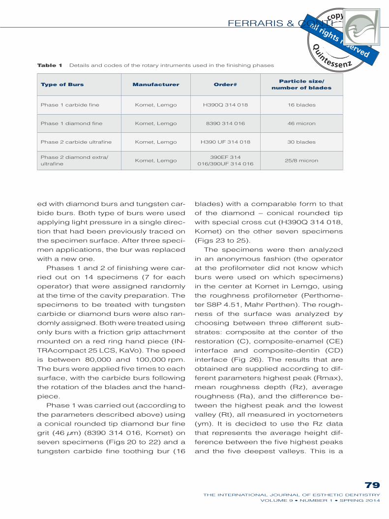
phase 1, fine grit or toothing (Table 1),
phase 2, ultrafine grit or toothing
(Table 1), both phases 1 and 2 will be
distinguished between specimens treat-
Fig 16 The dentin composite resin is layered,
leaving a superficial space for enamel composite.
Fig 17 The enamel composite resin is applied to
complete the layering phases.
Fig 18 Rebonding phase: the margins of the res-
toration are sealed with a specific low viscosity resin.
Fig 19 The last polymerization is done with a
translucent air-block agent.
FERRARIS & CONTI
79THE INTERNATIONAL JOURNAL OF ESTHETIC DENTISTRY
VOLUME 9 • NUMBER 1 • SPRING 2014
ed with diamond burs and tungsten car-
bide burs. Both type of burs were used
applying light pressure in a single direc-
tion that had been previously traced on
the specimen surface. After three speci-
men applications, the bur was replaced
with a new one.
Phases 1 and 2 of finishing were car-
ried out on 14 specimens (7 for each
operator) that were assigned randomly
at the time of the cavity preparation. The
specimens to be treated with tungsten
carbide or diamond burs were also ran-
domly assigned. Both were treated using
only burs with a friction grip attachment
mounted on a red ring hand piece (IN-
TRAcompact 25 LCS, KaVo). The speed
is between 80,000 and 100,000 rpm.
The burs were applied five times to each
surface, with the carbide burs following
the rotation of the blades and the hand-
piece.
Phase 1 was carried out (according to
the parameters described above) using
a conical rounded tip diamond bur fine
grit (46 μm) (8390 314 016, Komet) on
seven specimens (Figs 20 to 22) and a
tungsten carbide fine toothing bur (16
blades) with a comparable form to that
of the diamond – conical rounded tip
with special cross cut (H390Q 314 018,
Komet) on the other seven specimens
(Figs 23 to 25).
The specimens were then analyzed
in an anonymous fashion (the operator
at the profilometer did not know which
burs were used on which specimens)
in the center at Komet in Lemgo, using
the roughness profilometer (Perthome-
ter S8P 4.51, Mahr Perthen). The rough-
ness of the surface was analyzed by
choosing between three different sub-
strates: composite at the center of the
restoration (C), composite-enamel (CE)
interface and composite-dentin (CD)
interface (Fig 26). The results that are
obtained are supplied according to dif-
ferent parameters highest peak (Rmax),
mean roughness depth (Rz), average
roughness (Ra), and the difference be-
tween the highest peak and the lowest
valley (Rt), all measured in yoctometers
(ym). It is decided to use the Rz data
that represents the average height dif-
ference between the five highest peaks
and the five deepest valleys. This is a
Table 1 Details and codes of the rotary intruments used in the finishing phases
Type of Burs Manufacturer Order#Particle size/
number of blades
Phase 1 carbide fine Komet, Lemgo H390Q 314 018 16 blades
Phase 1 diamond fine Komet, Lemgo 8390 314 016 46 micron
Phase 2 carbide ultrafine Komet, Lemgo H390 UF 314 018 30 blades
Phase 2 diamond extra/
ultrafineKomet, Lemgo
390EF 314
016/390UF 314 01625/8 micron

CLINICAL RESEARCH
80THE INTERNATIONAL JOURNAL OF ESTHETIC DENTISTRY
VOLUME 9 • NUMBER 1 • SPRING 2014
Fig 20 The appearance of the roughness surface
of a class V composite restoration before the first
phase of finishing with tungsten carbide burs.
Fig 21 First phase of carbide finishing: a tungsten
carbide fine toothing bur (16 blades), with conical
rounded cross cut tip, was applied using light pres-
sure in a single direction for five times.
Fig 22 The appearance of the roughness sur-
face of a class V composite restoration after the
first phase of finishing with a fine toothing tungsten
carbide bur.
Fig 23 The appearance of the roughness surface
of a class V composite restoration before the first
phase of finishing with diamond burs.
Fig 24 First phase of diamond finishing: a fine grit
diamond bur (46 μm) was applied using light pres-
sure in a single direction for 5 times.
Fig 25 The appearance of the roughness sur-
face of a class V composite restoration after the first
phase of finishing with a fine grit diamond bur, the
lines leaved from the bur are easily detectable.
FERRARIS & CONTI
81THE INTERNATIONAL JOURNAL OF ESTHETIC DENTISTRY
VOLUME 9 • NUMBER 1 • SPRING 2014
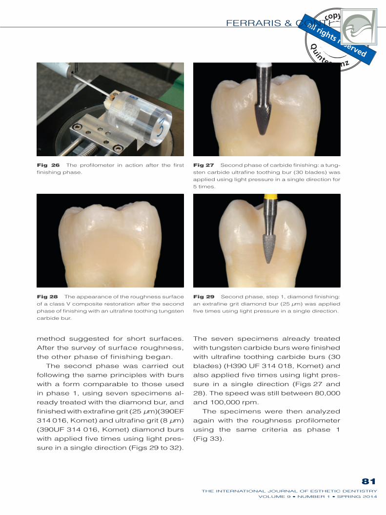
method suggested for short surfaces.
After the survey of surface roughness,
the other phase of finishing began.
The second phase was carried out
following the same principles with burs
with a form comparable to those used
in phase 1, using seven specimens al-
ready treated with the diamond bur, and
finished with extrafine grit (25 μm)(390EF
314 016, Komet) and ultrafine grit (8 μm)
(390UF 314 016, Komet) diamond burs
with applied five times using light pres-
sure in a single direction (Figs 29 to 32).
The seven specimens already treated
with tungsten carbide burs were finished
with ultrafine toothing carbide burs (30
blades) (H390 UF 314 018, Komet) and
also applied five times using light pres-
sure in a single direction (Figs 27 and
28). The speed was still between 80,000
and 100,000 rpm.
The specimens were then analyzed
again with the roughness profilometer
using the same criteria as phase 1
(Fig 33).
Fig 26 The profilometer in action after the first
finishing phase.
Fig 27 Second phase of carbide finishing: a tung-
sten carbide ultrafine toothing bur (30 blades) was
applied using light pressure in a single direction for
5 times.
Fig 28 The appearance of the roughness surface
of a class V composite restoration after the second
phase of finishing with an ultrafine toothing tungsten
carbide bur.
Fig 29 Second phase, step 1, diamond finishing:
an extrafine grit diamond bur (25 μm) was applied
five times using light pressure in a single direction.
CLINICAL RESEARCH
82THE INTERNATIONAL JOURNAL OF ESTHETIC DENTISTRY
VOLUME 9 • NUMBER 1 • SPRING 2014
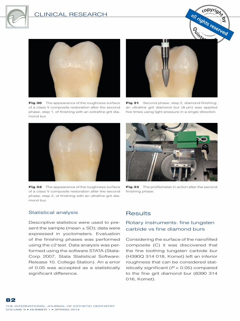
Statistical analysis
Descriptive statistics were used to pre-
sent the sample (mean ± SD); data were
expressed in yoctometers. Evaluation
of the finishing phases was performed
using the c2 test. Data analysis was per-
formed using the software STATA (Stata-
Corp 2007, Stata Statistical Software:
Release 10. College Station). An a error
of 0.05 was accepted as a statistically
significant difference.
Results
Rotary instruments: fine tungsten
carbide vs fine diamond burs
Considering the surface of the nanofilled
composite (C) it was discovered that
the fine toothing tungsten carbide bur
(H390Q 314 018, Komet) left an inferior
roughness that can be considered stat-
istically significant (P < 0.05) compared
to the fine grit diamond bur (8390 314
016, Komet).
Fig 30 The appearance of the roughness surface
of a class V composite restoration after the second
phase, step 1, of finishing with an extrafine grit dia-
mond bur.
Fig 31 Second phase, step 2, diamond finishing:
an ultrafine grit diamond bur (8 μm) was applied
five times using light pressure in a single direction.
Fig 32 The appearance of the roughness surface
of a class V composite restoration after the second
phase, step 2, of finishing with an ultrafine grit dia-
mond bur.
Fig 33 The profilometer in action after the second
finishing phase.
FERRARIS & CONTI
83THE INTERNATIONAL JOURNAL OF ESTHETIC DENTISTRY
VOLUME 9 • NUMBER 1 • SPRING 2014
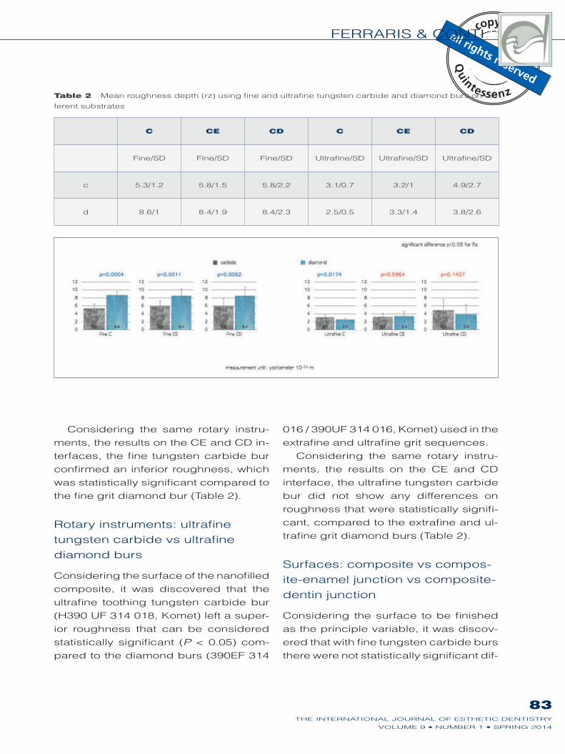
Considering the same rotary instru-
ments, the results on the CE and CD in-
terfaces, the fine tungsten carbide bur
confirmed an inferior roughness, which
was statistically significant compared to
the fine grit diamond bur (Table 2).
Rotary instruments: ultrafine
tungsten carbide vs ultrafine
diamond burs
Considering the surface of the nanofilled
composite, it was discovered that the
ultrafine toothing tungsten carbide bur
(H390 UF 314 018, Komet) left a super-
ior roughness that can be considered
statistically significant (P < 0.05) com-
pared to the diamond burs (390EF 314
016 / 390UF 314 016, Komet) used in the
extrafine and ultrafine grit sequences.
Considering the same rotary instru-
ments, the results on the CE and CD
interface, the ultrafine tungsten carbide
bur did not show any differences on
roughness that were statistically signifi-
cant, compared to the extrafine and ul-
trafine grit diamond burs (Table 2).
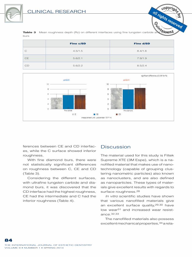
Surfaces: composite vs compos-
ite-enamel junction vs composite-
dentin junction
Considering the surface to be finished
as the principle variable, it was discov-
ered that with fine tungsten carbide burs
there were not statistically significant dif-
Table 2 Mean roughness depth (rz) using fine and ultrafine tungsten carbide and diamond burs on dif-
ferent substrates
C CE CD C CE CD
Fine/SD Fine/SD Fine/SD Ultrafine/SD Ultrafine/SD Ultrafine/SD
c 5.3/1.2 5.8/1.5 5.8/2.2 3.1/0.7 3.2/1 4.9/2.7
d 8.6/1 8.4/1.9 8.4/2.3 2.5/0.5 3.3/1.4 3.8/2.6
CLINICAL RESEARCH
84THE INTERNATIONAL JOURNAL OF ESTHETIC DENTISTRY
VOLUME 9 • NUMBER 1 • SPRING 2014
ferences between CE and CD interfac-
es, while the C surface showed inferior
roughness.
With fine diamond burs, there were
not statistically significant differences
on roughness between C, CE and CD
(Table 3).
Considering the different surfaces,
with ultrafine tungsten carbide and dia-
mond burs, it was discovered that the
CD interface had the highest roughness,
CE had the intermediate and C had the
inferior roughness (Table 4).
Discussion
The material used for this study is Filtek
Supreme XTE (3M Espe), which is a na-
nofilled material that makes use of nano-
technology (capable of grouping clus-
tering nanometric particles) also known
as nanoclusters, and are also defined
as nanoparticles. These types of mater-
ials give excellent results with regards to
surface roughness.28
In vitro scientific studies have shown
that various nanofilled materials give
an excellent surface quality,29,30 have
low wear31 and increased wear resist-
ance.32,33
The nanofilled materials also possess
excellent mechanical properties,34 a rela-
Table 3 Mean roughness depth (Rz) on different interfaces using fine tungsten carbide and diamond
burs
Fine c/SD Fine d/SD
C 4.5/1.5 8.4/1.6
CE 5.6/2.1 7.9/1.9
CD 5.6/2.2 8.5/2.4

FERRARIS & CONTI
85THE INTERNATIONAL JOURNAL OF ESTHETIC DENTISTRY
VOLUME 9 • NUMBER 1 • SPRING 2014
tively low shrinkage and high strength.35
Some observations found that even
the finest grade diamond finishing bur
caused extensive damage to surface
areas of enamel, and on composite. It
was concluded that composites finished
with diamond burs appeared rough and
uneven, which was particularly evident
with the hybrids because of the mixture
of large and small particles.36
The choice of different rotary instru-
ments to obtain smoother surfaces, has
been investigated in the past, with ex-
cellent polishing results in favor of the
tungsten carbide burs.21 On the contra-
ry, other authors have noticed that the
carbide tips did not perform at the same
level, causing damage to the peripheral
margins of the restoration.22 It can be
assumed that the cause is a non-homo-
geneous performance of the blades of
this type of bur.
The unfavorable roughness achieved
with the diamond bur has been under-
lined in the past, but it was considered
that the carbide burs left ridges that
were more difficult to eliminate later on
with the final polishing. Some authors
concluded that although the diamond
burs gave rougher surfaces, and were
not optimal, overall the gouges were not
as deep as with the carbide burs and
could therefore be more easily polished
to a smooth surface.37
In past studies driven with scanning
electron microscopy, it was observed
Table 4 Mean roughness depth (RZ) on different interfaces using ultrafine tungsten carbide and dia-
mond burs
Ultrafine c/SD Ultrafine d/SD
C 2.7/0.7 2.5/0.8
CE 3.6/1.5 3.2/1.2
CD 4.7/2.6 4.6/2.9

CLINICAL RESEARCH
86THE INTERNATIONAL JOURNAL OF ESTHETIC DENTISTRY
VOLUME 9 • NUMBER 1 • SPRING 2014
that diamond burs had a tendency to
tear filler particles and leave irregulari-
ties on the composite surface.38 On the
contrary, another showed that the hard-
ness of microfill complexes and their
weak bond to the organic matrix do not
permit the flutes of a carbide bur to cut
cleanly.39
In this study, the comparison was not
made between tungsten carbide and
diamond burs with the same colorimet-
ric code (red ring). The reason for this
stems from the fact that the carbide burs
that were used have a yellow code (with
fine grit according to the manufacturer)
there is an addition of horizontal notches
(blue ring), whose function is to subdi-
vide shavings in many microshavings.
According to the manufacturer, removal
should be greater on material of hard
consistency with respect to the same
bur without notches. When magnified,
small scrapes can be noticed due to the
action of the notches, which can be a
drawback with respect to the advantage
of greater removal and lesser bur knead-
ing with respect to the version without
notches. However, as it has been noticed
from data from this study, the grade of
finishing is advantageous if compared
to the fine grit diamond bur (red ring, 46
μm). Afterwards, the tungsten carbide
fine toothing bur with 16 blades with yel-
low-blue ring was used (like the normal
yellow ring that has 16/20 blades) and
not the bur with a red ring that gener-
ally has 8 to 12 blades. The compari-
son was done with a fine grit diamond
bur (red ring, 46 μm) and for the second
phase of the finishing it was decided to
use two steps for the diamond (extrafine
yellow ring, and ultrafine white ring). For
the cutting characteristic of the tungsten
carbide bur (yellow-blue ring), it was de-
cided to avoid the step with the extrafine
bur in the second phase of finishing (al-
so indicated by the manufacturer) and
to proceed with the ultrafine multiblade
white ring (30 blades).
A very important variable, other than
which type of bur to use (carbide or di-
amond), is with which cutting capacity
of the rotating instrument the finishing is
done, also because usually the clinician,
according to which grade of surface fin-
ishing and modification he would like to
make, begins with more aggressive in-
struments to then move on to finer blades
or grit. The results of this study show how
fine toothing carbide burs finish the res-
toration surfaces better in composite
than fine grit diamond burs. The study al-
so shows that with ultrafine grit, smoother
surfaces are achieved, but that there are
not significant differences between car-
bide and diamond burs.
Another question is with regards to
the speed of rotation of the burs during
finishing procedures. In the past, it has
been observed that carbide burs used
at high speed on hybrid composites cre-
ated a smooth surface,40 but it is also
true that for better control of the rotating
instrument, it is advisable to reduce ro-
tation speed. In fact, on natural enamel
it has been observed that the diamond
bur used at a high speed produced a
very rough enamel surface.26
The importance of a good finishing
on interface restorations is considered
by some to be fundamental to prevent
secondary decay.7,16
Not all studies agree on the correla-
tion between roughness on the interface
restoration tooth and the presence of
secondary decay.
FERRARIS & CONTI
87THE INTERNATIONAL JOURNAL OF ESTHETIC DENTISTRY
VOLUME 9 • NUMBER 1 • SPRING 2014
Regardless, it is common sense to re-
establish a smooth surface after a restor-
ation in composite, first and foremost to
impede the adhesion of bacteria on the
composite and to avoid microinfiltration
at the margins of the repair and to obtain
a suitable appearance. Some analysis
found, at both the enamel and the den-
tin margins, no statistically significant
differences in microleakage across bur
types. Further results show that dentin
margins leaked significantly more than
enamel margins for all bur types.41
Based on these observations, even
this study indicates that the surface
roughness is worse on the CD interface
than on the CE interface and therefore
the composite surface. This fact is of
great importance because it emphasiz-
es how factors that can be considered
relevant by the clinician with regards to
surface roughness are secondary with
regards to the type of tissue on which
the restoration terminates.
Conclusions
Within the limitations of this study, the
following conclusions can be drawn for
clinical purposes, relative to direct res-
torations in composite:
Comparing the fine grit and toothing
burs, the tungsten carbide had a bet-
ter capacity of finishing compared to
the diamond bur, thereby obtaining
surfaces with less surface roughness,
both on C surfaces and on the CE and
CD interfaces.
Comparing the ultrafine grit and tooth-
ing burs, the diamond bur had a better
capacity of finishing than the tungsten
carbide bur only on C surfaces, while
significant differences were not noted
on the CE and CD interfaces.
Comparing the three surfaces: C, CE,
and CD, it was noted that with fine grit
diamond burs there were no signifi-
cant variations, while with fine toothing
carbide burs, differences were noted
between C compared to CE and CD,
which gave equal results. With both
the carbide and diamond ultrafine grit
burs, there were analogous differenc-
es that were statistically significant: C
was the smoother surface, followed by
the CE interface and finally higher su-
perficial roughness was noted on the
CD interface.
The clinical perception, which does
not have scientific validity, notes that
with prismatic magnifying 4X, there
was a greater roughness on the sur-
faces finished with the fine grit dia-
mond bur.
Clinical relevance
Considering the measurement units to
analyze the roughness with the pro-
filometer and the yoctometer, this study
shows some statistical differences that
could not be clinically perceivable.
The null hypothesis that considered
that there were no significant differ-
ences on the roughness of composite
resin restorations finishing between
tungsten carbide and diamonds burs
can be rejected with regards to the
fine burs, because the tungsten car-
bide burs (Fig 22) showed better fin-
ishing compared to the diamond burs
(Fig 25).
Considering the ultrafine burs, the
null hypothesis can be accepted that
concluded there wouldn’t be any clin-

CLINICAL RESEARCH
88THE INTERNATIONAL JOURNAL OF ESTHETIC DENTISTRY
VOLUME 9 • NUMBER 1 • SPRING 2014
ical differences between the tungsten
carbide (Fig 28) and diamond burs
(Figs 32).
The null hypothesis that considered
that there would be no significant dif-
ferences on roughness between fin-
ishing on C surface, CE and CD in-
terfaces can be rejected, because
from a clinical point of view the CD
interface is the most difficult to finish
compared to C and CE. Furthermore,
the CD interface is usually a critical
point for hygiene procedures and for
potential secondary caries, and the
clinician should pay close attention
during the finishing of this area.
Acknowledgements
Many thanks to all those who participat-
ed and contributed to the realization of
this study. Komet, who in addition to sup-
plying the rotating instruments for the
finishing and polishing stages, carried
out all the analyses with the profilome-
ter. Thanks to Dr Dario Consonni for the
statistical analysis. Thanks to 3M-Espe,
Kerr, Ultradent and Ivoclar Vivident for
supplying the material to carry out the
restorations. Thanks to Drs Raffaele
Acunzo, Andrea Camurati, Samuele
Chiapparoli, Francesca Manfrini, Rob-
erto Rossi, Giovanni Sammarco, and
Ilaria Venuti for their help in locating ex-
tracted teeth. Thanks to Mrs Francesca
Vasile, Ms Daniela Enriquez and Mr Re-
nato Alcidi, for contributing in various
ways to the realization of the samples.
Thanks to Dr Domenico Massironi for
his advice and teachings. Thanks to Ms
Marina Conti and Mr Mauro Ferraris for
their support.
References
1. Kidd EAM, J-BS, Beighton
D. Diagnosis of secondary
caries: a laboratory study. Br
Dent J 1994;176:135–139.
2. Mjor IA. Clinical diagnosis of
recurrent caries. J Am Dent
Assoc 2005; 136:1426–1433.
3. Özer L, TA. What is known
about caries in relation to
restorations as a reason for
replacement? A review. Adv
Dent Res 1995;9:394–402.
4. Mjor IA, Toffenetti F. Second-
ary caries: a literature review
with case reports. Quintes-
sence Int 2000;31:165–79.
5. Hals E, Nernaes A. Histo-
pathology of in vitro caries
developing around silver
amalgam fillings. Caries Res
1971;5:58–77.
6. Boeckh C, Schumacher
E, Podielski A, Haller B.
Antibacterial activity of
restorative dental bioma-
terials in vitro. Caries Res
2002;36:101–107.
7. Fontana M Gonzalez-
Cabezas C. Secondary
caries and restoration
replacement: an unresolved
problem. Compend Contin
Educ Dent 2000;21:15–18,
21–24, 26 passim; quiz 30.
8. Cenci MS, Tenuta LM,
Pereira-Cenci T, Del Bel Cury
AA, ten Cate JM, Cury JA.
Effect of microleakage and
fluoride on enamel-dentine
demineralization around
restorations. Caries Res
2008;42:369–379.
9. Marsh P, Nyvad B. The oral
microflora and biofilm on
teeth; in: Fejerskov O, Kidd
EAM (Eds). Dental Caries:
The Disease and Its Clinical
Management. London: Wiley-
Blackwell, 2003:29–48.
10. Larato D. Influence of a com-
posite resin restoration on
the gingiva. J Prosthet Dent
1972;28:402–404.
11. Weitman R, Eames W.
Plaque accumulation on
composite surfaces after
various finishing procedures.
Oral Health 1975:65:29–33.
12. Wise M, Dykema R. The
plaque-retaining capacity of
four dental materials. J Pros-
thet Dent 1975;33:178–190.

FERRARIS & CONTI
89THE INTERNATIONAL JOURNAL OF ESTHETIC DENTISTRY
VOLUME 9 • NUMBER 1 • SPRING 2014
13. Chan K, Fuller J, Hormati A.
The ability of food to stain
two composite resins. J Pros-
thet Dent 1980;43:542–545.
14. Holmen L, Thylstrup A, Artun
J, Clinical and histological
features observed during
arrestment of active enamel
carious lesions in vivo. Car-
ies Res 1987;21:546–554.
15. Lima FG, Romano AR, Cor-
rea MB, Demarco FF. Influ-
ence of microleakage, sur-
face roughness and biofilm
control on secondary caries
formation around composite
resin restorations: an in situ
evaluation. J Appl Oral Sci
2009;17:61–65.
16. Montanaro L, Campoccia D,
Rizzi S, Donati ME, Breschi
L, Prati C. Evaluation of
bacterial adhesion of Strep-
tococcus mutans on dental
restorative materials. Bioma-
terials 2004;25:4457–4463.
17. Patel SB, Gordan VV, Barrett
AA, Shen C. The effect of
surface finishing and stor-
age solutions on the color
stability of resin-based com-
posites. J Am Dent Assoc
2004;135:587–594; quiz 654.
18. Lu H, Roeder LB, Lei L,
Powers JM. Effect of surface
roughness on stain resist-
ance of dental resin com-
posites. J Esthet Restor Dent
2005;17:102–108; discussion
109.
19. Heintze SD, Forjanic M,
Rousson V. Surface rough-
ness and gloss of dental ma-
terials as a function of force
and polishing time in vitro.
Dent Mater 2006;22:146–165.
20. Paravina RD, Roeder L,
Lu H, Vogel K, Powers JM.
Effect of finishing and polish-
ing procedures on surface
roughness, gloss and color
of resin-based composites.
Am J Dent 2004;17:262-266.
21. Berastegui E, Canalda C,
Brau E, Miquel C. Surface
roughness of finished com-
posite resins. J Prosthet Dent
1992;68:742–749.
22. Ashe MJ, Tripp GA, Eich-
miller FC, George LA, Meiers
JC. Surface roughness of
glass-ceramic insert-com-
posite restorations: assess-
ing several polishing tech-
niques. J Am Dent Assoc
1996;127:1495–1500.
23. Kaplan BA, Goldstein GR,
Vijayaraghavan TV, Nelson
IK. The effect of three polish-
ing systems on the surface
roughness of four hybrid
composites: a profilomet-
ric and scanning electron
microscopy study. J Prosthet
Dent 1996;76:34–38.
24. Brackett WW, Gilpatrick RO,
Gunnin TD. Effect of finishing
method on the microleakage
of Class V resin compos-
ite restorations. Am J Dent
1997;10:189–191.
25. Hondrum SO, Fernandez Jr
R. Contouring, finishing, and
polishing Class 5 restora-
tive materials. Oper Dent
1997;22:30–36.
26. Giampaolo ET, Machado AL,
Pavarina AC, Vergani CE.
Different methods of finishing
and polishing enamel. J Pros-
thet Dent 2003;89:135–140.
27. Breschi L, Mazzoni A, Nato
F, Carrilho M, Visintini E,
Tjäderhane L, et al. Chlor-
hexidine stabilizes the
adhesive interface: a 2-year
in vitro study. Dent Mater
2010;26:320–325.
28. Janus J, Fauxpoint G, Arntz
Y, Pelletier H, Etienne O.
Surface roughness and
morphology of three nano-
composites after two differ-
ent polishing treatments by
a multitechnique approach.
Dent Mater 2010;26:416–
425.
29. Yap AU, Yap SH, Teo CK, Ng
JJ. Comparison of surface
finish of new aesthetic re-
storative materials. Oper
Dent 2004;29:100–104.
30. Silikas N, Kavvadia K,
Eliades G, Watts D. Surface
characterization of modern
resin composites: a multi-
technique approach. Am J
Dent 2005;18:95–100.
31. Turssi CP, Ferracane JL,
Serra MC. Abrasive wear of
resin composites as related
to finishing and polishing
procedures. Dent Mater
2005;21:641–648.
32. Yap AU, Tan CH, Chung
SM. Wear behavior of new
composite restoratives. Oper
Dent 2004;29:269–274.
33. Xu HH, Quinn JB, Giusep-
petti AA. Wear and mechan-
ical properties of nano-silica-
fused whisker composites. J
Dent Res 2004;83:930–935.
34. Mitra SB, Wu D, Holmes BN.
An application of nanotech-
nology in advanced dental
materials. J Am Dent Assoc
2003;134:1382–1390.
35. Chen MH, Chen CR, Hsu SH,
Sun SP, Su WF. Low shrink-
age light curable nanocom-
posite for dental restora-
tive material. Dent Mater
2006;22:138–145.
36. Quiroz L Lentz D. The effect
of polishing procedures on
light-cured composite restor-
ations. Compendium Contin
Dent Educ 1985;6:437–439.
37. Goldstein GR, Waknine S.
Surface roughness evalua-
tion of composite resin pol-
ishing techniques. Quintes-
sence Int 1989;20:199–204.
38. Grundy JR. Finishing pos-
terior composites. An SEM
study of a range of instru-
ments and their effect on a
composite and enamel. Re-
storative Dent, 1985;1:148,
150, 152–158.
39. Lutz F, Jetcos JF, Phillips RW.
New finishing instruments for
composite resins. J Am Dent
Assoc 1983;107:575–580.
40. Boghosian AA, Randolph
RG, Jekkals VJ. Rotary
instrument finishing of
microfilled and small-
particle hybrid composite
resins. J Am Dent Assoc
1987;115:299–301.
41. Shook LW, Turner EW, Ross
J, Scarbecz M. Effect of
surface roughness of cavity
preparations on the micro-
leakage of Class V resin
composite restorations. Oper
Dent 2003 28:779–785.