superoxide dismutase (sod) and catalase (cat) activity assay protocols ... · the present protocol...
TRANSCRIPT

www.bio-protocol.org/e2505 Vol 7, Iss 16, Aug 20, 2017 DOI:10.21769/BioProtoc.2505
Copyright © 2017 The Authors; exclusive licensee Bio-protocol LLC. 1
Superoxide Dismutase (SOD) and Catalase (CAT) Activity Assay Protocols for Caenorhabditis elegans
Jing Zhang1, *, Rui Chen2, 3, Zhenyang Yu2, 3 and Lili Xue3
1Ecological Technique and Engineering College, Shanghai Institute of Technology, Shanghai, China;
2State Key Laboratory of Pollution Control and Resource Reuse, Key Laboratory of Yangtze River Water
Environment, Ministry of Education, College of Environmental Science and Engineering, Tongji
University, Shanghai, China; 3Jiaxing Tongji Institute for Environment, Jiaxing, China
*For correspondence: [email protected]
[Abstract] Assays for superoxide dismutase (SOD) and catalase (CAT) activities are widely employed
to indicate antioxidant responses underlying the toxic effects of test chemicals. Yet, earlier studies mainly
described the procedures as performed according to manufacturer’s instructions without modifications
that are specific to any organisms. The present protocol describes the steps in analyzing the superoxide
dismutase (SOD) and catalase (CAT) activities in C. elegans, which is a model organism that can be
used to study effects of pharmaceutical compounds and environmental pollutants. The main steps
include: (1) sample preparation; (2) total protein assay; (3) SOD activity assay; (4) CAT activity assay;
and (5) medium list and formula, and also data analysis and performance notes.
Keywords: SOD, CAT, Total protein, C. elegans, Protocol
[Background] Biomarkers are essential to examine biological and pathogenic processes in response
to a chemical, an agent or a therapeutic intervention. Various biological processes in organisms result
in reactive oxygen species (ROS) which cause oxidative stress. In response to such oxidative stress,
organisms can deploy superoxide dismutase (SOD) and catalase (CAT) to scavenge ROS so as to
protect the cellular homeostasis (Balaban et al., 2005). On the one hand, various chemicals (pollutants)
can retard such antioxidant responses, and disturb the health of organisms including human beings. On
the other hand, many pharmaceuticals aim to strengthen the antioxidant responses to improve health.
Therefore, activities of SOD and CAT are very important to reflect potential effects of chemicals or/and
pharmaceuticals.
Caenorhabditis elegans (C. elegans) is a model organism that has been used to study effects of
pharmaceutical compounds (Dengg and van Meel, 2004; Carretero et al., 2017) and environmental
pollutants (Yu et al., 2013a and 2017). Several studies have used SOD and CAT assays to indicate the
antioxidant responses and potential mechanism underlying the toxic effects of test chemicals (Feng et
al., 2015; Yu et al., 2012; 2016 and 2017). However, these studies simply described that the
determination was carried out, by using a generic kit protocol without species-specific modifications.
Therefore, the explicit protocols to perform SOD and CAT assays in C. elegans are still needed for better
specific instruction.

www.bio-protocol.org/e2505 Vol 7, Iss 16, Aug 20, 2017 DOI:10.21769/BioProtoc.2505
Copyright © 2017 The Authors; exclusive licensee Bio-protocol LLC. 2
In the present protocol, we provide a nematode protocol with experimental details to analyze SOD
and CAT activities in C. elegans.
Materials and Reagents
1. Pipette tips (https://online-shop.eppendorf.com)
2. Plate (60 mm)
3. Centrifuge tubes, 1.5 ml (Eppendorf, catalog number: 022364111)
4. Absorbent paper (KCWW, Kimberly-Clark, catalog number: 0131)
5. 96-well plate, with lids (Corning, Costar®, catalog number: 3599)
6. Sealing tape, for 96-well plates (Thermo Fisher Scientific, Thermo ScientificTM, catalog number:
15036)
7. Nematodes (wild type N2)
Note: These nematodes are treated according to each researcher’s experiments. In the present
protocol, the nematodes only have difference in numbers.
8. BCA protein assay kits (Thermo Fisher Scientific, Thermo ScientificTM, catalog number: 23225
or 23227)
a. Protein standard (5 mg/ml)
b. BCA reagent A and B
9. SOD assay kits (Beyotime Biotechnology, catalog number: S0101)
a. Reaction initiation solution
b. SOD detection buffer
c. WST-8
d. Enzyme solution
e. Reaction initiation solution (40x)
10. CAT assay kits (Beyotime Biotechnology, catalog number: S0051)
a. Enzyme conjugate
b. Wash solution 40x
c. Substrate A and B
d. Stop solution
11. Sodium hydrate (NaOH), Analytic Reagent (Sinopharm Chemical Reagent, catalog number:
10019762)
12. Sodium hypochlorite (Antiformin, NaOCl, with 6-14% active Cl), Analytic Reagent (ALADDIN,
catalog number: S101636-500 ml)
13. Potassium phosphate dibasic (K2HPO4), Analytic Reagent (Sinopharm Chemical Reagent,
catalog number: 20032118)
14. Potassium phosphate monobasic (KH2PO4), Analytic Reagent (Sinopharm Chemical Reagent,
catalog number: 10017618)
15. Clorox solution (see Recipes)

www.bio-protocol.org/e2505 Vol 7, Iss 16, Aug 20, 2017 DOI:10.21769/BioProtoc.2505
Copyright © 2017 The Authors; exclusive licensee Bio-protocol LLC. 3
16. Phosphate buffered saline/buffer (PBS), pH 7.0 (see Recipes)
Equipment
1. Pipettes
2. Microscope
3. Centrifuge, Eppendorf 5417R (Eppendorf, model: 5417 R, catalog number: 01396)
4. Pestles, Eppendorf (Eppendorf, catalog number: F0140010)
5. Motor-driven tissue grinder (Beijing Baiwan Electronic Technology, catalog number: HG215-LH-
A)
6. Ice bath, in centrifuge tube box
7. Microplate reader, BioTek (BioTek Instruments, model: Epoch)
8. Sterilized bottle, Fisherbrand (100 ml, Fisher Scientific, catalog number: FB800100; 250 ml,
Fisher Scientific, catalog number: FB800250; 500 ml, Fisher Scientific, catalog number:
FB800500)
9. Magnetic stir bar
10. Magnetic stir plate
11. pH meter
12. Incubator, Yiheng (Yiheng, model: LRH-1000F)
Procedure
A. Sample preparation
1. Sample collection
The entire protocol uses the L4 stage nematodes as the reference point. Follow the nematode
culture and age-synchronization according to earlier reports (Solis and Petrascheck, 2015; Yu
et al., 2017). Wash the L4 stage nematodes off each nematode growth medium plate (60 mm)
with 1.6 ml phosphate buffered saline (PBS, pH 7.0) into a 1.5 ml centrifuge tube. After a 30-
min settlement by gravity at 4 °C, carefully pipette out and discard the supernatants. Add 500 μl
ice-cold PBS, resuspend the pellets with gentle and repetitive pipetting followed by a further 15-
min settlement. Then, discard the supernatants, add 500 μl ice-cold PBS, resuspend the pellets
with gentle and repetitive pipetting and obtain groups with 20, 50, 100, 200, 300, 400 and 500
nematodes in 200 μl PBS in the centrifuge tubes using the following steps.
Take the group with 100 nematodes as an example.
a. Count the nematodes in 20 μl PBS under a microscope with three independent trials to
estimate the nematode density in the whole tube.
b. Based on the number of worms obtained, calculate and adjust the volume of PBS so that
the final density is ~9 to 11 worms per 20 μl. For example, if the number is more than 10,

www.bio-protocol.org/e2505 Vol 7, Iss 16, Aug 20, 2017 DOI:10.21769/BioProtoc.2505
Copyright © 2017 The Authors; exclusive licensee Bio-protocol LLC. 4
add more PBS accordingly; and if the number is less than 10, perform a 10-min settlement
and remove corresponding amount of PBS to achieve the desired density.
c. Transfer 260 μl PBS with the nematodes into new 1.5 ml tubes with pipette, and resuspend
the pellets with gentle and repetitive pipetting.
d. Count nematode numbers in 20 μl PBS in three independent trials, with gentle and repetitive
pipetting in between, to confirm the nematode density, leaving 200 μl in the tube. If the
nematode density is not consistent with the setting, repeat step A1b.
e. Prepare groups with other nematode numbers through the same way.
2. Sample storage
Centrifuge the tubes at 5,000 x g for 5 min (4 °C). Discard the supernatant PBS carefully with
pipette and store the pellets at -26 °C overnight or -80 °C for over a week.
3. Sample homogenization
a. Connect a clean pestle with the motor-driven tissue grinder. Wash the pestle in ice-cold PBS,
and clean it with the absorbent paper. Push the pestle tightly against the pellets in the 1.5
ml centrifuge tube. Keep the centrifuge tube in an ice bucket (see Figure 1). Start the tissue
grinder, grind the pellets for 10 sec with a 2-sec cooling down followed by another 10-sec
grinding. Then, use 200 μl ice-cold PBS to wash the residual liquids on the pestle back to
the centrifuge tube (see Figure 1), before taking it out of the tube.
b. Centrifuge the tubes at 5,000 x g for 5 min at 4 °C. Pipette out 200 μl supernatants from
each sample. Aliquot them into four tubes with 50 μl in each for subsequent determination.
Figure 1. Sample homogenization. The left picture shows that motor-driven tissue grinder
homogenizes the pellets in the centrifuge tube in an ice bath, and the right one shows that
residual liquids are washed off the pestle with 200 μl ice-cold PBS.
B. Total protein assay
Measure the total protein (TP) in each sample by enzyme-linked immunosorbent assay (ELISA) kits
based on BCA methods.
1. Preparation for protein standards

www.bio-protocol.org/e2505 Vol 7, Iss 16, Aug 20, 2017 DOI:10.21769/BioProtoc.2505
Copyright © 2017 The Authors; exclusive licensee Bio-protocol LLC. 5
Use the stock protein standard (5 mg/ml) to prepare 0.05, 0.1, 0.2, 0.4, 0.6, 0.8, 1.0, 1.2 mg/ml
dilutions, 100 μl each using PBS.
2. Preparation for BCA working solution
Prepare BCA working solution with BCA reagent A and B by a volume ratio of 50:1. Total volume
of the BCA working solution is calculated according to the amount of tested samples (200 μl per
sample).
3. Determination of protein concentration
a. For each protein standard, add 20 μl into a 96-well plate with at least two replicates. Use 20
μl PBS buffer as control.
b. For each nematode sample, add 20 μl from one aliquot into the 96-well plate with at least
two replicates. Use up this aliquot to ensure replicate and avoid repetitive freezing-
unfreezing of the samples.
c. Then, add 200 μl BCA working solution per well for all the protein standards and nematode
samples. Seal the wells by sealing tape to avoid the evaporation of water, and incubate the
plate for 30 min at 37 °C.
d. Measure the absorbance at 562 nm (A562) using a microplate reader.
4. Data calculation
Calculate the protein contents in the samples according to the standard curve.
One presentation of the relation between the nematode numbers and the protein concentrations
is shown in Figure 2.
Figure 2. The relationship between the nematode numbers and the nematode protein concentrations
www.bio-protocol.org/e2505 Vol 7, Iss 16, Aug 20, 2017 DOI:10.21769/BioProtoc.2505
Copyright © 2017 The Authors; exclusive licensee Bio-protocol LLC. 6
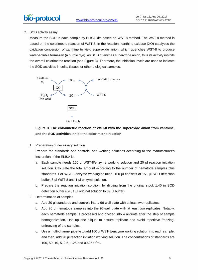
C. SOD activity assay
Measure the SOD in each sample by ELISA kits based on WST-8 method. The WST-8 method is
based on the colorimetric reaction of WST-8. In the reaction, xanthine oxidase (XO) catalyzes the
oxidation conversion of xanthine to yield superoxide anion, which quenches WST-8 to produce
water-soluble formazan (a purple dye). As SOD quenches superoxide anion, thus its activity inhibits
the overall colorimetric reaction (see Figure 3). Therefore, the inhibition levels are used to indicate
the SOD activities in cells, tissues or other biological samples.
Figure 3. The colorimetric reaction of WST-8 with the superoxide anion from xanthine, and the SOD activities inhibit the colorimetric reaction
1. Preparation of necessary solution
Prepare the standards and controls, and working solutions according to the manufacturer’s
instruction of the ELISA kit.
a. Each sample needs 160 μl WST-8/enzyme working solution and 20 μl reaction initiation
solution. Calculate the total amount according to the number of nematode samples plus
standards. For WST-8/enzyme working solution, 160 μl consists of 151 μl SOD detection
buffer, 8 μl WST-8 and 1 μl enzyme solution.
b. Prepare the reaction initiation solution, by diluting from the original stock 1:40 in SOD
detection buffer (i.e., 1 μl original solution to 39 μl buffer).
2. Determination of samples
a. Add 20 μl standards and controls into a 96-well plate with at least two replicates.
b. Add 20 μl nematode samples into the 96-well plate with at least two replicates. Notably,
each nematode sample is processed and divided into 4 aliquots after the step of sample
homogenization. Use up one aliquot to ensure replicate and avoid repetitive freezing-
unfreezing of the samples.
c. Use a multi-channel pipette to add 160 μl WST-8/enzyme working solution into each sample,
and then, add 20 μl reaction initiation working solution. The concentrations of standards are
100, 50, 10, 5, 2.5, 1.25 and 0.625 U/ml.

www.bio-protocol.org/e2505 Vol 7, Iss 16, Aug 20, 2017 DOI:10.21769/BioProtoc.2505
Copyright © 2017 The Authors; exclusive licensee Bio-protocol LLC. 7
d. There are two blank controls. For the blank control 1, 20 μl sample are replaced by SOD
detection buffer. For the blank control 2, 20 μl sample and 20 μl reaction initiation solution
are replaced by 40 μl SOD detection buffer.
e. Incubate the plate for 30 min at 37 °C.
f. Read the absorbance at 450 nm (A450) by a microplate reader.
3. Calculation in SOD activity assay
a. Inhibition (%) = (ABlank control 1 - AStandard or Sample)/(ABlank control 1 - ABlank control 2) x 100%. Use the
standards with known SOD activities and their inhibitions to establish a standard curve.
Then, use the curve to calculate the SOD activities of samples by their inhibitions. One
example for the standard curve is shown in Figure 4.
Figure 4. The relationship between SOD activities in logarithm and inhibition (%) on WST-8 colorimetric reaction
b. Express the SOD enzyme activity in each nematode sample as its proportion (P) in the total
protein (TP) of the same sample to eliminate the differences of nematode numbers among
samples. The normalized SOD activities in the nematodes are listed in Table 1. The
proportion values generally range from 4.04 to 4.35. The values in groups with 20 and 50
nematodes are quite greater than the general range. Although these values are within mean
value + 3 standard deviation (SD), we recommend not using them to avoid unreliability.
These results indicate that at least 100 nematodes should be used to ensure the feasibility
and stability in measuring SOD activities.

www.bio-protocol.org/e2505 Vol 7, Iss 16, Aug 20, 2017 DOI:10.21769/BioProtoc.2505
Copyright © 2017 The Authors; exclusive licensee Bio-protocol LLC. 8
Table 1. Connection between normalized SOD activities and nematode numbers
Nematode number
Total protein (mg/ml)
SOD activity (U/ml)
Normalized SOD activities (U/mg)
20 0.082 0.57 6.95 50 0.153 0.91 5.95 100 0.31 1.25 4.02 200 0.42 1.78 4.20 300 0.51 2.23 4.35 400 0.69 2.82 4.09 500 0.83 3.37 4.04
D. CAT activity assay
Measure the CAT activities in each sample with ELISA kits. The assay principle is based on the
reaction of catalase to decompose hydrogen peroxide (H2O2). The excess H2O2 can form
complexation with ammonium molybdate to produce light yellow solution. The solution’s adsorption
at 450 nm can be used to calculate the concentration of H2O2 and therefore to indirectly indicate the
CAT activities.
1. Reagents preparation
According to the manufacturer’s instruction of the ELISA kit, prepare all reagents before the
assay with careful calculation based on the numbers of samples, standards and controls.
2. Addition of standards or samples
Add 50 μl standards or samples into a 96-well plate. Use 50 μl PBS as blank controls. All
standards, samples and controls are recommended to have at least two replicates. Notably,
each nematode sample is processed and divided into 4 aliquots after the step of sample
homogenization. Use up one aliquot to ensure replicate and avoid repetitive freezing-unfreezing
of the samples. The concentrations of standards are 400, 100, 25, 6.25, 1.5625, 0.4 and 0.1
U/L.
3. Enzyme conjugation
Add 100 μl of enzyme conjugate to wells containing standards, samples and blanks. Then, seal
the wells by sealing tape to avoid the evaporation of water, and incubate the microplate statically
for 60 min at 37 °C.
4. Wash
a. Discard the liquid content and tap the assay plate on a piece of absorbent paper to remove
residual buffer.
b. Add 350 μl wash solution (1x, which was diluted from the 40x package with PBS) to each
well and keep the microplate static for 1 min. Then, discard the liquid content and tap the
assay plate on a piece of absorbent paper to remove residual buffer.
c. Repeat the above wash process for four times.
www.bio-protocol.org/e2505 Vol 7, Iss 16, Aug 20, 2017 DOI:10.21769/BioProtoc.2505
Copyright © 2017 The Authors; exclusive licensee Bio-protocol LLC. 9
5. Addition of substrates
Use a multi-channel pipette to add 50 μl substrate A and 50 μl substrate B to each well. Incubate
the microplate at 37 °C in the dark for 15 min.
6. Endpoint measurement
Add 50 μl stop solution to each well. After 1 min incubation, measure the absorbance at 450 nm
on a microplate reader within 15 min.
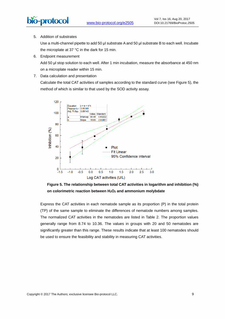
7. Data calculation and presentation
Calculate the total CAT activities of samples according to the standard curve (see Figure 5), the
method of which is similar to that used by the SOD activity assay.
Figure 5. The relationship between total CAT activities in logarithm and inhibition (%) on colorimetric reaction between H2O2 and ammonium molybdate
Express the CAT activities in each nematode sample as its proportion (P) in the total protein
(TP) of the same sample to eliminate the differences of nematode numbers among samples.
The normalized CAT activities in the nematodes are listed in Table 2. The proportion values
generally range from 8.74 to 10.36. The values in groups with 20 and 50 nematodes are
significantly greater than this range. These results indicate that at least 100 nematodes should
be used to ensure the feasibility and stability in measuring CAT activities.

www.bio-protocol.org/e2505 Vol 7, Iss 16, Aug 20, 2017 DOI:10.21769/BioProtoc.2505
Copyright © 2017 The Authors; exclusive licensee Bio-protocol LLC. 10
Table 2. Connection between normalized CAT activities and nematode numbers
Nematode number
Total protein (mg/ml)
CAT activity (U/l)
Normalized CAT activities (U/g)
20 0.082 2.33 28.41 50 0.153 2.39 15.62 100 0.31 3.22 10.36 200 0.42 3.85 9.08 300 0.51 4.59 8.96 400 0.69 6.12 8.88 500 0.83 7.29 8.74
Data analysis
1. The data analysis procedure refers to our previous report (Yu et al., 2013b). Firstly, the values
of the nematode total protein (TP) in one aliquot of each sample are measured in at least three
replicates, and the means of these TP values (TP mean) are calculated to represent the TP
concentration in the sample. Secondly, the SOD (or CAT) values in other aliquots of the same
sample are also measured in at least three replicates. Thirdly, the SOD (or CAT) values in each
replicate are calculated as their proportions to the TP mean value to obtain SOD (or CAT)
proportion values. Fourthly, the SOD (or CAT) proportion values are calculated to obtain the
mean SOD (or CAT) proportion in the sample and corresponding standard error.
2. In cases that the samples come from different treatments, the mean SOD (or CAT) proportion
in samples from the control are normalized to represent 100% (or 1.0); then, the SOD (or CAT)
proportions in samples from the chemical exposure are transformed to percentages of the
controls (POCs) (or fold changes against control); next, the POC values (or fold-changes) in the
samples are used to calculate the mean POC and corresponding standard error.
3. The nematode samples should be collected from at least three independently repeated
experiments, with one set of data in each experiment. The ANOVA (Origin 9.0, Origin Lab Corp.,
USA) was carried out between the mean SOD (or CAT) proportions in the control and those in
the chemical exposure from these independently repeated experiments. The probability levels
of 0.05 were considered statistically significant (P < 0.05).
Notes
1. When collecting the nematodes from nematode growth medium plates, the bacteria in the worm
slurry would cause problems to the assays. Therefore, don’t use centrifugation, but use gravity
settlement for sample collection. To fully get rid of the bacteria, gently wash the pellets (i.e.,
resuspend-settlement-discard supernatant) several times.
2. Remember to avoid unnecessary stress on the nematodes. On one hand, use ice-cold PBS to
lessen the stress on worms. On the other hand, it is better to spend less time on transfer and

www.bio-protocol.org/e2505 Vol 7, Iss 16, Aug 20, 2017 DOI:10.21769/BioProtoc.2505
Copyright © 2017 The Authors; exclusive licensee Bio-protocol LLC. 11
other performance with well-trained handling. Use a multi-channel pipette to make simultaneous
addition of all solutions, so as to shorten the differences of reaction time among samples. Also,
keep all the samples with the same performance to ensure the comparability between samples.
3. Considering that the worms will stick to the tip and they tend to settle with gravity, resuspend
the pellets with gentle and repetitive pipetting every time when transferring the worms. In this
way, we can ensure that the worm density is uniform in the tube, and also that the worms stuck
to the tip reach the equilibrium without further sticking.
4. To ensure reproducibility and variability, there should be at least 100 nematodes in each tube.
5. The supernatants from the homogenized PBS solutions of 100 nematodes are enough for four
aliquots, which are adequate for four biochemical assays including the total protein. If more
biochemical assays are performed simultaneously, the nematode numbers should increase.
6. The protein concentration range of the protein standard solutions can be altered according to
preliminary experiments, especially in cases where the nematode numbers are not counted.
7. When homogenizing nematode samples, handle the pestles in a uniform manner among
different samples. Keep the movement in the same strength with same time duration (10 sec
homogenizing + 2 sec cooling down + 10 sec homogenizing for each tube), so as to improve
the reproducibility of the data. Also, clean the pestle before using them for another tube, and
keep the clean procedure uniform throughout the whole homogenizing procedure.
Put pestles (for subsequent homogenization) in ice-cold PBS. After each use, wash the pestle
in ice cold water, and put it back to the clean ice-cold PBS. This performance will ensure the
cleanness of the pestle and also avoid potential influence of room-temperature pestles on
protein stability of the samples.
8. If the nematode number in each tube is much more than 500, the duration of homogenizing
procedure should be extended to at least twice the normal time (e.g., 10 sec grinding + 2 sec
cooling + 10 sec grinding + 2 sec cooling + 10 sec grinding + 2 sec cooling +10 sec grinding).
The pestles should be washed with 400 μl PBS. Through this way, the homogenizing efficiency
can be ensured, and the total protein concentration in the supernatants can be diluted to better
perform the biochemical assays.
9. In the prevention of water evaporation, a humid chamber will help.
Recipes
1. Clorox solution
Take 30 ml as an example. It consists of 0.6 g NaOH, 5 ml NaOCl (with 6-14% active Cl), and
25 ml deionized H2O, and the final concentrations are 0.5 M and ~1% for NaOH and NaOCl,
respectively
www.bio-protocol.org/e2505 Vol 7, Iss 16, Aug 20, 2017 DOI:10.21769/BioProtoc.2505
Copyright © 2017 The Authors; exclusive licensee Bio-protocol LLC. 12
2. Phosphate buffered saline/buffer (PBS), pH 7.0
If a ready-mix is not available, then make the solution as follows:
a. Solution A: dissolve 17.418 g of K2HPO4 (FW = 174.18) in 1,000 ml in sterilized deionized
water in sterilized bottle
b. Solution B: dissolve 13.689 g of KH2PO4 (FW = 136.89) in 1,000 ml in sterilized deionized
water in sterilized bottle
c. In a 2,000 ml flask with a magnetic stir bar on a magnetic stir plate, add 750 ml solution B.
After turning on the magnetic stirring apparatus, carefully put the probe of the pH meter
underneath the water surface and above the magnetic stirrer. Then, add solution A into the
flask slowly, and monitor the pH changes at the same time until the pH reaches 7.0
d. If the pH overgoes 7.0, add more solution B slowly until the pH backs to 7.0
Acknowledgments
The authors are grateful for the financial supports by the National Natural Science Foundation of
China (No. 21307095, No. 21407061), the International Science & Technology Cooperation
Program of China (No. 2016YFE0123700), the Collaborative Innovation Center for Regional
Environmental Quality, and the Swedish Research Council (contract Dnr. 639-2013-6913).
References
1. Balaban, R. S., Nemoto, S. and Finkel, T. (2005). Mitochondria, oxidants, and aging. Cell 120(4):
483-495.
2. Carretero, M., Solis, G.M., Petrascheck, M. (2017). C. elegans as model for drug discovery.
Curr Top Med Chem 17: 1-10.
3. Dengg, M. and van Meel, J. C. (2004). Caenorhabditis elegans as model system for rapid toxicity
assessment of pharmaceutical compounds. J Pharmacol Toxicol Methods 50(3): 209-214.
4. Feng, S., Cheng, H., Xu, Z., Shen, S., Yuan, M., Liu, J. and Ding, C. (2015). Thermal stress
resistance and aging effects of Panax notoginseng polysaccharides on Caenorhabditis elegans.
Int J Biol Macromol 81: 188-194.
5. Solis, G. M. and Petrascheck, M. (2015). Measuring Caenorhabditis elegans life span in 96 well
microtiter plates. J Vis Exp 49: e2496.
6. Yu, Z., Chen, X., Zhang, J., Wang, R. and Yin, D. (2013a). Transgenerational effects of heavy
metals on L3 larva of Caenorhabditis elegans with greater behavior and growth inhibitions in the
progeny. Ecotoxicol Environ Saf 88: 178-184.
7. Yu, Z., Sun, G., Liu, Y., Yin, D. and Zhang, J. (2017). Trans-generational influences of
sulfamethoxazole on lifespan, reproduction and population growth of Caenorhabditis elegans.
Ecotoxicol Environ Saf 135: 312-318.

www.bio-protocol.org/e2505 Vol 7, Iss 16, Aug 20, 2017 DOI:10.21769/BioProtoc.2505
Copyright © 2017 The Authors; exclusive licensee Bio-protocol LLC. 13
8. Yu, Z. Y., Zhang, J. and Yin, D. Q. (2012). Toxic and recovery effects of copper on
Caenorhabditis elegans by various food-borne and water-borne pathways. Chemosphere
87(11): 1361-1367.
9. Yu, Z., Zhang, J., Chen, X., Yin, D. and Deng, H. (2013b). Inhibitions on the behavior and growth
of the nematode progeny after prenatal exposure to sulfonamides at micromolar concentrations.
J Hazard Mater 250-251: 198-203.
10. Yu, Z., Zhang, J. and Yin, D. (2016). Multigenerational effects of heavy metals on feeding,
growth, initial reproduction and antioxidants in Caenorhabditis elegans. PLoS One 11(4):
e0154529.