supplementary information structure and mechanism of a ... fileeach sequencing and modification...
TRANSCRIPT

Supplementary Information
Structure and mechanism of a molecular rheostat, an RNA thermometer
that modulates immune evasion by Neisseria meningitidis
Ravi P Barnwal, Edmund Loh, Kate S Godin, Jordan Yip, Hayley Lavender,
Christoph M Tang and Gabriele Varani

RNA preparation
All transcriptions were optimized for Mg2+
, NTPs and DNA template concentrations
as well as T7 RNA polymerase to provide maximum yields. Typically, 5-20 mls of
RNA transcription reactions for unlabeled, 13
C/15
N-uniformly labeled, 13
C/15
N-AU
labeled and partially deuterated RNAs were performed. Unlabeled RNAs were
prepared using rNTPs (Carbosynth Inc) whereas (13
C/15
N; AGCU or AU)-labeled and
partially deuterated (H6/H8, H1′, H2′, D3′, D4′, D5′/D5′′ and D5) RNAs were
synthesized with (13
C,15
N)-labeled ribonucleotide triphosphates (rNTPs, Silantes Inc.)
and partially deuterated rNTPs, respectively (Supplementary Table S1). RNAs eluted
from chromatography were dialyzed directly into NMR buffer (10 mM NaCl, 10 mM
Phosphate pH 6.0, 0.1 M EDTA) and concentrated to 0.5-1 mM.
Secondary structure
All secondary structures drawing used in this study were generated either by VARNA
(1) or mfold (2).
SHAPE
Chemical acylation - Four microliters of 1µM RNA were diluted to 36µL with final
buffer content of 100mM HEPES pH 8.0 and 100mM NaCl. Three 9µL aliquots of
the RNA solution were distributed to individual Eppendorf tubes and combined with
either 1µL DMSO (control), 1µL 65mM NMIA (dissolved in DMSO), or 1µL of
130mM NMIA. Reactions were incubated at the desired temperature for 5 half-lives
of NMIA hydrolysis according to the equation (3):
half-life (minutes): 360e(-0.102x°C)
Following chemical modification, the RNA solution was diluted to 100µL,
adjusted to 0.2M NaCl, 20µg glycogen, and 2mM EDTA and finally precipitated with
3.5x ice cold absolute ethanol. Precipitation was carried out on dry ice for 15 minutes
and RNA was finally pelleted at 4°C and 16,000g for 30 minutes. The RNA pellet
was allowed to air dry before resuspension in 10µL MilliQ water.
Reverse Transcription and analysis - One picomole of DNA primer was 5′-end
labeled with 100µCi γ-[32
P]-ATP and 10 units of T4 polynucleotide kinase (PNK) in
50µL reaction adjusted to 1x PNK buffer. The labeling reaction was incubated at
37°C for 30 minutes. In order to separate the labeled RNA from unincorporated
nucleotides, the RNA solution was diluted to 100µL and applied to a 1.0mL G50 resin
spin column. The 32
P-labeled DNA was filtered through the resin at 2,773xg at room
temperature for 2 minutes. Five microliters of modified RNA (~0.5pmol) were
combined with 1.5pmol 32
P-labeled DNA primer and annealed by heating at 95°C for
5 minutes followed by 5 minutes on ice. Reverse transcription was initiated upon
addition of the enzyme mixture (100U Superscript III Reverse Transcriptase (RT),
1.5x RT buffer, 12.5mM DTT, 1.25mM of each dNTPs (dATP, dCTP, dGTP, dITP)
followed by incubation at 52°C for 10 minutes. Similarly, sequencing reactions were
prepared using 1pmol of unmodified RNA per each reaction. Each sequencing
reaction included either 0.5mM of ddATP or ddCTP or ddTTP or 12.5µM ddGTP.
The reaction was stopped by degrading the RNA with 200mM NaOH at 95°C for 5
minutes. The cDNA mixture was neutralized with 14.5µL acid solution (80mM
unbuffered Tris in 2x Novex TBE urea loading dye) at 95°C for 5 minutes. Four l of

each sequencing and modification reaction were loaded onto a pre-warmed 8%
denaturing polyacrylamide gel and separated by electrophoresis at 70 W for 2 hours.
The resulting gel was fixed with a solution of 40 % methanol and 10% acetic acid and
then transferred to Whatman paper. The fixed gel was dried at 80°C for 30 minutes
and exposed overnight onto a storage phosphor screen.
Autoradiographs were imaged with a Typhoon laser scanner and manipulated
with Semi-Automated Footprinting Analysis (SAFA) software (4). Secondary
structure analysis was conducted with RNAstructure (5), a software program that
generates a pseudo Gibbs free energy term from the SHAPE reactivities (i.e. band
intensities) to guide the secondary structure prediction.
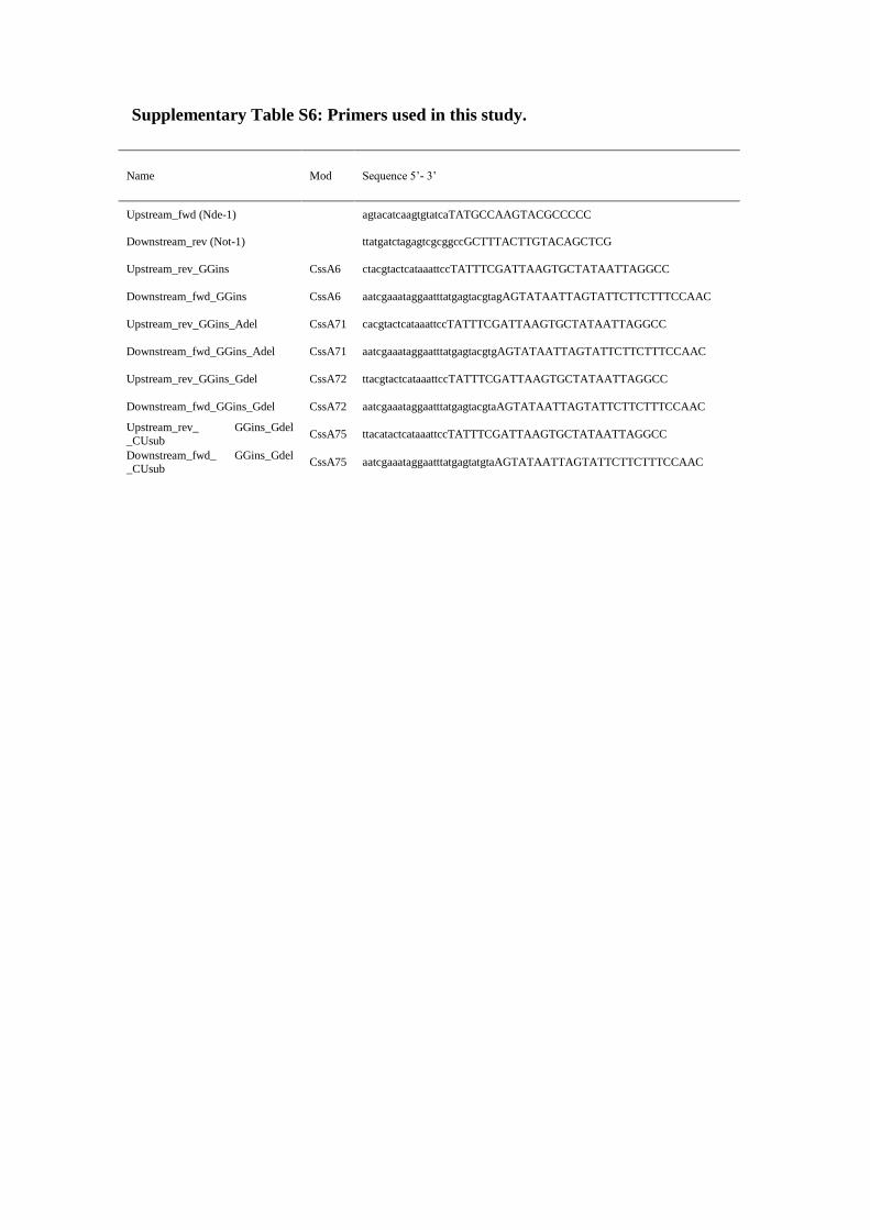
Generation of modified plasmids
NEBuilder® assembly tool was used to design primers using pEGFP-N2_S3_GFP
plasmid as a template per the manufacturer’s instructions. Overlap primers were
designed to include the mutated residues (Supplementary Tables S6 and S7).
Upstream and downstream fragments for each modification were ligated into pEGFP
plasmid digested with Not-1 and Nde-1 using the Gibson Assembly® master mix
(New England Biolabs, UK) as per manufacturer’s instructions. Assembled products
were transformed into E. coli DH5α.
Bacteria and growth conditions
Escherichia coli was grown in Luria-Bertani (LB) broth (2% w/v in dH2O, Oxoid,
UK) or on LB agar (1% w/v) plates. All liquid bacterial cultures were grown in 5 ml
of media inoculated from a single colony overnight at 37 ˚C with shaking (180
r.p.m.). Bacteria grown overnight were diluted 1/100 in media and grown to an abs600
of ~0.4 in the presence of Kanamycin (50µg/ml).
SDS-PAGE and Western blotting
Proteins were separated on polyacrylamide gels alongside Precision Plus All Blue
markers (Bio-Rad, USA) and transferred to immobolin-P polyvinylidine fluoride
(PVDF) membranes (Millipore, USA) using the semi-dry transfer system (Trans-Blot
Turbo system, Bio-Rad, USA). For Western blot analysis, membranes were washed
three times in 0.05% (w/v) dry milk/PBS with 0.05% (v/v) Tween-20 for 10 minutes,
then incubated with the primary antibody for one hour. Membranes were washed
again three times and incubated for a further hour with a secondary, HRP-conjugated
antibody. Binding was detected with an Amersham ECL Western Blotting Detection
kit and exposed to ECL Hyperfilm (GE Healthcare). An α-GFP mouse antibody (JL-
8, Clontech) was used at a final dilution of 1:16,000, α-RecA rabbit antibody (Abcam,
UK) was used at a final dilution of 1:5,000. Secondary Goat α-rabbit IgG HRP-
conjugated antibody (Santa Cruz, UK) and Goat α-mouse IgG HRP-conjugated
antibody (Dako, UK) were used at a final dilution of 1:3,000.
NMR spectral assignments
Base paired residues and exchangeable protons were identified based on imino-imino
and Cytidine NH2-Guanine NH1 and Adenine H2-Uridine H3 patterns in 2D 1H-
1H-
NOESYs (120 ms mixing time) acquired in H2O at both 7°C and 15°C. Sequential
assignments of non-exchangeable protons followed typical H1′-H6/H8 NOE patterns
observed in 2D 1H-
1H NOESYs (120ms mixing time) acquired in D2O at 7°C and
25°C. Further validation and structural restraints were obtained from H2′-H6/H8 and
H1′-H2′ NOEs of samples incorporating rNTPs deuterated at the ribose H3′, H4′, and
H5′/5′′ and pyrimidine base H5. Pyrimidine H5/H6 resonances were identified from 1H-
1H TOCSYs (30 and 70ms) acquired in D2O. Correlated
15N and
13C chemical
shifts were determined from 1H-
15N HSQC,
1H-
13C HSQC, and
1H-
13C edited
NOESY-HSQC experiments. Additional assignments for the UUCG tetraloop were
correlated with chemical shift values from previously reported structures (BMRB
5705).
Spectral assignments for CssA2 were guided by the assignments of the three
smaller fragments (Supplementary Figure S1). Imino patterns for the base pairs were
verified by comparison of the H2O 1H-
1H NOESY for CssA3, CssA4 and CssA5
acquired on Bruker Avance 800 MHz and 7°C. The 1H-
1H NOESY of CssA was done
on deuterated nucleotides acquired in the same conditions. The H1′-H6/H8 walk was
validated in a similar way by comparing the D2O 1H-
1H NOESYs. Non-exchangeable
protons at the positions H1′, H2′′, H5, H6, H8, and H2; the exchangeable imino
protons and the corresponding nitrogen atoms, the exchangeable NH2 protons, and
H3′ proton when resonances were obtained, but carbon resonances were not fully
assigned for CssA2 because increased relaxation broadened the peaks too
significantly.
Specific challenges and solutions:
CssA3: Assignments for CssA3 were somewhat challenging due to inherent
dynamics near the internal bulge (A5 and G6) and the upper bulge formed by U14,
U15, U28, C29, and C30. In the lower helix, weak base pairing of U4-A39 was
apparent from A39 (H2)-U4 (H3) NOEs at 7°C and the typical cross-strand and
sequential NOEs to A4H1′, A5H1′, and U40H1′, but base-pairing could not be
established for A7-U37 despite the typical cross strand and sequential NOEs from the
A7H2 to U37H1′, U38H1′ and G8H1′. Neither of the G3-U40 or G6-U38 wobble
pairs could be identified. A13-U31 showed typical cross-strand and sequential AH2 to
H1′ NOEs but no observable H2 to H3 NOE or HNN-COSY(6) transfer. At low
temperature (7°C), U15 and U28 form a U-U base pair which could be established
from the cross-strand NOEs from U15 (H3) to U28(H3) and U27(H3) (Supplementary
Figure S2). Assignments of CssA3 resulted in the identification of 56.4% of
nonexchangeable protons (all H1′, H2′′, H5, H6, H8, H2, and 35% of H3′) and 55% of
the exchangeable protons excluding the H2′ hydroxyl (70% of imino protons), 8.5%
of the ribose carbons (43 % of the C1′), 64% of the base carbons (C5, C6, C8, and
C2) and 54% of the imino nitrogens.
CssA4: The assignments of CssA4 were straightforward with a clear imino-
imino walk throughout the RNA. Strong NOEs from 9 AU and 2 GU base pairs in the
helix observed at 25 °C indicate the molecule is fully base paired. Additionally, the
unusual AC mismatch is also apparent even at 25 °C (Supplementary Figure S3).
Assignment of CssA4 resulted in identification of 61.5% of nonexchangeable protons
(all H1′, H2′′, H5, H6, H8, H2, and 72% of H3′) and 60% of the exchangeable protons
excluding H2′ (85% of H1 and H3 iminos), 13% of the ribose carbons (entirely from
assignment of 64% of the C1′), 81% of the base carbons (C5, C6, C8, and C2) and
85% of the imino nitrogen.
CssA5: CssA5 iminos could be assigned for the upper and lower helices
flanking C9-G13 and U32-A36; however the A-C mismatch could not be established,
as observed instead for CssA4 (Supplementary Figure S4). NMR assignment of
CssA5 resulted in the identification of 51.3% of nonexchangeable ribose and base
protons (all H1′, H2′′, H5, H6, H8, H2) and 41% of exchangeable protons excluding

H2′ (79% of iminos), 43% of the base carbons (C5, C6, C8, and C2) and 86% of the
imino nitrogens. The ribose carbons (C1′) were not assigned for CssA5.
Altogether, transfer of the CssA3, CssA4, and CssA5 assignments to CssA2
resulted in the identification of 56% of nonexchangeable protons (all H1′, H2′′, H5,
H6, H8, H2, and 41% of H3′) and 46% of the exchangeable protons excluding H2′
hydroxyls (82% of imino protons), and 68% of the imino nitrogens.
Structure determination
Experimental constraints
We generated a restraint table for CssA2 by combining distance restraint and
torsion angle restraints from the segments CssA3 and CssA4, while the CssA5
segment was used only for validation. Restraints from CssA3 spanned resonances
G16-U53; those for CssA4 spanned the resonances G1-A14 and U55-C68, while
CssA5 contributed resonances around the A-C mismatch, namely A14-U17 and A52-
U55. Structural calculations were performed first for CssA3 and CssA4, as described
in below, and for CssA5, which was used only for the AC mismatch and the GA
bulge, which were incorporated in the calculation of CssA2 (Supplementary Figure
S4).
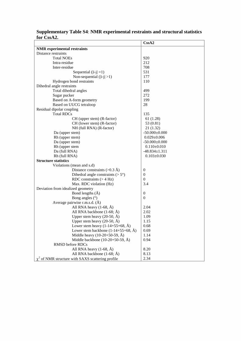
The NMR experimental constraints are summarized in Supplementary Table
S4. We obtained a total of 920 NOEs to calculate the structure of CssA2, which
includes 212 intra-residue, 531 sequential and 177 non-sequential NOE restraints
derived from NOESY spectra (mixing times of 50, 120, and 250ms). Distance
constraints were classified as strong (1.8-3.0 Å), medium (1.8-4.0 Å), weak (1.8-5.0
Å) and very weak (3.5-6.5 Å for NOEs observed in H2O spectra and 1.8-6.5 Å for
D2O NOEs) based on their peak intensities and comparison with peaks corresponding
to known interproton distances.
A total of 110 H-bond constraints were also used. The observation of JNN
couplings from HNN-COSY experiments were the predominant means of validating
hydrogen bonding restraints; however, restraints for weak base pairs (A-U, A-C, and
U-U base pairs) were also incorporated based on characteristic NOE patterns
observed in 2D 1H-
1H NOESYs (120ms mixing time recorded at 7°C and 25°C).
Potential A-U base pairs showing typical cross-strand H1′-H2, sequential H1′-H2, and
strong AH2-UH3 imino NOEs were considered base paired despite the lack of HNN-
COSY transfer.
Residual dipolar couplings (RDCs) were measured from modulation of the
resonance intensity between 1H-
13C or
1H-
15N couplings acquired for isotropic and
partially aligned samples using the ARTSY pulse sequence. RDCs were obtained for
the following internuclear vectors: H8-C8, H6-C6, H5-C5, H2-C2 (Adenine), H1′-C1′
(ribose), and 1H-
15N (base imino resonances).
Several less clearly defined base pairs could be established confidently from
the analysis of the smaller RNA segments due to more favorable spectral
characteristics. In the lower helix, base-pairing of U4-A39 was apparent from
U4(H3)-A39(H2) NOEs but the A7-U37 and A13-U31 base pair could not be
confirmed through inter-strand imino NOEs or HNN-COSY transfer, despite the
typical cross strand and sequential NOEs from the A7H2 to U37H1′, U38H1′ and
G8H1′. Neither the G3-U40 nor G6-U38 could be identified conclusively, but the
U15-U28 base pair could be established from the unusual cross-strand NOEs from
U15 (H3) to U28(H3) and U27(H3) (Figure 5E). However, base pairs found within

CssA4 could be easily assigned in the full RNA, identifying 9 AU and 2 GU base
pairs, and an AC mismatch apparent even at 25 °C (Supplementary Figure S3).
The sugar conformation and population of the major C2′-endo and C3′-endo
conformers were estimated from the observed peak intensity in DQF-COSY and 1H-
1H TOCSY (30ms and 70ms) of unlabeled samples in D2O. A-form torsion angles for
β, γ, and ε were assumed for all nucleotides that produced typical A-form NOE
patterns; nucleotides with atypical NOE patterns or that appeared to experience
conformational exchange were left unrestrained. The χ angle of unpaired bases were
constrained in the anti or syn conformation on the basis of the intensity of H1′-
H5/H6/H8 NOEs. Residues within the UUCG tetraloop were restrained with the
known UUCG tetraloop torsion angles for β, γ, δ, ε, and χ, obtained from the analysis
of structure deposited in the PDB.
Structure refinement
CssA2 structure refinement
CssA2 refinement was performed in two steps. First, local RDCs for the upper
and lower helices were introduced from CssA3 and CssA4, respectively, to regularize
the local structure. Second, global RDCs measured on CssA2 NH’s were introduced
to orient the upper and lower stems relative to each other. Both refinement procedures
were similar to that described below for CssA3 and CssA4, with the exception of the
initial bath temperature for torsion angle dynamics. The first refinement protocol
began with starting coordinates read from the lowest energy structure following the
initial CssA2 simulated annealing protocol. The first round of RDCs refinement
incorporated 1DCH and
1DNH of CssA3 and CssA4, with a final force constant of 0.25
kcal mol-1
Hz-1
, allowing Da and R to vary. High temperature dynamics at 2500K was
done to adjust the local environment of the upper and lower helices, then the bath was
cooled to 298K over 200 steps in 0.4ps, followed by Powell minimizations in torsion
angle and Cartesian space. The top 10 structures from the initial refinement were then
subjected to additional rounds of refinement to generate 10 structures each with
different seeds, for a total of 100 final structures.
The final round of CssA2 refinement used CssA3 and CssA4 1DCH for
retaining the local geometry, while 1DNH measured on CssA2 were used to globally
orient the upper and lower helices. The tensors for all RDCs were allowed to vary;
high temperature dynamics was conducted at 1500K to allow for reorientation of the
stems and cooled to 298K. The top 10 structures without violations in distance (>
0.3Å) or torsion angle (>5°) restraints were retained and used for further analysis
(Supplementary Table S4).
CssA3 and CssA4 refinement
Structure refinement for the segments also makes use of both torsion angle
dynamics and Cartesian coordinate minimization. The refinement protocol uses a
number of potential terms found in the simulated annealing protocol: NOEs and
Hydrogen bonds (final force constant = 50 kcal mol-1
Å2), dihedral torsion angles
(final force constant = 200 kcal mol-1
rad-2
), van der Waals repulsion (final force
constant of 4 kcal mol-1
Å2 and with a van der Waals radius scale factor of 0.8),
angles (final scale factor of 1; with a maximum deviation of 5°), impropers (final
scale factor of 1; with a maximum deviation of 5°), and RAMA (with final weighting
of 1). Additionally, the refinement protocol includes further potential terms for
correcting both local and overall geometry: planarity restraints for base pair
interactions (weight factor = 50), the ORIE database for relative positioning of base
pairs (final scale factor = 0.2), and residual dipolar couplings for orienting the entire
molecule (final force constant = 0.75 kcal mol-1
Hz-1
, with force constants for 1DCH
scaled to that for 1DNH). The starting coordinates are read from the lowest energy
structure without violations following the initial simulated annealing protocol. During
the first rounds of refinement, the RDC tensor was allowed to vary in order to find the
optimum values of Da and R; during the final structure calculations, these values were
fixed.
The procedure for refinement followed a simple protocol: initial high
temperature dynamics using only the phosphate-phosphate non-bonded terms,
followed by high temperature torsion angle dynamics (10,000 steps at 3,000K). The
bath temperature was gradually reduced from 3,000 to 298 K over 200 steps with
increases in a number of force constants (angles, impropers, dihedral angles, NOEs,
van der Waals repulsion, RAMA, ORIE, and RDCs). Cooling occurs over 200 steps
and, at each step, torsion angle dynamics is ran for 0.4ps followed by Powell
minimizations in torsional angle and Cartesian space. The calculation was repeated
for 100-200 structures and the 10 lowest energy structures without violations in
distance (> 0.3Å) or torsion angle (>5°) restraints were retained for analysis
(Supplementary Table S5).
CssA5 Refinement:
Refinement of CssA5 was limited to distance and torsion angle restraints. Refinement
followed the same protocol as described for CssA3 and CssA4 but omits
incorporation of RDCs, the RDC force constant, and evaluation of the tensor variables
Da and R. A full refinement was not undertaken because the flexibility of the
molecule reduced the usefulness of RDCs. The calculations were repeated for 100-
200 structures and the 10 lowest energy structures were retained for analysis.

Supplementary Table S1: Sequences of the RNA molecules used in the present
study.
S.No. Name of
the RNA
Length
(nt)
RNA Sequence
1 CssA 110 GGCCUAAUUAUAGCACUUAAUCGAAAUAAAUUUAUGAGUACGU
AGAGUAUAAUUAGUAUUCUUCUUUCCAACUUCCUUAUACUUAU
AUACUUAUAGAUUCUAAAAUCAUG
2 CssA-
Δ8bp
102 GGCCUAAUUAUAGCACUUAAUCGAAAUAAAUUUAUGAGUACGU
AGAGUAUAAUUAGUAUUCUUCUUUCCAACUUCCUUAUACUUAU
AGAUUCUAAAAUCAUG
3 CssA1 75 GGAAUUUAUGAGUACGUAGAGUAUAAUUAGUAUUCUUCUUUCC
AACUUCCUUAUACUUAUAUACUUAUAGAUUCC
4 CssA2 68 GGAAUUUAUGAGUACGUAGAGUAUAAUUAGUCUUCGGACUUCC
UUAUACUUAUAUACUUAUAGAUUCC
5 CssA3 42 GGGUAGAGUAUAAUUAGUCUUCGGACUUCCUUAUACUUAUCC
6 CssA4 36 GGAAUUUAUGAGUACCUUCGGAUACUUAUAGAUUCC
7 CssA5 43 GGUGAGUACGUAGAGUAUACUUCGGUAUACUUAUAUACUUACC
8 CssA6 73 GGAAUUUAUGAGUACGUAGAGUAUAAUUAGUAUUCUUCUUUCC
AACUUCCUUAUACUUAUAUACUUAUAGAUU
9 CssA7 66 GGAAUUUAUGAGUACGUAGAGUAUAAUUAGUCUUCGGACUUCC
UUAUACUUAUAUACUUAUAGAUU
10 CssA71 72 GGAAUUUAUGAGUACGUGAGUAUAAUUAGUAUUCUUCUUUCCA
ACUUCCUUAUACUUAUAUACUUAUAGAUU
11 CssA72 72 GGAAUUUAUGAGUACGUAAGUAUAAUUAGUAUUCUUCUUUCCA
ACUUCCUUAUACUUAUAUACUUAUAGAUU
12 CssA73 70 GGAAUUUAUGAGUACGUAGUAUAAUUAGUAUUCUUCUUUCCAA
CUUCCUUAUACUAUAUACUUAUAGAUU
13 CssA74 73 GGAAUUUAUGAGUAUGUAGAGUAUAAUUAGUAUUCUUCUUUCC
AACUUCCUUAUACUUAUAUACUUAUAGAUU
14 CssA75 72 GGAAUUUAUGAGUAUGUAAGUAUAAUUAGUAUUCUUCUUUCCA
ACUUCCUUAUACUUAUAUACUUAUAGAUU
15 CssA76 73 GGAAUUUAAUUCAUAUUAGAGUAUAAUUAGUAUUCUUCUUUCC
AACUUCCUUAUACUUAGCAUGAGUUAGAUU
16 CssA77 73 GGAAUUUAUGAGUACGAUUCAUAUAUUAGUAUUCUUCUUUCCA
ACUUCCUAUAUGAGAUUAUACUUAUAGAUU
17 CssA78 73 GGAAUUUAAUUCAUAUAUUCAUAUAUUAGUAUUCUUCUUUCCA
ACUUCCUAUAUGAGAUGCAUGAGUUAGAUU
18 CssA6-
Δ8bp
65 GGAAUUUAUGAGUACGUAGAGUAUAAUUAGUAUUCUUCUUUCC
AACUUCCUUAUACUUAUAGAUU
19 CssA6-
Δ8bp
(AU/GC)
65 GGAAUUUAUGAGUACGUAGAGUAUAAUUAGUAUUCUUCUUUCC
AACUUCCUUAUGCCUAUAGAUU
20 CssA6-
Δ8bp
(A/G)
65 GGAAUUUAUGAGUACGUAGAGUAUAAUUAGUAUUCUUCUUUCC
AACUUCCUUAUGCUUAUAGAUU
21 CssA6-
Δ8bp
(U/C)
65 GGAAUUUAUGAGUACGUAGAGUAUAAUUAGUAUUCUUCUUUCC
AACUUCCUUAUACCUAUAGAUU

Supplementary Table S2: Thermodynamic properties and translational activity
of mutations introduced to alter the stability of the CssA RNA thermometer.
S. No. Name of the RNA* Melting point (Tm, °C) Activity
1 CssA 39.0, 69.0 +
2 CssA6 40.6, 71.0 +
3 CssA71 41.6/69.8 +
4 CssA72 43.6/69.4 +
5 CssA73 43.4/69.8 ND
6 CssA74 40.1/70.0 ND
7 CssA75 44.9/71.3 +
8 CssA76 35.7/67.8 ND
9 CssA77 41.2/70.0 ND
10 CssA78 34.6/66.7 ND
11 CssA6- Δ8bp 37.2/69.8 Loh E. Nature (2013)(7)
12 CssA6- Δ8bp (AU/GC) 40.5, 69.6 Loh E. Nature (2013)(7)
13 CssA6- Δ8bp (A/G) 40.4, 69.3 Loh E. Nature (2013)(7)
14 CssA6- Δ8bp (U/C) 35.5/69.4 Loh E. Nature (2013)(7)
*Complete RNA sequences can be found in Supplementary Table S1
**ND- not determined
Supplementary Table S3: Thermodynamic melting points of the different RNAs
used in this study
. Name of the
RNA*
Melting point (Tm, °C)
1 CssA 39.0, 69.0
2 CssA-Δ8bp 35.0, 64.6
3 CssA1 45.2, 70.1
4 CssA2** 50.5
5 CssA3** 46.3
6 CssA4** 59.2
7 CssA5** 59.1
8 CssA6# 40.6, 71.0
9 CssA7**,# 46.0
10 CssA71# 41.6/69.8
11 CssA72# 43.6/69.4
12 CssA73# 43.4/69.8
13 CssA74# 40.1/70.0
14 CssA75# 44.9/71.3
15 CssA76# 35.7/67.8
16 CssA77# 41.2, 70.3
17 CssA78# 34.6/66.7
18 CssA6- Δ8bp 37.2/69.8
19 CssA6- Δ8bp
(AU/GC)
40.5, 69.6
20 CssA6- Δ8bp
(A/G)
40.4, 69.3
21 CssA6- Δ8bp
(U/C)
35.5/69.4
*Complete RNA sequences can be found in Supplementary Table S1
Note: **- with UUCG tetraloop, #- with two additional GG nucleotides at the 5′-end
Supplementary Table S4: NMR experimental restraints and structural statistics
for CssA2. CssA2
NMR experimental restraints
Distance restraints
Total NOEs
Intra-residue
Inter-residue
Sequential (|i-j| =1)
Non-sequential (|i-j| >1)
Hydrogen bond restraints
Dihedral angle restraints
Total dihedral angles
Sugar pucker
Based on A-form geometry
Based on UUCG tetraloop
Residual dipolar coupling
Total RDCs
CH (upper stem) (R-factor)
CH (lower stem) (R-factor)
NH (full RNA) (R-factor)
Da (upper stem)
Rh (upper stem)
Da (upper stem)
Rh (upper stem
Da (full RNA)
Rh (full RNA)
Structure statistics
Violations (mean and s.d)
Distance constraints (>0.3 Å)
Dihedral angle constraints (> 5°)
RDC constraints (> 4 Hz)
Max. RDC violation (Hz)
Deviation from idealized geometry
Bond lengths (Å)
Bong angles (°)
Average pairwise r.m.s.d. (Å)
All RNA heavy (1-68, Å)
All RNA backbone (1-68; Å)
Upper stem heavy (20-50, Å)
Upper stem heavy (20-50, Å)
Lower stem heavy (1-14+55+68, Å)
Lower stem backbone (1-14+55+68, Å)
Middle heavy (10-20+50-59, Å)
Middle backbone (10-20+50-59, Å)
RMSD before RDCs
All RNA heavy (1-68, Å)
All RNA backbone (1-68; Å)
2 of NMR structure with SAXS scattering profile
920
212
708
531
177
110
499
272
199
28
135
61 (1.28)
53 (0.81)
21 (1.32)
-50.000±0.000
0.029±0.006
-50.000±0.000
0.110±0.010
-48.834±1.311
0.103±0.030
0
0
0
3.4
0
0
2.04
2.02
1.09
1.15
0.68
0.69
1.14
0.94
8.20
8.13
2.34

Supplementary Table S5: NMR Structural statistics for CssA3, CssA4 and
CssA5
CssA3
Distance restraints
Total NOEs
Intra-residue
Inter-residue
Sequential (|i-j| =1)
Non-sequential (|i-j| >1)
Hydrogen bonds
475
108
367
279
88
64
Dihedral angle restraints
Total dihedral angles
Sugar pucker
Based on A-form geometry
Based on UUCG tetraloop
310
168
114
28
Residual dipolar coupling
Total RDCs
CH (R-factor)
NH (R-factor)
Da
Rh
64
55 (0.29)
9 (1.52)
28.150±0.027
0.093±0.000
Structure statistics
Violations (mean and s.d)
Distance constraints (>0.3 Å)
Dihedral angle constraints (> 5°)
RDC constraints (> 4 Hz)
Max. RDC violation (Hz)
Deviation from idealized geometry
Bond lengths (Å)
Bong angles (°)
Average pairwise r.m.s.d. (Å)
All RNA heavy (1-42, Å)
All RNA backbone (1-42; Å)
RNA upper stem heavy (15-28, Å)
RNA upper stem backbone (15-28, Å)
RNA lower stem heavy (1-12+32-42, Å)
RNA lower stem backbone (1-12+32-42, Å)
RMSD before RDC
All RNA heavy (1-42, Å)
All RNA backbone (1-42; Å)
RNA upper stem heavy (15-28, Å)
RNA upper stem backbone (15-28, Å)
RNA lower stem heavy (1-12+32-42, Å)
RNA lower stem backbone (1-12+32-42, Å)
2 of NMR structure with SAXS scattering profile
0
0
0
3.6
0
0
1.72
1.64
0.74
0.76
1.29
1.13
6.11
5.72
0.48
0.48
3.68
3.56
5.16

CssA4
Distance restraints
Total NOEs
Intra-residue
Inter-residue
Sequential (|i-j| =1)
Non-sequential (|i-j| >1)
Hydrogen bonds
544
129
415
297
118
74
Dihedral angle restraints
Total dihedral angles
Sugar pucker
Based on A-form geometry
Based on UUCG tetraloop
255
144
88
23
Residual dipolar coupling
Total RDCs
CH (R-factor)
NH (R-factor)
Da
Rh
90
73 (0.28)
17 (1.18)
25.324±0.469
0.025±0.000
Structure statistics
Violations (mean and s.d)
Distance constraints (>0.3 Å)
Dihedral angle constraints (> 5°)
RDC constraints (> 4 Hz)
Max. RDC violation (Hz)
Deviation from idealized geometry
Bond lengths (Å)
Bong angles (°)
Average pairwise r.m.s.d. (Å)
All RNA heavy (1-36, Å)
All RNA backbone (1-36; Å)
RMSD before RDC
All RNA heavy (1-36, Å)
All RNA backbone (1-36; Å)
2 of NMR structure with SAXS scattering profile
0
0
0
1.2
0
0
0.70
0.56
2.50
2.29
3.02

CssA5
Distance restraints
Total NOEs
Intra-residue
Inter-residue
Sequential (|i-j| =1)
Non-sequential (|i-j| >1)
Hydrogen bonds
456
102
354
265
89
72
Dihedral angle restraints
Total dihedral angles
Sugar pucker
Based on A-form geometry
Based on UUCG tetraloop
299
172
109
18
Residual dipolar coupling
Total RDCs
CH
NH
Da
Rh
0
0
0
Not Applicable
Not Applicable
Structure statistics
Violations (mean and s.d)
Distance constraints (>0.3 Å)
Dihedral angle constraints (> 5°)
RDC constraints (> 4 Hz)
Max. RDC violation (Hz)
Deviation from idealized geometry
Bond lengths (Å)
Bong angles (°)
Average pairwise r.m.s.d. (Å)
All RNA heavy (1-43, Å)
All RNA backbone (1-43; Å)
RNA upper stem heavy (15-30, Å)
RNA upper stem backbone (15-30, Å)
RNA lower stem heavy (1-7+37-43, Å)
RNA lower stem backbone (1-7+37-43, Å)
RMSD before RDCs
2 of NMR structure with SAXS scattering profile
0
0
Not Applicable
Not Applicable
0
0
2.57
2.47
0.49
0.45
0.47
0.44
Not Applicable
Not Applicable
Supplementary Table S6: Primers used in this study.
Name Mod Sequence 5’- 3’
Upstream_fwd (Nde-1) agtacatcaagtgtatcaTATGCCAAGTACGCCCCC
Downstream_rev (Not-1) ttatgatctagagtcgcggccGCTTTACTTGTACAGCTCG
Upstream_rev_GGins CssA6 ctacgtactcataaattccTATTTCGATTAAGTGCTATAATTAGGCC
Downstream_fwd_GGins CssA6 aatcgaaataggaatttatgagtacgtagAGTATAATTAGTATTCTTCTTTCCAAC
Upstream_rev_GGins_Adel CssA71 cacgtactcataaattccTATTTCGATTAAGTGCTATAATTAGGCC
Downstream_fwd_GGins_Adel CssA71 aatcgaaataggaatttatgagtacgtgAGTATAATTAGTATTCTTCTTTCCAAC
Upstream_rev_GGins_Gdel CssA72 ttacgtactcataaattccTATTTCGATTAAGTGCTATAATTAGGCC
Downstream_fwd_GGins_Gdel CssA72 aatcgaaataggaatttatgagtacgtaAGTATAATTAGTATTCTTCTTTCCAAC
Upstream_rev_ GGins_Gdel
_CUsub CssA75 ttacatactcataaattccTATTTCGATTAAGTGCTATAATTAGGCC
Downstream_fwd_ GGins_Gdel
_CUsub CssA75 aatcgaaataggaatttatgagtatgtaAGTATAATTAGTATTCTTCTTTCCAAC

Supplementary Table S7: Plasmids used in this study
Name Modification Plasmid
S3_GFP Wild Type pEGFP-N2
S3_CssA6_GFP GG insertion pEGFP-N2
S3_CssA71_GFP A deletion pEGFP-N2
S3_CssA72_GFP G deletion pEGFP-N2
S3_CssA75_GFP G deletion and C-U substitution pEGFP-N2

Supplementary Figure Legends:
Supplementary Figure S1: NMR Assignments for CssA2 and superposition of its
fragments CssA3, CssA4, and CssA5. H1′-H6/H8 assignment ‘walk’ for CssA2.
Spectral assignments matched to the smaller constructs (CssA3/4/5) are shown with
various colored lines. CssA3 assignments are colored in red; those for CssA4 are
shown in green and assignments for CssA5 are presented in magenta color.
Supplementary Figure S2: Assignment and NMR structure ensemble of CssA3.
(A) Complete H1′-H6/H8 walk in 2D 1H-
1H NOESY recorded in 99.9 %
2H2O on
Bruker Avance 800 MHz spectrometer at 25 °C. Ten out of 14 iminos were observed,
excluding the G1-C43 terminal base pair that frays. C2′-endo sugar pucker is
observed only in the tetraloop. (B) Superposition of 10 final NMR structures of
CssA3 with backbone rmsd superposition of 1.6 Å, calculated with NOEs and RDCs
constraints.
Supplementary Figure S3: Assignment and NMR structure ensemble of CssA4.
(A) Complete H1′-H6/H8 walk in 2D 1H-
1H NOESY recorded in 99.9 %
2H2O on
Bruker Avance 800 MHz spectrometer at 25 °C. All imino peaks predicted from the
secondary structure are observed. C2′-endo sugar pucker is observed only in the
tetraloop. (B) Superposition of 10 final NMR structures of CssA4 with backbone
rmsd superposition of 0.6 Å calculated with NOEs and RDCs constraints.
Supplementary Figure S4: Assignment and NMR structure ensemble of CssA5. (A) Complete H1′-H6/H8 walk in 2D
1H-
1H NOESY recorded in 99.9 %
2H2O on
Bruker Avance 800 MHz spectrometer at 25 °C. 15 out of 18 predicted iminos were
observed, excluding the terminal G1-C43 pair. C2′-endo sugar pucker is observed
only in tetraloop. (B) NMR ensemble structure of CssA5 with backbone rmsd
superposition of 2.5 Å calculated with NOE constraints only but without RDC
refinement.
Supplementary Figure S5: The wild type CssA thermometer forms a dimeric
structure at the concentrations required for chemical physical characterization.
(A) Results of small angle X-ray scattering (SAXS) analysis of the CssA1 and CssA2
variants. CssA1 has an extended shape with a long dimension of 140 Å,
approximately twice what is expected from the secondary structure of the stem-loop,
whereas CssA2, is around 67 Å long, the dimension expected for a monomeric RNA.
(B) Thermal melting of CssA1 and CssA2. CssA1 has two transitions with melting
temperatures of 45.2 and 70.1 °C, whereas a single transition with melting
temperature of 50.5 °C is observed for CssA2. Even at the concentration of these
experiments (about 1000-fold less than NMR), the CssA1 and CssA thermometers are
partially dimeric; the higher melting transition corresponds to the breaking of the
dimeric structure. (C) SHAPE studies on CssA1 and CssA2 executed in the
temperature range 4-42 °C, similar to the condition used for the CssA RNA. The
lower panel highlights nucleotides sensitive to SHAPE chemistry as a function of
temperature for both CssA1 and CssA2. (D) An overlay of 1H-
1H NOESYs recorded
in 95% H2O/5% 2H2O solution, depicting very similar imino spectra (except for the
UUCG tetraloop) for CssA1 and CssA2.

Supplementary Figure S6: SHAPE analysis of CssA-Δ8bp RNA. (A) Predicted
secondary structure of Δ8bp RNA; bold residues depict the single 8bp sequence
followed by the red-colored RBS. A81 and U83 are labeled in blue. (B) Selective 2′-
OH acylation analyzed by primer extension (SHAPE) for the Δ8bp RNA performed at
different temperatures, well below and above activation of protein synthesis (4-42
°C). A81 and U83 are shown with blue colored arrow.
Supplementary Video Legends:
Supplementary Video S1: Morphing of conformational changes within the core
of CssA thermometer near the apical loop and middle region during temperature
elevation. The CssA RNA core is represented with blue colored oval cartoon. The
RBS is shown with grey colored spheres whereas two copies of 8bp are shown with
orange and forest colored spheres. This morph video was created using in-house script
in pymol 1.8.0.

References
1. Darty, K., Denise, A. and Ponty, Y. (2009) VARNA: Interactive drawing and editing of the RNA secondary structure. Bioinformatics, 25, 1974-1975.
2. Zuker, M. (2003) Mfold web server for nucleic acid folding and hybridization prediction. Nucleic acids research, 31, 3406-3415.
3. Wilkinson, K.A., Merino, E.J. and Weeks, K.M. (2006) Selective 2'-hydroxyl acylation analyzed by primer extension (SHAPE): quantitative RNA structure analysis at single nucleotide resolution. Nature protocols, 1, 1610-1616.
4. Laederach, A., Das, R., Vicens, Q., Pearlman, S.M., Brenowitz, M., Herschlag, D. and Altman, R.B. (2008) Semiautomated and rapid quantification of nucleic acid footprinting and structure mapping experiments. Nature protocols, 3, 1395-1401.
5. Reuter, J.S. and Mathews, D.H. (2010) RNAstructure: software for RNA secondary structure prediction and analysis. BMC bioinformatics, 11, 129.
6. Dingley, A.J., Nisius, L., Cordier, F. and Grzesiek, S. (2008) Direct detection of N-H[...]N hydrogen bonds in biomolecules by NMR spectroscopy. Nature protocols, 3, 242-248.
7. Loh, E., Kugelberg, E., Tracy, A., Zhang, Q., Gollan, B., Ewles, H., Chalmers, R., Pelicic, V. and Tang, C.M. (2013) Temperature triggers immune evasion by Neisseria meningitidis. Nature, 502, 237-240.