supplementary material for · patterned ito glass substrates were first cleaned by ultrasonication...
TRANSCRIPT

science.sciencemag.org/content/365/6452/473/suppl/DC1
Supplementary Material for Stabilizing halide perovskite surfaces for solar cell operation with wide-
bandgap lead oxysalts
Shuang Yang, Shangshang Chen, Edoardo Mosconi, Yanjun Fang, Xun Xiao, Congcong Wang, Yu Zhou, Zhenhua Yu, Jingjing Zhao, Yongli Gao, Filippo De Angelis,
Jinsong Huang*
*Corresponding author. E-mail: [email protected]
Published 2 August 2019, Science 365, 473 (2019) DOI: 10.1126/science.aax3294
This PDF file includes:
Materials and Methods Figs. S1 to S16 Tables S1 to S5 References
Other Supplementary Material for this manuscript includes the following: (available at science.sciencemag.org/content/365/6452/473/suppl/DC1)
Movie S1

2
Materials and Methods
Device fabrication
Patterned ITO glass substrates were first cleaned by ultrasonication with soap,
acetone and isopropanol. The hole transport layer poly(bis(4-phenyl)(2,4,6-
trimethylphenyl)amine) (PTAA) with a concentration of 2 mg ml-1 dissolved in toluene
was spin-coated at the speed of 4,000 rpm for 35 s and then annealed at 100 oC for 10
min. Before depositing perovskite films, the PTAA film was pre-wetted by spinning 80
µl DMF at 4,000 rpm for 15 s to improve the wetting property of the perovskite precursor
solution. The perovskite precursor solution composed of mixed cations (lead (Pb),
cesium(Cs), formamidinium (FA) and methylammonium (MA)) and halides (I, Br) was
dissolved in mixed solvent (DMF/DMSO = 4:1) with a chemical formula of
Cs0.05FA0.81MA0.14PbI2.55Br0.45. Then 80 µl precursor solution was spun onto PTAA at
2,000 rpm for 2 s and 4,000 rpm for 20 s, and the film was quickly washed with 130 µl
toluene at 18 s during spin-coating. Subsequently, the sample was annealed at 65 oC for
10 min and 100 oC for 10 min. The ammonium sulfate solution was prepared by
dissolving methylamine (Sigma-Aldrich, 33 wt. % in ethanol) or octylamine ((Sigma-
Aldrich, 99 %) and sulfuric solution (Sigma-Aldrich, 99.999%) in mixed solvents
(toluene/isopropanol = 5:1) with the concentration of 4 mM. The sulfate precursor
solutions at different concentrations were prepared by serial dilution method.
Specifically, the amines and sulfuric solution were firstly diluted twice to a low
concentration by the mixed solvents. Then, the diluted amine and sulfuric solution were
mixed with certain amount of solvents to a desired concertation. To treat the surface of
perovskite films, 100 µl of precursor solution was loaded on the film for 20 s and was
then spun at 6,000 rpm for 30 s. During spin-coating process, extra 130 µl of toluene was
dropped to wash the unreacted precursors. The devices were finished by thermally
evaporating C60 (30 nm), BCP (8 nm) and copper (140 nm) in sequential order.
Encapsulation
A thin layer of CYTOP was firstly coated onto the back surface of device by blade
coating, followed by annealing at 75 oC for 45 min on a hot plate. Then, a cover glass
was attached onto the back surface for further protection by epoxy resin.
Surface treatment of single crystals
MAPbI3 single crystals were treated by dipping in 4 mM octylammonium sulfate
solution, followed by washing with toluene. The dipping time is 5 min and 20 s for the
water resistant and temperature-dependent conductivity tests, respectively. The sample
was then thermal annealed for 10 min in an oven at 100 °C.
Structure characterization
Crystallographic information for the as-synthesized crystals was obtained by a
Rigaku D/Max-B X-ray diffractometer with Bragg-Brentano parafocusing geometry, a
diffracted beam monochromator, and a conventional cobalt target X-ray tube set to 40 kV
and 30 mA. Cross-section FIB sample lamellae was prepared on a FEI Quanta 3D FEG
instrument, and a low beam current was applied to minimize ion beam damage to the
devices. The FIB lamellae were targeted with a final thickness of about 100 nm but this

3
may vary locally. Transmission electron microscopy (TEM) and scanning TEM were
performed on a FEI Talos F200X analytical scanning transmission electron microscope
operating at 200 kV, and a low electron dose was applied to minimize the electron beam
damage. The X-ray photoelectron spectroscopy (XPS) was measured (SPECS XR-MF)
by using a monochromatized Al source (hv=1486.6 eV). The Fourier transform infrared
(FT-IR) spectra of perovskite powder were collected in the transmittance mode on the
PerkinElmer IR spectrometer instrument in the 400 - 4,000 cm-1 region. The morphology
and structure of the samples were characterized by Quanta 200 FEG environmental
scanning electron microscope.
Optical characterization
Optical absorption spectra were measured by means of an Evolution 201/220
UV/visible Spectrophotometer. Time-resolved photoluminescence (TRPL) was
performed on the perovskite films grown on varied substrates by a Horiba DeltaPro
fluorescence lifetime system, which equipped with a DeltaDiode (DD-405) pulse laser
diode with wavelength of 404 nm. The laser excitation energy in the measurement was 20
pJ pulse-1.
Ion migration studies
Activation energy for ion migration was tested using lateral devices by a Keithley
2400 source meter at different temperatures. The electric field of the lateral device was
0.4 V/μm. The device was set in a Lakeshore Probe Station to obtain desired temperature.
Device characterizations
The J-V analysis of solar cells was performed using a solar light simulator (Oriel
67005, 150 W Solar Simulator) and the power of the simulated light was calibrated to
100 mW cm−2 by a silicon (Si) diode (Hamamatsu S1133) equipped with a Schott visible-
color glass filter (KG5 color-filter). All cells were measured using a Keithley 2400 source
meter with scan rate of 0.1 V s-1. The steady-state PCE was measured by monitoring
current with the largest power output bias voltage and recording the value of the
photocurrent. External quantum efficiency curves were characterized with a Newport QE
measurement kit by focusing a monochromatic beam of light onto the devices. The tDOS
of solar cells were derived from the frequency-dependent capacitance (C-f) and voltage-
dependent capacitance (C-V), which were obtained from the thermal admittance
spectroscopy (TAS) measurement performed by an LCR meter (Agilent E4980A). The
transient photovoltage was measured under 1 sun illumination. An attenuated UV laser
pulse (SRS NL 100 Nitrogen Laser) was used as a small perturbation to the background
illumination on the device. The laser-pulse-induced photovoltage variation and the VOC is
produced by the background illumination. The wavelength of the N2 laser was 337 nm,
the repeating frequency was about 10 Hz, and the pulse width was less than 3.5 ns.
Device operational stability tests
Long-term stability measurements of encapsulated perovskite devices were operated
under a plasma lamp with light intensity equivalent to AM 1.5G (Fig. S9), without
ultraviolet filter in air (relative humidity ∼60±10 %). The temperature of the devices
under illumination was measured to be ~65 °C. All devices were loaded with a resistance

4
so that they worked at maximum power point (MPP) at the beginning of the tests. Since
the sulfate-treated device is rather stable during the whole test, the voltage shift of MPP is
expected to be small. The J-V curves were automatically recorded with reverse scan rate
of 0.1 V s-1 every six hours.
CPMD computational details
To simulate the perovskite surfaces, we have properly cut 2 × 2 × 3 slabs from the
bulk tetragonal MAPbI3 crystal structure, which expose MAI-terminated surface. The
employed periodic cell dimension are a = b = 17.71 Å, corresponding to twice the
tetragonal experimental a = b cell parameters. A layer of 8 PbSO4 units was deposit on
the MAI-terminated perovskite slab and a Car-Parrinello molecular dynamics (32)
(CPMD) simulations have been carried out. The 2 × 2 × 1 (a = 13.918, b = 16.964 Å)
PbSO4 layer was generated by cutting the Anglesite experimental bulk structure (a =
6.959, b = 8.482, c = 5.398 Å). We use this slab dimension to have 8 PbSO4 units
corresponding to the 8 iodide undercoordinated atoms of the surface. By doing so we
have a lattice matching of 127% a and 104%, calculated as a percentage ratio between the
perovskite slab and the PbSO4 slab employed.
Along the c direction, a 10.0 Å of vacuum has been added for the slab calculation.
Quantum Espresso package (33) along with the GGA-PBE functional was used. Electron-
ion interactions were described by scalar relativistic ultrasoft pseudopotentials with
electrons from O, N and C 2s, 2p; H 1s; S 3s, 3p; I 5s, 5p; Pb 6s, 6p, 5d shells explicitly
included in the calculations. Plane-wave basis set cutoffs for the smooth part of the wave
functions and the augmented density were 25 and 200 Ry, respectively. CPMD
simulations have been performed with an integration time step of 10 au, for a total
simulation time of ca. 10 ps. The fictitious mass used for the electronic degrees of
freedom is 1000 au, and we set the atomic masses to an identical value of 5 amu to
enhance the dynamical sampling. Initial ions position randomization has been used to
reach temperature in the range of 350-400K, without further applying any thermostat.
Geometry optimizations of slabs
We also simulate the MAI- and PbI2-terminated interface for a full coverage of
PbSO4 layers (12 units). The MAI-terminated perovskite surface is the same adopted for
the CPMD simulation as specified above while the PbI2-terminate is a 2 × 2 × 5 slab. On
these slabs, we deposited a single layer of 12 PbSO4 units. The 3 × 2 × 1 PbSO4 layer was
generated by cutting the Anglesite experimental bulk structure. In this case, we adopt the
full coverage to evaluate the energetics of the interface formation with a lattice matching
of 85 % along a and 104 % along b.
On these systems, we performed geometry optimization using GGA-PBE functional.
Electron-ion interactions were described by scalar relativistic ultrasoft pseudopotentials
with electrons from O, N and C 2s, 2p; H 1s; S 3s, 3p; I 5s, 5p; Pb 6s, 6p, 5d shells
explicitly included in the calculations. Plane-wave basis set cutoffs for the smooth part of
the wave functions and the augmented density were 25 and 200 Ry, respectively.
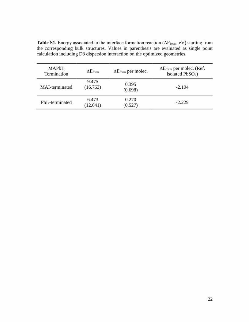
Formation energy (ΔEform) has been calculated as follows, see Table S1:
ΔEform = E(MAPbI3-nPbSO4) – E(MAPbI3) – E(nPbSO4)
Where E(MAPbI3-nPbSO4) is the energy of the interface with n PbSO4 units,
E(MAPbI3) is the energy of the isolated perovskite slab and E(nPbSO4) is the energy of

5
the optimized 1 × 1 × 1 PbSO4 bulk calculated at the same level of theory starting from
the experimental structure and using a k-point sampling grid of 2 × 2 × 2; n represents the
number of PbSO4 units involved. In Table S1 we reported also the ΔEform per molec
calculated as ΔEform/n and the ΔEform per molec referred to energy of the isolated PbSO4
molecules.
Model for Radial Distribution Function (RDF) analysis
To evaluate the RDF of the PbSO4 when interacting with perovskite surface we set
up a bulk model involving 2 × 2 × 5 PbI2-terminated perovskite bulk interacting with a 3
× 2 × 7 PbSO4 bulk, see Fig. S6. The 3 × 2 × 7 PbSO4 layer was generated from the
Anglesite experimental bulk structure. In this case, we adopt the full coverage to evaluate
the energetics of the interface formation with a lattice matching of 85% along a and
104% along b.
The a and b cell parameters are 17.7112 Å while the c dimension was set as sum of
the encumbrance of the separated parts (51 Å). The interface has been optimized using
CP2K program, using PBE functional, DZVP basis set, DFT-D3 dispersion interactions
and a cutoff of 300 Ry.

6
Fig. S1. XRD patterns of perovskite films reacted with octylammonium sulfate precursor
solution for 30 min, followed by 10 min annealing at 100 oC. Diffraction peaks of the
products could be ascribed to the formation of anglesite PbSO4. Inset is the photography
of the as-resulted PbSO4 film.

7
Fig. S2. XRD patterns of films treated with octylammonium phosphate precursor solution
for 60 min, followed by 10 min annealing at 100 oC. Diffraction peaks of the products
can be ascribed to Pb3(PO4)2. Inset is the photography of the as-resulted Pb3(PO4)2 film.
8
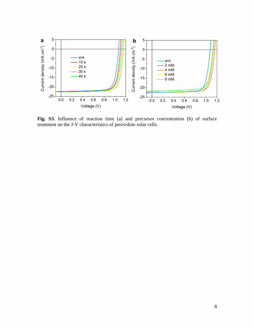
Fig. S3. Influence of reaction time (a) and precursor concentration (b) of surface
treatment on the J-V characteristics of perovskite solar cells.

9
Fig. S4. Scanning electron microscopy (SEM) images of the control (a) and sulfate-
treated (b) perovskite films deposited on ITO glass.

10
Fig. S5. AFM images of (a) control and (b) sulfate-treated perovskite films and root mean
squared (RMS) roughness were calculated to be 14.5 and 15.2 nm for control and treated
films, respectively. (c) Scanning TEM images of sulfate-treated perovskite solar cell
captured at various interface regions.

11
3500 3000 2500 2000 1500 1000
SO42-
PbSO4
Tra
nsm
itta
nce
Wavenumber (cm-1)
Fig. S6. FT-IR spectrum of PbSO4 powder. The FT-IR peaks at 964, 1040 and 1145 cm-2
represent the symmetric stretching (v1) and asymmetric stretching (v3) of sulfate ions,
respectively.

12
Fig. S7. (a) Optimized bulk MAPbI3-PbSO4 interface. RDF analysis of (b) O—S and (c)
O—Pb. Blue and red lines are the RDF calculated per the perfect PbSO4 crystal and
MAPbI3-PbSO4 interface, respectively.

13
400 500 600 700 8000
25
50
75
100
Wavelength (nm)
EQ
E (
%)
0
5
10
15
20
25
Inte
rgra
ted
Jsc (
mA
cm
-2)
Fig. S8. EQE spectra of the device based on sulfate-treated perovskite layer. The
integrated Jsc is 22.3 mA cm-2.

14
Fig. S9. Comparison of J-V metrics for 25 independent solar cells based on control and
sulfate-treated perovskite films.
15
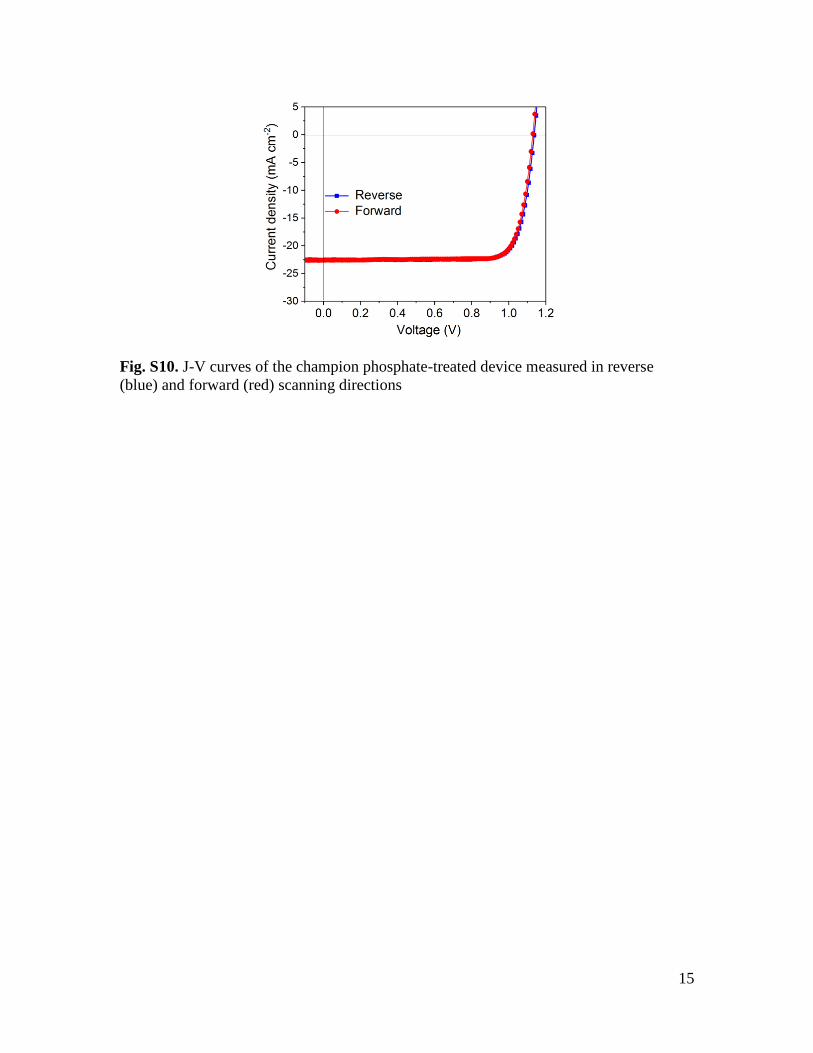
Fig. S10. J-V curves of the champion phosphate-treated device measured in reverse
(blue) and forward (red) scanning directions

16
Fig. S11. TPV spectra of control (red) and phosphate-treated (green) photovoltaic
devices.

17
200 400 600 800 1000
No
rma
llize
d irr
ad
ian
ce
(a
.u.)
Wavelength (nm)
Our light source
AM 1.5G
Fig. S12. Normalized spectra of our light source and the standard AM 1.5G.

18
Fig. S13. Images of the devices without (left) and with (right) a sulfate layer after
exposure to water droplet for 3 min.
19
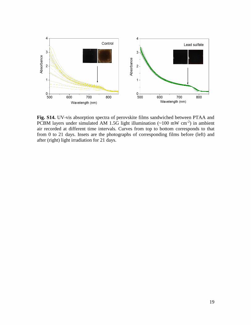
Fig. S14. UV-vis absorption spectra of perovskite films sandwiched between PTAA and
PCBM layers under simulated AM 1.5G light illumination (~100 mW cm-2) in ambient
air recorded at different time intervals. Curves from top to bottom corresponds to that
from 0 to 21 days. Insets are the photographs of corresponding films before (left) and
after (right) light irradiation for 21 days.

20
Fig. S15. UV-vis absorption spectra of perovskite films under light illumination (~70
mW cm-2) and dry air recorded at different time intervals. Insets are the photographs of
corresponding perovskite films before (left) and after (right) light irradiation for 4 days.
21
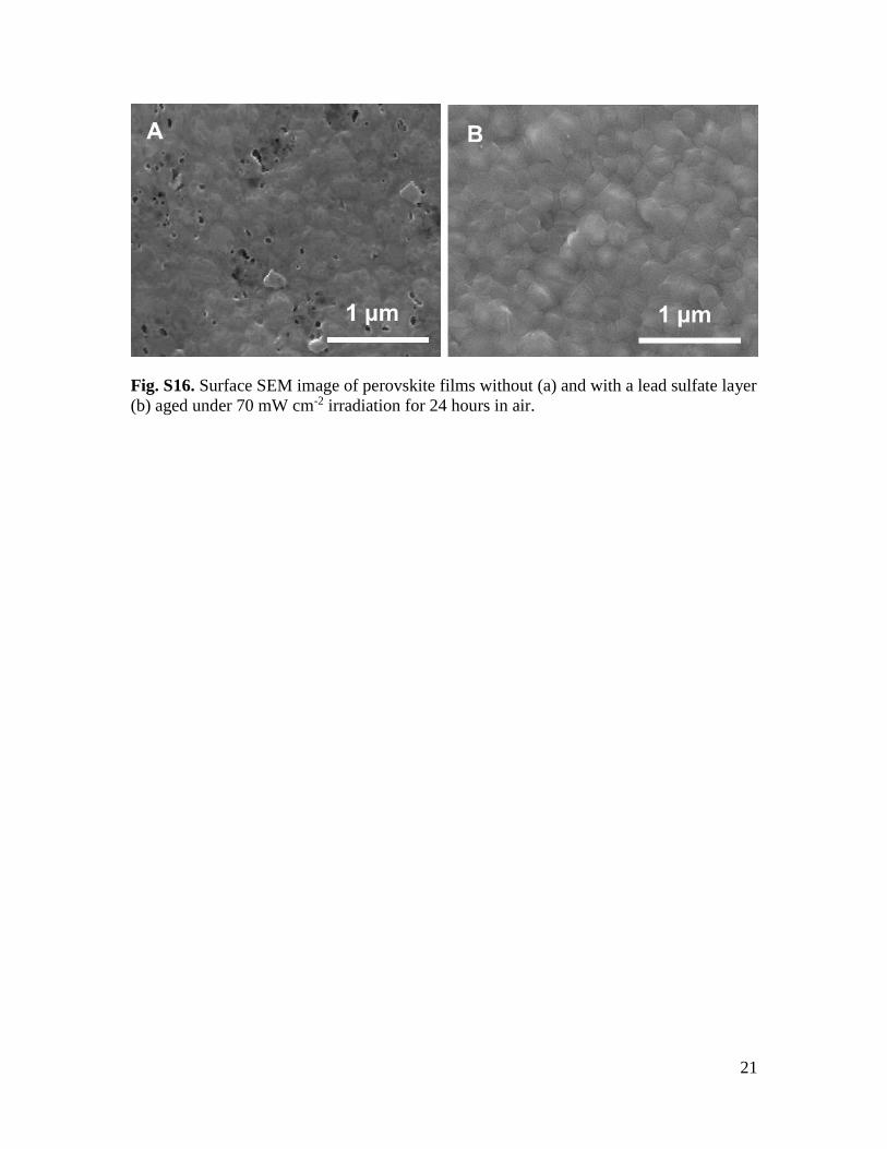
Fig. S16. Surface SEM image of perovskite films without (a) and with a lead sulfate layer
(b) aged under 70 mW cm-2 irradiation for 24 hours in air.
22
Table S1. Energy associated to the interface formation reaction (ΔEform, eV) starting from
the corresponding bulk structures. Values in parenthesis are evaluated as single point
calculation including D3 dispersion interaction on the optimized geometries.
MAPbI3
Termination ΔEform ΔEform per molec.
ΔEform per molec. (Ref.
Isolated PbSO4)
MAI-terminated
9.475
(16.763)
0.395
(0.698) -2.104
PbI2-terminated 6.473
(12.641)
0.270
(0.527) -2.229

23
Table S2. hotovoltaic parameters of the best sulfate-treated device with different
sweeping rates and directions. All the J-V curves were at a simulated AM 1.5 G solar
irradiation with a scan rate of 0.1 V s-1.
Jsc/mA cm-2 Voc/V FF PCE/%
Forward 22.65 1.16 0.803 21.09
Reverse 22.63 1.16 0.804 21.11
24
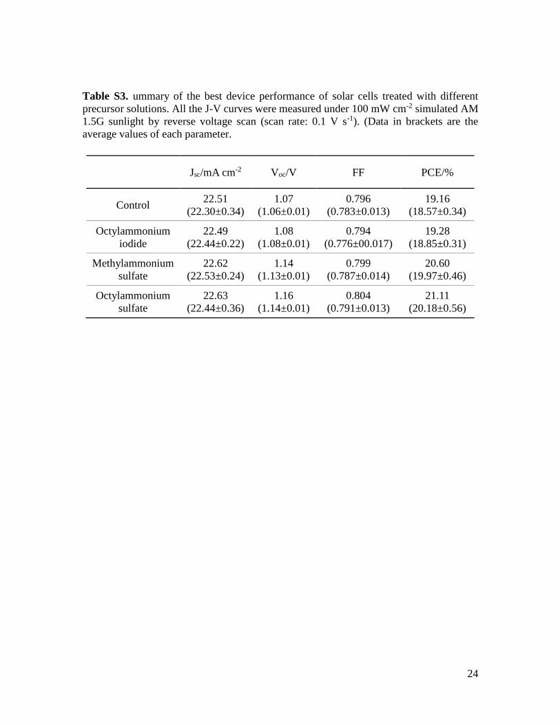
Table S3. ummary of the best device performance of solar cells treated with different
precursor solutions. All the J-V curves were measured under 100 mW cm-2 simulated AM
1.5G sunlight by reverse voltage scan (scan rate: 0.1 V s-1). (Data in brackets are the
average values of each parameter.
Jsc/mA cm-2 Voc/V FF PCE/%
Control 22.51
(22.30±0.34)
1.07
(1.06±0.01)
0.796
(0.783±0.013)
19.16
(18.57±0.34)
Octylammonium
iodide
22.49
(22.44±0.22)
1.08
(1.08±0.01)
0.794
(0.776±00.017)
19.28
(18.85±0.31)
Methylammonium
sulfate
22.62
(22.53±0.24)
1.14
(1.13±0.01)
0.799
(0.787±0.014)
20.60
(19.97±0.46)
Octylammonium
sulfate
22.63
(22.44±0.36)
1.16
(1.14±0.01)
0.804
(0.791±0.013)
21.11
(20.18±0.56)
25
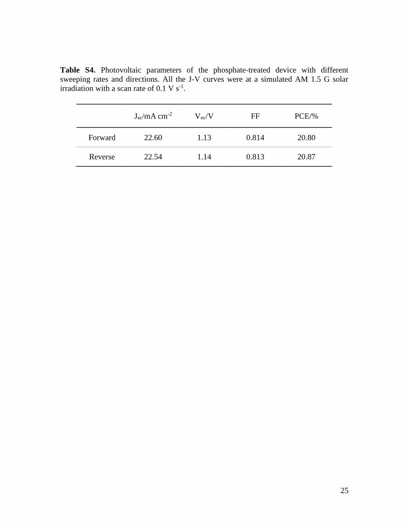
Table S4. Photovoltaic parameters of the phosphate-treated device with different
sweeping rates and directions. All the J-V curves were at a simulated AM 1.5 G solar
irradiation with a scan rate of 0.1 V s-1.
Jsc/mA cm-2 Voc/V FF PCE/%
Forward 22.60 1.13 0.814 20.80
Reverse 22.54 1.14 0.813 20.87

26
Table S5. Comparison of the stability of the solar cells in our work with some recent
reports. PCE0 and PCEt are the efficiency of devices before and after stability tests.
Environment Encapsulation Temperature Light source Testing duration PCE0 PCEt/PCE0 Reference
Air, RH
~60±10% Yes 65 °C
plasma lamp with
strong UV component,
equivalent to 1 sun
1200 h 19.44% 96.8% This work
Air, RH ~30% Yes 25 °C white LED, equivalent
to 1 sun 1370 h N.A. 95%
Nature, 2019,
567, 511.
N2 No 20 °C white LED, equivalent
to 1 sun 1000 h 19.54% 78.4%
Science 2018, 362, 449.
Not specified Not specified Not specified not specified,
equivalent to 1 sun 1500 h
19.17±0.4
2% 92%
Science 2019,
363, 265.
Air, RH ~10-20%
No 20-30 °C plasma lamp, equivalent
to 0.77 sun 1000 h 12.2±0.1% 94%
Nat. Energy, 2018, 3, 68.
N2 No ~60 °C white LED, equivalent
to 1 sun 1000 h N.A. >95%
Science, 2017,
358, 768.
Ar No 55-60 °C white LED, equivalent
to 1 sun 1000 h ~20.6% 98.1%
Nat. Common. 2018, 9, 4482.

27
Movie S1
Movie of dipping a MAPbI3 single crystal with a thick lead sulfate layer in water.
References and Notes 1. National Renewable Energy Laboratory, Best research-cell efficiencies chart (2019);
www.nrel.gov/pv/assets/pdfs/best-research-cell-efficiencies.20190703.pdf. 2. M. Grätzel, The light and shade of perovskite solar cells. Nat. Mater. 13, 838–842 (2014).
doi:10.1038/nmat4065 Medline 3. S. Yang, Y. Wang, P. Liu, Y.-B. Cheng, H. J. Zhao, H. G. Yang, Functionalization of
perovskite thin films with moisture-tolerant molecules. Nat. Energy 1, 15016 (2016). doi:10.1038/nenergy.2015.16
4. T. Leijtens, G. E. Eperon, N. K. Noel, S. N. Habisreutinger, A. Petrozza, H. J. Snaith, Stability of metal halide perovskite solar cells. Adv. Energy Mater. 5, 1500963 (2015). doi:10.1002/aenm.201500963
5. W. Nie, J.-C. Blancon, A. J. Neukirch, K. Appavoo, H. Tsai, M. Chhowalla, M. A. Alam, M. Y. Sfeir, C. Katan, J. Even, S. Tretiak, J. J. Crochet, G. Gupta, A. D. Mohite, Light-activated photocurrent degradation and self-healing in perovskite solar cells. Nat. Commun. 7, 11574 (2016). doi:10.1038/ncomms11574 Medline
6. K. Domanski, B. Roose, T. Matsui, M. Saliba, S.-H. Turren-Cruz, J.-P. Correa-Baena, C. R. Carmona, G. Richardson, J. M. Foster, F. De Angelis, J. M. Ball, A. Petrozza, N. Mine, M. K. Nazeeruddin, W. Tress, M. Grätzel, U. Steiner, A. Hagfeldt, A. Abate, Migration of cations induces reversible performance losses over day/night cycling in perovskite solar cells. Energy Environ. Sci. 10, 604–613 (2017). doi:10.1039/C6EE03352K
7. R. J. Sutton, G. E. Eperon, L. Miranda, E. S. Parrott, B. A. Kamino, J. B. Patel, M. T. Hörantner, M. B. Johnston, A. A. Haghighirad, D. T. Moore, H. J. Snaith, Bandgap-tunable cesium lead halide perovskites with high thermal stability for efficient solar cells. Adv. Energy Mater. 6, 1502458 (2016). doi:10.1002/aenm.201502458
8. J. W. Lee, D.-H. Kim, H.-S. Kim, S.-W. Seo, S. M. Cho, N.-G. Park, Formamidinium and cesium hybridization for photo- and moisture-stable perovskite solar cell. Adv. Energy Mater. 5, 1501310 (2015). doi:10.1002/aenm.201501310
9. C. Yi, J. Luo, S. Meloni, A. Boziki, N. Ashari-Astani, C. Grätzel, S. M. Zakeeruddin, U. Röthlisberger, M. Grätzel, Entropic stabilization of mixed A-cation ABX3 metal halide perovskites for high performance perovskite solar cells. Energy Environ. Sci. 9, 656–662 (2016). doi:10.1039/C5EE03255E
10. J. Zhao, Y. Deng, H. Wei, X. Zheng, Z. Yu, Y. Shao, J. E. Shield, J. Huang, Strained hybrid perovskite thin films and their impact on the intrinsic stability of perovskite solar cells. Sci. Adv. 3, o5616 (2017). doi:10.1126/sciadv.aao5616 Medline
11. Q. Wang, B. Chen, Y. Liu, Y. Deng, Y. Bai, Q. Dong, J. Huang, Scaling behavior of moisture-induced grain degradation in polycrystalline hybrid perovskite thin films. Energy Environ. Sci. 10, 516–522 (2017). doi:10.1039/C6EE02941H
12. Z. Fan, H. Xiao, Y. Wang, Z. Zhao, Z. Lin, H.-C. Cheng, S.-J. Lee, G. Wang, Z. Feng, W. A. Goddard III, Y. Huang, X. Duan, Layer-by-layer degradation of methylammonium lead tri-iodide perovskite microplates. Joule 1, 548–562 (2017). doi:10.1016/j.joule.2017.08.005

13. N. Aristidou, C. Eames, I. Sanchez-Molina, X. Bu, J. Kosco, M. S. Islam, S. A. Haque, Fast oxygen diffusion and iodide defects mediate oxygen-induced degradation of perovskite solar cells. Nat. Commun. 8, 15218 (2017). doi:10.1038/ncomms15218 Medline
14. J. Xu, A. Buin, A. H. Ip, W. Li, O. Voznyy, R. Comin, M. Yuan, S. Jeon, Z. Ning, J. J. McDowell, P. Kanjanaboos, J.-P. Sun, X. Lan, L. N. Quan, D. H. Kim, I. G. Hill, P. Maksymovych, E. H. Sargent, Perovskite-fullerene hybrid materials suppress hysteresis in planar diodes. Nat. Commun. 6, 7081 (2015). doi:10.1038/ncomms8081 Medline
15. N. K. Noel, A. Abate, S. D. Stranks, E. S. Parrott, V. M. Burlakov, A. Goriely, H. J. Snaith, Enhanced photoluminescence and solar cell performance via Lewis base passivation of organic-inorganic lead halide perovskites. ACS Nano 8, 9815–9821 (2014). doi:10.1021/nn5036476 Medline
16. X. Zheng, B. Chen, J. Dai, Y. Fang, Y. Bai, Y. Lin, H. Wei, X. C. Zeng, J. Huang, Defect passivation in hybrid perovskite solar cells using quaternary ammonium halide anions and cations. Nat. Energy 2, 17102 (2017). doi:10.1038/nenergy.2017.102
17. V. K. Ravi, R. A. Scheidt, A. Nag, M. Kuno, P. V. Kamat, To exchange or not to exchange. Suppressing anion exchange in cesium lead halide perovskites with PbSO4–oleate capping. ACS Energy Lett. 3, 1049–1055 (2018). doi:10.1021/acsenergylett.8b00380
18. H. Stephen, T. Stephen, Solubilities of Inorganic and Organic Compounds (Macmillan, 1963), vol. 1.
19. D. Peak, R. G. Ford, D. L. Sparks, An in situ ATR-FTIR investigation of sulfate bonding mechanisms on goethite. J. Colloid Interface Sci. 218, 289–299 (1999). doi:10.1006/jcis.1999.6405 Medline
20. E. Elzinga, D. Peak, D. Sparks, Spectroscopic studies of Pb (II)-sulfate interactions at the goethite-water interface. Geochim. Cosmochim. Acta 65, 2219–2230 (2001). doi:10.1016/S0016-7037(01)00595-6
21. J. H. Yun, I. Lee, T.-S. Kim, M. J. Ko, J. Y. Kim, H. J. Son, Synergistic enhancement and mechanism study of mechanical and moisture stability of perovskite solar cells introducing polyethylene-imine into the CH3NH3PbI3/HTM interface. J. Mater. Chem. A 3, 22176–22182 (2015). doi:10.1039/C5TA06008G
22. S. J. O. Hardman, D. M. Graham, S. K. Stubbs, B. F. Spencer, E. A. Seddon, H.-T. Fung, S. Gardonio, F. Sirotti, M. G. Silly, J. Akhtar, P. O’Brien, D. J. Binks, W. R. Flavell, Electronic and surface properties of PbS nanoparticles exhibiting efficient multiple exciton generation. Phys. Chem. Chem. Phys. 13, 20275–20283 (2011). doi:10.1039/c1cp22330e Medline
23. Y. Shao, Z. Xiao, C. Bi, Y. Yuan, J. Huang, Origin and elimination of photocurrent hysteresis by fullerene passivation in CH3NH3PbI3 planar heterojunction solar cells. Nat. Commun. 5, 5784 (2014). doi:10.1038/ncomms6784 Medline
24. D. W. deQuilettes, S. M. Vorpahl, S. D. Stranks, H. Nagaoka, G. E. Eperon, M. E. Ziffer, H. J. Snaith, D. S. Ginger, Solar cells. Impact of microstructure on local carrier lifetime in perovskite solar cells. Science 348, 683–686 (2015). doi:10.1126/science.aaa5333 Medline

25. W. S. Yang, B.-W. Park, E. H. Jung, N. J. Jeon, Y. C. Kim, D. U. Lee, S. S. Shin, J. Seo, E. K. Kim, J. H. Noh, S. I. Seok, Iodide management in formamidinium-lead-halide-based perovskite layers for efficient solar cells. Science 356, 1376–1379 (2017). doi:10.1126/science.aan2301 Medline
26. L. E. Black, New Perspectives on Surface Passivation: Understanding the Si-Al2O3 Interface (Springer, 2016).
27. M. Wang, Exploring stability of formamidinium lead trihalide for solar cell application. Sci. Bull. (Beijing) 62, 249–255 (2017). doi:10.1016/j.scib.2017.01.025
28. A. Rajagopal, R. J. Stoddard, S. B. Jo, H. W. Hillhouse, A. K.-Y. Jen, Overcoming the photovoltage plateau in large bandgap perovskite photovoltaics. Nano Lett. 18, 3985–3993 (2018). doi:10.1021/acs.nanolett.8b01480 Medline
29. J. Xing, Q. Wang, Q. Dong, Y. Yuan, Y. Fang, J. Huang, Ultrafast ion migration in hybrid perovskite polycrystalline thin films under light and suppression in single crystals. Phys. Chem. Chem. Phys. 18, 30484–30490 (2016). doi:10.1039/C6CP06496E Medline
30. Y. Yuan, J. Huang, Ion migration in organometal trihalide perovskite and its impact on photovoltaic efficiency and stability. Acc. Chem. Res. 49, 286–293 (2016). doi:10.1021/acs.accounts.5b00420 Medline
31. Y. Shao, Y. Fang, T. Li, Q. Wang, Q. Dong, Y. Deng, Y. Yuan, H. Wei, M. Wang, A. Gruverman, J. Shield, J. Huang, Grain boundary dominated ion migration in polycrystalline organic–inorganic halide perovskite films. Energy Environ. Sci. 9, 1752–1759 (2016). doi:10.1039/C6EE00413J
32. R. Car, M. Parrinello, Unified approach for molecular dynamics and density-functional theory. Phys. Rev. Lett. 55, 2471–2474 (1985). doi:10.1103/PhysRevLett.55.2471 Medline
33. P. Giannozzi, S. Baroni, N. Bonini, M. Calandra, R. Car, C. Cavazzoni, D. Ceresoli, G. L. Chiarotti, M. Cococcioni, I. Dabo, A. Dal Corso, S. de Gironcoli, S. Fabris, G. Fratesi, R. Gebauer, U. Gerstmann, C. Gougoussis, A. Kokalj, M. Lazzeri, L. Martin-Samos, N. Marzari, F. Mauri, R. Mazzarello, S. Paolini, A. Pasquarello, L. Paulatto, C. Sbraccia, S. Scandolo, G. Sclauzero, A. P. Seitsonen, A. Smogunov, P. Umari, R. M. Wentzcovitch, QUANTUM ESPRESSO: A modular and open-source software project for quantum simulations of materials. J. Phys. Condens. Matter 21, 395502 (2009). doi:10.1088/0953-8984/21/39/395502 Medline