supplementary movies for “purines induce directed migration and rapid homing of microglia to...
TRANSCRIPT

Supplementary Movies forSupplementary Movies for
““Purines Induce Directed Migration and Rapid Purines Induce Directed Migration and Rapid Homing of Microglia to Injured Pyramidal Homing of Microglia to Injured Pyramidal
Neurons in Developing Hippocampus”Neurons in Developing Hippocampus”
Kurpius D, Nolley E, and Dailey M.Kurpius D, Nolley E, and Dailey M.
Dept. of Biological SciencesDept. of Biological Sciences
Univ. of IowaUniv. of Iowa
Accepted for publication in GLIA © 2007Accepted for publication in GLIA © 2007

Figure 2Figure 2Supplementary Movie 1Supplementary Movie 1
To view in 3D, use To view in 3D, use redred--greengreen glasses (red over left eye). glasses (red over left eye).
SPSP
SRSR
30 30 mm
Time-lapse sequence showing activation and directional migration of MG into the pyramidal cell body layer (SP) in an acutely isolated neonatal mouse hippocampal tissue slice. MG in the stratum radiatum (SR) near the SP (lower right) polarize and migrate directionally into the SP. Note the slow wave of activation that progresses away from the SP through the SR (lower right to upper left). Total time is ~4hr.
Click to play

Figure 3Figure 3Supplementary Movie 2 & 3Supplementary Movie 2 & 3
Two-channel time-lapse imaging shows IB4-labeled MG (green) engaging nuclei of injured/dead neurons (red). (A) is a still image showing the relative position of fields ‘B’ and ‘C’. (B) Time-lapse sequence starting ~3.5 hr after tissue excision. (C) Time-lapse sequence starting ~6.5 hr after tissue excision. Note that MG appear to engulf the dead cell nuclei.
Click to play
Click to play
C.
B.
Supplementary Supplementary Movie 2Movie 2
Supplementary Supplementary Movie 3Movie 3

Figure 5CFigure 5CSupplementary Movie 4Supplementary Movie 4
Apyrase inhibits MG motility and migration in neonatal tissue slices. High magnification, time-lapse sequence shows highly mobile MG in and near the CA3 pyramidal cell body layer (SP). During the baseline, MG are highly locomotory. Application of apyrase (100 U/ml) inhibits MG movements. Following washout, MG movements rapidly recover. Total time = ~6 hr.
Experiment
Baseline (2 hr)
Apyrase (2.5 hr)
Washout (1.5 hr)
SP
SR
30 30 mm
Click to play

Suppl. Figure 3Suppl. Figure 3Supplementary Movie 5Supplementary Movie 5
Time-lapse sequence showing diffusion of BodipyTR-ADP (100M) from exterior of tissue slice (intense red region at left) to interior of slice (stratum radiatum, SR). Imaging starts shortly after application of ADP. Note the subtle increase in red fluorescence in the central and right portion of the field of view, especially during the first 60 min. In response to diffusion of ADP into the slice, microglia (green) in the SP and SR move toward the edge of the tissue slice.
Click to play
BodipyTR-ADP (100M)
+ADP (1mM)
3 hr
Experiment
IB4-Microglia
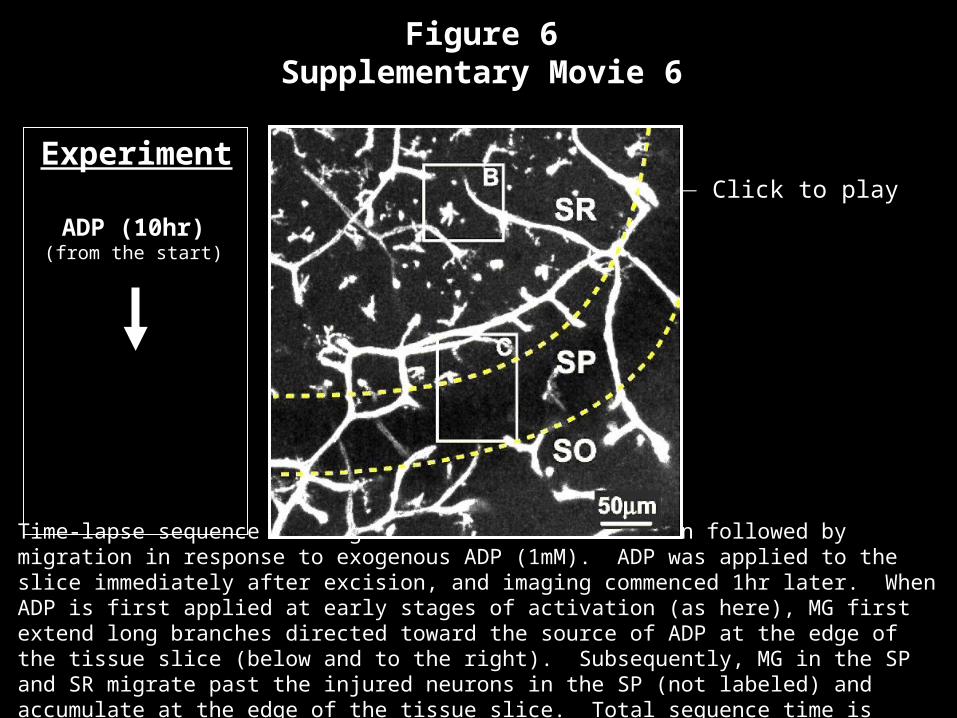
Figure 6Figure 6Supplementary Movie 6Supplementary Movie 6
ADP (10hr)ADP (10hr)(from the start)
Time-lapse sequence showing directed branch extension followed by migration in response to exogenous ADP (1mM). ADP was applied to the slice immediately after excision, and imaging commenced 1hr later. When ADP is first applied at early stages of activation (as here), MG first extend long branches directed toward the source of ADP at the edge of the tissue slice (below and to the right). Subsequently, MG in the SP and SR migrate past the injured neurons in the SP (not labeled) and accumulate at the edge of the tissue slice. Total sequence time is ~10hr.
Click to playExperiment

Figure 7AFigure 7ASupplementary Movie 7Supplementary Movie 7
Time-lapse sequence showing sequential responses of MG to application of apyrase and ATP in a neonatal rat hippocampal tissue slice. During the baseline, note the migration of MG and their accumulation near injured CA3 pyramidal neurons in the SP. Application of apyrase (indicated in upper right corner) inhibits MG movements. Following application of ATP (1mM), MG are drawn away from injured neurons in the SP and move toward the edge of the tissue slice (just below field of view). Following washout of ATP, MG show a low level of undirected movements.
Click to playBaseline (3.5 hr)
Apyrase (2 hr)
Washout (2 hr)
ATP (4.5 hr)
Experiment
Washout (5.5 hr)