supportive treatment for cancer – part 3: treatment … · supportive treatment for cancer –...
TRANSCRIPT
2013 www.kce.fgov.be
KCE REPORT 211
SUPPORTIVE TREATMENT FOR CANCER – PART 3: TREATMENT OF PAIN: MOST COMMON PRACTICES


2013 www.kce.fgov.be
KCE REPORT 211 GOOD CLINICAL PRACTICE
SUPPORTIVE TREATMENT FOR CANCER – PART 3: TREATMENT OF PAIN: MOST COMMON PRACTICES MARIJKE EYSSEN, NADIA BENAHMED, ANJA DESOMER

COLOPHON Title: Supportive treatment for cancer – Part 3: Treatment of pain: most common practices
Authors: Marijke Eyssen, Nadia Benahmed, Anja Desomer
Reviewers: Raf Mertens, Sabine Stordeur, Joan Vlayen
External experts: Cécile Avril (Fondation contre le cancer), Ahmad Awada (Institut Jules Bordet), Erik Briers (Patiëntenorganisatie “Wij Ook”), Tom Boterberg (UZ Gent), Annemarie Coolbrandt (UZ Leuven), Mieke Depril (UZ Leuven), Frederic Duprez (UZ Gent), Marie-Elisabeth Faymonville (CHU de Liège), Chantal Goossens (Fondation contre le cancer), Guy Hans (UZ Antwerpen), Lia Le Roy (Werkgroep Hersentumoren vzw), Johan Menten (UZ Leuven), Marc Peeters (UZ Antwerpen)
Stakeholders: Suzan Broekmans (UZ Leuven, NVKVV, BPS), Marc De Kock (Cliniques universitaires UCL Saint-Luc, BVAR-SBAR), Peter Demeulenaere (Huisartsengroep Groenenborg, palliative care specialist at GZA-ziekenhuizen, Domus Medica), Alain Dessard (Delta asbl, Fédération Wallonne des Soins Palliatifs), Jacques Devulder (UZ Gent, UGent, BVAR-SARB), Chantal Doyen (CHU. Mont Godinne, Fédération Wallonne des Soins Palliatifs), Koen Lauwers (AZ Klina, VAVP, BPS), Filomena Mazzeo (Cliniques universitaires UCL Saint-Luc, BSMO), Ivo Nagels (Stichting tegen Kanker), Patrick Paulus (CHR Citadelle Liège, BELNUC), Barbara Plehiers (CHU Ambroise Paré, Fédération Wallonne des Soins Palliatifs), Jan Poelaert (UZ Brussel, VUB, BVAR-SBAR), Dirk Schrijvers (Ziekenhuis Netwerk Antwerpen-Middelheim, BSMO), Marc Tanghe (CHRPBW Bois de la Pierre, Federatie Palliatieve Zorg Vlaanderen), Bart Van den Eynde (Gasthuiszusters Antwerpen, Domus Medica) In addition 1 patient participated on the stakeholder panel. For privacy reasons his/her name is not mentioned in this colophon.
External validators: Paul M. J. Clement (UZ Leuven, KU Leuven), Hilde Verbeke (UZ Leuven, Belgian Centre for Evidence-Based Medicine CEBAM), K.C.P. Vissers (Universitair Medisch Centrum St. Radboud – Radboud Universiteit Nijmegen Nederland)
Conflict of interest: External experts: A grant, fees or funds for a member of staff or another form of compensation for the execution of research: Annemarie Coolbrandt Stakeholders: Payments to speak, training remuneration, subsidised travel or payment for participation at a conference: Marc Tanghe (Guest lecturer HUB-Erasmus-Hogeschool Brussel) Presidency or accountable function within an institution, association, department or other entity on which the results of this report could have an impact: Susan Broekmans (president network Vlaamse pijnverpleegkundigen

– NVKVV), Koen Lauwers (Board member BPS, VAVP), Chantal Doyen (vice-president FWSP) Validators: Fees or other compensation for writing a publication or participating in its development: Kris Vissers A grant, fees or funds for a member of staff or another form of compensation for the execution of research: Paul Clement, Kris Vissers Consultancy or employment for a company, an association or an organisation that may gain or lose financially due to the results of this report: Paul Clement, Kris Vissers Payments to speak, training remuneration, subsidised travel or payment for participation at a conference: Paul Clement, Kris Vissers Presidency or accountable function within an institution, association, department or other entity on which the results of this report could have an impact: Kris Vissers Participation in scientific or experimental research as an initiator, principal investigator or researcher: Paul Clement, Kris Vissers Further, it should be noted that all experts and stakeholders, as well as the validators consulted within this report were selected because of their expertise in the field of cancer pain. Therefore, by definition, all consulted experts, stakeholders and validators have a certain degree of conflict of interest to the main topic of this report.
Layout: Ine Verhulst
Disclaimer: • The external experts were consulted about a (preliminary) version of the scientific report. Their comments were discussed during meetings. They did not co-author the scientific report and did not necessarily agree with its content.
• Subsequently, a (final) version was submitted to the validators. The validation of the report results from a consensus or a voting process between the validators. The validators did not co-author the scientific report and did not necessarily all three agree with its content.
• Finally, this report has been approved by common assent by the Executive Board. • Only the KCE is responsible for errors or omissions that could persist. The policy recommendations
are also under the full responsibility of the KCE.
Publication date: 18 November 2013
Domain: Good Clinical Practice (GCP)
MeSH: Neoplasms; Pain; Pain management; Analgesics; Radiotherapy
NLM Classification: QZ 266
Language: English
Format: Adobe® PDF™ (A4)
Legal depot: D/2013/10.273/84
Copyright: KCE reports are published under a “by/nc/nd” Creative Commons Licence http://kce.fgov.be/content/about-copyrights-for-kce-reports.
How to refer to this document? Eyssen M, Benahmed N, Desomer A. Supportive treatment for cancer – Part 3: Treatment of pain: most common practices. Good Clinical Practice (GCP) Brussels: Belgian Health Care Knowledge Centre (KCE). 2013. KCE Reports 211. D/2013/10.273/84.
This document is available on the website of the Belgian Health Care Knowledge Centre.

KCE Report 211 Treatment of cancer pain 1
TABLE OF CONTENTS
LIST OF FIGURES ............................................................................................................................................... 5 LIST OF TABLES ................................................................................................................................................. 5 LIST OF ABBREVIATIONS ................................................................................................................................. 6
SCIENTIFIC REPORT ......................................................................................................................... 10 1 INTRODUCTION .................................................................................................................................. 10 1.1 CONTEXT ............................................................................................................................................ 10 1.2 GENERAL SCOPE ............................................................................................................................... 11 1.3 TARGET USERS OF THE GUIDELINE ............................................................................................... 11 1.4 STATEMENT OF INTENT .................................................................................................................... 12 1.5 FUNDING AND DECLARATION OF INTEREST ................................................................................. 12 1.6 IMPLEMENTATION AND UPDATING OF THE GUIDELINE .............................................................. 12
1.6.1 Implementation ....................................................................................................................... 12 1.6.2 Monitoring the quality of care ................................................................................................. 12 1.6.3 Guideline update .................................................................................................................... 13
1.7 HOW TO USE THIS GUIDELINE? ....................................................................................................... 13 2 METHODS ............................................................................................................................................ 16 2.1 RESEARCH QUESTIONS ................................................................................................................... 16
2.1.1 Methodology ........................................................................................................................... 16 2.1.2 Research questions, included medical treatment options and outcomes .............................. 16 2.1.3 Excluded medical treatment options ...................................................................................... 17
2.2 DEFINITIONS ....................................................................................................................................... 17 2.3 LITERATURE SEARCH ....................................................................................................................... 18 2.4 SELECTION CRITERIA ....................................................................................................................... 18 2.5 SELECTION PROCESS ...................................................................................................................... 19 2.6 QUALITY APPRAISAL ......................................................................................................................... 20 2.7 DATA EXTRACTION AND GRADING OF EVIDENCE ........................................................................ 20 2.8 FORMULATION OF RECOMMENDATIONS ....................................................................................... 23
2.8.1 Methodology ........................................................................................................................... 23

2 Treatment of cancer pain KCE Report 211
2.8.2 Defining and interpreting the strength of a recommendation ................................................. 23 2.9 EXTERNAL REVIEW ........................................................................................................................... 25 2.10 FINAL VALIDATION ............................................................................................................................. 26 2.11 DEVELOPMENT OF THE GUIDELINE: PROJECT TEAM, INVOLVED EXPERTS, PANEL OF
STAKEHOLDERS ................................................................................................................................ 26 3 SEARCH RESULTS ............................................................................................................................ 27 3.1 SYSTEMATIC REVIEWS ..................................................................................................................... 27 3.2 RANDOMIZED CONTROLLED TRIALS .............................................................................................. 29 4 EVIDENCE REPORT ........................................................................................................................... 31 4.1 INTRODUCTION .................................................................................................................................. 31
4.1.1 Pain Assessment ................................................................................................................... 31 4.1.2 Pain assessment tools ........................................................................................................... 31 4.1.3 Severity of pain ...................................................................................................................... 32 4.1.4 The World Health Organization analgesic ladder .................................................................. 33
4.2 NSAIDS AND PARACETAMOL ........................................................................................................... 37 4.2.1 Introduction ............................................................................................................................ 37 4.2.2 Search results ........................................................................................................................ 38 4.2.3 Literature overview ................................................................................................................. 40 4.2.4 Other considerations .............................................................................................................. 47
4.3 OPIOIDS ............................................................................................................................................... 51 4.3.1 Introduction ............................................................................................................................ 51 4.3.2 Search results ........................................................................................................................ 60 4.3.3 Literature overview ................................................................................................................. 63 4.3.4 Other considerations .............................................................................................................. 93
4.4 CORTICOSTEROIDS ........................................................................................................................ 102 4.4.1 Introduction .......................................................................................................................... 102 4.4.2 Search results ...................................................................................................................... 102 4.4.3 Literature overview ............................................................................................................... 103 4.4.4 Other considerations ............................................................................................................ 104
4.5 ANTIDEPRESSANTS ........................................................................................................................ 105

KCE Report 211 Treatment of cancer pain 3
4.5.1 Introduction .......................................................................................................................... 105 4.5.2 Search results ...................................................................................................................... 105 4.5.3 Literature overview ............................................................................................................... 105 4.5.4 Other considerations ............................................................................................................ 107
4.6 ANTICONVULSANTS ........................................................................................................................ 109 4.6.1 Introduction .......................................................................................................................... 109 4.6.2 Search results ...................................................................................................................... 109 4.6.3 Literature overview ............................................................................................................... 110 4.6.4 Other considerations ............................................................................................................ 114
4.7 RADIOTHERAPY FOR PAINFUL BONE METASTASES.................................................................. 117 4.7.1 Introduction .......................................................................................................................... 117 4.7.2 Search results ...................................................................................................................... 118 4.7.3 Literature overview ............................................................................................................... 119 4.7.4 Other considerations ............................................................................................................ 123
4.8 RADIONUCLIDES FOR PAINFUL BONE METASTASES ................................................................ 127 4.8.1 Introduction .......................................................................................................................... 127 4.8.2 Search results ...................................................................................................................... 128 4.8.3 Literature overview ............................................................................................................... 129 4.8.4 Other considerations ............................................................................................................ 131
4.9 BISPHOSPHONATES FOR PAINFUL BONE METASTASES .......................................................... 133 4.9.1 Introduction .......................................................................................................................... 133 4.9.2 Search results ...................................................................................................................... 134 4.9.3 Literature overview ............................................................................................................... 136 4.9.4 Other considerations ............................................................................................................ 145
4.10 CELIAC PLEXUS BLOCK .................................................................................................................. 148 4.10.1 Introduction .......................................................................................................................... 148 4.10.2 Search results ...................................................................................................................... 149 4.10.3 Literature overview ............................................................................................................... 149 4.10.4 Other considerations ............................................................................................................ 150
5 DISCUSSION ..................................................................................................................................... 152
4 Treatment of cancer pain KCE Report 211
5.1 PAIN IMPROVEMENT: WHAT IS A CLINICALLY IMPORTANT CHANGE? .................................... 152 5.2 PREVENTION OF CANCER PAIN .................................................................................................... 153 5.3 OTHER CONSIDERATIONS ............................................................................................................. 154
5.3.1 Scope of the report............................................................................................................... 154 5.3.2 Quality of the studies ........................................................................................................... 154 5.3.3 Adverse events .................................................................................................................... 154 5.3.4 Use of pain medication in cancer patients with renal or liver impairment ............................ 154 5.3.5 Combinations of interventions .............................................................................................. 154 5.3.6 Aspects of costs and reimbursement of included interventions ........................................... 154 5.3.7 Multidisciplinary approach of pain treatment ....................................................................... 155 5.3.8 Patient-centered care ........................................................................................................... 155 5.3.9 Barriers and facilitators for implementation of this guideline ............................................... 155
REFERENCES ................................................................................................................................... 156

KCE Report 211 Treatment of cancer pain 5
LIST OF FIGURES Figure 1 – Study flow of selection of SRs (CDSR, Medline, PreMedline, Embase, DARE, HTA database) ...... 28 Figure 2 – Study flow of selection of RCTs ........................................................................................................ 30 Figure 3 – The 3-step analgesic ladder developed by the World Health Organization (WHO)36 ....................... 33
LIST OF TABLES Table 1 – In and exclusion criteria ...................................................................................................................... 19 Table 2 – Levels of evidence according to the GRADE system. ........................................................................ 21 Table 3 – Downgrading the quality rating of evidence using GRADE. ............................................................... 22 Table 4 – Standardized language used for formulating scientific conclusions. .................................................. 23 Table 5 – Strength of recommendations according to the GRADE system. ...................................................... 23 Table 6 – Factors that influence the strength of a recommendation. ................................................................. 24 Table 7 – Interpretation of strong and conditional (weak)* recommendations ................................................... 25 Table 8 – List of Professional Associations and Patient Associations to which the recommendations were communicated. ....................................................................................................... 26 Table 9 – Number of hits per database for systematic reviews search. ............................................................. 27 Table 10 – Number of hits per database for RCT search. .................................................................................. 29 Table 11 – Categorization of intensity of cancer pain on a numerical rating scale (0-10).................................. 32 Table 12 – Overview of RCTs per comparison ................................................................................................... 38 Table 13 – Comparison among opioids for mild-moderate and severe pain* .................................................... 53 Table 14 – Opioid availability and reimbursement of oral and transdermal opioids in Belgium (July 2013) ...... 59 Table 15 ─ Characteristics and quality appraisal of included systematic reviews dealing with opioids in cancer pain ......................................................................................................................................... 62 Table 16 – Characteristics of radiopharmaceuticals for bone pain palliation (Lewington 2005). ..................... 128 Table 17 – 22 RCTs included in bisphosphonate review, classified by intervention. ....................................... 135 Table 18 – Publications on bisphosphonates including ‘Pain response’ as an outcome. ................................ 138 Table 19 – Pooled publications on bisphosphonates including ‘Pain response’ as an outcome. .................... 139 Table 20 – Publications on bisphosphonates including ‘Pain score’ as an outcome. ...................................... 141 Table 21 – Pooled publications on bisphosphonates including ‘Pain score’ as an outcome. .......................... 142 Table 22 – Core outcomes for reporting in trials and reviews in chronic pain. ................................................. 152

6 Treatment of cancer pain KCE Report 211
LIST OF ABBREVIATIONS
ABBREVIATION DEFINITION 95% CI 95% confidence interval ADL Activities of Daily Living ADPI Average Daily Pain Intensity AI Aromatase Inhibitors AMSTAR Name of a measurement tool to assess systematic reviews ATC Around-The-Clock BELNUC Belgische Vereniging van Nucleaire Geneeskunde; Société Belge de Médecine
Nucléaire BPI Brief Pain Inventory BPI-SF Brief Pain Inventory Short Form BSMO Belgian Society of Medical Oncology CEBAM Center of Evidence-Based Medicine CENTRAL Cochrane Central Register of Controlled Trials CG Control group CI Confidence Interval CIPN Chemotherapy-induced Painful peripheral Neuropathies Cl2MDP Clodronate (dichloromethylenediphosphate) COX Cyclo-OXygenase enzyme CP Combination of codeine and paracetamol CPB Celiac Plexus Block CPG Clinical Practice Guideline CPSP Chronic Postoperative Pain CR Controlled Release formulation CS Conventional Strategy CYP2D6 Cytochrome P450 2D6 DARE Database of Abstracts of Reviews of Effects DPP Dextropropoxyphene ECOG Eastern Cooperative Oncology Group

KCE Report 211 Treatment of cancer pain 7
EMA European Medicibes Agency ENS Eastern cooperation oncology group Neuropathy Scale EORTC European Organization for Research and Treatment of Cancer quality of life
questionnaire EQ-5D EuroQoL EUS-guided Endoscopic Ultrasound-guided Celiac Plexus Block FBSF Fentanyl Buccal Soluble Film FBT Fentanyl Buccal Tablets FEM 5-fluorouracil, epirubicin and mitomycin C FIT-patch Fentanyl Improved Transdermal Patch FPNS Fentanyl Pectin Nasal Spray FSS Fentanyl Sublingual Spray GCP Good Clinical Pratice GI Gastrointestinal GRADE Grading of Recommendations Assessment, Development and Evaluation GY Gray (Internal system unit 1 GY = 1 Joule/Kg or = 1 rad) HTA Health technology assessment IASP International Association for Study of Pain IG Intervention group IM Intramuscular IMMPACT Initiative on Methods, Measurement, and Pain Assessment in Clinical Trials INAMI Institut national de l’assurance maladie-invalidité INSF Intranasal Spray Fentanyl IR Immediate-Release formulation IRMS Immediate-Release Morphine Sulfate IS Innovative Strategy ITT Intention to treat IV Intravenous IV-MO Intravenous morphine

8 Treatment of cancer pain KCE Report 211
KCE Belgian Healthcare Knowledge Centre LASA Linear Analogue Self-Assessment Scale M3G Morphine-3-Glucuronide M6G Morphine-6-Glucuronide MD Mean Difference MP Methylprednisolone NICE National Institute for health and Care Excellence NIHDI National Institute for Health and Disability Insurance NNH Number Needed to Harm NNT Number Needed to Treat NOISE Nurses’ Observation Scale of Inpatient Evaluation NOR Noradrenaline NRS Numerical Rating Scale ns Non significative NSAID Non-steroidal anti-inflammatory agent O Opioids OD Opioids and Dexamethasone OIS Optimal Information Size ONJ Osteonecrosis of the jaw OR Odds Ratio OTFC Oral Transdermal Fentanyl Citrate P Placebo PAC-SYM Patient Assessment of Constipation Symptoms PI Pain Intensity PID Pain Intensity Difference P.O. Per Os q Every QLQ-C30 Quality of Life Questionnaire-Core 36 QoL Quality of Life
KCE Report 211 Treatment of cancer pain 9
RCT Randomized Controlled Trial RIZIV Rijksinstituut voor ziekte- en invaliditeitsverzekering RoB Risk of Bias RR Risk Ratio RRR Relative Risk Reduction RT Radiotherapy SARB Society for Anesthesia and Resuscitation of Belgium SD Standard Deviation SE Standard Error SEM Standard Error of Mean SF-36 Short Form Health Survey SFODT Sublingual Fentanyl Orally Disintegrating Tablets SIGN Scottish Intercollegiate Guidelines Network SPID60 Weighted Sum of Pain Intensity Difference at 60 minutes SR Systematic review SRE Skeletal-Related Event SRM Sustained-Release Morphine SSRI Selective Serotonin Reuptake Inhibitors TCA Tricyclic Antidepressants TF Transdermal Fentanyl TOTPAR Total Pain Relief TTS-F Transdermal Therapeutic System Fentanyl VAS Visual Analogue Scale VRS Verbal Rating Scale WHO World Health Organization WHO QOL-BREF WHO quality of life assessment instrument
10 Treatment of cancer pain KCE Report 211
SCIENTIFIC REPORT 1 INTRODUCTION 1.1 Context The development of care pathways is one of the main items within the Belgian National Cancer Plan 2008-2010 and one of the tasks of the College of Oncology. KCE collaborates with the College of Oncology and provides scientific support in the development of clinical practice guidelines. Up to this date guidelines were jointly developed on breast cancer, colorectal cancer, testicular cancer, pancreatic cancer, upper gastrointestinal cancer and cervical cancer (www.kce.fgov.be). Since many cancer-specific guidelines also cover aspects of supportive care, which are often not specific to a certain cancer type, it was decided to develop a separate series of three reports on the supportive care of adult cancer patients receiving active treatment for their cancer. The first report (KCE report n° 185; 2012) deals with exercise treatment; the second report (KCE report n° 191; 2012) deals with prevention and treatment of adverse events related to chemotherapy and/or radiotherapy1, 2. This report is the third and last one in this series on supportive care in adult cancer patients. It aims to formulate, on the basis of current scientific evidence, recommendations relative to the treatment of cancer-related pain. It is intended to empower clinicians to interpret these recommendations in the context of individual patient values and preferences, and to make appropriate decisions regarding all aspects of disease management, tailored to the individual adult cancer patient.
KCE Report 211 Treatment of cancer pain 11
1.2 General scope A significant number of patients with cancer worldwide will, during the course of their disease, experience pain (Brinker 1998, Marcus 2011, van den Beuken-van Everdingen 2007)3-5. A meta-analysis of 52 studies calculated pooled prevalence rates of cancer pain, with over half of cancer patients experiencing a pain complaint (van den Beuken-van Everdingen 2007)5. Pooled prevalence including patients at all disease stages was 53%, patients under anticancer treatment 59%, patients characterised as suffering from advanced/metastatic/terminal disease 64%, and cancer survivors after curative treatment 33%. Pain may occur in cancer patients due to the cancer itself, due to cancer treatment, or from non-cancer health conditions (Caraceni 1999, Knudsen 2009)6, 7. This report focuses mainly on pain secondary to the cancer and the cancer treatment, but many of the principles outlined are applicable to other coexisting painful conditions. Pain treatment in patients suffering from cancer can be considered to be ‘supportive care’, and it is an integral part of cancer treatment. The focus of this report is on the effect of medical interventions to relieve pain in adults suffering from cancer of any type. The effect of rehabilitation interventions, physical exercise, or psychosocial interventions on pain, combined or not with medical interventions, is beyond the scope of the current report. Complementary or alternative treatment modalities e.g. acupuncture are also considered to be out of scope. During the course of their disease, patients can go through several phases: the phase of active curative cancer treatment, the phase after a patient has been cured of cancer, the phase of living with cancer as a chronic illness, the phase of palliative care etc. All phases of the disease are within the scope of this report, although most cancer guidelines developed by the KCE focus on the active curative treatment period only. However, the phase of ‘terminal care’ is excluded from this report. Terminal care aims to provide assistance and comfort during the process of dying; different definitions exist for the length of this period but mostly it varies from days to a few weeks before death (Dutch Guideline on cancer pain 2008)8. Further details on the definition of ‘palliative care’ and ‘terminal care’ can be found in Appendix I: see 1.3.
An important condition for adequate pain treatment is a systematic and comprehensive assessment of pain. Pain assessment is essential to determine pain intensity, other aspects of pain e.g. pain duration, and the impact of pain on a person’s global well-being and quality of life. It is also essential to determine the pathophysiology of pain, to plan for appropriate interventions, and to assess the effectiveness of these interventions after they have been initiated. It is beyond the scope of the present report to conduct a systematic literature review on the assessment of cancer pain. Rather, a narrative overview will be presented of information found in existing generic clinical guidelines on cancer pain (SIGN 2008, Dutch Guideline on cancer pain 2008, Guideline of the Ministry of Health (MoH) Malaysia 2010) which were retained from the literature search (see 3.1)8-10. This overview can be found in section 4.1.1 to 4.1.3. In general, the most effective treatment of a certain condition is prevention. However, the literature available on prevention of cancer pain is limited, and it is beyond the scope of the present report to conduct a systematic review on this subject. Some recent insights in this matter, and some references, are presented in chapter 5 (Discussion).
1.3 Target users of the guideline This guideline is intended to be used by all care providers involved in the management of patients suffering from any type of cancer, and in the provision of supportive care to these patients, including medical oncologists, surgeons, radiation oncologists, nuclear medicine specialists, anesthesiologists and pain specialists, palliative care specialists, general practitioners and other medical specialties, nurses, pharmacists etc. It could also be of particular interest for patients, for hospital managers and policy makers.
12 Treatment of cancer pain KCE Report 211
1.4 Statement of intent Clinical Guidelines are designed to improve the quality of health care and to decrease the use of unnecessary or harmful interventions. This guideline has been developed by clinicians and researchers for use within the Belgian healthcare context. It provides advice regarding the care and management of patients suffering from cancer pain. The recommendations are not intended to indicate an exclusive course of action or to serve as a standard of care. Standards of care are determined on the basis of all clinical data available for an individual case and are subject to change as scientific knowledge and technology advance and patterns of care evolve. Variations, which take into account individual circumstances, clinical judgement and patient choice, may also be appropriate. The information in this guideline is not a substitute for proper diagnosis, treatment or the provision of advice by an appropriate health professional. It is advised, however, that significant deviations from the national guideline should be fully documented in the patient’s file at the time the relevant decision is taken.
1.5 Funding and declaration of interest The KCE is a federal institution which is financed for the largest part by INAMI/RIZIV (NIHDI), but also by the Federal Public Service of Health, food chain safety and environment, and Federal Public Service of social security. The development of clinical practice guidelines is part of the legal mission of the KCE. Although the development of the guidelines is paid by KCE budget, the sole mission of the KCE is providing scientifically valid information. The KCE has no interest in companies (commercial or not, e.g. hospital, university), associations (e.g. professional association, syndicate), individuals or organisations (e.g. lobby group) on which the guidelines could have a positive or negative impact (financial or other). The affiliations of all members of the KCE expert team (see 2.8), members of the expert panel (see 2.8) and members of the stakeholder panel (see 1.1) as well as the affiliations of the validators of the report (see 2.10), are mentioned in Appendix I: see 6.1, 6.2, 6.3, 6.4.
All clinical and other experts involved in the peer-review process, all members of the stakeholder panel, and all validators completed a declaration of interest form. The information of possible conflicts of interest is published in the colophon of this report. All members of the KCE expert team make yearly declarations of interest and further details of these are available on request.
1.6 Implementation and updating of the guideline 1.6.1 Implementation The implementation of this guideline will be conducted by the National College of Oncologya. The implementation plan can include the development of specific tools for professionals and non professionals. It is beyond the scope of the present report to provide such tools. This can have an impact on the score of the AGREE evaluation of the guideline as it is presented in this report. 1.6.2 Monitoring the quality of care This guideline should be considered as a starting point to develop quality improvement programs that target all caregivers concerned. On the one hand it can be used as a tool to support health policies to improve the quality of care, e.g. through the support of actions to increase caregivers’ awareness and to improve their practice, or through the development (or revision) of sets of process and outcome quality indicators. On the other hand the scientific material of this guideline is intended to be disseminated by scientific and professional organisations. They can transform this material into attractive and user-friendly tools tailored to caregivers groups. They will also play a key role by a dissemination that makes use of diverse channels such as websites or sessions of continuing education.
a http://www.collegeoncologie.be/NL/Richtlijnen/;
http://www.collegeoncologie.be/FR/; http://www.collegeoncologie.be/EN/
KCE Report 211 Treatment of cancer pain 13
1.6.3 Guideline update This guideline should be updated every 5 years. If, in the meantime, important new evidence would become available, this should be taken into consideration. The KCE processes foresee that the relevance of an update would be yearly assessed for each published guideline by the authors. Decisions are made on the basis of new scientific publications on a specific topic (e.g. Cochrane reviews, RCTs on interventions). Potential interest for groups of health practitioners is also considered in this process. This appraisal leads to a decision on whether to update or not a guideline or specific parts of it to ensure the recommendations stay in line with the latest scientific developments.
1.7 How to use this guideline? The reader is invited to follow the chart to be guided to the ad hoc recommendations according to the cancer patient’s needs. At each step of the flow chart physicians should, depending on their own expertise, consider collaboration with a physician with expertise in pain treatment or palliative care.

14 Treatment of cancer pain KCE Report 211
Figure 1 – How to use this guideline? Continuation of Figure 1 (Table), see next page
KCE Report 211 Treatment of cancer pain 15
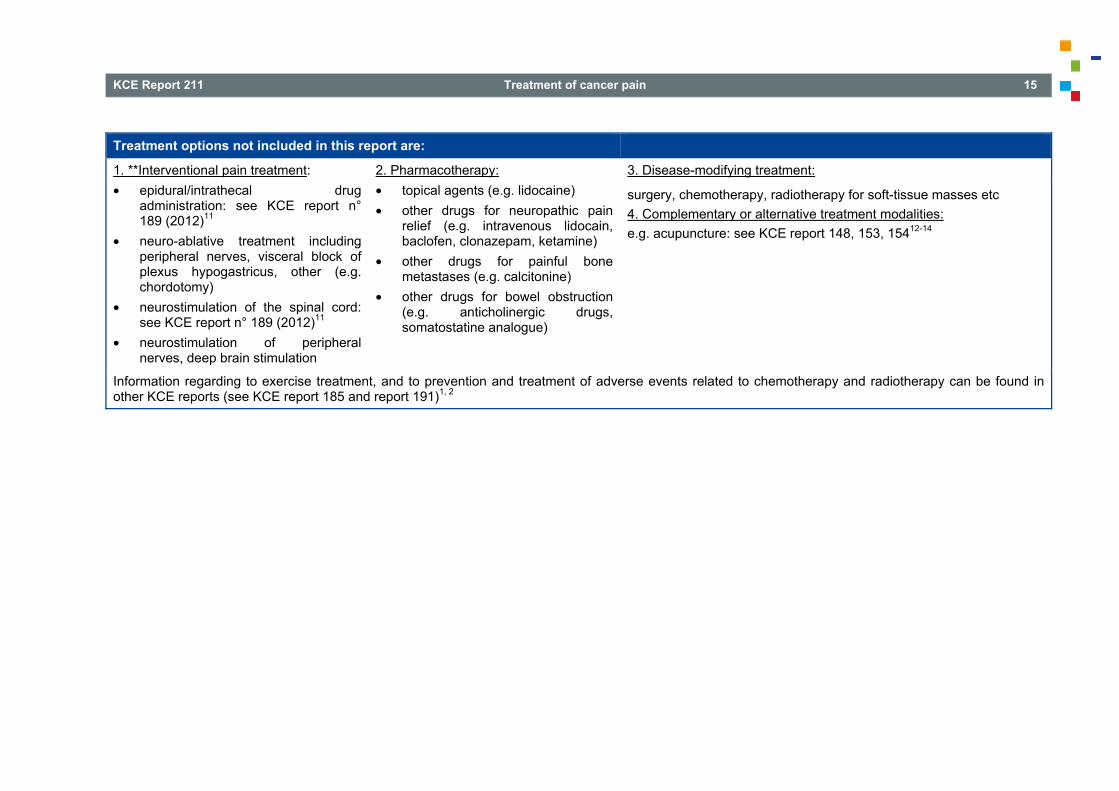
Treatment options not included in this report are:
1. **Interventional pain treatment: • epidural/intrathecal drug
administration: see KCE report n° 189 (2012)11
• neuro-ablative treatment including peripheral nerves, visceral block of plexus hypogastricus, other (e.g. chordotomy)
• neurostimulation of the spinal cord: see KCE report n° 189 (2012)11
• neurostimulation of peripheral nerves, deep brain stimulation
2. Pharmacotherapy: • topical agents (e.g. lidocaine) • other drugs for neuropathic pain
relief (e.g. intravenous lidocain, baclofen, clonazepam, ketamine)
• other drugs for painful bone metastases (e.g. calcitonine)
• other drugs for bowel obstruction (e.g. anticholinergic drugs, somatostatine analogue)
3. Disease-modifying treatment:
surgery, chemotherapy, radiotherapy for soft-tissue masses etc 4. Complementary or alternative treatment modalities: e.g. acupuncture: see KCE report 148, 153, 15412-14
Information regarding to exercise treatment, and to prevention and treatment of adverse events related to chemotherapy and radiotherapy can be found in other KCE reports (see KCE report 185 and report 191)1, 2

16 Treatment of cancer pain KCE Report 211
2 METHODS 2.1 Research questions 2.1.1 Methodology No universally accepted and clinically useful classification system for cancer pain exists, nor for the treatment modalities of cancer related pain (Hjermstad 2009, Knudsen 2009, Portenoy 2011)7, 15, 16. Therefore, an overview was made of the most relevant medical treatment options, based on a quick search for recent literature reviews and guidelines on this topic. Rehabilitation interventions, physical exercise or psychosocial interventions, combined or not with medical interventions, were considered to be out of scope (see 1.2), as were complementary or alternative treatment modalities e.g. acupuncture. Additionally, a list of potentially relevant outcome domains was included. This overview was presented to a group of Belgian experts in January 2012 (see also 2.11). The group consisted of health care professionals involved in the care for cancer patients (see colophon). They were asked to prioritize these lists by indicating which topics they considered to be of most interest to clinical practice. The experts could also complete the lists if necessary (in Appendix I: see 1). Based on this expert consultation, and taking into account the time schedule of the project, the nine most relevant treatment options and four most appropriate outcome domains were selected for inclusion in the report (see 2.1.2).
2.1.2 Research questions, included medical treatment options and outcomes
In collaboration with a group of Belgian experts (see also 2.10), the main research question to this report was defined as: • Which evidence exists on the treatment of cancer-related pain in adults
by: 1. Paracetamol and non-steroidal anti-inflammatory agents (NSAIDs); 2. Opioids; 3. Corticosteroids; 4. Antidepressants; 5. Anticonvulsants, especially gabapentin, pregabalin; 6. Radiotherapy for painful bone metastases; 7. Radionuclides for painful bone metastases; 8. Bisphosphonates for painful bone metastases; 9. Visceral plexus block of plexus coeliacus.
The list of most relevant outcomes to be studied was defined as: 1. Pain intensity, pain reduction, pain relief; 2. Quality of life, psychological well-being; 3. Functional impairment due to pain; 4. Side-effects of the treatment. The items 1-2-3 should be measured by a validated scale.

KCE Report 211 Treatment of cancer pain 17
2.1.3 Excluded medical treatment options As already mentioned, rehabilitation interventions, physical exercise or psychosocial interventions, combined or not with medical interventions, were considered to be out of scope (see 1.2); information regarding to exercise treatment, prevention and treatment of adverse events related to chemotherapy and radiotherapy can be found in other KCE reports (see KCE report 185 and 191)1, 2. Complementary or alternative treatment modalities e.g. acupuncture were also considered to be out of scope (see KCE report 148, 153, 15412-14. For the medical treatment options that were considered by the consulted experts (see 2.1.1) to be of relatively lower interest to clinical practice, as compared to the included medical treatment options, the reader is referred to the Appendix (Appendix I: see 1) for all details. The most important excluded treatment options are: Interventional pain treatment: • epidural/intrathecal drug administration: this topic has been included in
KCE report n° 189 (2012)11, • neuro-ablative treatment including peripheral nerves, visceral block of
plexus hypogastricus, other (e.g. chordotomy), • neurostimulation of the spinal cord: this topic has been included in
KCE report n° 189 (2012)11, • neurostimulation of peripheral nerves, deep brain stimulation. Pharmacotherapy: • topical agents (e.g. lidocaine), • other drugs for neuropathic pain relief (e.g. intravenous lidocain,
baclofen, clonazepam, ketamine), • other drugs for painful bone metastases (e.g. calcitonine), • other drugs for bowel obstruction (e.g. anticholinergic drugs,
somatostatine analogue). Disease-modifying treatment: surgery, chemotherapy, radiotherapy for soft-tissue masses etc.
2.2 Definitions Pain is a complex phenomenon, and it has many dimensions including physical, functional, psychological, social and spiritual aspects. All these aspects must be addressed in order to improve pain experience, functional ability and quality of life (Dutch Guideline on cancer pain 2008, Portenoy 2011, SIGN 2008)8, 10, 16. There have been many discussions between pain specialists about specific definitions. In this report, the overall definitions of the International Association for the Study of Pain (IASP)b are used, which have also been used in the previous KCE report on neuromodulation for the management of chronic pain (KCE report n° 189; 2012)11. According to the last IASP update in 2012, pain was defined as: ‘An unpleasant sensory and emotional experience associated with actual or potential tissue damage, or described in terms of such damage’. The full version of the definitions, underlining specific related aspects, is available at the IASP website. The evaluation of pain in an individual is inherently subjective, making interpretations of treatment effectiveness more difficult. Pain is termed nociceptive if the sustaining mechanisms are believed to be related to ongoing tissue injury, either somatic or visceral. Pain is termed neuropathic if it is associated with injury to neural tissues and is sustained by damage or dysfunction in the peripheral or central nervous system (Caraceni 1999, Guideline of the MoH Malaysia 2010, Portenoy 2011)6, 9,
16. The generic term, psychogenic pain, is used to label pain that is believed to be predominantly determined by psychological factors. Although psychological processes profoundly affect pain expression and consequences, the label psychogenic pain is rarely applied in patients with active cancer (Caraceni 1999, Portenoy 2011)6, 16. In cancer, nociceptive as well as neuropathic pain mechanisms are frequent, but mixed nociceptive-neuropathic syndromes are common as well (Caraceni 1999)6. Moreover, basic research in this domain has demonstrated that some pathophysiological mechanisms, e.g. induced by chemical mediators secreted by a tumour, can influence nociceptive and neuropathic pain processes at the same time (Hans 2009)17. More often than in other painful conditions, cancer pain shows an evolution in time, and it can evolve e.g. from one predominant type of pain into another, or
b www.iasp-pain.org

18 Treatment of cancer pain KCE Report 211
into a mixed nociceptive-neuropathic type of pain. This evolution can be related to the cancer process and/or related to the cancer treatment. Other co-existing painful conditions can add to the burden (Dutch Guideline on cancer pain 2008)8. Although the pathophysiology underlying a cancer pain syndrome often cannot be precisely determined, it is still conventional practice to infer the predominating type of mechanism or mechanisms on the basis of clinical information, and to use this to rationalize treatment(Caraceni 1999, Knudsen 2009, Portenoy 2011)6, 7, 16.
2.3 Literature search For all research topics, the search first focused on systematic reviews and meta-analyses. If guidelines were identified that were clearly based on a systematic review of the literature, they were included and treated as a systematic review. The following sources were used: • OVID Medline and PreMedline • EMBASE (Embase.com) • Cochrane Database of Systematic Reviews (Wiley) • DARE (Wiley) • HTA database (Wiley) • National Guideline Clearinghouse, Guidelines International Network,
and websites of organisations in oncology (for list, in Appendix I: see 4.2.1)
An additional search for randomized controlled trials (RCTs) was done (‘RCT Update’). The following sources were used: • OVID Medline and PreMedline • EMBASE (Embase.com) • CENTRAL (Wiley)
Medline and EMBASE searches for systematic reviews and meta-analyses were run on July 20th 2012. The search in the Cochrane Library (including DARE and the HTA database) was run on July 23th 2012. The Guideline websites and websites of organisations in oncology were searched on August 2nd 2012. The search for primary studies (RCTs) was run in Medline, EMBASE and CENTRAL on November 13th 2012. Detailed search strategies can be found in Appendix I: see 3. Given the high number of hits, and given the medical evolution in this domain, it was decided to limit all searches to the last 10 years (2001- 2012).
2.4 Selection criteria The selection criteria are summarized in Table 1. No a-priori criteria for the comparators of the intervention were defined.
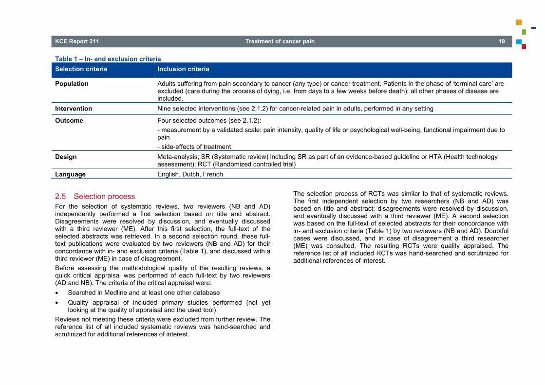
KCE Report 211 Treatment of cancer pain 19
Table 1 – In- and exclusion criteria Selection criteria Inclusion criteria
Population Adults suffering from pain secondary to cancer (any type) or cancer treatment. Patients in the phase of ‘terminal care’ are excluded (care during the process of dying, i.e. from days to a few weeks before death); all other phases of disease are included.
Intervention Nine selected interventions (see 2.1.2) for cancer-related pain in adults, performed in any setting
Outcome Four selected outcomes (see 2.1.2): - measurement by a validated scale: pain intensity, quality of life or psychological well-being, functional impairment due to pain - side-effects of treatment
Design Meta-analysis; SR (Systematic review) including SR as part of an evidence-based guideline or HTA (Health technology assessment); RCT (Randomized controlled trial)
Language English, Dutch, French
2.5 Selection process For the selection of systematic reviews, two reviewers (NB and AD) independently performed a first selection based on title and abstract. Disagreements were resolved by discussion, and eventually discussed with a third reviewer (ME). After this first selection, the full-text of the selected abstracts was retrieved. In a second selection round, these full-text publications were evaluated by two reviewers (NB and AD) for their concordance with in- and exclusion criteria (Table 1), and discussed with a third reviewer (ME) in case of disagreement. Before assessing the methodological quality of the resulting reviews, a quick critical appraisal was performed of each full-text by two reviewers (AD and NB). The criteria of the critical appraisal were: • Searched in Medline and at least one other database • Quality appraisal of included primary studies performed (not yet
looking at the quality of appraisal and the used tool) Reviews not meeting these criteria were excluded from further review. The reference list of all included systematic reviews was hand-searched and scrutinized for additional references of interest.
The selection process of RCTs was similar to that of systematic reviews. The first independent selection by two researchers (NB and AD) was based on title and abstract; disagreements were resolved by discussion, and eventually discussed with a third reviewer (ME). A second selection was based on the full-text of selected abstracts for their concordance with in- and exclusion criteria (Table 1) by two reviewers (NB and AD). Doubtful cases were discussed, and in case of disagreement a third researcher (ME) was consulted. The resulting RCTs were quality appraised. The reference list of all included RCTs was hand-searched and scrutinized for additional references of interest.

20 Treatment of cancer pain KCE Report 211
2.6 Quality appraisal For the quality appraisal of systematic reviews, the AMSTAR instrument was used (Appendix I: see 2.1). The first 30 SRs were evaluated by two researchers (NB and AD). Doubtful cases were discussed, and in case of disagreement a third researcher (ME) was consulted. All other SRs were evaluated by one researcher, the result was checked by the other reviewer. Three items of this checklist were considered key for labelling a review as high quality: • Item 3: Was a comprehensive literature search performed? • Item 7: Was the scientific quality of the included studies assessed and
documented? • Item 9: Were the methods used to combine the findings of studies
appropriate? For the quality appraisal of RCTs, the same evaluation process was applied. The first 30 RCTs were evaluated by two researchers (NB and AD). Doubtful cases were discussed, and in case of disagreement a third researcher (ME) was consulted. All other RCTs were evaluated by one researcher, the result was checked by the other reviewer. The evaluation instrument was the Cochrane Collaboration’s tool for assessing risk of bias18 (Appendix I: see 2.2). Judgement of each item includes three categories: ‘low risk of bias’, ‘high risk of bias’, and ‘unclear risk of bias’. For each criterion the definitions as described in the Cochrane Handbook18 were used. If applicable, risk of bias for the items regarding detection bias and attrition bias were assessed per class of outcomes. At the end, each study was labelled as low risk of bias, unclear risk of bias or high risk of bias according to the criteria described in the Cochrane Handbook; the three items ‘random sequence generation’, ‘blinding of participants and personnel’, ‘complete outcome data’ were considered key for labelling an RCT as high quality. An overview of the quality appraisal of the included SRs and RCTs is given in Appendix II and III: see 1. Additionally, the risk of bias is reported in the evidence tables for each individual study (Appendix II and III: see 4).
2.7 Data extraction and grading of evidence Data extraction was done by 1 researcher (AD or NB) using the standard KCE template for evidence tables, and checked by a second researcher (ME) (in Appendix II and III: see 4). Systematic reviews in which the link between included primary studies and overall review conclusions was not described in a transparent way, were used as a source of RCTs only. Other systematic reviews were extracted into evidence tables and discussed in full; if available, pooled results from meta-analyses were also extracted. For each of the included topics (e.g. bisphosphonates, celiac plexus block), the list of the newly identified RCTs (‘update RCTs’) was compared to a list of all RCTs included in the systematic reviews on that topic, and those RCTs that were not yet included in the systematic reviews were extracted into evidence tables and discussed in full. These RCTs were pooled in a meta-analysis if appropriate; data of meta-analyses from systematic reviews were included if appropriate and if the required data were readily available in the systematic review. Meta-analyses were performed according to the statistical guidelines described in the Cochrane Handbook (http://www.cochrane.org/training/cochrane-handbook) and by the use of Review Manager Software (Review Manager 2011). For each clinical question, conclusions were formulated at the level of individual treatment outcomes using standardized language (Table 4). A level of evidence was assigned by the research team (AD, NB, ME) to each conclusion using the GRADE system (Balshem 2011)19. The quality of evidence reflects the extent to which a guideline panel’s confidence in an estimate of the effect was adequate to support a particular recommendation. According to GRADE, we classified the quality of evidence into 4 categories: high, moderate, low, and very low (Table 2). GRADE for guidelines was used, meaning that the evidence across outcomes and across studies for a particular recommendation was assessed. The following quality elements for intervention studies were evaluated: study limitations, inconsistency, indirectness, imprecision and publication bias.

KCE Report 211 Treatment of cancer pain 21
As only RCTs were considered in this review, quality rating was initially considered to be of high level (Table 2). The rating was then downgraded if needed based on the judgement of the different quality elements by the assessors (Table 3). Each quality element considered to have serious or very serious risk of bias was rated down -1 or -2 points respectively. Judgement of the overall confidence in the effect estimate was also taken into account. We considered confidence in estimates as a continuum and the final rating of confidence could differ from that suggested by each separate domain (Guyatt 2013)20 Reasons for (no) downgrading are summarized in the GRADE profiles in Appendix II and III: see 2. Since upgrading of the level of evidence is primarily relevant to observational studies and our report focused on RCTs, upgrading was not considered applicable although theoretically possible. In practice this option never occurred.
Table 2 – Levels of evidence according to the GRADE system Quality level Definition Methodological Quality of Supporting Evidence
High We are very confident that the true effect lies close to that of the estimate of the effect
RCTs without important limitations or overwhelming evidence from observational studies
Moderate We are moderately confident in the effect estimate: the true effect is likely to be close to the estimate of the effect, but there is a possibility that it is substantially different
RCTs with important limitations (inconsistent results, methodological flaws, indirect, or imprecise) or exceptionally strong evidence from observational studies
Low Our confidence in the effect estimate is limited: the true effect may be substantially different from the estimate of the effect
RCTs with very important limitations or observational studies or case series
Very low We have very little confidence in the effect estimate: the true effect is likely to be substantially different from the estimate of the effect

22 Treatment of cancer pain KCE Report 211
Table 3 – Downgrading the quality rating of evidence using GRADE Quality element Reasons for downgrading
Risk of bias For each study reporting the selected outcome, possible risk of bias introduced by lack of allocation concealment, lack of blinding, lack of intention-to-treat analysis, loss of follow-up and selective outcome reporting were assessed. Additionally, other limitations such as stopping early for benefit and use of unvalidated outcome measures were taken into consideration. Level of evidence was downgraded if studies were of sufficiently poor quality. Downgrading was omitted if studies with low risk of bias were available that lead to similar conclusions as the studies with a high risk of bias.
Inconsistency Downgrading the level of evidence for inconsistency of results was considered in the following situations: point estimates vary widely across studies, confidence intervals show minimal or no overlap, the statistical test for heterogeneity shows a low p-value or the I2 is large. If large variability in magnitude of effect remained unexplained, the quality of evidence was rated down. If the body of evidence included only a single study, rating was downgraded with -2 points as consistency of results cannot be judged and there is no proof that results are reproducible. The only exception was the availability of one large multicentre trial without heterogeneity across sites.
Indirectness Quality rating was downgraded for indirectness in case the trial population or the applied intervention differed significantly from the population or intervention of interest. Also, the use of surrogate outcomes could lead to downgrading. A third reason for downgrading for indirectness occured when the studied interventions were not tested in a head-to-head comparison.
Imprecision Evaluation of the imprecision of results was primarily based on examination of the 95% CI. Quality was rated down if clinical action would differ if the upper versus the lower boundary of the 95% CI represented the truth. In general, 95% CIs around relative effects were used for evaluation, except when the event rate was low in spite of a large sample size. To examine the 95% CIs, the clinical decision threshold (CDT) was defined. When the 95% CI crossed this clinical decision threshold, the quality level was rated down. A relative risk reduction (RRR) of 25% was defined as CDT by default and adapted if deemed appropriate e.g. in case of a low risk intervention. Even if 95% CIs appeared robust, level of evidence could be rated down because of fragility. To judge fragility of results, it is suggested to calculate the number of patients needed for an adequately powered (imaginary) single trial, also called the optimal information size (OIS). If the total number of patients included in a systematic review was less than the calculated OIS, rating down for imprecision was considered. For calculations, a RRR of 25% was used, unless otherwise stated. When the OIS could not be calculated, a minimum of 300 events for binary outcomes and a minimum of 400 participants for continuous outcomes were used as a rule of thumb.
Reporting bias Quality rating was downgraded if publication bias was suggested by analysis using funnel plots or searching of trial registries. Publication bias was also suspected if results came from small, positive industry-sponsored trials only.
KCE Report 211 Treatment of cancer pain 23
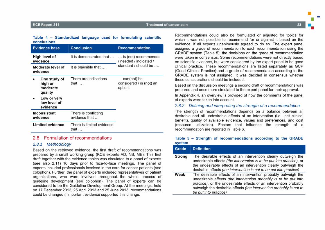
Table 4 – Standardized language used for formulating scientific conclusions Evidence base Conclusion Recommendation
High level of evidence
It is demonstrated that … … is (not) recommended / needed / indicated / standard / should be ….
Moderate level of evidence
It is plausible that …
• One study of high or moderate quality
• Low or very low level of evidence
There are indications that …
… can(not) be considered / is (not) an option.
Inconsistent evidence
There is conflicting evidence that …
Limited evidence There is limited evidence that …
2.8 Formulation of recommendations 2.8.1 Methodology Based on the retrieved evidence, the first draft of recommendations was prepared by a small working group (KCE experts AD, NB, ME). This first draft together with the evidence tables was circulated to a panel of experts (see also 2.11) 10 days prior to face-to-face meetings. The panel of experts included professionals involved in the care for cancer patients (see colophon). Further, the panel of experts included representatives of patient organizations, who were involved throughout the whole process of guideline development (see colophon). The panel of experts can be considered to be the Guideline Development Group. At the meetings, held on 17 December 2012, 25 April 2013 and 25 June 2013, recommendations could be changed if important evidence supported this change.
Recommendations could also be formulated or adjusted for topics for which it was not possible to recommend for or against it based on the evidence, if all experts unanimously agreed to do so. The expert panel assigned a grade of recommendation to each recommendation using the GRADE system (Table 5); the decisions on the grade of recommendation were taken in consensus. Some recommendations were not directly based on scientific evidence, but were considered by the expert panel to be good clinical practice. These recommendations are listed separately as GCP (Good Clinical Practice) and a grade of recommendation according to the GRADE system is not assigned. It was decided in consensus whether these considerations should be included. Based on the discussion meetings a second draft of recommendations was prepared and once more circulated to the expert panel for their approval. In Appendix 4, an overview is provided of how the comments of the panel of experts were taken into account. 2.8.2 Defining and interpreting the strength of a recommendation The strength of recommendations depends on a balance between all desirable and all undesirable effects of an intervention (i.e., net clinical benefit), quality of available evidence, values and preferences, and cost (resource utilization). Factors that influence the strength of a recommendation are reported in Table 6.
Table 5 – Strength of recommendations according to the GRADE system Grade Definition
Strong The desirable effects of an intervention clearly outweigh the undesirable effects (the intervention is to be put into practice), or the undesirable effects of an intervention clearly outweigh the desirable effects (the intervention is not to be put into practice)
Weak The desirable effects of an intervention probably outweigh the undesirable effects (the intervention probably is to be put into practice), or the undesirable effects of an intervention probably outweigh the desirable effects (the intervention probably is not to be put into practice)
24 Treatment of cancer pain KCE Report 211
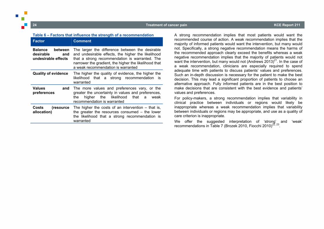
Table 6 – Factors that influence the strength of a recommendation Factor Comment
Balance between desirable and undesirable effects
The larger the difference between the desirable and undesirable effects, the higher the likelihood that a strong recommendation is warranted. The narrower the gradient, the higher the likelihood that a weak recommendation is warranted
Quality of evidence The higher the quality of evidence, the higher the likelihood that a strong recommendation is warranted
Values and preferences
The more values and preferences vary, or the greater the uncertainty in values and preferences, the higher the likelihood that a weak recommendation is warranted
Costs (resource allocation)
The higher the costs of an intervention – that is, the greater the resources consumed – the lower the likelihood that a strong recommendation is warranted
A strong recommendation implies that most patients would want the recommended course of action. A weak recommendation implies that the majority of informed patients would want the intervention, but many would not. Specifically, a strong negative recommendation means the harms of the recommended approach clearly exceed the benefits whereas a weak negative recommendation implies that the majority of patients would not want the intervention, but many would not (Andrews 2013)21. In the case of a weak recommendation, clinicians are especially required to spend adequate time with patients to discuss patients’ values and preferences. Such an in-depth discussion is necessary for the patient to make the best decision. This may lead a significant proportion of patients to choose an alternative approach. Fully informed patients are in the best position to make decisions that are consistent with the best evidence and patients’ values and preferences. For policy-makers, a strong recommendation implies that variability in clinical practice between individuals or regions would likely be inappropriate whereas a weak recommendation implies that variability between individuals or regions may be appropriate, and use as a quality of care criterion is inappropriate. We offer the suggested interpretation of ‘strong’ and ‘weak’ recommendations in Table 7 (Brozek 2010, Fiocchi 2010)22, 23.
KCE Report 211 Treatment of cancer pain 25
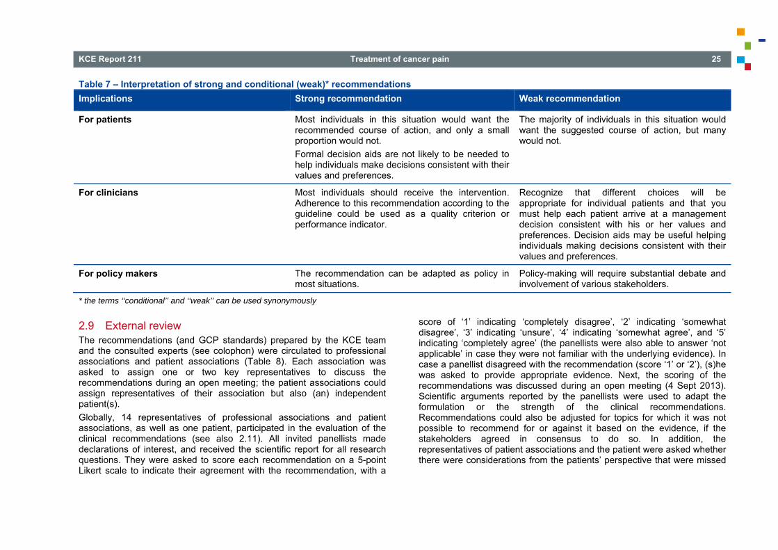
Table 7 – Interpretation of strong and conditional (weak)* recommendations Implications Strong recommendation Weak recommendation
For patients Most individuals in this situation would want the recommended course of action, and only a small proportion would not. Formal decision aids are not likely to be needed to help individuals make decisions consistent with their values and preferences.
The majority of individuals in this situation would want the suggested course of action, but many would not.
For clinicians Most individuals should receive the intervention. Adherence to this recommendation according to the guideline could be used as a quality criterion or performance indicator.
Recognize that different choices will be appropriate for individual patients and that you must help each patient arrive at a management decision consistent with his or her values and preferences. Decision aids may be useful helping individuals making decisions consistent with their values and preferences.
For policy makers The recommendation can be adapted as policy in most situations.
Policy-making will require substantial debate and involvement of various stakeholders.
* the terms ‘‘conditional’’ and ‘‘weak’’ can be used synonymously
2.9 External review The recommendations (and GCP standards) prepared by the KCE team and the consulted experts (see colophon) were circulated to professional associations and patient associations (Table 8). Each association was asked to assign one or two key representatives to discuss the recommendations during an open meeting; the patient associations could assign representatives of their association but also (an) independent patient(s). Globally, 14 representatives of professional associations and patient associations, as well as one patient, participated in the evaluation of the clinical recommendations (see also 2.11). All invited panellists made declarations of interest, and received the scientific report for all research questions. They were asked to score each recommendation on a 5-point Likert scale to indicate their agreement with the recommendation, with a
score of ‘1’ indicating ‘completely disagree’, ‘2’ indicating ‘somewhat disagree’, ‘3’ indicating ‘unsure’, ‘4’ indicating ‘somewhat agree’, and ‘5’ indicating ‘completely agree’ (the panellists were also able to answer ‘not applicable’ in case they were not familiar with the underlying evidence). In case a panellist disagreed with the recommendation (score ‘1’ or ‘2’), (s)he was asked to provide appropriate evidence. Next, the scoring of the recommendations was discussed during an open meeting (4 Sept 2013). Scientific arguments reported by the panellists were used to adapt the formulation or the strength of the clinical recommendations. Recommendations could also be adjusted for topics for which it was not possible to recommend for or against it based on the evidence, if the stakeholders agreed in consensus to do so. In addition, the representatives of patient associations and the patient were asked whether there were considerations from the patients’ perspective that were missed
26 Treatment of cancer pain KCE Report 211
in the formulating of the recommendations. It was decided in consensus whether these considerations should be included. In Appendix 4, an overview is provided of how the comments of the stakeholders were taken into account.
Table 8 – List of Professional Associations and Patient Associations to which the recommendations were communicated • Belgische Vereniging voor Medische Oncologie v.z.w.; Société Belge
d'Oncologie Médicale a.s.b.l. (BSMO Belgian Society of Medical Oncology),
• Belgian Pain Society vzw/asbl, • Domus Medica (General Practitioners, Flanders), • BVAR-SBAR Belgische Vereniging voor Anesthesie en Reanimatie;
Société Belge d’Anesthésie et de Réanimation (S.A.R.B. Society for Anesthesia and Resuscitation of Belgium),
• BELNUC Belgische Vereniging van Nucleaire Geneeskunde; Société Belge de Médecine Nucléaire,
• Federatie Palliatieve Zorg Vlaanderen vzw (Federation Palliative Care Flanders),
• Fédération Wallonne des Soins Palliatifs, • Stichting tegen Kanker; Fondation contre le Cancer, • Werkgroep Hersentumoren vzw.
2.10 Final validation As part of the standard KCE procedures, an external scientific validation of the report was conducted prior to its publication. Such validation process was done on 23 May 2013 and 24 Sept 2013. The current guideline was reviewed prior to its publication by 3 independent validators (see Appendix I: see 6.4; cf. names in the colophon), making use of the Agree II checklist. The validation process was chaired by CEBAM (Belgian Centre for Evidence-Based Medicine; Belgian Branch of the Dutch Cochrane Centre; www.cebam.be). The validation of the report results from a consensus or a voting process between the validators.
2.11 Development of the guideline: project team, involved experts, panel of stakeholders
The scientific report, including the literature search, evidence report and conclusions were written by a team of 3 KCE experts (Appendix I: see 6.3). The composition of the Guideline Development Group, i.e. the panel of consulted experts consisting of professional experts and representatives of patient associations, can be found in Appendix I: see 6.1; their conflicts of interest can be found in the colophon of the report. The professional experts were consulted to prioritize a list of medical treatment options and outcome domains; based on this prioritization the treatment options and outcome domains included in the report were defined (see also 2.1). Further, the professional experts and the representatives of patient associations were consulted three times (17 December 2012; 25 April 2013; 25 June 2013) about a (preliminary) version of the scientific report. Their comments were discussed during meetings, but they did not co-author the scientific report. This discussion also included all draft recommendations, and the professional experts and the representatives of patient associations decided in consensus on the grading of the recommendations (see also 2.8). The composition of the panel of stakeholders, consisting of representatives of professional associations, representatives of patient associations, and patients, can be found in Appendix I: see 6.2; their conflicts of interest can be found in the colophon of the report. The stakeholder meeting was helt on 4 Sept 2013. For the contribution of the panel of stakeholders, see 1.1.

KCE Report 211 Treatment of cancer pain 27
3 SEARCH RESULTS 3.1 Systematic reviews The searches yielded the following number of hits per database:
Table 9 – Number of hits per database for systematic reviews search Database Number of hitsCochrane Database of Systematic Reviews 209Medline 2 026PreMedline 19EMBASE 1 017DARE 31HTA database 4Duplicates were discarded and 3 140 hits were reviewed on title and abstract; 971 papers were selected for full-text review. Additionally, 19 guidelines based on systematic reviews were selected for full text evaluation from the Guideline websites and websites of organisations in oncology (Appendix I: see 4.2.1). Hand searching yielded 6 extra publications. Based on the full-text (and the quick critical appraisal) 45 papers fulfilled the inclusion criteria and were subsequently quality-appraised (Appendix II and III: see 1). The 45 papers were classified according to the 9 included medical interventions (e.g. bisphosphonates, celiac plexus block) and are listed in the chapter ‘Search results’ for the respective medical interventions; papers covering more than one medical intervention are reported in all chapters of interest. An overview of the finally included SRs per topic can be found in Appendix II (see 5) and Appendix III (see 5); an overview of the excluded SRs per topic can be obtained from the authors on request.

28 Treatment of cancer pain KCE Report 211
Figure 1 – Study flow of selection of SRs (CDSR, Medline, PreMedline, Embase, DARE, HTA database)

KCE Report 211 Treatment of cancer pain 29
3.2 Randomized controlled trials The searches yielded the following number of hits per database:
Table 10 – Number of hits per database for RCT search Database Number of hitsMedline 1 374PreMedline 67EMBASE 2 171CENTRAL 919Duplicates were discarded and 3 817 hits were reviewed on title and abstract; 303 papers were selected for full-text review. Based on the full-text, 37 papers fulfilled the inclusion criteria and were subsequently quality-appraised (Appendix II and III: see 1). Additionally, 153 RCTs were extracted for futher analyses from the finally included systematic reviews. The 190 papers were classified according to the 9 included medical interventions (e.g. bisphosphonates, celiac plexus block) and are listed in the chapter ‘Search results’ for the respective medical interventions; papers covering more than one medical intervention are reported in all chapters of interest. An overview of included and excluded RCTs per topic can be found in Appendix II (see 5) and Appendix III (see 5).

30 Treatment of cancer pain KCE Report 211
Figure 2 – Study flow of selection of RCTs
KCE Report 211 Treatment of cancer pain 31
4 EVIDENCE REPORT 4.1 Introduction 4.1.1 Pain Assessment Pain is a highly complex and subjective phenomenon, including physical, functional, psychological, social and spiritual aspects. An important condition for adequate pain treatment is a systematic and comprehensive assessment of pain, encompassing these multidimensional components. Pain assessment is a responsibility of all health care providers (physicians, nurses, pharmacists, etc), and team work or an interdisciplinary approach to cancer pain is essential (SIGN 2008, Dutch Guideline on cancer pain 2008, Guideline of the MoH Malaysia 2008, Portenoy 2011)8-10. Pain assessment should be performed prior to treatment in order to plan for appropriate interventions, and after treatment initiation to assess its effectiveness. It should aim to determine: • the pathophysiology of pain, • pain intensity as well as other aspects of pain, such as the type of
pain, its location, duration etc (see also 4.1.3.2), • the impact of pain on a person’s functions, psychosocial and spiritual
well-being, and quality of life, • the response to pain interventions. Similar to other clinical assessment, a complete pain assessment requires a detailed history and physical examination, as well as standardized assessment tools. This should be completed by laboratory tests, medical imaging or other diagnostic tests if these are necessary to determine appropriate clinical management. The assessment should be repeated if treatment does not alleviate the pain even after careful adjustment (SIGN 2008, Dutch Guideline on cancer pain 2008, Guideline of the MoH Malaysia 2008)8-10.
4.1.2 Pain assessment tools Many different pain assessment tools are used throughout the world, and there exists no universally accepted tool for the assessment of cancer pain (Dutch Guideline on cancer pain 2008)8. It is beyond the scope of this report to conduct a systematic literature review on pain assessment tools. Rather, a narrative overview will be presented of the information found in existing generic clinical guidelines on cancer pain (SIGN 2008, Dutch Guideline on cancer pain 2008, Guideline of the MoH Malaysia 2008) which were retained from the literature search (see 3.1) )8-10. A list of common pain assessment scales included in this report can be found in Appendix I: see 5.1 to 5.5. Pain assessment tools include unidimensional and multidimensional measures. Unidimensional pain scales only measure pain intensity; they are easy to implement in clinical practice. The most commonly used unidimensional assessment tools are the Visual Analogue Scale (VAS), Numerical Rating Scale (NRS) and Verbal Rating Scale (VRS), all of which are valid and reliable (Appendix I: see 5.1). In clinical practice, a numerical rating scale (NRS) is probably to be preferred over a VAS scale (and over a verbal rating scale VRS) because of its better responsiveness and ease of use (Hjermstad M 2011, Dutch Guideline on cancer pain 2008)8, 24. Multidimensional pain instruments measure pain intensity as well as other pain characteristics; they can be used for complex pain syndromes. Examples are the McGill Pain questionnaire and the Brief Pain Inventory (BPI), which incorporate NRS and VRS (Appendix I: see 5.1). These are reliable scales that have been validated in different cultures (SIGN 2008, Dutch Guideline on cancer pain 2008, Guideline of the MoH Malaysia 2008)8-10. It has been shown that health care professionals tend to underestimate the level of pain a patient is experiencing, while family members might overestimate it (SIGN 2008, Guideline of the MoH Malaysia 2008)8-10.

32 Treatment of cancer pain KCE Report 211
The patient, if competent and able to communicate, is the most reliable assessor of pain and should, where possible, be the prime assessor of his or her pain. A pain diary completed by the patient can be a useful tool to provide an overview of the evolution of the pain. Observational pain rating scales completed by health care professionals or family members should be preferred in patients who cannot complete a self assessment scale (SIGN 2008, Dutch Guideline on cancer pain 2008, Guideline of the MoH Malaysia 2008)8-10. When starting treatment for pain, patients should be given information about pain and instruction about pain management; and they should be encouraged to take an active role in their pain management. One of the keys to successful control of cancer pain is regular review to determine the effectiveness of treatment. The SIGN 2008 guideline suggests to carry out pain assessment at least daily when pain is not adequately controlled (SIGN 2008)10. A pain dairy completed by the patient can be a useful tool to provide an overview of the evolution of the pain. 4.1.3 Severity of pain
4.1.3.1 Mild, moderate or severe pain intensity Pain intensity is often described as mild, moderate or severe. A non-systematic review of RCTs investigated the classification of pain intensity into categories and the relationship of these to the continuous scale of the visual analogue scale (VAS) (0-100)10, 25. Individual pain scores for each patient were assessed by a categorical scale (none, mild, moderate or severe) and by a VAS (0-100). Comparisons between the scores showed that 85% of patients scoring moderate pain had a VAS of >30 mm and 85% of patients recording severe pain had a VAS of >54 mm. There was a significant difference between VAS scores for those reporting moderate or severe pain (p<0.001). In line with this publication, the SIGN guideline (2008)10 defined mild pain as <3 on a scale to 10; mild to moderate pain as 3-6; and severe pain as >6. In clinical practice, a numerical rating scale (NRS) is probably to be preferred over a VAS scale (and over a verbal rating scale VRS) because of its better responsiveness and ease of use, as already pointed out before (see 4.1.2 (Hjermstad M 2011, Dutch Guideline on cancer pain 2008))8, 24.
For the 11-point numerical rating scale (NRS), cut-off points to categorize the intensity of cancer pain have been described in the literature. Serlin 1995 and Li 2007 concluded, based on their research, that pain scores of (1- 4) on a numerical rating scale correlated with mild pain, (5 - 6) with moderate and (7 - 10) with severe pain; these cut-off points are used in the Guideline of the MoH Malaysia on cancer pain(2010)9, 26, 27. Other researchers (Paul 2005) found arguments to define mild pain as (1- 4) on a numerical scale (0 - 10); moderate pain as (>4 - 7); and severe pain as (>7 - 10), allowing for pain intensity scores that are not whole numbers28. In the present review the cut-offs presented in Table 11will be used, unless specified otherwise.
Table 11 – Categorization of intensity of cancer pain on a numerical rating scale (0-10) Pain severity Score on a numerical rating scale
(0-10)
mild pain 1 - 4
moderate pain >4 - 7
severe pain >7 - 10
4.1.3.2 Other characteristics of pain Although pain intensity is one of the major characteristics of pain and probably the most important one, other dimensions should be considered as well: the temporal pattern of pain (pain fluctuations, variations in intensity and occurrence), pain location, pain interference (how much components of health-related quality of life are reduced by the pain), the type of pain or pain quality (throbbing, lancinating or burning...), pain duration, the prevalence of sleep disturbances etc (Hjermstad 2008, Holen 2006, Knudsen 2012)29-31. These aspects also influence the burden of pain, but so far no pain assessment tool exists that covers all dimensions that are considered to be the most relevant ones to cancer patients receiving palliative care (Holen 2006)30.

KCE Report 211 Treatment of cancer pain 33
4.1.3.3 Clinically important improvement of pain Concerning pain intensity, recent research in the domain of chronic pain learned that, at the patient level, a score change on a 0 to 10 NRS or equivalent pain intensity scale of approximately 2 points or 30% to 36% represents ‘much better,’ ‘much improved,’ or ‘meaningful’ decreases in chronic pain. A decrease of 4 points or 50% appears to represent a substantial (‘very much improved’) change in pain, which patients have also considered ‘treatment success’ or ‘satisfactory improvement’. A change of approximately 1 point, or percentage changes of approximately 15% to 20% represent minimally but perhaps not very important decreases (Farrar 2001, Dworkin 2008)32, 33. Evidence is growing that chronic pain patients regard a pain intensity reduction of 50-70% as a clinical success and ideally want pain to be no worse than mild (Moore 2010; Dworkin 2008)32, 34. Further, there is evidence that a pain intensity score of ‘5’ or more has a major impact on functioning in daily life, which is also considered to be a critical threshold (Paul 2005, Serlin 1995)27, 28. 4.1.4 The World Health Organization analgesic ladder In 1986, the World Health Organization (WHO) launched a three-step analgesic ladder as a systematic approach to cancer pain control35. The regimen of analgesia is based on severity of pain starting with simple analgesics for mild or moderate pain, and progressing to opioid analgesics if the pain persists. If the patient presents with severe pain, opioids can be considered immediately, with or without the addition of non-opioids (see Figure 3). If opioids are considered for mild to moderate pain, weak opioids should be considered (Step II opioids, e.g. codeine). If the pain persists, or for the treatment of severe pain, strong opioids should be considered (Step III opioids, e.g. morphine). The type and cause of the pain influence the choice of adjuvant analgesics (for definition of adjuvant analgesic, see chapter 4.5 and 4.6 ). For the classification of pain intensity into mild, moderate or severe, see 4.1.3.
Figure 3 – The 3-step analgesic ladder developed by the World Health Organization (WHO)36
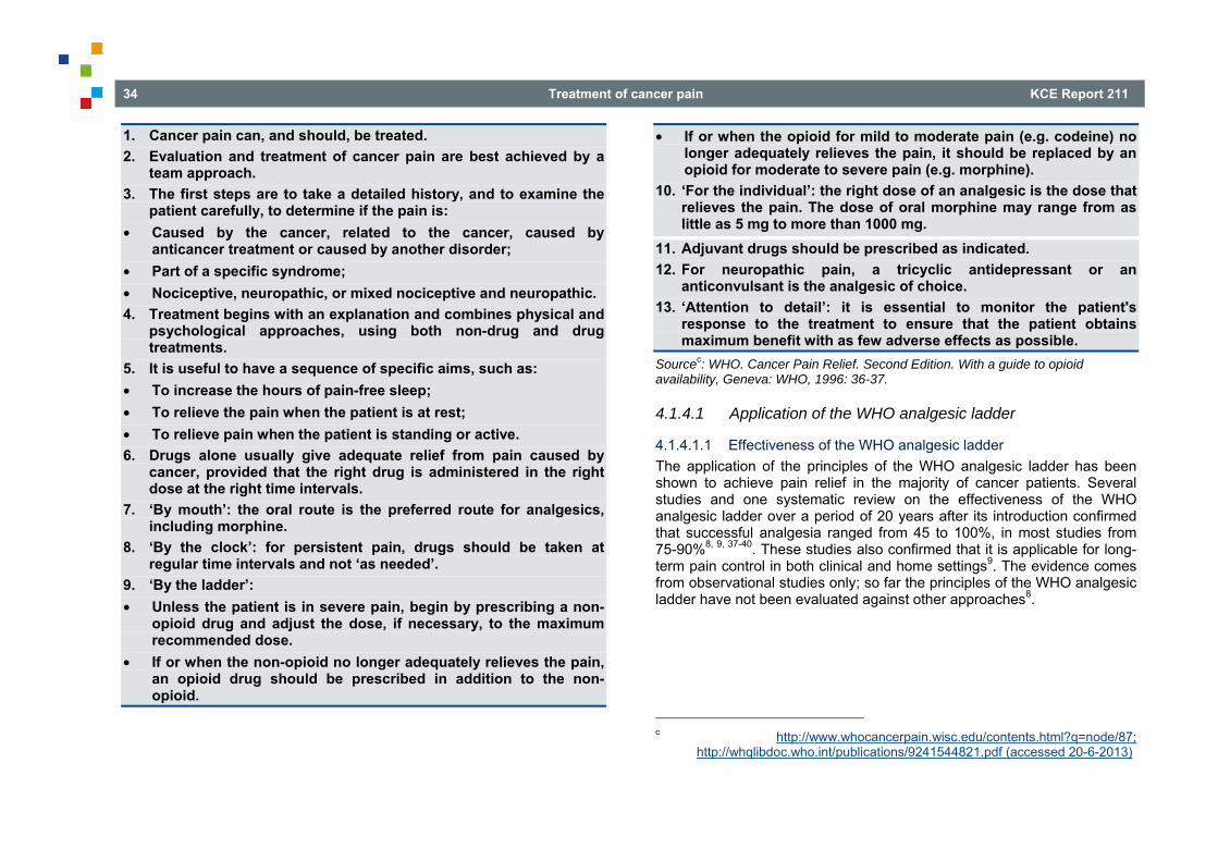
34 Treatment of cancer pain KCE Report 211
1. Cancer pain can, and should, be treated. 2. Evaluation and treatment of cancer pain are best achieved by a
team approach. 3. The first steps are to take a detailed history, and to examine the
patient carefully, to determine if the pain is: • Caused by the cancer, related to the cancer, caused by
anticancer treatment or caused by another disorder; • Part of a specific syndrome; • Nociceptive, neuropathic, or mixed nociceptive and neuropathic. 4. Treatment begins with an explanation and combines physical and
psychological approaches, using both non-drug and drug treatments.
5. It is useful to have a sequence of specific aims, such as: • To increase the hours of pain-free sleep; • To relieve the pain when the patient is at rest; • To relieve pain when the patient is standing or active. 6. Drugs alone usually give adequate relief from pain caused by
cancer, provided that the right drug is administered in the right dose at the right time intervals.
7. ‘By mouth’: the oral route is the preferred route for analgesics, including morphine.
8. ‘By the clock’: for persistent pain, drugs should be taken at regular time intervals and not ‘as needed’.
9. ‘By the ladder’: • Unless the patient is in severe pain, begin by prescribing a non-
opioid drug and adjust the dose, if necessary, to the maximum recommended dose.
• If or when the non-opioid no longer adequately relieves the pain, an opioid drug should be prescribed in addition to the non-opioid.
• If or when the opioid for mild to moderate pain (e.g. codeine) no longer adequately relieves the pain, it should be replaced by an opioid for moderate to severe pain (e.g. morphine).
10. ‘For the individual’: the right dose of an analgesic is the dose that relieves the pain. The dose of oral morphine may range from as little as 5 mg to more than 1000 mg.
11. Adjuvant drugs should be prescribed as indicated. 12. For neuropathic pain, a tricyclic antidepressant or an
anticonvulsant is the analgesic of choice. 13. ‘Attention to detail’: it is essential to monitor the patient's
response to the treatment to ensure that the patient obtains maximum benefit with as few adverse effects as possible.
Sourcec: WHO. Cancer Pain Relief. Second Edition. With a guide to opioid availability, Geneva: WHO, 1996: 36-37.
4.1.4.1 Application of the WHO analgesic ladder
4.1.4.1.1 Effectiveness of the WHO analgesic ladder The application of the principles of the WHO analgesic ladder has been shown to achieve pain relief in the majority of cancer patients. Several studies and one systematic review on the effectiveness of the WHO analgesic ladder over a period of 20 years after its introduction confirmed that successful analgesia ranged from 45 to 100%, in most studies from 75-90%8, 9, 37-40. These studies also confirmed that it is applicable for long-term pain control in both clinical and home settings9. The evidence comes from observational studies only; so far the principles of the WHO analgesic ladder have not been evaluated against other approaches8.
c http://www.whocancerpain.wisc.edu/contents.html?q=node/87;
http://whqlibdoc.who.int/publications/9241544821.pdf (accessed 20-6-2013)

KCE Report 211 Treatment of cancer pain 35
4.1.4.1.2 WHO analgesic ladder Step II: where are we now? Despite its success, the WHO ladder has been challenged especially with regard to Step II of the ladder (weak opioid for mild to moderate pain, e.g. codeine, currently also tramadol, see chapter 4.3.3.7). However, a novel approach suggests to by-pass the second step of the WHO analgesic ladder for the treatment of chronic cancer pain. Indeed, Portenoy 2011 argued in his narrative overview of the treatment of cancer pain)16 that, although common practice is still to follow the WHO recommendations, any of the single-entity, pure mu-agonist opioids, such as morphine or oxycodone (see 4.3.1), can be prescribed at doses low enough to be safe for the management of moderate pain and effectively eliminating the second rang of the analgesic ladder. They argued that there exists a genetically established variation in the effects of codeine, and that other evidence suggests that the most favourable opioid in an individual cannot be predicted. Therefore, Portenoy (2011)16 stated that the important principle is that treatment can be initiated with any of the commonly used pure mu-agonist drugs and that the clinician should be prepared to switch, if necessary, to identify the opioid drug that provides the best outcomes. In line with the position of Portenoy (2011)16, the Dutch Guideline on cancer pain (Dutch Guideline on cancer pain 2008)8 stated that it is not recommended to use weak opioids for the treatment of cancer pain. On the other hand, the SIGN 2008 guideline recommended the use of weak opioids for the treatment of mild to moderate cancer pain, in combination with a non-opioid analgesic10. Related evidence To test this novel approach, the SR of Tassinari 2011a retrieved 4 papers41 (Maltoni 2005, Marinangli 2004, Mercadante 2006, Mercadante 1998a)42-
45. Although in the present review no systematic search was performed on this research question, the overview of Tassinari 2011a will be presented narratively because of the interest of the topic41. The Dutch Guideline on cancer pain (Dutch Guideline on cancer pain 2008) underpinned its statement with the studies of Maltoni 2005 and Marinangli 2004; Portenoy 2011 referred to Maltoni 20058, 16, 42, 43. Marinangli 2004 included patients with terminal cancer (out of scope of this review)43 and Mercadante 2006 was a cohort study (design not included in the criteria of this review)44. However, because of the interest in this topic, narrative descriptions will be provided. Maltoni 2005 tested 2 strategies for
the treatment of mild to moderate chronic cancer pain in Italy42. Patients were randomized to conventional strategy (n=24) and innovative strategy (n=30). The conventional strategy (CS) consisted to initiate Step II drugs after ineffective therapy with NSAIDs. When Step II drugs became insufficient, Step III drug treatment was initiated. The innovative strategy (IS) consisted to pass directly from Step I to Step III drugs by-passing the Step II. Pain was measured each day during approximatively 3 years on a 10-point NRS. Side effects were assessed on 5-point scale (from 0=’none’ to 4=’very severe’). IS showed a lower percentage of days with worst pain ≥ 5 (IS 22.8% versus CS 28.6%, p<0.001) and ≥ 7 (IS 8.6% versus CS 11.2%, p=0.023). Grade 3 and 4 anorexia and constipation were more often reported in IS. The authors concluded that IS could reduce some pain scores but also required careful management of side effects. Marinangli 2004 included cancer patients with home palliative care43. They were randomized in a treatment group strategy according to the WHO ladder (CS, n=48) or in a treatment group strategy starting immediately on strong opioids (IS, n=44). The cumulative evaluated weeks of treatment were 503 for CS and 467 for IS. Pain intensity and general condition were measured on a 10-cm VAS. The intensity of side effects was assessed on the 3-point scale (from 0= mild to 2=severe). Quality of life was assessed by a multidimensional questionnaire (Ferrell 1989). Karnofsky Performance Status was also evaluated. Patients treated following IS had a significantly better pain relief than those treated with CS (Mean change from baseline CS -1.92 versus IC -2.61, p<0.041). The general condition assessed by the patients was better with IS than with CS (Mean (SD) CS 4.23±1.36 versus IS 4.98±1.26, p=0.007). No significant difference was observed in terms of quality of life and performance status. The number of episodes of nausea was significant higher in patients treated with IS than in those treated with CS (CS 315 episodes versus IS 437 episodes, p=0.0001). The authors concluded that strong opioids for first-line treatment of pain in patients with terminal cancer should be considered. Mercadante 2006 performed a multicenter prospective study testing the efficacy and tolerability of very low doses of morphine in 110 advanced cancer patients with pain during 4 weeks (95 completed the study)44. Pain intensity was measured using a 10-point numerical rating scale (NRS). Side effects were assessed on 4-point scale (from 0=’not at all’ to 3=’severe’). Significant pain intensity improvement was observed after 1 and 4 week (Mean (SD) pain intensity at baseline 6.1 (5.2-6.9) – at week1 3.2 (2.2-4.3), p<0.01 compared to

36 Treatment of cancer pain KCE Report 211
baseline – at week 4 3.0 (1.9-3.8), p<0.01 compared to baseline). No statistically significant difference was observed in side effects from baseline to week 1 and 4 excepted for constipation and dry mouth. The authors concluded that the WHO ladder should be still encouraged for advanced cancer patients with pain. However, the use of very low doses of morphine could be cost saving in comparison with the commonly used opioids for moderate pain. Mercadante 1998a tested the effectiveness of ‘weak’ opioids when administrated as second step in the analgesic WHO ladder in 32 opioid-naive cancer patients45. Dextropropoxyphene (DPP) was used as a weak opioid and compared to controlled-release morphine. No difference in pain intensity was observed between the 2 molecules after 10 days (VAS 10 cm median (range): DPP 3 (2-5) vs morphine 3 (2-5), ns). The symptom intensity for adverse effects was evaluated by the patients on a scale ranging from 0 to 3 for confusion, constipation, drowsiness, nausea and vomiting, xerostomia. The mean symptom intensity was higher (p<0.01) in the morphine group than in patients treated with DPP for drowsiness, nausea and vomiting and xerostomia. From the results, the authors supported the role of weak opioids as a second step of the analgesic WHO ladder for cancer patients. However, it should be noted that dextropropoxyphene has been withdrawn by the European Medicines Agency because of severe adverse effects and is no longer available in Belgium. Based on the GRADE approach, Tassinari 2011a assessed the level of evidence for supporting the use of the IS instead of CS as very low41. This assessment was mainly supported by the low statistical power of the trials, the use of surrogate endpoints to analyze the efficacy, and the safety. Consequently, according to Tassinari 2011a, the CS should remain the standard approach to the treatment of cancer pain41. Indeed, data were insufficient to recommend the routine use of IS in cancer-related pain. For recommendations related to Step II of the WHO analgesic ladder, see chapter on NSAIDs and paracetamol (see 2.10), and chapter on opioids (see 4.2).
Based on a narrative overview of the available evidence, there are indications that, when therapy with non-opioids is insufficient to treat mild to moderate cancer pain, initiating Step III opioids might provide better pain relief but at the cost of more adverse effects as compared to Step II opioids (very low level of evidence; Tassinari 2011a)41.
Conclusions • There are indications that initiating strong opioids (Step III)
instead of weak opioids (Step II) for mild to moderate cancer pain after conventional treatment with NSAIDs and/or paracetamol failed, might lead to better pain control but at the cost of more side effects (very low level of evidence; Tassinari 2011a).
4.1.4.2 A fourth step in the WHO analgesic ladder? As previously discussed, the WHO ladder approach can help to manage cancer pain for the vast majority of cancer patients, by assisting physicians in the selection of medications according to the level of pain intensity. However, Miguel et al. (2000) proposed a fourth step in the WHO analgesic ladder to address those patients who cannot reach pain relief with the proposed drug scheme, or those who develop undesirable or intolerable side effects36. The fourth step consists in ‘interventional’ procedures including spinal administration of drugs; neurostimulation of the spinal cord, the brain or peripheral nerves; or neuro-ablation including peripheral nerves, visceral block of plexus celiacus or plexus hypogastricus, or other neural structures. Before implementing these invasive analgesic methods as proposed in the Step IV, the risk/benefit ratio should be considered (Miguel 2000)36. Of all these interventional procedures, only celiac plexus block has been included in this report (see 1.7; 2.1.1; 4.10). Some of the other topics have been dealt with in previous KCE reports (see 1.7; 2.1.3).
KCE Report 211 Treatment of cancer pain 37
Good Clinical Practice
• Prior to treatment, an accurate assessment should be performed to determine the cause, type and severity of pain, and its functional and psychosocial impact on the patient. Validated assessment questionnaires should be used as well as clinical evaluation and, if necessary, medical investigations. The assessment should be repeated if treatment does not alleviate the pain even after careful adjustment.
• The patient is the most reliable assessor of pain and should, whenever possible, be the prime assessor of his or her pain.
• Patients with cancer pain should have their pain monitored regularly using unidimensional pain instruments such as visual analogue scales (VAS), numerical rating scales (NRS) or verbal rating scales (VRS). Multidimensional pain instruments should be used for complex pain syndromes. Examples are the McGill Pain questionnaire and the Brief Pain Inventory, which incorporate NRS and VRS. Observational pain rating scales should be preferred in patients who cannot complete a self assessment scale.
• The minimal objective of pain treatment should be a clinically relevant decrease of the pain (on a 0-10 scale, a decrease by 2 points, and/or a decrease of 30%, and preferentially a pain intensity <5).
• Patients should be given information about pain and instruction about pain management; they should be encouraged to take an active role in their pain management.
Recommendation The principles of treatment outlined in the WHO analgesic ladder should be followed when treating pain in patients with cancer (very low level of evidence; strong recommendation).
4.2 NSAIDs and paracetamol 4.2.1 Introduction Non-steroidal anti-inflammatory drugs (NSAIDs) and paracetamol are widely used and easy to administer as analgesics against pain of diverse etiologies. Paracetamol is considered a mild analgesic that is generally safe within a given range of dosages, but at high dosages it may cause considerable hepatic and renal toxicity. NSAIDs have a ceiling effect at which an increase of the dose does not further ameliorate the pain, and there are only few parenteral formulations available. They are also associated with a significant gastrointestinal risk on ulceration, as well as with cardiovascular, hematological, and renal adverse effects (McNicol 2011, Nabal 2012)46, 47. The NSAIDs exert their action mainly through inhibition of the cyclo-oxygenase enzyme (COX), of which two forms exist: COX-1 and COX-2. Most NSAIDs inhibit both forms; the so-called selective COX-2 inhibitors only inhibit the latter. For mild pain, the WHO analgesic ladder (see also chapter 4.1.4) recommends the use of paracetamol and/or NSAIDs, with or without an adjuvant analgesic (Step I). The same drugs are also proposed in addition to opioids for mild to moderate pain (Step II) or for moderate to severe pain (Step III). Many NSAIDs are available but it is unclear which agent and which opioid-NSAID combination are the most efficacious for relieving cancer pain. Following research questions were formulated: • What is the evidence for the efficacy of NSAID/paracetamol versus
placebo? • Is one NSAID more efficacious than another NSAID or paracetamol? • What is the evidence for the efficacy of the addition of a
NSAID/paracetamol to an opioid regimen (or vice versa) (in reduction in dose of initial drug and reduction of side effects)?

38 Treatment of cancer pain KCE Report 211
4.2.2 Search results Three reviews were identified on the effects of NSAIDs or paracetamol in patients with cancer-related pain (Alkhenizan 2004, Nabal 2012 and McNicol 2011)46-48. However, the RCTs in the review of Alkhenizan (2004)48 were also included in the more recent and up-to-date reviews of Nabal (2012)47 and McNicol (2011)46. We therefore decided to only focus on the two other reviews; the conclusions of the review of Alkhenizan 2004 will be mentioned in the discussion of this chapter48. McNicol (2011)46 searched for RCTs until 2003 and included 42 trials. This high quality review discussed the clinical efficacy of NSAIDs/paracetamol for relieveing cancer-related pain, and it evaluated the additional benefit of combining a NSAID/paracetamol with an opioid; the evidence table can be found in Appendix II: Table 8. Two of the RCTs included by McNicol (2011) were excluded from the present review, because these were studies on dosing of NSAIDs, which was considered to be out of scope for the present review. The review of Nabal (2012)47, a review of high quality, included 5 studies from the Cochrane review of McNicol (2011)46 and added 7 new found studies in his analysis on the role of paracetamol and NSAIDs as add-on to opioids for moderate to severe cancer pain (WHO Step III) (search date from 2002 up to 2010); the evidence table can be found in Appendix II: Table 8. Next to the retrieved systematic reviews, also 2 RCTs from the updated search were included (Rodriguez 2003, Sima 2012)49, 50. The results are summarized in evidence tables (see Appendix II: Table 9). The 4 generic reviews (Guideline of the MoH Malaysia 2010, Dutch Guideline on cancer pain 2008, SIGN 2008, Carr 2002)8-10, 51 did not provide detailed information on pooling of results of individual studies and therefore were used as a basis of RCTs only. One RCT (Jameel 1995)52 could be extracted from the guideline of the Dutch Working Group (Dutch Guideline on cancer pain 2008)8. The results of this study are summarized in the evidence tables (see Appendix II: Table 9). Table 12 provides an overview of all RCTs included in the present review. For those RCTs that were extracted from the SRs, only the information presented in these SRs will be discussed.
Table 12 – Overview of RCTs per comparison Head comparison
Sub comparison
NSAID vs placebo
• Ketorolac vs placebo: o Carlsson 1990 (<McNicol 2011) o Staquet 1989 (<McNicol 2011)
• Ketoprofen vs placebo: o Stambaugh 1988a (<McNicol 2011)
• Aspirin vs placebo: o Martino 1976 (<McNicol 2011) o Moertel 1971 (<McNicol 2011) o Moertel 1974 (<McNicol 2011) o Ventafridda 1975 (<McNicol 2011)
• Paracetamol vs placebo: o Stambaugh 1982b (<McNicol 2011)
• Paracetamol+opioid (oxycodone) vs placebo o Sima 2012 (<search for RCTs)50
NSAID vs NSAID • Diclofenac vs nimesulide: o Corli 1993 (<McNicol 2011)
• Nimesulide vs naproxen o Gallucci 1992(<McNicol 2011)
• Ketorolac vs diclofenac o Minotti 1998a (<McNicol 2011) o Panutti 1999 (<McNicol 2011) o Toscani 1994 (<McNicol 2011) o Wool 1991 (<McNicol 2011)
• Ketorolac vs dexketoprofen o Rodriquez 2003 (<search for RCTs)49
• Ketoprofen vs lysine acetylsaliculate o Sacchetti 1984 (<McNicol 2011)
• Naproxen vs diclofenac

KCE Report 211 Treatment of cancer pain 39
o Ventafridda 1990a (<McNicol 2011) o Ventafridda 1990b (<McNicol 2011)
• Naproxen vs indomethacin vs ibuprofen vs suprofen vs sulindac vs diclofenac vs aspirin vs paracetamol o Ventafridda 1990b (<McNicol 2011)
• Piroxicam vs aspirin o Saxena 1994 (<McNicol 2011)
• Naproxen vs aspirin o Turnbull 1986 (<McNicol 2011) o Ventafridda 1990b (<McNicol 2011)
• Ketorolac vs dipyron o Yalcin 1997 (<McNicol 2011)
• Diflunisal vs dipyron o Yalcin 1998 (<McNicol 2011)
NSAID vs opioids
• Ketorolac (tromethamine) vs morphine o Bosek 1994 (<McNicol 2011 o Jameel 1995 (< Dutch Working Group 2008)8,
52 • Ketorolac vs pentazocine
o Estape 1990 (<McNicol 2011) • Naproxen vs morphine
o Dellemijn 1994 (<McNicol 2011) • Piroxicam vs codeine
o Staquet 1993 (<McNicol 2011) • Ketoprofen vs morphine
o Sunshine 1988 (<McNicol 2011) • Diclofenac vs pentazocine
o Tonachella 1985 (<McNicol 2011) • Aspirin vs pentazocine
o Martino 1976 (<McNicol 2011) • Aspirin vs codeine
o Moertel 1971 (<McNicol 2011) • Dipyrone vs morphine
o Rodriguez 1994 (<McNicol 2011) • Paracetamol vs butorphanol
o Stambaugh 1982b (<McNicol 2011) NSAID vs NSAID+opioid
• Diclofenac vs diclofenac+ codeine o Minotti 1998b (<McNicol 2011)
• Diclofenac vs aspirin+codeine o Minotti 1989 (<McNicol 2011)
• Piroxicam vs piroxicam+codeine o Staquet 1993 (<McNicol 2011)
• Diclofenac vs diclofenac+codeine o Strobel 1992 (<McNicol 2011)
• Aspirin vs aspirin+ codeine/oxycodone/pentazocine/propoxyphene o Moertel 1974 (<McNicol 2011)
• Ketoprofen vs aspirin+codeine o Stambaugh 1988a (<McNicol 2011)
• Ketorolac vs paracetamol+ codeine: o Carlson 1990 (<McNicol 2011)
• Paracetamol vs paracetamol+ butorphanol o Stambaugh 1982b (<McNicol 2011)
Opioid vs opioid+NSAID
• Morphine vs morphine+diclofenac o Bjorkman 1993 (<McNicol 2011)
• Methadone vs methadone+ibuprofen o Ferrer-Brechner 1984 (<McNicol 2011)
• Morphine vs morphine+choline magnesium trisalicylate o Johnson 1994 (<McNicol 2011)
• Various vs various+flurbiprofen o Lomen 1986 (<McNicol 2011)
40 Treatment of cancer pain KCE Report 211
• Various vs various+ibuprofen o Weingart 1985 (<McNicol 2011)
• Morphine vs morphine+ketorolac o Mercadante 2002 (<Nabal 2012)
• Dipyrone (+morphine) vs placebo (+morphine) o Duarte Souza 2007 (<Nabal 2012)
• Codeine vs codeine+paracetamol o Chary 1994 (<McNicol 2011)
• Opioids vs opioids +paracetamol o Israel 2010 (<Nabal 2012) o Cubero 2010 (<Nabal 2012) o Tasmacioglu 2009 (<Nabal 2012) o Stockler 2004 (<Nabal 2012) o Axelsson 2003 (<Nabal 2012)
NSAID/opioid combination vs NSAID/opioid combination
• Doleron vs Doleron-S o Frankendal 1973 (<McNicol 2011)
• Tylox vs Percodan o Stambaugh 1980b (<McNicol 2011)
• Oxycodone/paracetamol vs oxycodone/paracetamol + ibuprofen o Stambaugh 1988b (<McNicol 2011)
For references see McNicol(2011)46 and Nabal (2012)47.
4.2.3 Literature overview In the included SRs, there were not enough studies for each individual NSAID to allow for subanalyses at this level, with the exception of aspirin. For NSAIDs other than aspirin, only comparisons including all types of NSAIDs versus a comparator were retained, without going into detail on the contribution of each individual NSAID within the NSAID group. Following comparisons were selected: • NSAID versus placebo • NSAID versus NSAID • NSAIDs and opioids:
o NSAID versus opioid o NSAID versus NSAID+opioid o Opioid versus opioid+NSAID o NSAID/opioid combination vs NSAID/opioid combination
The same comparisons were evaluated for paracetamol and dipyrone. Dipyrone (=metamizole, Novalgine®) was included in some publications; however its use is associated with severe adverse effects, notably an increased risk of agranulocytosis. In many countries its use is considered justified only in severe pain or fever where no alternative is available or suitable (Martindale 2009)53 (see also chapter on opioids 1.1). In line with McNicol 2011 and Nabal 2012, no pooling of the individual results was possible in most comparisons due to heterogeneity of study designs, duration of trials, different agents, baseline pain severity, type of pain and outcome measures46, 47. The dose comparisons were considered as out-of-scope for this report.
KCE Report 211 Treatment of cancer pain 41
4.2.3.1 NSAID versus placebo The following results were extracted from the included studies of the review of McNicol (2011)46 and from 1 RCT (Sima 2012)50 found in the updated search for RCTs.
Different NSAIDs/aspirin versus placebo In the review of McNicol 2011, 7 RCTs (total n= 555) were found which compared a NSAID (ketorolac (Carlson 1990, Staquet 1989), aspirin (Martino 1976, Moertel 1971, Moertel 1974, Ventafridda 1975) and ketoprofen (Stambaugh 1988a)) to placebo46. All studies evaluated a single dose of the NSAID. Baseline pain severity was mild in 2 studies and moderate to severe in 5 studies. Due to heterogeneity in outcomes, no pooling of results could be performed. However, all studies showed a superior pain reduction in the NSAID group and in the studies, which clearly reported on adverse events, it was demonstrated that there was no difference between both groups in incidence of adverse events.
Aspirin versus placebo Within these 7 RCTs, 4 studies (total n= 194) assessed the efficacy of aspirin compared to placebo (Martino 1976, Moertel 1971, Moertel 1974, Ventafridda 1975)46. A higher dose of aspirin (1000mg versus 600mg) lead to a significant difference compared to placebo for pain intensity, pain relief and side effects (similar results found in two studies (Martino 1976 and Ventafridda 1975))46. Another study (Moertel 1971) found already a significant pain relief in an aspirin dose of 650mg46. A second study of the same author (Moertel 1974) confirmed the latter results (a significant effect of aspirin on pain relief compared to placebo)46. The results on adverse events were poorly reported, which hampers to draw a conclusion on this outcome.
Dipyrone versus placebo No studies were found on the comparison of dipyrone versus placebo in cancer patients.
Paracetamol versus placebo The comparison of paracetamol versus placebo in cancer patients could only be retrieved in one study (included in the review of McNicol 2011) (Stambaugh 1982b)(total n=29)46. Baseline pain severity was moderate to severe. Only a slight but statistically not significant improvement in pain relief was noticed between the paracetamol and the placebo group. Adverse events were not reported.
Paracetamol + opioid versus placebo Only one RCT was retrieved that evaluated the combination of paracetamol and an opioid versus placebo. In the RCT of Sima et al, 2012 (retrieved in the updated search for RCTs)50 a combination of oxycodone (a WHO Step III opioid) and paracetamol (n=126) was compared to placebo (n=120) in patients with bone-cancer pain who were already on opioids. Baseline pain severity was rated at ≥4 on an 11-point numeric rating scale. After the three-day treatment, the mean pain intensity difference (PID), defined as the difference between the 24h NRS scores before and after therapy, was significantly increased compared to placebo (PID 1.5 (95% CI 1.2 to 1.7) versus PID 0.3 (95% CI 0.2 to 0.5), p<0.001). Next to the positive effect on pain relief, other outcomes significantly improved as well in the opioid/paracetamol group: significant decrease in number of patients who suffered breakthrough pain (30.2% vs 48.3%, p<0.001), significantly decrease in number of patients who needed rescue morphine for breakthrough pain (27.8% vs 43.3%, p=0.008) and a significantly better score for QoL (21.2% vs 22.4%, p=0.001). A total of 5 patients (4%) (without differentiation per group) experienced moderate to severe adverse events (2 asthenia, 2 dizziness, 1 nausea) and one patients was withdrawn from treatment (from opioid/paracetamol group). The authors conclude that oxycodone/paracetamol gives a further pain control in patients with bone-cancer pain who are already on opioids.

42 Treatment of cancer pain KCE Report 211
4.2.3.2 NSAID versus NSAID Thirteen RCTs comparing different NSAIDs (including aspirin), dipyrone and paracetamol with at least one other NSAID were extracted from the review of McNicol et al. (2011)46. McNicol et al. (2011) noted a large heterogeneity among the studies as to their design, precluding meaningful pooling46. One RCT was added from the updated search for RCTs (Rodriguez 2003)49.
Different NSAIDs/aspirin/dipyrone versus other NSAIDs/aspirin/dipyrone The 13 primary studies reviewed by McNicol 2011 evaluated the following drugs: naproxen, nimesulide, aspirin, diclofenac, ketorolac, ketoprofen, lysine acetylsalicylate, piroxicam, indomethacin, ibuprofen, suprofen, sulindac, diflunisal and dipyrone (Corli 1993, Gallucci 192, Minotti 1998a, Panutti 1999, Toscani 1994, Wool 1991, Sacchetti 1984, Saxena 1994, Turnbull 1986, Ventafridda 1990a&b, Yalcin 1997, Yalcin 1998) (n=989)46. Baseline pain severity was moderate, moderate to severe, or severe in 7 studies and not clearly stated in 6 studies. Only four studies found a statistically significant difference in pain relief. Two of these studies compared dipyrone and another NSAID and are discussed below. In one study (Sacchetti 1984), more pain relief was achieved with a high dose of ketoprofen (compared to a lower dose of ketoprofen or to aspirin) and in another study (Wool 1991) with ketorolac (compared to diclofenac). However, according to McNicol (2011) et al.46, these 2 studies did not use recommended doses and the clinical significance of the differences was minimal; therefore they questioned the results. Four of the 13 included studies found that one NSAID had more side effects than one or more other. One of these studies compared aspirin and another NSAID and is discussed below. The three other studies found more gastrointestinal side effects for nimesulide as compared to diclofenac, more sleepiness for ketorolac as compared to diclofenac, and less tolerability for ibuprofen as compared to several other NSAIDs. However, the long term follow-up (7 days or longer) on the incidence of adverse events was lacking. Rodriguez 2003 compared dexketoprofen trometamol to ketorolac in 115 patients with bone cancer pain49. Baseline pain severity was ≥40mm on the 100mm VAS and ≥10 in the pain rating index. In both groups the mean
VAS scores decreased over time (between baseline and day 7) and the majority of the participants (75% in dexketoprofen group and 65% in ketorolac group) reached a pain intensity difference from baseline ≥20mm but no significant differences between both NSAIDs could be found. Also a slight improvement was found in functional performance (Karnofsky performance score) between baseline and day 7 but no intergroup differences could be demonstrated. Most adverse events were of mild to moderate intensity with most frequently gastro-intestinal complaints. The incidence of treatment-related adverse events was slightly lower in the dexketoprofen group (16% vs 24%), 3.5% in both treatment groups suffered from severe adverse events. Six patients (1 in dexketoprofen and 5 in ketorolac) withdrew due to adverse events. The authors conclude that both NSAIDs are good analgesics in the early treatment of bone cancer pain. The lower incidence of adverse events and withdrawals found in the dexketoprofen group, could advantage the use of this NSAID but statistical differences are lacking to draw firm conclusions on these findings.
Aspirin versus other NSAIDs Within the 13 included studies in this paragraph from the review of McNicol 2011, 4 studies were focused on the efficacy of aspirin compared to other NSAIDs (Saxena 1994, Turnbull 1986, Ventrafridda 1990b, Martino 1976) (n=176)46. None of these four studies found a clear difference between the efficacy or the side effects of aspirin and the comparator (piroxicam, naproxen (2 studies), indoprofen); one of these studies found a slightly lower incidence of gastro-intestinal side effects for piroxicam as compared to aspirin.
Dipyrone versus other NSAIDs Within the 13 studies included in this paragraph from the review of McNicol 2011, 2 studies compared a NSAID to dipyrone (Yalcin 1997, Yalcin 1998) (n=100)46. In both studies the pain scores decreased in both groups but dipyrone was less effective for pain relief compared to the other NSAIDs. However, the clinical significance of these differences was minimal and was therefore questioned by McNicol et al. (2011)46. No differences between groups were found in the incidence of adverse events.

KCE Report 211 Treatment of cancer pain 43
Paracetamol versus other NSAIDs Within the 13 studies included in this paragraph from the review of McNicol 2011, one study compared 8 different NSAIDs to each other and to paracetamol in a crossover design (Ventafridda 1990) (n=65)46. No significant differences in pain relief were found in the paracetamol group compared to the other NSAIDs. Also no differences were found in incidence of adverse events. The reviewers warn for the different methodological flaws in this study and conclude that it should be interpreted as an observational study rather than an RCT.
4.2.3.3 NSAIDs and opioids From 1986, the standard approach to manage cancer pain in adults is based on the three-step WHO analgesic ladder (for more details see 4.1.4). The regimen of analgesia is based on severity of pain starting with simple analgesics (paracetamol, NSAIDs) for mild or moderate pain, and progressing to opioid analgesics if the pain persists. If the patient presents with severe pain, opioids can be considered immediately, with or without the addition of non-opioids. If opioids are considered for mild to moderate pain, weak opioids should be considered (Step II opioids, e.g. codeine). If the pain persists, or for the treatment of severe pain, strong opioids should be considered (Step III opioids, e.g. morphine). However, a novel approach suggests to by-pass the second step of the WHO analgesic ladder for the treatment of chronic cancer pain; for evidence related to this discussion, see 4.3.4. For a general introduction on opioids, and for more information on the use of opioids in the treatment of cancer pain, see chapter 4.2. In the review of McNicol 2011, some meta-analyses were performed on pain intensity and incidence of adverse events (see sections ‘NSAID versus opioid’ and ‘NSAID versus NSAID+opioid’)46. However, McNicol et al. (2011) noted a large heterogeneity among the studies as to their design, and for this reason they question the validity of the findings46.
4.2.3.3.1 NSAID versus opioid Ten studies on different NSAIDs (ketorolac, naproxen, piroxicam, ketoprofen and diclofenac and aspirin), dipyrone and paracetamol versus an opioid (8 studies on morphine, pentazocine, butorphanol (WHO Step III opioids); 2 studies on codeine (WHO Step II opioid)) were extracted from the review of McNicol (2011)46. In 9 of these studies baseline pain was moderate to severe (1 study did not mention baseline severity of pain). Only 3 of these studies lasted 7 days or longer. One RCT was found in the guideline of the Dutch Working Group (Dutch Guideline on cancer pain 2008)8 (Jameel 1995)52 on the comparison of ketorolac versus morphine; baseline pain was moderate to severe.
Different NSAIDs versus different opioids Within the 10 studies included in this paragraph from the review of McNicol 2011, six studies compared different NSAIDs (ketorolac (Bosek 1994, Estape 1990), naproxen (Dellemijn 1994), piroxicam (Staquet 1993), ketoprofen (Sunshine 1988) and diclofenac (Tonachella 1985)) to different opioids (morphine, pentazocine, codeine (1 study)) (n=385)46. McNicol 2011 pooled the results on pain intensity of 4 studies on NSAIDs versus strong opioids (also including one study on dipyrone versus morphine, further discussed in the next section)46. For a total of 190 participants, the mean difference (MD) was in favour of the NSAIDs but not statistically significant (-1.83 (95% CI from -7.72 to 4.06), p=0.54) but without differentiating between NSAIDs and paracetamol. Recalculation after exclusion of the studies on dipyrone and on aspirin, the 2 remaining studies (see Appendix II: Figure 1) showed very heterogeneous results (I²=72%), that were in line with the results of McNicol et al. (2011)46: a total of 96 participants, MD -2.96 (95% CI from -14.72 to 8.80), p=0.62. Based on the results of these meta-analyses, no difference on pain relief could be demonstrated between NSAIDs and opioids. Looking at all the 6 studies, 3 individual studies showed increased efficacy of a NSAID over an opioid but the differences were either of low clinical significance or were not quantified. An additional study was found in the guideline of the Dutch Working Group (Jameel 1995)52 comparing ketorolac to morphine; 51 persons with moderate to severe pain at baseline participated in this study that lasted less than one week. The pooling of the results together with the results of the included studies of the review of McNicol 2011 was

44 Treatment of cancer pain KCE Report 211
hampered due to the cross-over design and the lack of clear reporting on the results46. Even without pooling of the results of this study with the other included studies, a similar conclusion on the efficacy of NSAIDs compared to opioids can be drawn: a comparable potential analgesic effect is seen between NSAIDs and opioids, but the lower incidence of adverse events may advantage the use of NSAIDs as additional analgesic. In the review of McNicol 2011, a meta-analysis of 4 studies was performed on the number of patients reporting adverse events; one of these studies evaluated codeine46. This meta-analysis showed, for a total of 219 participants, a significant lower proportion of patients reporting adverse events in the NSAID group as compared to the opioid group (Odds Ratio OR 0.38 in favour of NSAIDs (95% CI from 0.15 to 0.97), p=0.043). In this section we recalculated these results excluding one study, which compared paracetamol to an opioid (and this study will be discussed in the section below) (see Appendix II: Figure 2). In contrast to the results of McNicol 2011, our pooling of results could not show any significant differences between both groups46. A trend towards a lower proportion of patients with adverse events in the NSAID group could be seen but the difference did not reach the statistically threshold of significance (total of 161 participants; OR 0.30, 95% CI from 0.08 to 1.08, p=0.07). Overall, the results on adverse events must be interpreted with caution: the potential for serious adverse events related to chronic use of NSAIDs may be underestimated because only 3/10 studies were conducted over 7 days or longer and moreover many adverse events of opioids decrease over time.
Aspirin versus different opioids Within the 10 studies included in this paragraph from the review of McNicol 2011, 2 studies assessed the efficacy of aspirin in comparison to opioids (pentazocine and codeine) (Moertel 1971, Martino 1976) (n=136)46. One study (Moertel 1971) assessed pain relief in both colon and pancreas cancer patients who received aspirin, codeine and placebo in a random order. In both cancer groups the proportion of patients with significant pain relief (>50%) was the highest with aspirin46. Also the patients ranked this drug as the most effective. Adverse events were not assessed. The results of Martino 1976 could not confirm the conclusions of Moertel 197146. A trend towards more pain relief was seen in the opioids group, but the difference did not reach the statistical threshold (mean difference 2.00,
95% CI from -6.22 to 10.22) (p=0.63). Also in this study the adverse events were not assessed.
Dipyrone versus opioids One study, included in the review of McNicol 2011, compared dipyrone to opioids (morphine) (Rodriguez 1994) (n=149)46. No differences in pain relief were found but a lower incidence of adverse events was noticed in the dipyrone group, however this trend did not reach the statistically threshold of significance.
Paracetamol versus opioids Only one RCT, included in the review of McNicol 2011, assessed the efficacy of paracetamol in comparison to opioids (butorphanol) and placebo (Stambaugh 1982) (n=29)46. Both analgesic drugs showed a slight but insignificant better pain relief compared to placebo but results on differences in pain relief between both drugs were not reported in the review. The combination of paracetamol and butorphanol showed a significant better pain relief compared to placebo or butorphanol alone. The results on adverse events were not reported in the review.
4.2.3.3.2 NSAID versus NSAID+opioid Eight studies on different NSAIDs (including aspirin), dipyrone and paracetamol versus a NSAID+opioid were extracted from the review of McNicol (2011)46. No other RCTs could be found on this topic. McNicol (2011)46 performed 2 meta-analyses on the incidence of adverse events, without differentiation between the different NSAIDs, dipyrone and paracetamol.
Different NSAIDs/aspirin/paracetamol versus NSAID/aspirin/paracetamol+opioids Overall, 8 studies compared a NSAID/paracetamol versus the combination of a NSAID/paracetamol with an opioid (Minotti 1989, Minotti 1998b, Moertel 1974, Stambaugh 1988a, Staquet 1993, Strobel 1992, Carlson 1990, Stambaugh 1982b) (n=997)46. Baseline pain severity was generally moderate to severe. Within these 8 studies, 3 studies included aspirin in at least one study arm, and 2 studies paracetamol. These studies are described in more detail in sections below. The other NSAIDS were

KCE Report 211 Treatment of cancer pain 45
diclofenac, ketoprofen, piroxicam, ketorolac. There were 6 studies on codeine (WHO Step II opioid), 1 study on butorphanol (WHO Step III opioid) and 1 study on codeine and several other WHO Step III opioids. Because of heterogeneity in the study design, no meta-analysis was performed on the analgesic effect. Overall, in 4 of the 8 studies (including one study on aspirin and the 2 studies on paracetamol) the combination of a NSAID/paracetamol and an opioid showed a superior analgesic effect compared to a NSAID/paracetamol alone, however this difference was less than 25% of the analgesic outcome measure, which makes this statistical difference less relevant for clinical practice. After pooling of the results related to adverse events, Mc Nicol 2011 found no significant difference for number of patients withdrawn due to adverse events (total participants=343; OR 0.45, 95% CI from 0.06 to 3.57) (p=0.45) and also no significant difference between groups was found for the number of patients reporting adverse events (total participants=838; OR 0.77, 95% CI from 0.35 to 1.67) (p=0.50)46. As only 3 of the 8 studies were of one weeks’ duration or longer, the results on adverse events must be interpreted with caution.
Aspirin versus NSAID+opioids Two of the 8 studies, included in the review of McNicol 2011, compared a combination of aspirin and codeine to different NSAIDs (diclofenac and ketoprofen) (n=260)46. Clear conclusion could not be drawn due to heterogeneous findings. In one study (Stambaugh 1988)46 the results were biased by the high number of patients withdrawn (only 26% of the patients completed the 10-day treatment). The second study (Moertel 1974)46 found a similar analgesic efficacy in ketoprofen compared to a combination of aspirin and codeine. The results on adverse events were not presented in the review. A third study on the efficacy of aspirin, compared aspirin to a combination of aspirin with a mixture of opioids. In this older study only subjective outcome measures were used. Based on the subjective maximum percentage of pain relief reported by each patient, the authors concluded that the combination of aspirin with a mixture of opioids was more effective than aspirin alone.
Dipyrone versus NSAID+opioids No studies were found which assessed the efficacy of dipyrone compared to a combination of a NSAID and an opioid.
Paracetamol versus NSAID+opioids Two of the 8 studies, included in the review of McNicol 2011, compared the efficacy of paracetamol to a combination of a NSAID and an opioid, but the different design of both studies hampered any pooling of the results (n=104)46. In the comparison of ketorolac versus a combination of paracetamol and codeine (Carlson 1990)46, a slightly lower pain relief and slightly higher incidence of adverse events was found in the group of patients which received only ketorolac. In Stambaugh 1982 paracetamol was compared to a combination of paracetamol and butorphanol and it was concluded that the combination of both drugs provided a superior analgesic effect compared to each drug alone.
4.2.3.3.3 Opioid versus opioid+NSAID Thirteen studies made a comparison between an opioid and a NSAID (including aspirin), dipyrone or paracetamol in addition to an opioid. These studies were extracted from the review of McNicol (2011)46 and the review of Nabal (2012)47. Heterogeneity in NSAIDs molecules employed, paracetamol dosages and follow-up period precluded the possibilitiy to perform a meta-analysis.
Different NSAIDs+opioids versus opioids Five studies, common in both reviews, assessed the efficacy of different NSAIDs (diclofenac (Bjorkman 1993), choline magnesium trisalicylate (Johnson 1994), flurbiprofen (Lomen 1986) and ibuprofen (Ferrer-Brechner 1984, Weingart 1985)) in addition to different Step III opioids (morphine, methadone and a mixture of opioids)46, 47. One additional study was included in the review of Nabal 2012, which compared ketorolac in combination with morphine to morphine alone (Mercadante 2002)47. In all 6 studies (n=162), patients used Step III opioids before the start of the study; in 4 of these 6 studies, patients were on a stable regimen of these opioids when the NSAID was added. In 3 of the 6 studies, study duration was less than 7 days. In the 2 studies which assessed the additional value of ibuprofen to opioids (methadone or morphine), an improvement in pain relief was found without a difference in incidence of adverse events between the combination of a NSAID with an opioid compared to an opioid alone. In one of these studies patients were on a stable regimen of opioids at baseline and this study
46 Treatment of cancer pain KCE Report 211
reported a baseline moderate to severe pain intensity; the other study reported a main baseline pain intensity of 44 mm (VAS scale). A potential reduction in opioid consumption was not reported. Two other studies reported a reduction in morphine consumption in the group that combined the NSAID with an opioid; in both studies baseline pain severity was <4 on a 10 point pain scale and in one of these studies patients were on a stable regimen of opioids at baseline. The two other studies could not demonstrate any potential benefits of the addition of a NSAID (choline magnesium trisalicylate or flurbiprofen) to an opioid regimen; in both studies patients were on a stable regimen of opioids at baseline and baseline pain severity was moderate. In 5 of the 6 studies, there was no evidence of a difference in adverse events. Nabal 2012 concludes that there is weak evidence (from 4/6 studies) that NSAIDs can improve analgesia or reduce opioid dose requirements for cancer patients on a WHO Step III regimen47. McNicol 2011 concluded that 4 of the 5 studies included in his review showed no difference or a clinically low difference (difference <25%); only in 1/5 studies (Weingart 1985) the difference was considered to be of clinical significance (difference >25%)46.
Aspirin+opioids versus opioids No studies were found on the efficacy of aspirin in addition to opioids.
Dipyrone+opioids versus opioids One study (Douarte Souza 2007) (n=34), included in the review of Nabal 2012, showed an improved analgesic effect when dipyrone was added to morphine47. Patients were naïve to Step III opioids but had used Step II opioids at baseline and baseline pain severity in this study was moderate to severe. The duration of this cross-over study was 4 days.
Paracetamol+opioids versus opioids McNicol 2011 included one four-day study on paracetamol in addition to codeine, a Step II opioid (Chary 1994) (n=24)46. The more updated review of Nabal 2012 revealed 5 additional studies evaluating paracetamol in addition to Step III opioids (Israel 2010, Cubero 2010, Tasmacioglu 2009, Stockler 2004, Axelsson 2003) (n=200)47. Only one of these studies lasted more than 7 days. In 4 of these 5 studies patients used Step III opioids
before the start of the study, and opioid regimen was stabilized in all 5 studies at baseline. Baseline pain severity was mild (≤4 on a 10 point pain scale) in 4/5 studies and severe in one study. Paracetamol dosage varied between studies from 3 to 5 g/day. In 4 of the 5 included studies in the review of Nabal 2012, no difference in pain relief between both groups could be found47. The fifth study found a slight mean difference (0.4 on a 10 point pain scale) in favour of the paracetamol-opioid combination but these results can be due to the very high paracetamol dose (5gr/day), the short follow-up period (only 96h) and the small sample size (total of 34 participants). The majority of studies found no differences in incidence of adverse events (reported in 4 of the 5 studies), only one study reported more somnolence in patients on methadone combined with paracetamol. The results of the study included in McNicol 2011 are in line with the results reported by Nabal et al. (2012): no potential benefit of paracetamol in addition to opioids could be demonstrated46, 47.
4.2.3.3.4 NSAID/opioid combination versus NSAID/opioid combination Three studies compared a combination of one or more NSAIDs (or aspirin) and an opioid, with another combination of one or more NSAIDs (or aspirin) and an opioid. All are extracted from the review of McNicol 2011 (Frankendal 1973, Stambaugh 1980b, Stambaugh 1988b) (n=117)46. The combination could also include paracetamol instead of one or more NSAIDs. The combinations under evaluation were usually available as commercial specialities (Doleron®, Tylox®, Doleron-S®) and the studies were of older date. No pooling of the results could be performed, so only a narrative description per study is presented. In the 4-day study of Frankendal 1973, included in the review of McNicol 2011, Doleron® (aspirin 325mg, fenazon 150mg, dextropropoxyphene 100mg) was compared to Doleron-S® (aspirin 325mg, fenazon 400mg, dextropropoxyphene 100mg) and to placebo in 22 patients with advanced gynaecological cancer. Patients suffered from severe, intermittent pain46. Pain relief was significantly better in both drug combinations compared to placebo but no differences between Doleron® and Doleron-S® could be found. Adverse events were not reported.

KCE Report 211 Treatment of cancer pain 47
The second study (Stambaugh 1980) compared a single dose of Tylox® (acetaminophen 500mg, oxycodone HCI 4.5mg, oxycodone terephthalate 0.38mg) to Percodan® (aspirin 224mg, phenacetin 160mg, caffeine 32mg, oxycodone HCI 4.5mg, oxycodone terephthalate 0.38mg) and to placebo in a mixed sample of 31 cancer patients with moderate to severe pain. A similar conclusion can be drawn from this study: both drug combinations are equally effective compared to placebo but no difference could be shown between Tylox® and Percodan®. A few, minor adverse events were reported, but results were not reported per intervention group. The only study included in McNicol 2011 on a paracetamol/oxycodone combination with in addition another NSAID (ibuprofen) (Stambaugh 1988) dealt with 64 patients suffering from moderate to severe cancer pain. It reported that adding ibuprofen lead to a decrease in the paracetamol/oxycodone combination requirement without an increase of adverse events46. 4.2.4 Other considerations The review of Alkhenizan (2004)48, of which all primary studies were included in the 2 other reviews (McNicol 2011 and Nabal 2012)46, 47 evaluated the efficacy of NSAIDs as add-on to opioids in the management of cancer pain in general (7 studies) and bone metastases in particular (5/7 studies). Their meta-analysis on pain intensity, including a total of 140 participants from the 7 studies, found a statistically significant improvement in pain score of 14.02% (95% CI: 26.72-1.32; p=0.031) which the authors considered a minimum clinically significant reduction. The results were in the advantage of the group using NSAIDs as add-on to opioids. The meta-analysis on the effect of NSAIDs on the dosage of opioids, for which 3 studies were included (60 participants), showed also a statistically significant reduction in the use of opioids (25.96% reduction (95% CI: 45.25-6.68%; p=0.008). When the meta-analysis was confined to the 5 studies on bone metastases (90 participants), both results were no longer statistically significant. The authors of the study warn for the possible side effects of NSAIDs, which were not quantified in their study. The small sample sizes and the large confidence intervals reflect the low precision of the results, and the authors conclude that the difference in effectiveness remains uncertain. This is in line with the conclusions of McNicol 2011 and Nabal 201246, 47.
Many of the included studies were of older date: 5 studies <1980; 14 studies 1980-1989; 24 studies 1990-1999; 9 studies >2000. Reporting of the results is not always optimal, which hampers pooling of the results or even correct interpreting of the findings. Only some small meta-analyses could be performed, but the majority of the studies are narratively described. Next to the lack of clear reporting of the results, also other factors, such as heterogeneity in outcome measures, heterogeneity in study designs, hampered pooling of the individual studies. Similar barriers were found for pooling of the results on adverse events. Often the incidence was not reported and the majority of the studies were of short duration, so severe adverse events might be underestimated. Indeed, research on gastrointestinal, cardiovascular, hematological, and renal adverse effects of NSAIDs in non-cancer patients has grown considerably. This is of major concern in view of the medical frailty of many cancer patients; and therefore Portenoy et al. (2011)16 concluded that it might be a prudent approach to view high baseline risk related to gastrointestinal, cardiovascular, hematological, or renal disease as a strong relative contraindication to NSAID administration. A prudent approach is probably also necessary in case of pre-existing hepatic disorders since hepatotoxicity of NSAIDs has been described as well. Moreover, from studies in non-cancer patients, high doses of paracetamol are well-known for their considerable hepatotoxic effect. In line with both described reviews, it was not possible to draw conclusions at the level of each individual NSAID (e.g. ketorolac). For most research questions there were not enough studies for each individual NSAID to allow for such subanalyses. Only the efficacy of aspirin is extracted from the results on different NSAIDs and the efficacy of dipyrone and paracetamol are described separately from the efficacy of NSAIDs. However, the difference in efficacy and adverse effects between individual NSAIDs has been extensively studied for other indications, and this might inform clinicians in their choice for one or another NSAID. The consulted expert panel (see colophon) added that some patients have better pain relief and/or less side-effects for one NSAID as compared to another, and that this individual sensitivity should be taken into account.
48 Treatment of cancer pain KCE Report 211
The information available in this review did also not allow to discuss separately advantages/disadvantages of NSAIDs of the group of the selective COX-2 inhibitors, as compared to NSAIDs of the group of the non-selective COX-1/COX-2 inhibitors. Based on the evidence in this review, it was also not possible to draw more detailed conclusions on the role of NSAIDs for specific subtypes of cancer pain, e.g. pain of bone metastases. However, the consulted experts (see colophon) stated that a NSAID can be preferred for specific indications, such as bone pain or inflammatory conditions (e.g. pain caused by radiotherapy induced mucositis). They also emphasized the importance of a correct dosing of paracetamol (for adults: 1 gr./ dose, up to 4 gr./day). However, they also warned that the recent introduction in Belgium of a large packaging of paracetamol 1g increased the number of accidental overdoses with repercussions on the liver function. Patient education on the use of paracetamol, or more broadly, of pain medication is a key issue for professionals. The consulted stakeholder panel suggested to add that for older patients a maximum of 3 gr./day should be respected. Other issues of dosing were considered to be out of scope for this review. For codeine, the Step II opioid suggested in the WHO analgesic ladder (see 4.1.4), the consulted expert panel (see colophon) pointed to the fact that in Belgium codeine is only available at a relatively low dose (30 mg) as a combination preparation with 500 mg paracetamol. This limits its use as a Step II opioid. The consulted expert panel suggested to add tramadol as a WHO Step II opioid. For further details, see chapter opioids (see 4.3.3.7.2).
Conclusions: • Based on a narrative review of single dose studies, it is plausible
that NSAIDs (including aspirin) show a superior pain reduction as compared to placebo in patients with mild or moderate to severe cancer pain (moderate level of evidence; McNicol 2011). The single dose study design precludes firm conclusions on the incidence of adverse events. The available evidence for the comparison of paracetamol against placebo in patients with cancer pain (1 small study) does not allow to draw conclusions (very low level of evidence; McNicol 2011).
• Based on a narrative overview of the available evidence, clear evidence is lacking to support superior efficacy or safety of one NSAID (including aspirin) over another in patients with cancer pain (very low level of evidence; McNicol 2011, Rodriguez 2003). The available evidence for the comparison of paracetamol against other NSAIDs in patients with cancer pain (1 small study) does not allow to draw conclusions (very low level of evidence; McNicol 2011).
• Based on a narrative overview of the available evidence, there are indications of a comparable analgesic effect between NSAIDs (including aspirin) and WHO Step II or Step III opioids in the treatment of moderate to severe cancer pain; however firm conclusions are not possible because of the large heterogeneity between the available studies. There are indications of a trend toward less adverse events in cancer patients treated with NSAIDs (including aspirin) as compared to WHO Step II or Step III opioids, but most studies were conducted over a short period (less than 7 days) which might lead to underestimation of serious side effects and precludes firm conclusions (very low level of evidence; McNicol 2011, Jameel 1995).
KCE Report 211 Treatment of cancer pain 49
• The available evidence for the comparison of paracetamol against WHO Step II or Step III opioids in patients with cancer pain (1 small study) does not allow to draw conclusions (very low level of evidence; McNicol 2011). The available evidence for the comparison of the combination of paracetamol and an opioid (oxycodone) against placebo in patients with cancer pain (1 small study) does not allow to draw conclusions (very low level of evidence; Sima 2012).
• Based on a narrative overview of the available evidence by McNicol et al. (2011), there is conflicting evidence as to the question whether combining a NSAID (including aspirin) or paracetamol with an opioid is superior to a NSAID (including aspirin) or paracetamol alone in patients with cancer pain. Most of the studies evaluated codeine, a WHO Step II opioid. For those studies showing a statistically significant difference in the advantage of the NSAID (including aspirin) or paracetamol combined with an opioid, the clinical significance of the difference can be questioned. There are indications of a trend toward a comparable incidence of adverse events in both groups; however, it is not possible to draw firm conclusions, since most studies were conducted over a short period (less than 7 days). The heterogeneity between the available studies was large. This evidence does not allow to confirm or refute the Step II WHO recommendation that a NSAID should be combined with a ‘weak’ opioid for the management of moderate cancer pain (very low level of evidence; McNicol 2011).
• There are indications that oral codeine and tramadol are effective and well-tolerated drugs as compared to placebo in the management of mild to moderate cancer pain never treated with opioids (low level of evidence; Quigley 2008, Tassinari 2011a, see chapter Opioids). There are indications that initiating strong opioids (Step III) instead of weak opioids (Step II) for mild to moderate cancer pain after conventional treatment with NSAIDs and/or paracetamol failed, might lead to better pain control but at the cost of more side effects (very low level of evidence; Tassinari 2011a, see chapter WHO analgesic ladder).
• Based on a narrative overview of the available evidence, there are indications that NSAIDs (not including aspirin) as add-on to WHO Step III opioids in comparison to Step III opioids alone for mild or moderate to severe pain in cancer patients show no difference or show a low clinical difference (<25% difference) to the advantage of the combination NSAIDs-opioids. There are indications of a trend toward a comparable incidence of adverse events in both groups; however, it is not possible to draw firm conclusions, since most studies were conducted over a short period (very low level of evidence, McNicol 2011, Nabal 2012).
• Based on a narrative overview of the available evidence, there are indications that paracetamol as add-on to a stabilized regimen of WHO Step III opioids for cancer pain of mild severity (≤4 on a 10 point pain scale) shows no difference in pain relief as compared to the Step III opioids alone (very low level of evidence, McNicol 2011, Nabal 2012).
• The specificity of the evaluated drug combinations of NSAIDs (or paracetamol) with opioids, mostly available as commercial specialities, precludes more generalised conclusions (McNicol 2011).
50 Treatment of cancer pain KCE Report 211
Recommendations
• Paracetamol at a correct dose (1 gr/dose; up to 4 gr/day for adults; up to 3gr/day for older patients) and/or a NSAID are recommended as the first-line treatment option for mild to moderate cancer pain (WHO step I) (paracetamol: very low level of evidence, NSAID: moderate level of evidence). A NSAID can be preferred for specific indications, such as bone pain or inflammatory conditions (very low level of evidence). Relative contra-indications such as pre-existing gastro-intestinal, cardiovascular, hematological, renal or hepatic conditions should be taken into consideration, and side-effects should be carefully monitored and instructed to the patient, especially with NSAIDs (strong recommendation).
• It is not possible to recommend for or against one NSAID over another in patients with cancer pain, but the individual sensitivity of each patient should be taken into account (very low level of evidence; weak recommendation).
• The available evidence does not allow to draw firm conclusions on the comparison of analgesic effect or short term side-effects between NSAIDs, and WHO step II or step III opioids in the treatment of moderate to severe cancer pain. Therefore it is not possible to recommend on whether a NSAID or a Step II or Step III opioid should be recommended as first-line treatment option for moderate to severe cancer pain (very low level of evidence).
• If paracetamol/NSAIDs no longer adequately relieve(s) the pain, an opioid drug should be considered. In line with the principles of the WHO analgesic ladder, weak opioids (Step II) should be considered in the management of mild to moderate cancer pain after conventional treatment with paracetamol/NSAIDs failed, and patient outcomes should be monitored (very low level of evidence; strong recommendation). This is based on the following evidence. There is conflicting evidence as to the question whether combining a NSAID or paracetamol with a WHO step II opioid is superior to a NSAID or paracetamol alone in patients with cancer pain (very low level of evidence). There are indications that oral codeine and tramadol are effective and well-tolerated drugs as compared to placebo in the management of mild to moderate cancer pain never treated with opioids (low level of evidence). There are indications that initiating strong opioids (Step III) instead of weak opioids (Step II) for mild to moderate cancer pain after conventional treatment with NSAIDs and/or paracetamol failed, leads to better pain control but at the cost of more side effects (very low level of evidence).
• There are indications that NSAIDs as add-on to WHO step III opioids in comparison to step III opioids alone for mild or moderate to severe cancer pain offer no advantage or offer a low clinical advantage (<25% difference) only. It is not possible to draw firm conclusions from the literature on the comparison of short term adverse events in both groups. Therefore the use of NSAIDs as add-on to a stabilized regimen of WHO step III opioids should not be considered as a routine treatment option (very low level of evidence; weak recommendation).
• There are indications that paracetamol as add-on to a stabilized regimen of WHO step III opioids for mild cancer pain shows no difference in pain relief as compared to the step III opioids alone. Therefore the use of paracetamol as add-on to a stabilized regimen of WHO step III opioids should not be considered as routine treatment (very low level of evidence; strong recommendation).
Good Clinical Practice
• Patients should be informed on the benefits and potential side-effects associated with the use of NSAIDs. Their preferences should be taken into account when deciding on the treatment.
KCE Report 211 Treatment of cancer pain 51
4.3 Opioids 4.3.1 Introduction In the first section, some background information on opioids is presented, including their main side effects (section 4.3.1.1). In section 4.3.1.1.2, some additional information on the pharmacological working principles and the opioid receptors is provided. In section 4.3.1.1.3, the basic principles of opioid analgesic use are explained. In the sections 4.3.1.1.4 to 4.3.1.1.11, some detailed information is given on opioids discussed in the literature review and currently available on the Belgian market. Section 4.3.1.2 discusses the availability and reimbursement of opioids in Belgium. From section 1.1.1 onwards, the literature review is presented. This review focuses on the relative efficacy and side effects of different opioid analgesics against each other, and on the relative efficacy of different administration routes (e.g. oral versus transdermal administration). Further, the relative efficacy and side effects of different opioid analgesics in breakthrough pain are evaluated. Finally, the literature on the substitution of one opioid by another (opioid switching, also called opioid ‘rotation’), on the use of combinations of opioids, and on their use in patients with renal or liver impairment is presented. At the end of section 4.3.4, an overview is given of all literature conclusions and other considerations related to this topic. Finally, the recommendations are presented.
4.3.1.1 Opioids: some background information
4.3.1.1.1 Opioids and opioid side effects Opioids have been used for thousands of years for the treatment of pain (Trescot 2008)54. Based on their analgesic potency, they have traditionally been classified as weak or strong opioids, however this classification is currently often replaced by the one used in the WHO analgesic ladder (see 4.1.4). In the WHO ladder, opioids are divided in those that are used for mild to moderate pain (WHO Step II), and those that are used for moderate tot severe pain (WHO Step III). Examples of WHO Step II opioids include codeine, dihydrocodeine and tramadol, these opioids are characterized by the existence of a ceiling effect implying that escalating the dose beyond a certain level does not increase the analgesic effect anymore. Examples of WHO Step III opioids are morphine, hydromorphone, oxycodone, methadone, fentanyl, buprenorphine (Martindale 2009)53; typical for these opioids is that their effect increases with dose and that dose increase is only limited by the appearance of hyperalgesia (see further). However, some opioids of this class also exhibit a ceiling effect due to their specific pattern of opioid receptor stimulation (see next paragraph). In addition to the relief of pain, opioids are used in anaesthesia, for the suppression of cough, for some forms of dyspnoea, or in the treatment of opioid dependence (Martindale 2009)53. The most frequent reason for opioid treatment failure is that a dose increase necessary to control pain is limited by intolerable side effects. In usual doses, the commonest adverse effects of opioid analgesics are nausea, vomiting, constipation, drowsiness, and confusion; tolerance to these (except to constipation) generally develops with long term use (Martindale 2009)53. Constipation should be prevented by the systematic use of laxatives. Other frequent side effects are itching, dry mouth, and myoclonus (at high doses) (Dutch Guideline on cancer pain 2008)8. The euphoric activity of opioids has led to their abuse. Larger doses of opioids typically produce respiratory depression, which can be life-threatening. Other side effects are difficulties with micturition, an antidiuretic effect, miosis, dizziness, headache, etc.; for a complete overview the reader is referred to standard pharmacological textbooks.

52 Treatment of cancer pain KCE Report 211
Pharmacological tolerance for an opioid refers to the diminishment of opioid effects caused by repeated exposure to the drug, requiring larger doses to sustain the analgesic effect (Wiffen 2010)55. A related phenomenon is cross-tolerance, or the tolerance to one opioid that develops as the result of the continued use of another substance with similar pharmacological action (Vissers 2010)56. In practice, cross-tolerance between opioid analgesics is mostly incomplete, due to differences in pharmacodynamics and receptor interaction between opioids (Vissers 2010)56. This incomplete cross-tolerance is one of the explanations of the success of opioid rotation and the combination of two strong opioids (see further). During chronic opioid therapy, physiological dependence can cause a withdrawal syndrome upon sudden cessation or upon the adminsitration of an antagonist (Wiffen 2010)55. Another clinical entity is opioid-induced hyperalgesia, which implies that patients, despite increasing doses of the opioid, experience worsening of pain and abnormal symptoms such as pain due to a stimulus which does not normally provoke pain (Vissers 2010)56. Opioid-induced hyperalgesia typically can arise after opioids have been used for a long time. The treatment of opioid side effects is beyond the scope of this review. General principles are symptomatic treatment; decrease of the opioid dose, associated or not with the introduction of adjuvant analgesics or other analgesic treatment options; switching to another opioid administration route or to another opioid (opioid rotation, see further) (Dutch Guideline on cancer pain 2008)8.
4.3.1.1.2 The opioid receptors Opioid analgesics possess some of the properties of naturally occurring or endogenous opioid peptides. Endogenous opioid peptides are widely distributed in the central nervous system and are also found in other parts of the body. Pharmacologically, the opioid analgesics are broadly similar but qualitative and quantitative differences are derived from their complex interaction with three main opioid receptor types: mu (µ); kappa (κ); and delta (δ). Other types of opioid receptors have been recognized as well, e.g. sigma (σ); their role remains less well understood. Opioids act at one or more of these receptors. In addition to different affinities for particular receptors, the degree of activation (or efficacy) once bound also differs (Martindale 2009)53. A full agonist has both affinity for and efficacy at a receptor; an antagonist has affinity for but no efficacy at a receptor while a partial agonist has affinity, but only partial efficacy (Trescot 2008)54. The full agonist morphine produces maximum activation at the mu-opioid receptor and its effect increases with dose; dose increase is only limited by the appearance of hyperalgesia. The same holds true for hydromorphone, oxycodone, fentanyl and methadone (Sarzi-Puttini 2012)57. Partial agonists (also called agonist-antagonists) may demonstrate a ceiling effect in that above a certain dose their effects do not increase proportionally with dose, e.g. buprenorphine (Martindale 2009)53. Some opioid analgesics have mixed working mechanisms, exhibiting an additional affinity for non-opioid receptors involved in analgesia (e.g. the N-methyl-D-aspartate (NMDA) receptor) or involved in related central nervous system pathways (e.g. the neuronal reuptake of norepinephrine and serotonin). An overview of opioid molecules and their main interaction mechanisms with the opioid receptors is given in Table 13. Other differences between opioid analgesics may relate to their lipid solubility and pharmacokinetics, speed of onset and duration of action may influence the choice of analgesic (Martindale 2009)53.

KCE Report 211 Treatment of cancer pain 53
Table 13 – Comparison among opioids for mild-moderate and severe pain* Opioid Main mode of action Precaution Typical starting dose (orally)
(Leppert (2011)58) Tramadol μ-Opioid receptor agonist, 5HT- and
NOR-reuptake blocker Nausea should be prevented by antiemetics; analgesia impaired in CYP2D6 poor metabolizers
25–50 mg q 4–6 h (IR); 50–100 mg q 12 h (CR)
Codeine μ-Opioid receptor agonist Constipation should be prevented by laxatives; should not be administered in CYP2D6 ultrarapid metabolizers
30 mg q 4–6 h (IR); 60 mg q 12 h (CR)
Morphine μ-Opioid receptor agonist Active metabolites may accumulate and cause adverse effects in renal failure
5–10 mg q 4 h (IR); 20–30 mg q 12 h (CR)
Fentanyl μ-Opioid receptor agonist Fever may increase absorption; should not be used for quick dose titration (unstable pain)
One patch 25 μg/h q 72 h; 12.5 μg/h q 72 h for older patients with liver or hepatic impairment
Oxycodone μ- and κ-Opioid receptor agonist May accumulate in renal failure 5 mg q 4–6 h (IR); 10–20 mg 12 h (CR)
Buprenorphine Partial μ-Opioid receptor agonist, weak κ-opioid receptor antagonist
Fever may increase absorption; should not be used for quick dose titration (unstable pain)
One patch 35 μg/h q 84 h; 17.5 μg/h q 84–96 h for older patients with liver or hepatic impairment
Hydromorphone μ-Opioid receptor agonist Parent compound and metabolites may accumulate in renal failure
1–2 mg q 4 h (IR); 2–4 mg q 12 h (CR)
Methadone μ - and δ-Opioid receptor agonist, NMDA-receptor antagonist, NOR- and 5HT-reuptake blocker
Possible QT interval prolongation; numerous drug interactions; long plasma half-life
3–5 mg q 8 h
Source: adapted from Leppert (2011)58 - CR controlled-release (i.e. slow release) formulations; IR immediate-release formulations; q every; NOR noradrenaline; 5HT serotonin; NMDA N-methyl-D-aspartate receptors; CYP2D6 cytochrome P450 2D6. - *Note: Typical starting doses for oral opioids can differ slightly from one publication to another. For further information on oral opioid starting doses, see 4.3.1.1.3.1. - *Note: for availability of molecules on the Belgian market, see Table 14.

54 Treatment of cancer pain KCE Report 211
4.3.1.1.3 Opioid dosing and titration
4.3.1.1.3.1 Initiating opioids: maintenance therapy and treatment for breakthrough pain
Strong interindividual differences in response to opioids are a well-known clinical phenomenon, underpinned by recent scientific insights in genetic variation in opioid metabolism (Dale 2010, Fallon 2011, Portenoy 2011, Dutch Guideline on cancer pain 2008)8, 16, 59, 60. Therefore, individualisation of the dose is the key to optimisation of the outcomes of opioid treatment, since the optimal dose cannot be determined in advance. Treatment initiation of the selected opioid should be followed by dose titration. This means looking for the optimal dose by progressively increasing (or reducing) the dose until adequate pain control is reached, and taking into account side effects. Titration is also necessary whenever readjustment of the dose is necessitated by worsening pain. The maintenance opioid therapy should be taken ‘by the clock’, i.e. at pre-defined regular time intervals. Dosing intervals depend in principle on the half-life elimination time of the opioid, and on the opioid formulation (immediate release, slow-release, transdermal…) (SIGN 2008, Dutch Guideline on cancer pain 2008, Guideline of the MoH Malaysia 2010)8-10. Breakthrough pain is a transient increase in pain intensity over background pain. It is a common and distinct component of cancer pain that can have a significantly negative impact on quality of life. Other terms that are used for breakthrough pain are episodic pain, transient pain etc. Breakthrough pain is typically of rapid onset, severe in intensity, and generally self-limiting with an average duration of 30 minutes (Zeppetella 2009)61. Two subtypes of breakthrough pain have been described: incident pain, which is precipitated by factors such as movement and is predictable; and spontaneous pain, which occurs in the absence of a relationship to specific activities, and which is not predictable. It is important to differentiate between breakthrough pain and end of dose failure, which results from an inadequate analgesic dose or too long an interval between administrations; this type of pain can be addressed by adjusting the maintenance (around-the-clock, ATC) dose9. The current management for breakthrough pain is 1. optimising ATC pain medication using the WHO ladder, to differentiate between breakthrough pain and end-of-dose pain; and 2. specific pharmacological interventions for the pain such as supplemental analgesia
(also known as rescue medication). Rescue medication is best administered before or soon after breakthrough pain has started, and ideally the medication should have a rapid onset, and a short duration of action (Zeppetella 2009)61. By their nature, rescue doses are given ‘on demand’, rather than ‘by the clock’. Rescue doses should be prescribed proactively from the start of the maintenance treatment with opioids, as breakthrough pain may occur at any moment. By way of illustration, information on a typical starting dose for oral administration of some opioids in an opioid-naive patient is given in Table 13 (based on Leppert 201158). Typical starting doses for oral opioids differ slightly from one publication to another. Conventional practice is to start with a formulation that is active during 4 to 6 hours, rather than with a slow-release formulation. This should allow background pain to be controlled more rapidly. It allows also easier titration up and down. The around-the-clock opioid formulation used for background pain relief should be supplemented by a short-acting opioid (the rescue medication) for episodes of breakthrough pain, typically at 1/12th to 1/6th of a normal daily dose of the ATC opioid (see also 4.3.3.9). The analgesic effect of the initial ATC opioid dose can be evaluated after 4-5 times the half-life elimination time of the opioid preparation used; typically 24 hours for morphine, hydromorphone and oxycodone, and 48h for transdermal fentanyl. If the pain persists, the dose is typically increased by 25-50% for moderate pain intensity; and by 50 to 100% for severe pain; however, dose escalation should also take into account the amount of rescue medication that is taken. In conventional practice, the use of 3 to 4 rescue doses within a 24 hour time period is interpreted as a sign that it should be considered to increase the dose of the maintenance opioid therapy by 30%. In case of persisting opioid side effects, the dose can be decreased by 25-50%. It is beyond the scope of this report to discuss in detail all different aspects of opioid dosing. For further practical considerations on dosing and dosing intervals, dose escalation, or discontinuation of opioids, the reader is referred to reference works, e.g. ‘Palliatieve zorg in de praktijk. Zakboekje voor hulpverleners’ (2009)62. Informative peer-reviewed publications are e.g. Vissers 2010, Sarzi-Puttini 201256, 57. For equianalgesic doses, see section 4.3.1.1.3.3.
KCE Report 211 Treatment of cancer pain 55
4.3.1.1.3.2 Opioid route of administration Based on clinical experience, oral delivery of opioids is effective and simple. Most guidelines recommend that the oral route should be used for the administration of opioids, if practical and feasible (SIGN 2008, Dutch Guideline on cancer pain 2008, Guideline of the MoH Malaysia 2010)8-10. In advanced illness, oral opioids can be administered by gastric tube, with the exception of sustained release oral opioids because it is not allowed to crush these formulations. However, one slow release oral formulation is an exception since the capsules have been specifically developed to be opened although they should not be crushed (slow release hydromorphone: Palladone Slow Release®). Transdermal opioids can be an alternative when oral drug administration is difficult or not possible (e.g. vomiting), or for non-compliant patients. On the other hand, in cachectic patients trandermal systems might not be effective after more than 4-8h, since they act through resoption by the subcutaneous fat. Transdermal patches typically provide continuous administration of the opioid for many hours after each application, e.g. 72 hours for fentanyl. They have a lag time of some hours to onset of action after application, require a few days before steady state is reached, and after removal a subcutaneous reservoir remains for a long time (e.g. up to 24 hours for fentanyl) (Trescot 2008)54. Therefore the use of patches is generally reserved for patients with stable opioid requirements (Quigley 2008)63. Intranasal drug delivery might have advantages as compared to the oral transmucosal route since the use of the latter might be compromised by oral problems such as xerostomia, which is common in patients with advanced cancer. Also, first-passage through the liver after gastro-intestinal absorption is avoided, but the absorbed drug dose might be variable due to nasal drip. Besides the oral, transdermal or intranasal route of administration, alternative routes can be considered for specific reasons. It is beyond the scope of the present review to provide an overview of the literature regarding these alternative routes of administration. Subcutaneous or intravenous infusion is often used in the setting of advanced illness. The intramuscular route is rarely used because it is painful and provides no pharmacological advantage. The rectal route is also considered rarely, when the oral route is unavailable and treatment duration will be short
(Portenoy 2011)16. Properly selected patients can benefit from intraspinal therapy (KCE report n° 189)11. For further practical considerations on doses in different routes of administration, the reader is referred to reference works, e.g. ‘Palliatieve zorg in de praktijk. Zakboekje voor hulpverleners’ (2009)62.
4.3.1.1.3.3 Equianalgesic opioid doses The equianalgesic dose of an opioid is the dose that produces equivalent analgesia to the reference compound. Knowledge of this dose is required when there are reasons to administer the same opioid by a different administration route, e.g. parenteral instead of oral administration. In the literature, equianalgesic opioid doses slightly differ between different publications. A second situation is the substitution of one opioid by another (opioid ‘rotation’), because of intolerable side effects or poor analgesia despite increasing doses of the first opioid. The success of opioid rotation is based on the fact that cross-tolerance between opioid analgesics is incomplete (Vissers 2010)56. Because of this incomplete cross-tolerance, the majority of patients needs a lower dosing (conventionally about 33%) than the dose theoretically calculated with an equianalgesic table. For principles underpinning a rational choice of the new opioid during rotation, see Vissers et al. (2010)56. An example of an opioid equianalgesic table is presented in Appendices II: see 6. An example accompanied by additional practical information can be found in ‘Palliatieve zorg in de praktijk. Zakboekje voor hulpverleners’ (2009)62. For other examples of opioid equianalgesic tables, see Dutch Guideline on cancer pain 2008; Vissers 2010; Sarzi-Puttini 20128, 56, 57. See also Wall and Melzack’s Textbook of Pain 2013 (6th edition)64. Inconsistencies in the reporting of conversion ratios make it difficult to interpret results. Moreover, the original equianalgesic tables were based on acute repeated cross-over administration and need to be re-evaluated in the scenario of chronic opioid administration (Mercadante 2011)65. Therefore, careful interpretation of such tables is mandatory. Further, the proposed opioid dose should be based on a theoretical dose calculation and titrated in accordance with the observed clinical efficacy and the patient’s individual characteristics such as age, renal function, side effects etc.

56 Treatment of cancer pain KCE Report 211
4.3.1.1.4 Morphine Morphine was first extracted from Papaver somniferum in 1804. It is a benzylisoquinoline alkaloid [(5α,6α)-7,8-didehydro-4,5-epoxy-17-methylmorphinan-3,6-diol] which is usually used as the sulphate or hydrochloride salt. Morphine acts on the central nervous system mainly by strong agonist activity at the mu-opioid receptor, although it also has some minor kappa and delta agonist activity (Martindale 2009)53. Morphine is available in four oral formulations: morphine solution, immediate release tabletsd, modified release tablets or capsules, and modified release suspensions. A great flexibility in the management of severe pain is allowed through this wide range of formulations in addition to the various dosages available. Besides its effect on pain, morphine also relieves anxiety, produces drowsiness and allows sleep (Martindale 2009)53.
4.3.1.1.5 Oxycodone Oxycodone is a semi-synthetic opioid agonist chemically related to codeine. It has agonist activity at the mu-opioid receptor along with a high affinity for the kappa-opioid receptor (Leppert 2011, Mercadante 2010)58, 66, which makes it suitable for use in opioid rotation schemes. Since many years, its immediate release formulation has been used in combination with paracetamol or aspirin in order to increase analgesic efficacy and to reduce both the amount of opioid required for pain relief and adverse events. These fixed dose combinations should not be used chronically in large doses because of dose-related toxicity from the non-opioid ingredients. Therefore, oxycodone has long been considered to have a ‘weak’ opioid effect since high doses for strong pain were not possible. However, studies conducted since 1990 have suggested that oxycodone used in single-entity formulations could be as effective as morphine, and since then it has been relaunched in different formulations for use as a strong opioid in moderate to severe pain (Reid 2006)67. The controlled
d In the Cochrane review on Oral morphine for cancer pain (Wiffen 2010)55,
‘immediate release’ morphine refers to morphine solution, or to morphine immediate release tablets. In the primary studies included in the Cochrane review, morphine immediate release tablets were typically given every four hours.
release formulation of oxycodone provides a biphasic absorption pattern (Cairns 2001)68. Its adverse effects are the usual opioid-related adverse effects.
4.3.1.1.6 Hydromorphone Hydromorphone is a potent semi-synthethic mu-opioid receptor agonist. It is a strong opioid and related to morphine but with a greater analgesic potency (Martindale 2009)53. Since a long time it has extensively been used for management of post-operative pain; it has been introduced in recommendations for management of moderate to severe cancer-related pain by the WHO in 1986 (Quigley 2009)69. It is available in different formulations. One of its applications is its use as an alternative to morphine for subcutaneous use, since its greater solubility in water allows a smaller dose volume (Leppert 2011)58. Its adverse effects are the usual opioid-related adverse effects.
4.3.1.1.7 Methadone Methadone is a synthetic opioid acting as a potent agonist at mu-opioid and delta-opioid receptors (Leppert 2011, Nicholson 2008)58, 70. Dextrorotatory (D-)methadone and levorotatory (L-)methadone are the 2 isomers of the molecule. Although the last isomer is the more potent analgesic, a racemic mixture is used in clinical practice. D-Methadone has also been demonstrated in animal studies to have antagonist activity at the N-methyl-D-aspartate (NMDA) receptor resulting in interest in the clinical application of the drug in neuropathic pain syndromes (Nicholson 2008, Portenoy 2011)16, 70. It also inhibits the reuptake of serotonin and noradrenalin in the central nervous system, a working mechanism that can also be found in antidepressants. Methadone has a unique pharmacological profile that has to be understood to encourage appropriate use and reduce risk. Its rapid onset of analgesic effect; its long half-life of around 24 hours (range 13 to 100 hours) resulting in infrequent dosing schedules; its lack of active metabolites which suggests reliable use in patients with renal failure; its low rate of induction of tolerance; its increased potency when administered after treatment with another opioid which makes it a candidate for opioid rotation schemes; and its low cost are characteristics that result in its use in the management of pain in profoundly ill patients. The perceived drawbacks of methadone

KCE Report 211 Treatment of cancer pain 57
include high potential for accumulation in peripheral tissues leading to delayed toxicity; highly variable pharmacokinetics between individuals and very long half-life in some individuals; possible drug interactions; prolongation of the cardiac QT interval, antidiuresis and respiratory depression; and concerns over safe dose titration and conversion from other opioids in opioid rotation schemes (Nicholson 2008, Portenoy 2011, Martindale 2009)16, 53, 70. Besides its use as an analgesic, it is often used as part of the treatment of opioid dependence. In Belgium, there is only one commercial preparation available for oral methadone at a dose of 5 mg.
4.3.1.1.8 Fentanyl Fentanyl is a potent synthethic µ-opioid receptor agonist. It is a strong opioid with a greater analgesic potency as compared to morphine (Martindale 2009)53. It is available in parenteral forms, and frequently used as an adjunct to general anaesthesia or in other peri-operative indications. Its highly lipophilic profile led to the development of a variety of transdermal, transbuccal and intranasal preparations for use in chronic pain. Transdermal patches typically provide continuous administration of fentanyl for 72 hours after each application. They have a lag time of some hours to onset of action after application, require a few days before steady state is reached, and after removal a subcutaneous reservoir remains for up to 24 hours (Trescot 2008)54. Therefore their use is generally reserved for patients with stable opioid requirements (Quigley 2008)63. Oral fentanyl developed for the treatment of breakthrough pain is partly absorbed across the buccal musoca, and partly swallowed with the saliva and absorbed through the gastro-intestinal system. Several forms of oral fentanyl for breakthrough pain exist; e.g. oral transmucosal fentanyl citrate (OTFC), fentanyl buccal tablets (FBT), sublingual fentanyl orally disintegrating tablets (SFODT). Some forms claim to have a more rapid onset of pain relief than others.
Intranasal spray fentanyl has the potential to offer a rapid, acceptable route of drug administration because nasal tissues are highly vascularised and easy permeable, but the absorbed drug dose might be variable due to nasal drip. The fentanyl pectin nasal spray (FPNS) is a special formula developed with the aim to avoid the latter problem. The adverse effects of fentanyl are the usual opioid-related adverse effects.
4.3.1.1.9 Buprenorphine Buprenorphine is a semi-synthetic opioid, usually considered to be a strong opioid (Martindale 2009)53. It acts as a partial agonist of the mu-receptor, implying that it has a high affinity for but low efficacy at this receptor. At the same time it has a kappa-receptor antagonist activity. Opioid partial agonists may have a ceiling to their analgesic effect, such that escalating the dose beyond a certain level does not increase the analgesic effect but yields greater opioid side effects (Trescot 2008, Martindale 2009)53, 54. It is currently debated whether this is true or not for buprenorphine. It is also debated whether there is a ceiling effect for buprenorphine, or not, to its side effects such as respiratory depression (Martindale 2009)53. In addition to its actitivity on the opioid receptors, buprenorphine has been described to interact with other central pain mechanisms, related to neuropathic pain and with the mechanisms involved in the phenomenon of hyperalgesia (Davis 2012)71. Buprenorphine is available in several formulations. It has a high liposolubility (but lower intrinsic activity than fentanyl) and therefore is available as sublingual tablets (onset of effect at 30-60 min.) and transdermal patches which have to be replaced after 72 to 96 hours only (Martindale 2009, Trescot 2008)53, 54. Buprenorphine is used for pain treatment, and also for substitution therapy in the management of opioid dependence, because its working mechanism as a partial agonist is associated with milder withdrawal symptoms (Martindale 2009)53. However, also due to its partial mu-opioid receptor activity, it may cause withdrawal symptoms when administered in persons on maintenance therapy with pure mu-receptor agonists (Trescot 2008)54. Its adverse effects are the usual opioid-related adverse effects.

58 Treatment of cancer pain KCE Report 211
4.3.1.1.10 Codeine Codeine is a methylated morphine derivate that is found naturally, along with morphine, in the poppy seed. It acts as a mu-opioid receptor agonist, but it is considered to be a weak opioid and is much less potent as an analgesic than morphine (Martindale 2009)53. It demonstrates a ceiling dose-response curve to pain relief; its maximal analgesic effect is typically achieved at a dose of 240 mg/day (SIGN 2008)10. Besides its use as an analgesic it has also antitussive activity. Codeine is available in several different formulations, oral route of administration is most common. Several preparations combining codeine and non-opioids such as paracetamol, aspirin or a NSAID exist. Cytochrome P450 2D6 (CYP2D6) catalyses the metabolisation of codeine to, among other, morphine. It has been suggested that the analgesic effect of codeine may be impaired in patients devoid of CYP2D6 (poor metabolizers) as compared to those with normal activity at this cytochrome (extensive metabolizers) (Leppert 2011, Martindale 2009)53, 58. On the other hand, adverse effects are unrelated to the conversion to morphine, and are observed in both genotypes. Approximately 7% of Caucasian people, 3% of black people and 1% of Asian people have poor or absent codeine metabolism, resulting in a reduced or absent analgesic effect (SIGN 2008)10. Drug interactions are possible for other drugs interacting with CYP2D6, a.o. antidepressants. In therapeutic doses codeine is much less liable than morphine to produce adverse effects, although constipation may be troublesome with long-term use (Martindale 2009)53.
4.3.1.1.11 Tramadol Tramadol is an atypical, centrally acting synthetic opioid agonist. It is available in several different formulations. Its mechanism of action is dual. First, its weak opioid effect is conferred by binding to mu-opioids receptors. Second, tramadol inhibits weakly the neuronal reuptake of norepinephrine and serotonin (Leppert 2011)58. Tramadol is used both in neuropathic and nociceptive pain (Duehmke 2009)72. Patients devoid of cytochrome P450 2D6 activity (CYP2D6) (poor metabolizers) need higher tramadol doses than those with normal activity at this cytochrome (extensive metabolizers) (Leppert 2011)58. Drug interactions are possible for other drugs interacting with CYP2D6, a.o. antidepressants. Tramadol should be used with care in patients susceptible to seizures, since it can lower the treshold for convulsions. It may produce fewer typical opioid adverse effects such as respiratory depression and constipation (Martindale 2009)53. It may often produce nausea and vomiting after initiation (Tassinari 2011a)41. Tramadol treatment may have a lower potential for causing tolerance and dependence (Duehmke 2009, Martindale 2009)53, 72.
4.3.1.2 Opioid availability and reimbursement in Belgium In Belgium, only a limited number of molecules and formulations are currently reimbursed by the national insurance system; the reimbursed oral and transdermal formulations (July 2013) are listed in Table 14. Some other formulations are available, but not reimbursed (e.g. morphine solution, morphine tablets (q 4h), fentanyl immediate release tablets, fentanyl intranasal spray, a combination preparation of 30 mg codeine/ 500 mg paracetamol).
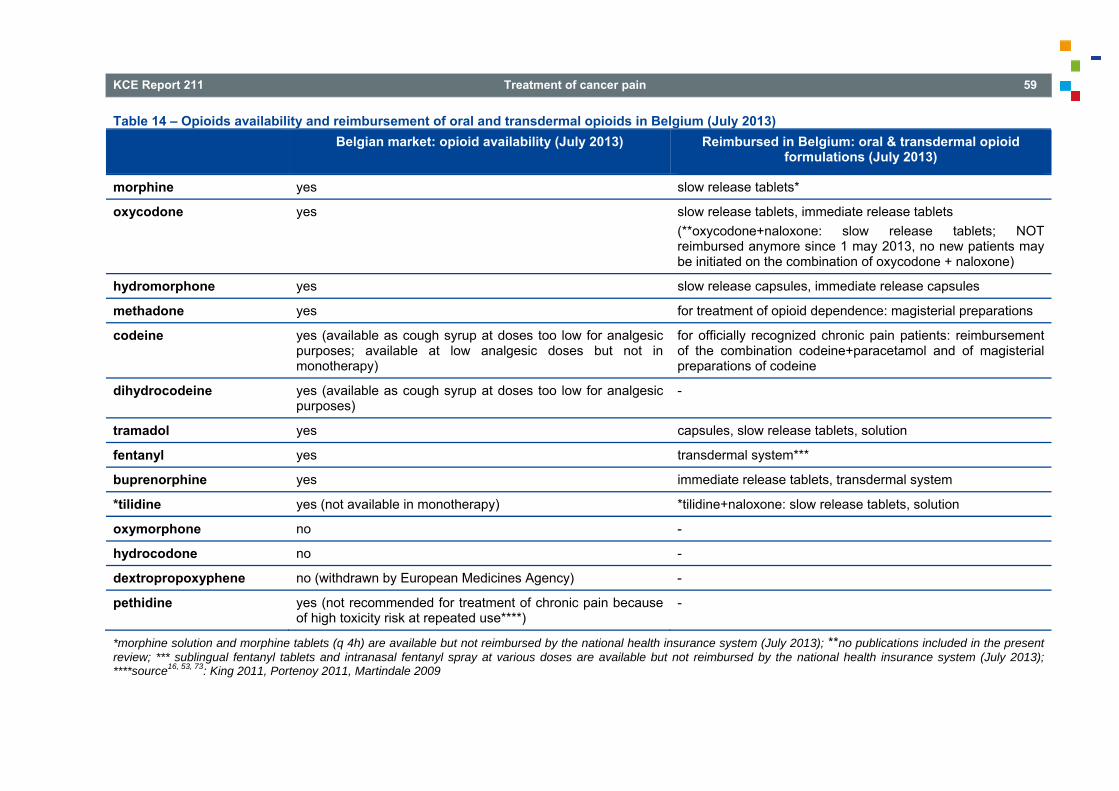
KCE Report 211 Treatment of cancer pain 59
Table 14 – Opioids availability and reimbursement of oral and transdermal opioids in Belgium (July 2013) Belgian market: opioid availability (July 2013) Reimbursed in Belgium: oral & transdermal opioid
formulations (July 2013)
morphine yes slow release tablets*
oxycodone yes slow release tablets, immediate release tablets (**oxycodone+naloxone: slow release tablets; NOT reimbursed anymore since 1 may 2013, no new patients may be initiated on the combination of oxycodone + naloxone)
hydromorphone yes slow release capsules, immediate release capsules
methadone yes for treatment of opioid dependence: magisterial preparations
codeine yes (available as cough syrup at doses too low for analgesic purposes; available at low analgesic doses but not in monotherapy)
for officially recognized chronic pain patients: reimbursement of the combination codeine+paracetamol and of magisterial preparations of codeine
dihydrocodeine yes (available as cough syrup at doses too low for analgesic purposes)
-
tramadol yes capsules, slow release tablets, solution
fentanyl yes transdermal system***
buprenorphine yes immediate release tablets, transdermal system
*tilidine yes (not available in monotherapy) *tilidine+naloxone: slow release tablets, solution
oxymorphone no -
hydrocodone no -
dextropropoxyphene no (withdrawn by European Medicines Agency) -
pethidine yes (not recommended for treatment of chronic pain because of high toxicity risk at repeated use****)
-
*morphine solution and morphine tablets (q 4h) are available but not reimbursed by the national health insurance system (July 2013); **no publications included in the present review; *** sublingual fentanyl tablets and intranasal fentanyl spray at various doses are available but not reimbursed by the national health insurance system (July 2013); ****source16, 53, 73: King 2011, Portenoy 2011, Martindale 2009
60 Treatment of cancer pain KCE Report 211
4.3.2 Search results From the literature search in the main databases, 12 systematic reviews were retrieved. Four SRs from the literature search in the main databases will no further be dealt with in this chapter for the following reasons: • 1 SR (McNicol 2011) concerned the effectiveness of NSAIDs or
paracetamol alone or combined with opioids and is discussed elsewhere in this report 46 (see chapter 4.2).
• 3 SRs concerned opioid dose titration, or equianalgesic doses for two or more different opioids. In line with the other topics in this review e.g. biphosphonates, issues related to drug dosing were considered to be out of scope. Therefore these SRs were excluded from further review. o 2 SRs concerned the conversion ratio between different opioids:
Mercadante 2011, Weschules 2008;65, 90 o 1 SR concerned dose titration: Davis 200491.
Three studies from the literature search in the main databases will be discussed in a narrative way only: • 2 SRs that included observational studies only, were considered to be
a topic of interest by the expert panel (see colophon). Because no further evidence on this topic could be included (no other SR and no RCTs retrieved in RCT update), these two SRs will be discussed in a narrative way (see 4.3.3.10.1), but they will be excluded from the overall conclusions. These SRs dealt with opioid switching (Dale 2010, Quigley 2010)59, 92
• 1 SR that included observational studies only, was considered to be a topic of interest by the expert panel (see colophon). Because no further evidence on this topic could be included (no other SR and no RCTs retrieved in RCT update), it will be discussed in a narrative way (see 4.3.3.12), but it will be excluded from the overall conclusions. This SR dealt with the use of opioids in case of renal impairment (King 2011)93. Likewise, one letter retrieved by hand searching concerned the use of opioids in liver impaired cancer patients (Hanna 2011)179, and because it was considered to be a topic of interest by the expert panel, it will be discussed in a narrative way (see 4.3.3.12), but it will be excluded from the overall conclusions.
In addition to the 12 SRs included from the literature search, 3 references were included by hand search (Tassinari 2011a, Tassinari 2011b, Agency for Healthcare Research and Quality AHRQ, Carr 2002)41, 51, 74. Moreover, 3 SRs from generic guidelines (Dutch Guideline on cancer pain 2008, SIGN 2008, Guideline of the MoH Malaysia 2008)8-10 were retrieved through the search for guidelines; these SRs did not provide detailed information on data extraction and therefore were used as sources of RCTs only. A guideline from NICE in 2012, based on a SR, discussed the utilization of opioids in palliative care75. This guideline included other pathologies than cancer and was thus excluded. Besides the 3 SRs from generic guidelines (Dutch Guideline on cancer pain 2008, SIGN 2008, Guideline of the MoH Malaysia 2008)8-10, fifteen SRs were finally included (see Table 15), and the results are presented below. The generic review by Quigley et al. (2008)63 will be discussed in all relevant sections. The generic review of Carr 2002 only evaluated whether different formulations and routes of administration of opioids are associated with different efficacy rates or patient preferences; however this research question is out of scope for the present review51. Therefore the SR of Carr 2002 has been used as a source of RCTs only and is not presented in the Evidence Tables; its conclusions will not be discussed further. The SRs of Reid 2006 and Cairns 2001 were fully comprised in other SRs on this topic67, 68; for these 2 SRs no Evidence Table is provided but their conclusions are presented in the relevant sections below. The 12 Evidence Tables of the remaining SRs can be found in Appendix II: Table10. The three included generic guidelines (Dutch Guideline on cancer pain 2008, Guideline of the MoH Malaysia 2008, SIGN 2008)8-10 did not provide additional RCTs and as already mentioned, they will not be discussed futher. The 15 SRs included 40 different RCTs (see Appendix II: Table 11); additionally, 16 RCTs were found in the seach for update RCTs.
KCE Report 211 Treatment of cancer pain 61
The included SRs contained few information on the results derived from pooling of primary studies. Mostly, due to heterogeneity of the included primary studies, the SRs presented a narrative overview of the available studies. The results presented in the overview below are based on these narrative overviews which often did not contain many details on the included primary studies. E.g. quantitative results of the included primary studies were not always provided; in some cases the authors of the SRs mentioned explicitly that the primary studies did not contain this information. Also information on the baseline pain intensity in the different trials was mostly not provided by the authors of the SRs. Likewise, it was often not mentioned whether patients entering the trial were opioid naive, or whether they had been using WHO Step II or III opioids before. For several opioids, different oral formulations are available, such as suspensions, immediate release or slow release tablets and capsules. Moreover, for some of them a number of different preparations exists. In the overview below, the terminology used in the original publication will be respected, e.g. modified release morphine, sustained released morphine, controlled release oxycodone, fentanyl buccal tablets, fentanyl buccal soluble film. In the rest of the text and in the recommendations, the term ‘slow release’ will be used for oral formulations that aim to allow for a longer dosage interval as compared to the traditional oral tablets. For a note on immediate release morphine, see 4.3.1.1.4. For reasons explained in the section ‘Other considerations’ (see 4.3.4), the (mean) drug doses used in the trials will not be mentioned systematically.

62 Treatment of cancer pain KCE Report 211
Table 15 ─ Characteristics and quality appraisal of included systematic reviews dealing with opioids in cancer pain First author, year Topic Number of included studies AMSTAR evaluation
Cairns 200168 Oxycodone 5 Yes, can’t answer, yes, no, no, yes, yes, yes, not applicable, not applicable, no
Caraceni 201194 Which oral opioid as first choice 18 Yes, yes, yes, yes, yes, yes, yes, yes, not applicable, not applicable, yes
Carr 200251 General overview 2 Yes, can’t answer, yes, yes, yes, yes, yes, not applicable, not applicable, no
Fallon 201160 Combination of Step III opioids 1 Yes, yes, yes, yes, yes, yes, yes, yes, not applicable, not applicable, yes
Hansen 201295 Breakthrough pain 3 Yes, yes, yes, yes, yes, yes, yes, yes, not applicable, yes, yes
King 201173 Oxycodone 14 Yes, yes, yes, yes, no, yes, yes, yes, not applicable, can’t answer, yes
Nicholson 200870 Methadone 9 Yes, yes, yes, yes, yes, yes, yes, no, not applicable, not applicable, yes
Pigni 201196 Hydromorphone 5 Yes, yes, yes, yes, yes, yes, yes, yes, not applicable, not applicable, yes
Quigley 200863 General overview 27 Yes, can’t answer, no, yes, yes, can’t answer, yes, yes, not applicable, not applicable, yes
Quigley 200969 Hydromorphone 3 Yes, no, yes, yes, yes, yes, yes, yes, not applicable, not applicable, yes
Reid 200667 Oxycodone 6 Yes, no, yes, yes, yes, yes, yes, no, yes, yes, yes
Tassinari 2011a41 Tramadol 7 Yes, yes, yes, yes, no, yes, yes, yes, not applicable, not applicable, yes
Tassinari 2011b74 Transdermal opioids 7 Yes, yes, yes, yes, no, yes, yes, yes, not applicable, not applicable, yes
Wiffen 201055 Oral morphine as first choice opioid
21 Yes, yes, yes, yes, yes, yes, yes, yes, not applicable, not applicable, yes
Zeppetella 200961 Breakthrough pain 4 Yes, yes, yes, yes, yes, yes, yes, yes, yes, no, yes
KCE Report 211 Treatment of cancer pain 63
4.3.3 Literature overview In this overview, the following topics will be dealt with: • Morphine versus placebo or other drugs
o placebo – oxycodone – hydromorphone - transdermal opioids – methadone – tramadol - other
o adverse effects o other considerations, conclusions
• Strong opioids: oxycodone versus placebo and other drugs o placebo – morphine - oxymorphone – hydromorphone - other o other considerations, conclusions
• Strong opioids: hydromorphone versus placebo and other drugs • Strong opioids: fentanyl versus placebo and other drugs • Strong opioids: methadone versus placebo and other drugs • Strong opioids: buprenorphine versus placebo and other drugs • Weak opioids versus placebo and other drugs
o codeine – tramadol - other o other considerations, conclusions
• Transdermal opioids o transdermal fentanyl - transdermal buprenorphine - transdermal
fentanyl and buprenorphine o other considerations, conclusions
• Opioids for breakthrough pain o morphine - fentanyl o other considerations, conclusions
• Opioid rotation o narrative overview (SRs, RCTs) o other considerations, conclusions
• Two or more opioids o narrative overview (SRs) o other considerations, conclusions
• Opioids: other aspects At the end of the chapter (see 4.3.4) an overview is presented of the conclusions and other considerations of all topics, followed by the recommendations. The comparison between opioids versus opioids + NSAIDs or paracetamol is discussed in the chapter on NSAIDs and paracetamol (see 4.2.3.3). Likewise, the comparison between NSAIDs/paracetamol versus NSAIDs/paracetamol + opioids is also discussed in the chapter on NSAIDs and paracetamol.
4.3.3.1 Morphine versus placebo or other drugs: is morphine still the opioid of first choice for moderate to severe cancer pain?
In this section, the use of morphine as background analgesia is discussed; a separate chapter will discuss the use of opioids for breakthrough pain (see 4.3.3.9). Eleven systematic reviews compared morphine to oxycodone, hydromorphone, methadone, tramadol, dextropropoxyphene or Brompton cocktail (Cairns 2001, Caraceni 2011, Fallon 2011, King 2011, Nicholson 2008, Quigley 2008, Quigley 2009, Reid 2006, Wiffen 2010, Pigni 2011, Tassinari 2011a)41, 55, 63, 67-70, 73, 94, 96, 97.
4.3.3.1.1 Morphine versus placebo No RCTs on this topic
4.3.3.1.2 Morphine versus oxycodone Seven systematic reviews (Cairns 2001, Caraceni 2011, Fallon 2011, King 2011, Quigley 2008, Reid 2006 and Wiffen 2010) )55, 63, 67-69, 73, 94, 97 dealt with the comparison between morphine and oxycodone in cancer related pain. Wiffen 2010 included 7 RCTs (Bruera 1998, Ferrell 1989, Heiskanen 1997, Heikanen 2000, Kalso 1990, Lauretti 2003, Mucci LoRusso 1998).98-
104 One additional RCT (Beaver 1978)105 was retrieved from other systematic reviews (King 2011, Reid 2006)67, 73. The other systematic reviews did not provide additional RCTs. The search for update RCTs yielded a new RCT (Mercadante 2010)66.
64 Treatment of cancer pain KCE Report 211
Morphine modified release versus oxycodone Six RCTs (Bruera 1998, Ferrell 1989, Heiskanen 1997, Heikanen 2000, Lauretti 2003, Mucci LoRusso 1998)98-101, 103, 104, including a total of 331 patients, reported adequate analgesia with both morphine modified release and oxycodone when doses were titrated. Wiffen 2010 did not perform pooling due to the heterogeneity of pain assessment tools and the variation in the usage of oxycodone formulations (oxycodone modified release or oxycodone sustained release)55. Wiffen 2010 assessed the quality of these RCTs. Three of them (Heiskanen 1997, Heikanen 2000, Mucci LoRusso 1998) were of good quality (Jadad Score 5)55. Bruera 1998, Ferrell 1989, and Lauretti 2003 were rated at a Jadad score of 4,1 and 3 respectively55. Wiffen 2010 also stated that a subanalysis between studies including opioid naive patients and studies including patients with previous exposure to morphine was planned, but that data did not allow to perform such subanalysis55. Bruera 1998 conducted a double-blind double-dummy randomized crossover (2 periods of 7 days) study in 32 cancer patients98. Sustained release oxycodone was compared to sustained release morphine. Rescue drugs were allowed (immediate release oxycodone or immediate release morphine). No difference was observed in pain intensity (VAS) between the use of oxycodone or morphine. In a 6 weeks RCT including 83 patients, Ferrell 1989 compared short acting analgesics (oxycodone, hydromorphone, codeine or immediate release morphine) with modified release morphine99. When measured with the PPI scale, pain intensity was lower in patients treated with modified release morphine than in patients treated with short acting analgesics. However, patients treated with modified release morphine experienced more often constipation. Heiskanen 1997 used a randomized double-blind double-dummy crossover design in 45 patients (27 evaluated) to compare modified release morphine 30 mg with sustained release oxycodone 20 mg100. Treatment duration was from 3 to 6 days, followed by cross-over. During breakthrough pain episodes, immediate release morphine or oxycodone were available. Modified release morphine provided a lower pain intensity on a four point categorical scale than sustained release oxycodone. Constipation occurred more commonly in patients treated with oxycodone and vomiting in patients treated with morphine.
Heiskanen 2000 used a randomized double-blind double-dummy crossover design in 45 patients to compare modified release morphine 30 mg with modified release oxycodone 20 mg101. Treatment duration was from 3 to 6 days, followed by cross-over. During breakthrough pain episodes, morphine or oxycodone solutions were available. Both opioids provided adequate, stable analgesia as measured by a four-point categorial pain intensity scale. Lauretti 2003 included 26 patients with cancer pain not adequately controlled with a tramadol/ NSAID combination, in a randomized double blind crossover study for 2 periods of 14 days103. Patients were treated alternatively (without wash-out period) with modified release morphine and modified release oxycodone and acted as their own control; during both phases they could use immediate release morphine as a rescue drug so that daily pain scores would be less than 4 cm during all the trial. Hence, all patients received modified release morphine combined with immediate release morphine during one period (morphine alone phase), and modified release oxycodone combined with immediate release morphine during the other period (combined morphine-oxycodone phase). VAS score was used to assess pain intensity. Patients treated with morphine had a higher consumption of immediate release morphine than those treated with oxycodone. Nausea and vomiting was less reported in patients treated with oxycodone. The authors concluded that the combination of modified release oxycodone and immediate release morphine was more efficient than the combination modified release morphine and immediate release morphine. Mucci LoRusso 1998 compared in a RCT modified release oxycodone (48 patients) with modified release morphine (52 patients) 12 hourly, for a duration of 12 days. Pain intensity was measured with a four-points scale and no difference was observed between the two groups104. In the SR by Reid et al. (2006)67 a meta analysis was performed on 3 of these 6 RCTs (Bruera 1998, Heiskanen 1997, Mucci LoRusso 1998)98, 100,
104 including 178 participants. The pooled standardized difference in pain score was 0.20 (95% CI, -0.04 to 0.44, I²=0%) when oxycodone is compared to morphine. Reid et al. (2006) 67 concluded that this difference was not clinically meaningful to patients because it represented only 2 mm on a 100 mm VAS scale; they assumed that clinically significance is reached when at least 20 mm on a 100 mm VAS scale is observed.

KCE Report 211 Treatment of cancer pain 65
Morphine immediate release versus oxycodone immediate release Kalso and Vanio (1990)102 have conducted a double-blind cross-over trial in 20 patients to compare morphine immediate release solution against oxycodone immediate release solution (study duration 96h). In the SR of King 2011, the risk of bias of this RCT was considered to be high73. No significant difference in VAS score between groups was noted (absolute numbers not mentioned). However, oral morphine caused significantly more nausea. Hallicinations occurred only with morphine. Morphine i.m. versus oxycodone i.m. Beaver et al. (1978)105 compared intramuscular morphine with intramuscular oxycodone in 34 hospital inpatients. No significant differences in pain relief and in occurrence of adverse effects were noted. The relative potency of oxycodone as compared to morphine was assessed at 0.68. Quality of this RCT is low because it is no intention-to-treat analysis and because allocation concealment was not performed (see SR of King 2011)73. Morphine sustained release versus oxycodone sustained release Mercadante et al. (2010)66 weekly assessed pain intensity in pancreatic cancer pain during 8 weeks in 60 patients receiving sustained release oral oxycodone or sustained release oral morphine. No significant differences in pain intensity or in adverse effects were noted at each time point. The risk of bias of this RCT is high (see Appendix II: Table 5).
4.3.3.1.3 Morphine versus hydromorphone Five systematic reviews (Caraceni 2011, Pigni 2011, Quigley 2008, Quigley 2009 and Wiffen 2010)55, 63, 69, 94, 96 dealt with the comparison between morphine and hydromorphone in cancer related pain. Pigni 2011 included 3 peer-reviewed RCTs (Moriarty 1999, Hanna 2008, Houde 1986)106-108 and one unpublished RCT (Napp Laboratories 2000)109; these authors specifically focussed on studies evaluating moderate to severe cancer pain. The other systematic reviews did not provide additional RCTs. Heterogeneity of the studies precluded meta-analysis (Pigni 2011, Quigley 2009)69, 96. The update search did not yield new RCTs. Caraceni 2011 and Pigni 2011 advocated for more studies to clarify the question whether hydromorphone would be a better or worse alternative to oral morphine as first line opioid, specifically for moderate to severe cancer pain94, 96.
Pigni 2011 reported the first RCT using hydromorphone in pain treatment in cancer patients96. In 1986, Houde 1986 used a crossover controlled trial design in 48 patients to compare i.m. hydromorphone with i.m. morphine107. He did not find a difference in efficacy and adverse events between the 2 treatment groups. According to Pigni 2011, the limitation of this trial was the lack of allocation concealment. 96 The Jadad score of this study in the SR by Quigley 2009 was 1 (study quality very low) and it was not clear whether it was blinded69. Moriarty et al (1999)108 recruited 100 patients, who had already been treated with WHO Step III opioids, in a double-dummy cross over study testing the efficacy of oral controlled-release hydromorphone and oral controlled-release morphine for 3 days followed by cross-over (without wash-out period). Eighty-nine patients completed the trial. Pain and adverse events were assessed by 100 mm VAS and VRS. No difference in efficacy was demonstrated between the 2 treatments. They showed a similar pattern of side effects. The need of rescue drugs was also similar in the 2 patients groups. Caraceni 2001 and Pigni 2011 pointed out that no allocation concealment was done in this study. In 2 systematic reviews (Quigley 2009 and Wiffen 2010)55, 69, the Jadad score rated the quality of the trial at respectively 4 and 5. Another trial compared oral control-release morphine with oral control-release hydromorphone (Napp Laboratories 2000)109. This unpublished double dummy cross over RCTs included 87 patients (49 completed the study) who were treated for 5 to 10 days, followed by cross-over. A better analgesia was found with morphine: median pain score (p<0.04) and the requirement of rescue analgesia (p=0.002) were higher in patients treated with hydromorphone. Moreover, diarrhea occurred more often in patients treated with hydromorphone than those treated with morphine (p=0.007). The lack of allocation concealment, the large losses to follow-up (44%) and the absence of intention-to-treat analysis are the limitations of this trial (Caraceni 2011, Pigni 2011). The Jadad score rated the quality of the trial at 3 (Quigley 2009).
66 Treatment of cancer pain KCE Report 211
In a RCT including 200 patients, Hanna et al (2008)106 compared OROS® hydromorphone (24-hourly dosing) with slow release morphine tablets (12-hourly dosing) for 10 to 15 days. They included 200 patients who had already been treated with WHO Step III opioids. They found equivalent efficacy and equivalent occurrence of adverse events in both treatment groups. Caraceni 2011 and Pigni 2011 noted large losses to follow-up in this trial.
4.3.3.1.4 Morphine versus transdermal opioids See section 4.3.3.8 Transdermal opioids versus placebo or other drugs.
4.3.3.1.5 Morphine versus methadone Four systematic reviews (Caraceni 2011, Nicholson 2008, Quigley 2008, and Wiffen 2010)55, 63, 69, 70, 94 were found to compare morphine with methadone in cancer related pain. Nicholson 2008 included 5 RCTs which compared morphine with methadone in cancer patients (Beaver 1967, Bruera 2004, Grochow 1989, Mercadante 1998a, Ventafridda 1986)110-114. Nicholson 2008 has found another RCT (Gourlay 1986)115 in 18 patients comparing morphine to methadone, but results are reported only for 2 patients without explanation about the selection of these patients. Because the quality score for this RCT (Gourlay 1986)115 is very low (Jadad score = 2), it will be no further discussed. The other systematic reviews and the update search did not provide additional RCTs. In none of the 4 included systematic reviews, a meta-analysis was performed due to the heterogeneity in the study methodology and the pain scoring system, and the incompleteness of data reporting. Sample sizes were in most primary studies small. One more RCT was retrieved in Caraceni (2011)94; this RCT by Mercadante et al. (2008)113 evaluated the use of oral morphine, oral methadone and transdermal fentanyl. It will be discussed in the section on transdermal opioids (see 4.3.3.8).
Oral morphine versus oral methadone Four systematic reviews (Caraceni 2011, Nicholson 2008, Quigley 2008, and Wiffen 2010) )55, 63, 70, 94 dealt with the comparison of oral morphine versus oral methadone including three RCTs (Bruera 2004, Mercadante 1998a, Ventafridda 1986) 111, 113, 114. Bruera et al (2004)111 recruited 103 cancer patients with neuropathic and non-neuropathic pain syndromes; those who were treated previously with strong opioids were excluded. Included patients were treated twice a day with 7.5 mg methadone or 15 mg sustained release morphine (in each group, an additional 5 mg every 4 hours for breakthrough was provided). The neuropathic element of pain was clinically assessed with Edmonton staging system. Pain was scored on a 0 to 10 patient reported on Numerical Rating Scale. Clinically responders were defined as patients with a least 20 % reduction in baseline pain score. At day 8 and at day 29, there was no difference of proportion of responders in the 2 groups (day 8 methadone 37/49 versus morphine 41/54, ns; day 29 methadone 24/49 versus morphine 30/54, ns). More adverse events were observed in the methadone group (11/49) than in the morphine group (3/54). Moreover, the results do not support a superior role for methadone in neuropathic pain. The Jadad score rated the quality of the study at 4 to 5, respectively by Wiffen 2010 and Nicholson 200855, 70. Ventafridda (1986)114 conducted a trial in 66 patients with advanced cancer and severe pain. This open label study compared 4 mg to 24 mg oral methadone every 4 hours with 8 mg to 28 mg sustained-release oral morphine every 6 hours for 3 days then every 8 hours. Pain was measured by integrated score using a categorical pain intensity 5-point scale. Baseline pain reduction was observed in 2/3 of the patients in the 2 groups (27 patients per group) after 2 days and after 14 days. Results reported graphically showed that morphine significantly increased dry mouth compared with methadone (p<0.01), whereas methadone significantly increased headache (p<0.01). The quality of trial was very low (Jadad scores from 1 (Wiffen 2010)55 to 2 (Nicholson 2008)70. Caraceni 2011 highlighted the large losses to follow-up (34%)94. Mercadante (1998a)113 included in an open label parallel study 40 patients with advanced cancer that used already strong opioids for pain. These patients were treated with sustained-release oral morphine or oral methadone, dosing was adjusted following the patients’ clinical needs.
KCE Report 211 Treatment of cancer pain 67
Pain was assessed during home care follow-up using 10 cm VAS, mean follow-up of 53 days (methadone) and 47 days (morphine). No significant difference between the 2 groups was noted (mean pain score (± S.D.) of all follow-up data: methadone 3.4 ± 0.1 versus morphine 3.3 ± 0.2). The quality of trial was very low: Jadad score 2 (Nicholson 2008)70. Morphine i.v. versus methadone i.v. Grochow et al. (1989)112 conducted a trial in 23 patients (18 evaluable: 10 morphine – 8 methadone; 12 patients evaluated) comparing morphine with methadone delivered by a patient controlled intravenous infusion analgesia system. Pain is assessed using the McGill Pain Intensity index. No significant difference in pain relief was noted between morphine i.v. and methadone i.v. in pain intensity at 4 hours after treatment on day 5 (p=0.94 – results presented graphically). Nicholson 2008 gave a Jadad score of 4 to assess the quality70. Morphine i.m. versus methadone i.m. Beaver et al. (1967)110 compared intramuscular morphine with intramuscular methadone in 37 cancer inpatients; none were opioid naive. This crossover trial alternated morphine 8–16 mg or methadone 16–48 mg in series for moderate to severe pain. Pain relief was defined as at least 50 % reduction in pain intensity measuring by VRS. Additional analgesic was allowed excluding morphine or methadone. The potency of methadone was similar to that of morphine at 2-6 hours after treatment (proportion of people with pain relief: 53% with morphine versus 55% with methadone; no further data reported). Nicholson 2008 gave a Jadad score of 3 to assess the quality; they noted that dose selection was not random (at physician’s choice)70.
4.3.3.1.6 Morphine versus tramadol Three systematic reviews (Quigley 2008, Tassinari 2011a, Wiffen 2010)41,
55, 63 dealt with the comparison between morphine and tramadol in cancer related pain. Quigley 2008 and Wiffen 2010 included 2 RCTs (Leppert 2001, Wilder-Smith 1994)116, 117. One additional RCT (Tawfik 1990)118 was retrieved from Tassinari 2011a41. It will be no further discussed because the reporting of this trial is an abstract. The other systematic reviews and the search for update RCTs did not provide additional primary studies. A meta-analysis was not performed due to the heterogeneity in the study methodology and the pain scoring system.
Morphine modified release versus modified release tramadol Leppert (2001)116 conducted a RCT in 40 cancer patients suffering from neuropathic pain. He found a significantly better neuropathic pain relief at 1 week in patients treated with morphine than in those treated with tramadol (100 mm VAS score: 19.25 with morphine versus 57.00 with tramadol, p<0.05). However, the superiority of morphine was not persistent after 2 weeks. Indeed, similar pain relief was noted in the 2 groups at week 2, 3, 4 and 5. Patients in the morphine group were more constipated and drowsy. Wiffen 2010 scored this RCT as low quality (Jadad Score 2); the study was not blinded55. Morphine solution 1% versus tramadol solution 5% Wilder-Smith et al. (1994)117 performed a double-blind, randomized, cross-over study in a small group of cancer patients (n=20). Pain intensity was measured on a 5-point verbal rating scale (0 = none, 4 = unbearable). The drugs were given for 8 days (cross-over at 4 days). The mean pain intensity (± SD) at day 1 was significantly reduced (p<0.01, absolute numbers not provided) in the morphine 16 mg group compared with the tramadol 50 mg group but similar pain intensity was found at day 4 (morphine 1.6 ± 1.2, n = 17 and tramadol 1.5 ± 1.3, n = 16; p-value not provided). Wiffen 2010 scored this RCT as good quality (Jadad Score 5)55.
4.3.3.1.7 Morphine versus other opioids One systematic review (Wiffen 2010)55 retrieved one RCT (Mercandante 1998b)45 comparing the effect on pain intensity of dextropropoxyphene with that of morphine. Wiffen 2010 scored this RCT as low quality (Jadad Score 2) due to the pour reporting of the results and the low sample size55. The trial included 32 opioids naive cancer patients treated at home. Pain was measured on a 10-cm VAS scale in the first 10 days of the treatment and during the last for weeks before death. Terminal care (support during the process of dying) is beyond the scope of this report (see chapter 1.2). Further, dextropropoxyphene is currently (July 2013) not available on the Belgian market, and it is generally not recommanded anymore for analgesic purposes because of its side effects (Portenoy 2011)16. It has been withdrawn by the European Medicines Agency (EMA) due to concerns over toxicity, particular in overdose. Therefore the results are no further discussed.
68 Treatment of cancer pain KCE Report 211
4.3.3.1.8 Morphine versus Brompton cocktail The systematic reviews of Wiffen 2010 and Nicholson 200855, 70 found 2 RCTs (Melzack 1979, Twycross 1977)119, 120 using ‘Brompton cocktails’ (generic term for mixtures or elixir containing diamorphine or morphine and cocaine with or without chlorpromazine). These cocktails are now obsolete and should not be used anymore (Wiffen 2010). Twycross 1977120 did not find, in 699 terminal patients, difference in efficacy or adverse events between a Brompton cocktail with morphine or a Brompton cocktail with diamorphine. This study is of historical interest only and has major methodological flaws with a drop-out of 553 patients after 2 weeks. Melzack 1979119 compared morphine solution with a Brompton mixture of morphine in 44 patients. No difference was found in pain score or adverse events. This study was valuable in demonstrating that cocaine did no enhance analgesia.
4.3.3.1.9 Adverse effects of morphine Due to the heterogeneity of reporting, it was not possible to pool the results for the adverse effects from the RCTs in the present review that compared morphine to other opioids. However, Wiffen 2010 reported on the adverse effects of morphine from the RCTs included in his review55. Wiffen 2010 found that adverse effects due to the use of oral morphine were common, but he concluded from 21 RCTs reporting this outcome that intolerable side effects leading to treatment withdrawal occurred in a small number of patients only (4%) and that non-response also was infrequent55.
4.3.3.1.10 Summary of findings from systematic reviews on morphine versus placebo or other drugs
The SRs dealing with morphine versus placebo or other drugs concluded as follows: Wiffen 2010 compared oral morphine (immediate-release or modified-released formulation) to oxycodone, hydromorphone, tramadol, or methadone in cancer patients55. (In Wiffen 2010, ‘immediate release’ morphine refers to morphine solution, or to morphine immediate release tablets. In the primary studies included in the Cochrane review, morphine immediate release tablets were typically given every four hours.) They found qualitative evidence for effectiveness of oral morphine which compares well to other available opioids; adverse effects were common
but intolerable side effects occurred in a small number of patients only (4%) and non-respons also was infrequent. They recommended morphine as a gold standard for moderate to severe cancer-related pain. Conclusions of Quigley 2008 are the following (based on the SR of Reid 2006 and one RCT)63, 67: “we do not know whether morphine is more effective than oxycodone at reducing cancer-related pain at 4–14 days (low-quality evidence).” (Based on 1 RCT): we do not know whether hydromorphone as compared to morphine is more effective at reducing the need for rescue analgesia over the last 24 hours of treatment in people with cancer-related pain (very low-quality evidence). At present there is no consistent evidence to suggest that hydromorphone is superior to morphine in terms of analgesic effect or adverse-effect profile. (Based on 5 RCTs): we do not know whether oral morphine is more effective than oral methadone at reducing cancer-related pain (very low-quality evidence). We do not know whether parenteral morphine is more effective than parenteral methadone at reducing cancer-related pain (very low-quality evidence). The long half-life of methadone carries the risk of drug accumulation and latent toxicity; people taking methadone therefore need to be carefully monitored. (Based on 2 RCTs): we do not know whether morphine is more effective than tramadol at reducing cancer-related pain at 2–5 weeks (very low-quality evidence). Reid 2006 did not find differences in efficacy or adverse effects between oxycodone and morphine. The authors repeated the recommendation to use morphine as first-line treatment because oxycodone is 4 times more expensive than morphine in England; however the authors emphasized the need for larger trials of longer duration. However, since the efficacy and tolerability of oxycodone are similar to morphine, its use as an opioid for cancer-related pain can be supported according to these authors67. Cairns 2001 compared morphine to oxycodone and concluded that morphine remained the drug of choice to manage cancer pain. Rotation to oxycodone should be considered in case of side effects (i.e. hallucination) or renal impairment68. Caraceni 2011 concluded that oral morphine and oxycodone as well as hydromorphone have a similar efficacy and toxicity in cancer-related pain. They concluded that available evidence suggested that oral methadone and oral morphine offer similar pain relief, with a similar pattern of side effects. Caraceni 2011 did not recommend methadone as a first-line

KCE Report 211 Treatment of cancer pain 69
prescription, but concluded that, because of its very long and unpredictable half-life, the use of methadone must be left to skilled professionals94. Fallon 2011 did not conclude on the use of oxycodone versus morphine. They only concluded on opioid combinations, including one RCT (Lauretti 2003 combining oxycodone and morphine), and made a weak recommendation supporting the use of a combination of opioids60. King 2011 did not find differences in efficacy or adverse effects between oxycodone and morphine and stated that oxycodone can be recommended as an alternative to morphine73. Quigley 2009 compared morphine and hydrocodone and confirmed that morphine remains the gold standard for the management of moderate to severe cancer pain. They stated that hydromorphone is a potent analgesic but that there is no evidence to demonstrate the superiority or the inferiority in comparison with morphine, and that adverse events are comparable69. Pigni 2011 compared morphine and hydrocodone and stated that hydromorphone is efficient and well tolerated in moderate to severe cancer pain. Hydromorphone can be considered as an alternative to morphine and oxycodone although its inferiority or superiority in comparison with morphine is not demonstrated96. Nicholson 2008 concluded that study results suggested a similar efficacy of methadone and morphine in the treatment of cancer pain. Results of one RCT did not support a specifically beneficial role for methadone in neuropathic pain. Side effects were similar for methadone and morphine in the trials but the side effects of methadone may become more prominent with repeated dosing. That is the reason why Nicholson recommended that the responsibility for initiation and careful dose adjustment and monitoring of methadone should be left to an experienced clinician70. Tassinari 2011a did not conclude on the comparison tramadol-morphine but stated that tramadol should not be used as standard approach in opioid-naive patients with moderate to severe cancer pain41. Several of these SRs warned for methodological weaknesses (small study samples, statistical methods not always appropriate etc.) but they noted that the study results were mostly concordant.
4.3.3.1.11 Other considerations In Belgium, there is only one commercial preparation available for oral methadone at a dose of 5 mg. This is a relatively low dosage which makes its use in monotherapy more difficult. Moreover, it is not reimbursed by the national health insurance system (however, the magisterial preparations are reimbursed for the treatment of opioid dependence where it is used to substitute the (illegal) opioid use). Further, the consulted expert panel (see colophon) advises to avoid high dosages, because of one of the specific although rare adverse effects of methadone, prolongation of the QT interval with cardiac dysrhythmias (Martindale 2009)53. It is possible that this adverse effect did not occur in the trials mentioned above, given their limited number of participants and their short duration. For these reasons, according to the expert panel, methadone for analgesic purposes should preferably be used as add-on to other opioids.

70 Treatment of cancer pain KCE Report 211
Conclusions • The available evidence could not demonstrate the superiority of
oxycodone against morphine. Whatever the formulation considered, the two drugs can provide comparable analgesia when titration is performed. Most trials did not show differences in frequency of side-effects between morphine and oxycodone, but the small number of participants in the trials precludes firm conclusions. Morphine remains the gold standard in the management of cancer pain (very low level of evidence; Cairns 2001, Caraceni 2011, Fallon 2011, King 2011, Quigley 2008, Reid 2006, Wiffen 2010; Mercadante 2010).
• The available evidence could not demonstrate the superiority or inferiority of hydromorphone against morphine in moderate to severe cancer pain. The trials of best quality showed comparable efficacy and did not show differences in frequency of side-effects between oral morphine and oral hydromorphone. However, the small number of trials precludes firm conclusions. At present, morphine remains the gold standard in the management of cancer pain (very low level of evidence; Caraceni 2011, Quigley 2008, Quigley 2009, Wiffen 2010, Pigni 2011).
• The available evidence suggested a similar efficacy of methadone and morphine in the treatment of cancer pain, with a similar pattern of adverse effects. The findings of one RCT (Bruera 2004) did not support superiority of methadone to morphine in patients with neuropathic pain, but further evidence confirming this finding is needed. In the included studies, the pattern of adverse effects was similar for methadone and morphine (very low level of evidence; Caraceni 2011, Nicholson 2008, Quigley 2008, and Wiffen 2010). It is well-known that the side effects of methadone may become more prominent with repeated dosing because of its pharmacokinetics and pharmacodynamics.
• Based on the available evidence, it is not possible to demonstrate superiority or inferiority of tramadol against morphine (very low level of evidence; Quigley 2008, Tassinari 2011a, Wiffen 2010).
4.3.3.2 Strong opioids: Oxycodone versus placebo or other drugs For SRs and RCTs on the comparison of oxycodone to morphine: see section 4.3.3.1.2. Five systematic reviews (King 2011, Pigni 2011, Quigley 2008, Quigley 2009, Reid 2006)63, 67, 69, 73, 96 found one RCT (Hagen and Babul 1997)121 comparing oxycodone with hydromorphone. King 2011 found another RCT (Gabrail 2004)122 using oxycodone versus oxymorphone. The search for update RCTs yielded an additional RCT including the combination of oxycodone with naloxone (Ahmedzai 2012)123. In this section, the use of oxycodone as background analgesia is discussed; a separate chapter will discuss the use of opioids for breakthrough pain (see 4.3.3.9).
4.3.3.2.1 Oxycodone versus placebo No RCTs on this topic.
4.3.3.2.2 Oxycodone versus morphine See section 4.3.3.1.2.
4.3.3.2.3 Oxycodone versus oxymorphone Gabrail 2004122 conducted compared in 44 patients the efficacy of controlled released oxycodone with that of controlled released oxymorphone administered for 7 to 10 days. Mean pain intensity score (± S.D.) (measurement instrument not mentioned) was similar in the two groups (oxymorphone: 2.5 ± 1.3, n=10; oxycodone: 2.8 ± 1.3, n=10). No significant difference was also noted in side effects or level of pain interfering with function. The quality of this trial is low because of the lack of intention-to-treat analysis and allocation concealment. Oxymorphone is currently (July 2013) not available on the Belgian market. Therefore, the results of this trial are no further discussed.

KCE Report 211 Treatment of cancer pain 71
4.3.3.2.4 Oxycodone versus hydromorphone Hagen and Babul 1997121 conducted a double-dummy RCT in 44 patients with moderate to severe cancer pain testing controlled release oxycodone versus controlled release hydromorphone; these patients had already been treated with opioids before. Pain intensity was measured with 100 mm VAS and a 5-point categorical scale. Pain intensity after 7 days was similar in the two groups whatever the measurement tool (VAS: oxycodone 28 mm vs hydromorphone 30.6 mm, p = 0.11; 5-point categorical scale: oxycodone 1.4 vs hydromorphone 1.5, p = 0.10). No significant difference in side effects was observed, although oxycodone produced less sedation than hydromorphone. Using Jadad scoring, Quigley 2009 rated the quality of the study at 469. Pigni 2011 pointed out a large loss to follow-up and no intention-to-treat analysis as limitations96. King 2011 reported the same limitation as Pigni 201173.
4.3.3.2.5 Oxycodone versus oxycodone + naloxone Ahmedzai 2012123 conducted a double-dummy trial in 124 adult patients from 9 countries with moderate to severe chronic cancer pain to examine whether a combination of oxycodone and naloxone prolonged-release tablets can improve constipation and maintain analgesia compared with oxycodone prolonged-release tablets alone. Pain intensity was measured at 4 weeks using the Brief Pain inventory – Short Form. No significant difference was noted between the two groups (Mean (SD) BPI-SF scores combination oxycodone - naloxone: 3.50 ± 1.88 vs oxycodone: 3.52 ± 1.80, ns). The use of rescue drugs was the same in the combination group as compared to the oxycodone alone group in terms of both frequency (p=0.4) and dose (p=0.22). Quality of life was measured with EQ-5D and QLQ-C30 and no significant difference was observed (Mean (SD) index scores EQ-5D combination oxycodone - naloxone: 0.55 ± 0.33 vs oxycodone: 0.49 ± 0.38, ns). The assessment of constipation was measured with PAC-SYM (Patient assessment of constipation symptoms). Mean score was lower in the combination oxycodone – naloxone than in oxycodone alone (Mean (SD) PAC-SYM scores combination oxycodone - naloxone: 1.47 ± 1.07 vs oxycodone: 2.03. ± 1.29, p<0.01). However, after 4 weeks, the total laxative intake (oral bisacodyl) was 20 % lower in the combination group than in the oxycodone alone group but the difference was not statistically
significant (Mean (SD) dose in mg combination oxycodone - naloxone: 26.10 ± 27.60 vs oxycodone: 32.69 ± 31.26, ns). The authors concluded that the combination of oxycodone - naloxone provided superior bowel function without compromising analgesic efficacy and safey in cancer patient. The overall risk of bias for this study is high (see Appendix II: Table 5).
4.3.3.2.6 Summary of findings from systematic reviews on oxycodone versus placebo or other drugs
For conclusions of SR on oxycodone versus morphine, see section 4.3.3.1.2. For conclusions of SR on the use of oxycodone in breakthrough pain, see section 4.3.3.9. The other SRs dealing with this topic concluded as follows: Reid 2006 concluded that the difference measured in one RCT between oxycodone and hydromorphone was too small (2 to 3 mm on a 100 mm VAS scale) to be of clinical significance. The authors recommended to use morphine rather than oxycodone as first-line treatment in cancer pain because oxycodone is 4 times more expensive than morphine in England; however the authors could support the use of oxycodone as well and they emphasized the need for larger trials of longer duration. The authors did not recommend on hydromorphone67. Pigni 2011 concluded that oxycodone can give similar analgesic results in comparison to hydromorphone in patients with moderate to severe cancer pain. The overall toxicity profile was the same in oxycodone and hydromorphone. They concluded that there is evidence to support the efficacy and tolerability of hydromorphone for moderate to severe cancer pain as an alternative to oxycodone (or morphine), while there is no evidence to demonstrate its superiority or inferiority in comparison with morphine as the first choice opioid for the same indication96. King 2011 concluded that there is no significant difference in efficacy and tolerability between oxycodone and hydromorphone or morphine73. Recommendations of Quigley 2008 are the following: we do not know whether oxycodone is more effective than morphine at reducing cancer-related pain at 4–14 days (low-quality evidence, based on the SR of Reid 2006 and 1 RCT). We do not know whether oxycodone is more effective

72 Treatment of cancer pain KCE Report 211
than hydromorphone at reducing pain intensity as measured on a 5-point scale in people with cancer pain (low-quality evidence based on 1 RCT)63. Quigley 2009 concluded that clinical superiority of hydromorphone was not demonstrated over other strong opioid analgesics, including oxycodone. At present morphine remains the ’gold standard’ for the management of moderate to severe cancer pain69.
4.3.3.2.7 Other considerations No other considerations.
Conclusions • For conclusions on oxycodone versus morphine, or on the use of
oxycodone in breakthrough pain, see sections 4.3.3.1.2, 4.3.3.9 • The available evidence could not demonstrate the superiority or
inferiority of oxycodone against hydromorphone in cancer pain (very low level of evidence; King 2011, Pigni 2011, Quigley 2008, Quigley 2009, Reid 2006). The available trials showed comparable efficacy and did not show differences in frequency of side-effects. However, the small number of trials precludes firm conclusions. At present, morphine remains the gold standard in the management of cancer pain. One trial showed comparable efficacy but lower frequency of side-effects (constipation) in favour of the combination of oxycodone and naloxone as compared to oxycodone alone; more studies are needed (very low level of evidence; Ahmedzai 2012).
4.3.3.3 Strong opioids: Hydromorphone versus placebo or other drugs
In this section, the use of hydromorphone as background analgesia is discussed; a separate chapter will discuss the use of opioids (including hydromorphone) for breakthrough pain (see 4.3.3.9). No RCTs were retained comparing hydromorphone versus placebo. For SRs and RCTs on the comparison of hydromorphone versus morphine, see section 4.3.3.1.3. One RCT (Hagen and Babul 1997)121 compared hydromorphone with oxycodone and is presented in section 4.3.3.2.4.
For SRs and RCTs on the comparison of hydromorphone versus transdermal opioids, see section 4.3.3.8.
Conclusions • See sections 4.3.3.1.3, 4.3.3.2.4, 4.3.3.8
4.3.3.4 Strong opioids: Fentanyl versus placebo or other drugs The evidence for the use of fentanyl in breakthrough pain is discussed in the section on the management of breakthrough cancer-related pain (see 4.3.3.9). The evidence for the transdermal formulation of fentanyl is presented in the section transdermal opioids (see 4.3.3.8.1). No evidence was found about the use of fentanyl for other indications related to cancer pain treatment.
Conclusions • See sections 4.3.3.9, 4.3.3.8.1
4.3.3.5 Strong opioids: Methadone versus placebo or other drugs Four SRs deal with the use of methadone for cancer pain (Caraceni 2011, Nicholson 2008, Quigley 2008, and Wiffen 2010)55, 63, 70, 94. In his systematic review, Nicholson 2008 identified 9 RCTs (Beaver 1967, Bruera 2004, Ferrer-Brechner 1984, Gourlay 1986, Grochow 1989, Matts 1964, Mercadante 1998a, Twycross 1977, Ventafridda 1986)99, 110-115, 120, 124. No meta-analysis was feasible due to the lack of reporting and quality of the trials. Six of these RCTs had morphine as comparator (Beaver 1967, Bruera 2004, Gourlay 1986, Grochow 1989, Mercadante 1998a, Ventafridda 1986) 110-115 and are discussed in the section on morphine (see 4.3.3.1.5). Twycross 1977 is presented in the section dedicated to the Brompton cocktail (see 4.3.3.1.8)120. Matts 1964 will not be discussed because of inclusion of cancer and non cancer patients.124 In 1984, Ferrer-Brechner and Ganz studied the combination of ibuprofen and methadone as compared to methadone alone99. This RCT is presented in NSAID chapter (see 4.2.3.3).
KCE Report 211 Treatment of cancer pain 73
One more RCT was retrieved in Caraceni (2011)94; this RCT by Mercadante et al. (2008)113 evaluated the use of oral morphine, oral methadone and transdermal fentanyl. It will be discussed in the section on transdermal opioids (see 4.3.3.8). The other SRs and the search for update RCTs did not provide additional RCTs. Other considerations on the use of methadone in Belgium are presented in the section on morphine (see 4.3.3.1.11)
Conclusions • See sections 4.2.3.3, 4.3.3.8
4.3.3.6 Strong opioids: Buprenorphine versus placebo or other drugs
Buprenorphine is a partial agonist-antagonist of the central opioid receptors, which is in most references considered to be a strong (WHO Step III) opioid. For oral buprenorphine see section 4.3.3.7.2.3 (tramadol versus buprenorphine). Buprenorphine is often used in a transdermal application; see section 4.3.3.8.2 (transdermal buprenorphine). For other considerations on buprenorphine, see section 4.3.3.8.5 (transdermal opioids, other considerations).
Conclusions • See sections 4.3.3.7.2.3, 4.3.3.8.5
4.3.3.7 Weak opioids versus placebo or other drugs From 1986, the standard approach to manage cancer pain in adults is based on the three-step WHO analgesic ladder (for more details see chapter 4.1.4). Weak opioids are central in the second step of this ladder. In the section below, codeine, tramadol and other weak opioids will discussed. However, a novel approach suggests to by-pass the second step of the WHO analgesic ladder for the treatment of chronic cancer pain. For the discussion related to the usefulness of the second step of the WHO ladder, see chapter 4.1.4.
4.3.3.7.1 Codeine From the SR of Quigley 200863, one RCT could be found that compared codeine with placebo in cancer patients (Dhaliwal 1995)125. One additional RCT comparing codeine combined with paracetamol to tramadol (and hydrocodone) was found in the SR of Tassinari 2011a; it will be discussed in the section on tramadol (see 4.3.3.7.2.4). The other SRs and the search for update RCTs yielded no additional publications. The SR of Quigley 200863 conducted an evaluation of the quality of the evidence based on the GRADE system. Since the evidence on codeine in the present review has been derived from this SR, the GRADE evaluation of Quigley 2008 will be used63. Despite the central role of codeine in the Step II of the WHO analgesic ladder for the treatment of cancer pain, very little evidence could be retrieved on its effectiveness and potential side effects for this indication. In this review, no RCTs were found that compared codeine with morphine, dihydrocodeine, hydromorphone, oxycodone, methadone or transdermal fentanyl.
4.3.3.7.1.1 Codeine versus placebo In the RCT of Dhaliwal 1995125, a double-blind study based on a crossover design, 35 patients no longer responsive to non-steroidal antiinflammatory drugs received controlled-release codeine or placebo, and after 1 week crossed over to the other arm. Results before cross-over were not reported. At 2 weeks, a significant statistical reduction in pain was observed in patients treated with codeine compared to those with placebo (100 mm VAS scale – mean score (± S.D.): codeine 22 ± 18 versus placebo 36 ± 20, p = 0.0001). Less rescue analgesia was needed in the codeine group than in placebo group (mean daily tablets needed: codeine 2.2±2.3 versus placebo 4.6±2.8, p=0.001). Nausea was more frequent in patients treated with codeine (proportion of patients with nausea: codeine 40% versus placebo 15%, p = 0.01). No difference in other adverse events was noted. Quigley assessed the quality of this trial using the GRADE tool at a low level. Moreover, Quigley 2008 stated than the efficacy of codeine in cancer pain treatment is not known because the only RCT found provided insufficient evidence to draw firm conclusions63.

74 Treatment of cancer pain KCE Report 211
4.3.3.7.1.2 Codeine combined with paracetamol versus tramadol and hydrocodone
See section 4.3.3.7.2.4 on tramadol. Note that hydrocodone is currently (July 2013) not available on the Belgian market. Therefore, it will no further be discussed.
4.3.3.7.1.3 Codeine combined with paracetamol versus transdermal opioids
See section 4.3.3.8 on transdermal opioids.
4.3.3.7.2 Tramadol For the SRs and RCTs on the comparison of morphine and tramadol, see section 4.3.3.1.6. In 2011, Tassinari et al. made a review targeting the question if oral tramadol is better than placebo or other opioids in the management of mild to moderate cancer pain never treated with opioids41. The authors identified four RCTs (Arbaiza 2007, Brema 1996, Rodriguez 2007, Rodriguez 2008)126-129. The RCT of Arbaiza and Vidal (2007)126 compared tramadol with placebo and was also reported in the review of Quigley 200863. The other RCTs (Brema 1996, Rodriguez 2007, Rodriguez 2008)127-129 compared tramadol with other opioids (oral buprenorphine, hydrocodone or codeine, a combination of hydrocodone and paracetamol). One other SR, Quigley 2008, also discussed the role of tramadol in cancer pain relief but did not include additional RCTs that corresponded to the selection criteria for the present review63. The search for update RCTs yielded no additional publications. The SR of Tassinari 2011a presented a narrative overview of the included primary studies, and also conducted an evaluation of the quality of the evidence based on the GRADE system.41 Since the evidence on tramadol in the present review has been derived from this SR, the GRADE evaluation of Tassinari 2011a will be used41.
4.3.3.7.2.1 Tramadol versus placebo Arbaiza and Vidal (2007)126 randomized 36 patients with moderate to severe cancer pain or cancer treatment-related neuropathic pain in 2 groups. The first group received tramadol every 6 hours, the second group received placebo. When measured on a 10-point scale, pain at 45 days
was significantly lower with tramadol than with placebo (Pain score tramadol 2.9 versus placebo 4.3, p<0.001). Moreover, the need of rescue drug is significantly reduced with tramadol in comparison with placebo (p<0.05, absolute numbers not reported). Better quality of life was reached with tramadol than with placebo (Activities of Daily Living (ADL) scale: proportion of people with serious limitations in ADL: tramadol 4/13 versus placebo 10/12; p = 0.008). The quality assessment of this trial using the GRADE tool is low (Quigley 2008, Tassinari 2011a)41, 63.
4.3.3.7.2.2 Tramadol versus morphine See section 4.3.3.1.6.
4.3.3.7.2.3 Tramadol versus buprenorphine Brema et al (1996)127 conducted a multicenter RCT comparing slow-release oral tramadol with oral sublingual buprenorphine in 131 cancer patients with pain resistant to NSAIDs with or without adjuvant. The proportion of patients with strong/unbearable pain at week 1 had fallen significantly in the tramadol group (from 98.4% to 48.1%, p<0.05) as compared to a drop from 92% to 66.7% for buprenorphine. No change in the quality of life was noted in the tramadol group when measured with Karnofsky’s and Spitzer’s indices for functional capacities and quality of life respectively. After 2 weeks, Karnofsky index dropped slightly (p<0.05 between treatments). Good safety was noted in both treatment groups. According to the GRADE system, Tassinari 2011a assessed this trial at a low level of quality41.
4.3.3.7.2.4 Tramadol versus other weak opioids (codeine, hydrocodone) Rodriguez et al (2007)128 randomized 117 cancer patients in 3 groups: 62 patients received hydrocodone, 59 patients received codeine combined with paracetamol and 56 patients received tramadol. No significant statistical difference in the analgesic efficacy was observed between the 3 treatment groups (χ² = 0.73; p = 0.69). The safety of tramadol was lower than the 2 others opioids. Significant more adverse events were produced by tramadol than by the 2 others opioids (p<0.05). According to the GRADE system, Tassinari 2011a assessed this trial at a low level of quality41.

KCE Report 211 Treatment of cancer pain 75
The analgesic efficacy and tolerability of tramadol was compared to those of a combination of hydrocodone and paracetamol in 118 patients with chronic cancer pain (Rodriguez 2008)129. No significant statistical difference in analgesic efficacy was found between the 2 opioids. However, as demonstrated in Rodriguez 2007, tramadol produced more adverse events than the hydrocodone/paracetamol combination. For instance, the relative risk of nausea (95% CI) was 1.69 (1.03-2.77) – p = 0.03, the relative risk of vomiting (95% CI) was 2.21 (1.14-4.32) – p = 0.02, the relative risk of dizziness (95% CI) was 2.12 (1.17-3.86) – p = 0.03, the relative risk of loss of appetite (95% CI) was 3.27 (1.12-9.55) – p = 0.02 and the relative risk of weakness (95% CI) was 7.75 (0.98-61.05) – p = 0.019. According to the GRADE system, Tassinari 2011a assessed this trial at a low level of quality41. Note that hydrocodone is currently (July 2013) not available on the Belgian market. Therefore, it will no further be discussed.
4.3.3.7.3 Other WHO Step II opioids Hydrocodone: see section 4.3.3.7.2.4 (tramadol versus other weak opioids). Note that hydrocodone is currently (July 2013) not available on the Belgian market. Therefore, it will no further be discussed. Dextropropoxyphene: see section 4.3.3.1.7(morphine versus dextropropyphene). Dextropropoxyphene is currently (July 2013) not available on the Belgian market, and it is generally not recommanded anymore for analgesic purposes because of its side effects (Portenoy 2011)16. It has been withdrawn by the European Medicines Agency (EMA) due to concerns over toxicity, particular in overdose. Therefore the results are no further discussed.
4.3.3.7.4 Summary of findings from systematic reviews on weak opioids versus placebo or other drugs
The SRs dealing with this topic concluded as follows: Conclusions of Quigley 2008 for codeine are the following: (based on 1 RCT): codeine may be more effective as compared to placebo at reducing chronic cancer pain at 2 weeks; it may also be more effective at reducing the need for rescue analgesia (low-quality evidence)63. We do not know whether codeine is more effective as compared to tramadol at reducing cancer-related pain (very low-quality evidence). We do not know whether
people prefer codeine to tramadol (very low-quality evidence). We found no clinically important results about codeine compared with morphine, dihydrocodeine, transdermal fentanyl, hydromorphone, methadone, or oxycodone in people with cancer-related pain. Conclusions of Quigley 2008 for tramadol are the following: (based on 3 RCTs): tramadol may be more effective as compared with placebo at reducing pain intensity in people with moderate to severe cancer pain at 45 days, and it may be more effective at reducing the need for rescue analgesia63. It may also be more effective at reducing the proportion of people with serious limitations in Activities of Daily Living (a measure of quality of life) (low-quality evidence). We do not know whether tramadol as compared with codeine is more effective at reducing cancer-related pain (very low-quality evidence). We do not know whether tramadol as compared with dihydrocodeine is more effective at relieving prostate cancer-related pain (very low-quality evidence based on a RCTof 32 participants published in Spanish). Patient preference: tramadol may be the less preferred opioid as compared with morphine when balancing between pain control and adverse effects (very low-quality evidence). We do not know whether people prefer tramadol to codeine or to dihydrocodeine (very low-quality evidence).We found no clinically important results about tramadol compared with transdermal fentanyl, hydromorphone, methadone, or oxycodone in people with cancer-related pain. Tassinari 2011a: Tramadol is an effective and well-tolerated drug in moderate cancer pain but its superiority in comparison with codeine, hydrocodone or buprenorphine cannot be demonstrated; at present the codeine/paracetamol combination remains the standard41. There are insufficient data to support routine use of tramadol as an alternative to codeine/paracetamol in mild to moderate cancer pain. For conclusions of SR on tramadol versus morphine see section 4.3.3.1.10.
4.3.3.7.5 Other considerations For codeine, the Step II opioid suggested in the WHO analgesic ladder (see4.1.4), the consulted expert panel (see colophon) pointed to the fact that in Belgium codeine is only available at a relatively low dose (30 mg) as a combination preparation with 500 mg paracetamol. This limits its use as
76 Treatment of cancer pain KCE Report 211
a Step II opioid. The consulted expert panel suggested to add tramadol as a WHO Step II opioid. After initiation tramadol often causes nausea and vomiting but according to the experts, this is a temporary effect. Tramadol has partially an opioid working mechanism, and partially it has other central working mechanisms namely reuptake inhibition of serotonin and norepinephrine. The experts emphasized that for this reason, the opioid side effects of tramadol such as drowsiness and constipation are usually less severe than for codeine. The experts added that codeine probably also carries a higher risk of addiction given its stronger opioid effect; and because its metabolisation differs between individuals some patients react too much or do not respond at all. Another alternative to the use of the Step II opioids codeine or tramadol could be, according to the consulted expert panel (see colophon), tilidine. In Belgium, tilidine is only available in combination with the opioid antagonist naloxone, to prevent abuse. However, this can hinder good analgesic effect in severe pain, when high tilidine doses might be required. No publications on tilidine corresponding to the inclusion criteria of the present review have been found. Further, the consulted expert panel suggested to use buprenorphine, as an alternative to Step II opioids if codeine, tramadol or tilidine are not suitable. Buprenorphine is usually considered to be a strong (WHO Step III) opioid. For other considerations on buprenorphine, see also section 4.3.3.8.5 (transdermal opioids). The consulted expert panel also suggested that nefopam, an atypical centrally-acting non-opioid analgesic, could act as an alternative to Step II opioids; however, nefopam was not included in the systematic literature search and is considered to be out of scope of this review.
Conclusions • There are indications that codeine is an effective and well-
tolerated drug as compared to placebo in the management of cancer pain never treated with opioids (low level of evidence; Quigley 2008).
• There is conflicting evidence as to the question whether combining a NSAID (including aspirin) or paracetamol with codeine, a WHO Step II opioid, is superior to a NSAID (including aspirin) or paracetamol alone in patients with cancer pain. There are indications of a trend toward a comparable incidence of adverse events in both groups; however, it is not possible to draw firm conclusions, since most studies were conducted over a short period (less than 7 days). This evidence does not allow to confirm or refute the Step II WHO recommendation that a NSAID should be combined with a ‘weak’ opioid for the management of moderate cancer pain (very low level of evidence; McNicol 2011, see chapter NSAIDs and paracetamol).
• There are indications that initiating strong opioids (Step III) instead of weak opioids (Step II) for mild to moderate cancer pain after conventional treatment with NSAIDs and/or paracetamol failed, might lead to better pain control but at the cost of more side effects (very low level of evidence; Tassinari 2011a, see chapter WHO analgesic ladder).
• There are indications that oral tramadol is an effective and well-tolerated drug as compared to placebo in the management of mild to moderate cancer pain never treated with opioids (low level of evidence; Tassinari 2011a).
• There are indications that there is no difference in efficacy between tramadol and codeine combined with paracetamol in the management of mild to moderate cancer pain never treated with opioids; however, there are also indications that mild adverse effects such as nausea or vomiting are more frequent in tramadol (low level of evidence; Tassinari 2011a).
• Based on the available evidence, it is not possible to conclude on the relative effectiveness of tramadol and oral buprenorphine (low level of evidence; Tassinari 2011a).
• For conclusions on tramadol versus morphine, see section 4.3.3.1.6.

KCE Report 211 Treatment of cancer pain 77
4.3.3.8 Transdermal opioids versus placebo or other drugs Four systematic reviews (Tassinari 2011b, Caraceni 2011, Wiffen 2010 and Quigley 2008)55, 63, 74, 94 dealt with transdermal opioids. Tassinari 2011b focused on patients suffering from moderate to severe cancer pain who had never been treated with strong opioids74; Caraceni 2011 focused on patients suffering from moderate to severe cancer pain94. Wiffen 2010 and Quigley 2008 did not make this restriction. Tassinari 2011b included 11 RCTs74 (Ahmedzai 1997, Hunt 1999, Mercadante 2008, Pace 2007, Poulain 2008, Kongsgaard 1998, Sittl 2003, Sorge 2004, van Seventer 2003, Wirz 2009, Wong 1997)130-140. Caraceni 2011 identified no additional RCTs, and no additional RCTs were retrieved from the 2 other systematic reviews94. Two RCTs (Sittl 2003, Sorge 2004) included cancer and non cancer patients and three RCTs (Ahmedzai 1997, Hunt 1999, Wong 1997)130, 131, 136, 137, 140 included terminal cancer patients. Those RCTs are not within the scope of the present review and will be not discussed further. The update yielded to 3 additional RCTs (Kress 2008, Mystakidou 2005, Pistevou-Gompaki 2004)141-143 (see evidence tables in Appendix II: Table 12). This brings the total of RCTs discussed below on 9. The SRs of Tassinari 2011b and Quigley 2008 presented a narrative overview of the included primary studies63, 74, and also conducted an evaluation of the quality of the evidence based on the GRADE system. Therefore, for the evidence in the present review that has been derived from these two SRs, the GRADE evaluation of these two SRs will be used.
4.3.3.8.1 Transdermal fentanyl
Transdermal fentanyl versus placebo Kongsgaard 1998 conducted a double blind study including 138 patients with chronic cancer-related pain (72 were evaluated)132. The treatment was administered during 9 days. Pain on walking was measured on a 10 cm VAS scale. No significant difference was observed between transdermal fentanyl and placebo (Median: fentanyl 1.0 versus placebo 1.1, p>0.05). Immediate-release morphine was allowed as rescue medication; the use of rescue medication was the same in the placebo group and the treatment group (Mean: fentanyl 47.7 mg versus placebo 51.0 mg, p= 0.21). The RCT did not directly compare adverse effects of fentanyl versus placebo; it found that fentanyl was associated with nausea in 4% of the participants.
The results of this trial might be explained by a high placebo analgesic response, or by the fact that participants with relatively well-controlled pain were recruited (Quigley 2008)63. Using the GRADE system, Quigley 2008 and Tassinari et al. 2011 assessed the quality of evidence as very low63, 74.
Transdermal fentanyl versus oral morphine Mercadante 2008 included 108 cancer patients no longer responsive to opioids for moderate pain (70 patients completed the 4 weeks trial)133. After a titration phase, they were randomly assigned to one of the 3 following groups: 60 mg of oral sustained-release morphine, 15 mg of oral methadone, or 0.6 mg (25 microg/h) of transdermal fentanyl. Oral morphine was allowed as rescue medication. No differences in pain, need of rescue drugs and symptom intensity were observed between the three treatments but methadone showed advantages in cost analysis (p<0.0001) and in opioid escalation index (p<0.0001) although requiring up and down changes in doses. No relevant differences in adverse effects were observed among the groups. Caraceni 2011 pointed out large losses to follow-up and the lack of allocation concealment occurring in this trial94. Using the GRADE system, Tassinari et al. 2011b assessed the quality of evidence as low74. Van Seventer 2003 included 131 patients with cancer-related pain randomized in 2 groups treated for 4 weeks with transdermal fentanyl (TF) or sustained-release morphine (SRM). Immediate release morphine was allowed as rescue medication138. Pain was assessed on a scale from 1 (no pain) to 10 (as bad as you can imagine). No significant difference in pain intensity was found (mean decrease in pain scale (points): morphine 1.1 versus transdermal fentanyl 1.5, p= 0.31). Statistically significantly more patients in the SRM group discontinued the trial prematurely (59% vs 27%; p < 0.001), particularly due to adverse events (36% vs 4%; p < 0.001). Fewer patients in the TF group reported constipation during the trial (27% vs 57%; p = 0.003). The global patient evaluation of the treatment was also in favour of TF. Caraceni 2011 pointed for this study to the lack of allocation concealment94. Wiffen 2010 gave a 2 points Jadad score to assess the quality of this trial55. Using the GRADE system, Quigley 2008 and Tassinari et al. 2011b assessed the quality of evidence as very low63,
74.
78 Treatment of cancer pain KCE Report 211
The SR of Tassinari 2011b referred to a meta-analysis performed by the same authors on 4 RCTs (transdermal fentanyl: van Seventer 2003, Ahmedzai 1997, Wong 1997; transdermal buprenorphine: Pace 2007) )130,
134, 138 comparing transdermal opioids and slow-release oral morphine74. On a total of 425 patients (4 RCTs), a significant difference in favour of transdermal opioids was observed for constipation (OR=0.38, p<0.001); for overall side-effects no difference was found. On a total of 373 patients (3 RCTs on transdermal fentanyl), a significant difference in favour of transdermal opioids was observed for patient preference (OR=0.43, p=0.014).
Fentanyl improved transdermal patch (FIT-patch) versus other oral and transdermal opioids The international study of Kress 2008 included 220 patients during 30 days (for more detail see evidence table in Appendix II: Table 12)141; patients already using strong opioids before the start of the trial were not excluded and mild baseline pain intensity was not clearly stated as an exclusion criterium. The study evaluated the fentanyl improved transdermal patch or FIT-patch, a variant with a different drug delivery system as compared to the classical transdermal fentanyl patch. The FIT-patch was compared with standard oral or transdermal opiate treatments in this open-label trial. Pain was assessed using a numerical rating scale from 0 to 10. No difference was observed between FIT-patch and oral opioids or standard transdermal opioids (Mean difference (95% IC) FIT-patch versus oral opioids: -1.1 (-0.92, 7.0), p=0.795, FIT-patch versus standard transdermal opioids: -5.6 (-11.8, 0.5), p=0.072). No difference in adverse events was observed when measured with tolerability score from 0 (none) to 3 (severe). The authors concluded that FIT-patch is as efficiency and as safe as standard treatment (oral or other transdermal opioids). The risk of bias of this study is high (see Appendix II: Table 5).
Transdermal fentanyl versus a combination of codeine and paracemol This comparison was identified in 2 RCTs (Mystakidou 2005, Pistevou-Gompaki 2004)142, 143 retrieved from the update of the systematic reviews. Mystakidou 2005 included 422 opioid-naive patients with moderate to severe metastatic bone pain in an open-label trial142. Transdermal therapeutic system fentanyl (TTS-F) combined with radiotherapy was randomly administered in 201 patients. The others patients received a combined treatment composed of radiotherapy and a combination of codeine and paracetamol (CP). The study lasted 2 months. Pain relief at 2 months was better achieved with TTS-F (Mean difference from baseline at 2 months ± SD from the Brief Pain Inventory - Question 5: TTS-F 5.39 ± 1.54 vs CP 5.26 ± 1.46, p<0.05, Question 9i: TTS-F 5.38 ± 1.65 vs CP 5.22 ± 1.40, p<0.05, Question 9ii: TTS-F 5.60 ± 1.87 vs CP 5.33 ± 1.63). Mean VAS scores improved gradually in both groups throughout the study, without significant difference between the 2 groups. However, more patients in the CP group increased their dose, and 2.3% withdrew because of intolerable pain (number not given for TTS-F group). No difference in side effects (constipation, nausea, vomiting, sleep disturbance) and quality of life was noted. The authors concluded that TTS-F was more effective in reducing metastatic bone pain than CP. However, the clinical significance of the differences found can be questioned. The risk of bias of this study is high (see Appendix II: Table 5). Pistevou-Gompaki 2004 in a smaller trial included 23 patients with the same comparators143. Pain intensity was measured with a 10 cm VAS scale. A better pain relief was observed at 1 month (Mean VAS score ± SD: TTS-F 2.0±1.2 versus CP 4.7±1.8, p<0.01) in the TTS-F group. Also, a greater quality of life was observed when the Brief Pain Inventory tool was used (Global quality of life score on 100: TTS-F: 22/100 to 96/100 versus CP 22/100 to 51/100, p<0.001). No difference in nausea and vomiting was observed. The risk of bias of this study is high (see Appendix II: Table 5).
KCE Report 211 Treatment of cancer pain 79
4.3.3.8.2 Transdermal buprenorphine
Transdermal buprenorphine versus placebo Poulain 2008 compared transdermal buprenorphine with placebo in 289 patients with severe cancer pain in the advanced stages of the disease135. The study started with an open-label stabilisation phase for transdermal buprenorphine which was only completed by 189 participants due to adverse events or lack of efficacy. These 189 participants entered the blinded randomized trial that lasted for 2 weeks. Buprenorphine sublingual tablets 0.2mg were allowed as rescue drug. Mean pain intensity of <5 (0-10 scale) and mean daily buprenorphine sublingual tablet intake of ≤ 2 tablets were the 2 conditions to be identified as a treatment responder. More treatment responders were identified in the treatment group than in the placebo group (number of responders: transdermal buprenorphine 70 (74.5%, 65.7-83.3) versus placebo 47 (50%, 39.9-60.1), p=0.0003). A slightly higher incidence of adverse events was observed in treatment group in comparison with placebo group. The authors concluded that transdermal buprenorphine 70 microg/h is an efficient and safe treatment for patients with severe cancer pain. Using the GRADE system, Tassinari et al. 2011 assessed the quality of evidence as very low74.
Transdermal buprenorphine versus oral morphine Pace 2007 performed an open-label randomized study to compare transdermal buprenorphine (35 microg/h) with sustained-released morphine (60 mg/day)134. Oral tramadol was used as rescue drug. Significant differences in physical pain (p = 0.01), mental health (p = 0.03) and vitality (p = 0.001) were observed to the advantage of buprenorphine compared with morphine. The authors concluded that transdermal buprenorphine showed an improvement of pain and a positive effect on the quality life in comparison with morphine treatment. Using the GRADE system, Tassinari et al. 2011 assessed the quality of evidence as very low74.
4.3.3.8.3 Different transdermal opioids versus oral hydromorphone Wirz 2009 compared the effect of transdermal opioids and oral hydromorphone on nausea, emesis and constipation in an open-label randomized trial139. The authors selected 174 outpatients who were assigned to a long-term treatment with oral sustained-release hydromorphone, transdermal fentanyl, and transdermal buprenorphine. Nausea was experienced by 21% of all patients; it was measured with NRS scale from 0 to 10. The mean NRS score did not differ significantly for nausea (Mean score Nausea: transdermal fentanyl 1.3; transdermal buprenorphine 1.2; oral hydromorphone 1.5; p=0.6). The number of patients with emesis was lower in both transdermal groups (transdermal fentanyl: 16%; transdermal buprenorphine: 13%; oral hydromorphone: 33%; p=0.02). The consumption of antiemetics and laxatives did not differ significantly (Proportion of patients – Antiemetics: transdermal fentanyl 42%; transdermal buprenorphine 33%; oral hydromorphone 36%; p=0.6 – Laxatives: transdermal fentanyl: 53%; transdermal buprenorphine: 66%; oral hydromorphone: 61%; p=0.2). The incidence of stool-free periods was significantly higher with transdermal opioids (transdermal fentanyl: 22%; transdermal buprenorphine: 21%; oral hydromorphone: 2%; p=0.003). The authors concluded that transdermal opioids showed no benefit over oral controlled-release hydromorphone with regard to gastrointestinal symptoms. Using the GRADE system, Tassinari et al. 2011 assessed the quality of evidence as very low74.
4.3.3.8.4 Summary of findings from systematic reviews on transdermal opioids versus placebo or other drugs
The SRs dealing with this topic concluded as follows: Tassinari 2011b concluded that, at the time of his SR, no definitive data existed to support an extensive use of transdermal opioids in all strong-opioid patients with moderate to severe cancer pain, and that no literature data justified the extensive use of transdermal formulations reported in clinical practice by several authors. Tassinari 2011b concludes that slow release oral morphine remains the standard treatment of moderate to severe cancer pain. However, the use of transdermal opioids can be reserved to selected patients74.

80 Treatment of cancer pain KCE Report 211
Recommendations of Quigley 2008 are the following (resulting from 4 RCTs): transdermal fentanyl may be no more effective as compared to placebo at reducing pain intensity in people with chronic cancer (very low-quality evidence)63. We do not know whether transdermal fentanyl is more effective compared to morphine at relieving cancer-related pain (very low-quality evidence). We do not know whether transdermal fentanyl is more effective compared to morphine at reducing the need for rescue analgesia for breakthrough pain in people with cancer (very low-quality evidence). We do not know whether people with cancer prefer transdermal fentanyl to morphine (very low-quality evidence). Transdermal fentanyl may cause fewer adverse effects (particularly constipation, and drowsiness) compared to morphine, and may decrease the proportion of people who withdraw from treatment because of adverse effects (low-quality evidence). We found no clinically important results about transdermal fentanyl compared with codeine, dihydrocodeine, hydromorphone, methadone, oxycodone, or tramadol in people with cancer-related pain. Caraceni 2011 and Wiffen 2010 did not make a conclusion specifically for transdermal medication55, 94.
4.3.3.8.5 Other considerations There is only limited evidence of two trials, that transdermal fentanyl and oral sustained-release morphine show comparable efficacy for moderate to severe cancer pain in patients naive to strong opioids; and it is not possible to conclude on their relative profile of adverse effects (Mercadante 2008, Van Seventer 2003)133, 138. Based on the available evidence it is not possible to conclude on the relative efficacy and side-effects of transdermal buprenorphine as compared to sustained-released morphine for this patient group (Mystakidou 2005, Pistevou-Gompaki 2004). According to the expert panel (see colophon), clinical practice in this patient group learns that transdermal fentanyl and oral sustained-release morphine show comparable efficacy and side-effects. Also, in their experience some patients explicitly prefer transdermal instead of oral formulations, because of its ease of administration. In most publications buprenorphine is considered to be a strong (WHO Step III) opioid. It has a mixed working mechanism (see 4.3.1). The consulted expert panel (see colophon) admits the paucity of well-conducted RCTs on the use of buprenorphine in cancer pain. However,
based on the principles of its working mechanism the experts suppose it might have advantages in the treatment of neuropathic pain. It might also have advantages when used as add-on to pure mu-receptor agonist opioids, to limit opioid-related adverse effects or to prevent opioid-induced hyperalgesia. Further, the consulted expert panel suggested to use buprenorphine, as an alternative to Step II opioids if codeine, tramadol or tilidine are not suitable. The long duration of action of the available transdermal opioid systems should be taken into account, and therefore transdermal opioids are mostly used after a stable opioid regimen has been established.
Conclusions • Based on the limited evidence of two trials, there are indications
that transdermal fentanyl and oral sustained-release morphine show comparable efficacy for moderate to severe cancer pain in patients naive to strong opioids. Based on these two trials, it is not possible to conclude on their relative profile of adverse effects in this patient group (very low level of evidence; Mercadante 2008, Van Seventer 2003).
• Based on the available evidence it is not possible to conclude on the relative efficacy and side-effects of transdermal fentanyl and oral methadone for moderate to severe cancer pain in patients naive to strong opioids (low level of evidence; Mercadante 2008).
• Based on the available evidence it is not possible to conclude on the relative efficacy and side-effects of transdermal buprenorphine as compared to sustained-released morphine for patients with moderate to severe cancer pain who have never been treated with strong opioids (very low level of evidence; Pace 2007).
• Based on the available evidence it is not possible to conclude on the relative efficacy and side-effects of transdermal fentanyl as compared to a combination of codeine and paracetamol when added to radiotherapy for painful bone metastases in opioid naive patients with moderate to severe cancer pain who have never been treated with strong opioids (very low level of evidence; Mystakidou 2005, Pistevou-Gompaki 2004).
KCE Report 211 Treatment of cancer pain 81
• Based on the available evidence it is not possible to conclude on the relative amount of side-effects of transdermal opioids (fentanyl or buprenorphine) as compared to sustained-released hydromorphone for patients with cancer pain (very low level of evidence; Wirz 2009).
4.3.3.9 Opioids for breakthrough pain The current chapter is on the role of opioids as rescue medication for breakthrough pain (see also section 4.3.1.1.3.1). Although comparisons between different routes of administration of a certain drug are generally considered to be out of scope for the present review, for breakthrough pain route of administration might be an issue as it can influence the time to onset of the analgesia. Therefore this topic has been included in the chapter below. Three systematic reviews (Hansen 2012, Wiffen 2010, Zeppetella 2009)55,
61, 95 reported RCTs dealing with the use of opioids as treatment of breakthrough cancer pain. Zeppetella 2009 included 4 RCTs (Christie 1998, Coluzzi 2001, Farrar 1998 and Portenoy 1999)144-147. Dosing is out of the scope of this present review. That is why the RCT of Portenoy 1999, based on a dose design, will be no further discussed147. The review of Hansen 2012 provided 3 additional RCTs (Davies 2011, Kress 2009, Portenoy 2010)141, 148, 149. The search for update RCTs yielded 8 additionnal references (Elner 2005, Mercadante 2007, Mercadante 2009, Portenoy 2006, Rauck 2009, Rauck 2010, Rauck 2012, Slatkin 2007)150-157 (see evidence tables in Appendix II: Table 12). This brings the total number of RCTs for this section to 14. All studies evaluated breakthrough episodes in cancer patients who were on a stable treatment regimen of strong opioids (in most studies a total daily dose equivalent to or greater than 60 mg/day oral morphine for background cancer-related pain).
4.3.3.9.1 Morphine in breakthrough cancer pain Elsner et al. (2005) compared intravenous versus subcutaneous morphine in 39 patients experiencing cancer breakthrough pain, more precisely persisting pain exacerbations for more than 24h not responding to concomitant rescue medication and due to opioid sensitivity150. Pain was assessed with VAS score (from 0 to 100). No difference in pain intensity was observed after 4 days (intravenous morphine 41 ± 31 vs subcutaneous morphine: 31 ± 16; p= 0.37). Mean time up to adequate analgesia was 53 (intravenously) and 77 min (subcutaneously), respectively (not significant, p=0.051). The authors concluded that intravenous and subcutaneous morphine titration is adequate for the treatment of patients with persisting exacerbations of cancer pain. The risk of bias of this study is high (see Appendix II: Table 5). Besides this study on the route of administration of morphine for breakthrough pain, another study comparing intravenous morphine to oral transmucosal fentanyl citrate (OTFC) is reported in the section below. (see: 4.3.3.4 Fentanyl versus morphine).
4.3.3.9.2 Fentanyl in breakthrough cancer pain
Oral fentanyl versus placebo Six RCTs (Farrar 1998, Portenoy 2006, Slatkin 2007, Rauck 2009, Rauck 2010, Rauck 2012)146, 153-157 studied the comparison between oral fentanyl with placebo. One of them (Farrar 1998)146 was identified by Zeppetella 2009 and the other were retrieved from the update61. Despite a certain homogeneity in the measurement scales used to describe the effectiveness of analgesics, the results of these trials cannot be pooled because of their poor reporting (data reported in graph, standard deviation not provided…). All studies were preceded by an open-label dose-titration phase of oral fentanyl.
82 Treatment of cancer pain KCE Report 211
Farrar et al (1998) studied the effect of oral transmucosal fentanyl citrate (OTFC) in comparison with placebo in 86 patients in 23 centres in the USA146. Each patient received a sequence of 7 OTFC and 3 tablets of placebo in similar formulation and packaging. Five hundred and fifty seven breakthrough episodes were treated with OTFC and 247 with placebo. Pain intensity was measured on a 11-point NRS and pain relief on a 5-point scale. Pain intensity and pain relief were measured at 15, 30, 45 and 60 minutes. At all time points, mean pain intensity differences from the baseline and mean pain relief were statistically significant higher in episodes treated with OTFC than those treated with placebo. Moreover, additional rescue medication was more often needed for the breakthrough pain episodes treated with placebo than in episodes treated with OTFC (percentage of episodes: OTFC 15% versus placebo 34%, relative risk (95% CI) = 2.27 (1.51-3.26); p<0.0001). The authors concluded that the OTFC drug-delivery system was a safe and efficacious treatment for treatment of cancer-related breakthrough pain. Using the Jadad score, Zeppetella 2009 scored the quality of this study at 4 points61. Portenoy et al (2006) included 77 cancer patients of 32 outpatients centres in Israel (68 completed the trial)153. Patients were treated with a randomized sequence of 10 tablets including 7 fentanyl buccal tablets (FBT) and 3 placebo tablets. Four hundred and ninety three breakthrough pain episodes were treated with FBT and 208 with placebo. Pain intensity was measured on a 11-point NRS at 15, 30, 45 and 60 minutes. At 30 minutes, the mean decrease in pain score was significantly higher with FBT than with placebo (mean decrease FBT: 2.3±0.2 vs placebo 1.4±0.2; p≤0.0001). The summed pain intensity differences at 30 minutes was still in favour of FBT (least squares mean: FBT 3.0±0.12 vs placebo 1.8±0.18; p<0.0001). At each time points, the number of episodes with ≥ 33 % or with 50% improvement in pain were significantly higher with FBT than in placebo (data are presented in evidence table in Appendix II: Table 12). Pain relief was also assessed on a 5-point numeric scale. At each time point, pain relief were significantly higher for FBT than for placebo (results presented in graph and not reported, p<0.003). The most reported adverse events were nausea, dizziness and headache. The authors concluded that FBT was an efficacious and safe treatment for cancer-related breakthrough pain and provided a rapid-onset analgesia. The risk of bias of this study is high (see Appendix II: Table 5).
Slatkin et al (2007) performed a RCT in 82 adult cancer patient in 30 centres in USA157. The efficacy of fentanyl buccal tablet (FBT) was tested during 493 breakthrough pain episodes and compared with 223 episodes treated with placebo. Pain intensity was measured on a 11-point NRS. Mean pain intensity difference from the baseline to 10 minutes was in favour FBT (mean decrease: FBT: 0.9 vs placebo 0.5, p<0.0001). Differencial between FBT and placebo increased up to 90 minutes and then was maintained through 2 hours (p<0.0001, results presented in graph). Weighted sum of pain intensity difference at 60 minutes (SPID60) was in favour of FBT (Mean SPID60 (±SE): FBT 9.7±0.63 vs 4.9±0.50, p<0.0001 – details of weighted procedure is provided in Appendix II: Table 12). Pain relief was assessed with 5-point Likert scale. Pain relief at 10 minutes was in favour of FBT (FBT: 0.815 vs placebo 0.606, p<0.0001). Differential between FBT and placebo increased up to 90 minutes and then was maintained through 2 hours (p<0.0001, results presented in graph). From 10 minutes to 2 hours, the number of episodes with ≥ 33 % or with 50% improvement in pain were significantly higher with FBT than in placebo (data are presented in evidence table in Appendix II: Table 12). Moreover, additional rescue medication was more often needed for the breakthrough pain episodes treated with placebo than in episodes treated with FBT (FBT 53/493 vs placebo 67/223). The study did not mention if adverse events occurred during the placebo treatment, and did not compare these to adverse events during active treatment. The authors concluded that FBT was an efficient and well tolerated treatment for breakthrough pain episodes in cancer-related chronic pain. The risk of bias of this study is low (see Appendix II: Table 5). Rauck et al (2009) included 66 adult cancer patients from 36 centres in the USA in a RCT multi-dose phase III study156. The efficacy and the safety of sublingual fentanyl orally disintegrating tablets (SFODT) were compared to placebo. Overall, 393 breakthrough pain episodes were treated with SFODT and 168 with placebo. Pain intensity was measured on a 11-point NRS. Mean pain intensity differences were in favour of SFODT from 10 to 60 minutes (p≤0.0055, results reported in graph). The sum of pain intensity difference (SPID) at 60 minutes post-dose calculated with area under the curve is in favour of SFODT (SFODT: 143.0 vs placebo 104.5, p=0.0002). Pain relief is measured on a 5-point scale. Mean pain relief was in favour of SFODT from 10 to 60 minutes (p≤0.049, results reported in graph).

KCE Report 211 Treatment of cancer pain 83
Responders were defined as patients with pain reduction ≥ 30 % between baseline and 30 minutes. There were more responders in patients treated with SFODT (FSS: 53/61 vs placebo 37/57). Moreover, additional rescue medication was more often needed for the breakthrough pain episodes treated with placebo (SFODT: 44/393 vs placebo 46/168). The authors concluded that SFODT is an efficacious, well-tolerated treatment option for opioid-tolerant patients suffering cancer related breakthrough pain.The risk of bias of this study is unclear (see Appendix II: Table 5). Rauck et al (2010) used in their RCT fentanyl buccal soluble film (FBSF) in 82 adult cancer patients with breakthrough pain from 30 centres in the USA154. During the double-blinding period, patients received a randomized sequence of 6 doses of FBSF and 3 doses of placebo. Three hundred and ninety four breakthrough pain episodes were treated with FBSF and 197 with placebo. Mean pain intensity difference from the baseline to time points was calculated using a 11-point NRS. FBSF is statistically significant superior to placebo at 30 min (p<0.05), at 45 min (p<0.01) and at 60 min (p<0.001).These results were presented in graph. The weighted sum of pain intensity differences over the 30 minutes post dose was in favour of FBSF (least squares mean (±SEM): FBSF: 47.6±3.9 vs placebo 38.1±4.3, p=0.004). The result was confirmed starting at 15 min post dose (P < 0.05) through 60 min post dose (P < 0.001). At 15 minutes, the number of episodes with ≥ 33 % or with 50% improvement in pain were similar in both groups (number of episodes with ≥ 33 % improvement: FBSF: 26.4 (3.55) vs placebo 21.3 (3.66); p=0.100 – number of episodes with ≥ 50 % improvement: FBSF: 14.9 (2.81) vs placebo 14.7 (3.35), p=0.963). From 30 to 60 minutes, the number of episodes with ≥ 33 % or with 50% improvement in pain were higher when FBSF was used as treatment (data are presented in evidence table in Appendix II: Table 12). Pain relief was also assessed on a 5-point numeric scale. FBSF is superior to placebo from 30 min to 60 min (p< 0.01). Moreover, additional rescue medication more often needed for the breakthrough pain episodes treated with placebo was than in episodes treated with OTFC (mean of episodes (±SEM): FBSF: 30.0%±3.5% vs placebo 44.6%±4.4%, p=0.002). The 3 most frequent adverse events were nausea, vomiting and headache. The authors concluded than FBSF is an effective and well tolerated option for control of breakthrough pain in patients receiving ongoing opioid therapy. The risk of bias of this study is unclear (see Appendix II: Table 5).
Rauck et al (2012) included 98 adult cancers patients from 24 centres in USA to compare the efficacy of fentanyl sublingual spray (FSS) with placebo155. Pain intensity was assessed in 100 mm VAS. The means pain intensity differences from the baseline were in favour of FSS at 5 minutes (p<0.05) and from 10 to 60 minutes (p<0.0001, results reported in graph). Pain relief was also assessed on a 5-point numeric scale. At 30 and 60 minutes, pain relief was significantly higher in episodes treated FSS than those treated with placebo (mean pain relief score ± SE: at 30 minutes FSS: 2.8±0.08 vs placebo 2.0±0.08, mean difference: 0.8±0.09, p<0.0001– at 60 minutes: FSS 3.1±0.08 vs placebo 2.2±0.08, mean difference: 0.9±0.1, p<0.0001. The cumulative sum of pain differences across time (SPID) was in favour of FSS from 5 to 60 minutes (data are presented in evidence table in Appendix II: Table 12). At 60 minutes, additional rescue medication more often needed for the breakthrough pain episodes treated with placebo was than in episodes treated with FSS (percentage of episodes needed rescue medication: FSS 10 % vs placebo 28%, p<0.0001). Adverse events occurred in 47/98 patients. The 3 most frequent adverse events were nausea, peripheral edema and hyperhidrosis. The authors concluded that FSS is effective and well tolerated in opioid-tolerant cancer patients with breakthrough pain. The risk of bias of this study was unclear (see Appendix II: Table 5). Intranasal fentanyl versus placebo Hansen et al (2012) performed a review about the use of intranasal spray fentanyl (INSF) in the treatment of acute pain95. The authors identified 16 eligible RCTs of which 3 were dealing with cancer patients (Davies 2011, Kress 2009, Portenoy 2010)148, 149, 158. Kress 2009 and Portenoy 2010 are two RCTs evaluating INFS and placebo149, 158. The 2 studies were preceded by an open-label titration phase of intranasal fentanyl. Davies 2011 compared INSF with morphine and will be described in the section below dedicated to the comparison between fentanyl and morphine in breakthrough pain148. Kress et al (2009) included 110 adult in- and outpatients with cancer from 6 European countries158. In this crossover study, patients were randomized to treatment sequences composed of 2 sets of 4 administrations including 3 INFS and 1 placebo administration. Six hundred and fifty nine breakthrough pain episodes were treated with INFS and 219 with placebo. Pain intensity was measured in NRS from 0 to 10. Pain intensity difference

84 Treatment of cancer pain KCE Report 211
(PID) was calculated by the difference between pain intensity at baseline at pain intensity at one point time. Mean PID scores at 10 min (± SD) was 2.56±1.38 for episodes treated with INFS versus 1.28±1.45 for those treated placebo. Difference between the 2 treatment group was estimated to 1.26 (95% IC 1.03-1.48; p<0,001). This difference is statistically and clinically in favour of INFS. The authors stated that clinical significance is reached when a difference of 0.5 is observed. The sum of PID from baseline to 60 minutes (SPID60) was significant higher with INFS than with placebo (Mean SPID60 (± SD): INFS 3.63±1.51 versus placebo 1.89±1.75). Difference between the 2 treatment groups was estimated to 1.70 (95% IC 1.45-1.95; p<0,001). Moreover, a reduction in pain intensity by more than 33 % at 10 min occurred in 380/659 episodes treated with INFS in comparison to 62/219 episodes treated with placebo (p<0.001). This difference was also observed at all other time points or when a reduction in pain intensity by more than 50 % was considered (data reported in graph). The number of episodes needing additional rescue medication was higher in placebo group (99/219) than in INFS group (99/662). During the efficacy phase of the study, 5 patients experienced nausea (3 related to treatment) and 2 experienced vertigo due to the treatment158. Authors concluded that INFS (in 5 to 200 µg dosing) was well tolerated and clinically efficacious in breakthrough pain in opioid-tolerant cancer patients. Using a three-item instrument (randomization, blinding, dropouts/withdrawals) on a 0-5 point quality scale, Hansen et al (2012) 95 assessed the quality of this study at 5. Portenoy et al (2010) included 73 adult patients with cancer from 3 centers in United States, Costa Rica, and Argentina)149. In this crossover study, patients received 10 ‘blinded’ bottles identified by a number including 7 filled with fentanyl pectin nasal spray (FPNS) and 3 filled with placebo. Four hundred and fifty nine breakthrough pain episodes were treated with FPNS and 200 with placebo. Pain intensity was measured in NRS from 0 to 10. Pain intensity was lower with FPNS than with placebo at 5 min (p<0.05), at 10 min (p<0.001) and at all time points from 15 to 60 min (p<0.0001). Data were reported in graph. PID at 5 minutes trended to be in favour of FPNS (p=0.07) and this trend became significant from 10 minutes (p<0.01). Cumulative sum of PID at 30 min (SPID30) was higher in episode treated with FPNS than those treated with placebo (Mean SPID30 (± SD): INFS 6.57±4.99 versus placebo 4.45±5.51). Difference between the 2 treatment group was estimated to 2.12±3.91 (95% IC 1.21-3.03; p<0,001).
This observation was shown at for all time points from 10 to 60 min (data reported in graph). The percentage of breakthrough pain episodes with at least 2-point reduction in pain intensity from baseline was higher with FPNS than with placebo from 10 min (percentage of episodes: INFS 50.8 versus placebo 32.0; p=0.01) to 60 min (percentage of episodes: INFS 76.3 versus placebo 4.85; p<0.0001). Within 60 min, the number of episodes needed rescue medication was significant higher (p>0.001) in placebo group (40/200) than in FPNS group (43/459). More adverse events were observed in patients treated with FPNS (58/113) than placebo (0/78). The authors concluded that FPNS was a safe, well tolerated and rapidly efficacious treatment for breakthrough pain in cancer patients. Using a three-item instrument (randomization, blinding, dropouts/withdrawals) on a 0-5 point quality scale, Hansen et al (2012)95 assessed the quality of this study at 4. Fentanyl: oral versus intranasal route of administration Mercadante 2009 was an open-label crossover international study. Intranasal fentanyl spray (INFS) was compared to oral transmucosal fentanyl citrate (OTFC) in 139 adults with breakthrough cancer pain (86 completed the trial)151. Pain intensity was assessed on a standard 11-point numerical rating scale (from 0 to 10). Pain intensity difference was larger with INFS than with OTFC both at 10 and 30 minutes (Median (range) at 10 minutes: INFS 2.27 (1.98-2.56) vs OTFC 1.08 (0.79-1.36), p<0.001; at 30 minutes: INFS 4.15 (3.82-4.48) vs OTFC 3.39 (3.06-3.72), p<0.001). The sum of the pain intensity differences at different time points was calculated based on the area under the curve for pain intensity difference/time interval in minutes. This was again larger for INFS than for OTFC (Median (range) at 15 minutes: INFS 1.66 (1.46-1.87) vs OTFC 0.85 (0.64-1.05), p<0.001; at 60 minutes: INFS 3.52 (3.26-3.79) vs OTFC 2.83 (2.56-3.09), p<0.001). Proportions of episodes with pain intensity reduction ≥33% and ≥50% were in favour of INFS (pain intensity reduction ≥33% at 5 minutes: INFS 25.3% vs OTFC 6.8%, p<0.001; at 30 minutes: INFS 51.0% vs OTFC 23.6% , p<0.001/ pain intensity reduction ≥50% at 5 minutes: INFS 12.8% vs OTFC 2.1%, p<0.001; at 30 minutes: INFS 36.9% vs OTFC 9.7% , p<0.001). The authors concluded that intranasal fentanyl spray (INFS) produced a larger pain intensity difference from 5 minutes post-dosing onwards than oral transmucosal fentanyl citrate (OTFC). The risk of bias of this study is high (see Appendix II: Table 5).

KCE Report 211 Treatment of cancer pain 85
Fentanyl versus morphine Three systematic reviews (Hansen 2012, Wiffen 2010,Zeppetella 2009)55,
61, 95 retrieved 2 RCTs concerning the comparison between fentanyl and morphine (Coluzzi 2001, Davies 2011)145, 148. The update of literature search yielded 1 additional RCT (Mercadante 2007)152. The RCT dealing with the comparison between intranasal fentanyl and morphine (Davies 2011) is analyzed in this section and not in the section on intranasal fentanyl versus placebo148. Davies 2011 reported a double blind, double dummy trial including 106 patients148. This study analysed the analgesia of immediate-release morphine sulphate (IRMS) compared to fentanyl pectin nasal spray (FPNS) in breakthrough cancer pain. The immediate-release morphine sulphate (IRMS) was a formula with an average peak effect at 60 minutes after administration. The study was preceded by an open-label titration phase. Pain intensity was measured on a 11-point NRS; a baseline pain score assessment was not perfomed. The proportion of episodes with clinically significant (≥ 2-point) reduction in pain intensity was higher in FPNS than in IRMS at 10 and 15 minutes (percentage of episodes with ≥ 2-point reduction in pain intensity: at 10 minutes FPNS 52.4 vs IRMS 45.4, p<0.05; at 15 minutes FPNS 75.5 vs IRMS 69.3, p<0.05). No statistical difference was observed at 5, 30, 45 and 60 minutes. Pain relief was measured on a 11-point NRS. The proportion of episodes with ≥ 2-point reduction in pain relief was higher in FPNS than in IRMS at 15 and 30 minutes (percentage of episodes with ≥ 2-point reduction in pain relief: at 15 minutes FPNS 60.2 vs IRMS 53.4, p<0.05; at 30 minutes FPNS 82.4 vs IRMS 71.4, p<0.0001). No statistical difference was observed at 5, 10, 45 and 60 minutes. Total pain relief (TOTPAR) was calculated by summing pain relief of the previous time points. The proportion of episodes with TOTPAR ≥ 33% was higher in FPNS than in IRMS at 15, 30, 45 and 60 minutes (percentage of episodes with TOTPAR ≥ 33%: at 15 minutes FPNS 52.3 vs IRMS 43.5, p≤0.01; at 30 minutes FPNS 59.3 vs IRMS 51.0, p≤0.01; at 45 minutes FPNS 76.2 vs IRMS 64.3, p<0.001; at 60 minutes FPNS 83.4 vs IRMS 74.9, p<0.01). No statistical difference was observed at 5 and 10 minutes. The percentage of episodes requiring rescue medication was similar between the 2 treatment groups (FPNS 3.0% vs IRMS 3.8%, p=0.57). More treatment-emergent adverse events were reported after FPNS than after IRMS treatment (FPNS 68/270 vs IRMS
13/80). A dose-effect relation was noted. Indeed, more treatment-emergent adverse events were observed in higher doses of FPNS (400 or 800 µg) than in lower doses (100 or 200 µg). Nausea, vomiting, somnolence or dehydration were the most commonly reported treatment-emergent adverse events. No significant nasal events were reported. The authors concluded that FPNS is a safe and efficacious treatment for breakthrough cancer-related pain. Using a three-item instrument (randomization, blinding, drop-outs/withdrawals) on a 0-5 point quality scale, Hansen et al (2012)95 assessed the quality of this study at 3. Coluzzi et al (2001) used immediate release morphine sulfate capsules (IRMS, mean peak effect at 40-60 minutes after administration) as comparator of transmucosal fentanyl citrate (OTFC)145. Mercadante et al (2007) used i.v.-morphine (IV-MO) as comparator of OTFC152. Moreover, Coluzzi et al (2001)145 reported detailed results by time points in a graph only, prohibiting pooling with Mercadante 2007152. Coluzzi et al (2001) included 134 adult cancer outpatients from 19 centres in USA145. The study was preceded by an open-label titration phase. Each patient received 5 doses of active OTFC paired with dummy IRMS and 5 doses of active IRMS with dummy OTFC. Pain intensity was measured with an 11-point NRS. Mean pain intensity scores from 15 to 60 minutes were lower for OTFC than for IRMS (p≤0.033). Mean pain intensity differences across all time points were in favour of OTFC (p<0.008). Pain relief was measured with 5-point NRS. Means pain relief scores for each time point were higher for OTFC than for IRMS (p≤0.009). At 15 minutes, pain intensity reduction ≥ 33 % was observed in 42.3 % of episodes treated with OTFC and 31.8 % of those treated IRMS (p<0.001). Additional rescue medication was needed in the same percentage of breakthrough episodes for the 2 treatment group (percentage of episodes needed rescue medication: OTFC 2% vs IRMS 1%). Somnolence, nausea, constipation, and dizziness were the most common reported side effects; since all patients were on a stabilized opioid scheme before the start of the study, it was difficult to attribute or not the side-effects to the opioids for breakthrough pain. The authors concluded that OTFC was more effective than IRMS in treating breakthrough cancer pain. It should be noted that in the study only 75 patients (56%) were available for final evaluation. Also, the reporting by time points in a graph only, makes interpretation and thorough re-evaluation of their results by other scientists difficult. Using the
86 Treatment of cancer pain KCE Report 211
Jadad scoring, Wiffen 2010 and Zeppetella 2009 attributed a score of 5 for the quality of this study55, 61. Mercadante et al (2007) performed a crossover RCT in 25 adult cancer patient in Italy152. Patients received alternatively OTFC and IV-MO for each couple of breakthrough pain events. The OTFC was not titrated before the start of the study, since the authors argued that many patients are reluctant to go through the dose-titrating phase. A fixed dose of OFTC was given, proportional to the daily dose of the stable opioid medication taken by the patient. The IV-MO was a fixed dose used as a standard at the hopital unit. A wash-out period was respected. Pain intensity was measured with 4-point NRS. Mean pain intensity scores at 15 minutes were in favour of IV-MO (Mean scores (95% CI): OTFC 4.1 (3.5-4.7) vs IV-MO 3.3 (2.7-3.8), p=0.013) but this advantage was no longer observed at 30 minutes (Mean scores (95% CI): OTFC 2.4 (1.8-2.9) vs IV-MO 1.7 (1.2-2.3), p=0.059). The proportion of patients with > 33 % pain reduction was identical between the 2 treatments (percentage of patients with >33% of pain reduction: at 15 min OTFC 30 (57%) vs IV-MO 39 (74%), p=0.066; at 30 min OTFC 45 (85%) vs IV-MO 46 (87%), p=0.23). The proportion of patients with >50 % pain reduction was also identical between the 2 treatments. Nausea and drowsiness were the most common reported side effects. The authors concluded that IV-MO and OTFC were as effective to treat breakthrough pain episodes. Despite the fact that the effect of IV-MO worked faster, the treatment with OTFC could be feasible for outpatients or home patients without requiring complex titration procedures. The risk of bias of this study is high. Due to the small study sample and the specific treatment conditions, with fixed, proportional doses of OTFC and fixed doses of IV-MO, it is not possible to draw more generalized conclusions from this study.
Fentanyl versus oxycodone, hydromorphone, and other opioids
Zeppetella (2009)61 identified one RCT: Christie 1998144. This RCT, including 62 adult cancer patients, compared oral transmucosal fentanyl citrate (OTFC) for breakthrough pain with other rescue medications (oxycodone, hydromorphone, hydrocodone and propoxyphene). Pain intensity was measured by an 11-point categorical rating scale. Pain relief was assessed by a 5-point VRS. OTFC produced markedly lower pain intensity scores and higher pain relief scores than usual rescue medication at 15, 30, 60 minutes. Moreover, OTFC produced a faster onset of relief than patients treated with usual breakthrough medication. The most common side effects associated with OTFC were somnolence, nausea, and dizziness. Using the Jadad score, Zeppetella 2009 scored the quality of this study at 5 points; however the first part of the study including the comparison of fentanyl against other rescue medications was not blinded (very low level of evidence)61. Hydrocodone and propoxyphene are currently (July 2013) not available on the Belgian market and will no further be discussed.
4.3.3.9.3 Summary of findings from systematic reviews on opioids for breakthrough pain
The SRs dealing with this topic concluded as follows: Zeppetella 2009 concluded that the practice of delivering a fixed proportion of the ATC dose as rescue medication was not supported by the review61. Therefore the authors concluded that it is appropriate to titrate the dose of rescue medication in the same way as the around-the-clock opioid medication is titrated until a successful dose is found, rather than using a dose proportional to the total around-the-clock (ATC) opioid dose. These authors also concluded that oral transmucosal fentanyl citrate (OTFC) is safe and effective (compared to both placebo and morphine) in relieving breakthrough pain, and that the side effect profile of OTFC is similar to other opioids. Hansen 2012 concluded that significant analgesic effect of intranasal fentanyl was demonstrated in the treatment of breakthrough pain in cancer patients, without any major side effect in the included studies95.

KCE Report 211 Treatment of cancer pain 87
Wiffen 2010 concluded that there is evidence that transmucosal fentanyl may be superior for breakthrough pain as compared to immediate release morphine sulfate55.
4.3.3.9.4 Other considerations In Belgium, only a limited choice of reimbursed opioid preparations for breakthrough pain is available (July 2013) (see also 4.3.1.2). Sublingual fentanyl tablets and intranasal fentanyl spray at various doses are available but not reimbursed by the national health insurance system. Sublingual buprenorphine tablets are available and reimbursed; no publications were retrieved in the current review on the efficacy of sublingual buprenorphine tablets in the treatment of breakthrough pain. Further, morphine solution and morphine tablets (q 4h) are available but not reimbursed; immediate release hydromorphone capsules and instant tablets oxycodone are available and reimbursed. According to the expert panel (see colophon), transmucosal or intranasal fentanyl tends to work faster as compared to the immediate release formulations of the more conventional opioids used for breakthrough pain episodes. This is an important aspect in breakthrough pain relief. Further, it tends to have a shorter duration of action, which is important when one wants to avoid an increase in opioid side effects (e.g. somnolence) due to accumulation with the established maintenance dose of opioids. This is in line with the available evidence for the comparison between transmucosal or intranasal fentanyl and immediate release morphine. According to the stakeholder panel (see colophon), oral morphine e.g. prescribed as a syrup can be a cheap alternative to oral of intranasal fentanyl. According to the expert panel, the rescue medication should be started at a dose proportional to the total around-the-clock (ATC) opioid dose; and then titrated in the same way that the around-the-clock opioid medication is titrated. However, in the current literature there is not much information on proportional dosing of rapid-onset fentanyl preparations, since most studies have applied a titration process instead of proportional dosing.
Conclusions • Based on the available evidence, it is not yet possible to draw
conclusions on the question whether intravenous and subcutaneous morphine titration are equally adequate for the treatment of patients with persisting exacerbations of cancer pain not responding to concomitant rescue medication (very low level of evidence; Elsner 2005).
• There are indications that different formulations of oral fentanyl, after a phase of dose-titration, provide an efficacious and safe treatment for the treatment of breakthrough cancer pain with rapid-onset analgesia (from 5 to 15 minutes depending on the formulation). Based on the available evidence, it is not yet possible to draw conclusions on the relative efficacy of different oral fentanyl formulations against each other. No pooling of the results was possible because of differences in study design and heterogeneity in outcome measures, and because in several studies data were poorly reported (data reported in graph, mean without standard deviation,…). However, the result is consistent throughout the 6 trials included in this review (low level of evidence; Farrar 1998, Portenoy 2006, Slatkin 2007, Rauck 2009, Rauck 2010, Rauck 2012).
• There are indications that intranasal fentanyl, after a phase of dose-titration, is an efficacious and safe treatment for the treatment of breakthrough cancer related pain. Based on the available evidence, it is not possible to draw conclusions on the relative efficacy of different intranasal formulations against each other. Pooling of the results of these two trials was not possible because of differences in study design and because data were poorly reported (data reported in graph, mean without standard deviation,…) (low level of evidence; Kress 2009, Portenoy 2010).
• Based on the available evidence, it is not yet possible to conclude on the question whether intranasal fentanyl spray (INFS) or oral transmucosal fentanyl citrate (OTFC) is more efficient in relieving cancer breakthrough pain (very low level of evidence; Mercadante 2009).

88 Treatment of cancer pain KCE Report 211
• There are indications that, after an initial dose-titration phase, oral transmucosal fentanyl citrate (OTFC) is more efficacious for breakthrough cancer pain than immediate release morphine sulfate (IRMS) while side-effects are comparable (low level of evidence; Coluzzi 2001).
• There are indications that, after an initial dose-titration phase, fentanyl pectin nasal spray (FPNS) produces better pain relief at 10 minutes for breakthrough cancer pain than immediate release morphine sulfate (IRMS); however there are indications that FPNS might have more side-effects especially at higher doses (nausea, vomiting, somnolence) (low level of evidence; Davies 2011).
• Based on the available evidence, it is not yet possible to conclude on the question whether oral transmucosal fentanyl (OTFC) produces faster and/or better pain relief than other rescue medications for breakthrough cancer pain (oxycodone, hydromorphone, hydrocodone and propoxyphene) (very low level of evidence; Christie 1998).
4.3.3.10 Opioid rotation Opioid rotation or switching is ‘the term given to the clinical practice of substituting one strong opioid with another, in an attempt to achieve a better balance between pain relief and side effects’ (Quigley 2010)92. When opioids are switched, analgesia is often achieved at doses lower than equianalgesic dose conversions would suggest necessary. Opioid rotation or switching is an established clinical practice for patients with cancer pain (Quigley 2010)92. Although opioid rotation is common clinical practice, no systematic review related to this topic was found according to the selection criteria preset in this study (see 1.1.1). However given the interest of the topic, we give a short description of findings of the two reviews of Dale 2011 and Quigley 2010, based on non-randomized primary studies59, 92. The update yielded 2 RCTs (Cubero 2010, Moksness 2011)159, 160 dealing with switching from morphine to methadone. The design of these RCTs did not allow to evaluate the overall effectiveness of the switching, but rather to evaluate different strategies on how to perform the switching (add
paracetamol or not, use a stop-and-go strategy or switch progressively). The generic guidelines dealing with this topic (SIGN 2008, Dutch Guideline on cancer pain 2008) did not provide additional RCTs.
4.3.3.10.1 Narrative overview of SRs Dale 2011 and Quigley 2010 did not find any RCT dealing with opioid switching. 59, 92 While Dale et al. (2011) included 11 uncontrolled studies59, Quigley et al. (2010) included 52 papers of which 14 prospective reports, 15 retrospective studies and 23 case reports92. No study was reported in both systematic reviews. Quigley (2010) considered switching between the following molecules: morphine, hydromorphone, diamorphine, methadone, fentanyl, sufentanyl and transdermal fentanyl92. Due to the low quality of the available evidence and based on clinical practice, the authors concluded that switching to an alternative opioid may be considered for patients with inadequate pain relief and intolerable opioid-related toxicity or adverse events. Dale 2011 also pointed out the lack of firm evidence for the efficacy of opioid switching59. However, Dale et al did not exclude the usefulness of opioid switching in some patients. In most patients on low opioid doses, the question of the choice between increasing dose of the same opioid analgesic or opioid switching is not yet answered in this review. As Quigley 2010, Dale 2011 advocated for crossover design trials to draw the evidence on this topic.
4.3.3.10.2 Results of RCTs Cubero and Del Giglio (2010)159 studied switching from morphine to methadone using a ‘stop and go’ strategy in 50 adult cancer patients already using morphine. They compared the pain intensity before and after the switch to methadone, and found a significant improvement in pain intensity (mean (SD) pain intensity rated on a numerical rating scale (NRS) from 0 to 10: morphine 4.26 (2.33) versus methadone 3.31 (2.71); p=0.03). Substitution of morphine by methadone provided a significant reduction in constipation (mean (SD) constipation rated on a 0 to 3 scale: morphine 1.81 (1.03) versus methadone 0.70 (1.06); p<0.001) and xerostomia, but not a significant reduction in somnolence, nausea or vomiting. Once methadone was introduced, addition of acetaminophen did not improve pain control (mean reduction (SD) pain intensity rated on a NRS from 0 to

KCE Report 211 Treatment of cancer pain 89
10: methadone + placebo -0.79 (2.32) versus methadone + acetaminophen -1.09 (3.19); p=0.57). Time to reach equianalgesia is defined as reaching a pain intensity equal or lower than the baseline. Addition of acetaminophen did not reduce this time period (using NRS baseline pain intensity, median (range) time in hours: methadone + placebo 24 (24-144) versus methadone + acetaminophen 24 (24-120); p=0.70). The risk of bias of this trial is high (see Appendix II: Table 5). Therefore, firm conclusions based on this single trial are not possible. Moksnes et al. (2011) compared two methods of switching morphine or oxycodone to methadone in 42 cancer patients160. Patients were randomized in 2 groups. The ‘stop and go’ method was applied to one group while the second group was switched progressively over three days. No differences between groups were found in day 3 and day 14 (mean average pain intensity difference between groups – day 3: 0.5 (95%IC -1.2 – 2.2), day 14: 2.1 (95%IC -0.8 – 5.0)). More dropouts occurred in the ‘stop and go’ strategy (RR = 3.3 (95% CI 1.1-8.5)). The three dropouts in the ‘stop and go’ strategy were associated serious adverse events. No serious adverse event was observed in the ‘progressive’ strategy. The number needed to harm in the ‘stop and go’ strategy is 7. Because of the dropout rate and the occurrence of serious adverse events, the authors concluded the ‘progressive’ strategy should be preferred rather than the ‘stop and go’ strategy when switching from high doses of morphine or oxycodone to methadone. The risk of bias of this trial is high (see Appendix II: Table 5). Therefore, firm conclusions based on this single trial are not possible.
4.3.3.10.3 Summary of findings from systematic reviews on opioid rotation The SRs dealing with this topic concluded as follows: Dale 2011 concluded that firm evidence for the efficacy of opioid switching is lacking. However, the authors stated that opioid switching may well be a useful clinical manoeuvre in some patients59. Quigley 2010 concluded that switching to an alternative opioid may be an option in patients with inadequate pain relief and intolerable opioid-related toxicity or adverse effects. However, evidence supporting this practice is anecdotal and RCTs are needed92.
4.3.3.10.4 Other considerations According to the expert panel (see colophon), there is a lot of clinical evidence that points to the usefulness of opioid rotation in patients with inadequate pain relief and intolerable opioid-related adverse effects. Drug dosing is considered to be out of scope of the present review, however it is an important issue when switching from one opioid to another. More information on equi-analgesic doses of different opioids, and on opioid dose adjustment in case of opioid rotation, can be found elsewhere (see 4.3.3.10). For principles underpinning a rational choice of the new opioid during rotation, see Vissers et al. (2010)56.
Conclusions • The available evidence does not allow to conclude on the
effectiveness of opioid rotation in patients with inadequate pain relief and/or intolerable opioid-related toxicity or adverse effects (very low level of evidence).
4.3.3.11 Two or more opioids The use of combinations of opioids is not advocated by the World Health Organization (WHO) analgesic ladder. However, in clinical practice there can be reasons why strong opioids are used in combination. The topic of adding a short-acting strong opioid, e.g. a fast-acting fentanyl preparation, to a (stable) regimen of another strong opioid in the treatment of breakthrough pain has been discussed before (see 4.3.3.9). The rationale for ‘combination opioid therapy’ in the sense of the concurrent use of two strong opioids for background analgesia, is to improve analgesia, limit the development of opioid tolerance, or decrease opioid side effects by using opioids which together have a lesser effect on the central mu opioid receptors than individually (reducing nausea and vomiting, constipation, respiratory depression)60. One of the reasons of the potential advantages of opioid combination therapy is the incomplete cross-tolerance between opioids (see also xxx). In the present review, one systematic review related to this topic was found (Fallon 2010)60 that included 1 RCT (Lauretti 2003)103 (see Appendix II: Table 12). The search for update RCTs yielded no additional publications. The generic reviews included in the present review did not deal with this topic.

90 Treatment of cancer pain KCE Report 211
4.3.3.11.1 Narrative overview The RCT by Lauretti et al. (2003)103, also included in the SR of Wiffen (2010)55, has been dealt with before (see section ‘Morphine modified release versus oxycodone’); it included 26 patients. The authors concluded that the combination of modified release oxycodone and immediate release morphine was more efficient than the combination modified release morphine and immediate release morphine. Wiffen 2010 rated this publication with a Jadad score 3 (Wiffen 2010) 55, Fallon 2011 rated the quality of this publication as low and the overall level of evidence for the topic under review as very low60.
4.3.3.11.2 Summary of findings from systematic reviews on the combination of strong opioids
The SR dealing with this topic concluded as follows: Fallon 2011 concluded that there is a paucity of clinical evidence supporting combination opioid therapy in cancer, and that combination opioid therapy is only weakly recommended in the treatment of opioid-responsive cancer pain that is poorly controlled with the use of one strong opioid alone60. The authors highlight that other analgesic treatment combinations can be considered as well, such as the combination of opioids with adjuvant analgesics for neuropathic pain, but they considered that topic as out of scope for their review.
4.3.3.11.3 Other considerations According to the expert panel (see colophon), there is clinical evidence that points to the usefulness of adding a second strong opioid for background analgesia in cancer patients with inadequate pain relief and/or intolerable opioid-related adverse effects while using a single strong opioid. Addition of a second strong opioid can also be considered when one wants to prevent opioid-related hyperalgesia. The second strong opioid should be selected carefully, e.g. no second pure mu-receptor agonist should be added, but rather an opioid with a mixed working mechanism, e.g. methadone, buprenorphine.
Conclusions • The available evidence does not allow to conclude on the
effectiveness of the concurrent use of two strong opioids for background analgesia in cancer patients with inadequate pain relief and/or intolerable opioid-related toxicity or adverse effects while using a single strong opioid (very low level of evidence, Fallon 2011).
4.3.3.12 Opioids: other aspects Celiac or visceral plexus block with or without opioids In the search for update RCTs, one RCT (Johnson 2009) was found comparing three treatment modalities in patients with irresectable malignancy of the pancreas or upper abdominal viscera161. The first treatment options was a combination of opioid analgesia and celiac plexus block (n=20). The second treatment consisted in a combination of opioid analgesia and thoracoscopic splanchnicectomy (n=21). Finally, the third treatment modality was a simple opioid analgesia (n=24). No difference between the 3 groups was found for pain intensity after 2 weeks (Brief Pain Inventory – mean pain score ± SD: opioids: 4.16 ± 1.78, opioids + celiac plexus block: 4.02 ± 1.40, opioids + thoracoscopic splanchnicectomy: 3.86 ± 2.44; ns) or after 2 months (Brief Pain Inventory – mean pain score ± SD: opioids: 4.16 ± 1.78, opioids + celiac plexus block: 4.02 ± 1.40, opioids + thoracoscopic splanchnicectomy: 3.86 ± 2.44; ns). The authors concluded that there was no evidence to support the systematic use of surgical interventions (celiac plexus block or thoracoscopic splanchnicectomy) for effective pain relief in irresectable pancreatic or upper abdominal visceral cancer. The risk of bias of this study is high (see Appendix II: Table 5). For conclusions and recommendations on celiac plexus block, see chapter 4.10.
KCE Report 211 Treatment of cancer pain 91
Opioid medication for moderate to severe cancer pain in patients with renal impairment Deteriorating kidney function occurs approximately in 60 % of cancer patients (creatinine clearance < 90 ml/min). The prevalence of moderate to severe renal impairment is 4 times greater in cancer patients than in the general population (King 2011)93. Moreover, often dialyzed patients have a increasing risk to develop cancer (Dutch Guideline on cancer pain 2008)8. In this context, special attention will be paid to the use of analgesics in cancer patients with renal impairment. Despite the interest of the topic, no systematic reviews or RCTs responding to our inclusion criteria were retrieved. However, two guidelines (Dutch Guideline on cancer pain 2008, SIGN 2008)8, 10 and one systematic review retrieved in the update (King 2011)93 dealt with the treatment of cancer pain in renal impaired patients. The 2 guidelines reported two reviews (Dean 2004, Launay-Vacher 2005)162, 163. These two reviews and that of King 2011 did not retrieve any RCT93. All studies included in the reviews were prospective or retrospective designs. Some were reported as letters or abstracts. King et al (2011) identified 8 prospective observational studies and 7 retrospective studies, including 1135 cancer and non cancer renal impaired patients93. The quality appraisals of the guidelines and the review are presented in Appendix II: Tables 1 and 4. Due to the paucity of the literature available on this topic, only a narrative description of the findings will be presented in this section. Codeine and dihydrocodeine Cancer patients with renal impairment have reduced clearance of codeine, dihydrocodeine and their metabolites. In addition, codeine is metabolized in morphine (see below). King et al (2001) did not identify studies on this topic that met their inclusion criteria93. However, Dean et al (2004) discouraged the use of codeine because of the accumulation of its actives metabolites and severe adverse events reported in patients with renal impairment162. Morphine Based on pharmacokinetic evidence reported in King 2011, the Dutch guideline on cancer pain 2008 and SIGN 2008, morphine is converted principally 2 active metabolites (M3G: morphine-3-glucuronide and M6G: morphine-6-glucuronide)8, 10, 93.The accumulation of these 2 metabolites
are probably responsible of adverse events but the conflicting evidence were reported. King 2011 identified 6 prospective studies (Wood 1998, Ashby 1997, Tiseo 1995, Somogyi 1993, Klepstad 2003 Riley 2006)164-169 and 1 retrospective review (Riley 2004)170 reported the usage of morphine in treatment of cancer related pain in patients with renal impairment93. However, all these studies failed to prove a relationship between the concentration of the metabolites (M3G and M6G) with pain intensity, toxicity of need to switch. Only two studies (Wood 1998 and Ashby 1997)164, 169 highlighted a statistically significant relationship between nausea/vomiting with M3G and M6G93. Oxycodone Oxycodone and its principal metabolites are excreted by the kidneys. Renal impairment reduces the excretion but the clinical effects of metabolites accumulation is unknown. King et al (2011) identified a prospective observational study (Narabayashi 2008)171 that reported a higher adequate pain control when morphine is switched for oxycodone in a small sample of patients (9 cancer patients with renal impairment were compared with 18 without impairment) 93. Hydromorphone Hydromorphone and its metabolites are excreted renally. The evidence about their toxicity in case of renal impairment is inconsistent. King et al (2011) 93 have retrieved 1 retrospective study (Lee 2001)172 and conference abstract (Twomey 2006)173. Due to the imprecision of data reporting, no firm conclusion can be drawn from Twoney 2006173. In Lee 2001, no significant difference between patients with or without renal impairment was observed for drowsiness and hallucinations when morphine was switched for hydromorphone172. However, confusion improved in 77 % of the renal impaired cancer patients compared to 90% in cancer patient without renal impairment after switching morphine to hydromorphone. Methadone Dutch Guideline on cancer pain 2008 recommended methadone as an alternative of morphine for patients with renal impairment based on pharmacokinetic data)8. On the same basis, no dosage adjustment is needed. No studies were identified by SIGN 2008 and King 201110, 93.
92 Treatment of cancer pain KCE Report 211
Fentanyl After liver metabolism, the metabolites of fentanyl are inactive and non-toxic. Therefore, SIGN 2008 reported two studies (Mercadante 2004 and Launay-Vacher 2005)163, 174 that concluded that there is no need to adjust dose for patients with renal impairment10. King 2011 reported another retrospective study in 53 cancer patients with renal impairment reporting 85 % complete or partial pain relief and 57 % complete or partial improvement of adverse events (Mazzacato 2006)175. Dutch Guideline on cancer pain 2008 recommended also fentanyl as an alternative of morphine for patients with renal impairment)8. Synthetic derivates of fentanyl Sufentanil and alfentanil are synthetic derivates from fentanyl. Alfentanyl is used for intravenous, epidural, intrathecal or intramuscular administration; sufentanyl is used for intravenous and epidural administration or rarely for intrathecal, intranasal or sublingual administration (Martindale 2009)53. Sufentanyl is an analogue to fentanyl for which the metabolism in humans is not clearly documented. Its use is only mentioned in King 201193. The authors found a retrospective study in 48 patients published as a letter (White 2008)176 and reported a generally favourable result (no additional information provided). Alfentanyl is a short acting analgesic derivative from fentanyl. This synthetic component is excreted in the urine in form of inactive compounds. King et al (2011) retrieved 2 publications93. The first was a retrospective series of 4 patients published as a letter and reported a improvement in agitation when alfentanil was used (Kirkham 1995)177. The second publication in a retrospective study including cancer and non cancer patients and concluded a decrease of side effects with alfentanil in comparison with other opioids (Urch 2004)178. Pethidine Pethidine is considered only by King 201193. A retrospective study in 67 patients (19 of whom had cancer) reported a higher concentration of norpethidine, a metabolite of pethidine known for its CNS toxicity (Kaiko 1983). Pethidine is generally not recommended because of its high risk of toxicity (King 2011, Portenoy 2011)16, 93.
Buprenorphine Only SIGN 2008 reported that buprenorphine can be considered as safe for renal impaired patients because of its largely unchanged pharmacokinetics (Mercadante 2004 and Launay-Vacher 2005) 10, 163, 174. Tramadol King et al (2011) did not find any study concerning the use of tramadol in cancer pain treatment for renal impaired patients93. SIGN 2008 and Dutch Guideline on cancer pain 2008 advised however to reduce the dosage and to increase the dosing interval according to the degree of impairment8, 10. NSAIDs Dutch Guideline on cancer pain 2008 recommended paracetamol as first choice because this molecule is dialyzable8. The dosage has to be adapted in function of the degree of the renal impairment. Moreover, the authors recommended to avoid the use of NSAIDs (as acetylsalicylic acid) in renal impaired patients excepted in dialyzed patients. Patients undergoing renal dialysis Dean et al (2004) recommended not to use morphine and codeine in patients with severe renal impairment (creatinine clearance <20 ml/min) 162. Hydromorphone is theoretically excreted by dialyze and can be used for this population group (Dutch Guideline on cancer pain 2008)8 . Methadone and fentanyl are excreted by dialyze and can be a good alternative of morphine in dialyzed patients (Dutch Guideline on cancer pain 2008)8. NSAID can be used in this target population. Conclusions King 2011 et al93 concluded that it was impossible to formulate recommendations because of the lack of good quality studies including renal impaired cancer patients. Based on pharmacokinetic data, extrapolation from non-cancer pain studies and from clinical experience, the authors stratified the risk of opioid use in renal impairment according to the activity of opioid metabolites, potential for accumulation and reports of successful or harmful use. King 2011 et al93 concluded that fentanyl, alfentanil and methadone are identified, with caveats, as the least likely to cause harm when used appropriately. Morphine may be associated with toxicity in patients with renal impairment. This toxicity can be satisfactorily dealt with by increasing the dosing interval or reducing the 24 hour dose or

KCE Report 211 Treatment of cancer pain 93
by switching to an alternative opioid. The use of hydromorphone in renal impaired cancer patients was associated with toxicity. However, switching from morphine to hydromorphone suggested a greater analgesia and reduced adverse effects in one study93. Opioid medication for pain relief in patients with liver impairment Only 1 letter retrieved by hand searching concerned the use of opioids in liver impaired cancer patients (Hanna 2011)179. The liver plays a pivotal role in the metabolism of most opioids. Liver dysfunction can affect the analgesic efficacy and the toxicity of opioids. The authors recommended to avoid oxycodone, codeine, methadone, tramadol and oxymorphone. Morphine and hydromorphone have to be used cautiously with an increasing dosing interval. Fentanyl appears to be safe without dose adjustment. The authors highlighted however that monitoring is needed when transdermal fentanyl is used. 4.3.4 Other considerations As already mentioned in the introduction to this chapter (see 4.3.1), we did not systematically mention the (mean) drug doses used in the trials. In principle, this might have lead to a wrong interpretation of results, since some trial outcomes might have been influenced by under-dosing in one trial arm. However, strong interindividual differences in response to opioids are a well-known clinical phenomenon, underpinned by recent scientific insights in genetic variation in opioid metabolism (Dale 2010, Fallon 2011, Portenoy 2011, Dutch Guideline on cancer pain 2008)8, 16, 59, 60. Therefore, opioids should always be titrated according to individual analgesic response and occurrence of side-effects. Most trials explicitly stated that an open dose titration phase preceded the actual trial phase. Overall, the evidence on the use of opioids for the treatment of cancer pain is hampered by a mostly limited methodological quality: small study samples, poor reporting of study results, statistical methods not always appropriate etc. Moreover, for several clinically relevant topics the number of publications corresponding to the inclusion criteria of this review was low or even zero (e.g. buprenorphine). Another general remark is that those reviews reporting on pharmaceutical funding noted a considerable number of RCTs being funded by a pharmaceutical company.
Other considerations: overview chapter Opioids General principles • As already mentioned in the introduction to this chapter (see 4.3.1), we
did not systematically mention the (mean) drug doses used in the trials. In principle, this might have lead to a wrong interpretation of results, since some trial outcomes might have been influenced by under-dosing in one trial arm. However, strong interindividual differences in response to opioids are a well-known clinical phenomenon, underpinned by recent scientific insights in genetic variation in opioid metabolism (Dale 2010, Fallon 2011, Portenoy 2011, Dutch Guideline on cancer pain 2008)8, 16, 59, 60. Therefore, opioids should always be titrated according to individual analgesic response and occurrence of side-effects. Most trials explicitly stated that an open dose titration phase preceded the actual trial phase.
Strong opioids • In Belgium, there is only one commercial preparation available for oral
methadone at a dose of 5 mg. This is a relatively low dosage which makes its use in monotherapy more difficult. Moreover, it is not reimbursed by the national health insurance system (however, the magisterial preparations are reimbursed for the treatment of opioid dependence where it is used to substitute the (illegal) opioid use). Further, the consulted expert panel (see colophon) advises to avoid high dosages, because of one of the specific although rare adverse effects of methadone, prolongation of the QT interval with cardiac dysrhythmias (Martindale 2009)53. It is possible that this adverse effect did not occur in the trials mentioned above, given their limited number of participants and their short duration. For these reasons, according to the expert panel, methadone for analgesic purposes should preferably be used as add-on to other opioids.
• Dextropropoxyphene is currently (July 2013) not available on the Belgian market, and it is generally not recommanded anymore for analgesic purposes because of its side effects (Portenoy 2011)16.
• Brompton cocktails (generic term for mixtures or elixir containing diamorphine or morphine and cocaine with or without chlorpromazine) are now obsolete and should not be used anymore (Wiffen 2010).
94 Treatment of cancer pain KCE Report 211
• Wiffen 2010 found that adverse effects due to the use of oral morphine were common, but he concluded from 21 RCTs reporting this outcome that intolerable side effects leading to treatment withdrawal occurred in a small number of patients only (4%) and that non-response also was infrequent.
• Besides the oral and transdermal route of administration, alternative routes can be considered for specific reasons. Subcutaneous or intravenous infusion is often used in the setting of advanced illness. The intramuscular route is not used because it is painful and provides no pharmacological advantage, and the rectal route is considered rarely when the oral route is unavailable and treatment duration will be short. Properly selected patients can benefit from intraspinal therapy16. It is beyond the scope of the present review to provide an overview of the literature on these topics.
• There is only limited evidence of two trials, that transdermal fentanyl and oral sustained-release morphine show comparable efficacy for moderate to severe cancer pain in patients naive to strong opioids; and it is not possible to conclude on their relative profile of adverse effects (Mercadante 2008, Van Seventer 2003). Based on the available evidence it is not possible to conclude on the relative efficacy and side-effects of transdermal buprenorphine as compared to sustained-released morphine for this patient group (Mystakidou 2005, Pistevou-Gompaki 2004). According to the expert panel (see colophon), clinical practice in this patient group learns that transdermal fentanyl and oral sustained-release morphine show comparable efficacy and side-effects. Also, in their experience some patients explicitly prefer transdermal instead of oral formulations, because of its ease of administration. Besides this, it is obvious that transdermal opioids can be an alternative when oral drug administration is difficult or not possible (e.g. vomiting). On the other hand, in cachectic patients trandermal systems might not be effective after 4-8h, since they act through resoption by the subcutaneous fat.
• The long duration of action of the available transdermal opioid systems should be taken into account, and therefore transdermal opioids are mostly used after a stable opioid regimen has been established.
Weak opioids • Hydrocodone is currently (July 2013) not available on the Belgian
market. • For codeine, the Step II opioid suggested in the WHO analgesic
ladder, the consulted expert panel (see colophon) pointed to the fact that in Belgium codeine is only available at a relatively low dose (30 mg) as a combination preparation with 500 mg paracetamol. This limits its use as a Step II opioid. The consulted expert panel suggested to add tramadol as a WHO Step II opioid. After initiation tramadol often causes nausea and vomiting but according to the experts, this is a temporary effect. Tramadol has partially an opioid working mechanism, and partially it has other central working mechanisms namely reuptake inhibition of serotonin and norepinephrine. The experts emphasized that for this reason, the opioid side effects of tramadol such as drowsiness and constipation are usually less severe than for codeine. The experts added that codeine probably also carries a higher risk of addiction given its stronger opioid effect; and because its metabolisation differs between individuals some patients react too much or do not respond at all.
• Another alternative to the use of the Step II opioids codeine or tramadol could be, according to the consulted expert panel (see colophon), tilidine. In Belgium, tilidine is only available in combination with the opioid antagonist naloxone, to prevent abuse. However, this can hinder good analgesic effect in severe pain, when high tilidine doses might be required. No publications on tilidine corresponding to the inclusion criteria of the present review have been found.
• Further, the consulted expert panel suggested to use buprenorphine, as an alternative to Step II opioids if codeine, tramadol or tilidine are not suitable. Buprenorphine is usually considered to be a strong (WHO Step III) opioid.
• The consulted expert panel also suggested that nefopam, an atypical centrally-acting non-opioid analgesic, could act as an alternative to Step II opioids; however, nefopam was not included in the systematic literature search and is considered to be out of scope of this review.

KCE Report 211 Treatment of cancer pain 95
Breakthrough cancer pain • In Belgium, only a limited choice of reimbursed opioid preparations for
breakthrough pain is available (July 2013). Sublingual fentanyl tablets and intranasal fentanyl spray at various doses are available but not reimbursed by the national health insurance system. Sublingual buprenorphine tablets are available and reimbursed; no publications were retrieved in the current review on the efficacy of sublingual buprenorphine tablets in the treatment of breakthrough pain. Further, normal release morphine tablets are available but not reimbursed; immediate release hydromorphone capsules and instant tablets oxycodone are available and reimbursed.
• According to the expert panel (see colophon), transmucosal or intranasal fentanyl tends to work faster as compared to the immediate release formulations of the more conventional opioids used for breakthrough pain episodes. This is an important aspect in breakthrough pain relief. Further, it tends to have a shorter duration of action, which is important when one wants to avoid an increase in opioid side effects (e.g. somnolence) due to accumulation with the established maintenance dose of opioids. This is in line with the available evidence for the comparison between transmucosal or intranasal fentanyl and immediate release morphine. According to the stakeholder panel (see colophon), oral morphine e.g. prescribed as a syrup can be a cheap alternative to oral of intranasal fentanyl.
• According to the expert panel, the rescue medication should be started at a dose proportional to the total around-the-clock (ATC) opioid dose; and then titrated in the same way that the around-the-clock opioid medication is titrated. However, in the current literature there is not much information on proportional dosing of rapid-onset fentanyl preparations, since most studies have applied a titration process instead of proportional dosing.
Opioids rotation • According to the expert panel (see colophon), there is a lot of clinical
evidence that points to the usefulness of opioid rotation in patients with inadequate pain relief and intolerable opioid-related adverse effects. Drug dosing is considered to be out of scope of the present review, however it is an important issue when switching from one opioid to another. More information on equi-analgesic doses of different opioids, and on opioid dose adjustment in case of opioid rotation, can be found elsewhere (see 4.3.3.10).
Combination of opioids • According to the expert panel (see colophon), there is clinical evidence
that points to the usefulness of adding a second strong opioid for background analgesia in cancer patients with inadequate pain relief and/or intolerable opioid-related adverse effects while using a single strong opioid. Addition of a second strong opioid can also be considered when one wants to prevent opioid-related hyperalgesia. The second strong opioid should be selected carefully, e.g. no second pure mu-receptor agonist should be added, but rather an opioid with a mixed working mechanism, e.g. methadone, buprenorphine.

96 Treatment of cancer pain KCE Report 211
Conclusions: overview chapter Opioids Strong opioids • The available evidence could not demonstrate the superiority of
oxycodone against morphine. Whatever the formulation considered, the two drugs can provide comparable analgesia when titration is performed. Most trials did not show differences in frequency of side-effects between morphine and oxycodone, but the small number of participants in the trials precludes firm conclusions. Morphine remains the gold standard in the management of cancer pain (very low level of evidence; Cairns 2001, Caraceni 2011, Fallon 2011, King 2011, Quigley 2008, Reid 2006, Wiffen 2010; Mercadante 2010).
• The available evidence could not demonstrate the superiority or inferiority of hydromorphone against morphine in moderate to severe cancer pain. The trials of best quality showed comparable efficacy and did not show differences in frequency of side-effects between oral morphine and oral hydromorphone. However, the small number of trials precludes firm conclusions. At present, morphine remains the gold standard in the management of cancer pain (very low level of evidence; Caraceni 2011, Quigley 2008, Quigley 2009, Wiffen 2010, Pigni 2011).
• The available evidence suggested a similar efficacy of methadone and morphine in the treatment of cancer pain, with a similar pattern of adverse effects. The findings of one RCT (Bruera 2004) did not support superiority of methadone to morphine in patients with neuropathic pain, but further evidence confirming this finding is needed. In the included studies, the pattern of adverse effects was similar for methadone and morphine (very low level of evidence; Caraceni 2011, Nicholson 2008, Quigley 2008, and Wiffen 2010). It is well-known that the side effects of methadone may become more prominent with repeated dosing because of its pharmacokinetics and pharmacodynamics.
• Based on the available evidence, it is not possible to demonstrate superiority or inferiority of tramadol against morphine (very low level of evidence; Quigley 2008, Tassinari 2011a, Wiffen 2010).
• The available evidence could not demonstrate the superiority or inferiority of oxycodone against hydromorphone in cancer pain (very low level of evidence; King 2011, Pigni 2011, Quigley 2008, Quigley 2009, Reid 2006). The available trials showed comparable efficacy and did not show differences in frequency of side-effects. However, the small number of trials precludes firm conclusions. At present, morphine remains the gold standard in the management of cancer pain. One trial showed comparable efficacy but lower frequency of side-effects (constipation) in favour of the combination of oxycodone and naloxone as compared to oxycodone alone; more studies are needed (very low level of evidence; Ahmedzai 2012).
• Based on the limited evidence of two trials, there are indications that transdermal fentanyl and oral sustained-release morphine show comparable efficacy for moderate to severe cancer pain in patients naive to strong opioids. Based on these two trials, it is not possible to conclude on their relative profile of adverse effects in this patient group (very low level of evidence; Mercadante 2008, Van Seventer 2003).
• Based on the available evidence it is not possible to conclude on the relative efficacy and side-effects of transdermal fentanyl and oral methadone for moderate to severe cancer pain in patients naive to strong opioids (low level of evidence; Mercadante 2008).
• Based on the available evidence it is not possible to conclude on the relative efficacy and side-effects of transdermal buprenorphine as compared to sustained-released morphine for patients with moderate to severe cancer pain who have never been treated with strong opioids (very low level of evidence; Pace 2007).

KCE Report 211 Treatment of cancer pain 97
• Based on the available evidence it is not possible to conclude on the relative efficacy and side-effects of transdermal fentanyl as compared to a combination of codeine and paracetamol when added to radiotherapy for painful bone metastases in opioid naive patients with moderate to severe cancer pain who have never been treated with strong opioids (very low level of evidence; Mystakidou 2005, Pistevou-Gompaki 2004).
• Based on the available evidence it is not possible to conclude on the relative amount of side-effects of transdermal opioids (fentanyl or buprenorphine) as compared to sustained-released hydromorphone for patients with cancer pain (very low level of evidence; Wirz 2009).
Weak opioids • There are indications that codeine is an effective and well-
tolerated drug as compared to placebo in the management of cancer pain never treated with opioids (low level of evidence; Quigley 2008).
• There is conflicting evidence as to the question whether combining a NSAID (including aspirin) or paracetamol with codeine, a WHO Step II opioid, is superior to a NSAID (including aspirin) or paracetamol alone in patients with cancer pain. There are indications of a trend toward a comparable incidence of adverse events in both groups; however, it is not possible to draw firm conclusions, since most studies were conducted over a short period (less than 7 days). This evidence does not allow to confirm or refute the Step II WHO recommendation that a NSAID should be combined with a ‘weak’ opioid for the management of moderate cancer pain (very low level of evidence; McNicol 2011, see chapter NSAIDs and paracetamol).
• There are indications that initiating strong opioids (Step III) instead of weak opioids (Step II) for mild to moderate cancer pain after conventional treatment with NSAIDs and/or paracetamol failed, might lead to better pain control but at the cost of more side effects (very low level of evidence; Tassinari 2011a, see chapter WHO analgesic ladder).
• There are indications that oral tramadol is an effective and well-tolerated drug as compared to placebo in the management of mild to moderate cancer pain never treated with opioids (low level of evidence; Tassinari 2011a).
• There are indications that there is no difference in efficacy between tramadol and codeine combined with paracetamol in the management of mild to moderate cancer pain never treated with opioids; however, there are also indications that mild adverse effects such as nausea or vomiting are more frequent in tramadol (low level of evidence; Tassinari 2011a).
• Based on the available evidence, it is not possible to conclude on the relative effectiveness of tramadol and oral buprenorphine (low level of evidence; Tassinari 2011a).
Breakthrough cancer pain • Based on the available evidence, it is not yet possible to draw
conclusions on the question whether intravenous and subcutaneous morphine titration are equally adequate for the treatment of patients with persisting exacerbations of cancer pain not responding to concomitant rescue medication (very low level of evidence; Elsner 2005).
• There are indications that different formulations of oral fentanyl, after a phase of dose-titration, provide an efficacious and safe treatment for the treatment of breakthrough cancer pain with rapid-onset analgesia (from 5 to 15 minutes depending on the formulation). Based on the available evidence, it is not yet possible to draw conclusions on the relative efficacy of different oral fentanyl formulations against each other. No pooling of the results was possible because of differences in study design and heterogeneity in outcome measures, and because in several studies data were poorly reported (data reported in graph, mean without standard deviation…). However, the result is consistent throughout the 6 trials included in this review (low level of evidence; Farrar 1998, Portenoy 2006, Slatkin 2007, Rauck 2009, Rauck 2010, Rauck 2012).

98 Treatment of cancer pain KCE Report 211
• There are indications that intranasal fentanyl, after a phase of dose-titration, is an efficacious and safe treatment for the treatment of breakthrough cancer related pain. Based on the available evidence, it is not possible to draw conclusions on the relative efficacy of different intranasal formulations against each other. Pooling of the results of these two trials was not possible because of differences in study design and because data were poorly reported (data reported in graph, mean without standard deviation,…) (low level of evidence; Kress 2009, Portenoy 2010).
• Based on the available evidence, it is not yet possible to conclude on the question whether intranasal fentanyl spray (INFS) or oral transmucosal fentanyl citrate (OTFC) is more efficient in relieving cancer breakthrough pain (very low level of evidence; Mercadante 2009).
• There are indications that, after an initial dose-titration phase, oral transmucosal fentanyl citrate (OTFC) is more efficacious for breakthrough cancer pain than immediate release morphine sulfate (IRMS) while side-effects are comparable (low level of evidence; Coluzzi 2001).
• There are indications that, after an initial dose-titration phase, fentanyl pectin nasal spray (FPNS) produces better pain relief at 10 minutes for breakthrough cancer pain as IRMS; however there are indications that FPNS might have more side-effects especially at higher doses (nausea, vomiting, somnolence) (low level of evidence; Davies 2011).
• Based on the available evidence, it is not yet possible to conclude on the question whether OTFC produces faster and/or better pain relief than other rescue medications for breakthrough cancer pain (oxycodone, hydromorphone) (very low level of evidence; Christie 1998).
Opioid rotation • The available evidence does not allow to conclude on the
effectiveness of opioid rotation in patients with inadequate pain relief and/or intolerable opioid-related toxicity or adverse effects (very low level of evidence).
Combination of Opioids • The available evidence does not allow to conclude on the
effectiveness of the concurrent use of two strong opioids for background analgesia in cancer patients with inadequate pain relief and/or intolerable opioid-related toxicity or adverse effects while using a single strong opioid (very low level of evidence, Fallon 2010).

KCE Report 211 Treatment of cancer pain 99
Recommendations
General principles • Strong interindividual differences in response to opioids are a well-known clinical phenomenon, underpinned by recent scientific insights in genetic
variation in opioid metabolism. Therefore, all opioids should be titrated according to individual analgesic response and occurrence of side-effects (very low level of evidence; strong recommendation).
• Based on clinical experience, oral delivery of opioids is effective and simple. Therefore, the oral route should be used for the administration of opioids, if practical and feasible. However, depending on the evolution of the patient’s condition and taking into account his/her preferences, the route of administration should be adjusted dynamically and transdermal, subcutaneous, or intravenous opioid administration should be considered. Rarely, intrarectal, intramuscular or intraspinal administration can be considered. Slow release oral opioids cannot be administered by gastric tube since it is not allowed to crush these formulations. However, one slow release oral formulation is an exception since the capsules have been specifically developed to be opened although they should not be crushed (slow release hydromorphone: Palladone Slow Release®) (very low level of evidence; strong recommendation).
Weak opioids • If paracetamol/NSAIDs no longer adequately relieve(s) the pain, an opioid drug should be considered. In line with the principles of the WHO analgesic
ladder, weak opioids (Step II) should be considered in the management of mild to moderate cancer pain after conventional treatment with paracetamol/NSAIDs failed, and patient outcomes should be monitored (very low level of evidence; strong recommendation). This is based on the following evidence. There is conflicting evidence as to the question whether combining a NSAID or paracetamol with a WHO step II opioid is superior to a NSAID or paracetamol alone in patients with cancer pain (very low level of evidence). There are indications that oral codeine and tramadol are effective and well-tolerated drugs as compared to placebo in the management of mild to moderate cancer pain never treated with opioids (low level of evidence). There are indications that initiating strong opioids (Step III) instead of weak opioids (Step II) for mild to moderate cancer pain after conventional treatment with NSAIDs and/or paracetamol failed, leads to better pain control but at the cost of more side effects (very low level of evidence).
• When weak opioids are considered in the management of mild to moderate cancer pain after conventional treatment with paracetamol/NSAIDs failed, codeine and tramadol can be considered as equivalent treatment options, and the choice for one of them should depend on the tolerance of each individual patient (very low level of evidence; weak recommendation). This is based on the following evidence. There are indications that there is no difference in efficacy between tramadol and codeine combined with paracetamol (low level of evidence). RCTs directly evaluating other combinations of weak opioids against each other are not available for this indication.
Strong opioids • Oral morphine should be considered as the drug of first choice and the gold standard for moderate to severe cancer pain (very low level of evidence;
strong recommendation). This is based on the following evidence. There are indications that the effectiveness of oral morphine in the treatment of cancer pain compares well to other available strong opioids (oxycodone, hydromorphone, methadone) when titration to effect is performed (very low level of evidence). There are indications that for oral morphine treatment side effects are common, but that intolerable adverse effects occur in a small number of patients only (4%) and that non-response is infrequent (very low level of evidence).
• Depending on the tolerability of each individual patient, other oral strong opioids in their equi-analgesic dose can be considered as an alternative to oral
100 Treatment of cancer pain KCE Report 211
morphine in the first-line treatment of moderate to severe cancer pain. Likewise, transdermal fentanyl, in an equi-analgesic dose, can be used as an alternative to oral opioids for moderate to severe cancer pain, after a stable opioid regimen has been established (very low level of evidence; strong recommendation). This is based on the following evidence. There are indications that oral morphine, oral oxycodone, oral hydromorphone and transdermal fentanyl, when titration to effect is performed, have a similar efficacy and toxicity in cancer-related pain (very low level of evidence).
• It is not possible to draw conclusions from the literature on the relative efficacy and side-effects of transdermal buprenorphine as compared to oral morphine, to other oral opioids or to transdermal fentanyl for moderate to severe cancer pain. Based on its pharmacological properties and mixed working mechanism, oral or transdermal buprenorphine might be considered as a treatment option (very low level of evidence; weak recommendation).
• It should be considered to restrict the initiation of a treatment with methadone for analgesic purposes (such as in cancer patients with moderate to severe pain) to medical experts in pain treatment or palliative care. Once optimal dosage has been identified, maintenance treatment can be carried out by another physician (very low level of evidence; strong recommendation). This is based on the following evidence. The pharmacological properties of methadone suggest that it might be useful in the treatment of neuropathic pain. However, based on the available evidence it is not possible to conclude on the superiority of methadone to morphine in patients with neuropathic cancer pain (very low level of evidence). There are indications that oral methadone and morphine have a similar efficacy in the treatment of nociceptive or mixed types of moderate to severe cancer pain (very low level of evidence). Because of its pharmacokinetics and pharmacodynamics, the adverse effects of methadone may become more prominent with repeated dosing. One of its specific although rare adverse effects is prolongation of the QT interval leading to cardiac dysrhythmias, especially at high doses.
• Individual patient assessment should determine the appropriate treatment option in cancer patients with neuropathic pain or with a mixed type of pain including neuropathic components, and opioids can be considered alone, or combined with adjuvant analgesics (antidepressants, anticonvulsants) (very low level of evidence; strong recommendation).
Breakthrough cancer pain Breakthrough pain is a transient increase in pain intensity over background pain. It is a common and distinct component of cancer pain that is typically of rapid onset, severe in intensity, and generally self-limiting with an average duration of 30 minutes. Two subtypes of breakthrough pain have been described: incident pain, which is precipitated by factors such as movement and is predictable; and spontaneous pain, which occurs in the absence of a relationship to specific activities, and which is not predictable. It is important to differentiate between breakthrough pain and end of dose failure, the latter resulting from an inadequate analgesic dose or too long an interval between administrations. • For cancer patients on a stable around-the-clock (ATC) regimen of opioids presenting with breakthrough pain, the first aim should be to optimize the ATC
regimen to differentiate between breakthrough pain and end of dose pain (very low level of evidence; strong recommendation). • Breakthrough pain in cancer patients on a stable and optimized ATC regimen of opioids can be treated by oral or intranasal fentanyl regardless of which
opioid is used for the maintenance therapy. In Belgium, sublingual fentanyl tablets and intranasal fentanyl spray are available but not reimbursed by the compulsory health insurance system (July 2013). Although there are indications that oral and intranasal fentanyl might be superior, oral morphine (e.g. as a syrup) can be considered as an effective and cheaper alternative in Belgium (weak recommendation). This is based on the following evidence. For cancer patients on a stable and optimized ATC regimen of opioids, different formulations of oral and intranasal fentanyl are effective and safe as compared to placebo in the treatment of breakthrough cancer pain (low level of evidence). Publications are lacking on the effectiveness and safety in this indication of immediate release oral morphine as well as other forms of oral opioids commonly used for breakthrough pain (oxycodone, hydromorphone). No conclusions can be drawn on whether one form of oral or intranasal fentanyl is superior to another (very low level of evidence). There are indications that oral transmucosal fentanyl citrate and fentanyl pectin nasal spray might be superior to immediate release morphine sulfate (low level of evidence).

KCE Report 211 Treatment of cancer pain 101
No conclusions can be drawn on whether one of the other oral opioids is superior to another (very low level of evidence). • Based on expert opinion, it should be considered to start the medication for breakthrough pain immediately at a dose proportional to the total ATC opioid
dose; and then titrate it further in the same way as the around-the-clock opioid medication is titrated (very low level of evidence; strong recommendation). • Both intravenous and subcutaneous morphine titration are adequate for the treatment of patients with persisting exacerbations of cancer pain, after
adequate oral opioid treatment has failed (very low level of evidence; weak recommendation). Opioid rotation Opioid rotation or switching is the term given to the clinical practice of substituting one strong opioid with another, in an attempt to achieve a better balance between pain relief and side effects. • The available evidence does not allow to conclude on the effectiveness of opioid rotation in patients with inadequate pain relief and intolerable opioid-
related toxicity or adverse effects. However, opioid rotation can be a treatment option in some of these patients, after a thorough reassessment of pain management has been performed (very low level of evidence; strong recommendation).
Combination of Opioids • The available evidence does not allow to conclude on the effectiveness of the concurrent use of two strong opioids for background analgesia in cancer
patients with inadequate pain relief and/or intolerable opioid-related adverse effects while using a single strong opioid. However, the concurrent use of two carefully selected strong opioids can be a treatment option in some of these patients, after a thorough reassessment of pain management has been performed. It should be considered to restrict the initiation of such treatment to medical experts in pain treatment or palliative care. Once optimal dosage has been identified, maintenance treatment can be carried out by another physician (very low level of evidence; weak recommendation).
Good Clinical Practice
Patients should be informed on the benefits and potential side-effects associated with the use of opioids. Their preferences should be taken into account when deciding on the treatment.
102 Treatment of cancer pain KCE Report 211
4.4 Corticosteroids 4.4.1 Introduction For decades already, corticoseroids have been used to relieve pain; it is generally assumed that this effect is mainly based on a decrease of local edema and on anti-inflammatory effects. Of all corticosteroids, the most commonly used drug is dexamethason, because of its limited mineralocorticoid side effects (hypertension, hypokalaemia and hypernatremia), but prednisone and methylprednisolon are often used as well (Dutch Guideline on cancer pain 2008)8. Side effects of this class of drugs are potentially serious and increase with prolonged use. Typical side effects are hyperglycemia, weight gain and ‘Cushingoid’ habitus, osteoporosis, immunosuppression, peptic ulceration (especially in association with aspirin or NSAIDs), myopathy and skin atrophy, and neuropsychological changes such as agitation or psychosis. Their usage in the management of cancer pain is largely based on clinical observations in advanced illness. They are commonly used for pain related to a mass effect of the tumour, such as headache caused by raised intracranial pressure due to brain metastases, nociceptive or neuropathic pain in spinal cord compression, abdominal pain from liver capsule distention, intestinal obstruction, etc. They are also used in combination with bisphosphonates as adjuvant treatment for multifocal bone pain (Portenoy 2011, Guideline of the MoH Malaysia 2010, Sorensen 1994)9, 16, 180. They may be used to help manage other symptoms in cancer patients such as anorexia, nausea, fatigue, general weakness or feelings of malaise and depression (Dutch Guideline on cancer pain 2008, Gomez-Hernandez 2010)8, 181, and can have a role as adjuvant in anticancer treatment, e.g. in hormone-refractory metastatic prostate cancer (Collins 2007)182. This review aims to summarize the existing evidence on the effectiveness of corticosteroids for pain relief or pain-related quality of life in cancer patients. In line with the predefined inclusion criteria (see 2.4), only evidence resulting from meta-analyses, systematic reviews or RCTs is accepted.
4.4.2 Search results No reviews meeting the inclusion criteria (see 2.4) were identified. Two generic reviews including this topic (Dutch Guideline on cancer pain 2008, Guideline of the MoH Malaysia 2010)8, 9 did not provide RCTs corresponding to the inclusion criteria of the present review. The update search for RCTs started from 2001 and did not yield papers related to corticosteroids. However, 6 RCTs were retrieved that were closely related to the subject of interest, and given the paucity of evidence in this domain, they are described below in a narrative way (Bruera 1985, Della Cuna 1989, Graham 2006, Mercadante 2007, Popiela 1989, Vecht 1989,)183-188. The RCTs of Mercadante 2007, Della Cuna 1989, Popiela 1989 and Bruera 1985 concerned patients in the terminal phase of life where the treatment was meant to alleviate pain in the last few weeks or months of life. This target population is out of scope for the review presented in this report (see 1.2), but given the paucity of RCTs in this domain, they will be mentioned below. The two last RCTs (Graham 2006, Vecht 1989) evaluated different bolus doses of dexamethasone combined with radiotherapy in patients with metastatic spinal cord compression, and dosing of drugs is strictly speaking also considered to be out of scope in this report. Popiela (1989)187, Della Cuna (1989)184, Vecht (1989)188 and Bruera (1985)183 were mentioned in Guideline of the MoH Malaysia (2010)9 and/or Dutch Guideline on cancer pain (2008)8. Mercadante (2007)186 and Graham (2006)185 were retrieved in the RCT update.
KCE Report 211 Treatment of cancer pain 103
4.4.3 Literature overview For reasons explained before, only narrative descriptions are provided. Mercadante et al (2007)186 evaluated the adjuvant effect of corticosteroids (dexamethasone 8 mg p.o.) to opioid therapy in 66 terminal cancer patients with pain. The median survival was 33 (2.6-40) days for patients treated with opioids (O) and 37 (28-45) days for patients treated with opioids and dexamethasone (OD). Average daily pain intensity (ADPI) was patient’s self-reported on a numeric scale from 0 (absence) to 10 (maximum). No significant difference in ADPI was found between the 2 groups at 2 weeks [ADPI (95% CI): O (n=28) 2.2 (1.8-2.7) vs OD (n=30) 1.9 (1.6-2.1)] and at 4 weeks [ADPI (95% CI): O (n=10) 2.5 (1.5-3.5) vs OD (n=13) 2.1 (1.5-2.5)]. Adverse effects were assessed by patients using a scale from 0 to 3 (not at all, slight, a lot, awful). Gastrointestinal symptoms (nausea and vomiting, constipation) were significatively less intensive in OD group than in O. This advantage persisted until 4 weeks. A symptom distress score was calculated by summing the patients’ assessment for nausea-vomiting, weakness, drowsiness, confusion and constipation. This score was improved in OD group patients compared to O group patients but this effect lasted only 2 weeks. The same conclusion was drawn for well-being sensation rated by means of a numerical scale from 0 to 10. The authors concluded that in advanced cancer patients, corticosteroids did not improve opioid analgesia but may be considered as adjuvant drugs able to limit some adverse effects associated with opioid therapy or illness. Two earlier studies (Della Cuna 1989, Popiela 1989)184, 187 focused on the effect of daily 125 mg i.v. dose methylprednisolone (MP) during 8 weeks in advanced disseminated or terminal stages of cancer, as compared to placebo (P). (Note: this is a high dosage for a non-acute indication). Della Cuna et al (1989)184 analyzed a sample of 403 patients from Belgium, Holland, Italy, Poland, Spain and Yugoslavia. In this trial, QoL was separately assessed by nurses and patients with specific tools. Nurses assessed social competence, social interest, irritability, retardation, sluggish and depression on a 5-point scale using a tool called NOISE (Nurses’ Observation Scale for Inpatient Evaluation). Patients used the Linear analogue Self-Assessment Scale (LASA). This QoL assessment tool scores on a 10-point scale the following factors: pain, appetite, well-being, nausea, sleepiness, weakness, drowsiness, anxiety, mood and vomiting. Methylprednisolone sodium succinate improved more, from the
baseline, the total LASA score and the NOISE total score (p<0.05) in comparison to placebo. The significantly improved components of the LASA score were pain, vomiting and well-being scores (p<0.05). Adverse effects were significantly more frequent in the MP group (MP: 38% vs. P: 28%; p<0.05); mortality was also higher in the MP group (MP: 40% vs. P: 30%; not significantly different). The authors stated that methylprednisolone sodium succinate should be considered as a therapeutic alternative in terminal cancer patients. Popiela et al (1989)187 included 173 female terminal cancer patients from 13 international centers. During the 8 weeks of observation, LASA score was significantly better for patients treated with corticosteroids than those treated with placebo at all follow-up weeks except week 1 and 6. There were no significant changes across time for pain but at week 7, fewer patients treated with steroids (9.7 %) initiated non-narcotic analgesics than in placebo group (17% – p<0.05). The observed improvement of QoL led the authors to recommend methylprednisolone as adjuvant treatment in this population. However, adverse effects were significantly more frequent in the MP group (no overall data given); mortality was also higher in the MP group (MP: 38% vs. P: 30%; not significantly different). Bruera et al (1985)183 performed a randomized double-blind cross-over trial comparing oral MP (32 mg) against P in 40 terminally ill cancer patients. They received MP or P for 5 days, followed by a 3-day wash-out period. Then they were crossed over to the other group. Only 28 patients were evaluated for pain. The intensity of pain was significantly lower for MP as compared with placebo: mean VAS scale p<0.01, mean daily consumption of analgesics p<0.05 (numerical values of results for placebo not given). Other parameters that showed significant improvement were depression and appetite; feelings of anxiety did not improve. On the other hand, performance status (ECOG) or nutritional status showed no difference. Five patients developed mild adverse effects (cushingoid facies, enhanced anxiety, mild fluid retention). The authors concluded that short courses of corticosteroids (e.g. 2 weeks) could be a treatment option in severely symptomatic advanced cancer patients who have no major contraindications.
104 Treatment of cancer pain KCE Report 211
Vecht et al (1989)188 studied the initial bolus dose effect of dexamethasone in 37 patients suffering from metastatic spinal cord compression. An intial bolus of 10 mg i.v. was compared to an higher dose of 100 mg i.v. This was followed by 16 mg/day dexamethasone orally in both patient groups. Pain score was assessed by a numeric rating scale. A significative decrease in average pain score occurred for both patient groups until 1 week but no significant difference between groups was observed. Because of toxicity of high-dose regimens of dexamethasone, the authors advocated the use of lower doses with an initial bolus of 10 mg i.v. Graham et al (2006)185 compared dexamethasone 96 mg versus 16 mg per day, gradually weaned to 0 mg by day 15, in 20 patients with malignant spinal-cord compression treated by radiotherapy. Pain was assessed by a visual analogue pain score from 0 to 10. No difference was observed between the high dose group (9 patients) and the low dose group (11 patients) at one and at two weeks (VAS week 1: high dose 2.1 versus low dose 3.2, p=0.23 ; VAS week 2: high dose 2.1 versus low dose 2.9, p=0.6). Staphylococcal sepsis related to the trial occurred in one patient. Firm conclusions cannot be drawn from this RCT because of methodological limitations (only 20 of 38 eligible patients included; small patient sample). 4.4.4 Other considerations After the search date of the current review, a systematic literature review has been published dealing with the use of corticosteroids in cancer pain management (Paulsen 2012)189. Paulsen 2012 included 4 studies, 3 of which are described in the section above (Bruera 1985Della Cuna 1989, Popiela 1989 see 4.3.3)183, 184, 187, 189. The authors rated the overall level of evidence as very low, mainly due to the availability of a few small studies only. They concluded that moderate doses of corticosteroids (equivalent to methylprednisolone 32 mg or dexamethasone 8 mg daily) are well tolerated while used for a short time and may have a moderate analgesic effect in cancer patients.
According to the expert panel (see colophon), for decades already there is ample clinical evidence of the usefulness of corticosteroids in the treatment of pain syndromes. Therefore they agree that corticosteroids alone or combined with other therapeutic options can be considered in cancer patients for pain related to a mass effect of the tumour. When corticosteroids are considered, pain management is usually only one of the therapeutic goals. They may be also be used to help manage other symptoms in cancer patients at an advanced or terminal stage of the disease, e.g. anorexia, nausea, fatigue, general weakness or feelings of malaise and depression. Side-effects should be monitored carefully.
Conclusions • Based on the currently available evidence, it is not possible to
conclude on the efficacy of corticosteroids in the treatment of cancer pain (very low level of evidence).
Recommendation Corticosteroids alone or combined with other therapeutic options can be considered in cancer patients for pain related to a mass effect of the tumour, e.g. headache caused by raised intracranial pressure, pain due to spinal cord compression, multifocal bone pain, abdominal pain from liver capsule distention, intestinal obstruction, etc. They may also be used to help manage other symptoms in cancer patients at an advanced or terminal stage of the disease, e.g. anorexia, nausea, fatigue, general weakness or feelings of malaise and depression. Side-effects should be monitored carefully (very low level of evidence; strong recommendation).
Good Clinical Practice
Patients should be informed on the possible benefits and side-effects associated with the use of corticosteroids for the treatment of pain or other symptoms. Their preferences should be taken into account.
KCE Report 211 Treatment of cancer pain 105
4.5 Antidepressants 4.5.1 Introduction Neuropathic pain mechanisms are present in up to 40% of patients with cancer pain (Bennett 2011, Jongen 2013)190, 191. The incidence of neuropathic pain is particularly high after treatment in breast cancer patients (Saarto 2012, Tasmuth 2002)192, 193. Based on recent pathophysiological insights researchers assume that the incidence of neuropathic pain mechanisms might be much higher than previously estimated (Hans 2009)17. This pain syndrome often responds poorly to NSAIDs and opioids. Antidepressants belong to the group of the adjuvant analgesic drugs. This means that they are drugs that have a primary indication that is not analgesia, but that they can be used as analgesics under certain circumstances (Dutch Guideline on cancer pain 2008)8. Two main groups of antidepressants are commonly used for neuropathic pain: the tricyclic antidepressants (e.g. amitriptyline, nortriptyline) and the SSRIs /SNRIs (selective serotonine reuptake inhibitors/ serotonine and norepinephrine reuptake inhibitors, e.g. duloxetine, venlafaxine). Analgesia is often achieved within one week, and a lower dose is needed in comparison to the mood regulation goal. Furthermore, antidepressants can produce analgesia in patients with and without depression. Another category of adjuvant analgesic drugs are the anti-epileptics, which will be discussed in chapter 4.6. Chapter 4.6 will also present the comparison of adjuvant effect between anti-epileptics (anticonvulsants) and antidepressants. 4.5.2 Search results A Cochrane review (Saarto 2012)192 dealing with antidepressants for neuropathic pain contained 4 RCTs dedicated to cancer patients (Kalso 1996, Reuben 2004, Tasmuth 2002, Mercadante 2002)193-196. This review had a low risk of bias; the 4 RCTs had a high, low, high and unclear risk of bias respectively. Because Saarto 2012 also discussed several other diagnostic patient categories it will not be presented in the Evidence tables; the 4 RCTs can be found in Appendix III: Table 22192. Two other reviews specifically dealing with the role of adjuvant analgesic drugs in cancer patients (Bennett 2011, Librach 2006)190, 197 were retrieved but all
RCTs mentioned in these reviews were included in the review by Saarto 2012198. The three generic reviews that included this topic (SIGN 2008, Guideline of the MoH Malaysia 2010, and Dutch Guideline on cancer pain 2008)8-10 included Saarto 2007 that was updated in 2012 (see above) and did not add other RCTs199. The update search for RCTs yielded 5 new trials (Amr 2010, Arai 2010, Kautio 2008, Kautio 2009, Mishra 2012)200-203. All these trials have a high risk of bias. Of all 9 included RCTs, two RCTs (Arai 2010, Mercadante 2002)195, 201 studied the potential adjuvant effect of antidepressants in patients treated with opioids. Arai 2010 compared the adjuvant effect to opioids of a combination of gabapentin (anticonvulsant) and imipramine (antidepressant)201. The results of this trial are presented elsewhere (see 4.6). Four trials (Amr 2010, Kalso 1996, Reuben 2004, Tasmuth 2002)193,
194, 196, 200 concerned patients with breast cancer. Two of them (Kalso 1996, Tasmuth 2002)193, 194 analysed the effect of antidepressants on neuropathic cancer pain, the other two (Amr 2010, Reuben 2004)196, 200 focused on pain prevention after mastectomy. Kautio 2009 also investigated the area of pain prevention but in the field of chemotherapy-induced neuropathy204. The last two studies (Mishra 2012, Kautio 2008)202,
203, including all type of malignancy, analysed the use of amitriptyline in the treatment of cancer-related pain. In this chapter, the presented results are limited to the comparison between antidepressants and placebo. The comparisons between antidepressants and anticonvulsants are presented elsewhere (see 4.6). 4.5.3 Literature overview Prevention of pain after mastectomy Venlafaxine is the antidepressant used in 2 trials (Amr 2010, Reuben 2004)196, 200 focused on pain prevention after mastectomy. Reuben et al 2004 compared 75 mg of venlafaxine to placebo in a study sample of 95 patients to answer the question whether venlafaxine could prevent the occurrence of postmastectomy pain196. The drug, started the day before the mastectomy and continued for 2 weeks, had no effect on pain intensity at rest 1 day after surgery (venlafaxine: 1.8 ±1.0 vs placebo 2.1 ±1.6,) or 1 month later (venlafaxine: 1.6 ±1.0 vs placebo 1.7 ±1.2). Pain intensity at movement led to the same conclusions. However, pain intensity at
106 Treatment of cancer pain KCE Report 211
movement was significant lower in the venlafaxine group at 6 months in comparison to placebo (2.3 ±1.4 vs placebo 3.2 ±1.8, p=0.01) but there was no significant difference for intensity pain at rest. In the venlafaxine treatment group after 6 months, less patients used analgesics (venlafaxine: 8/48 vs placebo 26/47, p=0.002) or experienced chronic pain (venlafaxine: 14/48 vs placebo 34/47, p=0.0001). Side-effects were not reported. Amr et al (2010) used a lower dose of venlafaxine (37.5 mg per day) in the same patient population including 100 patients for a 10 days period starting the night before surgery200. During the first ten days postoperatively, the VAS scores at rest did not differ significantly among the groups, but the VAS scores after movement on day 8 to 10 were significantly lower in the venlafaxine group (quantitative results not provided). The mean consumption of oral paracetamol or oral codeine between 2 and 10 postoperative days was lower in the venlafaxine group (p<0.0001): 3305 ± 2166 mg (venlafaxine) vs 5216 ± 2320 mg (placebo) and 185 ± 54 mg (venlafaxine) vs 300 ± 156 mg (placebo), respectively for paracetamol and codeine. Side-effects were not different between venlafaxine and placebo group. At 6 months, the frequency of burning pain and stabbing or pricking pain were lower in venlafaxine group (venlafaxine 1/50 vs placebo 11/50, p=0.0018 and venlafaxine 7/50 vs placebo 18/50, p=0.02, respectively for burning pain and stabbing or pricking pain). In addition, the pain intensity after movement at 6 months was significant lower in the venlafaxine group (mean 100 mm VAS score: venlafaxine 12 ± 5 vs placebo 22 ± 9, p<0.0001)(pain intensity at rest at 6 months not given). Use of opioid analgesics was observed in fewer patients treated by venlafaxine (venlafaxine 8/50 vs placebo 18/50, p=0.02). Prevention of chemotherapy-induced neuropathy Kautio 2009 investigated amitriptyline as a potential preventive drug of chemotherapy-induced neuropathic symptoms, and compared it to placebo in a sample of 114 patients204. Amitriptyline was started at the start of the first chemotherapy cycle and continued until the end of the last chemotherapy cycle. However, it failed to prevent the appearance of neuropathic symptoms after 3 (mean time 9 weeks), 6 (mean time 18 weeks) or 9 cycles (mean time 27 weeks) of chemotherapy. The severity of sensory and motor neuropathy was not different between patients treated with amitriptyline and placebo. No significant impact of the treatment on
quality of life was found. Moreover, amitriptyline caused more severe side effects: dry mouth (p<0.001) and tremor (p=0.034). Treatment of neuropathic cancer pain Neuropathic pain following treatment of breast cancer was studied by Kalso 1996194. This cross-over trial analysed the effect of a tricyclic antidepressant (amitriptyline) in small group of 15 patients, the median time elapsed since surgery was 45 months. The results of the antidepressant were evaluated at the short term (up to 4 weeks). Whatever the scale used (10 cm VAS scale or VRS 8-point verbal rating scale), less pain intensity was observed in patients treated with amitriptyline 100 mg in comparison to placebo both at the ipsilateral arm (median (range): VAS: 0.5 (0-3.0) vs 5.0 (0.-9.4), p<0.05 / VRS: 1.8 (1-4) vs 3.0 (1-8), p<0.05) and at the breast scar area (VAS: 0.2 (0-4.3) vs 3.1 (0.7-5.5), ns/ VRS: 1.9 (1-5) vs 2.7 (1-6), p<0.05). Pain relief was measured with a 5-point verbal rating scale and showed better relief in the antidepressants group than in the placebo group (median (range): in the ipsilateral arm 3 (2-5) vs placebo 2 (1-4), p<0.05; and at the breast scar area 3 (2-5) vs placebo 1.5 (1-5), p<0.05). However, amitriptyline caused significantly more adverse events (tiredness, dry mouth, constipation and sweating) than placebo. The severity of adverse events was dose related. Kalso also participated in another study (Tasmuth 2002)193 including breast cancer patients. In this cross-over trial including 15 subjects, venlafaxine 75 mg was used instead of amitriptyline. Tasmuth et al (2002) did not found any difference in pain intensity and in pain relief after 2 weeks between venlafaxine and placebo193. The median time elapsed since surgery was 20 months. Mishra et al 2012 studied the effect of amitriptyline on neuropathic cancer pain whatever the type of malignancy203. After 4 weeks, the VAS score in patients treated with amitriptyline (gradual dose escalation to 100 mg/d; n=30) decreased from (mean ± S.D.): 7.77 (±1.1) to 3.23 (±0.70). In comparison, the VAS score in placebo (n=30) decreased from 7.47 (±1.0) to 3.4 (±0.66). The authors did not report the difference between the groups. The need of extra morphine as rescue drug was observed in fewer patients in amitriptyline group than in placebo (amytriptyline 56.7 % vs placebo 100 %). The authors concluded that amitrityline has a morphine-sparing effect.

KCE Report 211 Treatment of cancer pain 107
Kautio et al 2008 studied the effect of amitriptyline in 44 patients in patients with chemotherapy-induced neuropathy manifesting as numbness, tingling or pain202. After 8 weeks of treatment, the global improvement on a 5-point verbal rating scale was not significantly different between groups (results for numeric scale on pain not given). The quality of life (QoL) was assessed by the authors by the European Organization for Research and Treatment of Cancer Quality of Life Questionnaire (EORTC QLQ)-C30. They found a temporary effect of amitriptyline on quality of life in this cancer patients population. Treatment of neuropathic cancer pain: add-on to opioids Amitriptyline was used in the cross-over trial of Mercadante 2002 in 16 patients suffering from neuropathic cancer pain despite the use of systemic morphine195. The study was not focused on one specific type of cancer. At two weeks, the authors did not found advantage in amitriptyline as an adjuvant to opioids. Moreover, the intensity of side effects measured on a scale from 0 to 3 was significantly increased (drowsiness, confusion and dry mouth: p=0.036, 0.003 and 0.034 respectively). The authors concluded that the extensive use of amitriptyline for cancer pain should be questioned. 4.5.4 Other considerations Although neuropathic pain mechanisms are common in patients with cancer pain, the literature dealing with the efficacy of antidepressants for this specific target group is limited. The review by Saarto 2012, evaluating 61 RCTs, included 57 RCTs on the use of antidepressants in neuropathic pain in other diagnostic patient groups (e.g. diabetic neuropathy, postherpetic neuralgia, painful polyneuropathy)192. These authors concluded that for approximately every three patients with neuropathic pain who are treated with tricyclic antidepressants (TCAs) or venlafaxine, the drugs most studied, one will get at least moderate pain relief (NNT number needed to treat: 3). However, especially with TCAs adverse effects could be significant; and in the review, 20% of participants receiving antidepressants withdrew because of intolerable adverse effects. The TCA most studied was amytryptiline. According to these authors, there was still limited evidence for the role of SSRIs, and whether antidepressants prevent the development of neuropathic pain (pre-emptive use) was still unclear.
It is clear that, before starting treatment, relative and absolute contra-indications for the use of TCA should be taken into consideration, e.g. closed-angle glaucoma, myasthenia gravis, paralytic ileus, urinary retention, cardiac disorders and especially arrithmias, cognitive disorders. In the 57 RCTs in non-cancer patients included in Saarto 2012, the antidepressants were almost exclusively used in mono-therapy as first-line drugs, which means that patients were not taking other drugs (e.g. opioids)192. The role of opioids as first-line drug in the treatment of neuropathic cancer pain is discussed in the chapter on opioids (see 4.2.1); the role of anti-epileptic drugs, another category of adjuvant analgesics, is discussed in chapter 4.6. However, neuropathic cancer pain is often part of a mixed pain syndrome and therefore concomitant use of other drugs e.g. opioids seems realistic (Caraceni A 1999)6. It is not known whether in patients taking concomitant other pain relieving drugs the NNT for TCAs or venlafaxine remains the same, as might be suggested by the results of Mercadante 2002 (very low level of evidence)195. This point has also been emphasized in the review by Bennett 2011190, 197. It should be noted that the guidelines on cancer pain included in this report extended the demonstrated effectiveness of antidepressants in non-cancer neuropathic pain related studies to advise on the use of antidepressants in cancer pain (Dutch Guideline on cancer pain 2008, SIGN 2008, Guideline of the MoH Malaysia 2010)8-10. The debate on this matter continues, as is illustrated by another systematic review, published after the search date of the present report, which discusses the role of antidepressants (and anticonvulsants) in the treatment of neuropathic cancer pain (Jongen 2013)191. In another recent review, published after the search date of the present report, Piano et al. (2013)205 compared 9 CPGs (clinical practice guidelines) on the treatment of neuropathic cancer pain developed in European countries. In all CPGs, amitriptylin was mentioned as the drug of first choice. Six guidelines proposed also gabapentinoids. Only 18% of the citations in the CPGs were based on studies in patients with cancer. The majority of guideline development groups extrapolated their results from non-cancer publications to formulate recommendations in patients with cancer. Piano et al. (2013) recommend to create specific recommendations by an international expert group for the treatment for
108 Treatment of cancer pain KCE Report 211
neuropathic pain in patients with cancer supported by targeted research in patients with cancer205. The expert panel agreed that individual patient assessment should determine the appropriate treatment option in cancer patients with neuropathic pain or with a mixed type of pain including neuropathic components, and that antidepressants can be considered alone, sequentially, or with other analgesic agents, including opioids. Their effectiveness and side-effects should be monitored carefully. This expert conclusion is based on consensus that the proven benefit of antidepressants (e.g. amytriptiline) as first-line drug to treat neuropathic pain in non-cancer diagnostic patient groups can be extended to patients suffering from neuropathic cancer pain. It should be mentioned that currently available antidepressants in Belgium cannot be administered intravenously. For patients in whom oral treatment is not possible, other drugs- such as anticonvulsants or neuroleptics- should be considered.
Conclusions • There are indications that venlafaxine started the day before
mastectomy in breast cancer patients and continued for 10 to 14 days, significantly reduces pain intensity at movement, as well as opioid consumption, after 6 months in comparison to placebo. The existing evidence does not allow to conclude on side effects (low level of evidence; Reuben 2004, Amr 2010).
• The existing evidence does not allow to conclude on the efficacy and side effects of amytriptyline in the prevention of chemotherapy-induced neuropathy (very low level of evidence; Kautio 2009).
• There is conflicting evidence from 4 small studies with high risk of bias about the efficacy and side effects of antidepressants as first-line treatment for neuropathic pain in cancer patients. Therefore it is not yet possible to conclude on their effectiveness for this indication (very low level of evidence; Kalso 1996, Tasmuth 2002, Mishra 2012, Kautio 2008).
• The existing evidence does not allow to conclude on the efficacy and side effects of antidepressants as add-on to opioids for the treatment of neuropathic cancer pain (very low level of evidence, Mercadante 2002).
Recommendation It is not possible to recommend for or against the use of antidepressants for the treatment of neuropathic pain in cancer patients. Individual patient assessment should determine the appropriate treatment option in cancer patients with neuropathic pain or with a mixed type of pain including neuropathic components, and antidepressants can be considered alone, sequentially, or with other analgesic agents, including opioids. Their effectiveness and side-effects should be monitored carefully. This recommendation is based on consensus that the proven benefit of antidepressants (e.g. amytriptiline) as a first-line drug to treat neuropathic pain in non-cancer patients can be extended to patients suffering from neuropathic cancer pain (very low level of evidence, strong recommendation).
Good Clinical Practice
Patients should be informed on the potentially increased incidence of adverse events associated with the use of antidepressants. Their preferences should be taken into account when deciding on the treatment.

KCE Report 211 Treatment of cancer pain 109
4.6 Anticonvulsants 4.6.1 Introduction Anticonvulsants (also known as antiepileptics) belong to the group of adjuvant analgesic drugs. This means that it are drugs that have a primary indication that is not analgesia, but that they can be used as analgesics under certain circumstances (Dutch Guideline on cancer pain 2008)8. Anticonvulsants have been used in pain management since the 1960s, very soon after they were first used for their original indication. The clinical impression is that they are useful for neuropathic pain, especially when the pain is lancinating or burning, and there is evidence for the effectiveness of a number of antiepileptics including carbamazepine, phenytoin, valproate, gabapentin and pregabalin (Moore, 2011)206. The use of antiepileptic drugs in chronic pain has tended to be confined to neuropathic pain (like painful diabetic neuropathy or postherpetic neuralgia), rather than nociceptive pain (like arthritis). However, some trials have examined the utility of this class of drugs in acute pain conditions (Moore 2011)206. Antiepileptics are sometimes prescribed in combination with antidepressants, or other analgesics. When they are combined with opioids, an opioid-sparing effect is assumed. In this report, based on expert consultation and taking into account the time schedule of the project, only the anti-epileptics gabapentin and pregabalin will be discussed (see also chapter 0 and 2.1.2). The status of license of anticonvulsants for the indication ‘pain treatment’ varies between countries. In Belgium gabapentin is licensed and reimbursed as first-line drug for the indication ‘pain treatment’; pregabalin is also licensed for this indication but it is only reimbursed after antidepressants and gabapentin failed to releave sufficiently the pain or in cases of contra-indication to the use of tricyclic antidepressants (such as cardiac arrhythmias).
It should be noted that recent Cochrane reviews on the role of valproate (Gill 2011)207, carbamazepine (Wiffen 2011)208 and pregabalin (Moore 2011)206 in pain treatment did not include publications focusing on cancer pain. The Cochrane review on the role of lamotrigine (Wiffen 2011b) included one publication only on treatment of cancer-related neuropathic pain; the overall conclusion of this report was that lamotrigine has no place in pain treatment208. The interested reader is referred to these references for further information. 4.6.2 Search results The search for studies on the efficacy of anticonvulsants in patients with cancer-related pain revealed two specific systematic reviews (Bennett 2011 and Moore 2011)190, 206. However, these systematic reviews included also non-cancer patients and were therefore only used as source or primary studies. A total of 7 primary studies were included: 3 studies extracted from the SRs (Keskinbora 2007, Caraceni 2004, Rao 2007)209-211 and 4 studies from the RCT update search (Amr 2010, Arai 2010, Fassoulaki 2002, Mishra 2012)200, 201, 203, 212. The specific guideline (Librach 2006)197and generic guidelines (Dutch Guideline on cancer pain 2008, SIGN 2008)8, 10 dealing with this topic did not provide other studies than already mentioned in the systematic reviews or the update search for RCTs. Following comparisons were considered in this chapter: • Efficacy of an anticonvulsant (Gabapentin) versus placebo (in patients
who underwent breast surgery and in patients with cancer-related neuropathic pain)
• Efficacy of an anticonvulsant (Gabapentin) versus an antidepressant (Imipramine, Venlafaxine, Amytriptyline)
• Efficacy of an anticonvulsant (Gabapentin) versus other anticonvulsants (Pregabalin)
• Efficacy of an anticonvulsant (Gabapentin) versus opioids The selected outcomes were: pain relief at short term and at long term, change in analgesic use and the incidence of adverse events.

110 Treatment of cancer pain KCE Report 211
4.6.3 Literature overview Gabapentin versus placebo
Prevention of pain after mastectomy Two RCTs (Amr 2010, Fassoulaki 2002)200, 212, both with a high risk of bias, assessed the effectiveness of gabapentin in comparison to placebo, to prevent pain in patients who underwent breast surgery. Gabapentin 300 mg/day (Amr 2010)200 and 1200 mg/day (Fassoulaki 2002)212, were started the day before surgery and continued for 10 days. Amr 2010 included 50 patients in the gabapentin group and 50 patients in the placebo group200; Fassoulaki 2002 included 22 patients and 24 patients respectively212. Amr 2010 also assessed the effectiveness of venlafaxine extended release200; these results will be dealt with in the chapter on antidepressants. The following outcomes were assessed: postoperative pain relief (measured by a VAS scale) and change in analgesic use, incidence of adverse events and incidence of chronic pain at 6 respectively 3 months. Poor reporting of the results (mainly graphs without quantitative presentation) prevented pooling of the 2 studies. In both studies, pain relief at rest or after movement of the arm, defined by mean decrease in VAS-scores during the first 24hours postoperatively, was not significant different in the gabapentin group compared to placebo (Amr 2010, Fassoulaki 2002)200,212. However, Amr 2010 noted a significantly lower use of morphine in the first 24 hours in the gabapentin group as compared to the placebo (mean mg ± SD: 13.5 ± 0.5 vs 22 ± 2.1; p<0.0001)200. This finding did not confirm the results of Fassoulaki 2002, who found no significant difference in analgesic requirements the first 24h. During the next 9 consecutive days postoperatively Amr 2010 found that the pain after movement (but not at rest) was significantly reduced in the gabapentin group compared to placebo (p<0.0001; mean not given)200. Fassoulaki 2002 had similar findings in the first days postoperatively and at 3 months; they did not mention mean values either212. Moreover, Amr 2010 found that codeine and paracetamol consumption were lower in the gabapentin group compared to the placebo group (Codeine: mean mg ± SD: 180 ± 22 vs 300mg±156; authors mention p<0.0001 but overlapping SD) (Paracetamol: mean mg±SD: 3105mg ± 1998 vs 5216mg ± 2320; authors mention p<0.0001 but overlapping SD)200. Similar results were also found by Fassoulaki 2002212.
At 6 respectively and 3 months both authors found no difference anymore in the overall pain scores and in the number of patients using analgesics in the different groups (Amr 2010, Fassoulaki 2002)200, 212. The incidence of 5 out of 6 characteristics of chronic pain (throbbing, aching, tender, troublesome and stabbing or pricking) were similar between groups in both publications, but Fassoulaki 2002 reported a significantly higher number of patients with burning pain in the placebo group (1/22 vs 7/24, p=0.033)212 which was not confirmed by Amr 2010200. Adverse events were only reported by Amr 2010; the authors found that drowsiness and dizziness were more common in the gabapentin group (not significant)200. Both authors (Amr 2010 and Fassoulaki 2002)200, 212 concluded that gabapentin, started the day before mastectomy and continued for 10 days, had a beneficial effect on pain scores (especially after movement of the arm) and on medication use in the acute phase after surgery; however no effect could be found in the prevention of chronic pain (3 to 6 months). Treatment of neuropathic cancer pain Two RCTs assessed the effectiveness of gabapentin in comparison to a placebo in cancer patients with existing neuropathic pain (Mishra 2012, Rao 2007)203, 211 for the main outcomes: pain relief (measured on a numercial scale), change in analgesic use and incidence of adverse events. Next to the pain intensity, other pain-related symptomes were also assessed: dysesthesias, burning pain, shooting/lancinating pain and allodynia. All results are described in detail in the evidence tables in Appendix III: Table 23. Heterogeneity in patient inclusion criteria and in reporting precluded pooling of the studies. Mishra 2012, a study with a high risk of bias, compared different anticonvulsants and an antidepressant to a placebo, but in this results section only the comparison of gabapentin (n=30) versus placebo (n=30) is extracted203. The gabapentin dose was increased over three weeks to 1800mg/d. Concomittant use of other drugs at baseline is not clearly reported.

KCE Report 211 Treatment of cancer pain 111
Pain relief, assessed on a VAS, was at 4 weeks significantly better compared to baseline in both groups (from 7.5 ± 1.1 to 3.07 ± 0.80 in the gabapentin group and from 7.47 ± 1.0 to 3.4 ± 0.66 in the placebo group). The authors do not report the difference between the groups. The number of patients with lancinating pain was decreased in both groups at 4 weeks (from 66.75% to 26.7% in gabapentin group and from 70.05% to 43.3% in the placebo group); this effect was significantly better in the gabapentin group compared to the placebo group (p=0.095). Both the incidence of allodynia (from 2.47% to 0.97% in gabapentin group and from 2.60% to 1.87% in placebo group) and burning pain (from 4.0% to 1.47% in gabapentin group and from 4.17% to 2.87% in placebo group) decreased over time in both groups but differences between groups were not clearly reported. The requirement of extra morphine increased in both groups over time but already after 1 week all participants of the placebo group required extra morphine. In the gabapentin group the number of patients using extra morphine increased to 33.3% at 4 weeks. The most common adverse events were somnolence, dizziness, dryness of the mouth, nausea and constipation. A gradual increase in adverse events was shown in both groups, and might be more frequent in the gabapentin group; however, limited reporting prohibits further conclusions. The poor reporting of the results made it difficult to draw a firm overall conclusion on the potential beneficial effects of gabapentin compared to placebo. An analgesic effect over time (4 weeks) is found in both groups but might be higher in the gabapentin group; however moderate adverse events might be more frequent in the gabapentin group. The second RCT, Rao 2007, assessed the effectiveness of gabapentin (n=57) versus placebo (n=58) on pain relief specifically in patients with chemotherapy-induced peripheral neuropathy211. This study has a low risk of bias. Particular in this trial is the inclusion of patients under active cancer treatment (chemotherapy), the inclusion of patients with pain or with sensory symptoms (sensory loss, paresthesias), and the exclusion of patients with severe pain (no use of opioids; average baseline pain score 4 on a 0-10 numerical rating scale (NRS)). The crossover design of the trial consisted of two 6-week phases, separated by a 2-week ‘wash-out’ period. The gabapentin doses were incrementally increased over 3 weeks to a target dose of 2700 mg/d.
Pain relief, defined as the average daily score on the 0-10 NRS, or on the 0-3 Eastern Cooperative Oncology Group neuropathy scale (ENS), was assessed weekly but only the results at baseline, after 6 weeks and after 14 weeks were presented. No significant differences were found between both groups at baseline, after 6 weeks and after 14 weeks on both pain scales (ENS and NRS). Only for worst pain score, assessed with the NRS, a difference between groups could be found after 14 weeks (4.2 vs 3.2, p=0.05). The change in analgesic use was assessed by the change in opioid and nonopioid analgesic use. At baseline, none of the participants underwent opioid therapy (which was also a precondition for enrollment in the trial), but at 6 weeks and 14 weeks the use of opioids had similarly increased in both groups (p>0.05); likewise there was no statistical significant difference in the use of nonopioid analgesics at 6 and 14 weeks. The incidence of adverse events, defined as grade ≥2, was similar in both groups. Several other secondary outcomes measures were used to assess the severity of chemotherapy-related neuropathic symptoms by using the World Health Organization classification for neuropathy related symptoms, the Short Form-McGill Pain Questionnaire, Brief Pain Inventory-Short Form, the Subjective Global Impression of Change, the Symptom Distress Scale, the Profile of Mood States Short Form and a Quality of Life Uniscale. The detailed scores on each instrument at the different time points can be found in the publication. Overall, no significant differences were found between both groups. In conclusion, this study could not demonstrate a beneficial effect of gabapentin in patients with chemotherapy-induced neuropathies, after a treatment period of 6 weeks. However, the patient inclusion criteria (pain or sensory symptoms, no severe pain) might have precluded sensitivity to detect difference. Treatment of neuropathic cancer pain: add-on to opioids Caraceni 2004, a study with an unclear risk of bias, included in a 10-day trial patients with a pain score ≥5 on a 0-10 numerical scale and with a baseline stable dose of opioid medication209. Stable use of other drugs, e.g. antidepressants, anxiolytics was also allowed. Pain score and change in opioid use were assessed at the 10th day of treatment with either gabapentin (n=79) or placebo (n=41). Participants started the trial with 600 mg/d (gabapentin/placebo); if the pain score was ≥3 and no significant side effects were reported, the doses could be gradually increased to 1800 mg/d.

112 Treatment of cancer pain KCE Report 211
Pain intensity, defined as the mean pain score, was significant lower in patients taking gabapentin (mean pain score=4.6) compared to patients taking the placebo (mean pain score=5.4) (p=0.025). Only dysesthesias were less severe in the patients taking gabapentin compared to the placebo group (mean 4.3 versus mean 5.2) (p=0.0077). The other pain-related symptoms (allodynia, buring pain, shooting/lancinating pain) did not differ between both groups. The analgesic effect of gabapentin was also achieved more quickly (a significant higher percentage of patients with more than 33% pain intensity difference in the first days of treatment) (p=0.0048) and the effect lasted longer (significant higher average percentage of follow-up days per patient with more than 33% pain intensity difference)(p=0.039). However, at day 10 of treatment, the percentage of patients with a pain reduction of at least 33% was the same in the gabapentin group (62%) and in the placebo group (64%). The change in analgesic use was assessed by the use of additional analgesic doses and extra doses of opioids; however no statistically significant difference between the gabapentin and placebo group was noted (p=0.0999 and p=0.0559 respectively). Adverse events were more common in the gabapentin group compared to the placebo group (36.2% versus 17.0%). However, the number of patients who discontinued the treatment was similar in both groups (7.5% and 7.3% respectively); in 2 patients of the gabapentin group, both on a complex pharmacological regimen, this was because of respiratory depression and coma. Most frequent adverse events were mild to moderate somnolence and dizziness. A potential small beneficial effect of gabapentin adjuvant to a stable opioid therapy regimen is demonstrated in the short term (10 days) in the study of Caraceni 2004209. However, the higher incidence of adverse events leads to more cautious use of this anticonvulsant in patients with neuropathic cancer pain.
Gabapentin versus antidepressants Only three RCTs were found on the comparison between anticonvulsants and antidepressants, each study comparing another antidepressant. Gabapentin versus imipramine: add-on to opioids for treatment of neuropathic pain In a study of high risk of bias, including 52 patients, Arai et al (2010)201 compared a combination of a low-dose anticonvulsant and a low-dose antidepressant to different doses of each component: one group received a combination of 200mg gabapentin and 10mg imipramine every 12h orally, another group received 200mg gabapentin every 12h orally, a third group received 400mg gabapentin every 12h orally and the fourth group received 10mg imipramine every 12h orally. Only patients with neuropathic pain that was not completely controlled with opioids and NSAIDs were included. After 7 days a significant difference in pain intensity scores appeared between the group who received the combination of gabapentin and imipramine and the 400mg gabapentin group (mean pain score 2 versus 4.5, p<0.05) as well as the 10mg imipramine group (mean pain score 2 versus 5, p<0.05). Similar results were found for the number of daily paroxysmal (shooting or lancinating) pain episodes: at 7 days a signifcant difference between the group who received the combination of gabapentin and imipramine and the 3 other groups (mean number of episodes 1 versus 3, 3.5 and 4 respectively, p<0.05). The analgesic use, defined as the opioid rescue dose at day 7 (mg/day), was significantly lower in the group who received the combination of gabapentin and imipramine compared to the 400mg gabapentin group (p<0.05). Mild drowsiness and nausea were the most common adverse events in all groups, but the incidence of mild an severe dizziness was signficantly higher in the 400mg gabapentin group. In patients with neuropathic pain that was not completely controlled with opioids and NSAIDs, the combination of an anticonvulsant and an antidepressant showed more effectiveness (pain relief and analgesic use) after 7 days than one component alone at the same dose, without a higher incidence of adverse events. However, the small number of patients included in this study prohibits to draw firm conclusions.
KCE Report 211 Treatment of cancer pain 113
Gabapentin versus venlafaxine: prevention of pain after mastectomy Amr et al (2010), a study with a high risk of bias, compared an anticonvulsant (gabapentin) to a placebo and to an antidepressant (venlafaxine) in patients who underwent breast surgery200. The results on the comparison of gabapentin versus placebo are already mentioned in the 4.6.3. Here, only the results on the comparison of gabapentin versus venlafaxine are reported. Fifty patients received 300mg of gabapentin, once daily at bed time, in the other group (n=50) patients received 37.5mg of venlafaxine extended release, also once daily at bed time. Both medication therapies started the night before surgery and continued for 10 days postoperatively. Pain relief at rest or after movement of the arm, defined by mean decrease in VAS-scores during the first 24hours postoperatively, was not significant different between the 2 groups. The authors noted a significantly lower use of analgesics in the first 24hours in the gabapentin group as compared to the venlafaxine group (morphine mean mg +-SD: 13.5±0.5 vs 21±1.5, p<0.0001). Difference between groups for pain relief or analgesic use at 10 days postoperatively were not reported. At 6 months postoperatively, the pain scores were significantly lower in the venlafaxine group compared to the gabapentin group (12±5 vs 21±12)(p<0.0001). No differences were found between groups in chronic pain characteristics (throbbing, aching, tender, troublesome, burning), only the incidence of stabbing or pricking pain was significantly reduced in the venlafaxine group compared to the gabapentin group (7 vs 16)(p=0.0028). At 6 months, significantly more patients in the gabapentin group than in the venlafaxine group were using opioid analgesics (17 vs 8)(p=0.03). No significant differences in incidence of adverse events were found between both groups. The authors concluded that venlafaxine and gabapentin, started the day before mastectomy and continued for 10 days, had similar analgesic effects in the immediate postoperative phase, but in contrast to gabapentin was venlafaxine more efficient in preventing chronic pain at 6 months. Gabapentin versus amytriptyline: treatment of neuropathic pain Mishra 2012, a study with a high risk of bias, compared gabapentin (n=30) to amytriptyline (n=30) in patients with neuropathic pain203. The gabapentin dose was increased over three weeks to 1800mg/d, the amytriptyline dose was increased over three weeks to 100mg/d. Concomittant use of other
drugs at baseline is not clearly reported. In the same study gabapentin was also compared to placebo but these results are already described elsewhere (see 4.6.3). In both groups the pain intensity decreased over time (at 4 weeks compared to baseline) (measured on a 0-10 VAS): from 7.77 ± 1.0 to 3.23 ± 0.70 in the amytriptyline group and from 7.5 ± 1.1 to 3.07 ± 0.80 in the gabapentin group. Comparison between groups are not reported. The percentage of patients with lancinating pain was significantly less in the gabapentin group compared to the amytriptyline group (26.7% versus 56.7%) (p=0.009). The results on the other pain symptoms (dysesthesia, allodynia, burning pain) are poorly reported and comparisons between groups are lacking. The analgesic requirements increased over time in both groups (up to 56.7% of patients using morphine in the amytriptyline group and 33.3% in the gabapentin group) but still a morphine-sparing effect can be seen because all patients (100%) in the placebo group required extra analgesics. The comparison between both groups is not reported. No comparisons between groups are made for the incidence of adverse events. Based on this one study with poor reporting of differences or similarities of gabapentin and amytriptyline, no firm conclusion can be drawn on the efficacy of gabapentin in comparison to amytriptyline.
Gabapentin versus other anticonvulsants Only one study was found on the comparison between gabapentin and other anticonvulsants (Mishra 2012)203. Gabapentin versus pregabalin Mishra 2012, a study with a high risk of bias, compared gabapentin (n=30) to pregabalin (n=30) in patients with neuropathic pain. The gabapentin dose was increased over three weeks to 1800mg/d, the pregabalin dose was increased over three weeks to 600mg/d. Concomittant use of other drugs at baseline is not clearly reported. In both groups the pain intensity decreased over time (measured on a 0-10 VAS at baseline and every week until 4 weeks after start of treatment) and at 4 weeks the mean VAS in the pregabalin group was significantly lower than in the gabapentin group (2.5 ± 0.70 versus 3.07±0.80) (p=0.042). For lancinating pain and dysesthesias similar results were found: a significantly

114 Treatment of cancer pain KCE Report 211
lower percentage of patients with these symptoms in the pregabalin group compared to the gabapentin group (p=0.026 and p=0.021 respectively). The use of extra analgesics (morphine) increased over time, but the number of patients using extra morphine was at 4 weeks significantly lower in the pregabalin group as compared to the gabapentin group (17% versus 33%)(p-value not given). A gradual increase in incidence of adverse events was found in both groups, but the comparison between both groups was not clearly reported. The authors conclude that the positive results on pain relief, morphine-sparing effects, incidence of other neuropathic pain symptoms and of adverse events make pregabalin more favourable for use in clinical practice. However, the small sample sizes and poor reporting of the results, oppose a firm conclusion on the efficacy of both anti-convulsants.
Gabapentin versus opioids One RCT with a high risk of bias, Keskinbora 2007, was found on the comparison between an anticonvulsant as adjuvant to opioids and opioids alone in patients with neuropathic cancer pain210. In this open trial the pain intensity (focused on burning and shooting pain) was assessed at baseline and on the 4th and 13th day after start of treatment. One group (n=38) received gabapentin as adjuvant to ongoing opioid treatment (gabapentin doses gradually titrated according to pain response, up to 3600mg/day; opioid doses kept constant), the other group (n=37) received the opioid treatment alone (doses increased according to pain response and side effects). Rescue doses of both gabapentin in the first group or opioids in the second group were allowed. Similar results were found for burning and shooting pain. In both groups the mean score for pain decreased over time (compared to baseline), however the mean pain intensity score was significantly higher in the opioid group (on both the 4th and the 13th day, mean values not given, p<0.0001) and the absolute decrease in pain scores was significantly higher in the gabapentin group (burning pain: -7.39±2.86 vs -5.78±2.35, p=0.018; shooting pain:-6.77±3.37 vs -4.66±2.80, p=0.009). No significant differences were found between groups for allodynia, nor on the 4th day (p=0.08) nor on the 13th day (p=0.16). The change in analgesic use was only descriptively reported and no comparison between groups was made. One patient in the gabapentin group withdraw because of respiratory
depression. The number of other patients reporting adverse events was significantly lower in the gabapentin group (p=0.015). The authors conclude that the combination of gabapentin and an opioid gave better results on pain relief in patients with neuropathic cancer pain. However, the results of this study must be interpreted with caution due to its open design and the small sample size. 4.6.4 Other considerations The small number of studies, mostly with small sample sizes and methodological flaws in the design of the trial and the poor reporting of the results, hinders to draw a firm conclusion on the effectiveness of gabapentin or pregabalin in the management of neuropathic cancer pain. The systematic review of Moore 2011 on the effect of gabapentin for chronic neuropathic pain and fibromyalgia in adults206, included two RCTs on cancer-related neuropathic pain (Caraceni 2004 and Rao 2007)209, 211. Based on these two studies, Moore et al. (2011) concluded that the amount of evidence for gabapentin in cancer-related neuropathic pain was low, excluding any confidence that it works or does not work206. However, based on a meta-analysis of 12 studies with 2227 participants suffering from other conditions (mainly people with postherpetic neuralgia, painful diabetic neuropathy or neuropathic pain of mixed origins), gabapentin was associated with a moderate benefit (equivalent to at least 30% pain relief) in almost one in two patients (43%) as compared to 26% for placebo (NNT: 5.8 (4.8-7.2)). A substantial benefit (equivalent to at least 50% pain relief) was experienced in almost one in three (31%) as compared to 17% for placebo (NNT: 6.8 (5.6-8.7)). Over half of persons with these conditions would not have good pain relief. Adverse events were experienced by about two-thirds of the people taking gabapentin, mainly dizziness, somnolence, oedema, and gait disturbance, but only about 1 in 10 (11%) had to stop the treatment because of these side effects. The authors found insufficient data for comparison with other active treatments. Moore et al. (2011) reminded that results were consistent among the three main conditions included in the meta-analysis, but that different types of neuropathic pain might respond differently206. Bennett 2011, one of the two retrieved systematic reviews, studied the role of anticonvulsants (and antidepressants) as add-on to opioids in the treatment of cancer pain190, 197. It included 2 RCTs on anticonvulsants
KCE Report 211 Treatment of cancer pain 115
(Keskinbora 2007, Caraceni 2004)209, 210 as well as some observational studies. Bennett et al. (2011) concluded that at the short term (few weeks) anticonvulsants were likely to result in a small improvement in pain intensity but at the expense of a higher incidence of adverse events190. The authors noted that the effect size they observed, was much less than that seen in patients with non-cancer neuropathic pain, which emphasizes that results found in other conditions and/or not in combination with opioids might not be directly transferable. Studies with results at the longer term could not be included. Another remark is that gabapentin nor pregabalin can be administered intravenously. For patients in whom oral treatment is not possible, other drugs should be considered. The anticonvulsant valproate has the advantage that it can be administered intravenously, when oral treatment is not possible. It should be noted that the guidelines on cancer pain included in this report extended the demonstrated effectiveness of anticonvulsants in non-cancer neuropathic pain related studies to advise on the use of anticonvulsants in cancer pain (Dutch Guideline on cancer pain 2008, SIGN 2008, Guideline of the MoH Malaysia 2010)8-10. The debate on this matter continues, as is illustrated by another systematic review, published after the search date of the present report, which discusses the role of antidepressants and anticonvulsants in the treatment of neuropathic cancer pain (Jongen 2013)191. In another recent review, published after the search date of the present report, Piano et al. (2013)205 compared 9 CPGs on the treatment of neuropathic cancer pain developed in European countries (see also 4.5.4). Six guidelines proposed gabapentinoids. Only 18% of the citations in the CPGs were based on studies in patients with cancer. The majority of guideline development groups extrapolated their results from non-cancer publications to formulate recommendations in patients with cancer. Piano et al. (2013) recommend to create specific recommendations by an international expert group for the treatment for neuropathic pain in patients with cancer supported by targeted research in patients with cancer205. The expert panel (see colophon) agreed that individual patient assessment including sensory symptoms, should determine the appropriate treatment option in cancer patients with neuropathic pain or with a mixed type of pain including neuropathic components. In these patients, anticonvulsants,
including gabapentin and pregabalin, can be considered alone, sequentially, or with other analgesic agents, including opioids; their effectiveness and side-effects should be monitored carefully. This expert conclusion is based on consensus that the proven benefit of anticonvulsants as first-line drug to treat neuropathic pain in non-cancer diagnostic patient groups can be extended to patients suffering from neuropathic cancer pain. The expert panel (see colophon) also agreed that carbamazepine is the preferred anticonvulsant to treat neuropathic pain resulting from cancerous involvement of the trigeminal nerve; thereby extending the demonstrated effectiveness of carbamazepine for the treatment of trigeminal neuralgia in non-cancer patients.
Conclusions • There are indications that gabapentin, started in breast cancer
patients the day before mastectomy and continued for 10 days, can decrease pain and analgesic use in the first 10 days postoperatively, as compared to placebo. However, there are indications that this drug scheme, as compared to placebo, has no effect in the prevention of chronic pain (3 to 6 months) (very low level of evidence; Amr, 2010, Fassoulaki 2002).
• There are indications that gabapentin is not effective for pain and does not decrease analgesic use, as compared to placebo at 6 weeks, when it is used as first-line treatment in patients with mild neuropathic cancer pain (average pain score 4 on a 0-10 numerical rating scale) who are under active chemotherapy or not. There are indications that in this patient group adverse events are not reported more frequently as compared to placebo (low level of evidence, Rao 2007).
• The existing evidence does not allow to conclude on the efficacy and side effects of gabapentin as first-line treatment for moderate to severe neuropathic cancer pain (very low level of evidence, Mishra 2012).
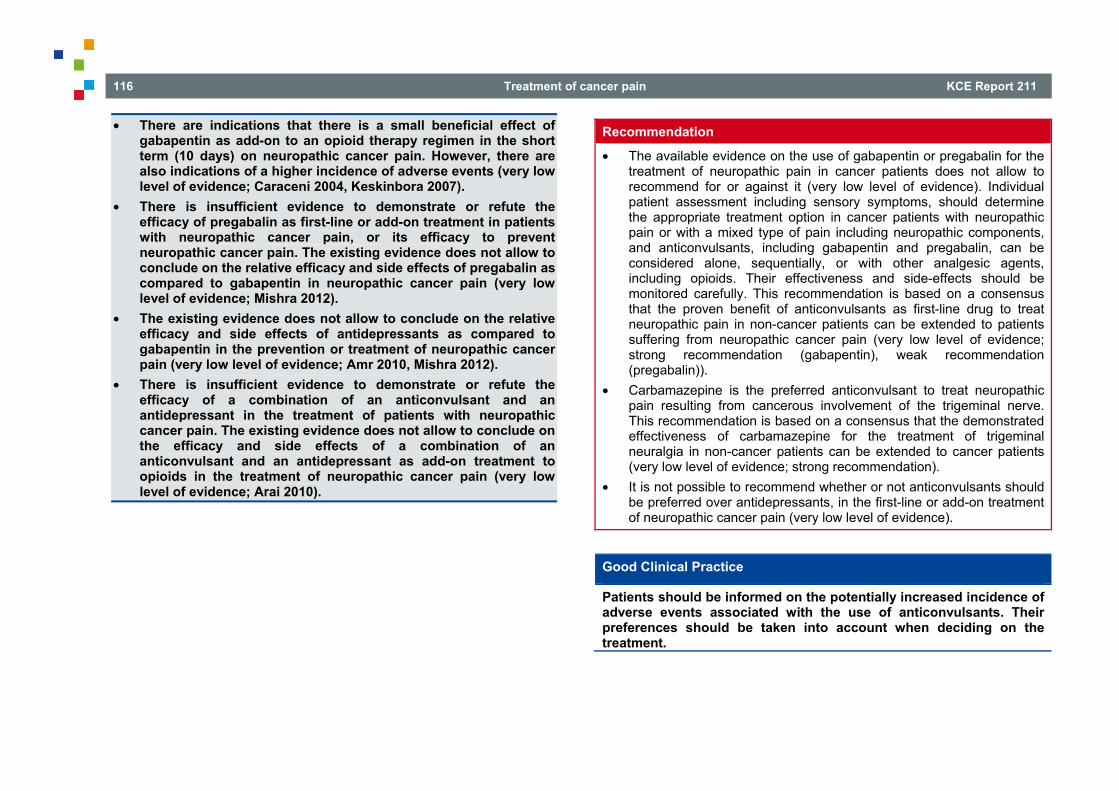
116 Treatment of cancer pain KCE Report 211
• There are indications that there is a small beneficial effect of gabapentin as add-on to an opioid therapy regimen in the short term (10 days) on neuropathic cancer pain. However, there are also indications of a higher incidence of adverse events (very low level of evidence; Caraceni 2004, Keskinbora 2007).
• There is insufficient evidence to demonstrate or refute the efficacy of pregabalin as first-line or add-on treatment in patients with neuropathic cancer pain, or its efficacy to prevent neuropathic cancer pain. The existing evidence does not allow to conclude on the relative efficacy and side effects of pregabalin as compared to gabapentin in neuropathic cancer pain (very low level of evidence; Mishra 2012).
• The existing evidence does not allow to conclude on the relative efficacy and side effects of antidepressants as compared to gabapentin in the prevention or treatment of neuropathic cancer pain (very low level of evidence; Amr 2010, Mishra 2012).
• There is insufficient evidence to demonstrate or refute the efficacy of a combination of an anticonvulsant and an antidepressant in the treatment of patients with neuropathic cancer pain. The existing evidence does not allow to conclude on the efficacy and side effects of a combination of an anticonvulsant and an antidepressant as add-on treatment to opioids in the treatment of neuropathic cancer pain (very low level of evidence; Arai 2010).
Recommendation
• The available evidence on the use of gabapentin or pregabalin for the treatment of neuropathic pain in cancer patients does not allow to recommend for or against it (very low level of evidence). Individual patient assessment including sensory symptoms, should determine the appropriate treatment option in cancer patients with neuropathic pain or with a mixed type of pain including neuropathic components, and anticonvulsants, including gabapentin and pregabalin, can be considered alone, sequentially, or with other analgesic agents, including opioids. Their effectiveness and side-effects should be monitored carefully. This recommendation is based on a consensus that the proven benefit of anticonvulsants as first-line drug to treat neuropathic pain in non-cancer patients can be extended to patients suffering from neuropathic cancer pain (very low level of evidence; strong recommendation (gabapentin), weak recommendation (pregabalin)).
• Carbamazepine is the preferred anticonvulsant to treat neuropathic pain resulting from cancerous involvement of the trigeminal nerve. This recommendation is based on a consensus that the demonstrated effectiveness of carbamazepine for the treatment of trigeminal neuralgia in non-cancer patients can be extended to cancer patients (very low level of evidence; strong recommendation).
• It is not possible to recommend whether or not anticonvulsants should be preferred over antidepressants, in the first-line or add-on treatment of neuropathic cancer pain (very low level of evidence).
Good Clinical Practice
Patients should be informed on the potentially increased incidence of adverse events associated with the use of anticonvulsants. Their preferences should be taken into account when deciding on the treatment.

KCE Report 211 Treatment of cancer pain 117
4.7 Radiotherapy for painful bone metastases 4.7.1 Introduction
4.7.1.1 Radiotherapy for painful bone metastases Skeletal metastases are common in advanced neoplastic diseases, especially in malignancies of the breast, prostate and lung. Metastases to the skeleton are usually multiple, and more than 80% are found in the axial skeleton (Falkmer 2003)213. Although some bone metastases are painless, many cause significant and debilitating pain. Cancer-induced bone pain exhibits components of inflammatory and nociceptive pain, as well as components of neuropathic pain (see 2.2), but the complete mechanism is not yet fully characterized (Delaney 2008, Lozano-Ondoua 2013, Falk 2012, Colvin 2008)214-217. Important aims of treatment are to palliate pain and to prevent future complications such as pathological fracture and spinal cord compression. Only exceptionally the treatment intention is curative, and then the patient usually has a solitary metastasis (Falkmer 2003)213. There is a general consensus among experts that treatment for bone metastasis requires a multimodal approach, and can include therapies such as NSAIDs and opioids, biphosphonates, cytotoxic chemotherapy, anticancer hormone therapy, etc (Falkmer 2003)213. Radiotherapy is one of the frequently used therapeutic options in bone metastasis, to decrease bone pain, or to cause tumor shrinkage or growth inhibition. Its effect on pain becomes fully apparent after 4 to 6 weeks. It is usually given as an outpatient treatment, however, it requires one or more daily hospital attendances, at a specialized radiation-oncological centre that may be some distance away from patient’s home. If the course of radiotherapy is protracted, this may cause considerable problems for the patient, especially those with poor performance status and limited life expectancy. From a health economic point of view, radiotherapy for bone pain constitutes a significant workload of a radiotherapy centre (Sze 2011)218. It is, therefore, important to strike a balance between the treatment efficacy, patient convenience and cost. There is yet no consensus regarding the most appropriate way of delivering radiotherapy for metastatic bone pain.
One of the controversies is whether single fraction radiotherapy is as effective as multiple fraction radiotherapy. Single fraction radiotherapy is more convenient for the patient and it is also less costly compared to multiple fraction radiotherapy. However, there are some important concerns relating to single fraction radiotherapy. Theoretically, the pain response to single fraction radiation may be inferior to multiple fraction radiotherapy, or it may not be durable enough to ensure that the patient remains asymptomatic. In addition, with a potentially reduced tumoricidal effect, single fraction radiotherapy may not be as effective in preventing complications, such as pathological fracture and spinal cord compression (Sze 2011)218. On the other hand, in case of complications or recurrence of the pain, oncologists are more reluctant to give retreatment after multiple fraction radiotherapy (Dutch Guideline on cancer pain 2008)8. Metastases in the spine can compress the spinal cord, leading to pain, neurological impairments (motor and sensory loss) and incontinence (George 2008)219. Spinal cord compression is defined as the compression of the dural sac and its content, spinal cord or cauda equine, or both by an extradural tumour mass. (Loblaw 1998)220. Radiotherapy is, next to surgery and steroids, one of the treatment options to reduce the pressure on the spinal cord. To answer some of the questions mentioned above, the research questions considered in this report are: 1. is radiotherapy more effective than no treatment or than other
treatment options to deal with painful bone metastates? 2. is there a difference in effectiveness and/or adverse events between
different single fraction radiotherapy schemes for the treatment of painful bone metastates?
3. is single fraction radiotherapy as effective as multiple fraction radiotherapy for treatment of painful bone metastates, and are adverse event rates comparable?
4. which radiation schedule is the most effective for pain relief in patients with neurological deficits caused by spinal cord compression?
The comparison of different multiple fraction radiotherapy schemes is considered as a rather technical aspect of the treatment, and is out of scope.

118 Treatment of cancer pain KCE Report 211
4.7.1.2 Hemibody irradiation for painful bone metastases In patients with widespread bone metastases, hemibody irradiation can be an option for the relief of bone pain. According to the expert panel (see colophon), it is not frequently used in Belgium. There could be a significant toxicity related to this treatment as it was applied initially, which required a one day hospitalization or close monitoring for several hours after the procedure. Therefore fractionated schedules of half-body irradiation were introduced, which minimised the side effects and allowed to increase the total dose delivered. So far, few studies are published on the optimal dose for palliation of pain in widespread bone metastases (Salazar 2001)221. The research questions of the section on hemibody irradiation are: • Is hemibody irradiation more effective than no treatment or to other
treatment options to deal with widespread painful bone metastases? • Which hemibody irradiation schedule is the most effective to deal with
painful bone metastases? 4.7.2 Search results
4.7.2.1 Radiotherapy for painful bone metastases Five reviews that met the inclusion criteria were identified on radiotherapy for pain relief in bone metastases. Only one review (Mc Quay 2008)222 dealt with the question whether radiotherapy is more effective than placebo; no review dealt with the comparison between radiotherapy and other treatment options. Mc Quay et al. (2008) did not find RCTs directly comparing radiotherapy with placebo; understandingly placebo is not seen as an ethical treatment option by the research community222. To find an indirect answer to their research question, the authors included 12 RCTs comparing different radiotherapy schemes (single fraction versus single fraction, single fraction versus multiple fraction, multiple fraction versus multiple fraction schemes). All the different treatment arms of the RCTs were used as separate cohorts to estimate the overall treatment response on pain relief in bone metastases. According to the authors, 25% of the included patients obtained complete pain relief at one month, and 41% obtained at least 50% pain relief at some time during the trials. Given the non-controlled
nature of these pooled data, they will not be used further in this report. However, the review by Mc Quay et al (2008) will be used as a basis of RCTs for the comparisons between single fraction versus single fraction, and single fraction versus multiple fraction schemes222. Between four of the five identified reviews (Sze 2003, Sze 2011, Falkmer 2003, and Mc Quay 2008)213, 218, 222, 223 there was a considerable overlap as to the included RCTs. For the subtopic ‘single versus single fraction radiotherapy’, Falkmer (2003) and Mc Quay (2008) each contained one RCT213, 222; these 2 RCTs (Jeremic, 1998 and Hoskin, 1992)224, 225 are used for pooling in this report (Appendix III: Figures 1-7). Both had a high risk of bias. In the update search for RCTs performed in this report, 1 additional RCT was found dealing with ‘single versus single fraction radiotherapy’ as add-on to bisphosphonates; it had an unclear risk of bias (Manas 2008)226. The evidence tables can be found in Appendix III: Table 24. The reviews will not further be described for this subtopic. For the subtopic ‘single versus multiple fraction radiotherapy’, Sze (2003)223 contained all RCTs available in these 4 SRs, and had –contra-intuitively- a more recent literature search date than Sze (2011)218 (November 2001). The overall risk of bias of this review was considered to be low; the overall risk of bias of the included RCTs was considered to be unclear. For one RCT (Sarkar 2002)227 no details on quality appraisal were available in the review and this RCT was evaluated by the Cochrane Collaboration’s tool for assessing risk of bias (Appendix III: Table 7). The evidence table of Sze (2003)223 can be found in Appendix III: Table 24. The other reviews will not further be described for this subtopic. The main topic of the 5th review by George et al. (2010) was treatment of metastatic spinal cord compression228. The literature search date for this review was July 2008 and the overall risk of bias was considered to be low. Of the 6 RCTs included in this review, 3 dealt with corticosteroid treatment for metastatic spinal cord compression but these RCTs did not correspond to the predefined inclusion criteria (see 2.4) of the present report. Only 1 RCT dealt with radiotherapy for metastatic cord compression (Maranzano 2005, see evidence table in Appendix III: Table 27, unclear risk of bias)229 and will be fully discussed below. The two other RCTs (Young 1980, Patchell 2005)230, 231 dealt with a combination of treatments: surgery combined with radiotherapy versus surgery alone for metastatic cord

KCE Report 211 Treatment of cancer pain 119
compression. One of these RCTs (Young 1980) dealt with laminectomy, a technique which has been abandonned if it is not accompagnied by local tumor resection and stabilizing techniques of the spine, for reasons of secondary complications231. Therefore this RCT will not further be discussed. Although surgery is not one of the selected interventions in this report, the comparison with radiotherapy might be relevant for clinicians. Therefore the results of Patchell (2005) will be shortly presented in a narrative way, without including them in the evidence tables or in further recommendations230. Two specific guidelines on radiotherapy for bone metastases were identified232, 233. They did not provide detailed information on pooling of results of individual studies and therefore were used as a basis of RCTs only. Only Alberta Health Services 2010 provided one RCT on radiotherapy for metastatic cord compression232 (Maranzano 2009, see evidence table in Appendix III: Table 27, unclear risk of bias)234 that had not yet been included in the systematic reviews, and a secondary analysis on combined treatment surgery-radiotherapy for the same indication (Chi 2009)235 that will only presented in a narrative way (see previous paragraph). The 4 generic reviews (Guideline of the MoH Malaysia 2010, Dutch Guideline on cancer pain 2008, SIGN 2008, Carr 2002)8-10, 51 did not provide detailed information on pooling of results of individual studies and therefore were used as a basis of RCTs only; no new RCTs that had not yet been included were retained. The update search for RCTs yielded 7 trials (Amouzegar 2008, Badzio 2003, Foro 2008, Hamouda 2007, Hartsell 2005, Kaasa 2006, Roos 2005)236-242, all dealing with the subtopic ‘single versus multiple fraction radiotherapy’. Two of these trials were excluded for the final analyses: the trial of Badzio et al. (2003) was excluded because it did not provide a clear definition of pain improvement237; the trial of Kaasa et al. (2006) because it did not report quantitative data on pain outcome measurements241. The evidence tables of the 5 RCTs can be found in Appendix III: Table 26. The overall quality of these RCTs was judged to be unclear.
4.7.2.2 Hemibody irradiation for painful bone metastases No systematic reviews were found on this topic. Only 1 RCT (Salazar 2001)221 was retained on this topic, selected from the update search for RCTs; the risk of bias for this RCT was high. 4.7.3 Literature overview
4.7.3.1 Single fraction versus single fraction: uncomplicated bone metastases
Two RCTs (Jeremic 1998, Hoskin 1992)224, 225, both with a high risk of bias, compared single fractions of 4-Gy with single fractions of 8-Gy in 489 cancer patients with painful bone metastases, regardless the primary tumour site. Both RCTs only included patients without established pathological fractures and with a life expectancy of more than 6-8 weeks. Only for complete pain response at different time points, and for retreatment rate, a pooled effect could be calculated. For the other outcomes, data were missing or were incomplete in one of the 2 RCTs. In this case the results are described in a narrative way. For the pooled results, complete pain response was defined as ‘no pain’ assessed with a 4 point categorical scale. It was not significantly different at 2, 4 or 8 weeks between the 4-Gy and the 8-Gy treatment (at 2 weeks: RR 0.77 [0.48-1.22], p=0.27; at 4 weeks: RR 0.71 [0.25-2.06], p=0.53; at 8 weeks: RR 0.97 [0.45-2.06], p=0.93). The overall pain response (sum of complete response rate and partial response rate) was only reported in the RCT of Jeremic (1998)225. The authors found significant differences between 4-Gy and 8-Gy at week 2 (RR 0.62 (0.46-0.84), p=0.002), at week 4 (RR 0.64 (0.51-0.80), p=0.0001) and at week 8 (RR 0.75 [0.62-0.90], p=0.003), resulting in significantly higher response rates in 8-Gy compared to 4-Gy. The pooled results showed no significant difference in retreatment rate between 4-Gy and 8-Gy treatment (RR 1.50 (0.74-3.07), p=0.26). In the study of Manas (2008), a single dose of 8Gy plus zoledronic acid (a bisphosphonate) (n=67) was compared to a single dose of 6Gy plus the same dose of zoledronic acid (n=51) in the treatment of painful bone metastases in a mixed cancer patients sample226. At 7 months, no differences were found for mean pain VAS scores (in supine and seated

120 Treatment of cancer pain KCE Report 211
position), QoL and performance status between both groups. The higher dose in the 8Gy group was associated with a higher incidence of adverse reactions (21% of patients showing adverse events versus 14% in the 6Gy group). The authors concluded that both doses of radiotherapy had overall similar analgesic effects and that only the incidence of adverse events made the difference in potential use for clinical practice.
4.7.3.2 Single fraction versus multiple fraction: uncomplicated bone metastases
The review of Sze 2003 included the most recent and complete list of RCTs (12 RCTs)223. One of the RCTs included in Sze 2003223 was not used for the final data pooling in this report 243, because there was no information on the timing of the pain assessment. For 8 of the 11 remaining studies one of the inclusion criteria stipulated that it should be uncomplicated bone metastases, meaning that patients with impending or established pathological fractures and/or spinal cord compression were excluded. The review of Sze 2003 was updated with 5 RCTs starting from 2002; also in these 5 RCTs only patients with uncomplicated bone metastases were included223. The forest plots of the pooled analyses can be found in Appendix III: Figures 8-15.
General remarks Several methodological differences complicated the pooling of the data, and should be taken into account for the interpretation of the results. First of all, the included trials were heterogeneous in several aspects: • No standard criteria for pain assessment
The authors of the different trials used different instruments to assess pain, hampering the pooling of data and its overall interpretation. Most used were a 10-point VAS or VRS but also a 4-or 5-point categorical pain score.
Also the time points for pain assessment differed between the studies. For the pooling we decided to set up general time points (4, 8 and 12 weeks), in line with the review of Sze (2003)223 and to include trial results at the time point itself or closest to that time-point. For example, Foro (2008) and Foro (1998) used 3 weeks as time-point and did not report on outcome at 4 weeks238, 244. In the pooling their results have been included at the 4 weeks
time-point. Third, no standardized criteria for defining partial pain improvement were used. In most studies, however, this was defined as 2 points improvement on the VAS/VRS or 1 point improvement in the categorical pain scores. This was accepted as corresponding roughly to the clinical significant decrease, described in the literature (Dutch Guideline on cancer pain 2008, Farrar 2001)8, 33, although recently more stringent criteria have been put forward. For further discussion on what are clinically significant changes on pain measurement instruments, see chapter 5 Discussion. • Differences in radiation dose and schedules
The most common radiation dose ranged from 8Gy to 10Gy in single fraction arm, whereas in the multiple fraction arm the schedules ranged from 15Gy in 3 sessions (5Gyx3) to 40Gy in 20 sessions (2Gyx20).
Other methodological problems were: • Design of the studies
Most of the included studies were two-arm studies (single fraction versus multiple fraction), whereas some of the studies (Foro 1998 and Ozsaran 2001)244, 245 were three-arm studies (single fraction versus two different multiple fraction schemes). This explains the double counting of the same trial in the pooled analysis (the results of each arm are presented). In some studies (Foro 1998 and Ozsaran 2001)244, 245, patients with metastases on several locations could be randomised more than once; in this case the studies reported the number of painful sites instead of the number of patients per trial arm. A total of 52 patients has been randomised more than once.
• Drop-outs The outcomes were measured based on an intention-to-treat analysis, but due to death of the patient or loss to follow-up, there was a significant amount of missing data at the different time points.
• No differentiation in tumour type Most trials included different types of tumours, and the proportions varied between studies. Usually, the data reported in the trials did not take into account the influence of the primary tumour type on bone metastases and its related pain effect. After discussion with the
KCE Report 211 Treatment of cancer pain 121
external experts that participated in this study (see colophon), it was decided not to conduct separate analyses on different primary tumour types, since this would probably not have a significant influence on the results.
The above-mentioned factors have been taken into account whenever possible, and have influenced the grading of the level of evidence (see Appendix III: Tables 16-18), resulting in a potential tempering of the effect of the intervention.
Results Overall pain response, defined as the sum of partial pain response and complete pain response, was not significant different between single fraction and multiple fraction at any of the time points: RR 1.02 (0.97-1.07) (p= 0.44) at 4 weeks; RR 0.90 (0.78-1.04) (p= 0.15) at 8 weeks and RR 1.01 [0.85-1.19] (p= 0.92) at 12 weeks (see forest plots in Appendix III: Figures 8-10). To obtain these results, 14, 3 and 2 studies, respectively, were pooled at 4, 8 and 12 weeks, respectively. At 4 weeks, 3 560 patients were included and 65% (single fraction) versus 64% (multiple fraction) of them obtained a partial or complete pain response. At 8 weeks, 452 patients were included and at 12 weeks, 267 patients were included; results were comparable. Regarding complete pain relief, similar lack of significant difference between single fraction versus multiple fraction was obtained: RR 1.05 [0.95-1.15] (p= 0.35) at 4 weeks, RR 0.95 [0.72-1.25] (p=0.70) at 8 weeks, and RR 0.89 [0.67-1.19] (p= 0.44) at 12 weeks. To obtain these results, eleven227, 236, 238, 239, 246-252 studies, respectively, were pooled at 4, 8 and 12 weeks respectively. At 4 weeks, 3 286 patients were included and 33% (single fraction) versus 32% (multiple fraction) of them obtained complete pain response. At 8 weeks, 452 patients were included and results were comparable. At 12 weeks, 1165 patients were included and 12% (single fraction) versus 14% (multiple fraction) of them obtained complete pain respons. Nine studies227, 238-240, 242, 246, 250-253 (3 913 patients) were pooled to calculate the retreatment rate: in the single fraction group the retreatment rate was 21%; in the multiple fraction group the retreatment rate was 8%. This means that retreatment was found more frequently in the single fraction group as compared to the multiple fraction group (RR 2.54 [2.14-
3.01], p<0.00001). It should be noted that some studies did not report on the time interval during which patients were followed-up for their need of retreatment, and for those studies reporting this time interval, the range was wide. However, within one study, the time interval can be assumed to be the same for the single and for the multiple fraction treatment. Six studies242, 246, 250-253 were pooled to calculate the rate of pathological fractures (2 748 patients): in the single fraction group the rate of pathological fractures was 3.1%; in the multiple fraction group the rate of pathological fractures was 1.8%. A significant higher rate of pathological fractures was found in the single fraction group compared to the multiple fraction group (RR 1.67 [1.04-2.69], p=0.04). Other adverse events, e.g. nausea and vomiting, mucositis were reported in a very heterogeneous way in the different publications. Also the concomitant use of analgesics, e.g. NSAIDs or opioids, was reported in a very heterogeneous way. These findings are in line with the findings in the review of Sze (2003)223.
4.7.3.3 Radiotherapy for metastatic spinal cord compression Comparison of radiotherapy schemes in patients with metastatic spinal cord compression The search for RCTs on the effectiveness of radiotherapy (RT) regimens for spinal cord compression due to metastatic malignancies revealed only two RCTs, one from the SR of George (2010)228, and one from a guideline (Alberta Health Services 2010) 232 (see 4.7.2). Both deal with the comparison of 2 different radiotherapy schemes (Maranzano 2005, Maranzano 2009)229, 234. Because the intervention RT regimens were different between both studies, pooling of the data was not possible. Maranzano (2005) compared a short course radiotherapy regimen, consisting of 8Gyx2 (total dose of 16Gy in 1 week), to a split-course radiotherapy regimen, consisting of 5Gyx3, followed by 3Gyx5 (total dose of 30Gy in 2 weeks) in a population of patients with metastatic spinal cord compression (ITT n=153 short-course, ITT n=147 split-course)229. Included patients had unfavourable histology or an estimated poor survival; those with indications for surgery were excluded. No p-values were mentioned in the comparisons between groups.

122 Treatment of cancer pain KCE Report 211
Pain relief, defined as the sum of partial and complete pain response, was achieved in half of the patients, without significant differences between short-course RT (80/153, 53%) and split-course RT (79/147, 54%). Complete pain response, defined as patients who had no pain after RT, was similar between both RT regimens: n=47/153 (31%) in short-course RT versus n=47/147 (32%) in split-course RT. In more than half of the patients who had a pain response, complete pain relief as achieved after the radiotherapy intervention (59% in short-course RT and 59% in split-course RT). None of the patients in the group with no analgesics pretreatment had pain after the radiotherapy intervention. Partial pain response, defined as patients using narcotics or minor analgesics before RT who had pain requiring minor analgesics after RT, was also similar between both RT regimens: n=33/153 (22%) in short-course RT versus n=32/147 (22%) in split-course RT. Also regarding non response after RT, no difference was found between short course and split-course radiotherapy. The non-responders were defined as patients with no pain before RT who developed pain or those with pain requiring minor analgesics who started taking narcotics after RT. Functional outcomes, such as walking ability, are within the scope of this report, as far as functionality is related to or a consequence of pain (see 2.1.2). It should be noted that for metastatic spinal cord compression, functional outcome (walking ability) after radiotherapy is rather a consequence of diminished compression due to tumour shrinkage than a consequence of diminished pain. In the study of Maranzano (2005), no significant difference (p-value not given) between short-course and split-course RT was found for walking ability (63% and 65% respectively)229. Sphincter control was considered as out-of-scope in the report. The results on this outcome are presented in the publication of the study and in the evidence tables (see Appendix III: Table 27). Following adverse events were observed: dysphagia, nausea, occurrence of vomiting in patients taking prophylactic anti-emetics (more detailed results can be found in the evidence tables, see Appendix III: Table 27). Late spinal cord morbidity (myelopathy) was never recorded. The authors conclude that no relationship can befound between RT regimen and acute adverse events (for details, Appendix III: Table 27)
• Maranzano (2009) compared a single dose of 8Gy to a short-course of 8Gy x2 (8Gy, 6-day rest and then 8Gy, total dose of 16Gy in 1 week) in a sample of patients with spinal cord compression due to metastatic malignancies (n= 150 in single-dose RT and n= 153 in short-course RT)234. As in Maranzano (2005)229, included patients had unfavourable histology or an estimated poor survival and were not eligible for surgery. The results of the comparison between the 2 RT schemes were comparable to the results of Maranzano (2005)229: there was no significant difference between short course RT and single dose RT (p-value not given) for complete pain response, partial pain response or pain relief, the latter being defined as the sum of partial and complete pain response. Details can be found in the Appendix III: Table 27 (Evidence tables). Following adverse events were observed: dysphagia, nausea, occurrence of vomiting in patients taking prophylactic anti-emetics, diarrhoea (more detailed results can be found in the evidence tables, see Appendix III: Table 27). Late spinal cord morbidity (myelopathy) was never recorded. The authors concluded that no relationship could be found between RT regimen and acute adverse events (for details, see Appendix III: Table 27).
• In conclusion, both radiotherapy schedules were equally effective. In order to minimise the inconvenience for the patients, a single fraction of 8Gy could be considered as the schedule of choice in clinical practice of patients with spinal cord compression and a short life expectancy.
Radiotherapy versus surgery in patients with metastatic spinal cord compression George et al. (2010)228 found one RCT (Patchell 2005)230, which for reasons explained before, will only be presented here in a narrative way (see 4.7.2). A secondary analysis of the study of Patchell 2005 was found (Chi 2009, in the guideline Alberta Health Services 2010)232, 235. Patchell 2005 compared direct decompressive surgical resection plus radiotherapy (n=50) versus radiotherapy alone in a group of patients with metastatic spinal cord compression (n=51) who should not have been paraplegic for more than 48hours; groups were stratified for tumour types230. The primary outcome was ‘motor function’, pain relief was not reported. The overall ambulatory rate after treatment was significantly
KCE Report 211 Treatment of cancer pain 123
higher in the group of surgery plus radiotherapy (84% vs 57%, p=0.001, OR 6.2 (95% CI 2.0-19.8)). In the surgery group significantly better results were also found in other outcomes such as maintenance of continence. A substantial reduction in use of corticosteroids and opioid analgesics was also found. The optimistic results achieved in the group of surgery plus radiotherapy induced an early stop of the study by the data safety and monitoring committee. The authors concluded that the best treatment for metastatic spinal cord compression is surgery as initial treatment followed by radiotherapy. However, factors such as surgical infection, prolonged hospital recovery times, need for reoperation and persistent pain after surgery counter these optimistic results after surgery. Secondary analysis on the influence of age on the results of the study of Patchell 2005 (Chi 2009)235 showed a decrease of the beneficial effect of surgery as age increases, so that it becomes equivalent to radiation therapy alone. These results suggest the use of an age cut off above which surgery may no longer be superior to radiation alone. Based on the analyses of Chi 2009, the cut off lies between 60 and 70 years of age, and statistically significant difference in outcome can be observed when using 65 years as a cut point to stratify patients235.
4.7.3.4 Hemibody irradiation for painful bone metastases The only RCT found, Salazar 2001, was a multi-centre study conducted in a mix of developed and developing countries221. The authors compared three different hemibody radiation schedules in a mixed population of cancer patients: short fractionation (2 fractions of 4-Gy, each in a single day)(n=56), accelerated hyperfractionation (2 fractions of 3Gy on 2 consecutive days) (n=49) and the control schedule (3Gy for 5 days) (n=51). The accelerated hyperfractionation schedule and the control schedules obtained similar results for pain response and QoL, in contrast to the short fractionation schedule after which significant less patients obtained complete pain response (32% vs 63%, p=0.016). A non-significant trend towards more non responders (11% vs 8%) and a trend towards more acute toxicities (16% vs 8%) was found. No differences between the three groups were found for time to pain relief (average of 3 days) and QoL, defined as patient’s remaining pain free % of life (65%, 75% and 72% respectively). The authors conclude that the accelerated radiation schedule (spread over 2 consecutive days instead of 5
consecutive days) could have its benefits to reach more patients and increase treatment adherence. 4.7.4 Other considerations
4.7.4.1 Other considerations: single/single or single/multiple fraction radiotherapy: uncomplicated bone metastases
The literature found on single versus single or single versus multiple fraction radiotherapy, almost invariably excluded patients with complicated bone metastases. This means that patients with impending or established pathological fractures and/or spinal cord compression were excluded. Some studies also specified that the life expectancy should be at least some weeks, excluding patients at the very end of their life. Consequently, firm conclusions from the literature can only be drawn on radiotherapy for uncomplicated bone metastases (see below). In Belgium, for this group of patients, a single fraction of 8-Gy currently is the preferred practice, according to the expert panel (see colophon). In their experience, the results of a single fraction of 8-Gy is better as compared to a single fraction of 4-Gy, whereas side-effects are comparable.They argue that, in a recent SR (Dennis 2013)254, published after the date of search of the present report, it was concluded that 8-Gy is the single fraction dose that is most frequently used in the included RCTs. The authors of this SR, which did not include a quality apprasal of the primary studies, described 3 RCTs that compared directly single and multiple fraction; besides the RCTs of Jeremic 1998 and Hoskin 1992 that are also included in the present report, they also included Altundag 2002 who reported a non-superior pain response within 60 days for 8-Gy as compared to 5-Gy224, 225, 255. Further, the experts refer to the Guideline from the American Society of Radiation Oncology (ASTRO) on palliative radiotherapy for bone metastases (2011), which also recommends the 8-Gy single fraction schedule over other single fraction schedules256. This guideline is based on a systematic review confined to a search in one database only (Medline), which is the reason why it was not included in this report.

124 Treatment of cancer pain KCE Report 211
The experts also conclude that aspects such as life expectancy of the patient and curability of the bone metastases should be considered when deciding on the preferred radiotherapy scheme for pain relief in bone metastases. The expert panel argues that patients with an uncomplicated solitary bone metastasis or with very few uncomplicated bone metastases are taken as an exception to the rule that a single fraction of 8-Gy is currently the preferred practice. For these patients, higher doses and/or multiple fractions might be considered, as the treatment intention is to cure them or at least to induce a long lasting local control. For patients with complicated bone metastases, i.e. impending or established pathological fractures and/or spinal cord compression, the experts consider a multiple fraction scheme, depending on the medical characteristics of each individual patient and taking into account his/her preferences after (s)he have been well informed on their situation. According to the experts, patients with bone metastases and an extensive soft tissue component also belong to the group of complicated bone metastases. A difficulty in interpreting the results of the present study arises from the fact that radiotherapy can be combined with other pain treatments. Heterogeneous reporting on concomitant changes in use of analgesics or other drugs (e.g. biphosphonates) prohibited pooling of the data with respect to this parameter. Indeed, changes in the use of these types of drugs during the study can influence significantly the pain measurements. Also, when these types of drugs can be diminished, it can be considered to be a favourable outcome of the radiotherapy. Taking this into account, it is clear that complete pain relief is reached in a minority of the patients only: in the literature mentioned above, a significant number of patients with uncomplicated bone metastases does not obtain total pain relief from single or multiple fraction radiotherapy. This means that in daily practice, there is room for a combination of treatments, adjusted to the needs of each individual patient. The expert panel agrees that a multimodal pain treatment approach should be considered for uncomplicated or complicated bone metastases: besides local treatment with single or multiple fraction radiotherapy, systemic therapies should also be considered throughout the treatment course, e.g. NSAIDs, opioids, cytotoxic chemotherapy, anticancer hormone therapy.
4.7.4.2 Other considerations: metastatic spinal cord compression and hemibody irradiation
The expert panel agrees that decompressive surgical tumor resection with or without stabilization of the spine and followed by radiotherapy is the option of first choice in patients with metastatic spinal cord compression, after aspects such as duration of the spinal cord compression, status and type of the disease, prior and available anticancer therapies, life expectancy of the patient, and patient preferences have been taken into account. The expert panel also agrees that hemibody irradiation can be a treatment option in patients with widespread bone metastases who suffer from pain after all systemic pain treatment options failed, and after life expectancy of the patient and patient preferences have been taken into account.

KCE Report 211 Treatment of cancer pain 125
Conclusions • Based on the available evidence, it is not yet known whether
radiotherapy by a single fraction of 4-Gy or by a single fraction of 8-Gy is most efficient in relieving bone pain completely at 4 or 8 weeks, in patients with uncomplicated bone metastases, regardless the primary tumour site (very low level of evidence; Hoskin 1992, Jeremic 1998).
• Based on the available evidence, it is not yet known whether, in patients with uncomplicated bone metastases, retreatment rate is higher after radiotherapy by a single fraction of 4-Gy or after radiotherapy by a single fraction of 8-Gy (low level of evidence; Hoskin 1992, Jeremic 1998).
• There is limited evidence from 1 RCT that in patients with uncomplicated bone metastases, regardless the primary tumour site, radiotherapy by a single fraction of 4-Gy results in a lower overall pain response, i.e. the sum of complete and partial response rates, as compared to a single fraction of 8-Gy at 2, 4 or 8 weeks. (very low level of evidence; Jeremic 1998).
• Based on the available evidence, it is not yet possible to conclude on the efficacy of single fraction of radiotherapy as add-on to bisphosphonates, in the treatment of painful bone metastases (very low level of evidence; Manas 2008).
• It is plausible that in patients with uncomplicated bone metastases, regardless the primary tumour site, radiotherapy by a single fraction of 8 to 10-Gy is as efficient in relieving bone pain completely at 4 weeks, as radiotherapy by multiple fractions (3x 5-Gy to 20x 2-Gy) (moderate level of evidence; Sze 2003, Amouzegar 2008, Foro 2008, Hamouda 2007).
• There are indications that in patients with uncomplicated bone metastases, regardless the primary tumour site, radiotherapy by a single fraction of 8 to 10-Gy is as efficient in relieving bone pain completely at 8 and 12 weeks, as radiotherapy by multiple fractions (3x 5-Gy to 20x 2-Gy) (low level of evidence; Foro 2008, Hamouda 2007, Hartsell 2005, Roos 2005, Sarkar 2002).
• It is plausible that in patients with uncomplicated bone metastases, regardless the primary tumour site, radiotherapy by a single fraction of 8 to 10-Gy is as efficient at 4 and 8 weeks in relieving bone pain completely or partially, as radiotherapy by multiple fractions (3x 5-Gy to 20x 2-Gy) (moderate level of evidence; Sze 2003, Amouzegar 2008, Foro 2008, Hamouda 2007, Roos 2005).
• There are indications that in patients with uncomplicated bone metastases, regardless the primary tumour site, radiotherapy by a single fraction of 8 to 10-Gy is as efficient at 12 weeks in relieving bone pain completely or partially, as radiotherapy by multiple fractions (3x 5-Gy to 20x 2-Gy) (low level of evidence; Foro 2008, Hamouda 2007).
• It is plausible that in patients with uncomplicated bone metastases, regardless the primary tumour site, there are more retreatments after radiotherapy by a single fraction of 8 to 10-Gy than after radiotherapy by multiple fractions (3x 5-Gy to 20x 2-Gy) (moderate level of evidence; Sze 2003, Foro 2008, Hamouda 2007, Hartsell 2005, Roos 2005).
• There are indications that in patients with uncomplicated bone metastases, regardless the primary tumour site, there are more pathological fractures after radiotherapy by a single fraction of 8 to 10-Gy than after radiotherapy by multiple fractions (3x 5-Gy to 20x 2-Gy) (low level of evidence; Sze 2003, Foro 2008, Hamouda 2007, Hartsell 2005, Roos 2005).
• In the current literature search, no studies have been found focusing on radiotherapy for bone metastases complicated by impending or established pathological fractures.
• Both RCTs comparing different radiation schedules in patients with metastatic spinal cord compression and poor prognosis, could not differentiate between both intervention arms. Based on the available evidence, no conclusion can be drawn on the greater effect on pain of one radiation schedule compared to another (very low level of evidence; Maranzano 2005, Maranzano 2009).

126 Treatment of cancer pain KCE Report 211
• There are no indications of a higher incidence of adverse events related to a specific radiation schedule in patients with metastatic spinal cord compression and poor prognosis (very low level of evidence; Maranzano 2005, Maranzano 2009).
• There is insufficient evidence to demonstrate or refute the superiority of a short fractionation schedule (2 x4-Gy, in 1 day), a hyperfractionation schedule (2x 3Gy, in 2 days) or a standard schedule (5x3Gy, in 5 days) in hemibody irradiation for widespread bone metastases (very low level of evidence; Salazar 2001).
Recommendation
• Aspects such as life expectancy, clinical condition of the patient and curability of the bone metastases should be considered when deciding on the preferred radiotherapy scheme for pain relief in bone metastases (very low level of evidence; strong recommendation).
• The use of single fraction radiotherapy for short term complete or partial pain relief in patients with uncomplicated bone metastases is recommended as treatment of first choice. However, the definitive decision on the treatment modality should also take into consideration the preferences of the patient after he/she has been well-informed; and aspects such as life expectancy, clinical condition of the patient and curability of the bone metastases should be considered (strong recommendation). This is based on the following evidence. It is plausible that single and multiple fraction radiotherapy are equally effective for short term (4 to 8 weeks) complete or partial pain relief in patients with uncomplicated bone metastases, regardless the primary tumour site (moderate level of evidence). It is also plausible that radiotherapy by a single fraction is associated with more retreatments (moderate level of evidence) and more pathological fractures (low level of evidence), but the overall rate of retreatment and pathological fractures is relatively low.
• The use of a 8-Gy single fraction radiotherapy rather than a 4-Gy single fraction radiotherapy for short term (4 to 8 weeks) complete or partial pain relief in patients with uncomplicated bone metastases is the option of first choice, after aspects such as life expectancy and clinical condition of the patient, curability of the bone metastases and patient preferences have been taken into account (very low level of evidence; weak recommendation).
• A multimodal pain treatment approach should be considered for uncomplicated or complicated bone metastases. Besides local treatment with single or multiple fraction radiotherapy, systemic therapies should also be considered throughout the treatment course, e.g. NSAIDs, opioids, cytotoxic chemotherapy, anticancer hormone therapy. This is supported by the significant number of patients with uncomplicated bone metastases who do not obtain total pain relief from single or multiple fraction radiotherapy (very low level of evidence; strong recommendation).
• Decompressive surgical tumor resection with or without stabilization of the spine and followed by radiotherapy is the option of first choice in patients with metastatic spinal cord compression, after aspects such as duration of the spinal cord compression, status and type of the disease, prior and available anticancer therapies, use of corticosteroids, life expectancy, and patient preferences have been taken into account (very low level of evidence; weak recommendation).

KCE Report 211 Treatment of cancer pain 127
• There is insufficient evidence to advise on the preferred radiation dose for the treatment of pain due to metastatic spinal cord compression (very low level of evidence).
• Hemibody irradiation can be a treatment option in patients with widespread bone metastases who suffer from pain after all systemic pain treatment options failed, and after life expectancy and patient preferences have been taken into account (very low level of evidence; weak recommendation).
• There is insufficient evidence to advise on the preferred schedule for hemibody irradiation as treatment of pain in patients with widespread bone metastases (very low level of evidence).
Good Clinical Practice
Patients should be clearly informed on the benefits and side-effects of the different radiotherapy schemes for pain relief in bone metastases, so that their preferences can be taken into account when deciding on the treatment modality.
4.8 Radionuclides for painful bone metastases 4.8.1 Introduction The main consequence of bone metastases is severe pain. Other clinical manifestations are pathological fractures, hypercalcemia and spinal cord compression. The radiopharmaceuticals discussed in this chapter, also called radionuclides or radio-isotopes, are beta-emitting agents administered intravenously, with a particular affinity to bone turnover sites and especially to sites with increased osteoblastic activity (Table 16). Advantages of radioisotopes include the ability to simultaneously treat multiple sites of disease, ease of administration, and the potential integration with other treatments like radiotherapy or chemotherapy. They might also exert less generalized side-effects, because of their selective and focused activity at the site of the bone metastases. The most frequently used radioactive substances are strontium-89 (89Sr) and samarium-153 (153Sm). Other agents considered are rhenium-186 (186Re) and rhenium-188 (188Re), which are still in the experimental phase. Strontium-89 is a pure beta
emitter and has a long physical half-life (50 days), while 153Sm, 188Re and 186Re have much shorter physical halflives (less than four days) and are gamma emitters, enabling posttreatment scintigraphic imaging and dosimetry but also having greater implications for radiation protection (Roqué 2011, Dutch Guideline on cancer pain 2008)8, 257. Radionuclides are one of the treatment options for the palliation of painful bone metastases. Other treatment options are analgesia, radiotherapy, chemotherapy and hormone therapy, surgery, and bisphosphonates. According to the expert panel (see colophon), the use of radionuclides for painful bone metastases is relatively limited in Belgium. However, the efficacy of radionuclides, their relative value as compared to other treatment options and their potential contribution in multimodal treatments is not yet clear. The present report evaluates the efficacy of radionuclides to relieve pain and in terms of quality of life. Their overall effect on patient survival and their efficacy in the treatment of complications such as pathological fractures, hypercalcemia or spinal cord compression, is out of scope.
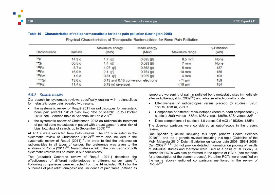
128 Treatment of cancer pain KCE Report 211
Table 16 – Characteristics of radiopharmaceuticals for bone pain palliation (Lewington 2005)
4.8.2 Search results Our search for systematic reviews specifically dealing with radionuclides for metastatic bone pain revealed two results: • the systematic review of Roqué 2011 on radioisotopes for metastatic
bone pain (overall risk of bias: low; date of search: up to October 2010, see Evidence table in Appendix III: Table 29)257
• the systematic review of Christensen 2012 on radionuclide treatment of painful bone metastases in patient with breast cancer (overall risk of bias: low; date of search: up to September 2009) 258
All RCTs were extracted from both reviews. The RCTs included in the systematic review of Christensen (2012)258 were also included in the systematic review of Roqué (2011)257. In order to find the evidence on radionuclides in all types of cancer, the preference was given to the analyses of Roqué (2011)257. Nevertheless a link to the conclusions of both systematic reviews will be made in our conclusions. The (updated) Cochrane review of Roqué (2011) described the effectiveness of different radio-isotopes in different cancer types257. Following comparisons were extracted from the 14 included RCTs for the outcomes of pain relief, analgesic use, incidence of pain flares (defined as
temporary worsening of pain in radiated bony metastatic sites immediately after radiotherapy (Hird 2009259) and adverse effects, quality of life: • Effectiveness of radioisotopes versus placebo (8 studies): 89Sr,
186Re, 153Sm, 223Ra • Comparison of different radio-isotopes (head-to-head comparisons) (3
studies): 89Sr versus 153Sm, 89Sr versus 186Re, 89Sr versus 32P • Dose-comparisons (4 studies): 1.0 versus 0.5 mCi of 153Sm, 188Re The dose-comparisons were considered as out-of-scope in the present review. One specific guideline including this topic (Alberta Health Services 2010)232, and the 4 generic reviews including this topic (Guideline of the MoH Malaysia 2010, Dutch Guideline on cancer pain 2008, SIGN 2008, Carr 2002)8-10, 51 did not provide detailed information on pooling of results of individual studies and therefore were used as a basis of RCTs only. A search for RCTs was also performed in the update of RCTs (see methods for a description of the search process). No other RCTs were identified on the same above-mentioned comparisons mentioned in the review of Roqué-257.

KCE Report 211 Treatment of cancer pain 129
Besides the literature on radio-isotopes versus placebo or versus another radio-isotope, a few studies were found that compared radio-isotopes to other medical treatment. The evidence on this type of comparisons is scarce and the evidence is limited due to small number of studies on the same comparison and small sample sizes. Within the list of update search for RCTs, 2 small RCTs (Wang 2003, Nilsson 2005)260, 261 were retrieved on the efficacy of radionuclides adjuvant to another medical treatment (bisphoshonate, chemotherapy). The details on the analyses are presented in appendix (see Appendix III: Table 30). 4.8.3 Literature overview Effectiveness of radio-isotopes compared to placebo The results from the systematic review of Roqué 2011 were extracted; the authors noticed that the studies included in their review all had recruited specifically participants that had failed conventional treatment; radio-isotopes were considered as a last treatment option257. In addition to the results of the Cochrane review sub analyses on the efficacy of 89Sr versus placebo were performed by the authors of this report. Pain control, defined as complete, partial or any amount of pain relief (see further), was assessed in 8 of the 14 included studies (n=499 participants) and included 89Sr, 186Re, 153Sm and 223Ra (Lewington 1991, Porter 1993, Sartor 2004, Nilsson 2007, Maxon 1991, Smeland 2003, Buchali 1988, Han 2002)262-269. Six trials concerned patients with prostate cancer, 2 studies concerned patients with any primary tumour histology (Maxon 1991, Smeland 2003)265, 269. Pain was measured with a VAS scale or a nominal scale of four to five categories; the percentage of patients reducing their pain from baseline was reported. Four of the 8 included studies assessed pain at short-term (1 month), 3 studies at medium-term (3 to 6 months) and 1 study at long-term (12 months). At short and medium term a beneficial effect for pain control was found, however at long-term this effect was not significant. Overall, when all possible time-frame evaluations were included, a small but significant improvement in pain was noticed for both complete and partial pain response: RR 2.10 (95% CI 1.32 to 3.35)(p=0.0018) for complete pain relief (100% pain relief) and RR 1.72 (95% CI 1.13 to 2.63)(p=0.012) for partial pain relief (50-100% pain relief). The NNT for complete pain relief is 5 (range 2 to 44) and for partial pain
relief is 8 (range 1 to 54). The results for the outcome ‘any amount of relief’ (between 0 and 100% pain relief) is more heterogeneous and the pooled result is not significant (RR 1.36, 95% CI 0.77 to 2.40) (p=0.29). We looked also more into detail which radio-isotopes were compared to placebo. Four studies of the 8 included studies compared 89Sr versus placebo. A sub-analysis of these 4 studies on 89Sr (n=189 participants) (Lewington 1991, Porter 1993, Buchali 1988, Smeland 2003)262, 264, 267, 269, performed by the authors of this report, is presented. The comparisons on the available data (ITT-analysis) showed different results as compared to the above-mentioned comparisons including all different radio-isotopes. For complete pain relief, no significant effect of 89Sr was found compared to placebo (based on 2 studies) (p=0.08)(RR 1.87, 95% CI 0.93 to 3.77). The results for partial pain relief were similar to the global comparisons (based on 2 studies): a significant effect of 89Sr on partial pain relief compared to placebo (RR 1.62 (95% CI 1.02 to 2.68), p=0.04). Also the non-significant difference between all radio-isotopes versus placebo for any amount of relief was found for the comparison ‘89Sr versus placebo’ (based on 3 studies): RR 1.03 (95% CI 0.60 to 1.79), p=0.90. The analysis on available data did not differ from the ITT-analysis (results are presented in the forest plots in Appendix III: Figures 18-23). In conclusion, the studies included in this sub-analysis on the effectiveness of 89Sr on pain relief showed a significant effect for a partial pain response, but complete pain relief could not be obtained. The difference with the overall results for all radio-isotopes together might be explained by a lower power in this sub-analysis. A sensitivity analysis on 3 small studies (Nilsson 2007, Porter 1993, Smeland 2003)264, 266, 267, 269 was also performed in the systematic review of Roqué 2011on the effects of radio-isotopes adjuvant to radiotherapy versus placebo adjuvant to radiotherapy on pain relief257. The total number of patients included in these 3 trials was 188. Only a significant difference was found for partial pain relief (RR 1.64 (95% CI 1.05 to 2.55)) (p=0.029). No significant differences were found between radio-isotopes adjuvant to radiotherapy versus placebo for complete pain relief (RR 2.55 (95% CI 0.52 to 12.63)) (p=0.25) and for any amount of relief (RR 0.80 (95% CI 0.50 to 1.27)) (p=0.34).

130 Treatment of cancer pain KCE Report 211
The four studies in Roque 2011, reporting on analgesic use, are heterogeneous in reporting the results, which makes it difficult to pool the results257. One study (Porter 1993)267 found a greater proportion of patients who stopped taking analgesics at three months compared to the placebo group. In the second study (Han 2002)263 the request for palliative radiotherapy is seen as a kind of use of analgesics, but no significant differences were found between the intervention group and the placebo group. The study of Nilsson 2007 found no differences on analgesic consumption after the addition of radio-isotopes of placebo in participants receiving external beam radiotherapy266. The fourth study (Maxon 1991)265 found a greater reduction in analgesic use, but these findings are difficult to generalize due to the small samples sizes (10 participants in each group). The few studies, reporting on analgesic use and the heterogeneity in reporting of the results, hinder to draw a firm conclusion on the effects of radio-isotopes on analgesic use. Two studies in Roqué 2011 assessed quality of life, but the poor reporting of the results, hampered further analysis and conclusion (Porter 1993, Smeland 2003)257, 267, 269. Only two studies in Roqué 2011 (Sartor 2004, Serafini 1998)268, 270 reported on the incidence of pain flares, but no significant effects were found on the incidence of these pain flares (RR 0.74, 95% CI 0.27 to 2.06) (p=0.57). Five studies in Roqué 2011 reported on severe adverse effects of (grade III to IV) 89Sr or 153Sm (Porter 1993, Buchali 1988, Sartor 2004, Serafini 1998 Lewington 1991)257, 262, 264, 267, 268, 270. Four trials concerned patients with prostate cancer, 1 study concerned patients with any primary tumour histology (Serafini 1998)270. The pooled analysis of four studies showed a significantly higher incidence of leucocytopenia in participants treated with radio-isotopes, compared to the placebo group (Risk difference 0.07, 95% CI 0.04 to 0.11) (p=0.00012). The NNH to observe one case of leucocytopenia due to radio-isotopes is 14 (95% CI 9 to 25). For the incidence of thrombocytopenia, the difference does not reach statistical significance, but a trend towards a higher incidence can be seen in participants treated with radio-isotopes (RR 0.04, 95% CI -0.03 to 0.11) (p=0.25). No significant difference in incidence of anemia was found between participants treated with radio-isotopes or the placebo group by
pooling of the 2 studies reporting on this adverse event (Risk difference 0.03, 95% CI -0.04 to 0.09)(p=0.44). An analysis of the studies on 89Sr compared to placebo, performed by the authors of this report, showed similar results as mentioned above. A significant higher incidence of leucocytopenia was found in patients treated with 89Sr compared to placebo (based on 2 studies)262, 267 (Risk difference 0.10, 95% CI 0.03 to 0.17) (p=0.006) and the difference in incidence of thrombocytopenia did not reach the threshold of statistical difference (based on 2 studies)262, 271 (Risk difference 0.17, 95% CI -0.02 to 0.36) (p=0.08). No results were reported on the incidence of anemia. These results confirm the higher incidence of severe adverse effects in patients treated with radio-isotopes, such as 89Sr.
Head-to-head comparisons of different radio-isotopes Roqué 2011 found three RTCs comparing the relative efficacy of 89Sr to other radio-isotopes (153Sm, 186Re and 32P)257. Each RCT made a different comparison, so no meta-analysis could be performed. Only descriptive results per comparison are presented. In the comparison of 89Sr versus 153Sm (Baczyk 2007)272 (n= 50 in 89Sr group and n=50 in 153Sm group) in patients with prostate or breast cancer, both radionuclide therapies had significant effects on complete pain relief (at 2 months post-treatment; p<0.05), on improvement of functional performance (Karnofsky scale) and on decrease of consumption of analgesic drugs (p<0.05). For complete pain relief, there was no significant difference between the two radio-isotopes (RR 0.75 (95% CI 0.44-1.29), ns). The overall analgesic effect (including the complete and partial pain response) was significantly better in patients with osteoblastic metastases, independently of the radionuclide therapy (p<0.01). Following adverse events were observed with a comparable frequency for both radionuclides: (severe) pancytopenia, (moderate) granulocytopenia and/or thrombocytopenia, and hypercalcaemia. The authors concluded that both radionuclide therapies had similar analgesic effects. Only the type of metastases could influence this effect: a better response was observed in patients with osteoblastic metastases compared to patients with mixed metastases.
KCE Report 211 Treatment of cancer pain 131
In the comparison of 89Sr (n=15) versus 32P (n=16)(Nair 1999)273 in patients with any primary tumour histology, similar results as in the comparison with 153Sm were found with no significant difference between both radionuclide therapies for pain relief (RR 1.07 (95% CI 0.49-2.32), ns) and toxicity. Also the pain improvement was followed by a reduction in consumption of analgesics and an improvement of mobility. In spite of the similar analgesic effects of both radionuclide therapies, the authors recommend the use of 32P instead of the expensive 89Sr. A similar lack of differences in analgesic effect and toxicities was found comparing 89Sr (n=25 patients) versus 186Re (n=25 patients) in breast cancer patients (Sciuto 2001)274. Regarding the analgesic effects, there was no significant difference for number of patients experiencing complete pain relief (RR 1.00 (95% CI 0.45-2.24), ns). Only the onset of the pain response was significantly shorter in the 186Re group (p<0.0001) while the duration and the degree of response were not significant different. Also no differences were found in the incidence of adverse effects, assessed by the decrease of leucocytes and platelets. However, the time to recovery was significantly lower in the 186Re group (p<0.001). The authors recommend the use of 186Re in patients with more compromised haematological function, more unbearable pain and with a lower estimated life expectancy because of the early onset of pain relief and the shorter recovery time. The moderately longer duration of effect advantages the use of 89Sr in patients with moderate pain, less compromised general conditions and a reasonable estimated life expectancy. The scarcity of data per comparison and the lack of differences in analgesic effects make it difficult to formulate general conclusions and recommendations on the preference for one specific radio-isotope compared to the others.
Radionuclides compared to other medical treatments Only two small RCTs were included, which are presented narratively. One trial assessed the analgesic effects of a radionuclide (89Sr) (n=18) compared to chemotherapy (5-FU, epirubicin and mitomycin C, FEM) (n=17) in prostate cancer patients (Nilsson 2005)260. In both groups the pain intensity decreased significantly over time (p<0.05 at 3, 6, 9 and 12 weeks) compared to baseline. One explanation for the unexpected pain reduction in the chemotherapy group is its anti-inflammatory effect. However, the side effects and hospitalizations were more frequent in the FEM group compared to the 89Sr group. The authors conclude that, despite comparable analgesic effects for both interventions, 89Sr treatment is preferable. Wang et al (2003)261 compared a group of cancer patients treated with 153Sm (n=9) to a group of cancer patients treated with a bisphoshonate (pamidronate disodium) (n=9). The greater analgesic efficacy, also characterised by a longer duration of palliative effect, in the 153Sm group was however accompanied with a higher incidence of side effects, such as haematological toxicities and myelosuppression. 4.8.4 Other considerations Methodologically, the available evidence on the efficacy of radio-isotopes in cancer patients with painful bone metastases shows different weaknesses. Only a limited number of studies was found per comparison and per outcome and most studies were characterized by small sample sizes and unclear or high risk of bias due to methodological flaws in the study design. Also the pooling of the results was hampered by the important heterogeneity between studies, due to the use of different radio-isotopes and different dose schedules, different outcome scales, difference in timing of the endpoints and different criteria for defining the outcome. The overall quality of the evidence according to the GRADE-system can be considered to be low.

132 Treatment of cancer pain KCE Report 211
Next to the methodological weaknesses of the included studies and the overshadowing of the beneficial effects on pain by an increased incidence of severe adverse events, the clinical usefulness of these findings can only be considered for patients who had failed conventional treatment. All included studies recruited particularly on these participants and radio-isotopes were seen as last option (Rocqué 2011)257. To consider the administration of radio-isotopes earlier in the disease process, they should prove to have antitumor activity, besides their pain control effect. Given the wide number of treatments options available to patients with bone metastases, all of them presenting differential effectiveness and safety profiles, detailed cost effectiveness analyses would be required to evaluate the place of radioisotopes in the array of treatment options. In the review of Christensen 2012, the authors included only studies on patients with bone metastases due to breast cancer258. The 3 included RCTs were also included in the analyses of Roqué 2011257. Methodological weaknesses prevented firm conclusions from these 3 RCTs. According to the expert panel (see colophon), thrombocytopenia and pain flares are serious side effects that should be taken into account, although in the (limited) literature included in this report the frequency of these adverse effects did not reach statistical difference as compared to placebo. In the stakeholders meeting, the specialist in nuclear medicine mentioned some new publications on the effectiveness of more recent radionuclides (alpha-emitters) in pain treatment. These studies were published after our search in literature and were therefore not retrieved and mentioned in this report. In a future report on the effectiveness of radionuclides an up-to-date search for primary studies is needed, especially on the new molecules such as the alfa-emitters.
Conclusions • The conclusion on the efficacy of bone-seeking radioisotopes in
relieving pain from bone metastases is focused on patients who have failed conventional treatment, because the included studies only recruited participants at this stage. No conclusions can be drawn on the use of radioisotopes early in the natural history of metastatic bone disease (Rocqué 2011).
• There are indications that the radioisotopes 89Sr, 153Sm, 186Re and 223Ra have a beneficial effect in the short and medium term (up to 6 months) on complete or partial (more than 50%) pain relief in a mixed population of cancer patients with metastatic bone pain who have failed conventional treatment. The number needed to treat (NNT) for complete pain relief is 5 (range 2 to 44) and for partial pain relief is 8 (range 1 to 54) (low level of evidence; Rocqué 2011). Due to a scarcity of evidence, it is not known whether this effect persists beyond 6 months.
• The available evidence prohibits firm conclusions on whether bone-seeking radioisotopes as compared to placebo modify the use of analgesics in cancer patients with metastatic bone pain (very low level of evidence; Rocqué 2011).
• There are indications that patients from a mixed cancer population with metastatic bone pain and treated with 89Sr or 153Sm after conventional treatment failed, may suffer more from severe adverse events as compared to placebo. The available evidence shows an increase in incidence of leucocytopenia (low level of evidence, Roqué 2011). The number needed to treat in order to observe one case of leucocytopenia due to radio-isotopes (NNH) is 14 (95% CI 9 to 25).
• No indications could be found that the incidence of pain flares after treatment with bone-seeking radionuclides in cancer patients with painful bone metastases is increased as compared to placebo (low level of evidence; Rocqué 2011).

KCE Report 211 Treatment of cancer pain 133
• Based on scarce evidence, no indications were found in the head-to-head comparisons of 89Sr versus 153Sm or 32P or 186Re, that 89Sr obtained more pain relief compared to the other radio-isotopes in a mixed population of cancer patients with painful bone metastases who have failed conventional treatment (very low level of evidence; Rocqué 2011).
• The available evidence prohibits firm conclusions on the relative effectiveness of bone-seeking radioisotopes adjuvant to external beam radiotherapy as compared to bone-seeking radioisotopes alone in obtaining pain relief in a mixed cancer population with metastatic bone pain after conventional treatment failed (low level of evidence; Rocqué 2011). The available evidence also prohibits firm conclusions on the relative effectiveness of bone-seeking radioisotopes as compared to other pain treatment options for metastatic bone pain (very low level of evidence).
• The available evidence prohibits firm conclusions on the relative effectiveness of bone-seeking radioisotopes as compared to chemotherapy or bisphosphonates in cancer patients with metastatic bone pain (very low level of evidence; Nilsson 2005, Wang 2003).
Recommendation
• When conventional treatment failed, bone-seeking radio-isotopes can be considered as a secondary option to obtain complete or partial (more than 50%) pain relief in the short and medium term (up to 6 months) for widespread painful osteoblastic bone metastases in cancer patients regardless of the primary tumour site (low level of evidence; weak recommendation).
• It is recommended to inform patients who this treatment can be associated with an increased incidence of severe adverse events, such as hematological toxicity and in particular leucocytopenia (low level of evidence; strong recommendation).
Good Clinical Practice
Patients should be informed on the benefits and potential side-effects associated with the use of bone-seeking radio-isotopes. Their preferences should be taken into account when deciding on the treatment.
4.9 Bisphosphonates for painful bone metastases 4.9.1 Introduction Bone metastases occur in up to 80% of patients with advanced breast or prostate cancer, and they are also common in several other malignancies such as lung or thyroid cancer (Diel 2004, Dearnaly 2003, Wong 2004)275-
277. Different types of bone metastases exist: those inducing increased bone formation (‘osteoblastic’ lesions), those inducing increased bone resorption (‘osteolytic’ or ‘osteoclastic’ lesions), and mixed types. Lesions in breast cancer are mostly of the osteolytic or mixed type (Wong 2012)278. Lesions in prostate cancer are typically of the osteoblastic type, but have been shown to include osteoclastic phenomena as well (Meulenbeld 2012)279. A slow and progressive bone damage is also present in the majority of patients suffering from multiple myeloma (Mhaskar 2012)280, causing typically osteolytic lesions. Bone metastases result in considerable morbidity, including bone pain, hypercalcemia, and so-called skeletal-related events (SREs) such as pathologic fractures, spinal cord compression, or the need for surgery or radiation to bone e.g. because of an impending fracture (Wong 2004, Wong 2009)277, 281. Despite the use of analgesic drugs, local therapy with radiation or surgery, or systemic therapy with hormonal or chemotherapeutic agents, many patients continue to experience symptoms of progressive bone destruction and deterioration of quality of life. Bisphosphonates are structural analogues of pyrophosphates, a naturally occurring component of bone crystal deposition. They inhibit osteoclastic bone resorption and have been shown to be effective in the management of tumour-induced hypercalcemia. They have also been suggested to be effective in preventing SREs and decreasing bone pain related to bone metastases, especially metastases of the osteolytic type. The exact
134 Treatment of cancer pain KCE Report 211
mechanisms as regards pain relief are not yet fully understood (Wong 2012)278. Bisphosphonates are broadly classified into two categories (amino- and nonaminobisphosphonates) based on their chemical structure and molecular mechanism of action. Aminobisphosphonates (alendronate, risedronate, ibandronate, pamidronate and zoledronate) are considered to be more potent than nonaminobisphosphonates (clodronate, etidronate and tiludronate). Based on in vitro data, zoledronate is considered the most potent and etidronate the least potent among bisphosphonates (Mhaskar 2012)280. All bisphosphonates are poorly absorbed after oral administration, but effective plasma levels can be achieved with clodronate. Aminobisphosphonates such as pamidronate have caused gastrointestinal (GI) ulceration when given orally. The other adverse effects associated with the use of bisphosphonates typically consist of renal functional impairment, myalgias and hypocalcemia. Another serious complication associated with bisphosphonates is osteonecrosis of the jaw (ONJ); it has been described in various malignancies, including multiple myeloma, breast cancer and prostate cancer, and can be a debilitating problem associated with significant morbidity (Mhaskar 2012)280. This review aims to summarize the existing evidence on the effectiveness of bisphosphonates for pain relief in patients with bone metastases (see also 2.4). The effectiveness of bisphosphonates in preventing SREs secondary to bone metastases is considered to be out of scope, since the search strategy used in this report (Appendix I: see 3.) systematically used ‘pain’ as a search term. This implies that publications dealing with SREs but not including pain as an outcome, might have been missed. Recently denosumab has been introduced as a promising new treatment option for patients with bone metastases. Denosumab is a monoclonal antibody against one of the proteins (RANK-L) involved in osteoclast formation and function, and is considered to be out of scope in this report (see also 2.1.2).
4.9.2 Search results Seven reviews that met the inclusion criteria (see 2.4) were identified about bisphosphonates. The review by Wong et al. (2009)281 can be found in Appendix III: Table 31; it addressed the effectiveness and the safety of the bisphosphonate agents for the management of pain secondary to bone metastases. In this review, the primary tumour could be of any type; also studies including different types of primary tumours were accepted. The date of literature search was February 2002. The overall risk of bias of this review was considered to be low; the risk of bias of the included RCTs was considered to be variable. All RCTs mentioned in the review by Wong et al. (2009)281 were included in the previous review by Wong et al. (2004)277, whose last search date was contra-intuitively more recent (December 2002) than the last search date of Wong (2009)281 (February 2002). However, the review by Wong et al. (2004)277 contained some results based on abstracts instead of full text; therefore this review was used as a basis of RCTs only. The review by Yuen et al. (2010)282 can be found in Appendix III: Table 31.; it addressed the role of bisphosphonates in pain relief for advanced prostate cancer. The date of literature search was 2005. The overall risk of bias of this review was considered to be low; the risk of bias of the included RCTs was considered to be variable. All RCTs mentioned in the review by Berry et al. (2006)283 were included in the review by Yuen et al. (2011)282. The review by Mhaskar et al. (2012)280 can be found in Appendix III: Table 31; it addressed the effects of bisphoshonates on pain in multiple myeloma. The date of literature search was 2011. The overall risk of bias of this review was considered to be low; the risk of bias of the included RCTs was considered to be variable. The review by Wong et al. (2012)278 can be found in Appendix III: Table 31; it reported on bisphosphonates and other bone agents in women with breast cancer. The date of literature search was April 2011. The overall risk of bias of this review was considered to be low; the overall risk of bias of the included RCTs was considered to be low to moderate. Lei 2012 reported on the effect of denosumab (a monoclonal RANK-L antibody) versus zoledronic acid (a bisphosphonate) in patients with bone
KCE Report 211 Treatment of cancer pain 135
metastases; denosumab was considered to be out of scope in the present report (see also 2.1.2 and 4.9.1); this review was excluded 284. The 4 generic reviews including this topic (Guideline of the MoH Malaysia 2010, Dutch Guideline on cancer pain 2008, SIGN 2008, Carr 2002)8-10, 51 did not provide detailed information on pooling of results of individual studies and therefore were used as a basis of RCTs only. From these reviews, no new RCTs that had not yet been included were retained. There was a considerable overlap as to the included RCTs between Wong 2009, on the one hand, and Yuen 2010, Mhaskar 2012 and Wong 2012, on the other hand278, 280-282. There was also a considerable overlap between these RCTs and the RCTs extracted from the SRs of Wong 2004, Berry 2006, and the generic reviews277, 283 . Therefore it was decided to extract RCTs from all the reviews, and to pool them with the RCTs from the literature update. From the SRs, 59 different RCTs were extracted. The update search for RCTs started from 2001 and yielded 74 RCTs related to bisphosponates; of which 71 RCTs were excluded based on full text; only 3 RCTs were retained: 2 on breast cancer (Body 2007, Diel 2004)276, 285, and one on prostate cancer (Meulenbeld 2012)279. Therefore the results of the SR that was retained on breast cancer (Wong 2012)278, the SR on prostate cancer (Yuen 2010)282 and the SR on multiple myeloma (Mhaskar 2012)280, will not be updated and will be presented as such. In a next step, the 59 RCTs obtained from all the SRs and the 3 RCTs from the RCT update, were ranked according to the different bisphosphonate molecules and the comparators in the control group. The full text of the RCTs was further evaluated (Appendix I: see 4.3.1.) according to the following parameters: • use of a standardized pain scale (e.g. reporting of ‘mild or severe pain’
without the use of a scale is excluded); • use of a composite pain score including also e.g. performance status;
in this case the results of the pain score have to be mentioned separately;
• reporting of the quantitative data (e.g. reporting of p-value only, or reporting of the results in a figure without reporting the data, is excluded).
After this selection process, of the 62 RCTs from SRs and from the RCT update, 22 RCTs were definitively included in the literature overview. Only 4/22 RCTs had a low risk of bias, 13/22 RCTs had an unclear and 5/22 RCTs had a high risk of bias (see Appendix III: Table 11). An overview of the included studies specified by molecule and by tumour type can be found in the Appendix I: see 4.3.1. The evidence tables of the 22 RCTs can be found in Appendix III: Tables 32-37.
Table 17 – 22 RCTs included in bisphosphonate review, classified by intervention Comparisons Included RCTs
Pamidronate versus placebo
Hortobagyi (1996)286, Hultborn (1999)287, Small (2003)288, Theriault (1999)289
Pamidronate versus control condition
Conte (1996)290
Pamidronate versus zoledronate
Berenson (2001)291
Clodronate versus placebo
Tubiana-Hulin (2001)292, Robertson (1995)293, Piga (1998)294, O’Rourke (1995)295, Lahtinen (1992)296, Kylmälä (1997)297, Ernst (2003)298, Dearnaley (2003)275, Ernst (1992)299, Ernst (1997)300
Ibandronate versus placebo
Diel (2004)276, Tripathy (2004)301
Ibandronate versus zoledronic acid
Body (2007)285
Zoledronate versus placebo
Saad (2002)302 Comments: Sample of Saad (2002)302 was the subject of multiple publications (Saad (2004)303, Weinfurt (2006)304). These were included and are reported here as one publication with Saad (2002)302
136 Treatment of cancer pain KCE Report 211
Zoledronate versus pamidronate
Berenson (2001)291 (see pamidronate versus zoledronate)
Zoledronate versus ibrandronate
Body (2007)285 (see ibandronate versus zoledronate)
Etidronate versus placebo
Daragon (1993)305
Risedronate + docetaxel + prednisone versus docetaxel + prednisone
Meulenbeld (2012)279
4.9.3 Literature overview
4.9.3.1 Effectiveness of bisphosphonates: overall results Statistical analysis of the included RCTs was limited by a number of factors. First there was a lack of standardized measurement of pain. • Different types of tools have been used for pain measurement:
continuous scales (e.g. VAS or visual analogue scales); ordinal scales (e.g. Brief Pain Inventory scale (10 points)), Berenson (2001)291; four point scale, Diel (2004)276; six point scale (none, mild, moderate, severe, very severe, unbearable), Conte (1996)290; scales combining several aspects (e.g. pain intensity and pain duration, Hortobayi (1996)286), etc.
• Furthermore there were different definitions of pain response. These include e.g. no pain (e.g. Lahtinen 1992296), two-point reduction in Present Pain Intensity scale (e.g. Ernst 2003)298, retreatment rate by radiotherapy because of bone pain (e.g. Small 2003)288 etc.
• Results were also reported in different ways, including pain score mean values (e.g. O’Rourke 1995)295, mean or median pain score changes (e.g. Small 2003)288, proportions of patients with no pain (e.g. Kylmala 1997)297, proportions of patients with a specific pain response (e.g. Ernst 2003, Berenson 2001)291, 298.
• Only patient reported outcomes were included; physician reported outcomes were excluded.
Similarly, the reporting of analgesic use varied between studies. • Different types of tools have been used for measurement of anagesic
consumption: oral morphine equivalent (Small 2003, Ernst 1992)288,
299, proportion of patients with or without analgesic (Hultborn 1999, Kylmälä 1997)287, 297, analgesic score based on not standardized scales on 6 levels (Diel 2004, Tripathy 2004)276, 301, on 4 levels (Saad 2002, Saad 2004)302, 303 or 3 levels (Piga 1998, Daragon 1993)294, 305, analgesic score combined with absence of pain (Ernst 2003)298, daily number of tablets/capsules of non-narcotic analgesic (Lahtinen 1992)296, mean of days on analgesics (Piga 1998)294, variation in analgesic use not futher defined (Small 2003, Robertson 1995, Tubiana-Hulin 2001)288, 292, 293
• Results were also reported in different ways, including mean or median pain score changes (e.g. Diel 2004)276, proportions of patients without analgesic (e.g. Kylmala 1997)297, proportions of patients using opioids (e.g. Hultborn 1999)287.
Second, the time points at which results were reported varied; this could be • short term: 7 days to one month, • medium term: one month to 6 months, • long term: 6 months to 2.9 years. To structure the available information, the reported results will be classified according to the classification proposed by Wong 2009, and independent from the bisphosphonate molecule or the tumour type281: • Pain response: proportion of all patients with a specific pain response:
e.g. proportion of all patients without pain, proportion of all patients with a prespecified amount of improvement on the pain scale: comparison between intervention and control;
• Pain score: change in mean/median pain score before and after treatment: e.g. mean pain score on a measurement instrument before and after treatment, change in mean pain score from baseline: comparison between intervention and control;
KCE Report 211 Treatment of cancer pain 137
• Analgesic response: proportion of all patients with a prespecified reduction/increase in analgesics: e.g. proportion of patients who had increased their analgesic use by the end of the study; proportion of patients who had ever used opioids at study start versus proportion of patients who had ever used opioids at the end of the study: comparison between intervention and control;
• Analgesic score: change in mean/median analgesic consumption: • e.g. increase in main daily number of tablets by the end of the study:
comparison between intervention and control. Other outcomes for bisphosphonates considered in this report are: • Functional outcomes, reported in publications that also evaluated
pain-related outcomes; • Comparison of two or more bisphosphonates against each other, for
each of the above mentioned outcomes. Although Quality of life was also systematically looked for in the selected publications, few data were found, and usually it was reported in a qualitative way only. The category to which the outcomes of each of the 22 included studies have been classified, can be found in the GRADE table (see Appendix III: Table 20).
4.9.3.2 Efficacy of bisphosphonates on pain: Pain response Seven of the 22 included studies reported on the proportion of patients who experienced a prespecified pain response at the end or at a specific time point during the study (Hortobagyi 1996, Small 2003, Conte 1996, Ernst 2003, Kylmala 1997, Lahtinen 1992, Tubiana-Hulin 2001, Meulenbeld 2012)279, 286, 288, 290, 292, 296-298. An overview is given in Table 17; details can be found in the Evidence tables (see Appendix III: Tables 32-37). Overall, 14 comparisons were made; one author reported on two different prespecified pain responses (Conte 1996)290, some authors reported at more than one time point (Small 2003, Kylmala 1997, Lahtinen 1992)288, 296, 297.
Three studies reported on the proportion of patients who were pain free at some time point during the study (Kylmala 1997, Lahtinen 1992, Tubiana-Hulin 2001, 7 comparisons)292, 296, 297. None of these comparisons could demonstrate a statistically significant result in favour of bisphosphonates. The results of the three studies for the proportion of patients who was free of pain at 12 months were pooled (N= 530 participants; see forest plot in Appendix III: Figures 29-30). There is a trend toward a higher proportion of patients who is free of pain in the bisphosphonate groups at 12 months, but the result is not statistically significant: RR[CI95%] 1.21 [0.99-1.49] (P=0.07) (low level of evidence). Five studies reported on the proportion of patients who reached a prespecified partial pain improvement at some time point during the study (Hortobagyi 1996, Small 2003, Conte 1996, Ernst 2003, Meulenbeld 2012)279, 286, 288, 290, 298. Only 2 of these comparisons showed a statistically significant result in favour of bisphoshonates: Hortobagyi 1996 and Conte 1996 (Table 17)286, 290. Due to the diversity of the pain scales used, the difference in time points, and the difference in definition of ‘partial pain improvement’, pooling of results was only possible for three studies (N=1114 participants; see forest plot in Appendix III: Figure 31.): Conte 1996, Ernst 2003, Meulenbeld 2012279, 290, 298. These authors reported the proportion of patients who reached a prespecified partial pain improvement that was maintained during a prespecified observation period, at any time during the follow-up period of the study. The difference between bisphosphonates and placebo is not statistically significant: RR [CI 95%] 1.07 [0.92-1.25] (P=0.36) (low level of evidence). Wong 2009 presented results for proportion of patients with pain relief (complete or partial pain improvement), by pooling the best response within 12 weeks for 8 studies (N= 723 participants)281. They found a statistically significant result in favour of bisphosphonates: OR [CI 95%]: 2.37 [1.61-3.5]; number needed to treat NNT [CI 95%]: 6 [5-11]. Reasons that might explain the difference between their results and the results of this report are explained in the paragraph ‘Other considerations’.
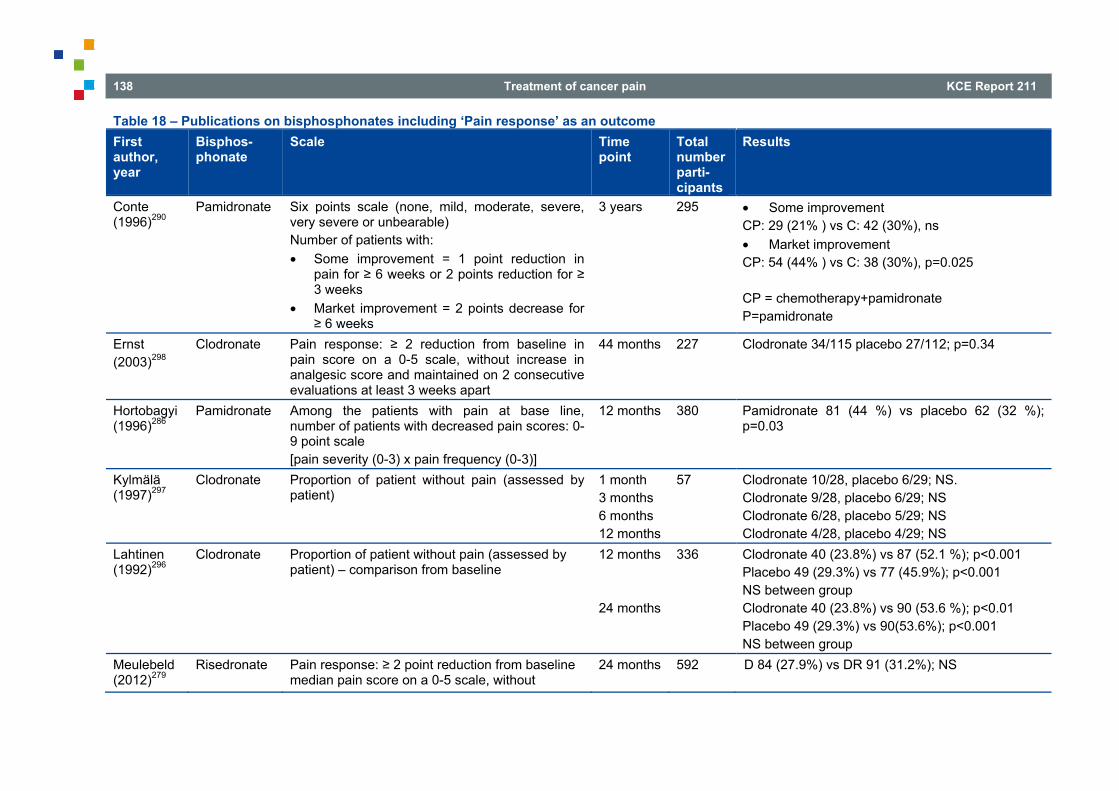
138 Treatment of cancer pain KCE Report 211
Table 18 – Publications on bisphosphonates including ‘Pain response’ as an outcome First author, year
Bisphos-phonate
Scale Time point
Total number parti-cipants
Results
Conte (1996)290
Pamidronate Six points scale (none, mild, moderate, severe, very severe or unbearable) Number of patients with: • Some improvement = 1 point reduction in
pain for ≥ 6 weeks or 2 points reduction for ≥ 3 weeks
• Market improvement = 2 points decrease for ≥ 6 weeks
3 years 295 • Some improvement CP: 29 (21% ) vs C: 42 (30%), ns • Market improvement CP: 54 (44% ) vs C: 38 (30%), p=0.025 CP = chemotherapy+pamidronate P=pamidronate
Ernst (2003)298
Clodronate Pain response: ≥ 2 reduction from baseline in pain score on a 0-5 scale, without increase in analgesic score and maintained on 2 consecutive evaluations at least 3 weeks apart
44 months 227 Clodronate 34/115 placebo 27/112; p=0.34
Hortobagyi (1996)286
Pamidronate Among the patients with pain at base line, number of patients with decreased pain scores: 0-9 point scale [pain severity (0-3) x pain frequency (0-3)]
12 months 380 Pamidronate 81 (44 %) vs placebo 62 (32 %); p=0.03
Kylmälä (1997)297
Clodronate Proportion of patient without pain (assessed by patient)
1 month 3 months 6 months 12 months
57 Clodronate 10/28, placebo 6/29; NS. Clodronate 9/28, placebo 6/29; NS Clodronate 6/28, placebo 5/29; NS Clodronate 4/28, placebo 4/29; NS
Lahtinen (1992)296
Clodronate Proportion of patient without pain (assessed by patient) – comparison from baseline
12 months 24 months
336 Clodronate 40 (23.8%) vs 87 (52.1 %); p<0.001 Placebo 49 (29.3%) vs 77 (45.9%); p<0.001 NS between group Clodronate 40 (23.8%) vs 90 (53.6 %); p<0.01 Placebo 49 (29.3%) vs 90(53.6%); p<0.001 NS between group
Meulebeld (2012)279
Risedronate Pain response: ≥ 2 point reduction from baseline median pain score on a 0-5 scale, without
24 months 592 D 84 (27.9%) vs DR 91 (31.2%); NS
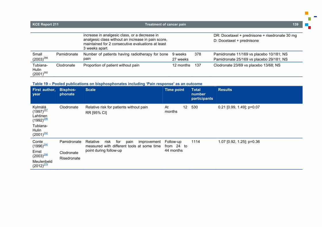
KCE Report 211 Treatment of cancer pain 139
increase in analgesic class, or a decrease in analgesic class without an increase in pain score, maintained for 2 consecutive evaluations at least 3 weeks apart.
DR: Docetaxel + prednisone + risedronate 30 mg D: Docetaxel + prednisone
Small (2003)288
Pamidronate Number of patients having radiotherapy for bone pain
9 weeks 27 weeks
378 Pamidronate 11/169 vs placebo 10/181; NS Pamidronate 25/169 vs placebo 29/181; NS
Tubiana-Hulin (2001)292
Clodronate Proportion of patient without pain 12 months 137 Clodronate 23/69 vs placebo 13/68; NS
Table 19 – Pooled publications on bisphosphonates including ‘Pain response’ as an outcome First author, year
Bisphos-phonate
Scale Time point Total number participants
Results
Kylmälä (1997)297 Lahtinen (1992)296 Tubiana-Hulin (2001)292
Clodronate Relative risk for patients without pain RR [95% CI]
At 12 months
530 0.21 [0.99, 1.49]; p=0.07
Conte (1996)290 Ernst (2003)298 Meulenbeld (2012)279
Pamidronate Clodronate Risedronate
Relative risk for pain improvement measured with different tools at some time point during follow-up
Follow-up from 24 to 44 months
1114 1.07 [0.92, 1.25]; p=0.36

140 Treatment of cancer pain KCE Report 211
4.9.3.3 Efficacy of bisphosphonates on pain: Pain score Twelve of the 22 included studies reported mean pain scores or changes in mean pain scores on a measurement instrument (Theriault 1999, Diel 2004, Piga 1998, O’Rourke 1995, Tubiana-Hulin 2001, Ernst 1992, Ernst 1997, Robertson 1995, Daragon 1993, Tripathy 2004, Small 2003, Saad 2002)276, 288, 289, 292-295, 299-302, 305. An overview is given in Table 17; details can be found in the Evidence tables (see Appendix III: Table 32-37). Overall, 16 comparisons were made; Small 2003 reported at 2 different time points and Saad 2002 at 4 different time points288, 302. Six of the 12 studies reported statistically significant outcomes in favour of the bisphosphonate group (Theriault 1999, Diel 2004, Tubiana-Hulin 2001, Ernst 1992, Robertson 1995, Saad 2002)276, 289, 292, 293, 299, 302; in Saad 2002 two of the four reported comparisons reached statistical significance302. As can be noticed in Table 17, there was a large diversity in pain measurement instruments used, and in outcome time points, which made pooling of results possible for 3 studies only: Daragon 1993 O’Rourke 1995, Piga 1998 (n=180 participants; see forest plot in Appendix III: Figure 32)294, 295, 305. The results of Robertson 1995 could not be used in this pooling because the authors presented only median and range in their publication293. For the results of Diel 2004 and Tripathy 2004, pooling was not possible because the authors presented no standard deviation276, 301. The pooled studies used VAS scales at a time frame between 1 and 4 months. For the 3 pooled studies, the difference between bisphosphonates and placebo is statistically significant in favour of the bisphosphonates: mean difference [CI95%]: -0.63 [-1.02 to -0.24] (P=0.001) (low level of evidence). It should be noted that the baseline mean pain scores were not comparable between the 3 studies (Bisphosphonate/ Placebo: Daragon 1993 (3.8 ± 2.7/ 3.7 ± 2.9)305; O’Rourke 1995 (3.4 ± 0.7/4.9 ± 0.8) 295; Piga 1998 (7.5 ± 5.4/5.1 ± 4.8)294. This makes it questionable whether these studies could be pooled in a meaningful way. Another question remains whether the difference found represents a clinically significant result (see 4.9.4)
Wong 2009 did not pool the studies they included for this domain, because of the heterogeneity in different domains and because of other methodological difficulties, as also described in the previous paragraph281. They reported a general trend showing the main pain score was lower for the treatment arm, but the magnitude of difference between the treatment and control arms showed a wide range. = mean
= median

KCE Report 211 Treatment of cancer pain 141
Table 20 – Publications on bisphosphonates including ‘Pain score’ as an outcome First author (year)
Bisphos-phonate
Scale Time point
Total number parti-cipants
Results
Daragon (1993)305
Etidronate Huskisson index (10 cm analogue visual scale) [ ± SD]
4 months 94 Etidronate 1.7 ±2 vs placebo 2±2.3; NS
Diel (2004)276
Ibandronate Average decrease in pain scores on a 5 points scale from baseline [ ± SD]
96 weeks/ 24 months
312 Ibandronate 60 mg -0.28±1.11 vs control 0.19±0.11p<0.001
Ernst (1992)299
Clodronate Average decrease in pain VAS 10 cm score [ ± SD (95% CI)]
7 days 24 Cl2MDP vs. placebo -0.89 ± 0.27 (-1.43 to -0.35) p=0.004
Ernst (1997)300
Clodronate Average decrease from baseline on 150 mm VAS [ ± SE]
14 days 60 Cl2MDP: 22 ± 7.2 vs placebo: 13.2 ± 7.5 p=0.51
O’Rourke (1995)295
Clodronate Average 10 cm VAS [ ± SD]
4 weeks 40 Cl2MDP (1600 mg): 2.7 ± 0.6 vs placebo 3.4 ± 0.8
Piga (1998)294
Clodronate Average decrease from baseline on Huskisson index (10 cm analogue visual scale) [ ± SD]
3 months 50 Cl2MDP 7.5 ± 5.4 vs 5.1 ± 4.8 Placebo 6.4 ± 5.4 vs 6.4 ± 5.9; p=0.424
Robertson (1995)293
Clodronate Median decrease from baseline on 10 points VAS [ (range)]
1 month 55 Cl2MDP -0.9 (-2.6 ─ -0.4) vs placebo +0.4 (-0.1 ─ +4.0); p=0.03
Saad (2002)302
Zoledronate 10 points scale Brief Pain Inventory mean variation from the baseline (15 months); average least-squares change from baseline on 10 points scale Brief Pain Inventory (18-21-24 months) [ (95% CI)]
15 – 18 – 21 – 24 months
422 • 15 months: -0.30 p=0.134 • 18 months: -0.37 (-0.78 to -0.04) p=0.075 • 21 months: -0.51 (-0.91 to -0.10) p=0.014 • 24 months: -0.47 (-0.88 to -0.06) p=0.024
Small (2003)288
Pamidronate Average decrease from baseline on 10 points VAS [ ± SD]
9 weeks + 27 weeks
378 at week 9: Pamidronate -0.61 ± 0.17; placebo -0.44 ± 0.16 p=0.46; at week 27: Pamidronate -0.40 ±0. 25; placebo -0.27 ± 0.24 p=0.71
Theriault (1999)289
Pamidronate Average increase from baseline on 0-9 point scale [pain severity (0-3) x pain intensity (0-3)]; 24 weeks 374 Pamitronate +0.5 vs placebo +1.6; p=0.007

142 Treatment of cancer pain KCE Report 211
Tripathy (2004)301
Ibandronate Average change from baseline on 5-point bone pain scale from 0 (none) to 4 (intolerable); 96 weeks/
24 months 287 Ibandronate(20 mg) -0.06 vs placebo 0.21;
p=0.071
Tubiana-Hulin (2001)292
Clodronate Average difference from baseline (VAS scale) [ ± SD]
12 months 137 Cl2MDP -11.8 ± 3.2 vs placebo 4.5 ± 4.7; p=0.007
: mean variation
Table 21 – Pooled publications on bisphosphonates including ‘Pain score’ as an outcome First author (year)
Bisphos-phonate
Scale Time point
Total number parti-cipants
Results
Daragon (1993)305 O’Rourke (1995)295 Piga (1998)294
Etidronate Clodronate
Huskisson index (10 cm analogue visual scale) or 10 cm VAS [95% CI]
From 4 weeks to 4 months
184 -0.63 [-1.02, -0.24]; p = 0.001
4.9.3.4 Efficacy of bisphosphonates on analgesic use: Analgesic
response Seven of the 22 included studies reported on analgesic response, i.e. the proportion of all patients with a prespecified reduction/increase in analgesics (Hultborn 1999, Small 2003, Ernst 2003, Kylmala 1997, Piga 1998, Robertson 1995, Tubiana-Hulin 2001)287, 288, 292-294, 297, 298. Examples of criteria used to define a positive response related to the use of analgesics are: decrease or stable analgesic use as compared to baseline (Small 2003)288; proportion of patients who had ever used opioids at study start versus proportion of patients who had ever used opioids at the end of the study (Hultborn 1999)287; 50% decrease in analgesic score (which is the total number of analgesics units ; 1 unit = standard doses of non-opioid; 2 units = morphine 10-mg equivalents) from the baseline with no increase in pain and maintained on two consecutive evaluations at least 3
weeks apart (Ernst 2003)298. Details can be found in the GRADE tables (see Appendix III: Table 20) and in the Evidence tables (see Appendix III: Tables 32-37). Overall, 13 comparisons were made; Small 2003 reported at 2 different time points and Kylmala 1997 at 4 different time points; Tubiana-Hulin 2001 reported on 3 different analgesic categories (responses) 288, 292, 297. Two of the 7 studies reported statistically significant outcomes in favour of the bisphosphonate group. Piga 1998 described a statistically significant difference between placebo and bisphosphonate treatment in favour of the latter, for the proportion of patients who had increased at 3 months their analgesic score (analgesic score: 0 none; 1 NSAIDs; 2 opioids) (placebo 10/22, bisphosphonates 5/27, p=0.042) 294. Tubiana-Hulin 2001 described a statistically significant difference between placebo and bisphosphonate treatment in favour of the latter, for the use of analgesics at 12 months (not
KCE Report 211 Treatment of cancer pain 143
further explained) (placebo 57/68, bisphosphonates 46/69, p=0.02)292. Overall, 5 comparisons (4 studies) reported results at the short or medium term (1 to 3 months); 4/5 comparisons were not statistically significant (Small 2003, Kylmala 1997, Piga 1998, Robertson 1995).288, 293, 294, 297 Due to the diversity of the analgesic scales and the definitions for analgesic response, due to the difference in time points, and due to some difficulties in the reporting of the data, pooling of results was only possible for two studies (N= 194 participants; see forest plot in Appendix III: Figures 29-30): Kylmala 1997 and Tubiana-Hulin 2001292, 297. These authors reported the proportion of patients who did not use analgesics at 12 months. The difference between bisphosphonates and placebo is statistically significant in favour of the bisphosphonates: RR [CI95%] 1.79 [1.02-3.15] (P=0.04) (low level of evidence). Wong 2009 presented results for the proportion of patients who reduces analgesics at 4 and 12 weeks, by pooling results of 3 trials (N=150 participants) and 3 trials (N=182 participants) respectively281. They found statistically significant results in favour of bisphosphonates: at 4 weeks Odd’s Ratio OR[CI95%]: 2.81 [1.24-6.38] (p=0.013); at 12 weeks OR [CI95%]: 2.37 [1.10-5.12] (p=0.028).
4.9.3.5 Efficacy of bisphosphonates on analgesic use: Analgesic score
Nine of the 22 included studies reported on change in analgesic score, i.e. change in mean or median analgesic consumption (Small 2003, Diel 2004, Tripathy 2004, Lahtinen 1992, Piga 1998, Ernst 1992, Ernst 1997, Saad 2002, Daragon 1993)276, 288, 294, 296, 299-302, 305. Examples of scales to measure analgesic consumption are: consumption of oral morphine equivalents (Small 2003)288; daily number of tablets of non-narcotic analgesics (Lahtinen 1992)296; 6 points scale of analgesic use (0:none, 1:mild analgesic [aspirin or paracetamol/acetaminophen] or NSAID, 2:mild analgesic + NSAID, 3:moderate analgesic, 4:opiates <40 mg morphine [or equivalent] daily, 5:opiates ≥40 mg, but <100 mg morphine [or equivalent] daily, or 6: opiates ≥100 mg morphine [or equivalent] daily) (Diel 2004)276. Details can be found in the GRADE tables (see Appendix III: Table 20) and in the Evidence tables (see Appendix III: Tables 32-37).
Overall, 12 comparisons were made; Lahtinen reported at 2 different time points, using each time two different measures of analgesic consumption296. Two of the 9 studies reported statistically significant outcomes in favour of the bisphosphonate group. Ernst 1997 reported a statistically significant difference for average change in daily morphine equivalent doses between placebo and bisphosphonate treatment in favour of the latter: placebo -6.4 (SE 2.9) versus bisphosphonates +24.6 (SE 14.9) (p=0.03)300. Tripathy 2004 reported a statistically significant difference (p=0.006) between placebo and bisphosphonate treatment in favour of the latter, but only for the lower dose of ibandronate (20 mg) and not for the higher dose (50 mg)301. For reasons already mentioned before, more precisely the diversity of the scales to measure analgesic consumption, and of outcome time points, pooling of results was not possible. E.g. for the results of Diel 2004 and Tripathy 2004, pooling was not possible because the authors presented no standard deviation276, 301. Also, for most studies it was difficult to compare the baseline analgesic consumption between the different studies due to the different definitions used for analgesic consumption. Wong 2009 did not present results for pooled studies on this topic, but presented in a narrative way the results from 3 studies reporting outcomes in favour of bisphosphonates and from 3 studies reporting no difference in outcome between placebo and bisphosphonate281.
4.9.3.6 Bisphosphonates: other outcomes of interest Two of the 22 included studies reported on functional performance of patients while on bisphosphonate therapy and compared it to placebo; both studies used the Karnofsky scale to measure performance (see Evidence tables in Appendix III: Tables 32-37). Piga 1998 included 50 patients and evaluated them at 3 months294. There was no statistical difference between the bisphosphonate group versus the placebo group for the percentage of patients with stable or minor change in performance status, for the percentage of patients with 20% decrease nor for the patients with 20% increase in performance status. Daragon 1993 evaluated 78 patients at 4 months305. There was no statistical difference for the mean score on the Karnofsky scale between the bisphosphonate and the placebo group. The overall level of evidence for these two publications is very low (see Appendix III: Table 10), and given the paucity of publications retrieved on this topic, no firm conclusions are allowed. It
144 Treatment of cancer pain KCE Report 211
should also be mentioned that functional performance might be influenced by non-pain related factors. Berenson 2001 reported on the relative efficacy of pamidronate as compared to different doses of zoledronic acid291. The mean pain scores after 1 year by the end of the study were not statistically different between the 2 bisphosphonates. Body 2007 compared oral ibandronate and intravenous zoledronate285; the mean pain scores at 12 weeks were not statistically different between the 2 bisphosphonates (see Evidence tables in Appendix III: Tables 32-37); The overall level of evidence for these two publications is very low (see Appendix III: Table 21), and given the paucity of publications retrieved on relative efficacy of bisphosphonates against each other, no firm conclusions are allowed.
4.9.3.7 Adverse events of bisphosphonates Fourteen of the 22 included RCTs presented information on adverse events related to the use of bisphosphonates. Withdrawal of patients from the trial because of adverse events related to the treatment was reported in 11 studies275, 286, 288-290, 292, 297, 299, 301, 302 (N participants=3078); the combined results are presented in Appendix III: Figure 33. The relative risk (RR) for bisphosphonates compared to placebo is 1.41 [95% CI 1.08, 1.85]. This implies that adverse events severe enough to cause withdrawal from the trial are significantly more frequent in patients treated with bisphosphonates in comparison to placebo (p=0.01) (moderate level of evidence). The adverse event most frequently reported was nausea; it was reported in 5 RCTs288, 296, 297, 301, 302 (N participants=1476). The combined results (see Appendix III: Figure 34) show a RR of 1.10 [95% CI 0.92-1.32] and indicate a non significant trend for a higher incidence of nausea in bisphosphonate therapy as compared to placebo (p=0.31) (moderate level of evidence). Nausea combined with vomiting was reported in 8 RCTs288,
292, 295-298, 301, 302 (N participants=1924) (see Appendix III: Figure 35); the combined results show a RR of 1.07 [95 % IC 0.94,1.21]. However, the trend remains non significant (p=0.33) (moderate level of evidence). Other adverse events (anemia, gastrointestinal discomfort, allergic reaction, hypocalcemia,…) were mentioned in publications but insufficient data or insufficient consistency in reporting between studies do not permit to draw a conclusion. None of the 22 included RCTs reported osteonecrosis of the jaw, one of the most severe adverse events related to bisphosphonates. One trial explicitly mentioned that renal functional impairment was the
reason for lowering the dose of zoledronic acid in one treatment arm (Saad 2002)302. These results are in line with the SR of Wong 2009281. These authors also found that discontinuation of therapy because of adverse effects was significantly more frequent in the bisphosphonate group, and that there was a non-significant trend for increased nausea and vomiting.
4.9.3.8 Efficacy of bisphosphonates in specific tumor types The above results combined all selected RCTs on bisphosphonates, without differentiating between different primary tumor types. Given the fact that bone metastases can be more or less ‘osteolytic’ or ‘osteoblastic’ depending on the primary tumor (see 4.9.1), and given the fact that bisphosphonates seem to exert their activity preferentially in metastases of the osteolytic type, it might be interesting to compare efficacy of bisphosphonates in bone metastases from different primary tumor types. Three recent SRs were selected dealing with the effect of bisphosphonates on pain from bone metastases from one specific primary tumor type: Wong 2012 (breast cancer)278, Yuen 2010 (prostate cancer)282, Mhaskar 2012 (multiple myeloma)280 (see Evidence tables in Appendix III: Table 31). All these reviews also discussed other outcomes, such as effect of bisphosphonates on SREs and overall survival, which is out of scope for this report. Wong 2012 included 11 RCTs but conducted no meta-analysis278. In 6 of these RCTs, bisphosphonates improved bone pain significantly as compared to placebo or no bisphosphonates. Improvements in global QoL were reported in 2 out of 5 studies, both for ibandronate. Reported toxicity was generally mild, but renal toxicity and osteonecrosis of the jaw had been identified. The authors concluded that some bisphosphonates (intravenous ibandronate or pamidronate, and oral ibandronate or possibly clodronate) may reduce bone pain in bone metastates in advanced breast cancer, and that they may improve QoL.

KCE Report 211 Treatment of cancer pain 145
Yuen 2010 included 5 RCTs and pooled results for data reported as ‘pain response’, independent of the definition that was given to the pain response in the primary study282. They found that the proportion of patients reporting pain response was not significantly different between bisphosphonate therapy and placebo: OR 1.54 (95% CI 0.97 - 2.44; p=0.07). The same was true for the pooled results of the proportion of patients reporting decreased analgesic consumption: OR 1.27 (95% CI 0.82 - 1.98; p=0.28). Yuen et al (2010)282 pooled also results of the individual studies for ‘adverse events’ and found a significantly higher proportion of patients having nausea (2 primary studies): OR 1.35 (95% CI 1.02 - 1.77; p=0.03). No significant results were found for pooled results of vomiting and anemia. One of the primary studies reported a case of renal failure. Yuen et al (2010)282 concluded that bisphosphonates may have a role in decreasing pain in patients with metastatic prostate cancer, but that statistical analysis is limited by relatively small sample size and heterogeneity in study design. The results of the RCT by Meulenbeld 2012279, selected in the update RCTs in this report and of more recent date than the RCTs included by Yuen 2010282, are in line with the results of Yuen 2010282 (see Evidence table in Appendix III: Table 31). Mhaskar 2012 included 8 RCTs; pooled results for the effect on pain showed a statistically significant advantage for bisphosphonate treatment as compared to placebo or no treatment: RR:0.75 (95% CI 0.60 - 0.95; p= 0.01) 280. The authors of the SR mentioned that from the primary studies all types of methods used to report pain were included in the their meta-analysis, e.g. pain index at 12 months, analgesic use at 4 months, bone pain reported by authors at 29 months. This introduced heterogeneity: p= 0.01, I2=63%. For adverse events, no difference in frequency between intervention and control was noticed (gastrointestinal symptoms, hypocalcemia, renal dysfunction). ONJ was reported in 2 primary studies (3 cases). The authors concluded that adding bisphosphonates to the treatment of multiple myeloma probably reduces pain. In their discussion they highlighted the results of 9 observational studies (N=1400 patients) for osteonecrosis of the jaw, indicating that it may be a common event (range 0% to 51%) and indicating that the frequency might be highest in the most potent bisphosphonate (zoledronic acid). Mhaskar et al (2012)280 recognized that the results from observational studies may be an overestimation due to their non-controlled
design, but existing RCTs did not consistently report on ONJ and were not of sufficient power to detect this rare but serious adverse event. 4.9.4 Other considerations The results found in the present report were in line with the results from the systematic review by Wong 2009, for the outcome domains ‘pain score’, ‘analgesic response’, ‘analgesic score’ and ‘adverse events’. However, for the domain ‘pain response’ the present review found non-statistically significant results at 12 months, whereas Wong 2009281 reported statistically significant results at 12 weeks based on 8 RCTs. The difference might be related to the time-frame used. However, the 2 RCTs included in the present review that explicitly reported on pain response within 3 months (Small 2003, Kylmala 1997), showed non-significant results (Table 18). It should be noted that the inclusion criteria for the RCTs in the present review were more stringent as compared to the inclusion criteria used by Wong 2009281. Wong 2009 presented overall results for the effectiveness of bisphosphonates on pain-related outcomes, independent from tumour type281; it included 30 RCTs and it’s last search date was February 2002. In the present report 22 RCTs were included; 13 RCTs overlapped with Wong 2009281. As compared to Wong 2009281, 8 more recent RCTs found in the other reviews and in the update search for RCTs were added. One RCT excluded by Wong 2009281 was included in the present report: Tubiana-Hulin 2001 (full publication found instead of abstract)292. On the other hand, the selection criteria applied in this report were more stringent on certain criteria, as presented before (see 4.9.2). Based on these criteria, 17 RCTs included in Wong 2009 were excluded for the present report281; the reasons can be found in Appendix I: Table 31. For example, Vinholes 1997: excluded because of the use of a composite pain score including performance status and number of days off306; Siris 1983 and Elomaa 1992: excluded because pain evaluation methodology is not provided307, 308.
146 Treatment of cancer pain KCE Report 211
The Wong 2009 study specifically focused on short to medium term pain outcomes (up to 3 months)281. Although patients experiencing pain expect pain relief in the short term (Wong 2004)277, it might also be interesting to know the effect of bisphosponates in the long term, because these drugs are also used for other indications, e.g. prevention of SREs. For this indication, bisphosphonates are often continued during a long time period. It should be remembered that in the present review the effectiveness of bisphosphonates in preventing SREs secondary to bone metastases, or to prevent hypercalcemia, is considered to be out of scope. Depending on the available evidence in these domains, bisphosphonates could be considered for treatment even if there would be no indication to start it because of bone pain. Overall, the literature in this domain reports poorly on the length of the time period that bisphosphonates should be taken. An important question remains whether the differences found in this report represent a clinically significant result. This holds especially true for the results reported by a pain score. Farrar et al (2001) demonstrated that on an 11-point pain intensity numerical scale (0= no pain, 10= worst possible pain), a clinically important difference in pain is represented by a reduction of approximately 2 points or a reduction of approximately 30%; the percent change was consistent regardless of baseline pain score33. The difference between mean pain scores for bisphosphonates and placebo found in this report for the pooled results of 3 studies (Daragon 1993, O’Rourke 1995, Piga 1998)294, 295, 305 was -0.63 (CI95%:-1.02 - -0.24) (see 1.1.1.1), which would imply that the difference found is not clinically relevant or at best very modest. Moreover, according to Wong 2009, mean or median pain scores used as an endpoint to evaluate pain relief should be interpreted with caution because of methodological issues281. Methodological issues in the use of mean or median pain scores as an endpoint have also been pointed out by Moore et al. (2010) and Dworkin et al. (2008)32, 34; this will be further discussed in chapter 5 Discussion. The KCE breast cancer guideline (2013)309 included bisphosphonates and discussed overall and disease-free survival as well as adverse events in women with early non-metastatic breast cancer without clinically evident bone metastases. They selected three RCTs to calculate the pooled overall RR for osteonecrosis of the jaw after treatment with zoledronic acid. Based on 18 cases in the 3 studies (N=5269 participants), the RR was 18.8 (95% CI 2.5 – 139.9) (p=0.004). The median follow-up time in these
studies was from 59 to 90 months, whereas the longest follow-up time for the studies included in this report was 33 months (2.9 years). Besides bisphosphonates, other treatment options exist for painful bone metastases: analgesic drugs; local therapy with radiation, radionuclides or surgery; or systemic therapy with hormonal or chemotherapeutic agents. The evidence retrieved on bisphosphonates in this report does not allow to conclude on relative efficacy compared to other interventions for metastatic bone pain. From the trials included in this report, the overall level of evidence for the effectiveness of each separate type of bisphosphonate is very low, and for most types of bisphosphonates only a small number of trials was available (see Appendix III: Tables 32-37). Only for Clodronate some pooling of results has been possible (see 4.9.3.2 Pain response (Kylmala 1997, Lahtinen 1992, Tubiana-Hulin 2001)292, 296, 297 and 1.1.1.1 Analgesic response (Kylmala 1997, Tubiana-Hulin 2001)292, 297). Results for different types of bisphosphonates will not further be discussed. Questions of dosing or route of administration were considered to be out of scope. The already mentioned heterogeneity of the RCTs as to pain or analgesic measurement instrument, definition of response, reporting of outcomes and time frame seem to reflect a more general problem in the domain of pain measurement that has also been observed in many other trials in other areas of medicine (Wong 2009)281. It reflects a lack of consensus on which pain endpoints should be used and how they should be reported. Still other methodological difficulties hampered meaningful interpretation of the available studies, e.g. some of the data could not be incorporated into a pooled analysis because crucial information, such as standard deviation of the mean, was not reported (e.g. Tripathy 2004)301. Finally, it should be mentioned that 13 of the 22 selected RCTS had been industry sponsored: (N=13; 60%): Berenson (2001)291, Body (2007)285, Conte (1996)290, Dearnaley (2003)275, Diel (2004)276, Hortobagyi (1996)286, Meulenbeld (2012)279, O’Rourke (1995)295, Robertson (1995)293, Saad (2002)302, Small (2003)288, Theriault (1999)289, Tripathy (2004)301. Three studies had been sponsored by independent bodies (e.g. Cancer Foundation, Academy of sciences) (N=3; 13%): Daragon (1993)305, Kylmälä (1997)297, Lahtinen (1992)296. Six studies did not mention their sources of funding (N=6; 27%): Ernst (1992)299, Ernst (1997)300, Ernst (2003)298, Hultborn (1999)287, Piga (1998)294, Tubiana-Hulin (2001)292.

KCE Report 211 Treatment of cancer pain 147
Conclusion • Based on the available evidence from RCTs in which stringent
criteria were applied as to the application of a pain measurement instrument, as well as to the quality of the reporting on pain outcome, it is not possible to conclude on the efficacy of bisphosphonates on pain response (proportion of patients with complete pain relief or proportion of patients with some pain relief) in patients with metastatic bone pain at the short to medium term (up to 6 months) (very low level of evidence; Small 2003, Kylmala 1997).
• There are indications that bisphosphonates, as compared to placebo, have no statistically significant effect on pain response (proportion of patients with complete pain relief or proportion of patients with some pain relief) in patients with metastatic bone pain at the long term (6 months or longer). This conclusion arises from RCTs in which stringent criteria were applied as to the application of a pain measurement instrument, as well as to the quality of the reporting on pain outcome (low level of evidence; Kylmala 1997, Lahtinen 1992, Tubiana-Hulin 2001, Conte 1996, Ernst 2003, Meulenbeld 2012).
• There are indications that bisphosphonates, as compared to placebo, have a statistically significant effect on the pain score in patients with metastatic bone pain at the short to medium term (up to 6 months). However, the clinical significance of this change is not clear. This conclusion arises from RCTs in which stringent criteria were applied as to the application of a pain measurement instrument, as well as to the quality of the reporting on pain outcome (low level of evidence; Daragon 1993, O’Rourke 1995, Piga 1998).
• There are indications that bisphosphonates, as compared to placebo, have no statistically significant effect on analgesic response (predefined decrease in use of analgesics) in patients with metastatic bone pain at the short to medium term (up to 6 months). This conclusion arises from RCTs in which stringent criteria were applied as to the application of a pain measurement instrument, as well as to the quality of the reporting on pain outcome (very low level of evidence; Small 2003, Kylmala 1997, Piga 1998, Robertson 1995).
• There are indications that bisphosphonates, as compared to placebo, have a statistically significant effect on analgesic response (predefined decrease in use of analgesics) in patients with metastatic bone pain at the long term (6 months or longer). This conclusion arises from RCTs in which stringent criteria were applied as to the application of a pain measurement instrument, as well as to the quality of the reporting on pain outcome (low level of evidence; Kylmala 1997, Tubiana-Hulin 2001).
• Due to methodological issues, it is not possible to conclude on the efficacy of bisphosphonates on analgesic score (consumption of analgesics) in patients with metastatic bone pain. This conclusion arises from RCTs in which stringent criteria were applied as to the application of a pain measurement instrument, as well as to the quality of the reporting on pain outcome (very low level of evidence; Small 2003, Diel 2004, Tripathy 2004, Lahtinen 1992, Piga 1998, Ernst 1992, Ernst 1997, Saad 2002, Daragon 1993).
• It is plausible that adverse events severe enough to cause withdrawal of patients from therapy are significantly more frequent in patients with metastatic bone pain treated with bisphosphonates as compared to placebo (moderate level of evidence; 11 trials).
• It is plausible that nausea, or nausea combined with vomiting, is not significantly more frequent in patients with metastatic bone pain treated with bisphosphonates as compared to placebo (moderate level of evidence; 5 trials and 8 trials respectively).

148 Treatment of cancer pain KCE Report 211
• Based on the available evidence, it is not possible to conclude on the prevalence of severe side effects, such as renal impairment or osteonecrosis of the jaw.
• The existing evidence does not allow to conclude on the relative efficacy of bisphosphonates compared to other interventions for metastatic bone pain.
Recommendation
• Bisphosphonates cannot be considered as first-line, purely analgesic treatment option of metastatic bone pain in the short, medium or long term, given the available evidence on efficacy (very low level of evidence), and on possible adverse events (moderate level of evidence) (strong recommendation). This report does not deal with the usefulness of bisphosphonates for other indications in patients with metastatic bone involvement, e.g. hypercalcemia, pathological fractures, impending fractures, spinal cord compression.
Good Clinical Practice
Patients should be informed on the benefits and potential side-effects associated with the use of bisphosphonates. Their preferences should be taken into account when deciding on the treatment.
4.10 Celiac Plexus Block 4.10.1 Introduction Pancreatic cancer causes severe pain in 50 to 70% of patients (Arcidiacono 2011)310. This type of pain is often difficult to treat, and is generally transmitted through the celiac plexus, a neural structure located in the upper abdomen. Celiac plexus neurolysis or block (CPB) by injecting alcohol or phenol into the nervous structures, has been developed as an alternative to the classical pharmacotherapeutic approach. The targets for celiac axis destruction are the celiac ganglia located along the midline and anterior to the aorta; or the splanchnic nerves that cross the diaphragm, enter the abdominal cavity and come together to form the celiac plexus. The effect of CPB is often transitory, and the procedure needs to be repeated after a few months. A common side-effect in CPB is diarrhea, which can be considered the hallmark of the procedure, due to the effect on the sympathetic nerve bundles. The procedure can be performed percutaneously, under local anesthesia and with radiologic fluoroscopic or CT guidance of the needle. Besides alcohol and phenol, local anesthetics or steroids can be used as injection fluids as well; the injected volume plays a role in the effect. To be effective, a bilateral approach is necessary. Different routes exist for needle insertion; in the standard technique the patient is in the prone position. For this so-called ‘posterior approach’, two variations exist that target the celiac ganglia: retrocrural (needle posterior to the crus of the diaphragm) vs. transaortic (passing through the aortic wall). Another posterior variant, the bilateral splanchnic nerve block technique, targets the splanchnic nerves (Ischia 1992)311. Major complications are possible during these procedures: intra-abdominal bleeding due to damage to the adjacent large vessels among which the aorta, paraparesis or lower extremity paraesthesia due to involuntary damage to adjacent nervous structures or infection. Although the percutaneous approach can be performed under local anesthesia, a general anesthesia is often necessary for practical reasons.

KCE Report 211 Treatment of cancer pain 149
The needle can also be inserted anteriorly, with the patient in the supine position. This ‘anterior’ percutaneous approach is uncommon; one of the main reasons is that it is often precluded by the position of the tumoral process. Sometimes CPB is performed during abdominal surgical procedures for the cancer process; in this case the neurolytic solutions can be injected directly into the celiac plexus(Arcidiacono 2011)310. A newly described technique uses endoscopic ultrasonography to guide a needle into the celiac plexus. It is hypothesized that this method might be safer since it offers real-time ultrasound visualisation of the procedure, allows for visualisation of the surrounding blood vessels by Doppler imaging, and because the transgastric positioning of the needle minimizes the risk of neurological complications (lower extremity paraesthesia, paraparesis) associated with the classical posterior approach (Arcidiacono 2011, Wyse 2011)310, 312. As an alternative to the use of injection fluids, radiofrequency ablation (RFA) has been described as well. This technique uses a high frequency alternating current to heat tissues leading to thermal coagulation. CPB can also be used for other indications than pancreatic cancer, e.g. chronic pancreatitis, intestinal neoplasms. However, indications not dealing with cancer are excluded in this report. Plexus hypogastricus block, another type of neurolysis that nowadays is used to treat cancer pain, is also excluded (see 2.4). 4.10.2 Search results Three reviews that met the inclusion criteria were identified about CPB. They all dealt specifically with pancreatic cancer. The review by Arcidiacono et al. (2011)310 can be found in Appendix III: Table 38.; it addressed the effectiveness of percutaneous posterior CPB or CPB performed during surgical procedures for the cancer process. The date of literature search was December 2010. The overall risk of bias of this review was considered to be low; the overall risk of bias of the included RCTs was considered to be high. All RCTs mentioned in the review by Yan et al. (2007)313 were included in the review by Arcidiacono et al. (2011)310 (see comments in Appendix III: Table 38). The review by Sharma et al. (2011) was excluded because it was partly based on an SR that included abstracts instead of full texts314. The 3 generic reviews including this topic (Guideline of the MoH Malaysia 2010, Dutch Guideline on cancer pain 2008, SIGN 2008)8-10 did not provide
detailed information on pooling of results of individual studies and therefore were used as a basis of RCTs only. From these reviews, 2 new RCTs that had not yet been included were retained; these RCTs dealt with comparing different percutaneous posterior nerve block techniques: transaortic CPB, retrocrural CPB, bilateral chemical splanchniectomy. The risk of bias of the study by Ischia et al. (1992) was considered to be unclear311. The risk of bias of the study by Süleyman Ozyalçin et al. (2004) was considered to be high315. The update search for RCTs yielded 1 trial (Wyse 2011)312 dealing with early endoscopic ultrasound-guided CPB in pancreatic cancer patients. The risk of bias of this RCT was judged low. The evidence tables of the 3 RCTs can be found in Appendix III: Table 39. No RCTs on percutaneous anterior CPB nor on radiofrequency ablation (RFA) were retrieved. Moreover, the update search for RCTs yielded another RCT (Johnson 2009)161 comparing simple opioid analgesia with the combination of opioid analgesia with celiac plexus block or thoracoscopic splanchnicectomy in irresectable pancreatic or upper abdominal visceral cancer patients. The results are discussed in the section 4.3.3.12. 4.10.3 Literature overview The review by Arcidiacono et al. (2011) included 6 RCTs and 358 participants310. Although included in the search strategy, no RCTs on percutaneous anterior CPB were retrieved. Percutaneous posterior CPB (5 RCTs) or CPB performed during cancer surgery (1 RCT) had a small but significant effect on pain as compared to standard treatment with NSAIDs and morphine, measured at 4 weeks by a 10 points VAS scale (absolute mean difference -0.42 (95 % CI -0.71– -0.14; p=0.004)). The clinical significance of this difference is unclear. Indeed, scientific evaluation showed that a decrease of pain intensity by 2 points on a 0-10 scale, or a pain decrease by 30%, can be considered to be a clinical significant decrease (Dutch Guideline on cancer pain 2008, Farrar 2001)8, 33. At 8 weeks, the effect was not significant anymore (-0.44 (95% CI -0.89– 0.01; p=0.06)). Opioid use in mg of opioids at 4 weeks was significantly lower in the intervention group (-34.33 (95% CI -44.42– -24.24; p<0.000001)). There were no major complications due to the intervention such as intra-abdominal bleeding, paraparesis or infection; therefore the procedure appeared to be safe. Main adverse effects were diarrhea and constipation; adverse effects were only reported on 121 and 161 patients for diarrhea

150 Treatment of cancer pain KCE Report 211
and constipation respectively. Diarrhea was more frequently reported in the CPB group, but the rate of patients with diarrhea was not significantly different between the 2 groups. Constipation was significantly more frequent in the control group, who used more opioids. One recent trial (Wyse 2011) including 96 patients compared endoscopic ultrasound-guided CPB (EUS-guided CPB) and conventional pain management with narcotics alone312. Patients were included at the time of diagnosis, assuming that CPB done as early as possible in the course of the disease would be more effective and require lower opioid consumption. Pain control, measured with a 7-point Likert scale, improved significantly in the EUS-guided CPB group as compared to the control group: -1.0 point in Likert scale (95% IC -1.7 - -0.1); p=0.01 at 1 month; -2.2 point in Likert scale (95% IC -3.1 - -1.4); p<0.001 at 3 months. The effects at 3 months might be considered to fall within clinically significant ranges. Opioid consumption and quality of life, measured at 1 and 3 months, were not significantly different between the two groups. The authors did not report on adverse events. Ischia et al. (1992) compared three posterior nerve block techniques (celiac plexus retrocrural vs. celiac plexus transaortic vs. bilateral splanchnic nerve block technique) in 61 patients with pancreatic cancer311. They found no statistical significant differences among the 3 techniques for immediate partial or total pain relief. Süleyman Özyalçin et al. (2004) postulated that classical CPB might be more effective in patients with cancer of the pancreatic head whereas bilateral splanchnic nerve block technique might be more effective in patients with cancer of the pancreatic body or tail315. The latter two parts of the pancreas are located more laterally from the midline. The authors compared celiac plexus transaortic vs. bilateral splanchnic nerve block technique in 39 patients with cancer of the pancreatic body or tail. They found a significant difference in pain rating at 4 and 8 weeks in favour of the bilateral splanchnic nerve block technique (p<0.001).
4.10.4 Other considerations As previously discussed in the opioids chapter (see 4.3.3.12), Johnson et al (2009)161 concluded that there was no evidence to support the systematic use of celiac plexus block or thoracoscopic splanchnicectomy for pain relief in irresectable pancreatic or upper abdominal visceral cancer patients. The literature studies included in this review mainly dealt with pancreatic cancer. Therefore it is not possible to comment on the effectiveness of CPB in these other types of cancer. The expert panel (see colophon) recommended a multimodal pain treatment approach for cancer pain. In patients who underwent CPB, other analgesic therapies should still be considered throughout the treatment course, e.g. NSAIDs, opioids.
Conclusions • There are indications that percutaneous posterior celiac plexus
block (CPB) or CPB performed during cancer surgery as treatment for patients with pancreatic cancer, has a limited but significant effect on pain as compared to standard treatment with NSAIDs and opioids, measured at 4 weeks. However, the clinical significance of this effect is not clear (low level of evidence; Arcidiacono 2011).
• There are indications that percutaneous posterior CPB or CPB performed during cancer surgery as treatment for patients with pancreatic cancer is associated, at 4 weeks, with a lower use of opioids (very low level of evidence; Arcidiacono 2011).
• There are indications that at 8 weeks, there is no significant difference anymore in opioid use between patients with pancreatic cancer treated with percutaneous posterior CPB or CPB during cancer surgery, and those treated with standard analgesic therapy (very low level of evidence; Arcidiacono 2011).
KCE Report 211 Treatment of cancer pain 151
• There are indications that percutaneous posterior CPB in patients with pancreatic cancer, or CPB performed during cancer surgery, are relatively safe procedures. There are indications that these procedures are associated with a statistically significant reduction in opioid-related adverse effects such as constipation (low level of evidence; Arcidiacono 2011). There are indications that these procedures are associated with a non-significant increase in diarrhea (very low level of evidence; Arcidiacono 2011).
• There is limited evidence from 1 RCT that endoscopic ultrasound-guided CPB (EUS-guided CPB) performed at the time of pancreatic cancer diagnosis reduces pain at 1 and 3 months, as compared to standard analgesic treatment. At 3 months, the effect might fall within clinically significant ranges but there are indications that it is not associated with a significant decrease in opioid consumption (low level of evidence; Wyse 2011).
• There is limited evidence from 1 RCT that there is no difference in quality of life between patients treated with EUS-guided CPB and patients treated with standard analgesic treatment for pancreatic cancer pain at 1 or at 3 months (very low level of evidence; Wyse 2011).
• Different percutaneous posterior nerve block techniques for treatment of pancreatic cancer pain have been compared in single, small trials of less than 30 patients per trial arm. The level of evidence is very low (Ischia 1992, Süleyman Özyalçin 2004). More research is necessary.
Recommendation
• Celiac plexus block (CPB) can be considered as a pain treatment option for patient suffering from pancreatic cancer, although CPB should not be the treatment of first choice. Patients should be timely informed on this treatment option. This recommendation is based on the limited short term (around 4 weeks) effectiveness of CPB on pain as compared to standard analgesic treatment, on its short term possible positive effect on opioid consumption, on the need to repeat the procedure and on the rare but important complications that can be associated with the procedure; it takes into account that the life expectancy of patients with pancreatic cancer is mostly limited (very low level of evidence; weak recommendation).
• It is recommended to inform patients with pancreatic cancer clearly on the limited short term (around 4 weeks) effectiveness of CPB on pain as compared to standard analgesic treatment, but also on the short term (around 4 weeks) possible positive effect on opioid dosing. They should also be informed on the need to repeat the procedure over time, and on the possible side-effects and complications of CPB (very low level of evidence; strong recommendation).
• A multimodal pain treatment approach should be considered for cancer pain. In patients who underwent CPB, other analgesic therapies should still be considered throughout the treatment course, e.g. NSAIDs, opioids (very low level of evidence; weak recommendation).
Good Clinical Practice
Patient preferences should be taken into account when deciding on the use of a celiac plexus block in the treatment of pain related to pancreatic cancer.

152 Treatment of cancer pain KCE Report 211
5 DISCUSSION 5.1 Pain improvement: what is a clinically important
change? Recent research on what constitutes pain improvement and pain relief, led to novel insights in what can be considered as a clinically meaningful outcome to patients. First, it has become clear that the population distribution of pain intensity or pain relief tends not to be bellshaped (Gaussian), where many patients would be close to the average result. Instead, results often have a highly skewed distribution with maximum frequencies at the two extremes of the range of a variable, i.e. ‘U-shaped’ distributions; patients tend to have either very good pain relief or very poor pain relief. Other measures like additional analgesic requirements also appear to be skewed. When few patients are ‘average’ the use of average values can be misleading, and a 10/100 mm difference between treatment and placebo on VAS or equivalent scales can hide the fact that 60% of patients on treatment obtain at least moderate benefit (Moore 2010)34. This implies that reporting of average results of continuous data (e.g. mean change in pain intensity score) poses problems when these results are to be translated to what this outcome really means to patients. Concerning pain intensity, recent research learned that, at the patient level, a score change on a 0 to 10 NRS or equivalent pain intensity scale of approximately 2 points or 30% to 36% represents ‘much better,’ ‘much improved,’ or ‘meaningful’ decreases in chronic pain (see also 4.1.3.3). A decrease of 4 points or 50% appears to represent a substantial (‘very much improved’) change in pain, which patients have also considered ‘treatment success’ or ‘satisfactory improvement’. A change of approximately 1 point, or percentage changes of approximately 15% to 20% represent minimally but perhaps not very important decreases (Farrar 2001, Dworkin 2008)32, 33. Evidence is growing that chronic pain patients regard a pain intensity reduction of 50-70% as a clinical success and ideally want pain to be no worse than mild (Moore 2010; Dworkin 2008)32, 34. Further, there is evidence that a pain intensity score of ‘5’ or more has a major impact on functioning in daily life, which is also considered to be a critical threshold (Paul 2005, Serlin 1995)27, 28.
Several expert groups in pain, among other the IMMPACTe group and the Cochrane Pain, Palliative, and Supportive Care groupf, reflected on these and other issues related to which outcomes matter most in this domain, and how these outcomes should be reflected in clinical trials and systematic reviews. This led to general recommendations for core outcomes at the level of the individual patient that should be included in evidence-based reporting on chronic pain, defined as pain lasting three to six months or more (Table 1) (Moore 2010)32, 34. The cut-offs mentioned in Table 1 should be reported as the proportion of all patients in the trial/review that achieve the cut-off. Although the scope of the present report is not confined to chronic pain alone, these recommendations can probably be extended to most of the studies specifically dealing with the treatment of cancer pain.
Table 22 – Core outcomes for reporting in trials and reviews in chronic pain. Core outcomes for reporting in trials and reviews in chronic pain. Pain • At least 50% pain reduction • At least 30% pain reduction • Proportion of patients below 30/100 mm on VAS or equivalent scale
(no worse than mild pain) • Patient global impression (very much improved) Function General • Quality of life measure • Patient global impression Adverse Events • Withdrawal due to adverse event • Serious adverse events • Death Core outcomes of analyses at the level of the individual patient. Source: Moore R et al. (Pain 2010 Sep;150(3):386-9)34.
e www.immpact.org (accessed April 29th, 2013) f http://papas.cochrane.org/ (accessed April 29th, 2013)
KCE Report 211 Treatment of cancer pain 153
Another recent insight is that measured efficacy of pain interventions decreases over 2-12 weeks, especially for less effective interventions, and that response to placebo is greater in longer trials (Moore 2010)34. Moore et al. (2010) propose to include trial duration in the overall appraisal of efficacy, and foresee that a 12-week measurement point is likely to become standard34. However, older clinical trials were often shorter, lasting 2-6 weeks. It is clear that most of the publications selected in the current review did not report the recommended outcomes on pain, and often relied on mean changes in pain intensity. Time frames used also varied considerably. Exceptions were some recent Cochrane reviews (e.g. Moore 2011, Roqué 2011)206, 257 and a minority of the included primary studies, which reported on at least some of the recommended pain outcomes. Although specifically looked for in the current review, reporting of functional outcome (in relation to pain treatment) and quality of life was only rarely included in the selected publications; and if included, the reporting was of poor quality.
5.2 Prevention of cancer pain The most effective treatment of a certain condition is prevention. This is also true for cancer pain. The literature available on prevention of cancer pain is limited, and it is beyond the scope of the present report to present a full review on this subject. As an introduction, a first impression of available evidence is presented in the next paragraphs, and the interested reader is referred to more comprehensive reviews for further details. Cancer pain can be due to the cancer itself, or it can be due to cancer treatment6, 7. Prevention is specifically related to pain secondary to cancer treatment. Surgery, radiotherapy, chemotherapy, hormonal therapy and other treatments, in combination or alone, all have the potential to lead to severe persistent pain states (Paice 2011)316. The awareness is growing that attention to prevention of these pain syndromes is necessary. However, so far the available literature remains limited, and more research is necessary to develop effective preventive strategies. First, surgeral procedures are common in cancer treatment, and chronic postoperative pain (CPSP) is a well-known clinical phenomenon in cancer patients or in others. Examples are chronic post-thoracotomy pain after lung cancer surgery or chronic pain after breast cancer surgery (Wildgaard 2009, Amr 2010, Fassoulaki 2002)200, 212, 317. Chronic postoperative pain is generated by variable, complex and poorly understood interactions
between the surgical procedure and patient-related factors. Research is going on to find out how specific analgesic measures or other treatments before, during and after the surgical intervention could prevent the development of CPSP, but so far results are mostly inconclusive. (Van de Ven 2012, Dahl 2011, Andersen 2011)318-320. Radiation induced pain syndromes are a second type of treatment induced cancer pain. Despite its benefits in reducing tumor burden and relieving pain, radiotherapy can result in late effects that may lead to chronic pain. These included plexopathies, osteoradionecrosis and fractures, pelvic pain etc. Treatment options as well as preventive measures have not been systematically evaluated so far (Paice 2011)316. Chemotherapy-induced painful peripheral neuropathies (CIPN) are increasing in frequency as more neurotoxic agents are introduced to treat cancer. Optimally, CIPN might be prevented through the concommitant use of neuroprotective agents, but due to limitations in trial design no drug has been proven yet to prevent CIPN in human trials (Paice 2011)316. Hormonal therapy is an essential component of cancer treatment, especially in the management of breast, prostate and gynaecological cancers. A newer class of agents is associated with significant pain in the form of arthralgias, e.g. aromatase inhibitors (AI). The treatment of AI induced arthralgias has been largely empiric. Understanding the mechanism of this syndrome warrants further research, and might lead to preventic and/or therapeutic strategies (Paice 2011)316.
154 Treatment of cancer pain KCE Report 211
5.3 Other considerations 5.3.1 Scope of the report For reasons of feasibility, the scope of this report needed to be focused. The authors selected a list of 9 interventions which are discussed in the report. Choices were made in collaboration with health professionals involved in the care for cancer patients. More information on this selection process can be found in the methodology section of this report (see 2.1.1; 2.1.3). Consequently, the report is not comprehensive and does not discuss all treatment options for cancer patients suffering from pain. For an overview of in- and excluded treatment options, see 1.7. 5.3.2 Quality of the studies For most of the included interventions, the evidence supporting the recommendations was of low or very low quality. The selected trials were often poorly designed, and/or the outcomes were poorly reported. All this is reflected in the level of evidence as evaluated with the GRADE system. This highlights the need for well-conducted high-quality research. It can be considered as a limitation that this report focused on (systematic reviews of) RCTs. For some outcome domains of the selected interventions, no or very few RCTs with several methodological flaws were identified, leading to gaps in the evidence base. An additional search for observational studies would have covered these gaps, but was not feasible within this project. 5.3.3 Adverse events The incidence of adverse events was one of the main outcomes reported in this report, but for several interventions the data on adverse events was very limited. Often the authors of the primary studies only reported the incidence at short-term (up to some weeks after the intervention) but long-term data was lacking. A specific search for observational studies might have revealed additional information, but was not feasible within this project. This lack of long-term data on adverse events was often discussed in the expert and stakeholder meetings. For some interventions, the experts and stakeholders agreed to take into account long-term adverse effects that were considered to be well-known from research in non-cancer patients
and/or from clinical practice (e.g. use of NSAIDs). Recommendations were adjusted accordingly. In future research, more attention should be given to this problem. 5.3.4 Use of pain medication in cancer patients with renal or liver
impairment According to King et al (2011), decline of renal function occurs in 60% of cancer patients (creatinine clearance < 90 ml/min.), and 20 % of cancer patients experience moderate renal failure (creatinine clearance < 60 ml/min.)93. Therefore, attention has to be paid when in this large group of patients pain medication is used. However, high quality evidence is lacking in cancer patients with renal impairment, probably due to difficulties in recruiting patients. Recommendations are then based on pharmacokinetic data and on extrapolation from non-cancer pain evidence and from clinical experience. The liver has a key role in the metabolization of many drugs, including pain medication. Toxicity in cancer patients with liver impairment can thus occur, and clinicians should adjust their prescribing in these patients. However, little evidence is available in this domain which makes it difficult to formulate evidence based recommendations. 5.3.5 Combinations of interventions Results on combinations of the included interventions have only rarely been mentioned. With a few exceptions, (e.g. combination of opioids and paracetamol/NSAIDs), publications on combined interventions corresponding to the inclusion criteria of the report could not be found in the literature. This could be a shortcoming for the clinician who wants to combine different treatments in order to obtain an optimal pain relief. In future research, more attention should be given to the effectiveness of combined therapies. 5.3.6 Aspects of costs and reimbursement of included
interventions Cost-effectiveness analysis of the included interventions was considered to be beyond the scope of this report. Further, the experts and stakeholders involved in this report (see 2.8; 1.1) mentioned that the current reimbursement mechanisms in Belgium and their implications for the patient sometimes influence the choice for one or

KCE Report 211 Treatment of cancer pain 155
another drug. These aspects have been mentioned in the paragraphs ‘introduction’ or ‘other considerations’ of each chapter. For instance, only a limited choice of reimbursed opioid preparations for breakthrough pain is available in Belgium (July 2013). Sublingual fentanyl tablets and intranasal fentanyl spray at various doses are available but not reimbursed by the national health insurance system. Sublingual buprenorphine tablets are available and reimbursed; but no publications were retrieved in the current review on the efficacy of sublingual buprenorphine tablets in the treatment of breakthrough pain. Further, normal release morphine tablets are available but not reimbursed; immediate release hydromorphone capsules and instant tablets oxycodone are available and reimbursed. 5.3.7 Multidisciplinary approach of pain treatment In this report we focused on the effectiveness of specific (medical) interventions, without taking into account the organization of health services. In clinical practice, a multidisciplinary approach by different health care professionals should be encouraged. This approach should not only cover the medical needs of the patient but should also consider the psychosocial needs related to cancer pain. 5.3.8 Patient-centered care The choice of a treatment should not only consider medical aspects related to cancer pain, but should also take into account patient preferences. Patients should be well and timely informed about all treatment options and the advantages and disadvantages related to these treatments. Indeed, patients and patient representatives involved in the development of this report emphasized the need for patient information. This information should ideally be clear and repeated over time. Also more emphasis should be put on potential adverse events related to each treatment. In the Dutch guideline on cancer pain treatment (2008)8 an overview is given of different topics which should be discussed with the patient. In addition to aspects related to pharmacological and non-pharmacological treatments, also self-management should be promoted. In this report no studies are included on the effectiveness of patient-oriented interventions.
5.3.9 Barriers and facilitators for implementation of this guideline During the stakeholders meeting (see 1.1), the potential barriers and facilitators related to the use of this guideline were discussed. The main barriers, mentioned by the stakeholders, were related to the format of the report. Especially the current ‘scientific report’ is too voluminous for hands-on use in clinical practice. Also the interpretation of the evidence, reported in English, can be hampered by a language barrier. Besides the scientific report, each GCP guideline published by the KCE includes a summary with all recommendations, the ‘abstract’. The abstract is available on the KCE website in English, French and Dutch, to facilitate interpretation by clinicians and patients. However, the stakeholders asked for more elaborated electronic tools. The tools could contain the recommendations, formulated in clear short messages, but the person who wants to retrieve more background information on the evidence behind each recommendation should be able to retrieve this information by clicking on the recommendation. This approach demands several formats of the same guideline: a short summary with short and clear messages for direct use in clinical practice (for example all recommendations summarized on one sheet), a more elaborated summary with more information on the evidence for each recommendation (an electronic format is preferred) and the complete scientific report with its appendices on the applied methodology. Next to the specific report-related barriers, more general barriers were mentioned, such as accessibility of information on the website of the KCE. The search engine on the website is not up-to-date and hampers a simple and quick search through the list of KCE reports. A more developed search engine would facilitate the retrieval of information by clinicians and patients. The identification of potential barriers and facilitators related to the use of this guideline is limited to a discussion held during the stakeholders meeting. More elaborative methods could be used, but this would go beyond the scope of this project. More information on the identification of barriers and facilitators in guidelines can be found in a recent KCE-report (see KCE website).
156 Treatment of cancer pain KCE Report 211
REFERENCES
1. Holdt K, Desomer A, Hanssens S, Vlayen J. Supportive treatment for cancer : Part 1 - exercice treatment. Good Clinical Practice (GCP. Brussels: Belgian Health Care Knowledge Centre (KCE); 2012. KCE Reports 185C (D/2012/10.273/61) Available from: https://kce.fgov.be/sites/default/files/page_documents/KCE_185C_supportive_treatment_cancer.pdf
2. Verleye L, van de Wetering F, Heus P, Scholten R, Vlayen J. Supportive treatment for cancer - Part 2 : Prevention and treatment of adverse events related to chemotherapy and radiotherapy. Good Clinical Practice (GCP). Brussels: Belgian Health Care Knowledge Centre (KCE); 2012. KCE Reports 191C (D/2012/10.273/88) Available from: https://kce.fgov.be/sites/default/files/page_documents/KCE_191C_supportive_treatment_cancer_Part2.pdf
3. Brincker H, Westin J, Abildgaard N, Gimsing P, Turesson I, Hedenus M, et al. Failure of oral pamidronate to reduce skeletal morbidity in multiple myeloma: a double-blind placebo-controlled trial. Danish-Swedish co-operative study group. British Journal of Haematology. 1998;101(2):280-6.
4. Marcus DA. Epidemiology of cancer pain. Current pain and headache reports. 2011;15(4):231-4.
5. van den Beuken-van Everdingen MH, de Rijke JM, Kessels AG, Schouten HC, van Kleef M, Patijn J. Prevalence of pain in patients with cancer: a systematic review of the past 40 years. Annals of oncology : official journal of the European Society for Medical Oncology / ESMO. 2007;18(9):1437-49.
6. Caraceni A, Portenoy RK. An international survey of cancer pain characteristics and syndromes. Pain. 1999;82(3):263-74.
7. Knudsen AK, Aass N, Fainsinger R, Caraceni A, Klepstad P, Jordhoy M, et al. Classification of pain in cancer patients--a systematic literature review. Palliative medicine. 2009;23(4):295-308.
8. Pijn bij kanker: Landelijke richtlijn. 2008. Dutch Association of Comprehensive Cancer Centres- Integraal Kankercentrum Nederland. Available from: http://www.oncoline.nl/; http://www.cbo.nl/

KCE Report 211 Treatment of cancer pain 157
9. Ministry of Health Malaysia. Management of Cancer Pain. Putrajaya: Ministery of Health Malaysia; 2010.
10. SIGN. Control of pain in adults with cancer: a national clinical guideline. Scottish Intercollegiate Guidelines Network; 2008. 106 (978 1 905813 38 4) Available from: www.sign.ac.uk
11. Camberlin C, San Miguel L, Smit Y, Post P, Gerkens S, De Laet C. Neuromodulation for the management of chronic pain : implanted spinal cord stimulators and intrathecal analgesic delivery pumps. Health Technology Assessment (HTA). Brussels: Belgian Health Care Knowledge Centre (KCE); 2012. KCE Reports 189 (D/2012/10.273/76) Available from: https://kce.fgov.be/sites/default/files/page_documents/KCE_189C_neuromodulation_chronic_pain.pdf
12. De Gendt T, Desomer A, Goossens M, Hanquet G, Léonard C, Mélard F, et al. Ostheopathy and chiropractic: state of affairs in Belgium. Health Services Research (HSR). Brussels: Belgian Health Care Knowledge Centre (KCE); 2010 13/01/2011. KCE Reports 148 Available from: https://kce.fgov.be/publication/report/ostheopathy-and-chiropractic-state-of-affairs-in-belgium
13. De Gendt T, Desomer A, Goossens M, Hanquet G, Léonard C, Mertens R, et al. Acupuncture: State of affairs in Belgium. Health Services Research (HSR). Brussels: Belgian Health Care Knowledge Centre (KCE); 2011. KCE Reports 153C (D/2011/10.273/06) Available from: https://kce.fgov.be/sites/default/files/page_documents/kce_153c_acupuncture_in_belgium(2)_0.pdf
14. De Gendt T, Desomer A, Goossens M, Hanquet G, Léonard C, Mertens R, et al. Homeopathy : State of affairs in Belgium. Health Services Research (HSR). Brussels: Belgian Health Care Knowledge Centre (KCE); 2011. KCE Reports 154 (D/2011/10.273/14) Available from: https://kce.fgov.be/publication/report/homeopathy-state-of-affairs-in-belgium
15. Hjermstad MJ, Fainsinger R, Kaasa S. Assessment and classification of cancer pain. Current opinion in supportive and palliative care. 2009;3(1):24-30.
16. Portenoy RK. Treatment of cancer pain. Lancet. 2011;377(9784):2236-47.
17. Hans G, Schmidt B, Strichartz G. Nociceptive sensitization by endothelin-1. Brain Res Rev. . 2009 60(1):36-42.
18. Higgings J, Green S. Cochrane Handbook for Systematic Reviews of interventions. Version 5.1.0 [updated March 2011]. The Cochrane Collaboration. In; 2011.
19. Balshem H, Helfand M, Schunemann HJ, Oxman AD, Kunz R, Brozek J, et al. GRADE guidelines: 3. Rating the quality of evidence. Journal of clinical epidemiology. 2011;64(4):401-6.
20. Guyatt G, Oxman AD, Sultan S, Brozek J, Glasziou P, Alonso-Coello P, et al. GRADE guidelines: 11. Making an overall rating of confidence in effect estimates for a single outcome and for all outcomes. Journal of clinical epidemiology. 2013;66(2):151-7.
21. Andrews J GG, Oxman AD, Alderson P, Dahm P, Falck-Ytter Y, et al. . GRADE guidelines: 15. Going from evidence to recommendations: the significance and presentation of recommendations. . J Clin Epidemiol. 2013.
22. Brozek JL, Bousquet J, Baena-Cagnani CE, Bonini S, Canonica GW, Casale TB, et al. Allergic Rhinitis and its Impact on Asthma (ARIA) guidelines: 2010 revision. Journal of Allergy & Clinical Immunology. 2010;126(3):466-76.
23. Fiocchi A, Schunemann HJ, Brozek J, Restani P, Beyer K, Troncone R, et al. Diagnosis and Rationale for Action Against Cow's Milk Allergy (DRACMA): a summary report. Journal of Allergy & Clinical Immunology. 2010;126(6):1119-28.e12.
24. Hjermstad MJ, Fayers PM, Haugen DF, Caraceni A, Hanks GW, Loge JH, et al. Studies comparing Numerical Rating Scales, Verbal Rating Scales, and Visual Analogue Scales for assessment of pain intensity in adults: a systematic literature review. J Pain Symptom Manage. 2011;41(6):1073-93.
25. Collins SL, Moore RA, McQuay HJ. The visual analogue pain intensity scale: what is moderate pain in millimetres? Pain. 1997;72(1-2):95-7.

158 Treatment of cancer pain KCE Report 211
26. Li KK, Harris K, Hadi S, Chow E. What should be the optimal cut points for mild, moderate, and severe pain? J Palliat Med. 2007;10(6):1338-46.
27. Serlin RC, Mendoza TR, Nakamura Y, Edwards KR, Cleeland CS. When is cancer pain mild, moderate or severe? Grading pain severity by its interference with function. Pain. 1995;61(2):277-84.
28. Paul SM, Zelman DC, Smith M, Miaskowski C. Categorizing the severity of cancer pain: further exploration of the establishment of cutpoints. Pain. 2005;113(1-2):37-44.
29. Hjermstad MJ, Gibbins J, Haugen DF, Caraceni A, Loge JH, Kaasa S. Pain assessment tools in palliative care: an urgent need for consensus. Palliat Med. 2008;22(8):895-903.
30. Holen JC, Hjermstad MJ, Loge JH, Fayers PM, Caraceni A, De Conno F, et al. Pain assessment tools: is the content appropriate for use in palliative care? J Pain Symptom Manage. 2006;32(6):567-80.
31. Knudsen AK, Aass N, Heitzer E, Klepstad P, Hjermstad MJ, Schippinger W, et al. Interviews with patients with advanced cancer--another step towards an international cancer pain classification system. Support Care Cancer. 2012;20(10):2491-500.
32. Dworkin RH, Turk DC, Wyrwich KW, Beaton D, Cleeland CS, Farrar JT, et al. Interpreting the clinical importance of treatment outcomes in chronic pain clinical trials: IMMPACT recommendations. Journal of Pain. 2008;9(2):105-21.
33. Farrar JT, Young JP, Jr., LaMoreaux L, Werth JL, Poole RM. Clinical importance of changes in chronic pain intensity measured on an 11-point numerical pain rating scale. Pain. 2001;94(2):149-58.
34. Andrew Moore R, Eccleston C, Derry S, Wiffen P, Bell RF, Straube S, et al. ‘Evidence’ in chronic pain--establishing best practice in the reporting of systematic reviews. Pain. 2010;150(3):386-9.
35. WHO. Cancer Pain Relief. With a guide to opioid availability. Geneva: 1996. Available from: http://www.whocancerpain.wisc.edu/contents.html?q=node/87; http://whqlibdoc.who.int/publications/9241544821.pdf
36. Miguel R. Interventional treatment of cancer pain: the fourth step in the World Health Organization analgesic ladder? Cancer Control. 2000;7(2):149-56.
37. Azevedo Sao Leao Ferreira K, Kimura M, Jacobsen Teixeira M. The WHO analgesic ladder for cancer pain control, twenty years of use. How much pain relief does one get from using it? Support Care Cancer. 2006;14(11):1086-93.
38. Mercadante S. Pain treatment and outcomes for patients with advanced cancer who receive follow-up care at home. Cancer. 1999;85(8):1849-58.
39. Ventafridda V, Tamburini M, Caraceni A, De Conno F, Naldi F. A validation study of the WHO method for cancer pain relief. Cancer. 1987;59(4):850-6.
40. Zech DF, Grond S, Lynch J, Hertel D, Lehmann KA. Validation of World Health Organization Guidelines for cancer pain relief: a 10-year prospective study. Pain. 1995;63(1):65-76.
41. Tassinari D, Drudi F, Rosati M, Tombesi P, Sartori S, Maltoni M. The second step of the analgesic ladder and oral tramadol in the treatment of mild to moderate cancer pain: a systematic review. Palliative Medicine. 2011;25(5):410-23.
42. Maltoni M, Scarpi E, Modonesi C, Passardi A, Calpona S, Turriziani A, et al. A validation study of the WHO analgesic ladder: a two-step vs three-step strategy. Supportive Care in Cancer. 2005;13(11):888-94.
43. Marinangeli F, Ciccozzi A, Leonardis M, Aloisio L, Mazzei A, Paladini A, et al. Use of strong opioids in advanced cancer pain: a randomized trial. Journal of Pain & Symptom Management. 2004;27(5):409-16.
44. Mercadante S, Porzio G, Ferrera P, Fulfaro F, Aielli F, Ficorella C, et al. Low morphine doses in opioid-naive cancer patients with pain. Journal of Pain & Symptom Management. 2006;31(3):242-7.
45. Mercadante S, Salvaggio L, Dardanoni G, Agnello A, Garofalo S. Dextropropoxyphene versus morphine in opioid-naive cancer patients with pain. Journal of Pain & Symptom Management. 1998;15(2):76-81.
46. McNicol E, Strassels SA, Goudas L, Lau J, Carr DB. NSAIDS or paracetamol, alone or combined with opioids, for cancer pain. Cochrane Database of Systematic Reviews. 2011;1.
KCE Report 211 Treatment of cancer pain 159
47. Nabal M, Librada S, Redondo MJ, Pigni A, Brunelli C, Caraceni A. The role of paracetamol and nonsteroidal anti-inflammatory drugs in addition to WHO Step III opioids in the control of pain in advanced cancer. A systematic review of the literature. Palliative Medicine. 2012;26(4):305-12.
48. Alkhenizan A, Librach L, Beyene J. NSAIDS: are they effective in treating cancer pain? European Journal of Palliative Care. 2004;11(1):5-8.
49. Rodriguez MJ, Contreras D, Galvez R, Castro A, Camba MA, Busquets C, et al. Double-blind evaluation of short-term analgesic efficacy of orally administered dexketoprofen trometamol and ketorolac in bone cancer pain. Pain. 2003;104(1-2):103-10.
50. Sima L, Fang WX, Wu XM, Li F. Efficacy of oxycodone/paracetamol for patients with bone-cancer pain: A multicenter, randomized, double-blinded, placebo-controlled trial. Journal of Clinical Pharmacy and Therapeutics. 2012;37(1):27-31.
51. Carr D, Goudas L, Lawrence D. Management of Cancer Symptoms: Pain, Depression, and Fatigue. Evidence Report/technology Assessment No.61. Rockville: Agency for Healthcare Research and Quality; 2002.
52. Jameel A, Stein RC, Rawson NSB. Comparative study of intramuscular ketorolac tromethamine and morphine in patients experiencing cancer pain. Int J Oncol. 1995;6:1307-11.
53. Martindale: The Complete Drug Reference. 36 ed.: Pharmaceutical Press; 2009.
54. Trescot AM, Datta S, Lee M, Hansen H. Opioid pharmacology. Pain Physician. 2008;11(2 Suppl):S133-53.
55. Wiffen PJ, McQuay HJ. Oral morphine for cancer pain. Cochrane Database of Systematic Reviews. 2010;4.
56. Vissers KC, Besse K, Hans G, Devulder J, Morlion B. Opioid rotation in the management of chronic pain: where is the evidence? Pain Pract. 2010;10(2):85-93.
57. Sarzi-Puttini P, Vellucci R, Zuccaro SM, Cherubino P, Labianca R, Fornasari D. The appropriate treatment of chronic pain. Clin Drug Investig. 2012;32 Suppl 1:21-33.
58. Leppert W. Pain management in patients with cancer: focus on opioid analgesics. Current pain and headache reports. 2011;15(4):271-9.
59. Dale O, Moksnes K, Kaasa S. European Palliative Care Research Collaborative pain guidelines: opioid switching to improve analgesia or reduce side effects. A systematic review. Palliative Medicine. 2011;25(5):494-503.
60. Fallon MT, Laird BJ. A systematic review of combination step III opioid therapy in cancer pain: an EPCRC opioid guideline project. Palliative Medicine. 2011;25(5):597-603.
61. Zeppetella G, Ribeiro MD. Opioids for the management of breakthrough (episodic) pain in cancer patients. Cochrane Database of Systematic Reviews. 2009;1.
62. Palliatief-Support-Team, Menten J. Palliatieve zorg in de praktijk. Zakboekje voor hulpverleners. In; 2009.
63. Quigley C. Opioids in people with cancer-related pain. Clinical Evidence. 2008.
64. McMahon S, Koltzenburg M, Tracey I, Turk D. Wall & Melzack's Textbook of Pain. 6th ed.; 2013.
65. Mercadante S, Caraceni A. Conversion ratios for opioid switching in the treatment of cancer pain: a systematic review. Palliative Medicine. 2011;25(5):504-15.
66. Mercadante S, Tirelli W, David F, Arcara C, Fulfaro F, Casuccio A, et al. Morphine versus oxycodone in pancreatic cancer pain: a randomized controlled study. Clinical Journal of Pain. 2010;26(9):794-7.
67. Reid CM, Martin RM, Sterne JAC, Davies AN, Hanks GW. Oxycodone for cancer-related pain: Meta-analysis of randomized controlled trials. Archives of Internal Medicine. 2006;166(8):837-43.
68. Cairns R. The use of oxycodone in cancer-related pain: a literature review. International Journal of Palliative Nursing. 2001;7(11):522-7.
69. Quigley C. Hydromorphone for acute and chronic pain. Cochrane Database of Systematic Reviews. 2009;1.
70. Nicholson AB. Methadone for cancer pain. Cochrane Database of Systematic Reviews. 2008;4.
160 Treatment of cancer pain KCE Report 211
71. Davis MP. Twelve reasons for considering buprenorphine as a frontline analgesic in the management of pain. J Support Oncol. 2012;10(6):209-19.
72. Duehmke R, Hollingshead J, Cornblath D. Tramadol for neuropathic pain. Cochrane Database of Systematic Reviews. 2006(3. Art. No.: CD003726).
73. King SJ, Reid C, Forbes K, Hanks G. A systematic review of oxycodone in the management of cancer pain. Palliative Medicine. 2011;25(5):454-70.
74. Tassinari D, Drudi F, Rosati M, Maltoni M. Transdermal opioids as front line treatment of moderate to severe cancer pain: a systemic review. Palliative Medicine. 2011;25(5):478-87.
75. NICE. Opioids in palliative care: safe and effective prescribing of strong opioids for pain in palliative care of adults - CG140. 2012.
76. Cherny N. Is oral methadone better than placebo or other oral/transdermal opioids in the management of pain? Palliative Medicine. 2011;25(5):488-93.
77. Dy SM, Asch SM, Naeim A, Sanati H, Walling A, Lorenz KA. Evidence-based standards for cancer pain management. Journal of Clinical Oncology. 2008;26(23):3879-85.
78. McNicol E, Horowicz-Mehler N, Fisk RA, Bennett K, Gialeli-Goudas M, Chew PW, et al. Management of opioid side effects in cancer-related and chronic noncancer pain: a systematic review. Journal of Pain. 2003;4(5):231-56.
79. Vissers D, Stam W, Nolte T, Lenre M, Jansen J. Efficacy of intranasal fentanyl spray versus other opioids for breakthrough pain in cancer. Current Medical Research & Opinion. 2010;26(5):1037-45.
80. Colson J, Koyyalagunta D, Falco FJ, Manchikanti L. A systematic review of observational studies on the effectiveness of opioid therapy for cancer pain. Pain Physician. 2011;14(2):Mar-Apr.
81. Wang Y-M, Liu Z-W, Liu J-L, Zhang L. Efficacy and tolerability of oxycodone in moderate-severe cancer-related pain: Α meta-analysis of randomized controlled trials. Experimental and therapeutic medicine. 2012;4:249-54.
82. Yang Q, Xie DR, Jiang ZM, Ma W, Zhang YD, Bi ZF, et al. Efficacy and adverse effects of transdermal fentanyl and sustained-release oral morphine in treating moderate-severe cancer pain in Chinese population: a systematic review and meta-analysis. Journal of Experimental & Clinical Cancer Research. 2010;29(67).
83. Carr DB, Goudas LC, Balk EM, Bloch R, Ioannidis JP, Lau J. Evidence report on the treatment of pain in cancer patients. Journal of the National Cancer Institute. Monographs. 2004;32:23-31.
84. Tassinari D, Sartori S, Tamburini E, Scarpi E, Raffaeli W, Tombesi P, et al. Adverse effects of transdermal opiates treating moderate-severe cancer pain in comparison to long-acting morphine: a meta-analysis and systematic review of the literature. Journal of Palliative Medicine. 2008;11(3):492-501.
85. Tassinari D, Sartori S, Tamburini E, Scarpi E, Tombesi P, Santelmo C, et al. Transdermal fentanyl as a front-line approach to moderate-severe pain: a meta-analysis of randomized clinical trials. Journal of Palliative Care. 2009;25(3):172-80.
86. Ballantyne JC, Carwood CM. Comparative efficacy of epidural, subarachnoid, and intracerebroventricular opioids in patients with pain due to cancer. Cochrane Database of Systematic Reviews. 2005;1.
87. Hayek SM, Deer TR, Pope JE, Panchal SJ, Patel VB. Intrathecal therapy for cancer and non-cancer pain. Pain Physician. 2011;14(3):219-48.
88. Kurita GP, Kaasa S, Sjogren P, European Palliative Care Research C. Spinal opioids in adult patients with cancer pain: a systematic review: a European Palliative Care Research Collaborative (EPCRC) opioid guidelines project. Palliative Medicine. 2011;25(5):560-77.
89. Myers J, Chan V, Jarvis V, Walker-Dilks C. Intraspinal techniques for pain management in cancer patients: A systematic review. Supportive Care in Cancer. 2010;18(2):137-49.
90. Weschules DJ, Bain KT. A systematic review of opioid conversion ratios used with methadone for the treatment of pain. Pain Medicine. 2008;9(5):595-612.

KCE Report 211 Treatment of cancer pain 161
91. Davis MP, Weissman DE, Arnold RM. Opioid dose titration for severe cancer pain: a systematic evidence-based review. Journal of Palliative Medicine. 2004;7(3):462-8.
92. Quigley C. Opioid switching to improve pain relief and drug tolerability. Cochrane Database of Systematic Reviews. 2010;3.
93. King S, Forbes K, Hanks GW, Ferro CJ, Chambers EJ. A systematic review of the use of opioid medication for those with moderate to severe cancer pain and renal impairment: a European Palliative Care Research Collaborative opioid guidelines project. Palliative Medicine. 2011;25(5):525-52.
94. Caraceni A, Pigni A, Brunelli C. Is oral morphine still the first choice opioid for moderate to severe cancer pain? A systematic review within the European Palliative Care Research Collaborative guidelines project. Palliative Medicine. 2011;25(5):402-9.
95. Hansen MS, Mathiesen O, Trautner S, Dahl JB. Intranasal fentanyl in the treatment of acute pain - a systematic review. Acta Anaesthesiologica Scandinavica. 2012.
96. Pigni A, Brunelli C, Caraceni A. The role of hydromorphone in cancer pain treatment: a systematic review. Palliative Medicine. 2011;25(5):471-7.
97. Fallon M, Reale C, Davies A, Lux AE, Kumar K, Stachowiak A, et al. Efficacy and safety of fentanyl pectin nasal spray compared with immediate-release morphine sulfate tablets in the treatment of breakthrough cancer pain: A multicenter, randomized, controlled, double-blind, double-dummy multiple-crossover study. Journal of Supportive Oncology. 2011;9(6):224-31.
98. Bruera E BM, Pituskin E, et al. Randomized, doubleblind, cross-over trial comparing safety and efficacy of oral controlled-release oxycodone with controlled-release morphine in patients with cancer pain. . Journal of Clinical Oncology 1998;16(10):3222-9.
99. Ferrell B WC, Wenzl C, Brown J. . Effects of controlled released morphine on quality of life for cancer pain. Oncology Nursing Forum. 1989;16(4):521-6.
100. Heiskanen T, Kalso E. Controlled-release oxycodone and morphine in cancer related pain. Pain 1997;73(1):37-45.
101. Heiskanen T, Ruismaki P, Seppala T, Kalso E. Morphine or oxycodone in cancer pain?. Acta Oncologica. 2000;39(8):941-7.
102. Kalso E VA. Morphine and oxycodone hydrochloride in the management of cancer pain. . Clinical Pharmacology and Therapeutics. 1990;47(5):639-46.
103. Lauretti GR, Oliveira GM, Pereira NL. Comparison of sustained-release morphine with sustained-release oxycodone in advanced cancer patients. British Journal of Cancer. 2003;89(11):2027-30.
104. Mucci LoRusso P BB, Silberstein PT, et al. Controlled-release oxycodone compared with controlled release morphine in the treatment of cancer pain: A Oral morphine for cancer pain (Review). . European Journal of Pain & Symptom Management. 1998;2(239-49).
105. Beaver WT, Wallenstein SL, Rogers A, Houde RW. Analgesic studies of codeine and oxycodone in patients with cancer. I. Comparisons of oral with intramuscular codeine and of oral with intramuscular oxycodone. Journal of Pharmacology & Experimental Therapeutics. 1978;207(1):92-100.
106. Hanna M, Thipphawong J. A randomized, double-blind comparison of OROS(registered trademark) hydromorphone and controlled-release morphine for the control of chronic cancer pain. BMC Palliative Care. 2008;7(1).
107. Houde R. Clinical analgesic studies of hydromorphone. In: Foley KM and Inturrisi CE (eds) Advances in pain research and therapy. 1986;8 New York: Raven Press:129-35.
108. Moriarty M MCaMA. A randomised crossover comparison of controlled release hydromorphone tablets with controlled release morphine tablets in patients with cancer pain. J Clin Res 1999;2:1-8.
109. NappLaboratories. A comparative efficacy and tolerability study of palladone capsules and MST continus tablets in patients with cancer pain. Data on file (unplubished) 2000.
110. Beaver WT, Wallenstein SL, Houde RW, Rogers A. A clinical comparison of the analgesic effects of methadone and morphine administered intramuscularly, and of orally and parenterally administered methadone. Clinical Pharmacology & Therapeutics. 1967;8(3):415-26.

162 Treatment of cancer pain KCE Report 211
111. Bruera E, Palmer JL, Bosnjak S, Rico MA, Moyano J, Sweeney C, et al. Methadone versus morphine as a first-line strong opioid for cancer pain: a randomized, double-blind study. Journal of Clinical Oncology. 2004;22(1):185-92.
112. Grochow L, Sheidler V, Grossman S, Green L, Enterline J. Does intravenous methadone provide longer lasting analgesia than intravenous morphine? A randomized, double-blind study. Pain. 1989;38(2):151-7.
113. Mercadante S, Casuccio A, Agnello A, Serretta R, Calderone L, Barresi L. Morphine versus methadone in the pain treatment of advanced-cancer patients followed up at home. Journal of Clinical Oncology. 1998;16(11):3656-61.
114. Ventafridda V, Ripamonti C, Bianchi M, Sbanotto A, De Conno F. A randomized study on oral administration of morphine and methadone in the treatment of cancer pain. Journal of Pain & Symptom Management. 1986;1(4):203-7.
115. Gourlay GK, Cherry DA, Cousins MJ. A comparative study of the efficacy and pharmacokinetics of oral methadone and morphine in the treatment of severe pain in patients with cancer. Pain. 1986;25(3):297-312.
116. Leppert W. Analgesic efficacy and side effects of oral tramadol and morphine administered orally in the treatment of cancer pain. Nowotwory. 2001;51(3):257-66.
117. Wilder-Smith CH, Schimke J, Osterwalder B, Senn HJ. Oral tramadol, a mu-opioid agonist and monoamine reuptake-blocker, and morphine for strong cancer-related pain. Annals of Oncology. 1994;5(2):141-6.
118. Tawfik MO EKaNF. Tramadol hydrochloride in the relief of cancer pain. A double blind comparison against sustained release morphine. Proc VI World Congress on Pain Adelaide. Pain. 1990;S377:728a.
119. Melzack R, Mount BM, Gordon JM. The Brompton mixture versus morphine solution given orally: effects on pain. Canadian Medical Association Journal. 1979;120(4):435-8.
120. Twycross RG. Choice of strong analgesic in terminal cancer: diamorphine or morphine? Pain. 1977;3(2):93-104.
121. Hagen NA, Babul N. Comparative clinical efficacy and safety of a novel controlled-release oxycodone formulation and controlled-release hydromorphone in the treatment of cancer pain. Cancer. 1997;79(7):1428-37.
122. Gabrail NY, Dvergsten C, Ahdieh H. Establishing the dosage equivalency of oxymorphone extended release and oxycodone controlled release in patients with cancer pain: a randomized controlled study. Current Medical Research & Opinion. 2004;20(6):911-8.
123. Ahmedzai SH, Nauck F, Bar-Sela G, Bosse B, Leyendecker P, Hopp M. A randomized, double-blind, active-controlled, double-dummy, parallel-group study to determine the safety and efficacy of oxycodone/naloxone prolonged-release tablets in patients with moderate/severe, chronic cancer pain. Palliative Medicine. 2012;26(1):50-60.
124. Matts SG. Double Blind Trial of Dextromoramide, Methadone and Pethidine in the Treatment of Severe Pain. Postgraduate Medical Journal. 1964;40:103-5.
125. Dhaliwal H, Sloan P, Arkinstall W, Thirlwell M, Babul N, Harsanyi Z, et al. Randomized evaluation of controlled-release codeine and placebo in chronic cancer pain. J Pain Symptom Manage. 1995;10(8):612-23.
126. Arbaiza D, Vidal O. Tramadol in the treatment of neuropathic cancer pain: a double-blind, placebo-controlled study. Clinical Drug Investigation. 2007;27(1):75-83.
127. Brema F, Pastorino G, Martini MC, Gottlieb A, Luzzani M, Libretti A, et al. Oral tramadol and buprenorphine in tumour pain. An Italian multicentre trial. International Journal of Clinical Pharmacology Research. 1996;16(4-5):109-16.
128. Rodriguez RF, Bravo LE, Castro F, Montoya O, Castillo JM, Castillo MP, et al. Incidence of weak opioids adverse events in the management of cancer pain: a double-blind comparative trial. Journal of Palliative Medicine. 2007;10(1):56-60.
129. Rodriguez RF, Castillo JM, Castillo MP, Montoya O, Daza P, Rodriguez MF, et al. Hydrocodone/acetaminophen and tramadol chlorhydrate combination tablets for the management of chronic

KCE Report 211 Treatment of cancer pain 163
cancer pain: a double-blind comparative trial.[Erratum appears in Clin J Pain. 2008 Sep;24(7):649]. Clinical Journal of Pain. 2008;24(1):1-4.
130. Ahmedzai S, Brooks D. Transdermal fentanyl versus sustained-release oral morphine in cancer pain: preference, efficacy, and quality of life. The TTS-Fentanyl Comparative Trial Group. Journal of Pain & Symptom Management. 1997;13(5):254-61.
131. Hunt R, Fazekas B, Thorne D, Brooksbank M. A comparison of subcutaneous morphine and fentanyl in hospice cancer patients. Journal of Pain & Symptom Management. 1999;18(2):111-9.
132. Kongsgaard UE PP. Transdermal fentanyl for pain control in adults with chronic cancer pain. Eur J Pain. 1998;2(1):53-62.
133. Mercadante S, Porzio G, Ferrera P, Fulfaro F, Aielli F, Verna L, et al. Sustained-release oral morphine versus transdermal fentanyl and oral methadone in cancer pain management. European Journal of Pain. 2008;12(8):1040-6.
134. Pace MC, Passavanti MB, Grella E, Mazzariello L, Maisto M, Barbarisi M, et al. Buprenorphine in long-term control of chronic pain in cancer patients. Frontiers in Bioscience. 2007;12:1291-9.
135. Poulain P, Denier W, Douma J, Hoerauf K, Samija M, Sopata M, et al. Efficacy and safety of transdermal buprenorphine: a randomized, placebo-controlled trial in 289 patients with severe cancer pain. Journal of Pain & Symptom Management. 2008;36(2):117-25.
136. Sittl R, Griessinger N, Likar R. Analgesic efficacy and tolerability of transdermal buprenorphine in patients with inadequately controlled chronic pain related to cancer and other disorders: a multicenter, randomized, double-blind, placebo-controlled trial. Clinical Therapeutics. 2003;25(1):150-68.
137. Sorge J, Sittl R. Transdermal buprenorphine in the treatment of chronic pain: results of a phase III, multicenter, randomized, double-blind, placebo-controlled study. Clinical Therapeutics. 2004;26(11):1808-20.
138. van Seventer R, Smit JM, Schipper RM, Wicks MA, Zuurmond WWA. Comparison of TTS-fentanyl with sustained-release oral morphine in the treatment of patients not using opioids for mild-to-
moderate pain. Current Medical Research & Opinion. 2003;19(6):457-69.
139. Wirz S, Wittmann M, Schenk M, Schroeck A, Schaefer N, Mueller M, et al. Gastrointestinal symptoms under opioid therapy: a prospective comparison of oral sustained-release hydromorphone, transdermal fentanyl, and transdermal buprenorphine. European Journal of Pain. 2009;13(7):737-43.
140. Wong JO, Chiu GL, Tsao CJ, Chang CL. Comparison of oral controlled-release morphine with transdermal fentanyl in terminal cancer pain.[Erratum appears in Acta Anaesthesiol Sin 1007 Sep;35(3):191]. Acta Anaesthesiologica Sinica. 1997;35(1):25-32.
141. Kress HG, Von der Laage D, Hoerauf KH, Nolte T, Heiskanen T, Petersen R, et al. A randomized, open, parallel group, multicenter trial to investigate analgesic efficacy and safety of a new transdermal fentanyl patch compared to standard opioid treatment in cancer pain. Journal of Pain & Symptom Management. 2008;36(3):268-79.
142. Mystakidou K, Katsouda E, Kouloulias V, Kouvaris J, Tsiatas M, Vlahos L. Comparison of transdermal fentanyl with codeine/paracetamol, in combination with radiotherapy, for the management of metastatic bone pain. Journal of Opioid Management. 2005;1(4):204-10.
143. Pistevou-Gompaki K, Kouloulias VE, Varveris C, Mystakidou K, Georgakopoulos G, Eleftheriadis N, et al. Radiotherapy plus either transdermal fentanyl or paracetamol and codeine for painful bone metastases: a randomised study of pain relief and quality of life. Current Medical Research & Opinion. 2004;20(2):159-63.
144. Christie JM, Simmonds M, Patt R, Coluzzi P, Busch MA, Nordbrock E, et al. Dose-titration, multicenter study of oral transmucosal fentanyl citrate for the treatment of breakthrough pain in cancer patients using transdermal fentanyl for persistent pain. Journal of Clinical Oncology. 1998;16(10):3238-45.
145. Coluzzi PH, Schwartzberg L, Conroy JD, Charapata S, Gay M, Busch MA, et al. Breakthrough cancer pain: a randomized trial comparing oral transmucosal fentanyl citrate (OTFC) and morphine sulfate immediate release (MSIR). Pain. 2001;91(1-2):123-30.

164 Treatment of cancer pain KCE Report 211
146. Farrar JT, Cleary J, Rauck R, Busch M, Nordbrock E. Oral transmucosal fentanyl citrate: randomized, double-blinded, placebo-controlled trial for treatment of breakthrough pain in cancer patients. Journal of the National Cancer Institute. 1998;90(8):611-6.
147. Portenoy RK, Payne R, Coluzzi P, Raschko JW, Lyss A, Busch MA, et al. Oral transmucosal fentanyl citrate (OTFC) for the treatment of breakthrough pain in cancer patients: a controlled dose titration study. Pain. 1999;79(2-3):303-12.
148. Davies A, Sitte T, Elsner F, Reale C, Espinosa J, Brooks D, et al. Consistency of efficacy, patient acceptability, and nasal tolerability of fentanyl pectin nasal spray compared with immediate-release morphine sulfate in breakthrough cancer pain. Journal of Pain & Symptom Management. 2011;41(2):358-66.
149. Portenoy RK, Raffaeli W, Torres LM, Sitte T, Deka AC, Herrera IG, et al. Long-term safety, tolerability, and consistency of effect of fentanyl pectin nasal spray for breakthrough cancer pain in opioid-tolerant patients. Journal of Opioid Management. 2010;6(5):319-28.
150. Elsner F, Radbruch L, Loick G, Gartner J, Sabatowski R. Intravenous versus subcutaneous morphine titration in patients with persisting exacerbation of cancer pain. Journal of Palliative Medicine. 2005;8(4):743-50.
151. Mercadante S, Radbruch L, Davies A, Poulain P, Sitte T, Perkins P, et al. A comparison of intranasal fentanyl spray with oral transmucosal fentanyl citrate for the treatment of breakthrough cancer pain: An open-label, randomised, crossover trial. Current Medical Research and Opinion. 2009;25(11):2805-15.
152. Mercadante S, Villari P, Ferrera P, Casuccio A, Mangione S, Intravaia G. Transmucosal fentanyl vs intravenous morphine in doses proportional to basal opioid regimen for episodic-breakthrough pain. British Journal of Cancer. 2007;96(12):1828-33.
153. Portenoy RK, Taylor D, Messina J, Tremmel L. A randomized, placebo-controlled study of fentanyl buccal tablet for breakthrough pain in opioid-treated patients with cancer. Clinical Journal of Pain. 2006;22(9):805-11.
154. Rauck R, North J, Gever LN, Tagarro I, Finn AL. Fentanyl buccal soluble film (FBSF) for breakthrough pain in patients with cancer: A
randomized, double-blind, placebo-controlled study. Annals of Oncology. 2010;21(6):1308-14.
155. Rauck R, Reynolds L, Geach J, Bull J, Stearns L, Scherlis M, et al. Efficacy and safety of fentanyl sublingual spray for the treatment of breakthrough cancer pain: a randomized, double-blind, placebo-controlled study. Current Medical Research & Opinion. 2012;28(5):859-70.
156. Rauck RL, Tark M, Reyes E, Hayes TG, Bartkowiak AJ, Hassman D, et al. Efficacy and long-term tolerability of sublingual fentanyl orally disintegrating tablet in the treatment of breakthrough cancer pain. Current Medical Research and Opinion. 2009;25(12):2877-85.
157. Slatkin NE, Xie F, Messina J, Segal TJ. Fentanyl buccal tablet for relief of breakthrough pain in opioid-tolerant patients with cancer-related chronic pain. The Journal of Supportive Oncology. 2007;5(7):327-34.
158. Kress HG, Oro?ska A, Kaczmarek Z, Kaasa S, Colberg T, Nolte T. Efficacy and tolerability of intranasal fentanyl spray 50 to 200 microg for breakthrough pain in patients with cancer: a phase III, multinational, randomized, double-blind, placebo-controlled, crossover trial with a 10-month, open-label extension treatment period. Clinical therapeutics. 2009;31(6):1177-91.
159. Cubero DI, del Giglio A. Early switching from morphine to methadone is not improved by acetaminophen in the analgesia of oncologic patients: a prospective, randomized, double-blind, placebo-controlled study. Supportive Care in Cancer. 2010;18(2):235-42.
160. Moksnes K, Dale O, Rosland JH, Paulsen O, Klepstad P, Kaasa S. How to switch from morphine or oxycodone to methadone in cancer patients? A randomised clinical phase II trial. European Journal of Cancer. 2011;47(16):2463-70.
161. Johnson CD, Berry DP, Harris S, Pickering RM, Davis C, George S, et al. An open randomized comparison of clinical effectiveness of protocol-driven opioid analgesia, celiac plexus block or thoracoscopic splanchnicectomy for pain management in patients with pancreatic and other abdominal malignancies. Pancreatology. 2009;9(6):755-63.

KCE Report 211 Treatment of cancer pain 165
162. Dean M. Opioids in renal failure and dialysis patients. J Pain Symptom Manage. 2004;28(5):497-504.
163. Launay-Vacher V, Karie S, Fau J, Izzedine H, Deray G. Treatment of pain in patients with renal insufficiency: the World Health Organization three-step ladder adapted. J Pain. 2005;6(3):137-48.
164. Ashby M, Fleming B, Wood M, Somogyi A. Plasma morphine and glucuronide (M3G and M6G) concentrations in hospice inpatients. . J Pain Symptom Manage. 1997;14:157-67.
165. Klepstad P, Borchgrevink P, Dale O, Zahlsen K, Aamo T, Fayers P. Routine drug monitoring of serum concentrations of morphine, morphine-3-glucuronide and morphine-6-glucuronide do not predict clinical observations in cancer patients. Palliat Med. 2003;17:679-87.
166. Riley J, Ross J, Rutter D, Wells A, Goller K, Bois Rd. No pain relief from morphine? Individual variation in sensitivity to morphine and the need to switch to an alternative opioid in cancer patients. . Support Care Canc. 2006;14:56-64.
167. Somogyi A, Nation R, Olweny C, Tsirgiotis P, Crugten JV, Milne R. Plasma concentrations and renal clearance of morphine, Morphine-3-Glucuronide and Morphine-6-Glucuronide in cancer patients receiving morphine. . Clin Pharmacokinet 1993;24:413-20.
168. Tiseo P, Thaler H, Lapin J, Inturrisi C, Portenoy R, Foley K. Morphine-6-glucuronide concentrations and opioid-related side effects: a survey in cancer patients. Pain 1995;61:47-54.
169. Wood M AM, Somogyi A and Fleming B. Neuropsychological and pharmacokinetic assessment of hospice inpatients receiving morphine. . J Pain Symptom Manage 1998;16:112-20.
170. Riley J, Ross J, Rutter D, Shah S, Gwilliam B, Wells A. A retrospective study of the association between haematological and biochemical parameters and morphine intolerance in patients with cancer pain. . Palliat Med. 2004;18:19-24.
171. Narabayashi M, Saijo Y, Takenoshita S, Chida M, Shimoyama N, Miura T. Opioid rotation from oral morphine to oral oxycodone in cancer patients with intolerable adverse effects: an open-label trial. . Jpn J Clin Oncol 2008. 2008;38:296-304.
172. Lee M, Leng M, Tiernan E. Retrospective study of the use of hydromorphone in palliative care patients with normal and abnormal urea and creatinine. . Palliat Med. 2001;15:26-34.
173. Twomey F, Douglas C, Anthony A. A retrospective study of prescribing in palliative care patients with renal failure (abstract from the 4th Research Forum of the EAPC). . Palliat Med . 2006;20:274.
174. Mercadante S AE. Opioids and renal function. Opioids and renal function. J Pain. 2004;5(1):2-19.
175. Mazzocato C, Beauverd M, Anwar D. Subcutaneous fentanyl in severly ill patients with renal failure (abstract from the 4th Research Forum of the EAPC). . Palliat Med. 2006;20:301.
176. White C HJ, Boyd A and Hall A. Subcutaneous sufentanil for palliative care patients in a hospital setting. Palliat Med . 2002;22:89-90.
177. Kirkham S, Pugh R. Opioid analgesia in uraemic patients. Kirkham S and Pugh R. Opioid analgesia in uraemic patients. Lancet 1995; 345: 1185. 1995;345:1185.
178. Urch C, Carr S, Minton O. A retrospective review of the use of alfentanil in a hospital palliative care setting. Palliat Med 2004;18:516-9.
179. Hanna M. The effects of liver impairment on opioids used to relieve pain in cancer patients. Palliat Med. . 2011;25(5):604-5.
180. Sorensen S, Helweg-Larsen S, Mouridsen H, Hansen HH. Effect of high-dose dexamethasone in carcinomatous metastatic spinal cord compression treated with radiotherapy: a randomised trial. European Journal of Cancer. 1994;30A(1):22-7.
181. Gomez-Hernandez J, Orozco-Alatorre AL, Dominguez-Contreras M, Oceguera-Villanueva A, Gomez-Romo S, Alvarez Villasenor AS, et al. Preoperative dexamethasone reduces postoperative pain, nausea and vomiting following mastectomy for breast cancer. BMC Cancer. 2010;10(692).
182. Collins R, Fenwick E, Trowman R, Perard R, Norman G, Light K, et al. A systematic review and economic model of the clinical effectiveness and cost-effectiveness of docetaxel in combination with prednisone or prednisolone for the treatment of hormone-
166 Treatment of cancer pain KCE Report 211
refractory metastatic prostate cancer. Health Technology Assessment. 2007;11(2):1-179.
183. Bruera E, Roca E, Cedaro L, Carraro S, Chacon R. Action of oral methylprednisolone in terminal cancer patients: a prospective randomized double-blind study. Cancer Treat Rep. 1985;69(7-8):751-4.
184. Della Cuna GR, Pellegrini A, Piazzi M. Effect of methylprednisolone sodium succinate on quality of life in preterminal cancer patients: a placebo-controlled, multicenter study. The Methylprednisolone Preterminal Cancer Study Group. European Journal of Cancer & Clinical Oncology. 1989;25(12):1817-21.
185. Graham PH, Capp A, Delaney G, Goozee G, Hickey B, Turner S, et al. A pilot randomised comparison of dexamethasone 96 mg vs 16 mg per day for malignant spinal-cord compression treated by radiotherapy: TROG 01.05 Superdex study. Clinical Oncology. 2006;18(1):70-6.
186. Mercadante SL, Berchovich M, Casuccio A, Fulfaro F, Mangione S. A prospective randomized study of corticosteroids as adjuvant drugs to opioids in advanced cancer patients. American Journal of Hospice & Palliative Medicine. 2007;24(1):13-9.
187. Popiela T, Lucchi R, Giongo F. Methylprednisolone as palliative therapy for female terminal cancer patients. The Methylprednisolone Female Preterminal Cancer Study Group. European Journal of Cancer & Clinical Oncology. 1989;25(12):1823-9.
188. Vecht CJ, Haaxma-Reiche H, van Putten WL, de Visser M, Vries EP, Twijnstra A. Initial bolus of conventional versus high-dose dexamethasone in metastatic spinal cord compression. Neurology. 1989;39(9):1255-7.
189. Paulsen O, Aass N, Kaasa S, Dale O. Do Corticosteroids Provide Analgesic Effects in Cancer Patients? A Systematic Literature Review. J Pain Symptom Manage. 2012.
190. Bennett MI. Effectiveness of antiepileptic or antidepressant drugs when added to opioids for cancer pain: systematic review. Palliative medicine. 2011;25(5):553-9.
191. Jongen JL HM, Jessurun J, Ogenio K, Schipper D, Verkouteren DR, Moorman PW, van der Rijt CC, Vissers KC. The Evidence for
Pharmacologic Treatment of Neuropathic Cancer Pain: Beneficial and Adverse Effects. J Pain Symptom Manage 2013;12.
192. Saarto T, Wiffen PJ. Antidepressants for neuropathic pain. Cochrane Database of Systematic Reviews. 2012(4):CD005454.
193. Tasmuth T, Hartel B, Kalso E. Venlafaxine in neuropathic pain following treatment of breast cancer. European Journal of Pain. 2002;6(1):17-24.
194. Kalso E, Tasmuth T, Neuvonen PJ. Amitriptyline effectively relieves neuropathic pain following treatment of breast cancer. Pain. 1996;64(2):293-302.
195. Mercadante S, Arcuri E, Tirelli W, Villari P, Casuccio A. Amitriptyline in neuropathic cancer pain in patients on morphine therapy: a randomized placebo-controlled, double-blind crossover study. Tumori. 2002;88(3):239-42.
196. Reuben S, Makari-Judson G, Lurie S. Evaluation of efficacy of the perioperative administration of venlafaxine XR in the prevention of postmastectomy pain syndrome. Journal of Pain & Symptom Management. 2004;27(2):133-9.
197. Librach L, Lloyd NS, Jarvis V, Warr D, Jadad A, Wilson J, et al. The Use of Gabapentin and Tricyclic Antidepressants in the Treatment of Neuropathic Pain in Cancer Patients: a Clincial Practice Guideline. 2006.
198. Saarto T, Janes R, Tenhunen M, Kouri M. Palliative radiotherapy in the treatment of skeletal metastases. European Journal of Pain: Ejp. 2002;6(5):323-30.
199. Saarto T, Wiffen PJ. Antidepressants for neuropathic pain. Cochrane Database of Systematic Reviews. 2007(3):CD005454.
200. Amr YM, Yousef AA. Evaluation of efficacy of the perioperative administration of Venlafaxine or gabapentin on acute and chronic postmastectomy pain. Clinical Journal of Pain. 2010;26(5):381-5.
201. Arai YC, Matsubara T, Shimo K, Suetomi K, Nishihara M, Ushida T, et al. Low-dose gabapentin as useful adjuvant to opioids for neuropathic cancer pain when combined with low-dose imipramine. Journal of Anesthesia. 2010;24(3):407-10.
KCE Report 211 Treatment of cancer pain 167
202. Kautio AL, Haanpää M, Saarto T, Kalso E. Amitriptyline in the treatment of chemotherapy-induced neuropathic symptoms. Journal of pain and symptom management. 2008;35(1):31-9.
203. Mishra S, Bhatnagar S, Goyal GN, Rana SP, Upadhya SP. A comparative efficacy of amitriptyline, gabapentin, and pregabalin in neuropathic cancer pain: a prospective randomized double-blind placebo-controlled study. American Journal of Hospice & Palliative Medicine. 2012;29(3):177-82.
204. Kautio AL, Haanpaa M, Leminen A, Kalso E, Kautiainen H, Saarto T. Amitriptyline in the prevention of chemotherapy-induced neuropathic symptoms. Anticancer Research. 2009;29(7):2601-6.
205. Piano V VS, Schalkwijk A, Hekster Y, Kress H, Lanteri-Minet M, Burgers J, Treede RD, Engels Y, Vissers K. Treatment for Neuropathic Pain in Patients with Cancer: Comparative Analysis of Recommendations in National Clinical Practice Guidelines from European Countries. Pain Pract. 2013 30.
206. Moore RA, Wiffen PJ, Derry S, McQuay HJ. Gabapentin for chronic neuropathic pain and fibromyalgia in adults. Cochrane Database of Systematic Reviews. 2011;3.
207. Gill D, Derry S, Wiffen PJ, Moore RA. Valproic acid and sodium valproate for neuropathic pain and fibromyalgia in adults. Cochrane Database of Systematic Reviews. 2011(10):CD009183.
208. Wiffen PJ, Derry S, Moore RA, McQuay HJ. Carbamazepine for acute and chronic pain in adults. Cochrane Database of Systematic Reviews. 2011;1.
209. Caraceni A, Zecca E, Bonezzi C, Arcuri E, Tur RY, Maltoni M, et al. Gabapentin for neuropathic cancer pain: A randomized controlled trial from the gabapentin cancer pain study group. Journal of Clinical Oncology. 2004;22(14):2909-17.
210. Keskinbora K, Pekel AF, Aydinli I. Gabapentin and an opioid combination versus opioid alone for the management of neuropathic cancer pain: a randomized open trial. Journal of Pain & Symptom Management. 2007;34(2):183-9.
211. Rao RD, Michalak JC, Sloan JA, Loprinzi CL, Soori GS, Nikcevich DA, et al. Efficacy of gabapentin in the management of chemotherapy-induced peripheral neuropathy: a phase 3
randomized, double-blind, placebo-controlled, crossover trial (N00C3). Cancer. 2007;110(9):2110-8.
212. Fassoulaki A, Patris K, Sarantopoulos C, Hogan Q. The analgesic effect of gabapentin and mexiletine after breast surgery for cancer. Anesthesia & Analgesia. 2002;95(4):985-91.
213. Falkmer U, Jarhult J, Wersall P, Cavallin-Stahl E. A systematic overview of radiation therapy effects in skeletal metastases. Acta Oncologica. 2003;42(5-6):620-33.
214. Colvin L, Fallon M. Challenges in cancer pain management--bone pain. European Journal of Cancer. 2008;44(8):1083-90.
215. Delaney A, Fleetwood-Walker SM, Colvin LA, Fallon M. Translational medicine: cancer pain mechanisms and management. Br J Anaesth. 2008;101(1):87-94.
216. Falk S, Uldall M, Heegaard AM. The role of purinergic receptors in cancer-induced bone pain. J Osteoporos. 2012;2012:758181.
217. Lozano-Ondoua AN, Symons-Liguori AM, Vanderah TW. Cancer-induced bone pain: Mechanisms and models. Neurosci Lett. 2013.
218. Sze WM, Shelley M, Held I, Mason M. Palliation of metastatic bone pain: single fraction versus multifraction radiotherapy - a systematic review of the randomised trials. Cochrane Database of Systematic Reviews. 2011;2.
219. George R, Jeba J, Ramkumar G, Chacko AG, Leng M, Tharyan P. Interventions for the treatment of metastatic extradural spinal cord compression in adults. 2008(4).
220. Loblaw DA, Laperriere NJ. Emergency treatment of malignant extradural spinal cord compression: an evidence-based guideline. Journal of Clinical Oncology. 1998;16(4):1613-24.
221. Salazar OM, Sandhu T, Motta NW, Escutia MA, Lanzós-Gonzales E, Mouelle-Sone A, et al. Fractionated half-body irradiation (HBI) for the rapid palliation of widespread, symptomatic, metastatic bone disease: a randomized Phase III trial of the International Atomic Energy Agency (IAEA). International journal of radiation oncology, biology, physics. 2001;50(3):765-75.

168 Treatment of cancer pain KCE Report 211
222. McQuay H, Collins S, Carroll D, Moore R. Radiotherapy for the palliation of painful bone metastases. Cochrane Database of Systematic Reviews. 2008(4).
223. Sze WM, Shelley MD, Held I, Wilt TJ, Mason MD. Palliation of Metastatic Bone Pain: Single Fraction versus Multifraction Radiotherapy-A Systematic Review of Randomised Trials. Clinical Oncology. 2003;15:345-52.
224. Hoskin PJ, Price P, Easton D, Regan J, Austin D, Palmer S, et al. A prospective randomised trial of 4 Gy or 8 Gy single doses in the treatment of metastatic bone pain. Radiotherapy and Oncology. 1992;23:74-8.
225. Jeremic B, Shibamoto Y, Acimovic L, Milicic B, Milisavljevic S, Nikolic N, et al. A randomized trial of three single-dose radiation therapy regimens in the treatment of metastatic bone pain. International Journal of Radiation Oncology, Biology, Physics. 1998;42(1):161-7.
226. Manas A, Casas F, Ciria JP, Lopez C, Saez J, Palacios A, et al. Randomised study of single dose (8 Gy vs. 6 Gy) of analgesic radiotherapy plus zoledronic acid in patients with bone metastases. Clinical & Translational Oncology: Official Publication of the Federation of Spanish Oncology Societes & of the National Cancer Institute of Mexico. 2008;10(5):281-7.
227. Sarkar SK, Sarkar S, Pahari B, Majumdar D. Multiple and single fraction palliative radiotherapy in bone secondaries - A prospective study. Indian Journal of Radiology and Imaging. 2002;12(2):281-4.
228. George R, Jeba J, Ramkumar G, Chacko AG, Leng M, Tharyan P. Interventions for the treatment of metastatic extradural spinal cord compression in adults. Cochrane Database of Systematic Reviews. 2010(1).
229. Maranzano E, Bellavita R, Rossi R, De Angelis V, Frattegiani A, Bagnoli R, et al. Short-course versus split-course radiotherapy in metastatic spinal cord compression: results of a phase III, randomized, multicenter trial. Journal of Clinical Oncology. 2005;23(15):3358-65.
230. Patchell RA, Tibbs PA, Regine WF, Payne R, Saris S, Kryscio RJ, et al. Direct decompressive surgical resection in the treatment of spinal
cord compression caused by metastatic cancer: a randomised trial. Lancet. 2005;366(9486):643-8.
231. Young RF, Post EM, King GA. Treatment of spinal epidural metastases. Randomized prospective comparison of laminectomy and radiotherapy. Journal of Neurosurgery. 1980;53(6):741-8.
232. Alberta Health Services. Palliative Radiotherapy: clinical practice guideline. Alberta Health Services-Cancer Care 2010.
233. NICE. Mestastatic spinal cord compression. 2008. 234. Maranzano E, Trippa F, Casale M, Costantini S, Lupattelli M,
Bellavita R, et al. 8Gy single-dose radiotherapy is effective in metastatic spinal cord compression: results of a phase III randomized multicentre Italian trial. Radiotherapy & Oncology. 2009;93(2):174-9.
235. Chi JH, Gokaslan Z, McCormick P, Tibbs PA, Kryscio RJ, Patchell RA. Selecting treatment for patients with malignant epidural spinal cord compression-does age matter?: results from a randomized clinical trial. Spine. 2009;34(5):431-5.
236. Amouzegar-Hashemi F, Behrouzi H, Kazemian A, Zarpak B, Haddad P. Single versus multiple fractions of palliative radiotherapy for bone metastases: A randomized clinical trial in Iranian patients. Current Oncology. 2008;15(3):36-9.
237. Badzio A, Senkus-Konefka E, Jereczek-Fossa BA, Adamska K, Fajndt S, Tesmer-Laskowska I, et al. 20 Gy in five fractions versus 8 Gy in one fraction in palliative radiotherapy of bone metastases. A multicenter randomized study. Nowotwory. 2003;53(3):261-4.
238. Foro AP, Fontanals AV, Galcerán JC, Lynd F, Latiesas XS, Dios NR, et al. Randomized clinical trial with two palliative radiotherapy regimens in painful bone metastases: 30 Gy in 10 fractions compared with 8 Gy in single fraction. Radiotherapy and oncology : journal of the European Society for Therapeutic Radiology and Oncology. 2008;89(2):150-5.
239. Hamouda WE, Roshdy W, Teema M. Single versus conventional fractionated radiotherapy in the palliation of painful bone metastases. The gulf journal of oncology. 2007;1(1):35-41.
240. Harstell WF, Scott CB, Bruner DW, Scarantino CW, Ivker RA, Roach Iii M, et al. Randomized trial of short- versus long-course
KCE Report 211 Treatment of cancer pain 169
radiotherapy for palliation of painful bone metastases. Journal of the National Cancer Institute. 2005;97(11):798-804.
241. Kaasa S, Brenne E, Lund JA, Fayers P, Falkmer U, Holmberg M, et al. Prospective randomised multicenter trial on single fraction radiotherapy (8 Gy x 1) versus multiple fractions (3 Gy x 10) in the treatment of painful bone metastases. Radiotherapy & Oncology. 2006;79(3):278-84.
242. Roos DE, Turner SL, O'Brien PC, Smith JG, Spry NA, Burmeister BH, et al. Randomized trial of 8 Gy in 1 versus 20 Gy in 5 fractions of radiotherapy for neuropathic pain due to bone metastases (Trans-Tasman Radiation Oncology Group, TROG 96.05). Radiotherapy & Oncology. 2005;75(1):54-63.
243. Warde P, Kirkbride P, Panzarella T. Single fraction vs fractionated radiation therapy in the palliation of skeletal metastases-a randomized phase III trial. International Journal of Radiation Oncology Biology Physics. 2001;48(3 suppl).
244. Foro AP, Algara M, Reig A, Lacruz M, Valls A. Randomized prospective trial comparing three schedules of palliative radiotherapy. Preliminary results. Oncologica. 1998;21(11):55-60.
245. Ozsaran Z, Yalman D, Anacak Y, Esassolak M, Haydaroglu A. Palliative radiotherapy in bone metastases: results of a randomized trial comparing three fractionation schedules. Journal BUON. 2001;6:43-8.
246. BPTWP. 8 Gy single fraction radiotherapy for the treatment of metastatic skeletal pain: randomised comparison with a multifraction schedule over 12 months of patient follow-up. Bone Pain Trial Working Party. Radiotherapy & Oncology. 1999;52(2):111-21.
247. Gaze MN, Kelly CG, Kerr GR, Cull A, Cowie VJ, Gregor A, et al. Pain relief and quality of life following radiotherapy for bone metastases: a randomised trial of two fractionation schedules. Radiotherapy & Oncology. 1997;45(2):109-16.
248. Kagei K, Suzuki K, Shirato H, Nambu T, Yoshikawa H, Irie G. [A randomized trial of single and multifraction radiation therapy for bone metastasis: a preliminary report]. Gan No Rinsho - Japanese Journal of Cancer Clinics. 1990;36(15):2553-8.
249. Koswig S, Budach V. [Remineralization and pain relief in bone metastases after after different radiotherapy fractions (10 times 3 Gy vs. 1 time 8 Gy). A prospective study]. Strahlentherapie und Onkologie. 1999;175(10):500-8.
250. Nielsen OS, Bentzen SM, Sandberg E, Gadeberg CC, Timothy AR. Randomized trial of single dose versus fractionated palliative radiotherapy of bone metastases. Radiotherapy & Oncology. 1998;47(3):233-40.
251. Price P, Hoskin PJ, Easton D, Austin D, Palmer SG, Yarnold JR. Prospective randomised trial of single and multifraction radiotherapy schedules in the treatment of painful bony metastases. Radiotherapy & Oncology. 1986;6(4):247-55.
252. Steenland E, Leer J, van Houwelingen H, Post WJ, van den Hout WB, Kievit J, et al. The effect of a single fraction compared to multiple fractions on painful bone metastases: a global analysis of the Dutch Bone Metastasis Study. Radiotherapy and Oncology. 1999;52(2):101-9.
253. Cole DJ. A randomized trial of a single treatment versus conventional fractionation in the palliative radiotherapy of painful bone metastases. Clinical Oncology. 1989;1(2):59-62.
254. Dennis K, Makhani L, Zeng L, Lam H, Chow E. Single fraction conventional external beam radiation therapy for bone metastases: A systematic review of randomised controlled trials. Radiotherapy and Oncology. 2013;106(1):5-14.
255. Altundag M. Single (500cGy, 800 cGy) and multifraction (300 x 10 cGy) radiotherapy schedules in the treatment of painful bone metastases. Turk Hematoloji-Onkoloji Dergisi. 2002;12:16-21.
256. Lutz S LB, E Chang, E Chow, P Hoskin, D Howell, A Konski, L Kachnic, S Lo, A Sahgal, L Silverman, C von Gunten, E Mendel, A Vassil, D Watkins Bruner, W Hartsell. Palliative Radiotherapy for Bone Metastases: An ASTRO Evidence-Based Guideline. 2011.
257. Roque IFM, Martinez-Zapata MJ, Scott-Brown M, Alonso-Coello P. Radioisotopes for metastatic bone pain. Cochrane Database of Systematic Reviews. 2011;7.
170 Treatment of cancer pain KCE Report 211
258. Christensen MH, Petersen LJ. Radionuclide treatment of painful bone metastases in patients with breast cancer: A systematic review. Cancer Treatment Reviews. 2012;38(2):164-71.
259. Hird A, Zhang L, Holt T, Fairchild A, DeAngelis C, Loblaw A, et al. Dexamethasone for the prophylaxis of radiation-induced pain flare after palliative radiotherapy for symptomatic bone metastases: a phase II study. Clinical Oncology. 2009;21(4):329-35.
260. Nilsson S, Strang P, Ginman C, Zimmermann R, Edgren M, Nordstrom B, et al. Palliation of bone pain in prostate cancer using chemotherapy and strontium-89. A randomized phase II study. Journal of Pain & Symptom Management. 2005;29(4):352-7.
261. Wang RF, Zhang CL, Zhu SL, Zhu M. A comparative study of samarium-153-ethylenediaminetetramethylene phosphonic acid with pamidronate disodium in the treatment of patients with painful metastatic bone cancer. Medical Principles & Practice. 2003;12(2):97-101.
262. Buchali K, Correns HJ, Schuerer M, Schnorr D, Lips H, Sydow K. Results of a double blind study of 89-strontium therapy of skeletal metastases of prostatic carcinoma. European Journal of Nuclear Medicine. 1988;14(7-8):349-51.
263. Han SH, De Klerk JMH, Tan S, Van het Schip AD, Derksen BH, Van Dijk A, et al. The placorhen study: A double-blind, placebo-controlled, randomized radionuclide study with 186re-etidronate in hormone-resistant prostate cancer patients with painful bone metastases. Journal of Nuclear Medicine. 2002;43(9):1150-6.
264. Lewington VJ, McEwan AJ, Ackery DM, Bayly RJ, Keeling DH, Macleod PM, et al. A prospective, randomised double-blind crossover study to examine the efficacy of strontium-89 in pain palliation in patients with advanced prostate cancer metastatic to bone. European Journal of Cancer. 1991;27(8):954-8.
265. Maxon H, Schroder L, Hertzberg V, Thomas S, Englaro E, Samaratunga R. Rhenium-186(Sn)HEDP for treatment of painful osseous metastases: results of a double-blind crossover comparison with placebo. Journal of Nuclear Medicine. 1991;32(10):1877-81.
266. Nilsson S, Franzen L, Parker C, Tyrrell C, Blom R, Tennvall J, et al. Bone-targeted radium-223 in symptomatic, hormone-refractory
prostate cancer: a randomised, multicentre, placebo-controlled phase II study. Lancet Oncology. 2007;8(7):587-94.
267. Porter AT, McEwan AJ. Strontium-89 as an adjuvant to external beam radiation improves pain relief and delays disease progression in advanced prostate cancer: results of a randomized controlled trial. Seminars in Oncology. 1993;20(3 Suppl 2):38-43.
268. Sartor O, Reid RH, Hoskin PJ, Quick DP, Ell PJ, Coleman RE, et al. Samarium-153-lexidronam complex for treatment of painful bone metastases in hormone-refractory prostate cancer. Urology. 2004;63(5):940-5.
269. Smeland S, Erikstein B, Aas M, Skovlund E, Hess SL, Fossa SD. Role of strontium-89 as adjuvant to palliative external beam radiotherapy is questionable: Results of a double-blind randomized study. International Journal of Radiation Oncology Biology Physics. 2003;56(5):1397-404.
270. Serafini AN, Houston SJ, Resche I, Quick DP, Grund FM, Ell PJ, et al. Palliation of pain associated with metastatic bone cancer using samarium-153 lexidronam: a double-blind placebo-controlled clinical trial. Journal of Clinical Oncology. 1998;16(4):1574-81.
271. Lewington VJ. Bone-seeking radionuclides for therapy. Journal of Nuclear Medicine. 2005;46(1).
272. Baczyk M, Czepczynski R, Milecki P, Pisarek M, Oleksa R, Sowinski J. 89Sr versus 153Sm-EDTMP: comparison of treatment efficacy of painful bone metastases in prostate and breast carcinoma. Nuclear Medicine Communications. 2007;28(4):245-50.
273. Nair N. Relative efficacy of 32P and 89Sr in palliation in skeletal metastases. Journal of Nuclear Medicine. 1999;40(2):256-61.
274. Sciuto R, Festa A, Pasqualoni R, Semprebene A, Rea S, Bergomi S, et al. Metastatic bone pain palliation with 89-Sr and 186-Re-HEDP in breast cancer patients. Breast Cancer Research & Treatment. 2001;66(2):101-9.
275. Dearnaley DP, Sydes MR, Mason MD, Stott M, Powell CS, Robinson AC, et al. A double-blind, placebo-controlled, randomized trial of oral sodium clodronate for metastatic prostate cancer (MRC PR05 Trial). Journal of the National Cancer Institute. 2003;95(17):1300-11.

KCE Report 211 Treatment of cancer pain 171
276. Diel IJ, Body JJ, Lichinitser MR, Kreuser ED, Dornoff W, Gorbunova VA, et al. Improved quality of life after long-term treatment with the bisphosphonate ibandronate in patients with metastatic bone disease due to breast cancer. European journal of cancer (Oxford, England : 1990). 2004;40(11):1704-12.
277. Wong R, Shukla VK, Mensinkai S, Wiffen P. Bisphosphonate agents for the management of pain secondary to bone metastases: a systematic review of effectiveness and safety. Canadian Coordinating Office for Health Technology Assessment (CCOHTA). 2004:60.
278. Wong MH, Stockler MR, Pavlakis N. Bisphosphonates and other bone agents for breast cancer. Cochrane Database of Systematic Reviews. 2012;2.
279. Meulenbeld HJ, Van Werkhoven ED, Coenen JLLM, Creemers GJ, Loosveld OJL, De Jong PC, et al. Randomised phase II/III study of docetaxel with or without risedronate in patients with metastatic Castration Resistant Prostate Cancer (CRPC), the Netherlands Prostate Study (NePro). European Journal of Cancer. 2012;48(16):2993-3000.
280. Mhaskar R, Redzepovic J, Wheatley K, Clark OAC, Miladinovic B, Glasmacher A, et al. Bisphosphonates in multiple myeloma: a network meta-analysis. Cochrane Database of Systematic Reviews. 2012(5).
281. Wong RKS, Wiffen PJ. Bisphosphonates for the relief of pain secondary to bone metastases. Cochrane Database of Systematic Reviews. 2009(4).
282. Yuen KK, Shelley M, Sze WM, Wilt T, Mason MD. Bisphosphonates for advanced prostate cancer. Cochrane Database of Systematic Reviews. 2010;4.
283. Berry S, Waldron T, Winquist E, Lukka H. The use of bisphosphonates in men with hormone-refractory prostate cancer: a systematic review of randomized trials. Canadian Journal of Urology. 2006;13(4):3180-8.
284. Lei S, Shiying Y. Efficacy and safety of denosumab versus zoledronic acid in patients with bone metastases: A systematic
review and meta-analysis. American Journal of Clinical Oncology: Cancer Clin. 2012.
285. Body JJ, Lichinitser M, Tjulandin S, Garnero P, Bergstrom B. Oral ibandronate is as active as intravenous zoledronic acid for reducing bone turnover markers in women with breast cancer and bone metastases. Annals of Oncology. 2007;18(7):1165-71.
286. Hortobagyi GN, Theriault RL, Porter L, Blayney D, Lipton A, Sinoff C, et al. Efficacy of pamidronate in reducing skeletal complications in patients with breast cancer and lytic bone metastases. Protocol 19 Aredia Breast Cancer Study Group. New England Journal of Medicine. 1996;335(24):1785-91.
287. Hultborn R, Gundersen S, Ryden S, Holmberg E, Carstensen J, Wallgren UB, et al. Efficacy of pamidronate in breast cancer with bone metastases: a randomized, double-blind placebo-controlled multicenter study. Anticancer Research. 1999;19(4C):3383-92.
288. Small EJ, Smith MR, Seaman JJ, Petrone S, Kowalski MO. Combined analysis of two multicenter, randomized, placebo-controlled studies of pamidronate disodium for the palliation of bone pain in men with metastatic prostate cancer. Journal of Clinical Oncology. 2003;21(23):4277-84.
289. Theriault RL, Lipton A, Hortobagyi GN, Leff R, Gluck S, Stewart JF, et al. Pamidronate reduces skeletal morbidity in women with advanced breast cancer and lytic bone lesions: a randomized, placebo-controlled trial. Protocol 18 Aredia Breast Cancer Study Group. Journal of Clinical Oncology. 1999;17(3):846-54.
290. Conte PF, Latreille J, Mauriac L, Calabresi F, Santos R, Campos D, et al. Delay in progression of bone metastases in breast cancer patients treated with intravenous pamidronate: results from a multinational randomized controlled trial. The Aredia Multinational Cooperative Group. Journal of Clinical Oncology. 1996;14(9):2552-9.
291. Berenson JR, Rosen LS, Howell A, Porter L, Coleman RE, Morley W, et al. Zoledronic acid reduces skeletal-related events in patients with osteolytic metastases.[Erratum appears in Cancer 2001 May 15;91(10):1956]. Cancer. 2001;91(7):1191-200.

172 Treatment of cancer pain KCE Report 211
292. Tubiana-Hulin M, Beuzeboc P, Mauriac L, Barbet N, Frenay M, Monnier A, et al. Double-blinded controlled study comparing clodronate versus placebo in patients with breast cancer bone metastases. Bulletin du Cancer. 2001;88(7):701-7.
293. Robertson AG, Reed NS, Ralston SH. Effect of oral clodronate on metastatic bone pain: a double-blind, placebo-controlled study. Journal of Clinical Oncology. 1995;13(9):2427-30.
294. Piga A, Bracci R, Ferretti B, Sandri P, Nortilli R, Acito L, et al. A double blind randomized study of oral clodronate in the treatment of bone metastases from tumors poorly responsive to chemotherapy. Journal of Experimental & Clinical Cancer Research. 1998;17(2):213-7.
295. O'Rourke N, McCloskey E, Houghton F, Huss H, Kanis JA. Double-blind, placebo-controlled, dose-response trial of oral clodronate in patients with bone metastases. Journal of Clinical Oncology. 1995;13(4):929-34.
296. Lahtinen R, Laakso M, Palva I, Virkkunen P, Elomaa I. Randomised, placebo-controlled multicentre trial of clodronate in multiple myeloma. Finnish Leukaemia Group.[Erratum appears in Lancet 1992 Dec 5;340(8832):1420]. Lancet. 1992;340(8827):1049-52.
297. Kylmala T, Taube T, Tammela TL, Risteli L, Risteli J, Elomaa I. Concomitant i.v. and oral clodronate in the relief of bone pain--a double-blind placebo-controlled study in patients with prostate cancer. British Journal of Cancer. 1997;76(7):939-42.
298. Ernst DS, Tannock IF, Winquist EW, Venner PM, Reyno L, Moore MJ, et al. Randomized, double-blind, controlled trial of mitoxantrone/prednisone and clodronate versus mitoxantrone/prednisone and placebo in patients with hormone-refractory prostate cancer and pain. Journal of Clinical Oncology. 2003;21(17):3335-42.
299. Ernst DS, MacDonald RN, Paterson AH, Jensen J, Brasher P, Bruera E. A double-blind, crossover trial of intravenous clodronate in metastatic bone pain. Journal of Pain & Symptom Management. 1992;7(1):4-11.
300. Ernst DS, Brasher P, Hagen N, Paterson AH, MacDonald RN, Bruera E. A randomized, controlled trial of intravenous clodronate in
patients with metastatic bone disease and pain. Journal of Pain & Symptom Management. 1997;13(6):319-26.
301. Tripathy D, Lichinitzer M, Lazarev A, MacLachlan SA, Apffelstaedt J, Budde M, et al. Oral ibandronate for the treatment of metastatic bone disease in breast cancer: Efficacy and safety results from a randomized, double-blind, placebo-controlled trial. Annals of Oncology. 2004;15(5):743-50.
302. Saad F, Gleason DM, Murray R, Tchekmedyian S, Venner P, Lacombe L, et al. A randomized, placebo-controlled trial of zoledronic acid in patients with hormone-refractory metastatic prostate carcinoma. Journal of the National Cancer Institute. 2002;94(19):1458-68.
303. Saad F, Gleason DM, Murray R, Tchekmedyian S, Venner P, Lacombe L, et al. Long-term efficacy of zoledronic acid for the prevention of skeletal complications in patients with metastatic hormone-refractory prostate cancer. Journal of the National Cancer Institute. 2004;96(11):879-82.
304. Weinfurt KP, Anstrom KJ, Castel LD, Schulman KA, Saad F. Effect of zoledronic acid on pain associated with bone metastasis in patients with prostate cancer. Annals of Oncology. 2006;17(6):986-9.
305. Daragon A, Humez C, Michot C, Le Loet X, Grosbois B, Pouyol F, et al. Treatment of multiple myeloma with etidronate: results of a multicentre double-blind study. Groupe d'Etudes et de Recherches sur le Myelome (GERM). European Journal of Medicine. 1993;2(8):449-52.
306. Vinholes JJ, Purohit OP, Abbey ME, Eastell R, Coleman RE. Relationships between biochemical and symptomatic response in a double-blind randomised trial of pamidronate for metastatic bone disease. Annals of Oncology. 1997;8(12):1243-50.
307. Elomaa I, Kylmala T, Tammela T, Viitanen J, Ottelin J, Ruutu M, et al. Effect of oral clodronate on bone pain. A controlled study in patients with metastic prostatic cancer. International Urology & Nephrology. 1992;24(2):159-66.

KCE Report 211 Treatment of cancer pain 173
308. Siris ES, Hyman GA, Canfield RE. Effects of dichloromethylene diphosphonate in women with breast carcinoma metastatic to the skeleton. American Journal of Medicine. 1983;74(3):401-6.
309. Wildiers H, Stordeur S, Vlayen J, Scholten R, van de Wetering F, Bourgain C, et al. Breast cancer in women : diagnosis, treatment and follow-up. Brussels: Belgian Health Care Knowledge Centre (KCE); 2013. KCE Reports 143 (D/2013/10.273/38) Available from: https://kce.fgov.be/sites/default/files/page_documents/KCE_143_Breast_cancer.pdf
310. Arcidiacono PG, Calori G, Carrara S, McNicol ED, Testoni PA. Celiac plexus block for pancreatic cancer pain in adults. Cochrane Database of Systematic Reviews. 2011;3.
311. Ischia S, Ischia A, Polati E, Finco G. Three posterior percutaneous celiac plexus block techniques. A prospective, randomized study in 61 patients with pancreatic cancer pain. Anesthesiology. 1992;76(4):534-40.
312. Wyse JM, Carone M, Paquin SC, Usatii M, Sahai AV. Randomized, double-blind, controlled trial of early endoscopic ultrasound-guided celiac plexus neurolysis to prevent pain progression in patients with newly diagnosed, painful, inoperable pancreatic cancer. Journal of Clinical Oncology. 2011;29(26):3541-6.
313. Yan BM, Myers RP. Neurolytic celiac plexus block for pain control in unresectable pancreatic cancer. American Journal of Gastroenterology. 2007;102(2):430-8.
314. Sharma C, Eltawil KM, Renfrew PD, Walsh MJ, Molinari M. Advances in diagnosis, treatment and palliation of pancreatic carcinoma: 1990-2010. World Journal of Gastroenterology. 2011;17(7):867-97.
315. Suleyman Ozyalcin N, Talu GK, Camlica H, Erdine S. Efficacy of coeliac plexus and splanchnic nerve blockades in body and tail located pancreatic cancer pain. European Journal of Pain: Ejp. 2004;8(6):539-45.
316. Paice JA. Chronic treatment-related pain in cancer survivors. Pain. 2011;152(3 Suppl):S84-9.
317. Wildgaard K, Ravn J, Kehlet H. Chronic post-thoracotomy pain: a critical review of pathogenic mechanisms and strategies for prevention. Eur J Cardiothorac Surg. 2009;36(1):170-80.
318. Andersen KG, Kehlet H. Persistent pain after breast cancer treatment: a critical review of risk factors and strategies for prevention. Journal of Pain. 2011;12(7):725-46.
319. Dahl JB, Kehlet H. Preventive analgesia. Curr Opin Anaesthesiol. 2011;24(3):331-8.
320. Van de Ven TJ, John Hsia HL. Causes and prevention of chronic postsurgical pain. Curr Opin Crit Care. 2012;18(4):366-71.
