suppression of the transformed phenotype in somatic cell...
TRANSCRIPT

J. Cell Sci. 33, 171-190 (1978)Printed in Great Britain © Company of Biologists Limited
SUPPRESSION OF THE TRANSFORMED
PHENOTYPE IN SOMATIC CELL HYBRIDS
C. J. MARSHALL AND HARSHA DAVEDepartment of Cellular Pathology, Imperial Cancer Research Fund Laboratories,Lincoln's Inn Fields, London WC2A 3PX, U.K.
SUMMARY
Somatic cell hybrids between mouse mammary tumour cells (TA3B) and diploid rat embryofibroblasts (REF) or between TA3B and Syrian hamster sarcoma cells (BI) were examined forthe in vitro characteristics of transformed cells as soon as possible after cell fusion. Unlike theparental tumour cells three of four TA3B x REF and five BI x TA3B independent hybridlines had low colony-forming efficiencies in agar, exhibited density-dependent inhibition ofgrowth and did not form colonies on confluent monolayers of 3T3 cells, demonstrating that thetransformed phenotype was suppressed in these hybrids. In addition tests of some of the hybridlines for tumour production in nude mice showed that this was also suppressed. Suppressionwas more stable in the TA3B x REF than in the BI x TA3B hybrids, variants of the BI xTA3B hybrids with the properties of transformed cells could be readily isolated by subculturingcells that had grown in agar. Tumour growth selected for hybrids with the characteristics oftransformed cells, and derivatives of the hybrids selected to show the transformed phenotypereadily produced tumours. These correlations suggest that the transformed phenotype andmalignancy may be under the same control in these cells. The phenomenon of suppression maybe explained by the hypothesis that neoplastic transformation results from recessive mutationsin genes which control the normal phenotype. On this model the finding of suppression inhybrids between two different tumour lines is interpreted as complementation and indicatesthat the mutations are not the same in all cell lines.
INTRODUCTION
Cells cultured from tumours and cells neoplastically transformed in tissue cultureshare many in vitro characteristics which distinguish them from normal cells. Thesein vitro characteristics which are referred to as the transformed phenotype, includealterations in membrane proteins (Hynes, 1976), increased agglutinability by lectins(Nicholson, 1974), reduced serum requirements (Dulbecco, 1970), anchorage-independent growth in agar (Macpherson & Montagnier, 1964) and loss of density-dependent inhibition of growth (Stoker & Rubin, 1967). None of these characteristicsshows an absolute correlation with tumour production (Stiles et al. 1976) but at presentcolony formation in soft agar is often a reliable method for predicting tumorigenicity(Freedman & Shin, 1974; Shin, Freedman, Risser & Pollack, 1975).
In principle the properties of hybrid cells formed between normal and neoplasticallytransformed cells should allow a distinction to be made between two simple alterna-tive models of neoplastic transformation. In one model cells became transformed as theresult of the continuous expression of' transformation genes' with diffusible products.In some cases these transformation genes might be coded for by an oncogenic virus.

172 C. J. Marshall and H. Dave
This model predicts that hybrids will show dominant expression of the properties ofneoplastically transformed cells because the transforming genes would be expressedin hybrids. On the alternative model cells become neoplastically transformed becausecellular genes essential to the normal phenotype are no longer expressed, perhaps asthe result of somatic mutation. This model predicts that hybrids between normal andneoplastically transformed cells will have the properties of normal cells, that is therewill be suppression of neoplastic transformation.
In early experiments on tumour production in normal x tumour hybrids the hybridcell inocula produced tumours, indicating that malignancy is expressed dominantly(Barski & Cornefert, 1962; Defendi, Ephrussi, Koprowski & Yoshida, 1967; Scaletta& Ephrussi, 1965). Subsequently it was demonstrated that such an interpretation waserroneous because tumours arose from normal x tumour hybrids by selection for theoutgrowth of variant tumorigenic cells (Harris, 1971; Harris et al. 1969). Malignancyas measured by tumour production, is suppressed in the initial hybrids betweennormal and tumour cells. Suppression has been found with spontaneous, virus- andchemically induced tumours (Bregula, Klein & Harris, 1971; Klein, Bregula, Wiener& Harris, 1971; Klein, Friberg, Wiener & Harris, 1973; Stanbridge, 1976; Wiener,Klein & Harris, 1971, 1973, 1974a). However, the observations of Croce, Aden &Koprowski (1975 a, b) on hybrids between Simian virus 40-transformed human fibro-blasts and mouse macrophages demonstrates that suppression of malignancy may notbe a universal rule.
In contrast when the properties of hybrids have been determined by tests for invitro characteristics, dominant expression of the transformed phenotype has beengenerally found (Croce & Koprowski, 1974a, b; Jha & Ozer, 1976; Marin, 1971; VanDer Noordaa, Van Haagen, Walboomers & Van Someren, 1972; Wiblin & Macpher-son, 1973). In these experiments the transformed cell partner was a cell line trans-formed either by SV 40 or polyoma virus. From other studies on the properties ofthe A gene of Papova viruses dominant expression of the transformed phenotypemight be expected; although there is one report that hybrids between SV 40-transformed human cells and 3T3 mouse cells behave like the non-transformed3T3 parent (Weiss, 1970).
Both dominance and suppression of markers of neoplastic transformation havetherefore been found in different experimental systems. But it is not clear from theprevious studies whether this reflects different mechanisms of neoplastic transforma-tion or a discrepancy between tests in vivo for tumour production and in vitro for thetransformed phenotype. Suppression has been generally found when hybrids wereassayed for tumour production but in other experimental systems dominance has beenfound when hybrids made with virally transformed cells were examined for the trans-formed phenotype in vitro.
We have therefore asked whether the in vitro characteristics of transformed cellsare expressed dominantly when hybrids are made with cells which were neoplasticallytransformed by means other than a Papova virus. We have also asked if when thetransformed phenotype is suppressed whether malignancy is also suppressed. Hybridcells were made between mouse mammary tumour cells (TA3B) and normal rat

Suppression of transformation in cell hybrids 173
embryo fibroblasts (REF) and between TA3B and BI hamster cells which had spon-taneously transformed in vitro. BI and TA3B are tumorigenic in nude mice and havethe characteristics of transformed cells in vitro. The hybrid cell lines were the resultof crosses between tumour and normal cells (TA3B x REF) and between two dif-ferent tumour lines (BI x TA3B). The hybrid cells were examined for the transformedphenotype by tests for growth in agar (Macpherson & Montagnier, 1964), density-dependent inhibition of growth (Fisher & Yeh, 1967) and colony formation onconfluent monolayers (Aaronson & Todaro, 1968). Tumorigenicity was tested byinoculation into athymic nude mice. Tumour production in immune deficient nudemice is probably the most sensitive assay for malignant cells at present (Freedman& Shin, 1974).
MATERIALS AND METHODS
Cells and cell culture
TA3B cells, a gift from Professor Henry Harris, University of Oxford, U.K., are derivativesof the ascitic form of the TA3Ha mammary carcinoma (Klein, 1951) selected for deficiency inhypoxanthine-guanine phosphoribosyl transferase (HGPRT, EC 2.4.2.8) by cloning inmedium supplemented with 10/tg/ml thioguanine (Sigma Chemical Co., St Louis, Mo.,U.S.A.). Bi (Littlefield & Basilico, 1966) Syrian hamster cells are a clonal line derived fromBHK21 selected for thymidine kinase deficiency (TK, EC 2.7.17.5) by resistance to 30 fig/ml5-bromodcoxyuridine (Sigma). Unlike the original Bi cells (Marin, 1971) our subline appearsto have undergone spontaneous transformation since it grows well in agar suspension and istumorigenic in nude mice. REF cells were prepared from 16- to 17-day pooled Wistar ratembryos by trypsinization, they were used at the 2nd to 4th passage. Cells were grown inDF10 which consists of 9 vol. Dulbecco's medium and 1 vol. foetal calf serum (GIBCO, Paisley,U.K.) and passaged when they reached confluence by dispersing in 0125 % trypsin, 0-02%EDTA in phosphate-buffered saline. To eliminate the growth of revertant cells the drug-resistant lines were passaged in DF10 containing the appropriate selective agent. All experi-ments and routine cell culture were carried out at 37 °C in a humidified atmosphere of 10%CO2.
Cell fusion and isolation of hybrids
Approximately io6 cells of the tumour lines or io4 REF were mixed and treated with 1000-4000 haemagglutinating units of /?-propionolactone-inactivated Sendai virus (Harris & Wat-kins, 1965). The cells were then plated in 10x5 cm Petri dishes in DF10 and after 24 h themedium was replaced by DF10 supplemented with 136/tg/ml hypoxanthine (Sigma), 017/<g/ml aminopterin (K and K Laboratories, Hollywood, California, U.S.A.) and 3-87/tg/mlthymidine (Sigma). This medium DF10/HAT selects against the growth of BI and TA3Bcells while allowing the hybrids to proliferate (Littlefield, 1966). REF cells were selected againstby their low plating efficiency when seeded sparsely. DF10/HAT medium was replaced every3 days until the colonies were large enough to be isolated by trypsinization in stainless steelcylinders (10-14 days after fusion). After isolation the cells were passaged in DF10/HAT untilsufficient numbers were available to be stored in a liquid N2 refrigerator. Ampoules of cellswere thawed into DF10HT medium lacking aminopterin and grown in this medium for 2-3generations before passage in DF10 to produce sufficient numbers for experiment.
Assays for cell transformation
The parameters of cell transformation were conveniently determined in one experiment bypreparing single cell suspensions and using samples for measurement of growth in agar, density-dependent inhibition of growth, plating efficiency on 3T3 monolayers and in some cases tumourproduction. Cells were tested for growth in medium containing 0-3 % agar (Difco, West Mole-sey, U.K.) by the method of Macpherson & Montagnier (1964) with slight modifications
12 C E L 33

174 C. jf. Marshall and H. Dave
(Marshall, Franks & Carbonnel, 1977). At least six replicate 5-cm dishes were seeded with105, io4, io3, 500 and 100 cells per dish and colonies greater than o-i mm diameter countedafter 3 weeks incubation at 37 °C. Colony-forming efficiency (CFE) was expressed as thepercentage of cells forming colonies in agar/number of cells seeded. Colonies were isolatedfrom soft agar by picking with a Pasteur pipette and pipetting vigorously in DF10 to dispersethe cells. Density-dependent inhibition of growth was monitored on colonies of growing cellslabelled for 24 h in DF10 medium containing [3H]thymidine (1 /tCi/ml, 5 Ci/mmol, Radio-chemical Centre, Amersham, U.K.). Colonies were either labelled on days 7-8 after seedingor at IO- I I days with a medium change at day 7. Dishes were processed for autoradiographyas previously described (Marshall et al. 1977). As originally demonstrated by Fisher & Yeh(1967) colonies from cell lines known to exhibit density-dependent inhibition of growth haddividing cells only at the periphery of the colonies, while transformed cells showed labellingthroughout the colonies. Between 100-400 labelled colonies were examined for each cell lineand classified as edge labelled (showing density-dependent inhibition of growth) or throughlabelled (lacking density-dependent inhibition of growth). The plating efficiency of cells onconfluent monolayers (Aaronson & Todaro, 1968) was measured by seeding 100, 200 and 400test cells in 4 ml fresh DF10 on to a monolayer of Balb-c 3T3 cells which had been kept con-fluent for at least 5 days before the experiment. Colonies were counted with the aid of a dis-section microscope after fixation and Giemsa staining at 7-8 days. Each time the parametersof cell transformation were determined the absolute plating efficiency (APE = % no. ofcolonies/no, of cells seeded) of the cells in 5-cm plastic dishes (Nunclon Nunc, Denmark) wasmeasured by plating 100, 200 and 400 cells in 4 ml DF10 and counting colonies after 7-8 daysincubation. These plating efficiencies served as an internal control for the viability of the cells.Each hybrid was tested for all parameters of transformation in at least two independent experi-ments which always included all control tests on both parental lines.
Tumour production in nude mice
Male nude mice (nu/nu) 6-25 weeks old were inoculated in the flank with cells suspendedin phosphate-buffered saline as previously described (Marshall et al. 1977). The mice weresacrificed when the tumours had reached at least 0-5-1 cm diameter or after 4 months if notumour had grown.
Karyology
Chromosome preparations were made by harvesting cells which had been incubated in DF10containing 1 /tg/ml vinblastine sulphate (Sigma) for 10-40 min, swelling in 007 M KC1,fixation in 3 changes of 3:1 methanol:acetic acid and air drying on to clean dry slides. Thecentromeres of the chromosomes were differentially stained using the formamide-SSC/Giemsa C-banding technique (Dev, Miller, Allderdice & Miller, 1972). This allows easyrecognition of the chromosomes since mouse chromosomes have intensely stained blocks ofcentromeric heterochromatin while most Syrian hamster or rat chromosomes have uniformpale staining at both the arm and centromeric regions except for a few chromosomes whichshow more intense centromeric staining (Marshall, 1975). The majority of TA3B cells have41 chromosomes with intensely stained centromeres, rare cells have 40 or 42 chromosomes, BIhas an average number of 39-5 acrocentric and biarmed chromosomes per cell (range 38-42),REF cells had the normal diploid rat karyotype (42 chromosomes); 25 metaphases wereexamined for each cell line and classified as acrocentric or biarmed and as mouse (C-band atcentromere), rat or hamster (no C-bands).
Nomenclature
The original hybrids between BI and TA3B are termed BIT and those between REF andTA3B, TREF, hybrids were given a number to indicate the fusion and dish from which eachcolony was isolated. Colonies isolated from agar are given a number preceded by letters AC,when secondary colonies were isolated by reseeding these lines in agar this was denoted by astroke and a number. For example, BIT 4.5 was an original hybrid colony isolated from dish 5of fusion experiment 4, BIT 4.5 AC1/2 was produced by first isolating a colony from seedingBIT 4.5 in agar to give BIT 4.5 ACi and then recloning this line in agar to give BIT 4.5 AC1/2.

Suppression of transformation in cell hybrids 175
RESULTS
Properties of the parental cells
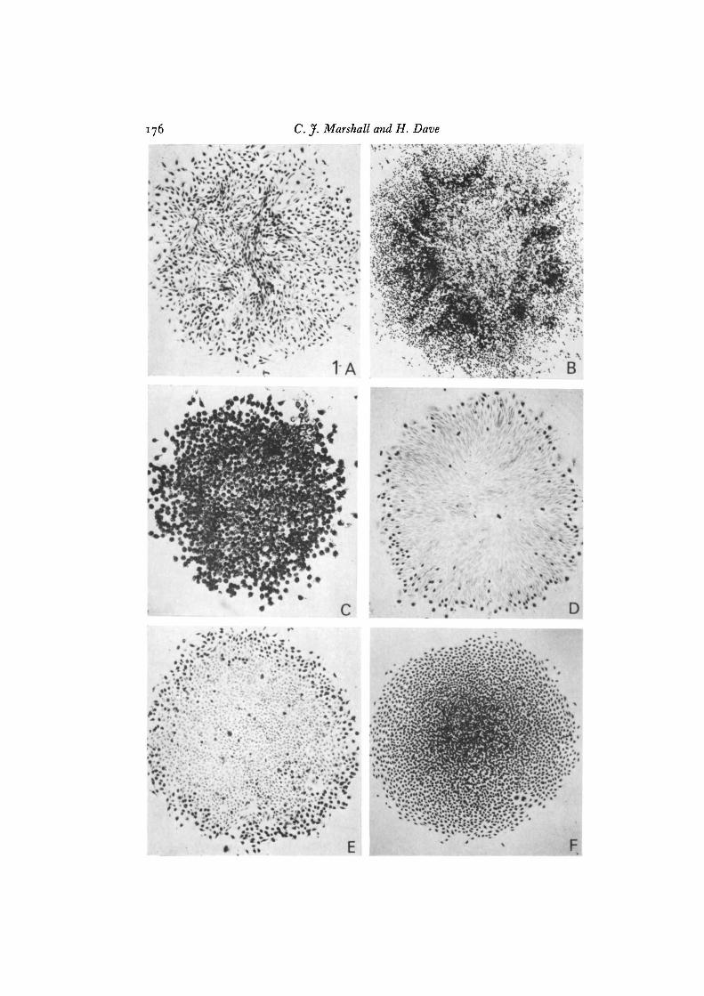
Table 1 (p. 178) demonstrates that normal diploid rat embryo cells did not growin agar or form colonies on confluent monolayers. However in culture conditions of10 % serum the cells did not show a marked inhibition of cell division as they reachedconfluence, typical of density-dependent inhibition of growth in cell lines such as3T3. In 10% serum the REF cells are multilayered and Fig. IA shows that cellsthroughout a colony continued to divide.
Table 2 (p. 179) shows that BI and TA3B have the characteristics of transformedcell lines because they grow in agar, lack density-dependent inhibition of growth, andform colonies on confluent 3T3 monolayers. The relative size and numbers of colonieson plastic and confluent monolayers are similar indicating that BI and TA3B growwith equal efficiencies on these 2 surfaces. Fig. 1B and c show the appearance ofcolonies labelled to detect density-dependent inhibition of growth. The absence ofdensity-dependent inhibition of growth in BI, which is TK~ and fails to incorporate[3H]thymidine, was demonstrated by labelling with [3H]adenosine (i/t Ci/ml, 20 Ci/mmol) and removing 3H-RNA with RNase. Colony formation in agar by TA3B is notas efficient as with BI but the average colony-forming efficiency (CFE) of 2-3 % doesnot indicate that the line consists of two types of cell of which a small fraction (2-3 %)has a 100 % CFE in agar, because colonies isolated from agar have CFEs similar to theparent line. Moreover the growth in agar of TA3B is comparable to that found forother epithelial tumour lines (Marshall et al. 1977).
Isolation of original hybrids
To obtain a representative sample of the phenotypes generated by cell hybridiza-tion it is necessary to avoid bias in the isolation of hybrid cells. Such bias may occurwhen only one parental cell can be directly selected against and hybrids are isolatedby virtue of having different properties from the non-selected parent (see for exampleVan Der Noordaa et al. 1972). Because HAT medium biochemically selects againstBI and TA3B cells while allowing their hybrids to grow, hybrid cell lines may beisolated without regard to the properties of the cells. In the TA3B x REF crossesonly TA3B could be selected against biochemically but controls showed that sparselyseeded REF formed very few colonies in HAT medium. Therefore colonies pickedwithout regard to morphology or growth properties have a high probability of beinghybrid. Precautions were taken against selection for the transformed phenotype byplating cells at moderate density during subculture and not allowing cultures to remainat confluence for longer than one day.
Between one and five colonies were found in each dish after cell fusion. Four wellseparated TA3B x REF colonies were obtained from independent fusion events(TREF 765, 3.2, 3.31, 13.5B). Two subclones of TREF 765 (765 SCi, 765 SC2)were subsequently isolated by micromanipulation. Six BI x TA3B hybrid lines (BIT2.1A, 2.1B, 2.4, 3.4, 4.2, 4.5) representing five independent fusion events wereisolated. The similar properties of BIT 2.1A and 2.iB, which were the only two lines
176 C. J. Marshall and H. Dave

Suppression of transformation in cell hybrids 177
derived from the same dish, suggest that they were sister colonies from the same fusionevent. We will refer to these BI x TA3B and TA3B x REF as original BIT and TREFhybrids.
Tables 1 and 2 present the chromosome analysis of the original hybrids. All of theBIT hybrids and 3 of 4 TREF contain almost a complete single set of chromosomesfrom each parent although there does appear to be a tendency to lose mouse chromo-somes. Unlike the other TREF hybrids TREF 13.5B appears to be a 2:1 hybridwhich contains two mouse chromosome sets to one rat although the number of mousechromosomes (mean 69-9) is lower than the expected 82.
Properties of the original hybrid lines
Light-microscopic examination of cultures of the 6 original BI x TA3B linesshowed that they were very different from the parental cells. The, BI and TA3Bhad typical characteristics of transformed cells: they were refractile and grew in multi-layers, the original hybrids were very flat, poorly refractile and did not criss-cross ormultilayer. This morphology and growth pattern, suggestive of a non-transformedphenotype, was also seen in the 1:1 TREF hybrids (765, 3.2, 3.31), while TREF13.5B resembled TA3B. The very flat morphology and the absence of multilayers inall but TREF 13-sB has been confirmed in electron-microscope studies (Stamatoglou& Marshall, 1978).
The hybrid cell lines were further characterized by measurements of growth inagar, density-dependent inhibition of growth and plating efficiency on 3T3 cells. Thesetests were performed as soon as possible after cell fusion to eliminate effects of longterm culture and in DF10 medium to remove any effect of HAT or deliberate selec-tion for retention of the chromosomes carrying HGPRT or TK.
Table 1 shows that the 1:1 hybrids: TREF 765, its subclones 765 SCi, 765 SC2,TREF 2/3.2, TREF 2/3.31 do not grow in agar or on confluent monolayers and pos-sess density-dependent inhibition of growth. The non-transformed phenotype of thehybrids shows that neoplastic transformation is suppressed in these hybrids. Curiouslythe 1:1 TREF hybrids more closely resembled a non-transformed cell line like 3T3rather than the parental REF cells because they exhibited density dependent inhibi-tion of growth (Fig. 1 E) while the normal REF cells (Fig. 1 A) continued to divideafter they had reached confluence. Table 1 also shows that the 2:1 hybrid TREF13.5B appeared to have a transformed phenotype like TA3B since it grew in agar,lacked density-dependent inhibition of growth and formed colonies on 3T3 cells.However, growth in agar is a selective assay for transformed cells, therefore it wasnecessary to test if the TREF 13.5B cells which grew in agar were selected variants,with high colony-forming efficiencies in agar. Agar colonies were picked and the
Fig. 1. Autoradiographs of cell colonies labelled with [3H]thymidine or [3H]adenosineto detect density-dependent inhibition of growth. A, REF cells, labelling throughoutcolony ( x 69 ) ; B, BI cells, labelling throughout colony ( x 69 ) ; c, TA3B cells, labellingthroughout colony (xo/3) ; D, BIT hybrid 4.2 cells, note labelling only at edge ofcolony ( x 73) ; E, TREF hybrid 765, note labelling only at edge of colony ( x 67 ) ;F, BIT hybrid 4.2AC2 cells, labelling throughout colony ( x 64).

Tab
le I
. P
rope
rtie
s of
RE
F c
ells
and
the
ir h
ybri
ds w
itlz
TA
3B
Par
amet
ers
of t
rans
form
ed p
heno
type
,
, D
ensi
ty-
% co
lony
-"
Ave
rage
no.
of
chro
mos
omcs
/cel
la
0/,
abso
lute
b %
pla
ting
C
dep
end
ent
form
ing
f A
7
plat
ing
effi
cien
cy
inhi
biti
on o
f ef
fici
ency
C
ell
line
h4
ouse
R
at
To
tal
effi
cien
cy
on
3T
3 c
ells
pr
owth
in
apa
r
RE
F
-
42
42
1.5
No
o
TA
3 B
4
I -
4 I
5 5
30
No
2 '3
9
Hyb
rids
L?
TR
EF
765
40
'5
38.8
79
'3
25
o Y
es
o
TR
EF
765
SC
I N
T
NT
N
Te
*
23
o
Yes
o
3 e C
lr
TR
EF
765
SC
2 N
T
NT
N
T e*
24
o
Yes
o
a
TR
EF
213
.2
36.9
42
'9
79.8
1.5
0
Yes
0
S T
RE
F 2
13.3
1 35
'3
38.6
73
'9
35
o Y
es
o
R a
TR
EF
13.
5R
69.9
34
'1
104
10
5.8
No
1-2
A
Lin
es d
eriv
ed f
rom
aga
r $
colo
nies
of
13.5
B
TR
EF
I~
.~B
/AC
I
68.1
35
'1
103.
2 3
NT
N
o
9
6'7
$
TR
EF
I~
.~
B/
AC
~
68.9
32
'4
101.
3 40
N
T
No
3.
6 T
RE
F 1
3.5B
lAC
3 71
.1
31'7
10
2.8
42
NT
N
o
4'6
TR
EF
13.
5BlA
C6
NT
N
T
NT
e"
15
NT
N
o
11.6
(a)
Sta
ndar
d de
viat
ions
for
all
chro
mos
ome
coun
ts f
ell
in t
he r
ange
of
1-3
chro
mos
omes
. (b
) %
no.
of
colo
nies
on
plas
tic/
no.
of c
ells
see
ded.
(c
) %
no.
of
colo
nies
on
con
flue
nt 3
T3
mon
olay
ers/
no.
of c
ells
see
ded.
(d
) %
no.
of
colo
nies
> 0
.1 m
m d
iam
eter
/no.
of
cell
s se
eded
in
agar
. Val
ues
are
the
aver
ages
fro
m 5
-cm
dis
hes
seed
ed w
ith
~o
~e
ell
s.
(e)
Kar
yoty
pes
not
an~
lyse
d in d
etai
l bu
t sh
own
to b
e hy
brid
.

Tab
le 2
. Pr
oper
ties
of B
I an
d T
A3B
cel
ls a
nd t
heir
ori
gina
l hyb
rids
tn
Par
amet
ers
of t
rans
form
ed p
heno
type
%
r
%
A
-----7
2 D
ensi
ty-
%
2.
Ave
rage
no.
of
chro
mos
omes
/cel
la
% ab
solu
teb
% p
lati
ng6
depe
nden
t fo
rmin
g r - plati
ng
effi
cien
cy
inhi
biti
on o
f ef
fici
ency
%
C
ell
line
M
ouse
T
otal
ef
fici
ency
on
3T
3 c
ells
gr
owth
h
H
amst
er
in a
gar
2 a
Bl
-
39'5
39
'5
53
5 8
hrc
3 3
TA
3B
4
I -
41
5 5
30
No
2'
3 5
T s
Hyb
rids
2
BIT
Z.I
A
37.0
38
.1
75'1
48
o
Yes
1'
3 5
. J
BIT
2.1
B
36.5
38
.6
75'1
45
o
Yes
1.
5 B
IT 2
.4
38.4
41
.I
79'5
45
o
Yes
0.0
1
n 3
BIT
3.4
39
'2
39'3
78
.5
47
o Y
es
0.05
2 -
BIT
4.2
38
.9
40.8
79
'7
4 1
o Y
es
0.13
B
IT 4
.5
39'5
37
'7
77'2
3 7
o
Yes
0.
07
2. F
or t
he m
eani
ng o
f (a) to
(d
), se
e fo
otno
te t
o T
able
1.
6

Tab
le 3
. Pro
pert
ies
of t
rans
form
ed d
eriv
ativ
es is
olat
ed b
y se
edin
g or
igin
al B
IT h
ybri
ds i
n ag
ar
Par
amet
ers
of t
rans
form
ed p
heno
type
A
I
> D
ensi
ty-
% co
lony
- A
vera
ge n
o. o
f ch
rom
osom
es/c
ell
% a
bsol
ute
% p
lati
ng
dep
end
ent
form
ing
?
A
c >
pl
atin
g ef
fici
ency
in
hibi
tion
of
effi
cien
cy 9
Cel
l lin
e M
ouse
H
amst
er
To
tal
effi
cien
cy
on 3
T3
cel
ls
grow
th
in a
gar
BIT
~.I
A/
AC
~
35'0
36
.6
71.6
4
0
o
No
10
5 Z B
IT z
.rA
/AC
8 33
'2
37'4
70
.6
3 8
o
No
I 2.
7 B
IT z
.IB
/AC
I 31
'7
43 '7
75
'5
5 5
o
No
1
2.2
a &
BIT
~.I
R/A
C~
31
'4
42'7
74
'1
44
o N
o
10.6
8
R,
BIT
~.
~/
AC
I
32'9
4'
'4
74'3
5 6
o
Yes
6.
9 B
IT 3
.4/A
C13
35
'8
42'7
78
.5
65
o
Yes
4'
3 9
BIT
4.2
/AC
z 35
'0
39'3
74
'3
3 8
o
No
16
.1
BIT
4.z
/AC
q 29
.2
41.6
70
.8
50
o
No
5.
8 F e (b
BIT
4.5
/AC
3 35
'7
4' '
3 77
'0
44
o N
o
64
BIT
4.5
/AC
1o
36.3
41
4
78.1
3 6
o
No
62
B
IT 4
.5/A
C14
37
.6
41 '3
78
.9
53
o
No
66
B
IT 4
.5/A
Czr
36
.6
40'5
77
'1
63
o N
o
5 9

Suppression of transformation in cell hybrids 181
characteristics of the cells tested. Four colonies picked from agar (TREF 13.5B, ACi,AC2, AC3, AC6) had colony-forming efficiencies only 2- to 5-fold higher than theoriginal TREF 13.5B, suggesting that cells that grew in agar were not selected variants.
Table 2 shows that the original BIT hybrids possessed density-dependent inhibi-tion of growth, did not grow on 3T3 cells and had low colony-forming efficiencies inagar. Fig. 1D illustrates the appearance of density-inhibited colonies of hybrids withnuclear DNA synthesis only at the periphery. The absence of colonies on confluentmonolayers and the small numbers of colonies in agar are not due to a low ability ofcells to grow when seeded sparsely because the absolute plating efficiencies of BIThybrids are similar to BI and TA3B. The properties of the hybrids show that thetransformed phenotype is suppressed but the colony-forming efficiencies in agar of theoriginal BIT hybrids (range 0-01-1-5 %) are greater than those we have measured fornon-transformed permanent cell lines such as 3T3, BHK or NRK where less than 1 inio5 or ioG cells form a colony in agar. This suggested that suppression of the trans-formed phenotype in the original BIT lines might not be as stable as in the TREFhybrids and that each BIT hybrid line might consist of a majority of hybrid cells inwhich the transformed phenotype is suppressed plus a small fraction of transformedhybrids. This possibility was tested by isolating the agar colonies and studying thecells' properties to determine if they are selected variants. This approach was especi-ally relevant to the BIT 2.1A, 2.1B lines which had CFEs in agar close to TA3B.
Evidence that BIx TAyB hybrid cells with transformed cell characteristics are variants
If the colonies in agar from the original BIT hybrids arise from selection for varianthybrids with the characteristics of transformed cells then the cells cultured from thesecolonies should grow in agar much more efficiently than the original hybrids. We in-vestigated this prediction by testing 18 agar colonies from 5 BIT lines. The criteriafor isolating these were that the colonies were visible to the unaided eye and wereseparated from others (although in dishes seeded at high density the colonies wouldnot be separated from other cells). Table 3 shows that 12 of the 18 lines from agarcolonies grew much better in agar than the original lines. Comparison of the colony-forming efficiencies in agar of these lines with the original hybrids shows that theefficiencies are increased 40- to 940-fold for all lines except the derivatives of BIT2.1A and 2.1B where there is about a 10-fold increase. Table 3 and Fig. 1F also showthat selection for ability to grow in agar also selected for the loss of density-dependentinhibition of growth but not for growth on confluent monolayers.
We conclude from the properties of these 12 lines that the colonies which arise inagar come from the selection of variant cells different from the majority of cells withinthe original BIT lines. We will refer to these hybrids which lack density-dependentinhibition of growth and grow well in agar as transformed derivatives. The originalBIT hybrid lines therefore consist of a majority of hybrid cells in which the trans-formed phenotype is suppressed and a variable fraction of transformed derivatives.In lines BIT 2.1A, 2.1B, which had colony forming efficiencies in agar similar toTA3B, the fraction of transformed derivatives is greater than in the other originalBIT lines but the cells which grow in agar are still selected variants, although the

Tab
le 4
. Pro
pert
ies
of n
on-t
rans
form
ed s
ztbl
ines
isol
ated
by
seed
ing
orig
inal
hyb
rids
in a
gar
Par
amet
ers
of t
ran
sfo
rmed
ph
enct
yp
e
Den
sity
- %
col
ony-
A
vera
ge n
o.
of c
hrom
osom
es/c
ell
% a
bsol
ute
% p
lati
ng
dep
end
ent
form
ing
?
A
f >
plat
ing
effi
cien
cy
inhi
biti
on o
f ef
fici
ency
9
Cel
l li
ne
Mou
se
Ham
ster
T
ota
l ef
fici
ency
o
n 3
T3
cell
s g
row
th
in a
gar
BIT
z.I
B/A
CQ
34
.1
40.8
74
'9
29
o
hTo
0.5
r b 3 ;3
- B
IT 3
.4/A
C9
33'4
4
1.6
75
'2
53
o Y
es
0.7
o
Yes
0.
07
!$ B
IT ~
.z/A
CI
33
'5
39.8
73
'3
43
R
BIT
4.2
/AC
5 32
.8
40'3
73
''
62
o
Yes
0.
16
BIT
~.
~/
AC
I
38.8
39
'5
78.3
43
o
Yes
0.
5 B
IT 4
.5/A
C2
39.0
39
'2
78
2
49
o
NT
a
0.05
$
k
Tru
e t
rans
form
ed d
eriv
ativ
es
isol
ated
fro
m r
esee
ding
in
agar
B
IT 4
.5/A
C1/
2 B
IT 4
.5/A
C1/
9
(a) T
his
lin
e w
as n
ot t
este
d si
nce
it a
ppea
red
to b
e T
K-.

Suppression of transformation in cell hybrids 183
transformed derivatives (BIT 2.1A/AC5, BIT 2.1A/AC8; BIT 2.1B/AC1, 2.1B/AC2) only show about 10-fold greater colony forming efficiencies in agar, because thecells which grow in agar lack density-dependent inhibition of growth shown by BIT2.1A and 2.1B.
The remaining 6 BIT lines isolated from agar colonies had colony forming effi-ciencies in agar similar to the original hybrid lines. Table 4 shows that some of theselines even had colony-forming efficiencies in agar, 2- to 3-fold lower than the originalhybrids. As might be expected most of these lines possessed density-dependentinhibition of growth and all failed to grow on confluent monolayers. The origin ofthese non-transformed cells from colonies which grew in agar is obscure. They wereonly isolated as small colonies in dishes seeded with at least io4 cells and may repre-sent growth of cells which were seeded as aggregates or attached to fibres of cottonshed from pipette plugs (Stoker, O'Neill, Berryman & Waxman, 1968). They couldalso arise from the limited proliferation of non-transformed cells (Kakunaga & Kama-hora, 1968). Typical transformed lines could be isolated from these sublines bypicking large agar colonies. For example Table 4 demonstrates that lines BIT 4.5AC1/2 and 4.5 AC1/9 derived from non-transformed 4.5 ACi grew well in agar andlacked density-dependent inhibition of growth.
Origin of transformed hybrids
Harris and his collaborators have shown that a necessary (but not sufficient) eventin the generation of malignant segregants from suppressed hybrids is the loss ofspecific chromosomes from the normal parent (Jonasson, Povey & Harris, 1977). Wetherefore attempted to determine if the origin of transformed BIT hybrids involvedchromosome loss.
Examination of the average number of chromosomes per cell line in Tables 2 and3 shows that most of the transformed derivatives have fewer mouse chromosomesthan the original BIT lines (range —2 to —9 chromosomes). Many of the lines alsoshow small increases in hamster chromosome numbers (range —1-5 to + 5-1 chromo-somes). However, comparison with the data in Table 4 for non-transformed BITsublines shows that mouse chromosomes may be reduced and hamster chromosomesincreased even in non-transformed BIT hybrids. These data therefore do not provideevidence for the involvement of chromosome loss in the generation of transformedderivatives. However, it is possible that a more refined analysis at the level of identifi-cation of individual chromosomes might detect the involvement of chromosome loss.
The only original hybrid in which the transformed phenotype is not suppressed isTREF I3-5B. The absence of suppression may be related to the genetic compositionof this hybrid which differed from the suppressed hybrids. While the suppressedhybrids contained a single chromosome set from each parent, TREF 13.5B contained2 chromosome complements from TA3B to one complement from REF. Dosagephenomena have been suggested to account for the difference in expression of dif-ferentiated characteristics in 1:1 and 2:1 hybrids between differentiated and non-differentiated cells (Ephrussi, 1972). However, the loss of rat chromosomes could alsobe a possible cause for the expression of the transformed phenotype in TREF 13.5B

Tab
le j
. T
umou
r pro
duct
ion
ifz n
ude
mic
e
Pro
pert
ies
of
Pro
per
ties
of
cell
lin
es
inoc
ulat
ed c
ells
N
o.
of c
ells
ino
cula
ted
cult
ure
d f
rom
tu
mo
urs
-
I
h
\ I
A
\
% co
lony
- D
ensi
ty-
I oe
I o
5
I o3
%
colo
ny-
Den
sity
- fo
rmin
g d
epen
den
t 5
x 10
-7
& --
form
ing
d
epen
den
t ef
icie
ncy
in
hibi
tion
T
ake"
T
aken
L
aten
tb
Tak
e"
Lat
entb
T
akea
L
aten
tb
Tu
mo
ur
effi
cien
cy
inh
ibit
ion
C
elll
ine
inag
ar
ofg
row
th
inci
denc
e in
cide
nce
peri
od
inci
dcnc
c p
erio
d
inci
denc
e pe
riod
no
. in
aga
r of
gro
wth
RE
F
o
No
01
4 -
-
-
-
TA
3B
2.
3 N
o
-
-
-
3/3
8
d
B I
33
No
-
-
-
313
zed
TR
EF
765
su
ppre
ssed
o
Yes
-
013
-
013
-
hy
bri
d
TR
EF
13
.jB
tr
ansf
orm
ed
1-2
N
o
-
3 /3
7
d
313
14d
hy
bri
d
BIT
4.2
su
pp
ress
ed
0.1
Y
es
-
3/4
45
d 3
/3
45d
hy
bri
d
BIT
4.z
AC
/3
tran
sfor
med
4
0
No
-
-
-
414
3od
deri
vati
ve
of 4
.2
(a)
No.
of
mic
e w
ith
tum
ours
/no.
in
ocul
ated
. (b
) M
ean
tim
e (d
ays)
tak
en t
o p
rod
uce
a t
um
ou
r ap
prox
imat
ely
0.5
cm i
n di
amet
er.

Suppression of transformation in cell hybrids 185
because Table 1 shows that TREF 13.5B and lines derived from its agar colonieshave fewer rat chromosomes than the suppressed hybrids.
Tumorigenicity of hybrids in nude mice
Our findings that the transformed phenotype is suppressed in the original hybridlines led us to ask if malignancy, as measured by tumour production, was also sup-pressed in these hybrids. Preliminary experiments shown in Table 5 demonstratedthat both BI and TA3B rapidly produced progressively growing tumours in nudemice even with small inocula (io3 cells), inocula of 5 x io6 REF failed to producetumours. The BI tumours were poorly differentiated sarcomas while TA3B tumourswere highly anaplastic carcinomas.
Two hybrid lines in which the transformed phenotype was suppressed (TREF765, BIT 4.2) and two hybrid lines with the characteristics of transformed cells (TREF13.5B, BIT 4.2 AC2/3) were chosen for tests of tumour production. The transformedhybrids were employed to serve as a positive control for the growth of hybrids in vivo.As Table 5 demonstrates these transformed hybrids produced tumours in all themice except those with a dose of io3 cells. The latent period for tumour production,which was the time for a tumour to reach 0-5 cm diameter, for TREF 13.5B and BIT4.2 AC2/3 was a little longer than with the parental cells. Tumour growth from trans-formed hybrids did not appear to involve selection since, as Table 5 shows, culturesderived from tumours had similar colony-forming efficiencies in agar to the inoculatedcells. The results establish that hybrid cells with transformed characteristics willreadily produce tumours in nude mice but that the tumours may have less vigorousgrowth than the parental tumour cells. The histology of the hybrid tumours was hardto classify because tumours were very poorly differentiated.
When TREF 765 and BIT 4.2, hybrids in which the transformed phenotype wassuppressed, were inoculated into nude mice very different results were obtained. Asseen in Table 5, TREF 765 did not produce tumours in any animal inoculated; thisclosely parallels the observation that no colonies were obtained when this cell linewas seeded in agar. These results suggest that both tumour production and the trans-formed phenotype are very stably suppressed in these hybrids. Like all the otherhybrids BIT 4.2 did not produce tumours at a dose of io3 cells but tumours did arisewith larger cell numbers. However, Table 5 shows that the latent period for thesetumours was clearly longer than with the parental cells or with the transformedderivative BIT 4.2 AC2/3. This longer latent period suggested that tumour produc-tion might be suppressed and that the tumours arose by selection for the outgrowthof variants with the characteristics of transformed cells. Alternatively the longerlatent period could have resulted from the slow growth of the whole inoculum. Thesetwo possibilities were tested by establishing whether cultures of tumours behavedlike the original BIT 4.2 or its transformed derivatives when tested for growth inagar and density dependent inhibition of growth. Table 5 shows that cultures from 5tumours from BIT 4.2 had high colony forming efficiencies in agar (10-40%) andlacked density-dependent inhibition of growth while the inoculated BIT 4.2 cellshad a colony-forming efficiency in agar of o-i % and showed density-dependent inhi-

186 C.J. Marshall and H. Dave
bition of growth. As Table 5 demonstrates the properties of these tumour cultureswere very similar to tumour cultures produced from the inoculation of BIT 4.2 AC2/3,a transformed derivative of BIT 4.2. Such results clearly show that tumours that areproduced from the inoculation of BIT 4.2 arise from the selective outgrowth ofvariant cells and not from the whole inoculum. These results establish that bothmalignancy, as measured by tumour production, and the transformed phenotype aresuppressed in BI x TA3B and TA3B x REF hybrids. The presence of the transformedphenotype was closely correlated with tumour production since selection for trans-formed hybrids in vitro led to the isolation of cells that rapidly produced tumourswhile selection for tumours led to the isolation of lines with the characteristics oftransformed cells.
DISCUSSION
We have shown that both the in vitro characteristics of transformed cells (the'transformed phenotype') and tumour production may be suppressed in somatic cellhybrids. A trivial explanation of this suppression is that the interspecific nature of thehybrids is incompatible with neoplastic transformation. However, this can be ruledout because we were able to readily isolate transformed derivatives of BI x TA3Bhybrids as well as a transformed TA3B x REF hybrid.
Another explanation that can be ruled out is that the hyperploidy of the hybrids leadsto the suppression of the transformed phenotype. Revertants of SV 40-transformed3T3 which no longer express the transformed phenotype are often hyperploid(Pollack, Vogel & Wolman, 1970) but hyperploidy cannot explain suppression in BITand TREF hybrids because the transformed derivatives often had only a few lesschromosomes than the suppressed hybrids and TREF 13.5B which had more chromo-somes than any other hybrid did not show suppression. Some of the characteristicsof transformed cells we have examined are dominantly expressed in hybrids of Papovavirus-transformed cells (e.g. Croce & Koprowski, 1974a, b, 1975; Jha & Ozer, 1976),therefore suppression or dominance is not a function of the characteristics themselvesbut is dependent on the type of cells which are studied. Harris (1971) has suggestedthat suppression can be interpreted as evidence that neoplastic transformation resultsfrom recessive mutation in gene(s) essential to the maintenance of the normal pheno-type, and is difficult to reconcile with models in which the transformed phenotype ismaintained by the continual expression of 'transformation genes'. On the basis of amutation hypothesis suppression in the BI x TA3B hybrids would involve comple-mentation between different recessive mutations. This implies that the mutations areat different loci in BI and TA3B and that neoplastic transformation follows mutationat any of these loci. Wiener, Klein & Harris (19746) were unable to find conclusiveevidence of complementation in crosses between mouse tumours but Levisohn &Thompson (1973) apparently found complementation resulting in the restoration ofdensity-dependent inhibition of growth in hepatoma x L cell hybrids. We believethat our observation of complementation was fortuitous and that other crosses wouldnot show complementation. Both BI x TA3B and TA3B x REF hybrids are the

Suppression of transformation in cell hybrids 187
products of crosses between epithelial and mesenchymal cells so that suppressioncould be the result of using cells with different embryological origins. However,Wiener et al. (19746) did not find suppression in hybrids between tumours from dif-ferent tissues while we have recently found suppression in hybrids between two celllines derived from mesenchyme (unpublished results of C. J. Marshall).
While we find the recessive mutation hypothesis of neoplastic transformationattractive, the alternative should be considered that genes essential to the normalphenotype are not expressed in transformed cells because of 'epigenetic' changes ingene regulation. Studies with hybrid cells do not at present distinguish between thesetwo hypotheses. The epigenetic hypothesis finds some support from the observationthat the transformed phenotype and malignancy was not suppressed in the 2:1TA3B x REF hybrid 13.5B. This suggests that gene dosage may be involved andparallels results on the expression of differentiated characteristics in hybrids (Ephrussi,1972). The role of dosage should be further investigated by back-cross experiments.
Whatever the mechanism of suppression, its stability is not the same in all hybrids.Suppression may be very stable as in the 1:1 TA3B x REF hybrids, where no trans-formed variants could be isolated or less stable as in the BI x TA3B hybrids whichreadily generated transformed derivatives. We do not understand the reasons for thedifferences in stability of suppression but such differences between crosses have beenfound by others (Wiener et al. 1971; Stanbridge, 1976). The stability of suppressionmay be related to the stability of the hybrid karyotype but we were unable to demon-strate that transformed derivatives arise by chromosome loss. Possibly such variantcells arise by new spontaneous transformation events. We are currently investigatingthis question by studying hybrids with cells in which the transformation event isgenetically marked by a temperature-sensitive mutation. Instability of suppressioncould account for previous observations that the transformed characteristics of hy-brids appeared to be intermediate between the normal and transformed parent. Forexample both Van Der Noordaa et al. (1972) and Wiblin & Macpherson (1973) foundthat the colony-forming efficiency of their hybrids was much lower than the parenttransformed lines, which suggests to us that their lines could have been a mixture ofsuppressed hybrids and transformed variants.
Irrespective of any mechanism of neoplastic transformation that the phenomenonof suppression may imply, we find that both tumorigenicity and the in vitro charac-teristics of tumour cells are coordinately suppressed in the hybrids. Furthermoreselection in vitro for transformed characteristics selects for hybrids which readilyform tumours while selection in vivo for tumour production concomitantly selectscells which express the transformed phenotype. The concordant expression of thetransformed phenotype and tumour production in these hybrids suggests that theyare under the same control and that the in vivo and in vitro properties of the cellsresult from the same events. These observations are further supported by results of afurther study which establish that the expression of LETS protein, a cell surfacemarker of transformation and of microfilament bundles, a cytoskeletal marker, corre-late with the growth characteristics of the hybrids (Marshall, Humphryes & Pollack,1978). However, we would like to stress that while there is probably a causal relation

188 C. y. Marshall and H. Dave
between the in vivo and some in vitro properties of the cells we have used - this maynot be true of all cell lines (Stiles et al. 1976). Indeed we have found that growth onconfluent monolayers, a characteristic of some transformed cell lines was dissociablefrom other transformed characteristics in these hybrids and was not a reliable markerof cell behaviour.
Finally we would like to point out that the experiments reported here are concernedwith the events which result in the altered growth regulation of transformed cells.They are not concerned with the transformation event which leads to the escape froma limited lifespan. The controls of these transformation events are probably differentsince Stanbridge (1976) has shown that the infinite potential to divide is not sup-pressible because hybrids between human tumour cells and fibroblasts with limitedlifespan show suppression of malignancy but no signs of a limited lifespan.
We would like to thank Angela Clarke and Eileen Simmons for preparation of the manu-script, Thelma Barnes for assistance with autoradiography and Drs Robert Shields, L. M.Franks and Robert Pollack for comments on the manuscript.
REFERENCES
AARONSON, S. A. & TODARO, G. J. (1968). Development of 3T3-like lines from Balb/c mouseembryo cultures: Transformation susceptibility to SV40. J. cell. Physiol. 72, 141-148.
BARSKI, G. & CORNEFERT, F. (1962). Characteristics of ' hybrid'-type clonal cell lines obtainedfrom mixed cultures in vitro. J. natn. Cancer Jnst. 28, 801-821.
BREGULA, U., KLEIN, G. & HARRIS, H. (1971). The analysis of malignancy by cell fusion. II .Hybrids between Ehrlich cells and normal diploid cells. J. Cell Sci. 8, 673-680.
CROCE, C. M., ADEN, D. & KOPROWSKI, H. (1975a). Somatic cell hybrids between mouseperitoneal macrophages and Simian Virus 40 transformed human cells. II. Presence ofhuman chromosome 7 carrying SV40 genome in tumours induced by hybrid cells. Proc.natn. Acad. Sci. U.S.A. 72, 1397-1400.
CROCE, C. M., ADEN, D. & KOPROWSKI, H. (19756). Tumorigenicity of mouse-human diploidhybrids in nude mice. Science, N. Y. 190, 1200-1202.
CROCE, C. M. & KOPROWSKI, H. (1974a). Positive control of transformed phenotype in hybridsbetween SV4o-transformed and normal human cells. Science, N.Y. 184, 1288-1289.
CROCE, C. M. & KOPROWSKI, H. (19746). Somatic cell hybrids between mouse peritonealmacrophages and SV40 transformed human cells. Positive control of the transformedphenotype by the human chromosome 7 carrying SV40 genome. J. exp. Med. 140, 1221-1224.
CROCE, C. M. & KOPROWSKI, H. (1975). Assignment of gene(s) for cell transformation tohuman chromosome 7 carrying the SV40 genome. Proc. natn. Acad. Sci. U.S.A. 72, 1658-1660.
DEFENDI, V., EPHRUSSI, B., KOPROWSKI, H. & YOSHIDA, M. C. (1967). Properties of hybridsbetween polyoma-transformed and normal mouse cells. Proc. natn. Acad. Sci. U.S.A. 57,299-3°5-
DEV, V. G., MILLER, D. A., ALLDERDICE, P. W. & MILLER, O. J. (1972). Method for locatingthe centromeres of mouse meiotic chromosomes and its application to T163H and T70Htranslocations. Expl Cell Res. 73, 259-262.
DULBECCO, R. (1970). Topoinhibition and serum requirement of transformed and untrans-formed cells. Nature, Lond. 227, 802-806.
EPHRUSSI, B. (1972). Hybridization of Somatic Cells. Princeton, New Jersey: Princeton Univer-sity Press.
FISHER, H. W. & YEH, J. (1967). Contact inhibition in colony formation. Science, N.Y. 155,581-582.

Suppression of transformation in cell hybrids 189
FREEDMAN, V. H. & SHIN, S. (1974). Cellular tumorigenicity in nude mice: correlation withcell growth in semi solid medium. Cell 3, 355-359.
HARRIS, H. (1971). Cell fusion and the analysis of malignancy. Proc. R. Soc. B 179, 1-20.HARRIS, H., MILLER, O. J., KLEIN, G., WORST, P. & TACHIBANA, T. (1969). Suppression of
malignancy by cell fusion. Nature, Lond. 223, 363-368.HARRIS, H. & WATKINS, J. F. (1965). Hybrid cells derived from mouse and man: artificial
heterokaryons of mammalian cells from different species. Nature, Lond. 205, 640-646.HYNES, R. O. (1976). Cell surface proteins and malignant transformation. Biochim. biopliys.
Acta 458, 73-107.JHA, K. K. & OZER, H. L. (1976). Expression of transformation in cell hybrids. I. Isolation
and application of density inhibited Balb/3T3 cells deficient in hypoxanthine phosphori-bosyl transferase and resistant to ouabain. Somatic Cell Genetics 2, 215-223.
JONASSON, J., POVEY, S. & HARRIS, H. (1977). The analysis of malignancy by cell fusion. VII.Cytogenetic analysis of hybrids between malignant and diploid cells and of tumours derivedfrom them. J . Cell Sci. 24, 217-254.
KAKUNAGA, T. & KAMAHORA, J. (1968). Properties of hamster embryonic cells transformed by4-nitroquinolinc-l-oxide in vitro and their correlations with the malignant properties of thecells. BikenJ. 11, 313-332.
KLEIN, G. (1951). Comparative studies of mouse tumours with respect to their capacity forgrowth as ascites tumours and their average nucleic acid content per cell. Expl Cell Res. 2,518-573.
KLEIN, G., BREGULA, U., WIENER, F. & HARRIS, H. (1971). The analysis of malignancy by cellfusion. I. Hybrids between tumour cells and L cell derivatives. J. Cell Sci. 8, 659-672.
KLEIN, G., FRIBERG, S., WIENER, F. & HARRIS, H. (1973). Hybrid cells derived from the fusionof TA3-HA ascites carcinoma with normal fibroblasts. I. Alalignancy, karyotype and forma-tion of isoantigenic variants. J. natn. Cancer Inst. 50, 1259-1268.
LEVISOHN, S. R. & THOMPSON, E. B. (1973). Contact inhibition and gene expression in H T C / Lcell hybrid lines. J. cell. Physiol. 81, 225-232.
LITTLEFIELD, J. W. (1966). The use of drug-resistant markers to study the hybridization ofmouse fibroblasts. Expl Cell Res. 41, 190-196.
LITTLEFIELD, J. W. & BASILICO, C. (1966). Infection of thymidine kinase deficient BHK cellswith polyoma virus. Nature, Lond. 211, 250-252.
MACPHERSON, I. & MONTAGNIER, L. (1964). Agar suspension culture for the selective assay ofcells transformed by polyoma virus. Virology 23, 291-294.
MARIN, G. (1971). Segregation of morphological revertants in polyoma-transformed hybridclones of hamster fibroblasts. J. Cell Sci. 9, 61-69.
MARSHALL, C. J. (1975). A method for analysis of chromosomes in hybrid cells employingsequential G-banding and mouse specific C-banding. Expl Cell Res. 91, 464-469.
MARSHALL, C. J., FRANKS, L. M. & CARBONNEL, A. W. (1977). Markers of neoplastic trans-formation in epithelial cell lines derived from human carcinomas, jf. TMtn. Cancer Inst. 53,1748-1751.
MARSHALL, C. J., HUMPHRYES, K. C. & POLLACK, R. E. (1978). Microfilament bundles, LETSprotein and growth control in somatic cell hybrids. J. Cell Sci. 33, 191-204.
NICHOLSON, G. L. (1974). The interaction of lectins with animal cell surfaces. Int. Rev. Cytol.39, 89-190.
POLLACK, R. E., VOGEL, A. & WOLMAN, S. (1970). Reversion of virus transformed cell lines:Hyperploidy accompanies retention of viral genes. Nature, Lond. 228, 938-967.
SCALETTA, L. J. & EPHRUSSI, B. (1965). Hybridization of normal and neoplastic cells in vitro.Nature, Lond. 205, 1169-1171.
SHIN, S.-I., FREEDMAN, V. H., RISSER, R. & POLLACK, R. (1975). Tumorigenicity of virus-transformed cells in nude mice is correlated specifically with anchorage independent growthin vitro. Proc. natn. Acad. Sci. U.S.A. 72, 4435-4439.
STAMATOGLOU, S. C. & MARSHALL, C. J. (1978). Ruthenium red-positive layer, extracellularfilamentous material and intercellular junctions in hybrids between tumour and normal cells:abundant gap junctions correlate with density-dependent inhibition of growth. J. Cell Sci.31. 323-339-
STANBRIDCE, E. J. (1976). Suppression of malignancy in human cells. Nature, Lond. 260, 17-20.
13 C E L 33

190 C. J. Marshall and H. Dave
STILES, C. D., DESMOND, W., CHUMAN, L. M., SATO, G. & SAIER, M. H. (1976). Relationshipof cell growth behaviour in vitro to tumorigenicity in athymic nude mice. Cancer Res. 36,3300-3305.
STOKER, M. G. P., O'NEILL, C, BERRYMAN, S. & WAXMAN, V. (1968). Anchorage and growthregulation in normal and virus-transformed cells. Int. J. Cancer 3, 683-693.
STOKER, M. G. P. & RUBIN, H. (1967). Density dependent inhibition of cell growth in culture.Nature, Lond. 215, 171-172.
VAN DER NOORDAA, J., VAN HAAGEN, A., WALBOOMERS, J. M. M. & VAN SOMEREN, H. (1972).Properties of somatic cell hybrids between mouse cells and simian virus 40-transformed ratcells. J. Virol. 10, 67-72.
WEISS, M. C. (1970). Further studies on loss of T-antigen from somatic hybrids between mousecells and SV40-transformed human cells. Proc. natn. Acad. Sci. U.S.A. 66, 79-86.
WIBLIN, C. N. & MACPHERSON, I. A. (1973). Reversion in hybrids between SV40-transformedhamster and mouse cells. Int.J. Cancer 12, 148-161.
WIENER, F., KLEIN, G. & HARRIS, H. (1971). The analysis of malignancy by cell fusion. III.Hybrids between diploid fibroblasts and other tumour cells. J. Cell Sci. 8, 681-692.
WIENER, F., KLEIN, G. & HARRIS, H. (1973). The analysis of malignancy by cell fusion. IV.Hybrids between tumour cells and a malignant L-cell derivative. .7. Cell Sci. 12, 253-261.
WIENER, F., KLEIN, G. & HARRIS, H. (19740). The analysis of malignancy by cell fusion. V.Further evidence of the ability of normal diploid cells to suppress malignancy. J. Cell Sci.15, 177-183.
WIENER, F., KLEIN, G. & HARRIS, H. (19746). The analysis of malignancy by cell fusion. VI.Hybrids between different tumour cells. J. Cell Sci. 16, 189-198.
{Received 28 February 1978)