suppression of vagal cardiac modulation by blue light in healthy … · suppression of vagal...
TRANSCRIPT
ORIGINAL ARTICLE Open Access
Suppression of vagal cardiac modulation byblue light in healthy subjectsEmi Yuda, Hiroki Ogasawara, Yutaka Yoshida and Junichiro Hayano*
Abstract
Background: In the contemporary life environments, our body is increasingly exposed to various sources of coloredlight, which may affect our physiological functions as non-image-forming effects. We examined the impacts of coloredlights on the autonomic functions by the analysis of heart rate variability (HRV).
Methods: A lighting device consisting of four organic light-emitting diode (OLED) modules (55 × 55 mm2) withadjustable red-green-blue color was secured 24 cm above the eyes of subject lying supine in a light-shieldedlaboratory. Following a 15-min supine rest, electrocardiogram and respiration were measured continuously during3-min darkness, 6-min colored OLED illumination, and 3-min darkness under paced breathing (15 breath/min).The measurements were repeated at a 45-min interval for red, green, and blue lights with melanopsin-stimulatingphoton flux density (MSPFD) of 0.00, 0.10, and 0.20 μmol/m2/s, respectively, in 12 healthy subjects (23 ± 2 years,two females). Additionally, the effects of blue lights with 0.20, 0.10, and 0.04 μmol/m2/s MSPFD were examined infour healthy subjects (25–39 years, two females). HRV was analyzed for low-frequency (LF, 0.04–0.15 Hz) and high-frequency (HF, 0.20–0.30 Hz) power and LF-to-HF ratio (LF/HF).
Results: Compared to darkness before lighting, HF power decreased (P < 0.001) and LF/HF increased (P = 0.024)during lighting on average of all color lights, whereas HF power showed a greater decrease with blue light thanwith red and green lights (P < 0.05 for both). The decrease in HF power lasted even during darkness after lighting(P < 0.001). HF power decreased with blue light with 0.20 μmol/m2/s MSPFD (P < 0.001) but not with that with 0.10 or 0.04 μmol/m2/s (P = 0.1 and 0.9, respectively).
Conclusions: Vagal cardiac modulation is suppressed by OLED blue light in healthy subjects most likely throughmelanopsin-dependent non-image-forming effect.
Keywords: Organic light-emitting diode, Non-image-forming vision, Melanopsin, Intrinsically photosensitive retinalganglion cell, Blue light, Heart rate variability
IntroductionIn the contemporary life environment, our body is increas-ingly exposed to various artificial lightings with variouscolors. Light affects many physiological parameters suchas melatonin, alertness, body temperature, heart rate, andheart rate variability (HRV) via its non-image-formingvisual functions [1–5]. The primary mediator of non-image-forming functions is the melanopsin system whosephotoreceptor is intrinsically photosensitive retinal gan-glion cells that have a specific sensitivity to blue lightaround 480 nm [6, 7]. While colored illuminations in our
life environments are often selected from esthetic or noti-ceability aspects, the melanopsin-stimulating property ofindividual colored illuminations may be important forconsidering their effects on health and wellbeing.In this study, we investigated the impacts of colored
lights on cardiac autonomic functions by the analysis ofHRV with particular interests in whether the melanopsin-stimulating spectral component is the determinant of theireffects. For this purpose, we developed a lighting deviceconsisting of organic light-emitting diode (OLED) withadjustable red-green-blue color, which allowed us to ge-nerate non-glaring surface illumination with differentcolors through a single lighting device. Although OLED isexpected to be used as a new lighting source for home,
* Correspondence: [email protected] of Medical Education, Nagoya City University Graduate School ofMedical Sciences, Kawasumi 1 Mizuho-cho Mizuho-ku, Nagoya 467-8601, Japan
© 2016 The Author(s). Open Access This article is distributed under the terms of the Creative Commons Attribution 4.0International License (http://creativecommons.org/licenses/by/4.0/), which permits unrestricted use, distribution, andreproduction in any medium, provided you give appropriate credit to the original author(s) and the source, provide a link tothe Creative Commons license, and indicate if changes were made. The Creative Commons Public Domain Dedication waiver(http://creativecommons.org/publicdomain/zero/1.0/) applies to the data made available in this article, unless otherwise stated.
Yuda et al. Journal of Physiological Anthropology (2016) 35:24 DOI 10.1186/s40101-016-0110-x
occupational, and healthcare environments, there is fewstudy on its physiologic effects.
MethodsSubjectsThe present study was performed according to the proto-col that was approved by the Ethics Review Committee ofNagoya City University Graduate School of MedicalSciences (No. 44-15-0001).The subjects of this study were recruited with the fol-
lowing inclusion criteria: healthy men or women who (1)were between 20 and 40 years old, (2) had normal colorvision, (3) were not taking any medications for >2 weeks,and (4) displayed a normal sinus rhythm on electrocardio-gram (ECG) at rest. There were 14 applicants who metthe inclusion criteria; 12 of them (mean age ± SD, 23 ±2 years, two females) participated in study 1, and four ofthem (age range, 25–39 years, two females) participated instudy 2. All subjects gave their written informed consentto participate in this study.
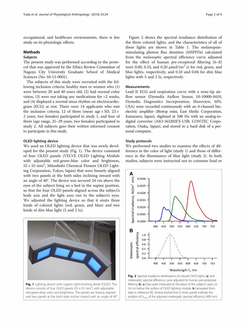
OLED lighting deviceWe used an OLED lighting device that was newly devel-oped for the present study (Fig. 1). The device consistedof four OLED panels (VELVE OLED Lighting Modulewith adjustable red-green-blue color and brightness,55 × 55 mm2, Mitsubishi Chemical Pioneer OLED Light-ing Corporation, Tokyo, Japan) that were linearly alignedwith two panels at the both sides inclining inward withan angle of 40°. The device was secured 24 cm above theeyes of the subject lying on a bed in the supine position,so that the four OLED panels aligned across the subject’sbody axis and the light axes run to the subject’s eyes.We adjusted the lighting device so that it emits threekinds of colored lights (red, green, and blue) and twokinds of dim blue light (5 and 2 lx).
Figure 2 shows the spectral irradiance distribution ofthe three colored lights, and the characteristics of all ofthese lights are shown in Table 1. The melanopsin-stimulating photon flux densities (MSPFDs) calculatedfrom the melanoptic spectral efficiency curve adjustedfor the effect of human pre-receptoral filtering [6–8]were 0.00, 0.10, and 0.20 μmol/(m2 s) for red, green, andblue lights, respectively, and 0.10 and 0.04 for dim bluelights with 5 and 2 lx, respectively.
MeasurementsLead II ECG and respiration curve with a nose-tip air-flow sensor (Dymedix Airflow Sensor, 10-10000-0410,Dymedix Diagnostics Incorporation, Shoreview, MN,USA) were recorded continuously with an 8-channel bio-electric amplifier (Biotop mini, East Medic Corporation,Kanazawa, Japan), digitized at 500 Hz with an analog-to-digital converter (AIO-163202FX-USB, CONTEC Corpo-ration, Osaka, Japan), and stored in a hard disk of a per-sonal computer.
Study protocolsWe performed two studies to examine the effects of dif-ference in the color of light (study 1) and those of differ-ence in the illuminance of blue light (study 2). In bothstudies, subjects were instructed not to consume food or
Fig. 1 Lighting device with organic light-emitting diode (OLED). Thedevice consists of four OLED panels (55 × 55 mm2) with adjustablered-green-blue color and brightness. The panels are linearly alignedand two panels at the both sides incline inward with an angle of 40°
Fig. 2 Spectral irradiance distributions of colored OLED lights (a) andmelanoptic spectral efficiency curve adjusted for human pre-receptoralfiltering (b). a Data were measured at the place of the subject’s eyes, i.e.,24 cm below the surface of OLED lighting module. b Generated fromdata in reference [8]. Vertical dashed lines in both panels indicate theposition of λmax of the adjusted melanoptic spectral efficiency (490 nm)
Yuda et al. Journal of Physiological Anthropology (2016) 35:24 Page 2 of 9

beverages containing caffeine or alcohol after 21:00 theprevious night. The studies were performed between 08:30and 13:00 in a calm, light-shielded, and air-conditioned(24 ± 2 °C) laboratory more than 2 h after a light meal.For both studies 1 and 2, data were collected with the
experimental schedule of dark and illumination condi-tions (Fig. 3). In study 1, measurement sessions withthree different color lights (red, green, and blue) wereperformed in 12 subjects with randomized orders amongsubjects at a 45-min interval. In study 2, sessions withthree different illuminance of blue lights (10, 5, and 2 lx)were performed in four subjects with randomized ordersat a 45-min interval. The 10-lx blue light used in study 2was the same as blue light used in study 1.At each session, the subjects lied on the bed in the su-
pine position, so that their eyes were right below theOLED device, and wore a headphone for the purpose ofpaced breathing. We used paced breathing method to pre-vent the changes in breathing frequency from confound-ing the assessment of vagal cardiac modulation by HRV[9, 10]. We developed a computer program that generatesan audio signal consisting of high (960 Hz)- and low(770 Hz)-pitched sounds, which appeared alternatively for2 s each (at an interval of 4 s), resulting paced breathing at15 cycle/min (0.25 Hz). The subjects were instructed tobreathe in when hearing high-pitched sound and tobreathe out during low-pitched sound. Before the experi-ment, all the subjects practiced the paced breathing untilthey become able to breathe comfortably in synchronywith the audio signal coming through the headphone.For both studies, measurement of each session was
started after a 15-min supine rest. The subjects wereinstructed to keep their eyes open, to gaze at the OLEDpanels, and to continue the paced breathing throughouteach measurement session.
Data analysesData were analyzed off-line on a personal computer.The temporal positions of all QRS waves in digitizedECG data were detected with a fast-peak detectionalgorithm. After all errors in the detection of QRSwaves were edited, time series of the R-R interval wereobtained. The R-R interval time series thus obtainedwere analyzed separately for the periods of three differ-ent conditions (3-min darkness before lighting, 6-minlighting, and 3-min darkness after lighting) in individ-ual measurement sessions with four different lightingcolors.For each data segment, frequency domain analyses of
the HRV were performed with fast Fourier transform-ation (FFT) with the original software in our laboratory[9]. Briefly, R-R interval time series were interpolatedwith a step function only using interval data consistingof consecutive QRS waves in sinus rhythm, resampled at256 and 512 equidistant time points for 3-min and 6-min data segments, respectively, filtered with a Han-ning window, and converted into frequency domain byFFT. After correcting for the losses of variance result-ing from the sampling and filtering processes, the abso-lute power of the low-frequency (LF, 0.04–0.15 Hz) andthe high-frequency (HF, 0.15–0.40 Hz) componentswere computed. The powers of LF and HF componentswere transformed into natural logarithmic values. Weused the HF power as an index of vagal cardiac modu-lation for cardiopulmonary resting [9, 11, 12].The respiration data were also analyzed by FFT, and
breathing frequency was estimated from the position ofthe dominant spectral peak. The synchronicity of respir-ation was evaluated from the breathing frequency, anddata were excluded from this analysis if it deviated fromthe range between 0.23 and 0.27 Hz.
Table 1 Characteristics of light sources
Red Green Bluea Blue 5 Blue 2
Illuminance, lx 39 71 10 5 2
Irradiance, W/m2 0.20 0.14 0.07 0.03 0.01
Chromaticity (x, y) (0.63, 0.34) (0.33, 0.62) (0.14, 0.16) (0.14, 0.16) (0.14, 0.16)
Photon flux density, μmol/(m2 s) 1.05 0.64 0.26 0.13 0.05
Melanopsin-stimulating photon flux density, μmol/(m2 s)b 0.00 0.10 0.20 0.10 0.04aBlue light with 10-lx illuminance was used for both studies 1 and 2bCalculated from melanoptic spectral efficiency adjusted for human pre-receptoral filtering [7, 8]
Fig. 3 Experimental schedule of dark and illumination conditions. Measurement sessions with three different color lights (red, green, and blue)were performed in all subjects with different orders randomized among subjects at an interval of 45 min
Yuda et al. Journal of Physiological Anthropology (2016) 35:24 Page 3 of 9

Statistical analysisStatistical analyses system version 9.4 (SAS institute, Cary,NC, USA) was used for the statistical analysis. Our primaryinterest in study 1 was to clarify whether the autonomicneural activities during OLED lighting differ with the colorof lighting and the secondary interest was to examine theeffects of colored lighting on the autonomic activity afterexposure to lighting. For these purposes, two-way repeatedmeasures ANOVAs of autonomic indices were separatelyperformed on the difference between before and duringlighting and on the difference between before and afterlighting. For study 2, to evaluate the effects of blue lightwith different illuminance, we used paired t test for eachilluminance. P < 0.05 was considered to be statistically sig-nificant, and Bonferroni method was used to keep type 1error level of <0.05 in multiple comparisons.
ResultsIn study 1, we used data in 10 subjects (mean age ± SD,24 ± 1 years) out of 12, because two subjects (one maleand one female) were excluded due to the loss of respira-tory synchronicity. The heart rate and HRV indices in in-dividual subjects before, during, and after exposures tored, green, and blue OLED lighting are shown in Fig. 4.Repeated measures ANOVA on the difference between
before and during lighting revealed that there was no sig-nificant main effect of color or time (before and duringlighting) or their interaction on heart rate or LF power,whereas significant main effects of color and time and theirinteraction on HF power and significant main effect oftime on LF-to-HF ratio (LF/HF) were observed (Table 2).As shown in Fig. 5, HF power decreased and LF/HF in-creased during lighting. Multiple comparisons of HF powerthat showed significant time × color interaction indicatedthat the decrease in HF power with blue light was greaterthan those with red and green lights (P < 0.05 for both)with no significant difference between those of red andgreen lights (Table 3).Repeated measures ANOVA on the difference between
before and after lighting revealed significant main effectsof time on LF and HF power and LF/HF (Table 4). Al-though significant main effect of color was also revealedon HF power, no significant interaction between timeand color was detected. As shown in Fig. 6, LF powerand LF/HF were increased and HF power was decreasedafter lighting.In study 2, respiratory synchronicity was maintained in
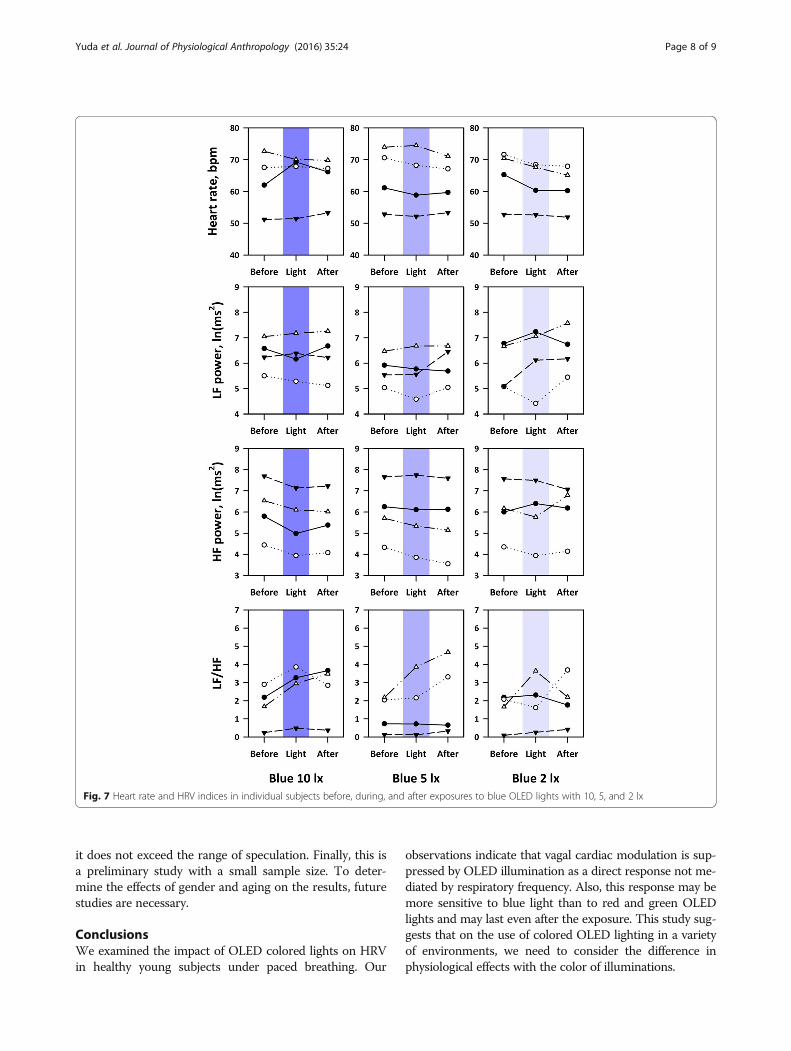
all subjects. Figure 7 shows the heart rate and HRV indi-ces in individual subjects before, during, and after expo-sures to blue lights with 10, 5, and 2 lx (MSPFD, 0.20,0.10, and 0.04 μmol/m2/s, respectively). Blue lights with 5and 2 lx caused no significant changes in heart rate or anyof HRV indices either during or after lighting, while bluelight with 10 lx increased HR during darkness after
lighting (P = 0.03), decreased HF power both during light-ing (P = 0.006) and darkness after lighting (P = 0.001), andincreased LF/HF during lighting (P = 0.02).
DiscussionTo investigate the acute physiological effects of coloredOLED lights in healthy young subjects, we looked intothe changes in HRV indices during and after exposure tolights with different color and illuminance. We foundthat blue light caused a greater decrease in HF powerthan red and green colors. While the blue light had alower illuminance (10 lx) than red and green (39 and71 lx, respectively), it had the highest MSPFD (0.20,0.10, and 0.00 μmol/(m2 s) for blue, green, and red, re-spectively). Furthermore, the decrease in HF power wasobserved for 10-lx blue light but not for 5- or 2-lx bluelights, indicating that the decrease in HF power withblue light is not attributable to its lower intensity. Thedecrease in HF power was also observed even duringdarkness after the termination of lighting. These findingssupport the hypothesis that blue light suppresses vagalcardiac modulation through melanopsin-dependent non-image-forming effect. Also, the sustained response of vagalsuppression during darkness after lighting seems also con-sistent with the property of the melanopsin-dependentnon-image-forming effect that is known to have sustainedresponse after light exposure [13, 14].Earlier studies have reported mixed results for the ef-
fects of colored fluorescent lights on HRV [15, 16]. Forexample, Schäfer et al. [15] have analyzed the changes inHRV with 10-min exposure to red, green, and blue fluor-escent light of 700 lx preceded and followed by 15-mindarkness in 12 healthy young subjects. Although theyfailed to detect significant change in absolute HF powerduring exposure to any color of lights, their results indi-cated a significant decrease in absolute HF power duringdarkness after exposure to blue light, while there wereno significant changes with exposures to red or greenlights. Their results seem partially consistent with ours,suggesting a specific sensitivity of the HF component toblue light. Choi et al. [16] have also analyzed HRV beforeand after 5-min exposure to blue, red, and white fluores-cent light in 92 healthy adults in the seating position withspontaneous breathing. They observed a decrease in abso-lute HF power after exposure to red light, while there wasno significant change with exposure to blue or white light.Although their results seem inconsistent with ours, thedifference in body positions during HRV measurementbetween the studies may be critical because the HF poweris strongly suppressed by gravitation stress [17].In the present study, we used paced breathing for the
autonomic assessment by HRV. Although HF compo-nent of HRV is widely used as an index of vagal cardiacmodulation [11, 18], the power of this component is also
Yuda et al. Journal of Physiological Anthropology (2016) 35:24 Page 4 of 9

affected by respiration frequency; HF power decreaseswith increasing respiration frequency independently ofcardiac vagal modulation [10, 19]. Furthermore, while theHF component is thought to be generated centrally by themechanism of cardiorespiratory coordination, earlier stu-dies have suggested that this central coordination may beaffected by light depending on its color [20]. Because thechanges in HF power in the present study were observedunder paced breathing, they may be interpreted to reflect
the direct effects of colored light on central vagal function.In contrast, the changes in HF power under spontaneousbreathing could be indirectly mediated by the changes inrespiratory frequency at least partly.In study 1, we compared the autonomic effects among
red, green, and blue OLED lights with different inten-sity (illuminance, irradiance, and photon flux density);the blue light was the lowest in these parameters(Table 1). Thus, we were unable to determine whether
Fig. 4 Heart rate and heart rate variability (HRV) indices in individual subjects before, during, and after exposures to red, green, and blue OLEDlights. HF high-frequency component, LF low-frequency component, LF/HF LF-to-HF ratio in power
Yuda et al. Journal of Physiological Anthropology (2016) 35:24 Page 5 of 9

the observed differences between blue light and othercolored lights were caused by the color of lights or theirintensity. The method for standardizing the light inten-sity for comparing the effects of colors of light has notbeen established [8]. Also, possible changes in pupillarysize with light could affect the amount of light reachingthe retina, even if the intensity of lights was standar-dized in some way. Thus, we instead investigated theeffects of intensity of blue light to examine whether theautonomic effect of blue light is caused by its low in-tensity or not (study 2). We observed that the suppres-sion of HF power occurred only with 10-lx blue lightbut not with 5-lx or 2-lx blue lights. Additionally, theestimated MSPFD of 5-lx blue light was the same asthat of 71-lx green light. These indicate that the greatersuppression of HF power with the 10-lx blue light thanwith the red and green lights is not attributable to thelower intensity of blue light and the pattern of responsessuggests that the suppression of HF power by blue light ismost likely explained by its higher MSPFD.While HF power decreased with 10-lx blue OLED light,
heart rate showed no significant change in study 1 andonly a modest increase in study 2. Although HF powerhas been often interpreted simply as an index of cardiacvagal tone to control heart rate, HRV in HF band, particu-larly under paced breathing at >0.15 Hz, is a quantitativereflection of respiratory sinus arrhythmia (RSA). We havepreviously reported that RSA is a cardiopulmonary restingfunction for saving cardiac and respiratory energy by sup-pressing unnecessary heartbeats during expiration andineffective ventilation during waning phases of perfusion[12, 21]. Although the magnitude of RSA and heart rateusually change reciprocally to each other with physicaland mental stress and relaxation, they are thought to beregulated separately by the vagal outflows from the nu-cleus ambiguous and the dorsal motor nucleus of vagus,respectively; the former generates phasic changes with res-piration, while the latter causes tonic pattern [22]. Thesesuggest that blue OLED light may suppress the vagalmechanisms generating RSA without affecting substan-tially the vagal mechanisms controlling heart rate. One
may speculate that blue OLED light might have an effectthat shifts the state of our body to arousal from resting.We used a newly developed OLED lighting device for
this study. Because we did not compare the OLED withconventional lighting sources including fluorescent lamps
Fig. 5 Average changes in heart rate and HRV indices from beforeto during OLED lighting. Error bars represent standard error of mean.*Significantly different from values for red and green with multiplecomparisons (P < 0.05)
Table 2 Repeated measures ANOVA on differences in heart rateand HRV indices between before and during lighting
Main effect Interaction
Color (DF = 2) Time (DF = 1)a Time × color (DF = 2)
F value P F value P F value P
Heart rate 0.07 0.93 2.83 0.12 1.30 0.30
LF power 0.52 0.60 0.01 0.93 0.58 0.57
HF power 4.62 0.030 42.04 <0.001 6.52 0.010
LF/HF 1.70 0.22 6.52 0.024 0.92 0.42
DF degree of freedom, HF high-frequency component, LF low-frequencycomponent, LF/HF LF-to-HF ratio in poweraEffect of time between before and during lighting
Yuda et al. Journal of Physiological Anthropology (2016) 35:24 Page 6 of 9

and light-emitting diode (LED), we were unable to de-termine whether the results we observed are specific toOLED or not. As shown in Fig. 2, however, the spectralirradiance of OLED blue light has a broader spectrumat the region of the melanoptic spectral efficiency curve[6–8] compared with those reported for blue LED [23]and for fluorescent lamps [24]. OLED is gathering atten-tions as non-glaring comfortable surface illumination andis expected to be used as a new lighting source at home,workplace, and healthcare environments. Our findings ofthe autonomic effects seem important characteristics ofcolored OLED illuminations, which should be consideredon their utilization.
LimitationsThis study has several limitations. First, we examined theeffects of lighting only for 6 min. Although we observed asignificant reduction in HF power both during and afterthe exposure of blue light, it is not clear whether the re-sponse had been saturated during 6 min or might haveprogressed more by longer exposure. The same is the casefor recovery of the response. We were unable to deter-mine the time course of the recovery. If the response lastlong, the 45-min interval between the measurements withdifferent color lights might have been insufficient forwashing out the effects of the previous exposure. Second,we did not standardize the intensity of illumination amongdifferent colors. Thus, we cannot exclude the effects ofthe intensity on our results. However, as discussed above,the suppression of HF power by blue light was not ex-plained by its low intensity in study 2. Third, because we
did not measure melatonin secretion in this study, wewere unable to determine whether the colored lights affectthe entrainment to environmental light-dark cycles or not.Although our findings seem consistent with the hypo-thesis that blue light suppresses vagal cardiac modulationthrough melanopsin-dependent non-image-forming effect,
Table 3 Multiple comparisons of the changes in HF power(during-before) between colors of lighting
Red Green Blue
Red – 0.75 0.007a
Green 0.75 – 0.010a
Blue 0.007a 0.010a –
Values are P value for the significance of difference between two colorsaSignificant after Bonferroni adjustment (P < 0.05)
Table 4 Repeated measures ANOVA on differences in heart rateand HRV indices between before and after lighting
Main effect Interaction
Color (DF = 2) Time (DF = 1)a Time × Color (DF = 2)
F value P F value P F value P
Heart rate 0.07 0.93 0.27 0.62 0.06 0.94
LF power 0.36 0.70 6.67 0.022 0.29 0.75
HF power 4.63 0.030 22.38 <0.001 1.98 0.18
LF/HF 1.70 0.22 12.71 0.004 0.17 0.84
Abbreviations are explained in the footnote found in Table 2aEffect of time between before and after lighting
Fig. 6 Average changes in heart rate and HRV indices from beforeto after OLED lighting. Error bars represent standard error of mean
Yuda et al. Journal of Physiological Anthropology (2016) 35:24 Page 7 of 9
it does not exceed the range of speculation. Finally, this isa preliminary study with a small sample size. To deter-mine the effects of gender and aging on the results, futurestudies are necessary.
ConclusionsWe examined the impact of OLED colored lights on HRVin healthy young subjects under paced breathing. Our
observations indicate that vagal cardiac modulation is sup-pressed by OLED illumination as a direct response not me-diated by respiratory frequency. Also, this response may bemore sensitive to blue light than to red and green OLEDlights and may last even after the exposure. This study sug-gests that on the use of colored OLED lighting in a varietyof environments, we need to consider the difference inphysiological effects with the color of illuminations.
Fig. 7 Heart rate and HRV indices in individual subjects before, during, and after exposures to blue OLED lights with 10, 5, and 2 lx
Yuda et al. Journal of Physiological Anthropology (2016) 35:24 Page 8 of 9

AbbreviationsECG: Electrocardiogram; FFT: Fast Fourier transformation; HF: High frequency;HRV: Heart rate variability; LF: Low frequency; LF/HF: LF-to-HF ratio;MSPFD: Melanopsin-stimulating photon flux density; OLED: Organic light-emitting diode; RSA: Respiratory sinus arrhythmia
AcknowledgementsThis study was performed as a part of the collaborative studies with Chemicalmaterials Evaluation and Research Base (CEREBA), Japan.
FundingThis study was supported by the New Energy and Industrial TechnologyDevelopment Organization (NEDO), Japan.
Availability of data and materialsAll data described in this study are freely available to any scientist wishing touse them in a way that ensures the participant confidentiality. The softwaredescribed in this study is also available to any scientist wishing to use them.In any case, the data and software described in the manuscript are availablefor testing by reviewers in a way that preserves the reviewers’ anonymity.
Authors’ contributionsEY participated in the design and coordination of the study and drafted themanuscript. HO developed the software for the study and participated in thecoordination of the study. YY participated in the design of the study andperformed the statistical analysis. JH conceived the study and helped to draftthe manuscript. All authors read and approved the final manuscript.
Competing interestsThe authors declare that they have no competing interests.
Consent for publicationThis manuscript does not contain any individual person’s data in any form.
Ethics approval and consent to participateThe present study was performed according to the protocol that was approvedby the Ethics Review Committee of Nagoya City University Graduate School ofMedical Sciences (No. 44-15-0001). All of them gave their written informedconsent to participate in this study.
Received: 5 July 2016 Accepted: 9 September 2016
References1. Cajochen C, Munch M, Kobialka S, Krauchi K, Steiner R, Oelhafen P, Orgul S,
Wirz-Justice A. High sensitivity of human melatonin, alertness, thermoregulation,and heart rate to short wavelength light. J Clin Endocrinol Metab. 2005;90(3):1311–6.
2. Yasukouchi A, Ishibashi K. Non-visual effects of the color temperature offluorescent lamps on physiological aspects in humans. J Physiol AnthropolAppl Hum Sci. 2005;24(1):41–3.
3. Chellappa SL, Steiner R, Blattner P, Oelhafen P, Gotz T, Cajochen C. Non-visualeffects of light on melatonin, alertness and cognitive performance: can blue-enriched light keep us alert? PLoS One. 2011;6(1):e16429.
4. Litscher D, Wang L, Gaischek I, Litscher G. The influence of new coloredlight stimulation methods on heart rate variability, temperature, and well-being: results of a pilot study in humans. Evid Based Complement AlternatMed. 2013;2013:674183.
5. Daneault V, Dumont M, Masse E, Vandewalle G, Carrier J. Light-sensitive brainpathways and aging. J Physiol Anthropol. 2016;35(1):9.
6. Enezi J, Revell V, Brown T, Wynne J, Schlangen L, Lucas R. A “melanopic”spectral efficiency function predicts the sensitivity of melanopsinphotoreceptors to polychromatic lights. J Biol Rhythms. 2011;26(4):314–23.
7. Bailes HJ, Lucas RJ. Human melanopsin forms a pigment maximally sensitiveto blue light (lambdamax approximately 479 nm) supporting activation ofG(q/11) and G(i/o) signalling cascades. Proc Biol Sci. 2013;280(1759):20122987.
8. Price LLA. Report on the first international workshop on circadian andneurophysiological photometry, 2013. International Commission onIllumination, 2015 CIE TN 003:2015.
9. Hayano J, Sakakibara Y, Yamada A, Yamada M, Mukai S, Fujinami T, YokoyamaK, Watanabe Y, Takata K. Accuracy of assessment of cardiac vagal tone by heartrate variability in normal subjects. Am J Cardiol. 1991;67(2):199–204.
10. Hayano J, Mukai S, Sakakibara M, Okada A, Takata K, Fujinami T. Effects ofrespiratory interval on vagal modulation of heart rate. Am J Physiol. 1994;267:H33–40.
11. Pomeranz B, Macaulay RJ, Caudill MA, Kutz I, Adam D, Gordon D, KilbornKM, Barger AC, Shannon DC, Cohen RJ, et al. Assessment of autonomicfunction in humans by heart rate spectral analysis. Am J Physiol. 1985;248(1Pt 2):H151–3.
12. Hayano J, Yasuma F, Okada A, Mukai S, Fujinami T. Respiratory sinus arrhythmia.A phenomenon improving pulmonary gas exchange and circulatory efficiency.Circulation. 1996;94(4):842–7.
13. Berson DM, Dunn FA, Takao M. Phototransduction by retinal ganglion cellsthat set the circadian clock. Science. 2002;295(5557):1070–3.
14. Kankipati L, Girkin CA, Gamlin PD. Post-illumination pupil response in subjectswithout ocular disease. Invest Ophthalmol Vis Sci. 2010;51(5):2764–9.
15. Schafer A, Kratky KW. The effect of colored illumination on heart ratevariability. Forsch Komplementmed. 2006;13(3):167–73.
16. Choi CJ, Kim KS, Kim CM, Kim SH, Choi WS. Reactivity of heart rate variabilityafter exposure to colored lights in healthy adults with symptoms of anxietyand depression. Int J Psychophysiol. 2011;79(2):83–8.
17. Mukai S, Hayano J. Heart rate and blood pressure variabilities during gradedhead-up tilt. J Appl Physiol. 1995;78:212–6.
18. Berger RD, Saul JP, Cohen RJ. Transfer function analysis of autonomicregulation. I: canine atrial rate response. Am J Physiol. 1989;256:H142–52.
19. Hirsch JA, Bishop B. Respiratory sinus arrhythmia in humans: how breathingpattern modulates heart rate. Am J Physiol. 1981;241:H620–9.
20. Edelhauser F, Hak F, Kleinrath U, Luhr B, Matthiessen PF, Weinzirl J, Cysarz D.Impact of colored light on cardiorespiratory coordination. Evid BasedComplement Alternat Med: eCAM. 2013;2013:810876.
21. Hayano J, Yasuma F. Hypothesis: respiratory sinus arrhythmia is an intrinsicresting function of cardiopulmonary system. Cardiovasc Res. 2003;58(1):1–9.
22. Taylor EW. The evolution of efferent vagal control of the heart invertebrates. Cardiosci. 1994;5:173–82.
23. Cao D, Nicandro N, Barrionuevo PA. A five-primary photostimulator suitablefor studying intrinsically photosensitive retinal ganglion cell functions inhumans. J Vis. 2015;15(1):15.1.27.
24. Romeo S, Viaggi C, Di Camillo D, Willis AW, Lozzi L, Rocchi C, Capannolo M,Aloisi G, Vaglini F, Maccarone R, Caleo M, Missale C, Racette BA, Corsini GU,Maggio R. Bright light exposure reduces TH-positive dopamine neurons:implications of light pollution in Parkinson’s disease epidemiology. Sci Rep.2013;3:1395.
• We accept pre-submission inquiries
• Our selector tool helps you to find the most relevant journal
• We provide round the clock customer support
• Convenient online submission
• Thorough peer review
• Inclusion in PubMed and all major indexing services
• Maximum visibility for your research
Submit your manuscript atwww.biomedcentral.com/submit
Submit your next manuscript to BioMed Central and we will help you at every step:
Yuda et al. Journal of Physiological Anthropology (2016) 35:24 Page 9 of 9