surfactant protein d interacts pneumocystis...
TRANSCRIPT
Surfactant Protein D Interacts with Pneumocystis carinhl and MediatesOrganism Adherence to Alveolar MacrophagesDeirdre M. O'Riordan,* Joseph E. Standing,* Kun-Young Kwon,* Donald Chang,* Edmond C. Crouch,*and Andrew H. Umper***Thoracic Diseases Research Unit, Division of Pulmonary, Critical Care and Internal Medicine and tDepartment of Biochemistry andMolecular Biology, Mayo Clinic and Foundation, Rochester, Minnesota 55905; and IDepartment of Pathology, Jewish Hospital atWashington University, St. Louis, Missouri 63110
Abstract
Pneumocystis carini interacts with glycoproteins present inthe lower respiratory tract through its mannose-rich surfaceantigen complex termed gpA. Surfactant protein D (SP-D)is a recently described component of the airspace liningmaterial that possesses a calcium-dependent lectin domaincapable of interacting with glycoconjugates present on mi-croorganisms and leukocytes. Accordingly, we evaluated theextent and localization of SP-D in the lower respiratory tractduring Pneumocystis pneumonia in an immunosuppressedrat model and examined its role in modulating interactionof P. carinji with macrophages. We report that SP-D is amajor component of the alveolar exudates that typify P.carinji pneumonia and is present bound to the surface of P.carinji organisms in vivo. Wefurther demonstrate that SP-Dbinds toP. carini through saccharide-mediated interactionswith gpA present on the surface of the organism. Lastly, weshow that SP-D augments binding of P. carinji to alveolarmacrophages, but does not significantly enhance macro-phage phagocytosis of the organism. The interaction of SP-D with gpA represents an additional important componentof the host-parasite relationship during P. carini pneumo-nia. (J. Clin. Invest. 1995. 95:2699-2710.) Key words: Pneu-mocystis carinji * surfactant protein D * gpA * macrophages* lectin
Introduction
Pneumocystis carinii pneumonia remains a common, life-threat-ening opportunistic infection in immunocompromised patients.It is particularly prevalent among those with acquired immuno-deficiency syndrome (AIDS), hematologic or solid malignan-cies, transplanted organs, or in patients receiving chronic immu-nosuppressive therapies, particularly corticosteroids (1-6). P.carinii pneumonia is characterized by filling of the alveolarspaces with distinctive protein-rich foamy exudates laden withorganisms (7). These proteinaceous exudates can be so markedas to mimic alveolar proteinosis (8). The exact chemical natureof the alveolar exudates in P. carindi pneumonia is not fullydefined, but prior studies indicate the presence of fibronectin,
Address correspondence to Dr. Limper, Thoracic Diseases ResearchUnit, 601A Guggenheim Building, Mayo Clinic, Rochester, MN55905.Phone: 507-284-2301; FAX: 507- 284-4521.
Received for publication 11 March 1994 and in revised fonn 15February 1995.
The Journal of Clinical Investigation, Inc.Volume 95, June 1995, 2699-2710
vitronectin, and surfactant components including surfactant pro-tein D (SP-D)' (8-13).
Alveolar macrophages play a significant role in host defenseby binding, phagocytizing, and degrading P. carini (14, 15).Recent investigations demonstrate that lung surfactant may par-ticipate in the host-organism relationship during P. carini pneu-monia and may modulate interaction of organisms with alveolarmacrophages (12, 13, 16, 17). Surfactant protein-A (SP-A) ispresent in enhanced amounts in the lower respiratory tract dur-ing P. carinii pneumonia and binds to the organism (12, 17).Despite the presence of enhanced amounts of surfactant proteinsin the lower respiratory tract during P. carinii pneumonia, physi-ologic studies suggest a relative deficiency of functional surfaceactive material during pneumonia (18, 19).
SP-D is a collagenous protein synthesized and secreted byalveolar type II cells and nonciliated bronchiolar cells (20-23).Structural studies demonstrate that SP-D possesses a calcium-dependent lectin-binding domain sharing high sequence homol-ogy with the group Ill mammalian C-type lectins (24-26). Sev-eral members of this family including bovine conglutinin,human mannose-binding protein, and SP-A are believed to par-ticipate in host defense against microorganisms (27-32). Previ-ous studies indicate that SP-A binds to P. carinii and Staphylo-coccus aureus (12, 16, 17, 33). Furthermore, SP-A enhancesphagocytosis of particles through Fc receptor and complementreceptor- I mechanisms (32). SP-D has also been shown to inter-act with lipopolysaccharide present on Escherichia coli and tomediate agglutination of the organism (34). Another C-typelectin, human mannose-binding protein, has been reported tofunction as an acute phase reactant and opsonin present in serum(29, 30). Additional studies indicate that SP-D interacts withleukocytes. In particular, recent studies demonstrate that SP-Dbinds to alveolar macrophages (23).
P. carindi possesses several carbohydrate-rich surface pro-teins capable of interacting with lectins and other glycoproteins(35-39). In particular, P. carinji contains a major surface glyco-protein complex which has been variously termed gpA, gp 95,gpl2O, or major surface glycoprotein (40-45). Differences inthe relative molecular mass of this complex have been relatedto the host species from which the P. carinii are derived (35).Therefore, a number of investigators have adopted the nomen-clature of gpA for this glycoprotein complex (44-47). Molecu-lar studies indicate that gpA is encoded by a family of genescontaining relatively conserved cysteine-rich regions (41, 42,45, 46). N-linked carbohydrate rich in mannose, glucose, andN-acetyl glucosamine residues represents approximately one-
1. Abbreviations used in this paper: BAL, bronchoalveolar lavage; gpA,the mannose-rich major surface glycoprotein complex of P. carinii; SP-A, surfactant protein A; SP-D, surfactant protein D.
Interaction of Surfactant Protein D with Pneumocystis carinii 2699
tenth of gpA's mass (35, 36, 48). gpA is known to interact withseveral glycoproteins including concanavalin A, fibronectin,and SP-A and likely represents a major ligand recognized duringthe interactions of P. carinii with alveolar epithelial cells andmacrophages (17, 39, 49, 50). In view of the lectin-bindingproperties of SP-D and gpA, we hypothesized that SP-D inter-acts with P. carini through binding of gpA present on theorganism.
The current investigation was therefore undertaken to ac-complish the following goals: (a) to determine the extent andlocalization of SP-D in the lung during P. carinji pneumonia;(b) to investigate whether SP-D binds to P. carini organismsin vivo and to determine which components of the organisminteract with SP-D; and (c) to determine the effect of SP-D onthe interaction of P. carinii with alveolar macrophages. Wepresent evidence that SP-D is present in enhanced amounts inthe lower respiratory tract during P. carinji pneumonia and thatSP-D specifically binds to gpA and modulates interaction ofthe organism with alveolar macrophages.
Methods
Materials. All organic chemicals were obtained from Sigma ChemicalCo. (St. Louis, MO) unless otherwise specified. "I-Bolton-Hunter re-agent was purchased from New England Nuclear (Boston, MA). Apolyclonal rabbit antiserum generated against rat SP-D has been de-scribed previously (21). Monoclonal antibody 5E12, a mouse IgM recog-nizing gpA derived from rodent, human, and ferret, was the gift of Dr.Francis Gigliotti (Department of Pediatrics, University of Rochester,Rochester, NY) (51). A polyclonal goat antiserum recognizing macro-phage mannose receptors was generously provided by Dr. Philip Stahl(Department of Cell Biology, Washington University, St. Louis,MO) (52).
Preparation of P. carinii. All studies described in this report wereapproved by the institutional animal care committee. P. carinii pneumo-nia was induced in Harlan Sprague-Dawley rats by immunosuppressionwith dexamethasone as reported previously (53, 54). Pathogen-free ratswere provided with drinking water containing dexamethasone (2 mg/liter), tetracycline hydrochloride (500 mg/liter), and nystatin (200,000U/liter). After S d, rats were intratracheally inoculated with P. carinii(500,000 cysts) prepared by homogenizing lung from rats with pneumo-nia using a Stomacher blender (Tekmar Co., Cincinnati, OH). After 6wk of additional immunosuppression, rats were killed, and whole lunglavage was performed with 50 ml of either HBSSor TBS containing 1mMcalcium as specified. P. carinii organisms were purified from thislavage by differential centrifugation (54, 55). The recovered lavagefluids were initially centrifuged (400 g for 10 min) to remove inflamma-tory cells. Some cysts were present in these initial cellular pellets, asidentified by Diff-Quik staining (Baxter Healthcare Corp., Dade Divi-sion, McGraw, IL), and were discarded along with the inflammatorycells. The supernatants containing predominantly suspended P. cariniiorganisms were recentrifuged (1,400 g for 30 min). The pellets resultingfrom the second centrifugation represent the P. carinii isolates used inthese studies. These isolates were resuspended in 1 ml of HBSS, andduplicate 10-ml aliquots of this suspension were spotted onto glassslides, stained with Diff-Quick, and P. carinii quantified (54, 55). Priorstudies in our laboratory revealed that these isolates contain both tropho-zoite and cyst forms in a ratio of 9:1 (55). P. carinii generally represented> 97% of the cellular material on Diff-Quik-stained smears with theremainder representing fragmented nonviable host cells. If other micro-organisms were noted in the lavage smear. the material was discarded.Whole lung lavage from control rats without P. carinii failed to yieldany material after the second centrifugation (54). Microbiologic cultureson selected P. carinii isolates demonstrated no growth of other bacteriaor fungi over 72 h.
Immunohistochemistry. Immunohistochemistry was performed to
evaluate the distribution of SP-D present in rat lung during P. cariniipneumonia. Lung specimens were fixed in 10%phosphate-buffered for-malin and embedded in paraffin. Serial 5-,im sections were deparaffin-ized with xylene and graded alcohols and submitted to either standardhematoxylin and eosin staining, immunohistochemistry, or methenaminesilver staining to visualize cysts. Tissue localization of SP-D was evalu-ated using a polyclonal rabbit antibody generated against purified ratSP-D (21). The deparaffinized tissue sections were sequentially incu-bated for 30 min each in methanol containing 0.3% hydrogen peroxideto quench endogenous peroxidase activity, and 1.5% normal goat serumto reduce nonspecific binding of antibodies (56). Subsequently, the sec-tions were incubated with primary antibody (10 jsg/ml) for 4 h. Thesections were washed and reacted with biotinylated goat anti-rabbitIgG (2 ag/ml for 30 min; Dako Corp., Carpinteria, CA). The sectionswere next treated with peroxidase-conjugated streptavidin (2 pg/ml;Dako Corp.) for 30 min, and bound antibodies detected with 3-amino-9-ethylcarbazole substrate (AEC substrate; Dako Corp.) in the presenceof 3%hydrogen peroxide for 15 min. Sections were counterstained withhematoxylin. To confirm the specificity of staining, semiserial sectionswere incubated with nonimmune IgG, and identical lung regions wereexamined.
Immunoelectron microscopy. To directly evaluate whether SP-D wasbound to the surface of both P. carinii cysts and trophozoites, immuno-electron microscopy was performed. P. carinii were isolated from bron-choalveolar lavage (BAL) of moribund rats using TBS with 1 mMcalcium to preserve surface-bound SP-D. Isolated organisms were fixedin periodate-lysine-paraformaldehyde buffer (57) and embedded in Low-acryl mounting medium (Ted Pella, Inc., Redding, CA). Ultrathin sec-tions were obtained, blocked with normal goat serum (2%) for 1 h, andincubated with either rabbit anti-rat SP-D or nonimmune rabbit IgG(25 ,ug/ml) overnight. After washing, the sections were subsequentlyincubated with goat anti-rabbit IgG conjugated to 15 nMcolloidal gold(Amersham Corp., Arlington Heights, IL). The sections were washedagain and examined on a transmission electron microscope (model 6400;JEOL USA Inc., Peabody, MA).
Detection of SP-D in cell-free BAL and on isolated P. carinii organ-isms. To determine the time course of SP-D accumulation during P.carinii pneumonia, rats were immunosuppressed and inoculated with P.carinii. Since corticosteroids are known to increase the expression ofsurfactant components in the lung, control rats were sham inoculatedand maintained on an identical immunosuppressive regimen of dexa-methasone. In addition, the corticosteroid-treated control animals alsoreceived trimethoprim, 160 mg/liter, and sulfamethoxazole, 800 mg/liter, added to the drinking water to prevent development of P. cariniipneumonia. At 2-wk intervals, rats from each group (four animals)were killed, lavaged with HBSS, and SP-D in the BAL assayed byimmunoblotting. The first 5-ml aliquot of BAL from P. carinii-infectedrats and from controls was centrifuged (400 g) to pellet cellular material.The supernatants were concentrated, dissolved in sample buffer, andseparated by SDS-PAGE using 3% stacking and 7% resolving gels.Subsequently, the separated lavage proteins were transferred to nitrocel-lulose, and SP-D was detected by immunoblotting using anti-SP-D(1:1,000 dilution).
To similarly evaluate whether freshly isolated P. carinii possesssurface bound SP-D, organisms were obtained from rats with fulminantP. carinii pneumonia by lavage with TBS containing 1 mMcalcium.The organisms were purified by differential centrifugation and washedin TBS with calcium. P. carinii were solubilized in 0.125 MTris, 4%SDS, and 4% 2-mercaptoethanol and separated with SDS-PAGEandtransferred to nitrocellulose. The membranes were washed with TBScontaining 0.05% Tween 20, and nonspecific binding sites were blockedwith TBS containing 3% dried milk. The presence of organism-associ-ated SP-D was assessed by immunoblotting using anti-SP-D (1:100dilution). To investigate the saccharide and divalent cation dependencyof SP-D's interaction with P. carinii, parallel experiments were per-formed where equal numbers of organisms were incubated for 30 minin either glucose (100 mM), mannose (100 mM), or EDTA (20 mM)and washed before lysis and immunoblotting for SP-D.
2700 O'Riordan et al.

Purification and radiolabeling of SP-D. SP-D was isolated from the10,000 g supernatant of lavage obtained from rats with silica-inducedalveolar lipoproteinosis using affinity chromatography on maltosyl-aga-rose followed by gel filtration chromatography (58). Purity of the SP-D preparation was verified by SDS-PAGEand silver staining. Radioio-dination of SP-D was undertaken by the method of Bolton and Hunter(59). In brief, benzene was removed from Bolton-Hunter iodinationreagent by evaporation under a stream of nitrogen. SP-D (5-10 yg)was added to 100 ul of 0.1 Msodium borate, pH 8.5, in the presenceof 2 mMmaltose and 2 mMcalcium chloride. This suspension wasadded to the dried Bolton-Hunter reagent and reacted for 2 h on ice,with agitation. Free iodine was removed by dialysis against 50 mMTris-HCl, pH 7.4. The specific activity of the recovered protein was
2 x 10⁶ cpm/,g protein.Binding of SP-D to separated P. carinii proteins. The interaction of
SP-D with specific components of P. carinii was further assessed byevaluating binding of 251I-SP-D to solubilized P. carinii separated bySDS-PAGEand immobilized onto nitrocellulose. P. carinii (20 x 106organisms), obtained by lavage with HBSS, were dissolved in 0.125 MTris, 4% SDS, 20% glycerol, and 4% 2-mercaptoethanol, separated bySDS-PAGE and transferred to nitrocellulose as described (55). Themembranes were washed with TBS containing 0.05% Tween 20 andnonspecific binding sites blocked by incubation with TBS containing1mg/ml BSA for 1 h at room temperature. Blocked membranes wereincubated overnight in the same buffer containing 2 X l0 cpm '5I-SP-D/ml. In some experiments, an identical number of P. carinii was blottedwith 1251-SP-D in the presence of EDTA(20 mM)or glucose (100 mM).The next day, membranes were extensively washed in TBS, and boundSP-D was visualized by autoradiography. To confirm the identity ofgpA in the P. carinii extracts, immunoblotting was also performed withantibody 5E12, a mouse IgM monoclonal antibody which recognizesgpA (51). The nitrocellulose membranes were incubated with mAb5E12(1:50 dilution) or with a similar concentration of nonimmune controlimmunoglobulin, washed and reincubated with a secondary peroxidase-conjugated goat anti-mouse IgM antibody (Sigma Chemical Co.).Bound antibodies were visualized by reaction of peroxidase with diami-nobenzidine in the presence of H202-
Interaction of purified gpA with SP-D. Prior studies indicate thatgpA can be purified from P. carinii using polyacrylamide gel electropho-resis (11, 39). P. carinii (1 X 10⁸) were solubilized in 125 mMTris,4% SDS, 4% 2-mercaptoethanol, 0.002% bromophenol blue, and 20%glycerol, pH 7.4. From this extract, gpA was purified by continuousflow electrophoresis over a 10% polyacrylamide preparative tube gel(PrepCell Apparatus; Bio-Rad Laboratories, Hercules, CA). The gel wasresolved over 48 h using 25 mAcurrent and continuously eluted with25 mMTris Base, 192 mMglycine, and 0.1% SDS. Fractions wereanalyzed by SDS-PAGEand silver staining on 4-15% gradient resolv-ing gels (Phast Gel System; Pharmacia LKB Biotechnology, Piscataway,NJ). Fractions containing the 120-kD complex were dialyzed and con-centrated. Immunoblotting of the purified protein with monoclonal anti-body 5E12 verified this material as gpA (51). To investigate SP-Dbinding to the purified gpA complex, we coated gpA (30 ,g/ml in 0.1MNaHCO3) on 96-well break apart ELISA plates (Removawell Plates;Dynatech Laboratories Inc., Chantilly, VA) for 8 h at 22TC. The plateswere washed and blocked by overnight incubation in TBS containing1 mg/ ml BSA at 40C. The next day the plates were washed again andallowed to bind '"I-SP-D in TBS containing calcium (1 mM)and BSA(1 mg/ml) at the indicated concentrations. To assess the effects of vari-ous sugar ligands on SP-D binding to gpA, additional incubations wereperformed in the presence of glucose, mannose, or lactose (100 mMeach). Nonspecific binding was determined in parallel by the presenceof 20 mMEDTAwhich inhibited binding through SP-D's carbohydraterecognition domain.
Role of SP-D in P. carinii adherence to alveolar macrophages. Wequantified the attachment of P. carinii to cultured alveolar macrophagesin the presence or absence of antibody to SP-D or with the addition ofpurified SP-D. Adherence of P. carinii to alveolar macrophages wasassayed by 5'Cr-labeling the organisms (60, 61). P. carini were isolated
from rats with TBS containing 1 mMcalcium to prevent loss of surface-bound SP-D. The organisms were radiolabeled by incubation for 8 h at370C in 2 ml of DMEcontaining 20%FCS and 200 OCi of 5"Cr-sodiumchromate (New England Nuclear). Normal alveolar macrophages werelavaged from healthy rats and plated in tissue culture plates (1 X 10⁵cells/well) which had been precoated with normal rat IgG (100 pg/mlx 60 min), in order to ensure firm adherence of the macrophages.After 1 h, the macrophages were gently washed with HBSS to removenonadherent cells. Wehave reported previously that > 95% of macro-phages are adherent after this wash (62). 5"Cr-P. carinii (1 X 10⁶)containing surface-associated SP-D were added to the macrophages andincubated at 370C for an additional hour. Subsequently, nonadherent P.carinii were removed by washing. The macrophage monolayers con-taining adherent P. carinii were solubilized in 1 NNaOHand quantified.Adherence of P. carinii was defined as: percentage of adherence = (A/A + B) x 100, where A = 5'Cr-P. carinii associated with the monolayer,and B = unattached 5'Cr-P. carinii. To assess the effect of SP-D on theattachment of P. carini to alveolar macrophage lung cells in culture,P. carinii adherence assays were conducted in the presence of nonim-mune rabbit IgG (100 Ag/ml), a polyclonal rabbit antibody generatedagainst SP-D (100 Ag/ml), or SP-D (5 [g/ml).
As a second method to evaluate SP-D-mediated binding of P. cari-nii to macrophages, we used specific sugars to dissociate SP-D fromthe surface of freshly isolated P. carinii. Interactions of SP-D withglycoconjugates are preferentially inhibited by maltose, but can also beinhibited by glucose and mannose, the principal sugar constituents ofgpA (9, 22, 34). In contrast, lactose is a much less effective inhibitorof SP-D interactions (21, 22, 24). Accordingly, freshly isolated P. cariniiwere incubated with either maltose, mannose, or lactose (100 mM) orin TBS containing 1 mMcalcium alone (control) for 1 h. Subsequently,the organisms were washed and collected by centrifugation (1,400 gX 5 min). SP-D released into the supernatants was assessed with immu-noblotting. The recovered organisms were additionally labeled with 51Crand assayed for their ability to adhere to alveolar macrophages.
Immunoprecipitation. Immunoprecipitation experiments were per-formed to determine whether the polyclonal antibody to SP-D mightalso crossreact with macrophage mannose receptors, another C-typelectin. Alveolar macrophages (12 x 106) were isolated from uninfectedrats by lavage, starved in methionine-free RPMI medium containing10% FBS for 1 h, and incubated with 35S-Trans label (250 GCi; NewEngland Nuclear) in 5 ml methionine-free RPMI containing 10% FBSovernight at 370C. The cells were washed, disrupted by repeated freeze-thaw cycles, and sonicated in 50 mMTris buffer (pH 7.4) containing1 mMleupeptin and 1 mMpepstatin. Membrane-associated proteinswere recovered by centrifugation at 100,000 g for 30 min and solubilizedin PBScontaining 1%Triton X-100 and 1 mMcalcium and magnesium.Radiolabeled macrophage protein extracts were divided into equal ali-quots, preabsorbed with protein A-Sepharose, and parallel 50-sLI ali-quots were incubated with either anti-SP-D or with nonimmune rabbitIgG (100 ,ug/ml). Protein-antibody complexes were precipitated usingprotein A-Sepharose, eluted with 1% SDS containing 5% /3-mercapto-ethanol, and analyzed by SDS-PAGEas described previously (63). Asa positive control, radiolabeled macrophage proteins were also immuno-precipitated using a polyclonal goat anti-mannose receptor antiserum,generously provided by Dr. Philip Stahl, or with nonimmune goat serum(52). For conditions using goat serum, protein G-Sepharose was substi-tuted for protein A-Sepharose in an identical fashion.
Effect of SP-D on macrophage phagocytosis of P. carinii. To deter-mine whether SP-D also mediates phagocytosis of P. carinii, radiola-beled P. carinii were allowed to bind to macrophages in the presenceor absence of adenosine and homocysteine thiolactone, which are potentinhibitors of macrophage phagocytosis (64). P. carinii were radiolabeledwith 5'Cr and incubated with alveolar macrophages for 2 h in the pres-ence or absence of adenosine (100 AM) and homocysteine thiolactone(250 MM). Nonbound organisms were removed by washing, and thecells were lysed in 1 N NaOHand quantified. The number of cpmsassociated with macrophages in the absence of inhibitors represents bothbound and internalized P. carini organisms. In contrast, the number of
Interaction of Surfactant Protein D with Pneumocystis carinii 2701
cpms associated with macrophages in the presence of adenosine andhomocysteine thiolactone represents only bound P. carinii. The differ-ence between these two determinations provides an estimate of phagocy-tosis. To evaluate the role of SP-D in binding and phagocytosis of P.carinii by macrophages, we compared the interaction of freshly isolatedP. carinii, EDTA-treated organisms, and EDTA-treated P. carinii withadditional SP-D. As an independent confirmation of the effectivenessof these agents in impairing phagocytosis, we and others have observedpreviously that adenosine and homocysteine thiolactone effectively in-hibit the phagocytosis of opsonized zymosan particles by alveolar mac-rophages using the luminol-dependent chemiluminescence method (61).
Role of mannose receptors in P. carinii adherence to alveolar mac-rophages. To investigate the potential role of mannose receptors inmediating the SP-D-dependent adherence of P. carinii to alveolar mac-rophages, we further assayed P. carinii binding to macrophages in thepresence of yeast a-mannan. Yeast a-mannan is an effective solubleinhibitor of macrophage mannose receptors (65, 66). Fresh untreatedand EDTA-treated P. carinii were radiolabeled with 51Cr as described.Alveolar macrophages were incubated with a-mannan (100 jig/ml) for20 min before the addition of radiolabeled 5"Cr-labeled P. carinii withor without additional SP-D (5 /ig/ml) and throughout a subsequent 60-min incubation. Unattached P. carinii were removed by washing, andthe percentage of organisms adherent to the macrophage cell layers wasdetermined by gammacounting.
Statistical methods. Data are expressed as mean±SEMfrom multipleexperiments. Differences between multiple data groups were first as-sessed using one-way ANOVA. Subsequently, individual treatmentgroups were compared using t tests with Bonferroni's correction formultiple comparisons. Differences between paired experimental datawere assessed with Student's t test for normal parameters and with theMann-Whitney U test for nonparametric data. Statistical testing wasperformed using Statview II package (Abacus Concepts, Inc., Berkeley,CA) on a Macintosh IIci computer, with P < 0.05 defined as a significantdifference.
Results
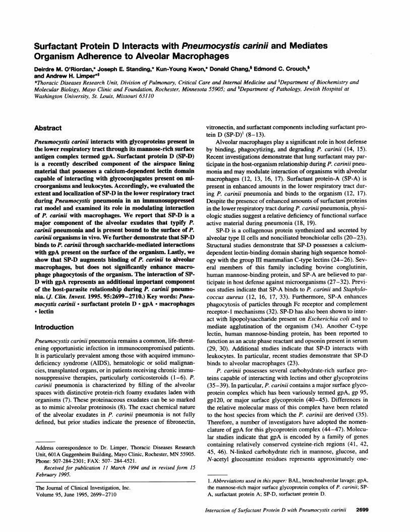
SP-D accumulates in the lung during P. carinii pneumonia. Todetermine whether SP-D participates in the pulmonary responseto P. carinii, we evaluated the extent and tissue distribution ofthis surfactant-associated glycoprotein by immunohistochemis-try using a rabbit polyclonal SP-D antibody (Fig. 1). Lungsamples were obtained from rats with P. carinii pneumonia 6-8 wk after inoculation. Abundant immunoreactive SP-D wasobserved in the proteinaceous exudates filling the alveolarspaces. Additional staining was noted in bronchiolar and typeII alveolar epithelial cells, and occasional cytoplasmic stainingwas also observed in macrophages. These exudates containednumerous P. carinii organisms as detected by silver staining.Examination of identical lung fields in semiserial sections withnonimmune IgG failed to demonstrate any staining, therebyconfirming the specificity of the SP-D antibody. Immunostain-ing of normal rat lung with SP-D antibody revealed reactivityof bronchiolar and alveolar type H epithelial cells and occasionalcytoplasmic staining of macrophages (67).
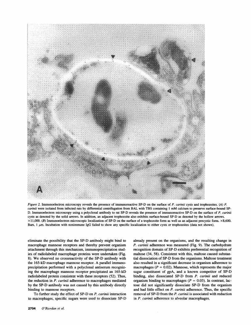
SP-D is present on the surface of P. carinii. P. cariniiexhibits a complex life cycle alternating between cystic formsand trophozoites (7). Accordingly, immunoelectron microscopywas performed to determine whether SP-D binds to the surfaceof these various P. carinii forms (Fig. 2). P. carinii were isolatedfrom BAL performed with TBS containing 1 mMcalcium topreserve surface-bound SP-D. Polyclonal rabbit antibody to ratSP-D interacted specifically with the surface of P. carinii cysts,precysts, and trophozoites, thus indicating that SP-D is presenton the surface of all forms of the organisms. Although immuno-
electron microscopy is not rigorously quantitative, relativegreater abundance of SP-D was found on the surface of P.carinii cysts compared with other forms. Parallel studies withnonimmune IgG exhibited no specific localization to either cystsor trophozoites.
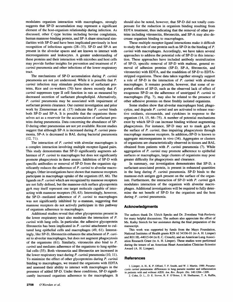
SP-D increases in BAL during the development of P. cariniipneumonia. Wenext sought to evaluate the time course overwhich SP-D accumulates during the development of pneumonia(Fig. 3). Rats were immunosuppressed with dexamethasone andinoculated with P. carinii. Since corticosteroid treatment itselfis known to increase expression of surfactant components, con-trol rats were also immunosuppressed with dexamethasone, butin addition received trimethoprim/sulfamethoxazole prophy-laxis to prevent development of pneumonia. No control animalsshowed evidence of P. carinii pneumonia during the study. At2-wk intervals, rats were killed, lavaged with HBSS, and solubleSP-D in the cell-free lavage supernatants was assessed by immu-noblotting. The amounts of lavage fluid recovered were equiva-lent between P. carinii and control rats. Total BAL proteinsare known to increase during the development of P. cariniipneumonia, likely reflecting alveolar capillary leak (10, 11).However, since SP-D is produced locally in the lung, any in-crease in total recoverable SP-D reflects enhanced local accu-mulation of this protein. Equal volumes of lavage fluid wererecovered from control rats and animals with P. carinii and aretherefore reflective of the total soluble SP-D present in thealveolar lining fluid (68). Accordingly, equal volumes of BALfrom controls and animals with P. carinii were concentratedand analyzed by SDS-PAGEand immunoblotting. After 4 wkof immunosuppression, enhanced recoverable SP-D was seenin the rats with P. carinii pneumonia compared with controls(Fig. 3). Scanning densitometry of the immunoblots at the 4-wk time point demonstrated a 170.4±17.1% increase in SP-Din rats with P. carinii pneumonia compared with controls (P= 0.05). After 8 wk of P. carinii pneumonia an 266.1±83.3%increase was observed (P = 0.04). It should be further notedthat we measured soluble SP-D present in the lavage supernatantafter a 400 g centrifugation. Even at these relatively low forces,a small amount of SP-D is lost in the cellular pellet. Therefore,the soluble SP-D determinations likely underestimate the totalSP-D present in the lung. Nevertheless, we observed significantenhancement of the relative SP-D present in the rats with P.carinii pneumonia compared with the steroid-treated controlsat these time points.
SP-D interacts with P. carinii through saccharide-mediatedmechanisms. To further determine the mechanisms by whichSP-D interacts with P. carinii organisms, we next performedimmunoblot analyses of whole organism extracts obtained fromfreshly purified P. carinii (Fig. 4). Organisms were obtainedfrom rats by lavage in TBS containing 1 mMcalcium to pre-serve surface-bound SP-D. The organisms were lysed, and theproteins were separated by SDS-PAGEand blotted with anti-body recognizing SP-D. Extracts from freshly isolated P. cariniicontained abundant immunoreactive SP-D indicating that SP-D is bound to these organisms in vivo (Fig. 4, lane A). Incuba-tion of equal numbers of P. carinii with glucose, mannose, or
EDTAbefore lysis and immunoblotting resulted in substantialremoval of SP-D associated with the organisms. Incubation ofP. carinii with glucose or mannose (100 mM) as competingsugars removed 59 or 47% of the SP-D, respectively. EDTA(20 mM) resulted in removal of 70% of the SP-D from P.carinii. Taken together, a significant fraction of SP-D's interac-
2702 O'Riordan et al.
?6 5'1iX ifr3fu voAnJ . -e, -,' \r~~~~~
Figure 1. Immunohistochemical localization of SP-D during P. carinii pneumonia in the rat. (A) Lung tissue obtained from rat with P. cariniipneumonia 8 wk after inoculation. Marked immunoreactive SP-D (reddish-brown pigment) is observed in the proteinaceous exudates filling thealveolar spaces, as detected by a rabbit polyclonal SP-D antibody. x 1,000. (B) Semiserial section of the identical lung field shows no reactivity ofthe intraalveolar exudates with nonimmune rabbit immunoglobulin verifying the specificity of the staining for SP-D. (C) Methenamine silver stainof this lung field demonstrates numerous P. carinii cysts. (D) Staining of normal rat lung with the rabbit antibody recognizing SP-D reveals reactivityof bronchiolar epithelial cells and type II cells. X 1,000.
tion with P. carinii is mediated by C-type lectin binding. Itshould be noted that a component of the SP-D associated withP. carinii was not displaceable by such manipulations. Simi-larly, prior studies by Lee et al. (69) indicate that the bindingof other multimeric C-type lectins is not totally reversible withsmall competitive ligands.
SP-D binds to gpA on ligand blot analysis. Next, lectinblot studies were performed to define which molecule(s) on thesurface of P. carinii interacts with SP-D (Fig. 5). Purified P.carinii were extracted and separated by SDS-PAGE. Coomassieblue staining of P. carinii extracts demonstrated a prominentband migrating at 120 kD with additional components between40 and 70 kD. This 120-kD band was identified as gpA byblotting with monoclonal antibody 5E12 which recognizes thismajor surface antigen of P. carinii (51). Incubating the mem-branes with '251-SP-D demonstrated predominantly interactionof the surfactant protein primarily with gpA. Again, EDTA(20mM)and glucose (100 mM)effectively inhibited the interactionof SP-D with the 120-kD molecule during lectin blotting. Thesefindings demonstrate that SP-D interacts with gpA present onP. carinii, through lectin-mediated binding.
Interaction of SP-D with purified gpA. To further character-ize the interaction of SP-D with gpA, the gpA glycoproteincomplex was purified with preparative gel electrophoresis. 12511SP-D exhibited concentration-dependent binding to immobi-lized gpA (Fig. 6). The binding of SP-D to gpA was significantlyinhibited by both glucose and mannose (100 mMeach) (P< 0.05 compared with control). In contrast, lactose causedlesser inhibition in 1251-SP-D binding to gpA. This pattern ofsugar inhibition is consistent with the interaction of SP-D'scarbohydrate recognition domain with carbohydrate moieties ongpA (24, 34, 58). As a further confirmation of specificity, '"I-
ISP-D (1 1sg/ml) binding to gpA immobilized on plastic wasalso studied in the presence of soluble gpA (500 jig/ml). Thebinding of 1251-SP-D to immobilized gpA was inhibited 60±5%by the presence of soluble gpA (P = 0.003). Thus, soluble gpAcan compete with immobilized ligand during this interaction.
SP-D enhances the interaction of P. carinii with alveolarmacrophages. After demonstrating that SP-D is bound to freshlyisolated P. carinii, we next sought to determine whether SP-Dmodulates the adherence of P. carinii to macrophages (Fig. 7).Freshly isolated P. carinii possessing surface-associated SP-Dwere radiolabeled with 51Cr and permitted to bind to alveolarmacrophages in the presence of no added antibody, or withantibody to SP-D or nonimmune immunoglobulins. Incubationof the radiolabeled P. carinii with surface-associated SP-D inthe presence of antibody recognizing SP-D resulted in signifi-cant reduction in P. carinii adherence to macrophages (asteriskin Fig. 7 denotes P = 0.0004 compared with control). In con-trast, incubation in the presence of nonimmune immunoglobulindid not alter adherence of the radiolabeled P. carinii to macro-phages. To further confirm the specificity of the anti-SP-Dantibody in these studies, parallel assays were performed inwhich P. carinii binding was evaluated in the presence of bothanti-SP-D and purified SP-D (5 jig/ml) (Fig. 7). Addition ofSP-D resulted in neutralization of the antibody, and P. cariniiadherence was returned to control levels. Of interest, the addi-tion of purified SP-D alone (5 jig/ml) resulted in little changein organism adherence to macrophages.
Since SP-D shares sequence homology with other C-typelectins, it is possible that the polyclonal antibodies recognizingSP-D may also crossreact with macrophage mannose receptors,another C-type lectin. Recent studies indicate that macrophagemannose receptors can mediate P. carinii uptake (65, 66). To
Interaction of Surfactant Protein D with Pneumocystis carinii 2703
...... A -.6 --,TV Z-
i_, _,..I. I,.
V 4
!,..t
A>.
-- ::. sumw v. In
X Mv
It.... A....
......
__
., :.:. .,:
be ., ;::' :: A,.: . :
4,.o . ce.
A imI-IN:M_w
Figure 2. Immunoelectron microscopy reveals the presence of immunoreactive SP-D on the surface of P. carinji cysts and trophozoites. (A) P.carinji were isolated from infected rats by differential centrifugation from BAL with TBS containing 1 mMcalcium to preserve surface-bound SP-D. Immunoelectron microscopy using a polyclonal antibody to rat SP-D reveals the presence of immunoreactive SP-D on the surface of P. carinjicysts as denoted by the solid arrows. In addition, an adjacent trophozoite also exhibits surface-bound SP-D as denoted by the hollow arrows.x l 1,000. (B) Immunoelectron microscopic localization of SP-D on the surface of a trophozoite form as well as an adjacent precystic form. x8,600.Bars, 1 Jtm. Incubation with nonimmune IgG failed to show any specific localization to either cysts or trophozoites (data not shown).
eliminate the possibility that the SP-D antibody might bind tomacrophage mannose receptors and thereby prevent organismattachment through this mechanism, immunoprecipitation stud-ies of radiolabeled macrophage proteins were undertaken (Fig.8). Weobserved no crossreactivity of the SP-D antibody withthe 165-kD macrophage mannose receptor. A parallel immuno-precipitation performed with a polyclonal antiserum recogniz-ing the macrophage mannose receptor precipitated an 165-kDradiolabeled protein consistent with these receptors (52). Thus,the reduction in P. carinji adherence to macrophages mediatedby the SP-D antibody was not caused by this antibody directlybinding to mannose receptors.
To further study the effect of SP-D on P. carinii interactionto macrophages, specific sugars were used to dissociate SP-D
already present on the organisms, and the resulting change inP. carinii adherence was measured (Fig. 9). The carbohydraterecognition domain of SP-D exhibits preferential recognition ofmaltose (34, 58). Consistent with this, maltose caused substan-tial dissociation of SP-D from the organisms. Maltose treatmentalso resulted in a significant decrease in organism adherence tomacrophages (P = 0.02). Mannose, which represents the majorsugar constituent of gpA, and a known competitor of SP-Dbinding, also dissociated SP-D from P. carinii and reducedorganism binding to macrophages (P = 0.03). In contrast, lac-tose did not significantly dissociate SP-D from the organismand had little effect on P. carinii adherence. Thus, the specificremoval of SP-D from the P. carinii is associated with reductionin P. carinji adherence to alveolar macrophages.
2704 O'Riordan et al.
A s:'.,:
.v-.:WI:,i:;, '; f:'-:...''n ":
-i %-4
q .. I.,
.A
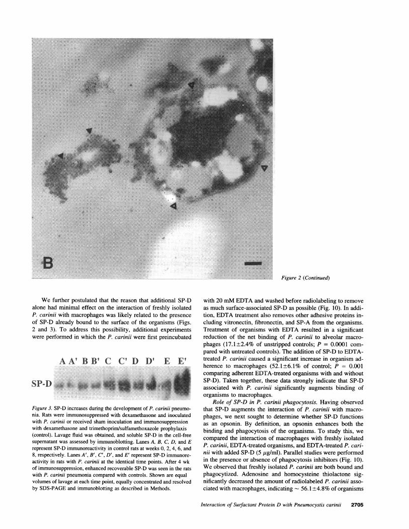
6Figure 2 (Continued)
We further postulated that the reason that additional SP-Dalone had minimal effect on the interaction of freshly isolatedP. carinii with macrophages was likely related to the presenceof SP-D already bound to the surface of the organisms (Figs.2 and 3). To address this possibility, additional experimentswere performed in which the P. carindi were first preincubated
AA' BB' C C' D D' F EE
SP-D W4**'U as
Figure 3. SP-D increases during the development of P. carinji pneumo-nia. Rats were immunosuppressed with dexamethasone and inoculatedwith P. carinii or received sham inoculation and immunosuppressionwith dexamethasone and trimethoprim/sulfamethoxazole prophylaxis(control). Lavage fluid was obtained, and soluble SP-D in the cell-freesupernatant was assessed by immunoblotting. Lanes A, B, C, D, and Erepresent SP-D immunoreactivity in control rats at weeks 0, 2, 4, 6, and8, respectively. Lanes A', B', C', D', and E' represent SP-D immunore-activity in rats with P. carinii at the identical time points. After 4 wkof immunosuppression, enhanced recoverable SP-D was seen in the ratswith P. carinii pneumonia compared with controls. Shown are equalvolumes of lavage at each time point, equally concentrated and resolvedby SDS-PAGEand immunoblotting as described in Methods.
with 20 mMEDTAand washed before radiolabeling to removeas much surface-associated SP-D as possible (Fig. 10). In addi-tion, EDTAtreatment also removes other adhesive proteins in-cluding vitronectin, fibronectin, and SP-A from the organisms.Treatment of organisms with EDTA resulted in a significantreduction of the net binding of P. carinii to alveolar macro-phages (17.1±2.4% of unstripped controls; P = 0.0001 com-pared with untreated controls). The addition of SP-D to EDTA-treated P. carinii caused a significant increase in organism ad-herence to macrophages (52.1±6.1% of control; P = 0.001comparing adherent EDTA-treated organisms with and withoutSP-D). Taken together, these data strongly indicate that SP-Dassociated with P. carinii significantly augments binding oforganisms to macrophages.
Role of SP-D in P. carinii phagocytosis. Having observedthat SP-D augments the interaction of P. carinii with macro-phages, we next sought to determine whether SP-D functionsas an opsonin. By definition, an opsonin enhances both thebinding and phagocytosis of the organisms. To study this, wecompared the interaction of macrophages with freshly isolatedP. carinii, EDTA-treated organisms, and EDTA-treated P. cari-nii with added SP-D (5 ug/ml). Parallel studies were performedin the presence or absence of phagocytosis inhibitors (Fig. 10).Weobserved that freshly isolated P. catinii are both bound andphagocytized. Adenosine and homocysteine thiolactone sig-nificantly decreased the amount of radiolabeled P. carinii asso-ciated with macrophages, indicating - 56. 1+4.8% of organisms
Interaction of Surfactant Protein D with Pneumocystis carinii 2705

A B C D E Figure 4. SP-D binds toP. carinii through sac-
.4 w wcharide-dependent mech-90t o f ;3;3; ;jj anisms. (Lane A) P. cari-
_~ "^ nii were isolated fromrats by lavage and puri-fied as described and
A 5 _ _ _ washed once in TBS con-
45J - taining 1 mMcalcium.The organisms (10x 106) were lysed in 2%SDS with 4% /3-mercap-
toethanol, separated with SDS-PAGE, transferred to nitrocellulose, andblotted with an antibody to SP-D (1:1,000 dilution). Freshly isolated P.carinii possess abundant immunoreactive SP-D bound to the surface ofthe organism. Monomeric SP-D migrates at 43 kD, the higher molecularmass bands represent dimeric SP-D (- 90 kD) and higher molecularmass multimers. (Lane B) SP-D purified from silicotic rats (10 ng)migrated in an identical fashion to the SP-D present on P. carinii. (LaneC) SP-D was partially removed from an equal number of P. carinii bywashing in glucose (100 mM). (Lane D) Similarly, incubation of anequal number of P. carinii with mannose (100 mM) results in loss ofSP-D from the organism. (Lane E) Incubation of an identical numberof P. carinii with EDTA (20 mM) resulted in substantial removal ofSP-D from the organisms.
were phagocytized during these assays (P = 0.0003 comparingfreshly isolated P. carinii in the presence and absence of phago-cytosis inhibitors). Treatment of the organisms with EDTAde-creased P. carinii interaction with macrophages, and the reinsti-tution of SP-D significantly enhanced P. carinii binding. Ofinterest, however, SP-D did not result in significant potentiationof P. carinii phagocytosis (P = 0.07 comparing the interactionof EDTA-treated P. carinii and added SP-D with macrophagesin the presence and absence of inhibitors). These data indicatethat, although SP-D promotes the binding of P. carinii to macro-phages, it does not act as a potent opsonin for organism uptake.
Role of mannose receptors in P. carinii adherence to alveo-lar macrophages. Recent studies indicate that mannose recep-
A BCDE
97.5
69
30
Figure 5. SP-D interactswith P. carinii gpA on li-gand blot analysis. Puri-fied P. carinii were ex-tracted in 2% SDS con-taining 4% /3-mercaptoethanol, and theextracted componentswere separated by SDS-PAGEand stained ortransferred to nitrocellu-lose. (Lane A) Coomassieblue staining of the sepa-rated P. carinii extractdemonstrates a promi-nent band at 120 kD. Ad-
ditional components between 40 and 70 kD are also visible. (Lane B)Radiolabeled SP-D binds to the 120-kD material, with minimal reactiv-ity to other P. carinii components. (Lane C) EDTA (20 mM) inhibitsthe interaction of SP-D with the 120-kD material. (Lane D) Glucose(100 mM) inhibits SP-D binding to P. carinii components. (Lane E)Immunoblotting of P. carinii extracts with mAb5E12 identifies the120-kD material as the major surface glycoprotein of P. carinii, termedgpA.
E 5,000 -c
X 4,000 *Guo-C * Mans
3,000
OL2,00
0.
Vc 1,000
0.0 0.5 1.0 1.5 2.0 2.5 3.0 3.5
SP-D added (ng)
Figure 6. Radiolabeled SP-D binds to purified gpA. The mannose-richsurface glycoprotein complex of P. carinii known as gpA was purifiedby preparative polyacrylamide electrophoresis and solid-phase ligand-binding assays performed as described in Methods. '25I-SP-D exhibiteddose-dependent binding to immobilized gpA. This interaction was sig-nificantly inhibited by glucose and mannose. Lactose caused much lessinhibition of binding. These findings are consistent with gpA bindingto the carbohydrate recognition domain of SP-D. Shown are mean±SEMfrom three determinations.
tors participate in macrophage uptake of P. carinii (65, 66).These investigations demonstrate that P. carinii interaction withmacrophages can be inhibited by yeast a-mannan, a solubleinhibitor of mannose receptors (65, 66). To study the role ofmannose receptors in the interactions of P. carinii-associatedSP-D and macrophages, additional P. carinii adherence assayswere performed in the presence or absence of a-mannan (100jtg/ml). Consistent with our previous studies, we observed thatthe adherence of freshly isolated P. carinii was significantlyinhibited by a-mannan. Freshly isolated P. carinii exhibited
Nonimmune
AntiSP-D
AnU.SP-D+ SPD
SP-DI
0 25 50 75 100 125P. cwiuti binding (% cono)
Figure 7. Role of SP-D in the adherence of P. carinii to alveolar macro-phages. 51Cr-labeled P. carinii were permitted to bind to confluent mono-layers of alveolar macrophages plated on nonimmune IgG over 1 h inDMEcontaining BSA (1 mg/ml) in the presence of no additional SP-D or antibody (Control) or in the presence of nonimmune IgG (100 Mg/ml) or with the polyclonal antibody recognizing SP-D (100 Mg/ml). Thespecific antibody to SP-D but not the nonimmune IgG resulted in sig-nificant reduction in P. carinii adherence to macrophages (*P = 0.0004compared with control). To further assess the specificity of this antibodyinteraction in parallel assays, binding was studied in the presence ofboth anti-SP-D and purified SP-D (5 jg/ml). The addition of exogenousSP-D resulted in neutralization of the antibody, and P. carinii adherencewas not significantly different from control. Interestingly, the additionof SP-D alone (5 Mg/ml) resulted in little change in organism adherenceto alveolar macrophages. Shown are the mean±SEMfrom six determi-nations.
2706 O'Riordan et al.

200f
97.5-
69-
45-i
A B C D E Untreated PC(control)
EDTA-treatedPC
EDTA-treatedPC + SPD
0
Figure 8. Immunoprecipitation verifies that the anti-SP-D antibodydoes not crossreact with macrophage mannose receptors. To determinewhether the anti-SP-D antibody crossreacted with macrophage mannosereceptors, another C-type lectin, immunoprecipitation studies were un-dertaken. Alveolar macrophages were isolated and radiolabeled as de-scribed in Methods. (Lane A) Total membrane-associated "S-labeledalveolar macrophage proteins. (Lane B) Nonimmune rabbit IgG (100jig/ml) does not precipitate any macrophage proteins. (Lane C) Likewisetreatment of the radiolabeled extracts with the SP-D antibody (100 jg!ml) also did not immunoprecipitate any macrophage proteins. (Lane D)Nonimmune goat serum (1:100 dilution) also yielded no precipitationproduct. (Lane E) In contrast, a polyclonal goat antiserum recognizingmacrophage mannose receptors (1:100 dilution) specifically precipitateda 165-kD protein consistent with macrophage mannose receptors. Notespecifically that the SP-D antibody (see lane C) did not crossreact withthese 165-kD macrophage mannose receptors.
26.0±6.7% adherence in the presence of a-mannan, comparedwith 100.0±5.4% maximal adherence in controls (P = 0.0001).Weadditionally observed that the residual adherence of EDTA-stripped P. carinii was dramatically reduced by similar concen-trations of a-mannan. In these experiments, the adherence of
U Qphagocytosis inhibitors
-- * ( phagocytosis inhibitors
20 40 60 80 100 120
P. carinhi binding (% control)
Figure 10. Role of SP-D in macrophage phagocytosis of P. carinii. Toevaluate the potential role of SP-D in the binding and phagocytosis ofP. carinii by macrophages, freshly isolated P. carinii were treated withEDTA to remove surface-associated adhesive proteins. The adherenceof 5tCr-labeled EDTA-treated P. carinii was assessed in the presenceor absence of SP-D (5 jig/ml) and in the presence or absence of thepotent inhibitors of phagocytosis, adenosine (100 AM) and homocysteinethiolactone (250 jAM). Treatment of P. carinii with EDTAsignificantlydecreased organism interaction with macrophages. The subsequent addi-tion of SP-D significantly enhanced P. carinii binding to macrophages.Interestingly, however, SP-D did not result in significant potentiationof organism phagocytosis. Shown are mean±SEMfrom five determina-tions.
EDTA-treated P. carinii to macrophages was reduced from13.7± 1.0 to 5.3±1.0% by a-mannan (P = 0.0002). Finally, weassessed whether mannose receptor inhibition would alter theSP-D-mediated adherence of P. carindi to macrophages. Al-though the adherence of EDTA-treated P. carinii with addedSP-D (5 jig/ml) was somewhat reduced from 43.9±9.7 to29.4±9.0% by the presence of a-mannan (100 jtg/ml), this didnot reach statistical significance (P = 0.33). These data confirmthe previous findings by ourselves and others that mannosereceptors do participate in P. carini adherence to macrophages.However, our studies further indicate that mannose receptorinhibition does not significantly effect SP-D-mediated bindingof P. carinii to alveolar macrophages.
120 Figure 9. Removal ofSP-D from P. carinji is
100 associated with de-g = | S S S | creased organism adher-c 0 * ence to macrophages. To
further evaluate the role0 .60C0 ~~~~~~~~~ofSP-D in adherence of
P. carinXi to alveolarmacrophages, freshly
20 isolated organisms wereincubated with specific
Cnol sugar ligands to removeControl Maltose Mannose Lactose SP-D from the organ-
isms. The adherence ofL~E _ _ .treated organisms to al-
Ce _ S:- W veolar macrophages was
assayed. In parallel, SP-D released into the media was assessed by immunoblotting. Both malt-ose and mannose caused the release of SP-D from the organisms andsignificantly decreased adherence of P. carinii to macrophages. In con-trast, treatment of P. carinii with lactose, which exhibits less interactionwith SP-D, caused minimal release of SP-D and did not significantlyalter P. carinii adherence to macrophages. Shown are mean±SEMfromfive determinations.
Discussion
This investigation demonstrates that SP-D, a component of theairspace lining material, interacts with gpA, a mannose-richglycoconjugate present on the surface of P. carinii. In addition,we demonstrated that enhanced quantities of SP-D are presentin the lower respiratory during the development of P. cariniipneumonia and that SP-D is bound to the surface of freshlyisolated P. carinii organisms. SP-D's interaction with P. cariniiis inhibited by glucose, mannose, maltose, and EDTA, consis-tent with binding by the carbohydrate-recognition domain ofSP-D. SP-D significantly participates in the adherence of P.carinii to alveolar macrophages. Removal of SP-D with EDTAor specific sugar inhibitors, or treatment of P. carinii with apolyclonal antibody to SP-D which recognizes epitopes in thecarbohydrate recognition domain, significantly reduces P. cari-nii adherence to alveolar macrophages. Interestingly, althoughSP-D augments the binding of P. carinii to macrophages, SP-D does not greatly enhance phagocytosis of the organism.
The presence of abundant SP-D in the lung during P. cariniipneumonia, coupled with the demonstration that this protein
Interaction of Surfactant Protein D with Pneumocystis carinii 2707
modulates organism interaction with macrophages, stronglysuggests that SP-D accumulation may represent a significantelement of the host-organism relationship during infection. Asdiscussed, other C-type lectins including bovine conglutinin,human mannose-binding protein, and SP-A share structural sim-ilarities with SP-D and have been implicated previously in hostrecognition of infectious agents (28-33). SP-D and SP-A arepresent in the alveolar spaces and are known to interact withmicroorganisms and leukocytes. A greater understanding ofthese proteins and their interaction with microbes and host cellsmay provide further insights for prevention and treatment of P.carinji pneumonia and other infections of the lower respiratorytract.
The mechanisms of SP-D accumulation during P. carinipneumonia are not yet understood. While it is possible that P.carinii infection may stimulate production of surfactant pro-teins, Rice and co-workers (70) have shown recently that P.carinii suppresses type II cell function in rats as measured bydecreased secretion of surfactant phospholipid. Alternatively,P. carinii pneumonia may be associated with impairment ofsurfactant protein clearance. Our current investigation and priorwork by Zimmerman et al. (17) indicate that P. carinii bindsboth SP-D and SP-A. It is possible that the organisms them-selves act as a reservoir for the accumulation of surfactant pro-teins during pneumonia. Data concerning the abundance of SP-D during other pneumonias are not yet available. Recent studiessuggest that although SP-A is increased during P. carinii pneu-monia, SP-A is decreased in BAL during bacterial pneumonia(12, 71).
The interaction of P. carinji with alveolar macrophages isa complex interaction involving multiple receptor-ligand pairs.This study demonstrates that SP-D significantly augments ad-herence of P. carinji to macrophages but does not significantlypromote phagocytosis in these assays. Inhibition of SP-D withspecific antibodies or removal of SP-D from the organism sig-nificantly reduces the adherence of P. carinii to alveolar macro-phages. Other investigations have shown that mannose receptorsparticipate in macrophage uptake of the organism (65, 66). Theligands on P. carindi which are recognized by mannose receptorsare not fully defined, but the mannose-rich surface glycoproteingpA may itself represent one target molecule capable of inter-acting with mannose receptors (50, 63). Interestingly, however,the SP-D-mediated adherence of P. carinji to macrophageswas not significantly inhibited by a-mannan, suggesting thatmannose receptors do not actively participate in this pathwayof organism adherence to macrophages.
Additional studies reveal that other glycoproteins present inthe lower respiratory tract also modulate the interaction of P.carindi with lung cells. In particular, the adhesive glycoproteinfibronectin has been implicated in P. carindi attachment to cul-tured lung epithelial cells and macrophages (49, 61). Interest-ingly, like SP-D, fibronectin enhances the attachment of P. cari-nii to alveolar macrophages, but does not augment phagocytosisof the organisms (61). Similarly, vitronectin also bind to P.carinji and mediates adherence of the organisms to lung epithe-lial cells (55). Both vitronectin and fibronectin are increased inthe lower respiratory tract during P. carini pneumonia (10, 11).To minimize the effect of other glycoproteins during P. cariniibinding to macrophages, we treated the organisms with EDTAand assessed their ability to interact with macrophages in thepresence of added SP-D. Under these conditions, SP-D signifi-cantly increased organism adherence to the macrophages. It
should also be noted, however, that SP-D did not totally com-pensate for the reduction in organism binding resulting fromEDTAtreatment, thus indicating that the removal of other pro-teins including vitronectin, fibronectin, and SP-A may also de-crease organism binding to macrophages.
These multiple receptor-ligand interactions make it difficultto study the role of one protein such as SP-D in the binding of P.carini with macrophages. Accordingly, we have taken severalapproaches to address the potential role of SP-D in this interac-tion. These approaches have included antibody neutralizationof SP-D, specific removal of SP-D with maltose, general re-moval of adhesion proteins (SP-D, SP-A, fibronectin, andvitronectin) with EDTA, and the readdition of SP-D to EDTA-stripped organisms. These data taken together strongly supporta role of SP-D in the interaction of P. carinji with alveolarmacrophages. It remains possible, however, that some of re-ported effects of SP-D, such as the observed lack of effect ofexogenous SP-D on the adherence of unstripped P. carinii tomacrophages (Fig. 7), may also be related to the presence ofother adhesive proteins on these freshly isolated organisms.
Some studies show that alveolar macrophages bind, phago-cytize, and degrade P. carinii and are activated to release reac-tive oxidants, eicosanoids, and cytokines in response to theorganism (14, 15, 66-75). A number of potential mechanismsexist by which SP-D can increase binding without augmentingphagocytosis. For instance, SP-D may act to mask gpA onthe surface of P. carindi, thus impairing phagocytosis throughmacrophage mannose receptors. In addition1>SP-D is known toaggregate microorganisms in vivo (34). Aggregates or clustersof organisms are characteristically observed in tissues and BALobtained from patients with P. carinji pneumonia (7). Whileaggregation of P. carinji may promote adherence to epithelialsurfaces or macrophages, large clusters of organisms may posegreater difficulty for phagocytosis and clearance.
In summary, our investigation demonstrates that SP-D, asurfactant-associated protein, is present in enhanced quantitiesin the lung during P. carinji pneumonia. SP-D binds to themannose-rich antigen gpA present on the surface of the organ-ism. Furthermore, the interaction of SP-D with P. carinii gpAmodulates interaction of the organism with alveolar macro-phages. Additional investigations will be required to fully deter-mine the net benefit of SP-D for the organism and the hostduring P. carinii pneumonia.
Acknowledgments
The authors thank Dr. Ulrich Specks and Dr. Zvezdana Vuk-Pavlovicfor many helpful discussions. The authors also appreciate the effort ofMs. Kathy Streich for her assistance during the final preparation of themanuscript.
This work was supported by funds from the Mayo Foundation,National Institutes of Health grants R29 AI 34336-01 (to A. H. Limper)and ROl HL-44015-04 (to E. C. Crouch), and an American Lung Associ-ation Research Grant (to A. H. Limper). These studies were performedduring the tenure of an American Heart Association Clinician-ScientistAward (A. H. Limper).
References
1. Limper, A. H., K. P. Offord, T. F. Smith, and W. J. Martin. 1989. Pneumo-cystis carinji pneumonia: differences in lung parasite number and inflammationin patients with and without AIDS. Am. Rev. Respir. Dis. 140:1204-1209.
2. Cohn, D. L., D. E. Stover, R. F. O'Brien, J. H. Shelhamer, T. A. Raffin,
2708 O'Riordan et al.

and P. C. Hopewell. 1988. Pulmonary complications of AIDS: advances in diagno-sis and treatment. Am. Rev. Respir. Dis. 135:1051-1052.
3. Murray, J. G., and J. Mills. 1990. Pulmonary infectious complications ofhuman immunodeficiency virus infection. Am. Rev. Respir. Dis. 141:1582-1598.
4. Sepkowitz, K. A., A. E. Brown, E. E. Telzak, S. Gottlieb, and D. Armstrong.1992. Pneumocystis carinji pneumonia among patients without AIDS in a cancerhospital. JAMA (J. Am. Med. Assoc.). 267:832-837.
5. Peters, S. G., and U. B. S. Prakash. 1987. Pneumocystis carinji pneumonia.Am. J. Med. 82:73-78.
6. Centers for Disease Control. 1989. Guidelines for prophylaxis against Pneu-mocystis carinji pneumonia for persons with human immunodeficiency virus infec-tion. MMWR(Morb. Mortal. Wkly. Rep.). 38:1-8.
7. Limper, A. H. 1991. Parasite adherence and host responses in the develop-ment of Pneumocystis carinii pneumonia. Sem. Respir. Infect. 6:19-26.
8. Tran Van Nheiu, J., A. M. Vojtek, J. F. Bernaudin, E. Escudier, and J.Fleury-Feith. 1990. Pulmonary alveolar proteinosis associated with Pneumocystiscarinji. Ultrastructural identification in bronchoalveolar lavage in AIDS and im-munocompromised non-AIDS patients. Chest. 98:801-805.
9. Crouch, E., A. Persson, and D. Chang. 1993. Accumulation of surfactantprotein D in human pulmonary alveolar proteinosis. Am. J. Pathol. 142:241-248.
10. Neese, L. W., and A. H. Limper. 1993. Vitronectin is increased in thelower respiratory tract of patients with P. carinji pneumonia. Chest. 104:16S.(Abstr.)
11. Neese, L. W., J. E. Standing, E. J. Olson, M. Castro, A. H. Limper. 1994.The role of vitronectin, fibronectin, and immune opsonization in augmentingtumor necrosis factor-a release during Pneumocystis carinji pneumonia. J. Immu-nol. 152:4549-4556.
12. Phelps, D. S., and R. M. Rose. 1991. Increased recovery of surfactantprotein A in AIDS-related pneumonia. Am. Rev. Respir. Dis. 143:1072-1075.
13. O'Riordan, D. M., J. E. Standing, E. C. Crouch, and A. H. Limper. 1993.Surfactant protein D interacts with Pneumocystis carinii. Am. Rev. Respir. Dis.147:33a. (Abstr.)
14. Von Behren, L. A., and E. L. Pesanti. 1978. Uptake and degradation ofPneumocystis carinji by macrophages in vitro. Rev. Respir. Dis. 118:1051-1059.
15. Masur, H., and T. C. Jones. 1978. The interaction in vitro of Pneumocystiscarinii with macrophages and L-cells. J. Exp. Med. 147:157-170.
16. Koziel, H., D. O'Riordan, D. Phelps, J. A. Fishman, M. Y. K. Armstrong,F. F. Richards, and R. M. Rose. 1992. Surfactant protein A inhibits binding andinternalization of Pneumocystis carinii by alveolar macrophages. Am. Rev. Respir.Dis. 145:247a. (Abstr.)
17. Zimmerman, P. E., D. R. Voelker, F. X. McCormack, J. R. Paulsrud, andW. J. Martin.1992. 120-kD surface glycoprotein of Pneumocystis carinii is aligand for surfactant protein A. J. Clin. Invest. 89:143-149.
18. Hoffman, A. G., M. G. Lawrence, F. P. Ognibene, A. F. Suffredini, G. Y.Lipschik, J. A. Kovacs, H. Masur, and J. H. Shelhamer. 1992. Reduction ofpulmonary surfactant in patients with human immunodeficiency virus infectionand Pneumocystis carinji pneumonia. Chest. 102:1730-1736.
19. Stokes, D. C., W. T. Hughes, P. 0. Alderson, R. E. King, and D. J.Garfinkel. 1986. Lung mechanics, radiography and 67Ga scintigraphy in experi-mental Pneumocystis carinji pneumonia. Br. J. Exp. Pathol. 67:383-393.
20. Persson, A., K. Rust, D. Chang, M. Moxley, W. Longmore, and E. Crouch.1988. CP4: a pneumocyte-derived collagenous surfactant-associated protein. Evi-dence for heterogeneity of collagenous surfactant proteins. Biochemistry.27:8576-8584.
21. Persson, A., D. Chang, K. Rust, M. Moxley, W. Longmore, and E. Crouch.1989. Purification and biochemical characterization of CP4 (SP-D): a collagenoussurfactant-associated protein. Biochemistry. 27:6361-6367.
22. Persson, A., D. Chang, and E. Crouch. 1990. Surfactant protein D (SP-D) is a divalent cation-dependent carbohydrate binding protein. J. Bio. Chem.265:5755-5760.
23. Voorhout, W. F., T. Veenendaal, Y. Kuroki, Y. Ogasawara, L. M. van-Golde, and H. J. Geuze. 1992. Immunocytochemical localization of surfactantprotein D (SP-D) in type II cells, Clara cells, and alveolar macrophages of ratlung. J. Histochem. Cytochem. 40:1589-1597.
24. Rust, R., L. Grosso, V. Zhang, D. Chang, A. Persson, W. Longmore, G.Kai, and E. Crouch. 1991. Human surfactant protein D: SP-D contains a C-typelectin carbohydrate recognition domain. Arch. Biochem. Biophys. 290:116-126.
25. Wiedemann, H., U. Holmskov, S. Theil, R. Timpl, and K. B. Reid. 1993.Structural similarity between lung surfactant protein D and conglutinin. Twodistinct, C-type lectins containing collagen-like sequences. Eur. J. Biochem.215:793-799.
26. Theil, S., and R. B. M. Reid. 1989, Structures and functions associatedwith the group of mammalian lectins containing collagen-like sequences. FEBS(Fed. Eur. Biochem. Soc.) Lett. 250:78-84.
27. Lee, Y. M., K. R. Leiby, J. Allar, K. Paris, B. Lerch, and T. B. Okarma.1991. Primary structure of bovine conglutinin, a member of the C type animallectin family. J. Biol. Chem. 266:2715-2723.
28. Friss-Christianson, P., 5. Theil, S. E. Svehag, R. Dessau, P. Svendsen, 0.Andersen, S. B. Laursen, and J. C. Jensenius. 1990. In vivo and in vitro antibacte-
rial activity of conglutinin, a mammalian plasma lectin. Scand. J. Immunol.31:453-460.
29. Ezekowitz, R. A., L. E. Day, and G. A. Herman. 1988. A human mannose-binding protein is an acute phase reactant that shares sequence homology withother vertebrate lectins. J. Exp. Med. 167:1034-1046.
30. Kuhlman, M., K. Joiner, and R. A. B. Ezekowitz. 1989. The humanmannose-binding protein functions as an opsonin. J. Exp. Med. 169:1733-1745.
31. Iwarden, F. W., T. Kawasaki, and I. Yamashina. 1989. A serum lectin(mannan-binding protein) has complement dependent bactericidal activity. J. Bio-chem. (Tokyo). 106:483-489.
32. Tenner, A. J., S. L. Robinson, J. Borchelt, and J. R. Wright. 1989. Humanpulmonary surfactant protein (SP-A), a protein structurally homologous to CIg,can enhance FcR- and CR1-mediated phagocytosis. J. BioL Chem. 264:13923-13928.
33. McNeely, T. B., and J. D. Coonrod. 1993. Comparison of the opsonicactivity of human surfactant protein A for Staphylococcus aureus and Streptococ-cus pneumonia with rabbit and human macrophages. J. Infect. Dis. 167:91-97.
34. Kuan, S. F., K. Rust, and E. Crouch. 1992. Interactions of surfactant proteinD with bacterial lipopolysaccharides. Surfactant protein D is an Escherichia coli-binding protein in bronchoalveolar lavage. J. Clin. Invest. 90:97-106.
35. Linke, M. J., M. T. Cushion, and P. D. Walzer. 1989. Properties of themajor antigens of rat and human Pneumocystis carinii. Infect. Immun. 57:1547-1555.
36. Radding, J. A., M. Y. K. Armstrong, E. Ullu, and F. F. Richards. 1989.Identification and isolation of a major cell surface glycoprotein of Pneumocystiscarinii. Infect. Immun. 57:2149-2157.
37. Pesanti, E. L,. and J. D. Shanley. 1988. Glycoproteins of Pneumocystiscarinii: characterization by electrophoresis and microscopy. J. Infect. Dis.158:1353-1359.
38. Lundgren, B., G. Y. Lipchik, and J. A. Kovacs. 1991. Purification andcharacterization of a major human Pneumocystis carinii surface antigen. J. Clin.Invest. 87:163-170.
39. Limper, A. H., S. T. Pottratz, and W. J. Martin. 1991. Modulation ofPneumocystis carinii adherence to cultured lung cells by a mannose-dependentmechanism. J. Lab. Clin. Med. 118:492-499.
40. Theus, S. A., M. J. Linke, R. P. Andrews, and P. D. Walzer. 1993.Proliferative and cytokine responses to a major surface glycoprotein of Pneumo-cystis carinii. Infect. Immun. 61:4703-4709.
41. Wada, M., K. Kitada, M. Saito, K. Egawa, and Y. Nakamura. 1993. cDNAsequence diversity and genomic clusters of major surface glycoprotein genes ofPneumocystis carinii. J. Infect. Dis. 168:979-985.
42. Kovacs, J. A., F. Powell, J. C. Edman, B. Lundgren, A. Martinez, B. Drew,and C. W. Angus. 1993. Multiple genes encode the major surface glycoprotein ofPneumocystis carinii. J. Biol. Chem. 268:6034-6040.
43. Gigliotti, F. 1992. Host species-specific variation of a mannosylated sur-face glycoprotein of Pneumocystis carinii. J. Infect. Dis. 165:329-336.
44. Haidaris P. J., T. W. Wright, F. Gigliotti, and C. G. Haidaris. 1992.Expression and characterization of a cDNA clone encoding an immunodominantsurface glycoprotein of Pneumocystis carinii. J. Infect. Dis. 166:1113-1123.
45. Wright, T. W., P. J. Simpson-Haidaris, F. Gigliotti, A. G. Harmsen, andC. G. Haidaris. 1994. Conserved sequence homology of cysteine-rich regions ingenes encoding glycoprotein A in Pneumocystis carinii derived from differenthost species. Infect. Immun. 62:1513-1519.
46. Stringer, S. L., T. Garbe, S. M. Suskin, and J. R. Stringer. 1993. Genesencoding antigenic surface glycoproteins in Pneumocystis carinii in humans. J.Eukaryotic Microbiol. 40:821-826.
47. Gigliotti, F., P. J. Haidaris, C. G. Haidaris, T. W. Wright, and K. R. Vander Meid. 1993. Further evidence of host species-specific variation in antigens ofPneumocystis carinii using the polymerase chain reaction. J. Infect. Dis. 168:191 -194.
48. Lundgren, B., G. Y. Lipchik, and J. A. Kovacs. 1991. Purification andcharacterization of a major human Pneumocystis carinii surface antigen. J. Clin.Invest. 87:163-170.
49. Pottratz, S. T., J. Paulsrud, J. Smith, and W. J. Martin. 1991. Pneumocystiscarinii attachment to cultured lung cells by gpl2O, a fibronectin binding protein.J. Clin. Invest. 88:403-407.
50. Limper, A. H., D. M. O'Riordan, and J. E. Standing. 1993. Pneumocystiscarinii gpl2O mediates organism attachment to alveolar macrophages but notrelease of tumor necrosis factor-a. Clin. Res. 41:680a. (Abstr.)
51. Gigliotti, F., D. C. Stokes, A. B. Cheatham, D. S. Davis, and W. T.Hughes. 1986. Development of murine monoclonal antibodies to Pneumocystiscarinii. J. Infect. Dis. 154:315-322.
52. Kery, V., J. J. Krepinsky, C. D. Warren, P. Capek, and P. D. Stahl. 1992.Ligand recognition of purified human mannose receptor. Arch. Biochem. Biophys.298:49-55.
53. Bartlett, M. S., J. A. Fishman, S. A. Queener, M. M. Durkin, M. A. Jay,and J. W. Smith. 1988. New rat model of Pneumocystis carinii pneumonia. J.Clin. Microbiol. 26:1100-1102.
54. Limper, A. H., and W. J. Martin. 1990. Pneumocystis carinii: Inhibitionof lung cell growth mediated by parasite attachment. J. Clin. Invest. 85:391-396.
Interaction of Surfactant Protein D with Pneumocystis carinii 2709
55. Limper, A. H., J. E. Standing, 0. A. Hoffman, M. Castro, and L. W.Neese. 1993. Vitronectin binds to Pneumocystis carini and mediates organismattachment to cultured lung epithelial cells. Infect. Immun. 61:4302-4309.
56. Limper, A. H., T. V. Colby, M. S. Sanders, S. Asakura, P. C. Roche, andR. A. DeRemee. 1994. Immunohistochemical localization of transforming growthfactor-fil in the non-necrotizing granulomas of pulmonary sarcoidosis. Am. J.Respir. Crit. Care Med. 149:197-204.
57. McClean, I. W., and P. K. Nakane. 1974. Periodate-lysine-paraformalde-hyde fixative. A new fixation for immunoelectron microscopy. J. Histochem.Cytochem. 22:1077-1083.
58. Crouch, E. C., A. Persson, D. Chang, and D. Parghi. 1991. Surfactantprotein D (SP-D): increased accumulation in silica-induced lipoproteinosis. Am.J. Pathol. 139:765-776.
59. Bolton, A. E., and W. M. Hunter. 1973. The labeling of proteins tohigh specific radioactivities by conjugation to a 125-I-containing acylating agent.Biochem. J. 133:529-539.
60. Pottratz, S. T., and W. J. Martin. 1990. Role of fibronectin in Pneunocystiscarinii attachment to cultured lung cells. J. Clin. Invest. 85:351-356.
61. Pottratz, S. T., and W. J. Martin. 1990. Mechanism of Pneumocystis carinjiattachment to cultured rat alveolar macrophages. J. Clin. Invest. 86:1678-1683.
62. Castro, M., T. I. Morgenthaler, 0. A. Hoffman, J. E. Standing, M. S.Rohrbach, and A. H. Limper. 1993. Pneumocystis carinji induces the release ofarachidonic acid and its metabolites from alveolar macrophages. Am. J. Respir.Cell Mol. Biol. 9:73-81.
63. O'Riordan, D. M., J. E. Standing, and A. H. Limper. 1995. Pneumocystiscarini glycoprotein A binds macrophage mannose receptors. Infect. Immun.63:779-784.
64. Sung, S. S. J., and S. C. Silverstein. 1985. Inhibition of macrophagephagocytosis by methylation inhibitors. J. Biol. Chem. 260:546-554.
65. Ezekowitz, R. A. B., D. J. Williams, H. Koziel, M. Y. K. Armstrong, A.Warner, F. F. Richards, and R. M. Rose. 1991. Uptake of Pneumocystis carinjimediated by the macrophage mannose receptor. Nature (Lond). 351:155-158.
66. Hoffman, 0. A., J. E. Standing, and A. H. Limper. 1993. Pneumocystiscarinji stimulates tumor necrosis factor-a release from alveolar macrophagesthrough a /3-glucan-mediated mechanism. J. ImmunoL 150:3932-3940.
67. Crouch, E., D. Parghi, S. F. Kuan, and A. Persson. 1992. Surfactantprotein D: subcellular localization in nonciliated bronchiolar epithelial cells. Am.J. PhysioL 263:L60-L66.
68. Limper, A. H., U. Specks, M. W. Brutinel, W. J. Martin II, and M. S.Rohrbach. 1993. Interlobar variations in the recovery of bronchoalveolar lavagefluid, cell populations, and angiotensin converting enzyme. J. Lab. Clin. Med.121:785-791.
69. Lee, R. T., Y. Ichikawa, T. Kowasaki, K. Drickamer, and Y. C. Lee. 1992.Multivalent ligand binding by serum mannose-binding protein. Arch. Biochem.Biophys. 298:129-136.
70. Rice, W. R., F. M. Singelton, M. J. Linke, and P. D. Walzer. 1993.Regulation of surfactant phosphatidylcholine secretion from alveolar type II cellsduring Pneumocystis carinii pneumonia in the rat. J. Clin. Invest. 92:2778-2782.
71. Baughman, R. P., R. I. Sternberg, W. Hull, J. A. Buchsbaum, and J.Whitsett. 1993. Decreased surfactant protein A in patients with bacterial pneumo-nia. Am. Rev. Respir. Dis. 147:653-657.
72. Hidalgo, H. A., R. J. Helmke, V. F. German, and J. A. Mangos. 1992.Pneumocystis carinji induces an oxidative burst in alveolar macrophages. Infect.Immun. 60:1-7.
73. Krishnan, V. L., A. Meager, D. M. Mitchell, and A. J. Pinching. 1990.Alveolar macrophages in AIDS patients: increased spontaneous tumour necrosisfactor-alpha production in Pneumocystis carinii pneumonia. Clin. Exp. ImmunoL80:156-160.
74. Chen, W., E. A. Havell, and A. G. Harmsen. 1992. Importance of tumornecrosis factor-alpha and gamma interferon in host resistance against Pneumo-cystis carinii infection. Infect. Immun. 60:1279-1284.
75. Kolls, J. K., J. M. Beck, S. Nelson, W. A. Summer, and J. Shellito.1993. Alveolar macrophage release of tumor necrosis factor-alpha during murinePneumocystis carinji pneumonia. Am. J. Respir. Cell Mol. BiW. 8:370-376.
2710 O'Riordan et al.