surgery the forefoot - bmj.com · in 400adthe rabbiushaiabegan a search for the seat ofthe soul....
TRANSCRIPT

276 BRITISH MEDICAL JOURNAL 29 JANUARY 1977
Bone and Joint Diseases
Surgery of the forefoot
B HELAL
British Medical Journal, 1977, 1, 276-280
The portion of the foot distal to the tarsus is the forefoot. It is a
small area packed with problems that often give rise to a degreeof misery and disability out of all proportion to the severity ofthe local condition. Kelikian' has written a 500-page monographon surgery of the forefoot. I can give only the broadest outlineof what I consider the best practice in surgical management.
Injury
Unusually, trauma does not play a large part in forefootsurgery. Most injuries are amenable to conservative management,and fatigue fracture (the march fracture of those unaccustomedto prolonged walking) of the metatarsals (fig 1) should beremembered when analysing forefoot pain. Foreign bodiesshould be removed under general anaesthesia with a tourniquetapplied and preferably with major theatre facilities and allpossible aids to location, such as an image intensifier and sonicdetector. Unnecessary physical injury to the patient andpsychological injury to the surgeon will thus be avoided.Subungual haematoma should be released by nail trephine,lacerations may require suture, and crushed devitalised toes or
forefoot may need excision. Unlike the hand, however, repair ofsevered tendons or nerves rarely seems necessary.
THE "OVERBONE"
The "overbone" is an annoying lump that lies on the dorsum of thefoot and commonly occurs in the young male. It abuts on the tongueof the male-pattern lace-up shoe and is caused by exostoses arisingfrom the dorsal margin of the first tarsometatarsal joint and mayacquire a "protective" adventitious bursa. Sometimes padding thetongue of the shoe with sponge rubber will allow the overlyingbursitis to settle and will relieve symptoms. If the exostoses are large,however, they are best excised together with the overlying bursa.
GANGLION
Ganglia may arise on the dorsum of the forefoot in relation to theextensor tendons and tarsometatarsal and metatarsophalangeal joints.Needle rupture should be tried, surgery being confined to the recurrenttroublesome ganglia.
HALLUX VALGUS AND BUNIONS (FIG 2)
The great toe drifts laterally at the metatarsophalangeal joint andalso rotates externally. The metatarsal head is "uncovered" medially,
and a bursa forms over the prominence of the metatarsal head thatmay become inflamed and occasionally becomes infected and dis-charges. This bursa is termed a bunion. The deformity presents in twoage groups-in the teens and in middle age. It is much more common
in the female. For the adolescent form alone over 130 differentoperations have been described.
FIG 1-Metatarsal stress fracture or "march fracture."
FIG 2-Hallux valgus.
An understanding of the pathological anatomy helps in devising a
rational approach to the problem. The condition gives rise to symptomsonly in the shod. Indeed, valgus of the great toe and the bunionformation, both of which are secondary phenomena, arise because offootwear. It is well known that external forces can alter the shape ofthe foot-witness the curious custom of the Chinese in binding thefoot to produce a small, high-arched foot that was apparently oferoticsignificanceL-a Far-eastern equivalent of "kinky boots." The forcesapplied, however, must be unremittent, and abandoning bindingwhile there is still growth potential in the foot will allow restorationto a normal shape.
In the hallux valgus deformity the basic "fault" is a separation ofthe first and second metatarsals-that is, a- varus deformity of the
London Hospital, Whitechapel El 1BB, and Enfield District Hospital,Enfield, Middx EN2 8JL
B HELAL, MCH(ORTH), FRCS, consultant orthopaedic surgeon
276 BRITISH MEDICAL JOURNAL 29 jANuARY 1977
on 14 August 2019 by guest. P
rotected by copyright.http://w
ww
.bmj.com
/B
r Med J: first published as 10.1136/bm
j.1.6056.276 on 29 January 1977. Dow
nloaded from

BRITISH MEDICAL JOURNAL 29 JANUARY 1977
first metatarsal. In the adolescent the metatarsus varus tendency isinherited3; in the older person it occurs as part of a general splayingof the metatarsals owing to loss of muscle tone, stretching of ligaments,and increase of stress on the foot caused by overweight (a form ofmiddle-aged foot spread). Once this has occurred, then the forcesacting across the metatarsophalangeal joint, principally the longtendons, are mechanically at an advantage to maintain the deformity.The condition of metatarsus primus varus is equally frequent in shodand unshod people.The bewildering number of operative options led me and my
colleagues to study the problem with special reference to adolescents.4The conclusions were that an osteotomy was the best procedure. Thisis carried out-at the level of the first metatarsal neck and designed soas to displace the head proximally, laterally, and plantarwards. Theadvantages are that the procedure is simple and does not interferewith the joint itself, which is usually painless and mobile, and remainsso. There is a minimum of soft-tissue dissection and a relaxation ofsoft tissues by the slight shortening that occurs, which helps to maintainmobility. The feature most valued by patients after freedom frompain is the ability to wear a fashionable shoe in comfort so that thisprocedure, which also narrows the forefoot, was found to be the mostsuccessful. In the adult type of hallux valgus the same procedure maybe used provided the joint is mobile and exhibits no signs of degenera-tion clinically or on x-ray examination.
Simple excision of the bunion and metatarsal head exostosis hasbeen widely practised but has proved only of temporary benefit; thevalgus deformity and degeneration of the joint is often accelerated bythe necessary encroachment on the joint and most come to further,more radical surgery. Gross deformation or arthritis of the joint is anindication for arthroplasty-that is, excision of the diseased joint withor without replacement by an artificial joint or "spacer." Keller5described an excision arthroplasty with removal ofthe proximal portionof the proximal phalanx of the great toe and Mayo6 described anexcision of the first metatarsal head. Both procedures produce reliefand mobility; the Mayo operation often has the edge cosmetically buthas to be carried out with more precision and skill to produce afunctional result comparable with the Keller procedure. There is botha functional and cosmetic improvement if the great toe can be keptout to length; also the sesamoids do not displace, the muscles maintaingood mechanical advantage, and there is less shift of stress on to thelateral metatarsals. A spacer of silicone rubber will achieve this effect.When there is a severe varus of the first metatarsal, an osteotomy ofthe metatarsal near its base and the insertion of a wedge of bone (themetatarsal head exostosis may be used for this) on the medial side willcorrect this and may be combined with an arthroplasty of the greattoe metatarsophalangeal joint. This procedure is especially appreciatedby the older woman who enjoys wearing an elegant shoe.7
HALLUX RIGIDUS
The great toe metatarsophalangeal joint is commonly rigid or verystiff and in some degree of fixed flexion. To compensate, the inter-phalangeal joint may adopt a position of hyperextension. The greattoe alignment is usually straight. The condition may arise secondaryto osteochondritis dissecans of the metatarsal head-a condition ofadolescence. There is infraction and sometimes separation of a wedgeof bone from the dome of the metatarsal head. The disease may alsoarise secondary to sesamometatarsal joint degeneration.
Arthrodesis or arthroplasty may be performed but arthroplasty isfavoured since both men and women's shoe styles may require altera-tion of the heel height that in turn necessitates a change of metatarso-phalangeal joint position. Simple excision arthroplasty may be
enhanced by suturing the great toe to the second toe while the greattoe is held out to length, thus ensuring a long fibrous and so more
mobile arthroplasty. Alternatively a silicone rubber spacer may be
used.
SESAMOIDS OF THE GREAT TOE
The sesamoids ofthe great toe have a large mythological significance.In 400 AD the Rabbi Ushaia began a search for the seat of the soul.
Because they are relatively the best preserved bones in ancient graves,the worthy Rabbi felt certain that they provided a suitable abode forthe soul, and that on the Day of Judgment they would be sprinkledwith celestial dew whereupon the rest of the body would "reconstitute"around them. He said this with such conviction that his statement still
appeared in Caspar Bauhinus's classical anatomical textbook 1000
years later (so typical of much textbook information).
277
These little bones lie like small kneecaps on the metatarsal heads andsuffer much the same diseases as the patella. These are presesamoidbursae, chondromalacia of the articular cartilage, degenerativearthritis of the sesamometatarsal joints, fractures, and osteomyelitis-any of which may require removal of the affected sesamoid.
MORTON'S METATARSALGIA
The patient complains of a sharp sometimes excruciating pain onweight bearing that radiates into the toes usually affecting one cleft-commonly the second or third. They may feel paraesthesia or havenumbness in the affected cleft. Occasionally transverse compressionof the metatarsal heads reproduces the symptoms. There is usually apain felt on the plantar aspect between the metatarsal heads orjust distal to them. Sometimes an intermetatarsal bursa can be feltthat, if sizable, will splay the affected toes on weight bearing. Explora-tion is carried out through a longitudinal plantar incision between theaffected metatarsal heads. The bifurcation of the nerve that suppliesthe adjacent sides of the toes to the affected cleft is identified; thisbifurcation may be flattened and elongated (fig 3).A study by Shepherd and Vernon-Roberts8 failed to show any true
neuroma formation histologically. In those patients in which bursaewere found they could show that microscopical evidence of rheumatoidarthritis was present in 40%. In 160' it was undiagnosed clinicallybut diagnosed histologically. Subsequently these patients developedthe clinical manifestations of rheumatoid arthritis, so that inter-metatarsal bursae may herald the onset of rheumatoid disease.
FIG 3-Morton's metatarsalgia showing area of numb-ness after excision of so-called neuroma.
METATARSALGIA WITH CALLOSITIES
Metatarsalgia with callosities is common and troublesome; loss ofintrinsic tone leads to dorsiflexion of the proximal phalanges of thetoes, which claw. The metatarsal fat pad migrates distally, and themetatarsal heads come to lie subdermally. The epidermis undergoes acompensatory hypertrophy and comifies in response to the excessivepressures. Local excision of callosities or trimming down or excision ofthe metatarsal heads has proved unrewarding or afforded only tem-porary relief since if the problem is confined to one or two metatarsalheads excision ofthese will throw an increased load on those remaining.A telescoping metatarsal osteotomy9 has helped in dealing with theproblem. In the rheumatoid forefoot it is seldom the joints or thedeformities that create the distress on weight bearing but the painfulcallosities that form under the metatarsal heads. If there is a shortage ofskin with ulcer formation then excision arthroplasty as described byFowler,'0 in which the heads and bases of the metatarsals are removedwith an ellipse of plantar skin, or a modified version described byKates et al,'1 in which only the metatarsal heads are removed, is theprocedure of choice.The remainder respond well if the bone section is carried out at the
level of the metatarsal neck and the obliquity of the osteotomy is suchthat it allows the metatarsal head to slide proximally and dorsalwardsaway from the sole. If the procedure is confined to any or all of themiddle three metatarsals the patient is allowed out of bed within 24hours. Surprisingly little discomfort occurs on weight bearing, whichhelps in correctly levelling the metatarsal heads.
BUNIONETM (FIG 4)
Bunionettes are caused by a prominence of the fifth metatarsal head,the fifth metatarsal being in valgus. Trimming the lateral portion of
on 14 August 2019 by guest. P
rotected by copyright.http://w
ww
.bmj.com
/B
r Med J: first published as 10.1136/bm
j.1.6056.276 on 29 January 1977. Dow
nloaded from
278
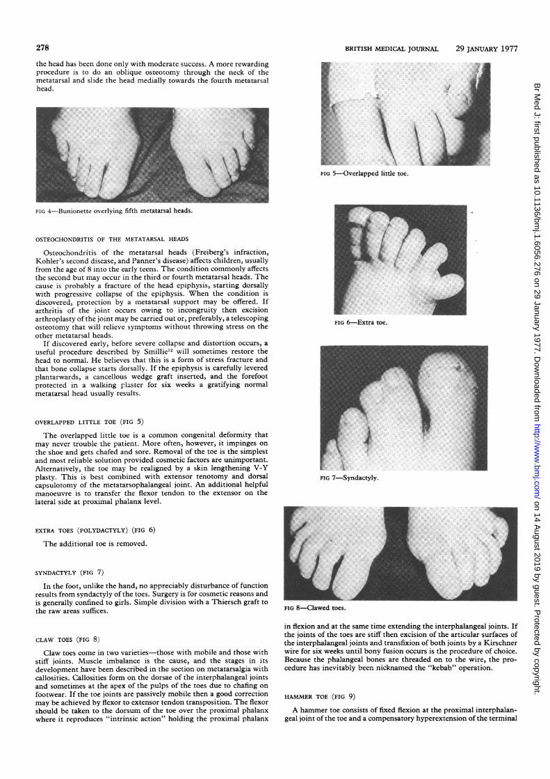
the head has been done only with moderate success. A more rewardingprocedure is to do an oblique osteotomy through the neck of themetatarsal and slide the head medially towards the fourth metatarsalhead.
BRITISH MEDICAL JOURNAL 29 JANUARY 1977_X t:: ~~~~~~~~~~~~~~~~~~~~~~~~~~~..........._~~~~~~~~~~~~~~~~.'i....:4.:_g11 ,9,~~~~~~~~~~~~~~~~~~~~~~~~~~~~~~~~~~~~~~~~~~~.. ..... ..... . . . l&,
FI 5Ovelpe little....toe.^ V
FIG 4-Bunionette overlying fifth metatarsal heads.
OSTEOCHONDRITIS OF THE METATARSAL HEADS
Osteochondritis of the metatarsal heads (Freiberg's infraction,Kohler's second disease, and Panner's disease) affects children, usuallyfrom the age of 8 into the early teens. The condition commonly affectsthe second but may occur in the third or fourth metatarsal heads. Thecause is probably a fracture of the head epiphysis, starting dorsallywith progressive collapse of the epiphysis. When the condition isdiscovered, protection by a metatarsal support may be offered. Ifarthritis of the joint occurs owing to incongruity then excisionarthroplasty of the joint may be carried out or, preferably, a telescopingosteotomy that will relieve symptoms without throwing stress on theother metatarsal heads.
If discovered early, before severe collapse and distortion occurs, auseful procedure described by Smillie12 will sometimes restore thehead to normal. He believes that this is a form of stress fracture andthat bone collapse starts dorsally. If the epiphysis is carefully leveredplantarwards, a cancellous wedge graft inserted, and the forefootprotected in a walking plaster for six weeks a gratifying normalmetatarsal head usually results.
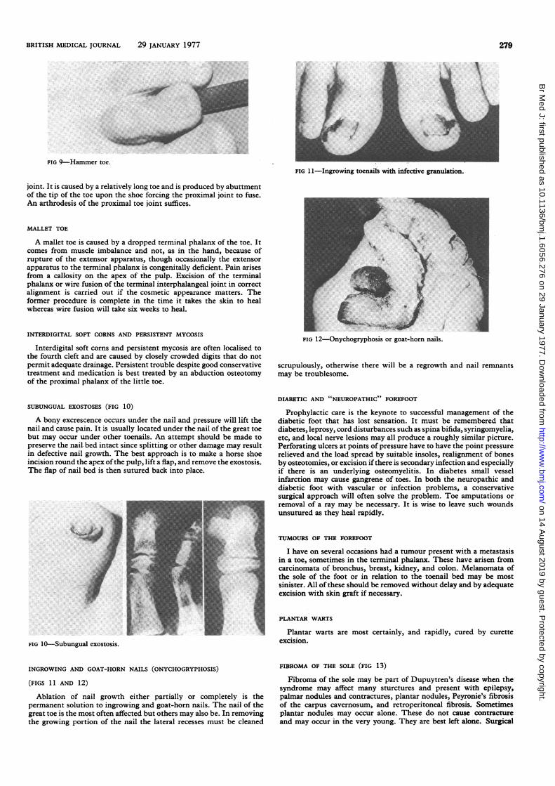
OVERLAPPED LITTLE TOE (FIG 5)
The overlapped little toe is a common congenital deformity thatmay never trouble the patient. More often, however, it impinges onthe shoe and gets chafed and sore. Removal of the toe is the simplestand most reliable solution provided cosmetic factors are unimportant.Alternatively, the toe may be realigned by a skin lengthening V-Yplasty. This is best combined with extensor tenotomy and dorsalcapsulotomy of the metatarsophalangeal joint. An additional helpfulmanoeuvre is to transfer the flexor tendon to the extensor on thelateral side at proximal phalanx level.
EXTRA TOES (POLYDACTYLY) (FIG 6)
The additional toe is removed.
SYNDACTYLY (FIG 7)
In the foot, unlike the hand, no appreciably disturbance of functionresults from syndactyly of the toes. Surgery is for cosmetic reasons andis generally confined to girls. Simple division with a Thiersch graft tothe raw areas suffices.
CLAW TOES (FIG 8)
Claw toes come in two varieties-those with mobile and those withstiff joints. Muscle imbalance is the cause, and the stages in itsdevelopment have been described in the section on metatarsalgia withcallosities. Callosities form on the dorsae of the interphalangeal jointsand sometimes at the apex of the pulps of the toes due to chafing onfootwear. If the toe joints are passively mobile then a good correctionmay be achieved by flexor to extensor tendon transposition. The flexorshould be taken to the dorsum of the toe over the proximal phalanxwhere it reproduces "intrinsic action" holding the proximal phalanx
FIG 6-Extra toe.
FIG 8-Clawed toes.
in flexion and at the same time extending the interphalangeal joints. Ifthe joints of the toes are stiff then excision of the articular surfaces ofthe interphalangeal joints and transfixion of both joints by a Kirschnerwire for six weeks until bony fusion occurs is the procedure of choice.Because the phalangeal bones are threaded on to the wire, the pro-cedure has inevitably been nicknamed the "kebab" operation.
HAMMER TOE (FIG 9)
A hammer toe consists of fixed flexion at the proximal interphalan-geal joint of the toe and a compensatory hyperextension of the terminal
on 14 August 2019 by guest. P
rotected by copyright.http://w
ww
.bmj.com
/B
r Med J: first published as 10.1136/bm
j.1.6056.276 on 29 January 1977. Dow
nloaded from
BRITISH MEDICAL JOURNAL 29 JANUARY 1977
FIG 9-Hammer toe.FIG I1-Ingrowing toenails with inifective granulation.
joint. It is caused by a relatively long toe and is produced by abuttmentof the tip of the toe upon the shoe forcing the proximal joint to fuse.An arthrodesis of the proximal toe joint suffices.
MALLET TOE
A mallet toe is caused by a dropped terminal phalanx of the toe. Itcomes from muscle imbalance and not, as in the hand, because ofrupture of the extensor apparatus, though occasionally the extensorapparatus to the terminal phalanx is congenitally deficient. Pain arisesfrom a callosity on the apex of the pulp. Excision of the terminalphalanx or wire fusion of the terminal interphalangeal joint in correctalignment is carried out if the cosmetic appearance matters. Theformer procedure is complete in the time it takes the skin to healwhereas wire fusion will take six weeks to heal.
INTERDIGITAL SOFT CORNS AND PERSISTENT MYCOSIS
Interdigital soft corns and persistent mycosis are often localised tothe fourth cleft and are caused by closely crowded digits that do notpermit adequate drainage. Persistent trouble despite good conservativetreatment and medication is best treated by an abduction osteotomyof the proximal phalanx of the little toe.
SUBUNGUAL EXOSTOSES (FIG 10)
A bony excrescence occurs under the nail and pressure will lift thenail and cause pain. It is usually located under the nail of the great toebut may occur under other toenails. An attempt should be made topreserve the nail. bed intact since splitting or other damage may resultin defective nail growth. The best approach is to make a horse shoeincision round the apex ofthe pulp, lift a flap, and remove the exostosis.The flap of nail bed is then sutured back into place.
FIG l-Subungual exostosis.
INGROWING AND GOAT-HORN NAILS (ONYCHOGRYPHOSIS)
(FIGS 1 1 AND 12)
Ablation of nail growth either partially or completely is thepermanent solution to ingrowing and goat-horn nails. The nail of thegreat toe is the most often affected but others may also be. In removingthe growing portion of the nail the lateral recesses must be cleaned
..).
iFIG 12-Onychogryphosis or goat-horn nails.
scrupulously, otherwise there will be a regrowth and nail remnantsmay be troublesome.
DIABETIC AND "NEUROPATHIC" FOREFOOT
Prophylactic care is the keynote to successful management of thediabetic foot that has lost sensation. It must be remembered thatdiabetes, leprosy, cord disturbances such as spina bifida, syringomyelia,etc, and local nerve lesions may all produce a roughly similar picture.Perforating ulcers at points of pressure have to have the point pressurerelieved and the load spread by suitable insoles, realignment of bonesby osteotomies, or excision ifthere is secondary infection and especiallyif there is an underlying osteomyelitis. In diabetes small vesselinfarction may cause gangrene of toes. In both the neuropathic anddiabetic foot with vascular or infection problems, a conservativesurgical approach will often solve the problem. Toe amputations orremoval of a ray may be necessary. It is wise to leave such woundsunsutured as they heal rapidly.
TUMOURS OF THE FOREFOOT
I have on several occasions had a tumour present with a metastasisin a toe, sometimes in the terminal phalanx. These have arisen fromcarcinomata of bronchus, breast, kidney, and colon. Melanomata ofthe sole of the foot or in relation to the toenail bed may be mostsinister. All of these should be removed without delay and by adequateexcision with skin graft if necessary.
PLANTAR WARTS
Plantar warts are most certainly, and rapidly, cured by curetteexcision.
FIBROMA OF THE SOLE (FIG 13)
Fibroma of the sole may be part of Dupuytren's disease when thesyndrome may affect many sturctures and present with epilepsy,palmar nodules and contractures, plantar nodules, Peyronie's fibrosisof the carpus cavernosum, and retroperitoneal fibrosis. Sometimesplantar nodules may occur alone. These do not cause contractureand may occur in the very young. They are best left alone. Surgical
279
on 14 August 2019 by guest. P
rotected by copyright.http://w
ww
.bmj.com
/B
r Med J: first published as 10.1136/bm
j.1.6056.276 on 29 January 1977. Dow
nloaded from

280 BRITISH MEDICAL JOURNAL 29 JANUARY 1977
K
... .:_' ~~~~~~~~~~~~~~~~~~~~~~~~~~~~~~~~..... .... .. 'FIG 13 Fibroma of plantar fascia.
excision carries a high recurrence rate, and the histology is difficult tointerpret as the nodules are cellular and may be reported as fibrosar-comata when needless amputation and irradiation may ensue.
Conclusion
Less than a century ago the type of surgery described abovewas considered beneath the dignity of the professional man andwas relegated to barbers and bootmakers; indeed, this attitudepersists in part today, for many delegate forefoot surgery to the
most junior and inexperienced surgical house officers. WhenC H Mayo, one of the famous Mayo brothers who founded theMayo Clinic, read his paper on hallux valgus in 1920,13 acolleague was so surprised that he commented that it wasstrange that so celebrated a surgeon should consider the treat-ment of bunion an important procedure. The adage that there isno such thing as minor surgery, only minor surgeons, is alasstill true.
I thank Miss D Bellamy, my secretary at Enfield District Hospital,for her help and Mr Martin Moor at Enfield District Hospital andMr Ray Ruiddick at the London Hospital for the photographs.
References
Kelikian, M, Hallux Valgus, Allied Deformities of the Forefoot andMetatarsalgia. Philadelphia and London, W B Saunders, 1965.
2 Levy, H S, Chinese Footbinding, the History of a Curious Erotic Custom.New York, Walton Rawls, 1966.
3 Helal, B, Gupta, S K, and Gojaseni, P, Acta Orthopaedica Scandinavica,1974, 45, 271.
4 Helal, B, Gupta, S K, and Gojaseni, P, Acta Orthopaedica Scandinavica,1974, 45, 271.
5 Keller, W L, New York Medical Journal and Philadelphia MedicalJournal, 1904, 80, 741.
6 Mayo, C H, Minnesota Medical Journal, 1970, 3, 326.7 Simmonds, F A, and Menelaus, M B, Journal of Bone and Joint Surgery,
1960, 42B, 761.8 Shepherd, E, Vernon Roberts, B, lecture at the Orthopaedic Foot Society
meeting, The London Hospital, Whitechapel, November 1975.9 Helal, B, Journal of Bone and Joint Surgery, 1975, 57B, No 2.
10 Fowler, A W, Journal of Bone and Joint Surgery, 1959, 41B, 80.' Kates, A, Kessel, L, and Kay, A,Jrournal of Bone and Joint Surgery, 1967,
49B, 452.12 Smillie, I S, Journal of Bone and Joint Surgery, 1957, 39B, 580.13 Mayo, C H, Minnesota Medical Journal, 1970, 3, 326.
Letter from ... Chicago
Buttering opsonins
GEORGE DUNEA
British Medical Journal, 1977, 1, 280-282
On 24 March 1976 at the height of the primary elections,President Ford wearing his TV-blue shirt and facing the camerasannounced his decision to ask Congress for $135m to vaccinateevery American man, woman, and child against swine influenza."We cannot afford to take a chance with the health of thenation," said the President, explaining the need to stimulate thephagocytes, butter the germs appetisingly with opsonins, andneutralise the deadly ptomaines arising from infected nuciformsacs. But it was reported-in this revisitation of George BernardShaw's Doctor's Dilemma-that the decision to march againstthe swine virus had not been made lightly; that there wereanxious moments of consultation with Sir Colenso Ridgeon,Sir Patrick Cullen, and Sir Ralph Bloomfield Bonington; that
Cook County Hospital, Chicago, IllinoisGEORGE DUNEA, MB, MRCP, attending physician
Mr Cutter Walpole, a surgeon, opposed the measure andadvocated wholesale amputations of uvulae and extirpations ofimmunologically incompetent nuciform sacs; and that DrBlenkinsop, an impoverished and overworked general practi-tioner, insisted that a pound of ripe greengages taken everyday half an hour before lunch would be just as good and far lessexpensive.
Reasons for vaccination
Yet the events precipitating the President's decision weresuspicious and alarming. A serious outbreak of influenza, thethird worst in 50 years, had raged during the winter months,and over 20 000 people had died from respiratory illness. Thevirus had found its usual fruitful soil among the nation'smilitary camps, and at Fort Dix, New Jersey, some half of therecruits had come down with upper respiratory complaints.Among these recruits was young private David Lewis, whoseillness at first had seemed to be an ordinary cold. Several dayslater, however, his malaise increasing, he wrote to his fiancee
on 14 August 2019 by guest. P
rotected by copyright.http://w
ww
.bmj.com
/B
r Med J: first published as 10.1136/bm
j.1.6056.276 on 29 January 1977. Dow
nloaded from