surgical pathology “biopsy”
TRANSCRIPT

SURGICAL PATHOLOGY
“BIOPSY”
I N O R A L S U R G E R Y
T his series of booklets are directed to undergraduate den-tal students and GDP. The booklets are aimed to serve as an outline guide to the user during reading in more compre-hensive textbooks.

2
CLINICAL PATHOLOGY
Forward
This issue prepared and printed by: Prof. Maged Lotfy,
Pyramids award coordinator and cosponsor March 2016
FOREWORD
B iopsy technique is among the key clinical skills imparted to a dentist during training as it has
great bearing on successful diagnosis of various oral conditions. Dental practitioners who under-
take this procedure without putting into consideration the key principles involved could miss the
opportunity to diagnose a serious condition such as a malignancy until it is too late. Although the
use of biopsy to assist in diagnosis is widely used in all medical fields, the procedure is not widely
used in dental practice. This is mainly because of lack of awareness of its importance and the rel-
evant techniques among dental practitioners.
This book acquaints the dental student and the general dental practitioner with the essential
basic information that would assist them decide on whether to take a biopsy, and if so, the prop-
er surgical techniques for the biopsy procedure.
Dr. Ochiba M. Lukandu BDS, PhD HOD, Oral, Maxillofacial, Oral Medicine, Oral Radiology And Oral Pathology Dept. SOD, MU

3
CLINICAL PATHOLOGY
INTRODUCTION TO DIAGNOSTIC PATHOLOGY ..... 4
History of specific lesion ..... 4
CLINICAL EXAMINATION ..... 6
PREBIOPSY MONITORING ..... 10
BASIC PROMCIPLES OF FOLLOW-UP AND REFERRAL ..... 13
Biopsy or Referral ..... 13
INSTRUMENTATION
Incising tissues ..... 15
Cutting tissues (scissors) ..... 17
Grasping tissues ...... 17
SOFT TISSUES BIOPSY TECHNIQUES
Incisional biopsy ..... 19
Excisional biopsy ..... 19
Tissue stabilization ..... 22
Hemostasis ..... 24
Wound CLOSURE ..... 24
HANDLING THE SPECIMEN ..... 26
TAGGING THE BIOPSY ..... 27
BIOPSY SUBMISSION FORM ..... 27
ORAL BIOPSY (WHY, WHEN AND HOW) ..... 30
REFERENCES, FURTHER READINDS AND LINKS ..... 44
CONTENT

4
CLINICAL PATHOLOGY
INTRODUCTION TO DIAGNOSTIC PATHOLOGY
D entists often find oral soft tissue abnormalities when examining their patients. Some of
these abnormal findings can be diagnosed based on their appearance and history, whereas
others require more diagnostic steps. One available option to reach a diagnosis is the perfor-
mance of a biopsy; namely, taking tissue for examination under a microscope.
HISTORY OF THE SPECIFIC LESION
It is generally accepted in medicine that many systemic diseases (up to 90%) can be diagnosed by
gathering a detailed medical history. The same can be true of many oral lesions when the diag-
nostician is familiar with the natural history of the more common diseases. Questioning the pa-
tient who has a potentially pathologic condition should include the following:
How long has the lesion been present?
A lesion that has been present for several years might be congenital and is more likely
benign.
A rapidly developing lesion is considered more ominous.
Duration must be taken in context with other elements of the history because the lesion
might have been present for an extended period before the patient became aware of its
presence.
Has the lesion changed in size?
A change in the radiographic or clinical size of a lesion, or both must determine.
An aggressive, enlarging lesion is more likely to be malignant
A slower-growing lesion suggests a possibly benign lesion.
By combining information on the growth rate with findings regarding the duration , one
can make a more accurate assessment of the nature of the lesion.
Has the lesion changed in character or features?
Noting changes in the physical characteristics of a lesion often can assist in the diagno-
sis. (e.g., a lump becoming an ulcer or an ulcer starting as a vesicle)
For example, if an ulcer began as a vesicle, then it could suggest a localized or systemic
vesiculobullous or viral disease.
What symptoms are associated with the lesion?
For example, pain, altered function, anesthesia or paraesthesia abnormal taste or odors,
dysphagia, tenderness of cervical lymph nodes

5
CLINICAL PATHOLOGY
If painful, is the pain acute or chronic, constant or intermittent. What increases or de-
creases the pain.
Lesions with an inflammatory component are most often associated with pain.
Cancers, believed by many to be painful, actually are typically painless unless secondari-
ly infected.
Sensory nerve changes, such as numbness or tingling, often occur with a malignant or
inflammatory process unless other identifiable causes can be ascertained.
Swelling often can result from and occur with oral lesions, indicating an expansile pro-
cess from any number of causes, including inflammation, infection, cysts, or tumor for-
mation. The patient might indicate a sensation of fullness even before the doctor can
actually visualize or verify the swelling during clinical examination.
Painful lymph nodes usually indicate an inflammatory or infectious cause, but also can
be a manifestation of malignancy.
What anatomic locations are involved?
Certain lesions have a predilection for certain anatomic areas or tissues.
Noting whether the lesion is confined to keratinized or non-keratinized tissues, regions
with salivary gland tissues, or areas of neural or vascular anatomy sometimes can pro-
vide clues to the diagnosis.
Are there any associated systemic symptoms?
The dentist should look for possible relations or manifestations from related systemic dis-
eases or conditions.
For example, many systemic viral conditions (e.g., measles, mumps, mononucleosis, her-
pes, and acquired immunodeficiency syndrome) can cause oral manifestations concur-
rent with the systemic involvement.
Autoimmune conditions also can manifest with oral lesions.
Many oral ulcerative conditions also can present lesions elsewhere in the body (e.g.,
pemphigus, lichen planus, erythema multiform, sexually transmitted infections).
Other factors could include drug abuse or injuries from domestic violence.
Is there any historical event associated with the onset of the lesions?
For example, trauma, recent treatment, exposure to toxins or allergens, or visits to foreign
countries
One of the initial steps the dentist should take when a lesion is noted is to seek a possi-
ble explanation based on the patient’s medical, dental, family, or social histories.
Frequently, oral and perioral lesions can be caused by parafunctional habits, hard or hot
foods, application of medications not intended for topical use, recent trauma, conditions
involving the dentition (eg, caries, periodontal disease, fractured teeth), or an identified
event or exposure.

6
CLINICAL PATHOLOGY
W hen a lesion is discovered, careful clinical, radiographic examinations and palpation of
regional lymph nodes are mandatory. Once the examination is complete, a detailed de-
scription of all objective and subjective findings should be documented in the patient’s chart. A
drawing or a graphic schematic of the location, orientation, general shape, and dimensions of the
lesion in the patient record is helpful. The use of standardized illustrations can simplify the docu-
mentation. In addition, good-quality digital photographs are useful for documentation and can
aid the pathologist. Details, descriptions, and drawings allow the dentist or subsequent referral
specialists to evaluate the course of the lesion over time and determine whether it is enlarging,
its features are changing, or if new lesions are appearing in different anatomic areas.
An examination is classically described as a process that includes inspection, palpation, percus-
sion, and auscultation. In the head and neck region, inspection and palpation are more commonly
used as diagnostic modalities, with inspection always preceding palpation. Early inspection facili-
tates creating a description of the lesion before it is handled because some lesions are so fragile
that manipulation of any kind might result in hemorrhage or rupture of a fluid-filled lesion or loss
of loosely attached surface tissues, which would compromise any subsequent examinations. Per-
cussion is reserved for examination of the dentition. Auscultation is used infrequently but is im-
portant when examining for suspected vascular lesions.
The following list includes some important additional points to be considered during the inspec-
tion of a lesion:
Anatomic location.
Pathologic lesions can arise from any tissue within the oral cavity, including the epitheli-
um, subcutaneous and submucosal connective tissues, muscle, tendon, nerve, bone,
blood vessels, lymphatic vessels, or salivary glands.
The dentist should attempt to ascertain, as much as possible, which tissues are contrib-
uting to the lesion, based on the anatomic location of the lesion:
o For example, if a mass appears on the dorsum of the tongue, then the dentist would
logically consider an epithelial, connective tissue, lymphatic, vascular, glandular, neu-
ral, or muscular origin.
o A mass on the inner aspect of the lower lip would prompt the dentist to include a mi-
nor salivary gland origin in the differential diagnosis, in addition to a connective tissue
origin and other possibilities.
C L I N I C A L E X A M I N A T I O N

7
CLINICAL PATHOLOGY
o Certain lesions can have unique anatomic characteristics, such as the linear tenden-
cies of herpes zoster lesions as they follow neural pathways.
o Pulpal, periapical, and periodontal pathologic or inflammatory conditions also cause a
large percentage of oral lesions.
o The role of trauma should always be entertained as a possible source of the lesion (ill-
fitting dental appliances, parafunctional habits such as cheek biting, sharp edges on
teeth or restorations, trauma from acts of domestic or other types of violence).
Overall physical characteristics.
Appropriate medical terminology should always be used to describe clinical findings in the
record because terminology can be misleading and nonspecific.
o Terms such as ulcer or nodule might be interpreted differently by different examiners.
Table 1 lists several common physical descriptions that are useful in describing oral
and maxillofacial pathologic entities. Lay terms such as swelling and sore are generally
not helpful and could be subject to misinterpretation.
Single versus multiple.
o The presence of multiple lesions is an important feature. When multiple ulcerations are
found within the mouth, the dentist should think of specific possibilities for the differ-
ential diagnosis. To find multiple or bilateral neoplasm in the mouth is unusual, where-
as vesiculobullous, bacterial, and viral diseases commonly present such a pattern. Simi-
larly, an infectious process can exhibit outward spread because lesion infects the adja-
cent tissues with which it has had contact.
Size, shape, and growth.
o The size of the lesion should be measured and documented.
o The shape of the lesion also should be noted, whether the lesion is flat or slightly ele-
vated, endophytic (growing inward) or exophytic (growing outward) from the epithelial
surface, and sessile (broad based) or pedunculated (on a stalk).
Surface appearance.
o The epithelial surface of a lesion can be smooth, lobulated (verruciform), or irregular.
o If ulceration is present, then the characteristics of the ulcer base and margins should be
recorded. Margins of an ulcer can be flat, rolled, raised, or everted.
o The base of the ulcer can be smooth, granulated, or covered with fibrin membrane,
slough, or hemorrhagic crust (scab) or can have the fungating appearance that is char-
acteristic of some malignancies.
Color.
The surface color(s) of a lesion can reflect various characteristics and even the origin of
many lesions.
o A dark bluish swelling that blanches on pressure suggests a vascular lesion.
o A lighter-colored, bluish lesion that does not blanch might suggest a mucus-retention
cyst.

8
CLINICAL PATHOLOGY
TABLE 1. DESCRIPTIVE PATHOLOGY TERMS
Bulla: A blister; an elevated, circumscribed, fluid containing lesion of
skin or mucosa.
Crust: (crusted): dried or clotted serum on the surface of the skin or
mucosa.
Dysplasia: any abnormal development of cellular size, shape, or organi-
zation in tissue.
Erosion: a shallow superficial ulceration.
Hyperkeratosis: an overgrowth of the cornified layer of epithelium.
Hyperplasia: an increased number of normal cells.
Hypertrophy: an increase in size caused by an increase in the size of
the cell, not the number of cells.
Keratosis: an overgrowth and thickening of cornified epithelium.
Leukoplakia: a slowly developing change in mucosa characterized by
firmly attached thickened white patches.
Macule: a circumscribed non-elevated area of color change that distinct
from adjacent tissues
Malignant: anaplastic; a cancer that is potentially invasive and meta-
static.
Nodule: a large elevated circumscribed solid, palpable mass of skin or
mucosa
Papule: a small, elevated, circumscribed solid palpable mass of skin or
mucosa.
Plaque: any flat, slightly elevated superficial lesion.
Scale: a thin compressed, superficial flake of cornified (keratinized) epi-
thelium.
Stomatitis: any generalized inflammatory condition of the oral mucosa.
Ulcer: a crater-like circumscribed surface lesion resulting from necrosis
of epithelium.
Vesicle: a small blister: a small circumscribed elevation of skin or muco-
sa containing serous fluid.

9
CLINICAL PATHOLOGY
o A pigmented lesion within the mucosa can suggest a ‘‘traumatic tattoo’’ of restorative
material or a more ominous melanotic tumor.
o Keratinized white lesions can reflect a reaction to repetitive local tissue trauma or rep-
resent potentially premalignant changes.
o An erythematous (or mixed red-and-white) lesion can represent an even more ominous
prognosis for dysplastic changes than a white lesion.
o Inflammation can be superimposed on areas of mechanical trauma or ulceration, re-
sulting in a varied color from one examination to the next.
Sharpness of borders and mobility.
o If a mass is present, then the dentist should determine whether it is fixed to the sur-
rounding deep tissues or freely movable.
o Determining the boundaries of the surface lesion will aid in establishing whether the
mass is fixed to adjacent bone, arising from bone and extending into adjacent soft tis-
sues, or only infiltrating the soft tissue.
Consistency when palpated.
o Consistency can be described as soft or compressible (eg, a lipoma or abscess), firm or
indurated (eg, a fibroma or neoplasm), or hard (eg, torus or exostosis).
o Fluctuant is a term used to describe the wavelike motion sensed during bi-digital palpa-
tion of a lesion with non-rigid walls and that contains fluid.
Presence of pulsation.
o Palpation of a mass can disclose a rhythmic pulsation that is suggestive of a major vas-
cular component. This sensation can be subtle and is especially important when dealing
with intrabony lesions.
o The pulsation can be accompanied by a palpable vibration, called a thrill. If a thrill is
palpated, auscultation of the area with a stethoscope might disclose a bruit, or audible
murmur, in the area.
o Invasive procedures on lesions with thrills, bruits, or both should be avoided, and pa-
tients should be referred to specialists for treatment because life-endangering hemor-
rhage can result if surgical intervention (biopsy) is attempted.
Examination of regional lymph nodes.
o This examination should be accomplished before any biopsy procedure. Sometimes,
lymphadenitis develops in the regional nodes after a surgical procedure such as biopsy,
thus creating a subsequent diagnostic dilemma. Then, it can become difficult to differ-
entiate reactive lymphadenitis as a surgical sequela from coincidental regional infection
or inflammation from metastatic spread of the tumor in question.

10
CLINICAL PATHOLOGY
A ny undiagnosed or suspicious change in oral tissues that cannot be explained by localized
trauma or other factors should be followed up in 7 to 14 days. If the lesion enlarges or ex-
pands, develops an altered appearance, or does not respond as expected to local therapy, a biop-
sy is usually indicated. Areas of leukoplakia can be problematic because up to 20% of those areas
(and 100% of erythroplakia lesions) exhibit histological evidence of dysplasia or frank malignancy.
High-risk areas of the mouth include the floor of the mouth, the lateral and ventral surfaces of
the tongue, and the buccal and lower lip mucosa. Areas of redness or pebbling within areas of
leukoplakia are especially troubling. Incisional biopsy specimens from at least 1 suspicious area
are generally indicated. Table 2 list the lesion characteristics that raise suspicion of malignancy.
During subsequent examinations, the patient record should provide details on whether the ob-
served lesion has improved or not improved and the dentist’s plan for subsequent management
(biopsy, or referral). (Fig. 1 and 2)■
PRE-BIOPSY MONITORING
TABLE 2. CHARACTERIST IC OF LES IONS THAT RAISE
SUSPIC ION OF MALIGNANCY
Bleeding: Lesions bleeds on gentle manipulation.
Duration: Lesions persisted for more than 2 weeks
Erythroplasia: Lesion is totally red or has a speckled
red and white appearance.
Fixation: Lesions attached to adjacent structures.
Growth rate: Lesions exhibits rapid growth.
Induration: Lesion and surrounding structure are
firm to palpation.
Ulceration: Lesion become ulcerated or presented
as an ulcer.

11
CLINICAL PATHOLOGY
FIG.1. Decision tree diagram for managing suspicious lesions. Reprinted
Lesion Detection
Lesion history, clinical and radiographic examinations, laboratory testing
Decision to biopsy
Perform biopsy
Refer to specialist
No further treatment required
Diagnosis indicates need for further treatment/
surgery
Needs are within capabil-ities of a general dentist
Need for referral to specialist
P a t i e n t m o n i t o r i n g / f o l l o w - u p / s u p p o r t
Observation or nonsurgical treatment not indicated; high suspicion of malig-
nancy
Differential Diagnosis
No improvement Improvement and no further treat-ment is required
Observation or nonsurgical treatment for 10-14 days

12
CLINICAL PATHOLOGY
FIG. 2. Examples of lesions that should considered for biopsy.
A, Ulcer on the lateral border of the tongue. In this case, it was a traumatic ulcer from bit-
ing. B, Another ulcer on the lateral border of the tongue. In this case, it was from a sharp edge
of a fractured tooth cusp. C, Large ulcer of the lower lip, especially if in a patient with a history of
smoking. This lesion was squamous cell carcinoma. D, Typical appearance of squamous cell
carcinoma of the alveolar ridge. .

13
CLINICAL PATHOLOGY
T he ultimate responsibility for the detection of pathologic conditions (including oral cancer
screening) rests with the dentist. Delegation of this duty is not permitted by law. The follow-
ing should also be considered:
If the dentist decides to refer the patient for a second opinion or specialty management, then
the referral appointment ideally should be arranged before the patient leaves the office. If left
to make the appointment themselves, many patients might fail to do so because of fear or de-
nial.
The arranged appointment should be followed with a letter or electronic message from the
referring general dentist to the specialist, outlining the details of the case, the concerns, and
the requested procedures.
A copy of this correspondence should be placed in the patient record. Copies of the specialist’s
findings, recommendations, procedures, and biopsy findings also should be placed in the pa-
tient record. These formal exchanges provide precise documentation that prevents miscom-
munications between offices and can provide some protection if litigation is initiated later.
The patient should be notified of the results, and if the results are unexpected or positive re-
quiring further treatment, then the patient should be counseled in person by the dentist.
BIOPSY OR REFERRAL
Some dentists might be comfortable performing biopsy procedures on their patients, whereas
others might refer their patients to a specialist. This is a personal choice and should take several
points into consideration.
Health of the patient. Greater number of medically compromised patients are now seen in
the dental clinic. Such patients may need a special medical care during biopsy taking.. Patients
can be referred to an oral and maxillofacial surgeon who is trained to manage patients with
special medical needs so that the procedure is carried out as safely as possible.
Surgical difficulty. The degree of surgical difficulty during taking the biopsy should be consid-
ered. Factors such as access, lighting, accessibility and the presence of nearby important ana-
tomical structures should be considered. Each dentist should use his or her best judgment
when deciding whether the biopsy is within the dentist’s surgical abilities or if the patient
would be better managed by a more experienced specialist.
Malignant potential. The dentist who suspects that a lesion is malignant has two choices.
The first is to perform a surgical biopsy after completion of comprehensive diagnostic workup
or to refer the patient to a specialist who can provide definitive treatment if the lesion is
BASIC PRINCIPLES OF FOLLOW-UP AND REFERRAL

14
CLINICAL PATHOLOGY
shown to be malignant. The latter choice usually represents better service to the patient as it
save time, which is important. In addition, it is better for the referral specialist to evaluate the
lesion before any surgical intervention has compromised its clinical features. Biopsy also can
produce reactive lymph nodes that might be unrelated to the original lesion and even spread
malignant tissue. Allowing the referral specialist to evaluate the patient before biopsy helps
toward a more accurate diagnosis and aids in the formulation of a suitable treatment plan.

15
CLINICAL PATHOLOGY
INC IS ING TISSUE
Performing a biopsy of oral soft tissue usually involves incising the tissue with a scalpel. The scal-
pel is composed of a reusable handle and a disposable, sterile, sharp blade. Scalpels also are
available as a single-use scalpel with a plastic handle and fixed blade. The most commonly used
handle for oral surgery is the number 3 handle (Fig 3).
The tip of a scalpel handle is prepared to receive a variety of differently shaped scalpel blades to
be inserted onto the slotted portion of the handle. The most commonly used scalpel blade for
intraoral surgery is the number 15 blade (Fig 3)
Loading and unloading the scalpel
The scalpel blade is carefully loaded onto the handle while holding the blade with a needle
holder. This lessens the chance of injuring the fingers.
The blade is held along the unsharpened edge, where it is reinforced with a small rib, and the
handle is held so that the male portion points upward (Fig . 4,A).
Then, the scalpel blade is slid slowly onto the handle along the grooves in the male portion
until it clicks into position (Fig 4,B).
The scalpel is unloaded similarly. The needle holder grasps the end away from the blade (Fig
4,C) and lifts it to disengage it from the male fitting portion.
Then, the scalpel is slid off the handle, always away from the body (Fig 4,D).
The used blade is immediately discarded into a specifically designed, rigid-sided sharps con-
tainer.
Handling the scalpel
When using the scalpel to make an incision, the surgeon typically holds it in the pen grasp (Fig 5)
to allow maximal control of the blade as the incision is made. Mobile tissue should be held firmly
in place under some tension so that as the incision is made, the blade will incise and not just push
away the mucosa. When incising depressible soft tissue, an instrument such as a retractor or a
tissue forceps should be used to hold the tissue taut while incising.
Scalpel blades and blade-and-handle sets are designed for single-patient use. Blades dull easily
after repeated strokes through keratinized tissue. Dull blades do not make clean, sharp incisions
in soft tissue and therefore should be replaced before they become overly dull.
INSTRUMENTATION

16
CLINICAL PATHOLOGY
FIG. 3. Scalpels
(Upper) Scalpels come in two forms. One has a reusable handle and one-time use disposable blade (top) and the other has a blade/handle combination in which the entire blade/handle unit is discarded after one-time use (bottom). In both cases the blade or blade/handle unit must be placed into a red sharps disposable box.
(Lower) Scalpel blades come in various shapes and sizes. The most common blade used for oral surgery is the #15 blade, the right-most in this figure, followed by #12, 11 and 10.

17
CLINICAL PATHOLOGY
CUTT ING SOFT TISSUE (SCI SSORS)
There are many types of scissors used while performing oral biopsies. They are designed for un-
dermining and cutting soft tissue. Two major types of tissue scissors are used intraorally; iris scis-
sors and Metzenbaum scissors (Fig 5). These scissors can have straight or curved blades. Iris scis-
sors are small, sharply pointed, delicate tools used for fine work. Metzenbaum scissors are used
for undermining soft tissue and for cutting. They can have sharp or blunt (rounded) tips. Tissue
scissors such as iris or Metzenbaum scissors should not be used to cut sutures because the suture
material will dull the edges of the blades and make them less effective and more traumatic when
cutting tissue. Surgical scissors are held in the same manner as needle holders.
GRASPING TISSUE
Adson forceps can be useful when performing a biopsy. They can be used to grasp the tissue to
stabilize the lesion while incising and then be used while suturing the resulting open wound. In
some types of biopsies, forceps with locking handles and teeth that will grip the tissue firmly are
necessary. In this situation, the Allis tissue forceps are used (Fig 6). The locking handle allows the
forceps to be placed in the proper position and then to be held by an assistant to provide the
necessary tension for proper dissection of the tissue. The Allis forceps should never be used on
tissue that is to be left in the mouth because they cause a relatively large amount of tissue de-
struction as a result of crushing injury.
Fig. 4. Loading (upper) and unloading (lower) the scalpel.

18
CLINICAL PATHOLOGY
Fig 6. Grasping the tissues
A, Allis tissue forceps are useful for grasping and holding tissue that will be excised. B, Allis forceps are held in the same fashion as the needle holder. C, Comparison of Adson beaks (right) with Allis beaks (left) shows the differences in their designs.
FIG. 5. Soft tissue scissors;
Iris (upper) and Metzenbaum (lower) scissors

19
CLINICAL PATHOLOGY
B iopsy of oral soft tissues is a useful competency for a general dentist to possess. When per-formed properly, most biopsies are straight forward procedures that can be readily per-
formed in the dental office using local anesthesia and minimal instrumentation. The only varia-bles of the technique relate to areas of anatomic risk or limitations imposed by the size and type of lesion.
ANESTHESIA
Block local anesthesia techniques are preferred over infiltration, whenever possible, so that the anesthetic solution is not inadvertently incorporated in the surgical specimen. Infiltration anes-thesia can cause distortion of the cellular architecture of the specimen and make pathologic diag-nosis more difficult, if not impossible. Peripheral infiltration of local anesthetic with a vasocon-strictor is often helpful, injecting at least 1 cm away from the lesion’s perimeter to prevent distor-tion of the lesion itself.
INCISIONAL BIOPSY
An incisional biopsy is a procedure that removes only a portion of the lesion. If the lesion is large or exhibits differing characteristics in different locations, then more than 1 area of the lesion might require sampling. Incisional biopsies are used when:
The lesion is large (>1 cm in diameter), or located in a hazardous location,
A definitive histopathologic diagnosis (eg, for suspected malignancy) is desired before a com-
plex removal or other treatment.
The following should be considered during taking an incisional biopsy:
The incisional biopsy is generally excised as a wedge of tissues that intentionally includes nor-
mal- and abnormal appearing tissues in the sample.
The only exception to this approach is when a malignant lesion is strongly suspected. In that
circumstance, including normal- appearing issue in the specimen could spread malignant cells.
(Fig. 7)
Avoid the central area of the lesion which is often necrotic and of little diagnostic values.
Care must be taken to include an adequate depth of tissue, so that cellular features from the
base of the lesion are included.
In general, it is better to take a narrow, deep specimen rather than a broad, shallow one. .
EXCISIONAL BIOPSY
An excisional biopsy consists of the removal of a lesion in its entirety, to include a 2- to 3-mm pe-rimeter of normal tissue around the lesion. An additional 2 to 3 mm of tissue might be required for specimens suspected of being malignant, including some pigmented lesions and lesions al-ready diagnosed as having dysplastic or malignant cells. (Fig. 8)
Complete excision often constitutes definitive treatment of the lesion biopsied. Excisional biopsy also is reserved for smaller lesions (<1 cm in diameter), taking care to avoid adjacent nerves or blood vessels unless they are thought to be a part of the lesion or the lesion has been deter-mined to be a malignancy.
SOFT TISSUE BIOPSY TECHNIQUES

20
CLINICAL PATHOLOGY
FIG.7. Principles of incisional biopsy.
(Upper) A, Obtaining a deep specimen, rather than a broad and shallow specimen, when inci-
sional biopsy is performed. If malignant cells are present only at the base of the lesion, then a
broad and shallow biopsy might not obtain these diagnostic cells. B, Obtaining incisional biopsy
at the margin of the soft tissue lesion. The junction of the lesion with normal tissue frequently
provides the pathologist with more diagnostic information than a biopsy specimen taken only
from the center of the lesion. This is particularly important when a biopsy of an ulcer is per-
formed.
(Lower) A, Frequently, one area of a lesion appears histologically different from another. There-
fore, it is often desirable to obtain more than 1 incisional biopsy to detect whether the charac-
teristics of the lesion differ from one area to another. B, When obtaining a biopsy on buccal or
labial mucosa, the incision is usually carried to the depth of the musculature.

21
CLINICAL PATHOLOGY
FIG.8. Excisional biopsy of soft tissue lesion.
A, Surface view. An elliptical incision is made around the lesion, at least 3 mm away from the le-
sion. B, Side view. The incision is made deep enough to remove the lesion completely. C, End
view. Incisions are made convergent to the depth of the wound. An excision performed this way
facilitates closure.
For other methods of tissues screening and examination, refer to the outline presenta-
tion on page 30.

22
CLINICAL PATHOLOGY
TISSUE STABILIZATION
Oral soft tissue biopsies frequently involve mobile surfaces and structures (e.g., lip, cheek, and
tongue). Accurate surgical incisions can be placed with greater ease when the involved tissues
are first stabilized. This can be accomplished by any of several methods. (Fig. 9 and 10)
The surgical assistant can grasp the lip on both sides of the biopsy site with his or her fingers,
which also retracts and immobilizes the lip. This also can help decrease bleeding by compress-
ing labial blood vessels and their tributaries.
Different retractors are available that can perform the same function.
When used, retraction sutures should be placed deeply into the tissues, away from the
planned biopsy site, so that they can function without pulling through or damaging the tissues.
HEMOSTASIS
FIG 9. Traction suture Use of traction suture placed through the specimen. While the lesion is incised, a traction suture is used to lift the specimen from the wound bed. Then, the suture can be tied and left attached to the le-sion to identify the orientation of the specimen

23
CLINICAL PATHOLOGY
Fig.10. Stabilization of tongue for biopsy.
A, Stabilization of tissue with traction sutures. Two silk sutures are used to stabilize the tongue
before excisional biopsy. They are placed through the substance of the tongue (mucosa and
muscle) to prevent pulling through tissue. B, The lesion is removed after an elliptical incision
has been made around it. C, Resorbable sutures are placed to approximate muscle. D, The
mucosa is closed. E, Alternative means of stabilization of tissue using a Chalazion-type device.

24
CLINICAL PATHOLOGY
FIG. 11. The principles used in closing an el-
liptical biopsy wound.
The mucosa should be undermined bluntly with
scissors to the width of the original ellipse in
each direction. Scissors are inserted under mu-
cosa tissue while closed and then opened to
spread tissues, undermining the mucosa in that
area. This is repeated around the periphery of
the wound. This allows approximation of wound
margins without tension.
The use of a suction device for keeping the surgical field free of blood during the procedure
should be minimized as much as possible, especially high-volume suction devices. The assistant
can usually use gauze sponges to blot the site during the procedure. Suctioning can increase not
only bleeding but also the risk of the biopsy tissue sample being accidentally aspirated into the
suction. If suction is needed, it is helpful to place gauze over the end of the suction tip to serve as
a filter.
WOUND CLOSURE
After removal of the tissue sample, primary closure of the wound is desirable and usually possi-
ble. The wound left after a larger biopsy often will not close without tension on the wound edges.
Undermining of adjacent surface tissue is used to help decrease the tension on wound edges dur-
ing closure. The following should be considered during wound closure:
Mucosa is undermined by using a spreading action of the tips of small scissors to separate the
mucosal from the submucosal tissues (Fig. 11). The submucosal layer is largely loose connec-
tive tissue that is easily dissected free from the overlying mucosa without sharp incision or
snipping.
This permits closure of the mucosa as a separate layer without regard to closure of the deeper
layers.
The extent to which this undermining is carried out is determined by the size of the wound
and the anatomic location.
Suture materials of choice are generally black silk or a resorbable material.
Biopsy wounds on the dorsum or lateral border of the tongue require deeply placed sutures at
close intervals to counteract inherent muscle movements and maintain closure. (Figs 12)■
25
CLINICAL PATHOLOGY
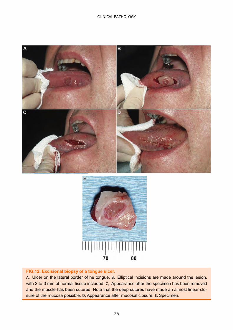
FIG.12. Excisional biopsy of a tongue ulcer.
A, Ulcer on the lateral border of he tongue. B, Elliptical incisions are made around the lesion,
with 2 to-3 mm of normal tissue included. C, Appearance after the specimen has been removed
and the muscle has been sutured. Note that the deep sutures have made an almost linear clo-
sure of the mucosa possible. D, Appearance after mucosal closure. E, Specimen.

26
CLINICAL PATHOLOGY
A ny tissue specimen must be maintained in a condition that is optimal for preserving the his-
tological and structural architecture of the cells of the lesion. Specimens that have been
crushed, frozen, desiccated, burned, or otherwise compromised might not be microscopically di-
agnostic. The following should be considered:
The removed tissue sample should not be wrapped in gauze (wet or dry) because it is at risk of
getting thrown out accidentally with the gauze.
The specimen should not be set on paper or linen drapes and allowed to dry out while the sur-
gery is being completed.
The specimen should be placed immediately in a glass or plastic container that contains a
quantity of 10% formalin solution (4% formaldehyde) that is at least 20 times the volume of
the specimen itself and that can be capped.
The specimen must be totally immersed in the preservative solution at all times, even if the
container is tilted sideways during transport.
Before turning attention to wound closure, the dentist should ensure that the tissue sample
does not adhere to the container wall above the level of the formalin.
If the specimen is mailed to the pathologist, then it must be labeled with a biohazard label ap-
proved by the Occupational Safety and Health Administration. (Fig. 13 and 14)
HANDLING THE SPECIMEN
FIG. 13. Handling the specimen. A specimen being dropped into a biopsy bot-tle filled with formalin. Do not re-enter the wound with the forceps unless they are first rinsed free of the fixative solution.

27
CLINICAL PATHOLOGY
FIG. 14. Typical biopsy kit that is available from pathology laboratories.
The kit includes a specimen bottle containing formalin, a biopsy requisition
form onto which information about the patient and specimen is documented,
and a mailer to send the specimen back to the laboratory
TAGGING OF SPECIMENS
If dysplasia or malignancy is suspected, it is helpful to the pathologist if the surgeon ‘‘tags’’ one of
the margins of a specimen with a loosely tied suture to orient the anatomic alignment of the
specimen. The orientation and location of the marker suture should be illustrated, documented,
or both on the oral and maxillofacial pathology service’s submission form (Fig. 15). Suture tagging
also can be used to identify multiple specimens from one lesion when accompanied by a drawing
that delineates from which area each specimen was removed and the orientation of each speci-
men. The first specimen receives 1 tagging suture and the second receives 2, and so on, for all
other specimens. However, each specimen should be submitted in its own container.
SUBMISSION OF SPECIMENS
Every dental office should prearrange a relationship with a local or regional oral and maxillofacial
pathology examination service where specimens can be submitted. In general, it is preferable to
have odontogenic tissues submitted to an oral and maxillofacial pathologist, whenever possible.
Highly competent, general (medical) pathologists might not be familiar with the subtleties of
odontogenic cysts and tumors, which occasionally can result in incorrect diagnoses and treat-
ment.

28
CLINICAL PATHOLOGY
FIG 15. Example of a biopsy requisition form.
Such forms vary from one laboratory to the next. Illustrations of oral cavity and perioral areas
that are useful when indicating the size and location of oral lesions are often on the datasheet
as shown here.

29
CLINICAL PATHOLOGY
BIOPSY SUBMISSION FORM
Each pathology laboratory has a form unique to its facility for use in submitting specimens for ex-
amination (Fig 15). As noted earlier, the specimen container must be labeled and identified with
the demographic data of the patient and the name and address of the submitting dentist in the
event it gets separated from the submission form, transporting container, or both. Most forms
are structured to gather supporting information and data, which generally include:
Demographic data about the patient; name and contact information for the submitting den-
tist; medical and family history of the patient.
Clinical description of the lesion, specimen, or both; and presumptive clinical differential diag-
noses. The dentist must take the time to provide as much information on the submission form
as possible to aid the pathologist. Insufficient information, incomplete data, or important
omitted historical notes often lead to wasted time and inaccurate diagnoses.
A negative (benign) pathology report should never be taken as a final assessment, and the den-
tist should not be lulled into a false sense of security when one is received. If the clinical behavior
of a lesion suggests that it is not benign, a second biopsy of the area should be considered. More-
over, a nondiagnostic or unrepresentative area of the lesion might have been sampled, and the
areas of pathologic cellular changes might not have been included in the specimen(s). Errors in
microscopic diagnosis also occur, especially if odontogenic tissues are examined by general
pathologists who might be unfamiliar with the nuances of oral and odontogenic lesions. It is not
inappropriate in such cases to ask for a second pathology opinion from an oral and maxillofacial
pathologist before contemplating ablative or disfiguring surgery. General dentists who submit
biopsies also must be conversant with the terminology used in reports to fully grasp the meaning
of the microscopic diagnosis and the course of treatment or follow-up that is appropriate for that
diagnosis. If any uncertainty about the contents of the report exists, then the dentist should seek
clarifications from the pathologist.

30
CLINICAL PATHOLOGY
ORAL BIOPSY . . . . .
WHY? . . . . .
WHEN . . . . .
HOW PRESENTATION - OUTLINE SUMMARY

31
CLINICAL PATHOLOGY

32
CLINICAL PATHOLOGY

33
CLINICAL PATHOLOGY

34
CLINICAL PATHOLOGY

35
CLINICAL PATHOLOGY

36
CLINICAL PATHOLOGY

37
CLINICAL PATHOLOGY

38
CLINICAL PATHOLOGY

39
CLINICAL PATHOLOGY

40
CLINICAL PATHOLOGY

41
CLINICAL PATHOLOGY

42
CLINICAL PATHOLOGY

43
CLINICAL PATHOLOGY

44
CLINICAL PATHOLOGY
REFERENCES AND FURTHER READING
Williams PM, Poh CF, Hovan AJ, Ng S, Rosin MP. Evaluation of a suspicious oral mucosal le-sion. J Can Dent Assoc 2008; 74(3):275–80.
Ellis E III. Principles of differential diagnosis and biopsy. In: Peterson L, Ellis E III, Hupp JR, Tucker MR, editors. Contemporary oral and maxillofacial surgery. St. Louis: Mosby; 1998. p. 515–32.
Moule I, Parsons PA, Irvine GH. Avoiding artefacts in oral biopsies: the punch biopsy versus the incisional biopsy. Br J Oral Maxillofac Surg 1995; 33(4):244–7.
Rosin MP, Poh CF, Elwood JM, Williams PM, Gallagher R, MacAulay C, and others. New hope for an oral cancer solution: together we can make a difference. J Can Dent Assoc 2008; 74(3):261–6.
Gale N, Pilch BZ, Sidransky D. Epithelial precursor lesions. In: Barnes L, Eveson J, Reichart P, Sidransky D, editors. World Health Organization classification of tumours: pathology and ge-netics of tumours of the head and neck. Lyon: IARC Press; 2005. p. 143.
Lumerman H, Freedman P, Kerpel S. Oral epithelial dysplasia and the development of inva-sive squamous cell carcinoma. Oral Surg Oral Med Oral Pathol Oral Radiol Endod 1995; 79(3):321–9.
The Early Detection of Oral Cancer Working Group. Guideline for the early detection of oral cancer in British Columbia 2008. BC Oral Cancer Prevention Program of the BC Cancer Agen-cy. March 2008. Available: www. cdsbc.org/pdf/OC_Guideline_Final_2008.pdf
Guide to suturing, with sections on diagnosing oral lesions and post-operative medication, Journal of Oral and Maxillofacial Surgery, Editor James R. Hipp, August 2015, Volume 73 • Supplement 1, www.joms.org
Amparo Mota-Ramírez, Francisco Javier Silvestre, Juan Manuel Simó: Oral biopsy in dental practice Med Oral Patol Oral Cir Bucal. 2007 Nov 1;12(7):E504-10
Biopsy an essential diagnostic tool. Dent. Toady 1998;17,83-85
Scully C, Felix D H: Update for the dental practitioners. Oral cancer. Br Dent J 2006; 14:13-8.
Pía López Jornet, Antonio Velandrino Nicolás, Yolanda Martínez Beneyto, Mercedes Fernán-dez Soria: Attitude towards oral biopsy among general dentists in Murcia. Med Oral Patol Oral Cir Bucal 2007;12:E116-21
R. J. Oliver, P. Sloan, M. N. Pemberton: Oral biopsies: methods and applications. British den-tal journal 2004;196:329
How competent our graduates feel. J. dent. Edu. 1998;62: 307-313.
Biopsy issues & procedures. Dent. Today 1995;14, 50-55
Diamanti N, Duxbury AJ, Ariyaratnam S, Macfarlane TV: Attitudes to biopsy procedures in general dental practice. Br Dent J 2002; 192: 588-92.
Biopsy an essential diagnostic tool. Dent. Toady 1998;17,83-85
Catherine F. Poh, Samson Ng, Kenneth W. Berean, P. Michele Williams, Miriam P. Rosin, Lewei Zhang: Biopsy and Histopathologic Diagnosis of Oral Premalignant and Malignant Le-sions. JCDA April 2008; 74: 3.
Michael A. Kahn, Lynn W. Solomon, J. Michael Hall, American Board of Oral and Maxillofacial Pathology http://dental.tufts.edu/tops.