surgical problems in primary care - ucsf cme · road map for our journey •gastrointestinal...
TRANSCRIPT
Surgical Problems in Primary Care
Ronald H. Labuguen, MD Clinical Professor
UCSF Department of Family and Community Medicine
-o-
UCSF Family Medicine Board Review Course
March 8, 2016

Faculty Disclosure
• I have nothing to disclose

The closest I’ll get to being a surgeon
Road Map for Our Journey
• Gastrointestinal Problems/Acute Abdominal Pain • Preop/periop/postop care, wounds, and infections • Other surgical specialties:
– Trauma surgery – Vascular surgery – Thoracic surgery – Otolaryngology/head and neck surgery – Urology – Neurosurgery

GASTROINTESTINAL PROBLEMS ACUTE ABDOMINAL PAIN
Right Upper Quadrant Pain
• 42 year old woman with right upper quadrant pain
• Worse with eating • Nausea, no vomiting • No fever • Exam:
– Tender to palpation in the RUQ – Murphy’s sign: reproducible pain & halts
breathing on inspiration on palpation at right costal margin at the midclavicular line

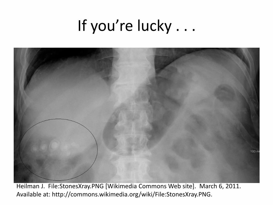
If you’re lucky . . .
Heilman J. File:StonesXray.PNG [Wikimedia Commons Web site]. March 6, 2011. Available at: http://commons.wikimedia.org/wiki/File:StonesXray.PNG.
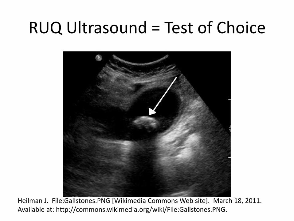
RUQ Ultrasound = Test of Choice
Heilman J. File:Gallstones.PNG [Wikimedia Commons Web site]. March 18, 2011. Available at: http://commons.wikimedia.org/wiki/File:Gallstones.PNG.
Right Upper Quadrant Pain
• 84-year-old woman
• 3 month history of diffuse abdominal pain
• 40 pound weight loss
• Exam:
– hard, nontender, baseball-sized mass in the right upper quadrant
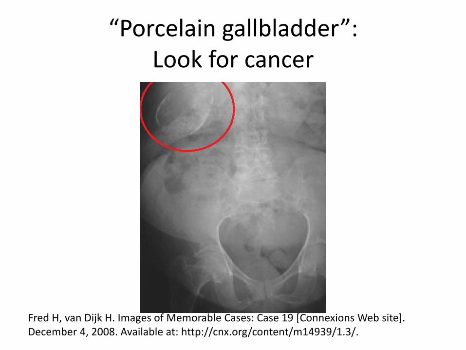
“Porcelain gallbladder”: Look for cancer
Fred H, van Dijk H. Images of Memorable Cases: Case 19 [Connexions Web site]. December 4, 2008. Available at: http://cnx.org/content/m14939/1.3/.
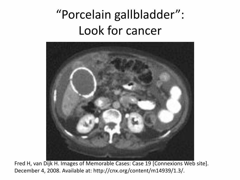
“Porcelain gallbladder”: Look for cancer
Fred H, van Dijk H. Images of Memorable Cases: Case 19 [Connexions Web site]. December 4, 2008. Available at: http://cnx.org/content/m14939/1.3/.
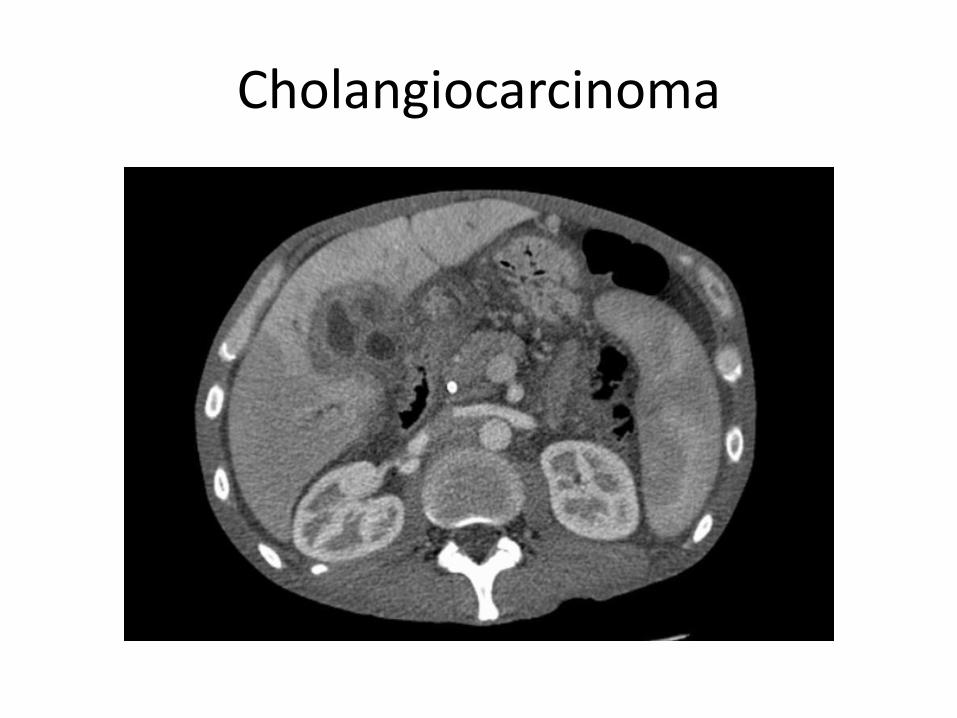
Cholangiocarcinoma
Cholangiocarcinoma
• Treatment: complete surgical resection • Generally poor prognosis
– Only 10% present at an early enough stage to consider curative resection
– 5-year survival rate up to 40% for patients with completely resected tumors
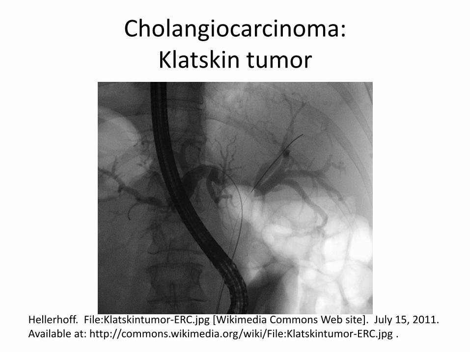
Cholangiocarcinoma: Klatskin tumor
Hellerhoff. File:Klatskintumor-ERC.jpg [Wikimedia Commons Web site]. July 15, 2011. Available at: http://commons.wikimedia.org/wiki/File:Klatskintumor-ERC.jpg .
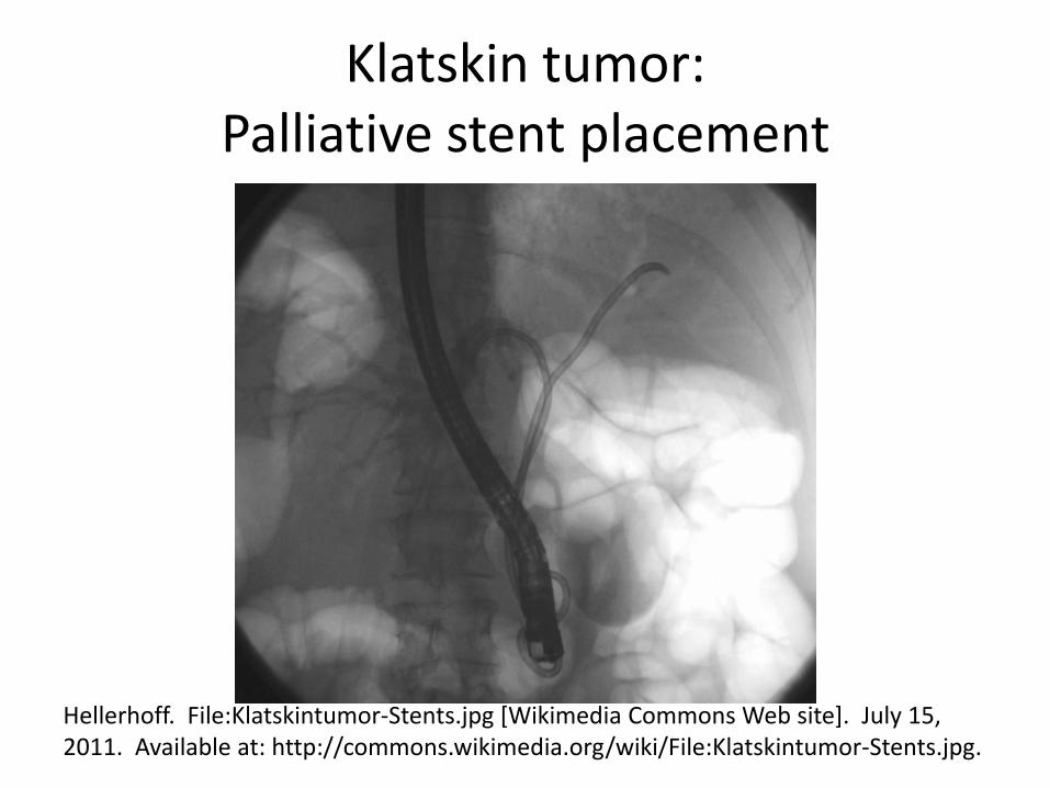
Klatskin tumor: Palliative stent placement
Hellerhoff. File:Klatskintumor-Stents.jpg [Wikimedia Commons Web site]. July 15, 2011. Available at: http://commons.wikimedia.org/wiki/File:Klatskintumor-Stents.jpg.

Right Lower Quadrant Pain
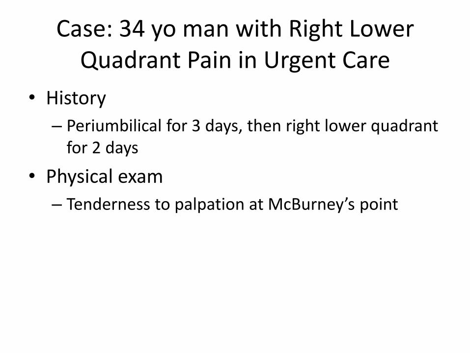
Case: 34 yo man with Right Lower Quadrant Pain in Urgent Care
• History
– Periumbilical for 3 days, then right lower quadrant for 2 days
• Physical exam
– Tenderness to palpation at McBurney’s point
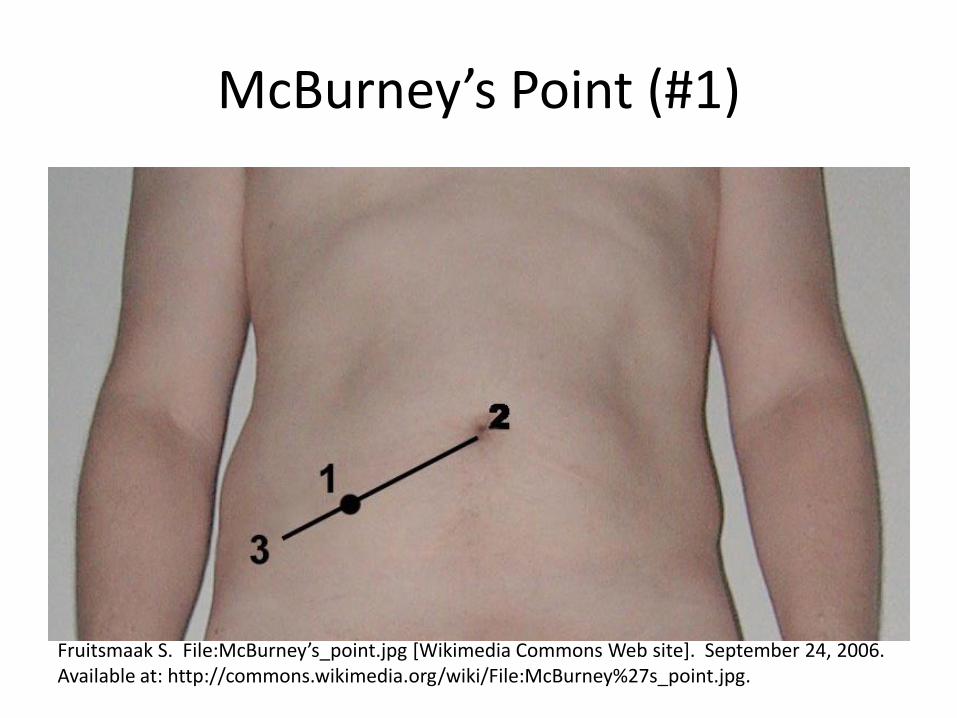
McBurney’s Point (#1)
Fruitsmaak S. File:McBurney’s_point.jpg [Wikimedia Commons Web site]. September 24, 2006. Available at: http://commons.wikimedia.org/wiki/File:McBurney%27s_point.jpg.
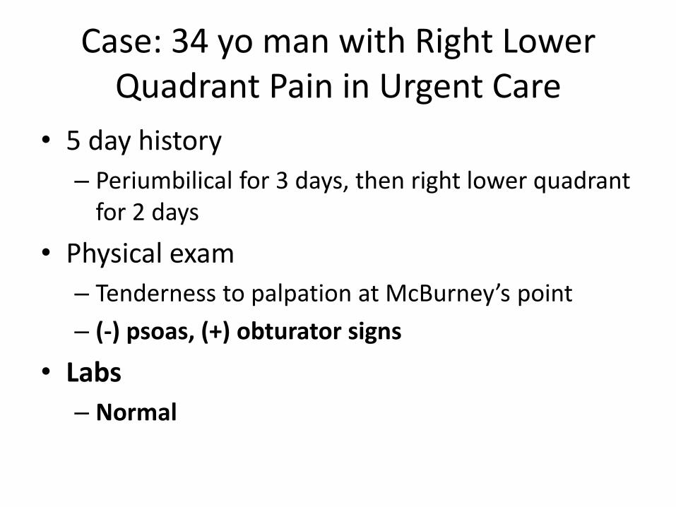
Case: 34 yo man with Right Lower Quadrant Pain in Urgent Care
• 5 day history
– Periumbilical for 3 days, then right lower quadrant for 2 days
• Physical exam
– Tenderness to palpation at McBurney’s point
– (-) psoas, (+) obturator signs
• Labs
– Normal
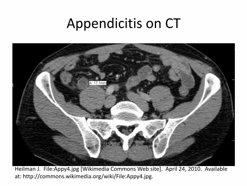
Appendicitis on CT
Heilman J. File:Appy4.jpg [Wikimedia Commons Web site]. April 24, 2010. Available at: http://commons.wikimedia.org/wiki/File:Appy4.jpg.
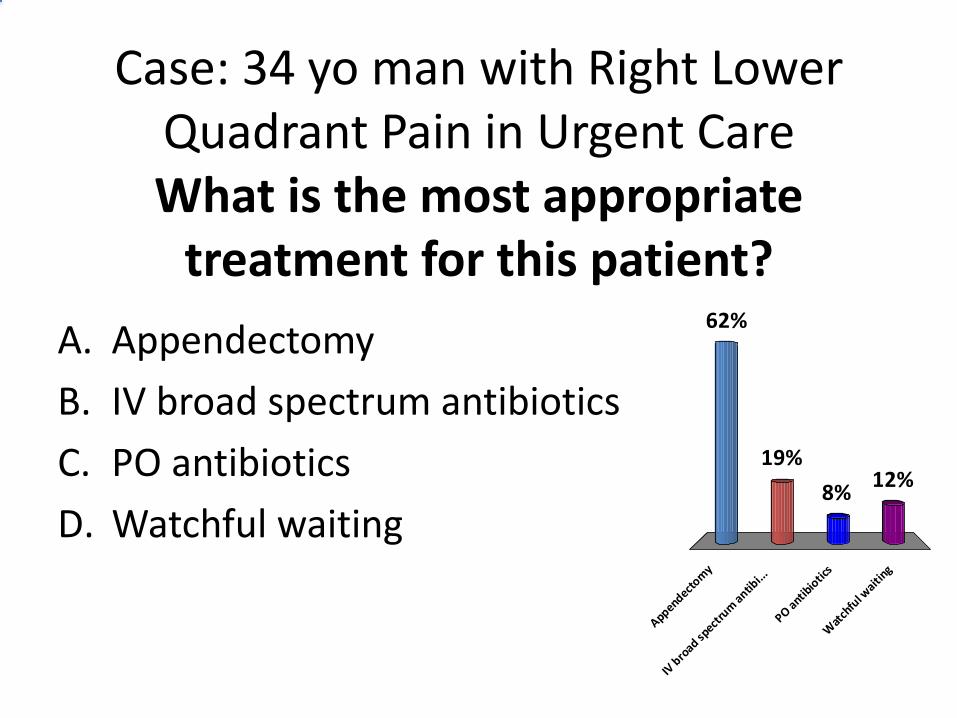
Case: 34 yo man with Right Lower Quadrant Pain in Urgent Care What is the most appropriate
treatment for this patient? A. Appendectomy
B. IV broad spectrum antibiotics
C. PO antibiotics
D. Watchful waiting
Appendectom
y
IV b
road sp
ectru
m antib
i...
PO antib
iotic
s
Wat
chfu
l waiti
ng
62%
12%8%
19%
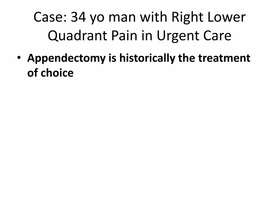
Case: 34 yo man with Right Lower Quadrant Pain in Urgent Care
• Appendectomy is historically the treatment of choice
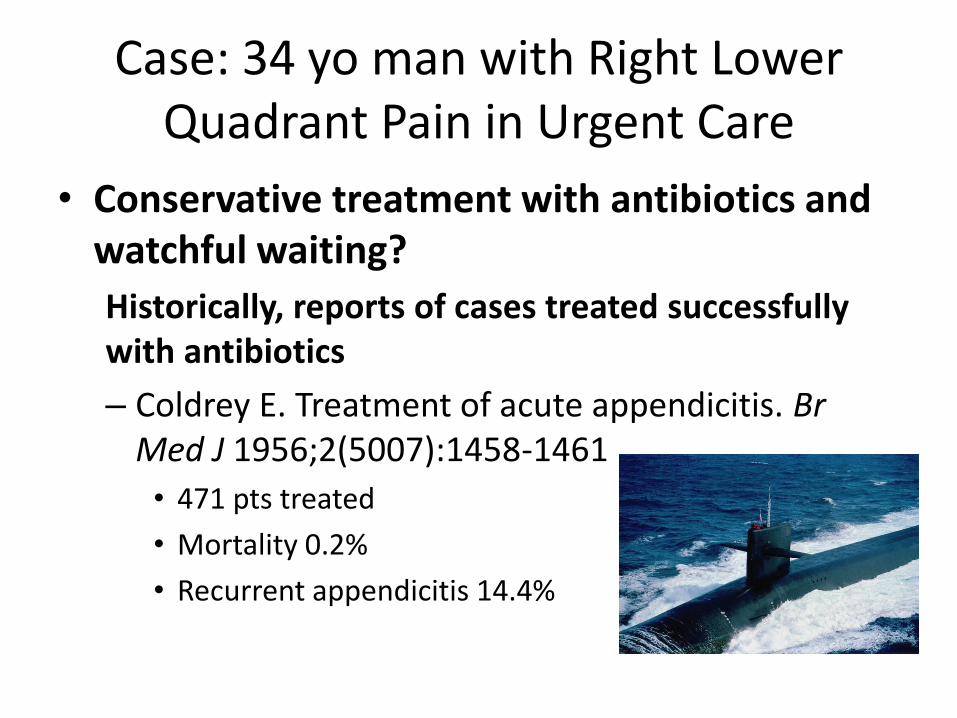
Case: 34 yo man with Right Lower Quadrant Pain in Urgent Care
• Conservative treatment with antibiotics and watchful waiting?
Historically, reports of cases treated successfully with antibiotics
– Coldrey E. Treatment of acute appendicitis. Br Med J 1956;2(5007):1458-1461
• 471 pts treated
• Mortality 0.2%
• Recurrent appendicitis 14.4%
Case: 34 yo man with Right Lower Quadrant Pain in Urgent Care
• Conservative treatment with antibiotics and watchful waiting?
– Antibiotics not definitively “non-inferior” to surgery
Wilms IM, de Hoog DE, de Visser DC, Janzing HM. Appendectomy versus antibiotic treatment for acute appendicitis. Cochrane Database Syst Rev. 2011 Nov 9;(11):CD008359.
Appendicitis: Antibiotics vs. Surgery
• APPAC: most recent RCT
– Excluded “complicated” appendicitis: appendicolith, perforation, abscess, or suspicion of tumor
– Did not demonstrate “noninferiority” of antibiotics:
27% randomized to antibiotics had surgery within 1 year of presentation (≤24% required for “noninferiority”)
– Surgical complication rates: Antibiotic group 7.0%, Surgery group 20.5% (p=0.02)
Salminen P, Paajanen H, Rautio T et al. Antibiotic Therapy vs Appendectomy for Treatment of Uncomplicated Acute Appendicitis: The APPAC Randomized Clinical Trial. JAMA. 2015;313(23):2340-2348.
Appendicitis: Red Flags
• Signs of rupture
– Change in condition:
• Fever
• Increased pain
• Abdominal rigidity
– Could see improvement in pain (think of a walled-off ruptured abscess) until peritonitis more fully develops
Appendicitis: Red Flags
• Higher proportion of patients with ruptured appendicitis at the extremes of age (early childhood, elderly)
– May be due to lower incidence, because absolute rate of rupture is constant across ages
Psychopoesie. File:Grandma&me_at_my_cousin’s_wedding.jpg [Wikimedia Commons Web site]. October 31, 2011. Available at: http://commons.wikimedia.org/wiki/File:Grandma%26me_at_my_cousin%27s_wedding.jpg .

Chan Ho Park
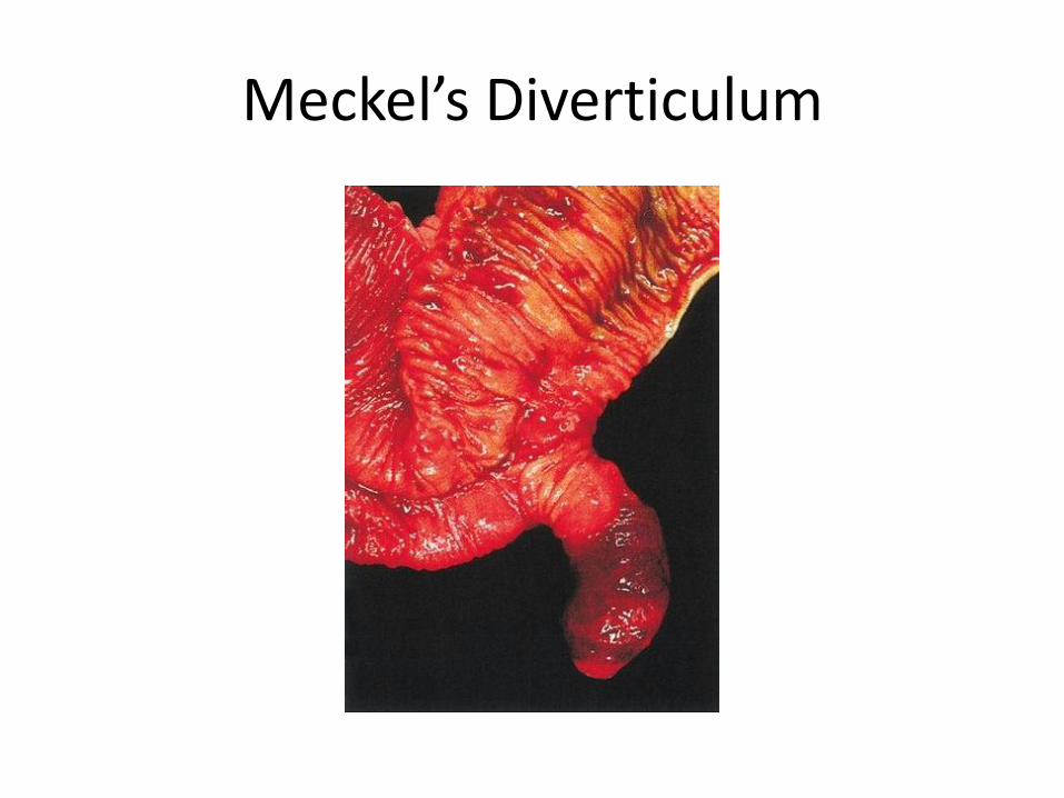
Meckel’s Diverticulum
Meckel’s Diverticulum: Rule of 2’s
• 2% prevalence
• 2 years of age at presentation
• 2 feet from the ileocecal junction
• 2 inches in length
• 2 types of common ectopic tissue – Gastric
– Pancreatic
• 2% symptomatic
• 2 times more symptomatic in boys

Left Lower Quadrant Pain
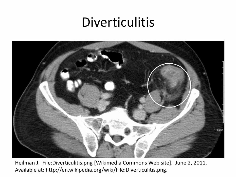
Diverticulitis
• Typical story: – Acute constant abdominal pain in LLQ
– Fever
– Can also see nausea, vomiting, constipation, diarrhea
– “Sympathetic cystitis”: dysuria and frequency caused by bladder irritation from inflamed colon
• Typical physical exam findings: – LLQ tenderness, guarding, rebound
Diverticulitis
• Diagnostics:
– Leukocytosis
– CT of abdomen and pelvis with contrast
Diverticulitis
Heilman J. File:Diverticulitis.png [Wikimedia Commons Web site]. June 2, 2011. Available at: http://en.wikipedia.org/wiki/File:Diverticulitis.png.
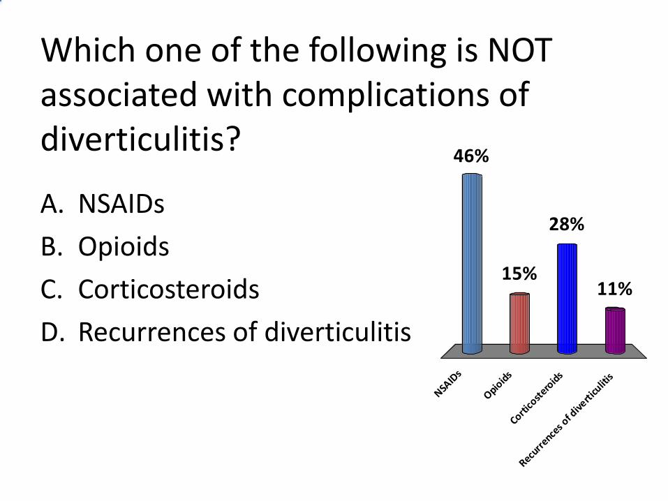
Which one of the following is NOT associated with complications of diverticulitis?
A. NSAIDs
B. Opioids
C. Corticosteroids
D. Recurrences of diverticulitis
NSAID
s
Opioid
s
Cortico
stero
ids
Recurr
ences o
f dive
rticu
litis
46%
11%
28%
15%
Diverticulitis
• Risk factors: Smoking, obesity
• Negative risk factor: Increased physical activity
• Associated with complications: – Yes: NSAIDs, opioids, corticosteroids
– No: Recurrences
• Recurrences are uncommon (13.3%) & not clustered
Morris AM, Regenbogen SE, Hardiman KM, Hendren S. Sigmoid Diverticulitis: A Systematic Review. JAMA. 2014;311(3):287-297.
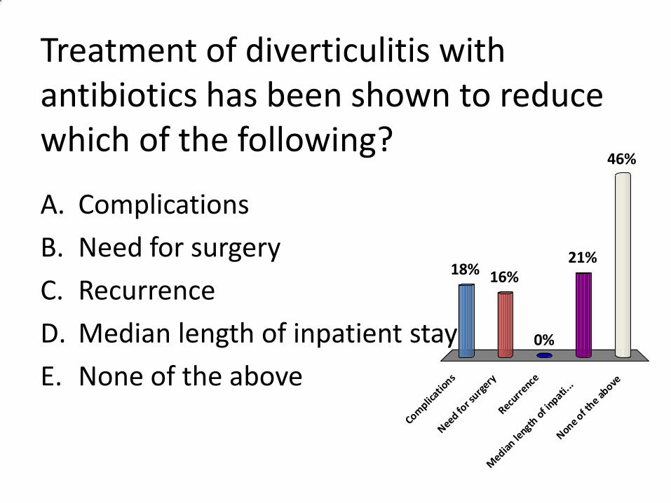
Treatment of diverticulitis with antibiotics has been shown to reduce which of the following?
A. Complications
B. Need for surgery
C. Recurrence
D. Median length of inpatient stay
E. None of the above
Complic
ations
Need for s
urgery
Recurr
ence
Median
length
of i
npati...
None of t
he above
18% 16%
46%
21%
0%
Diverticulitis: Treatment
• Uncomplicated – Stable, tolerating oral fluids: outpatient
Traditionally: PO antibiotics x 7-10 d., clear liquid diet
More recent evidence questions role of antibiotics
- Cochrane review – best available data do not support
- No effect on complications, need for surgery, recurrence, median length of inpatient stay
– Older or ill pts, not tolerating fluids: admit IV fluids, bowel rest/NPO, ? Antibiotics
Chabok A, Pahlman L, Hjern F et al. Randomized clinical trial of antibiotics for acute uncomplicated diverticulitis. Br J Surg 2012;99(4):532-539.
Shabanzadeh DM, Wille-Jorgensen P. Antibiotics for uncomplicated diverticulitis. Cochrane Database Syst Rev. 2012 Nov 14;11:CD009092.
Diverticulitis: Treatment
• Complicated (sepsis, perforation, abscess, fistula, obstruction) • stabilize, IV fluids, antibiotics, surgical consultation,
percutaneous drainage, intraperitoneal lavage
• Antibiotics to cover anaerobes, gram negative rods: – Metronidazole or clindamycin (Cleocin) –PLUS one of the
following: aminoglycoside, monobactam (aztreonam), or third generation cephalosporin
– Second generation cephalosporin
– Extended spectrum penicillin/beta-lactamase inhibitor combinations
– Newer evidence: ertapenem, rifaximin
Diverticulitis: Treatment
• Other nonoperative treatments
– Probiotics: reduce chronic symptoms but not recurrences
– Antiinflammatory medications: mesalamine + rifaximin reduces recurrences vs. rifaximin alone
Diverticulitis: Treatment
• Indications for surgery
– Sepsis, acute peritonitis
– No improvement with medical therapy, percutaneous drainage, or both
– Trend toward minimally invasive surgical techniques
Regenbogen SE, Hardiman KM, Hendren S, Morris AM. Surgery for Diverticulitis in the 21st Century: A Systematic Review. JAMA Surg. 2014;149(3):292-303.
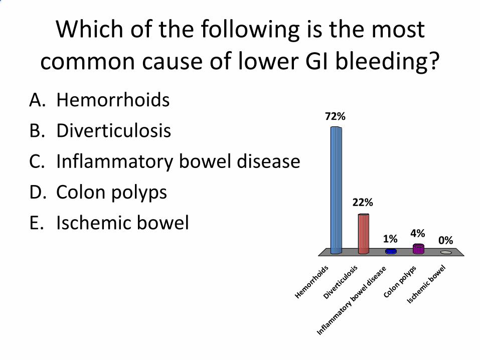
Which of the following is the most common cause of lower GI bleeding?
A. Hemorrhoids
B. Diverticulosis
C. Inflammatory bowel disease
D. Colon polyps
E. Ischemic bowel
Hemorrh
oids
Diverti
culo
sis
Infla
mm
atory
bow
el dise
ase
Colon p
olyps
Ischem
ic bow
el
72%
22%
0%4%1%
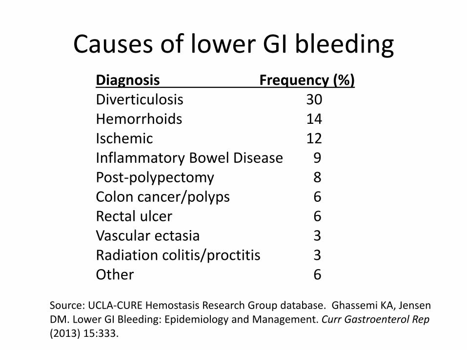
Causes of lower GI bleeding Diagnosis Frequency (%) Diverticulosis 30 Hemorrhoids 14 Ischemic 12 Inflammatory Bowel Disease 9 Post-polypectomy 8 Colon cancer/polyps 6 Rectal ulcer 6 Vascular ectasia 3 Radiation colitis/proctitis 3 Other 6
Source: UCLA-CURE Hemostasis Research Group database. Ghassemi KA, Jensen DM. Lower GI Bleeding: Epidemiology and Management. Curr Gastroenterol Rep (2013) 15:333.
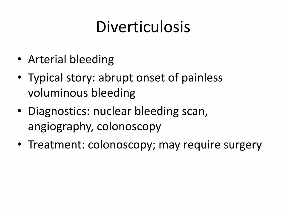
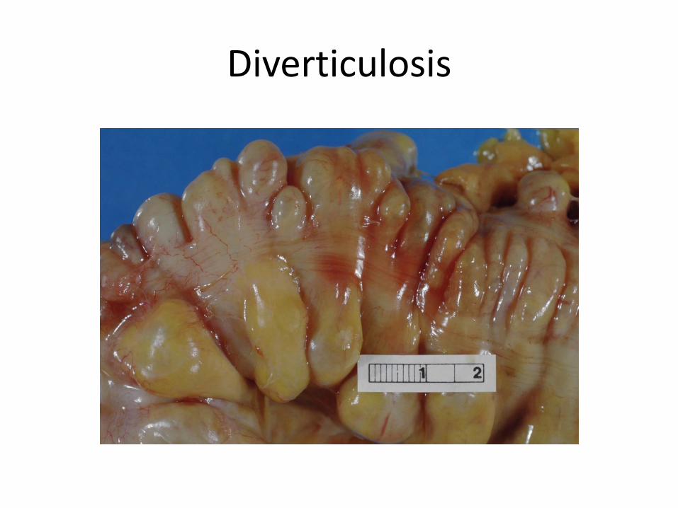
Diverticulosis
• Arterial bleeding
• Typical story: abrupt onset of painless voluminous bleeding
• Diagnostics: nuclear bleeding scan, angiography, colonoscopy
• Treatment: colonoscopy; may require surgery
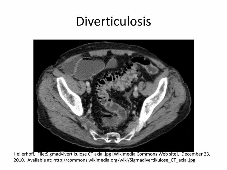
Diverticulosis
Hellerhoff. File:Sigmadvivertikulose CT axial.jpg [Wikimedia Commons Web site]. December 23, 2010. Available at: http://commons.wikimedia.org/wiki/Sigmadivertikulose_CT_axial.jpg.
Diverticulosis
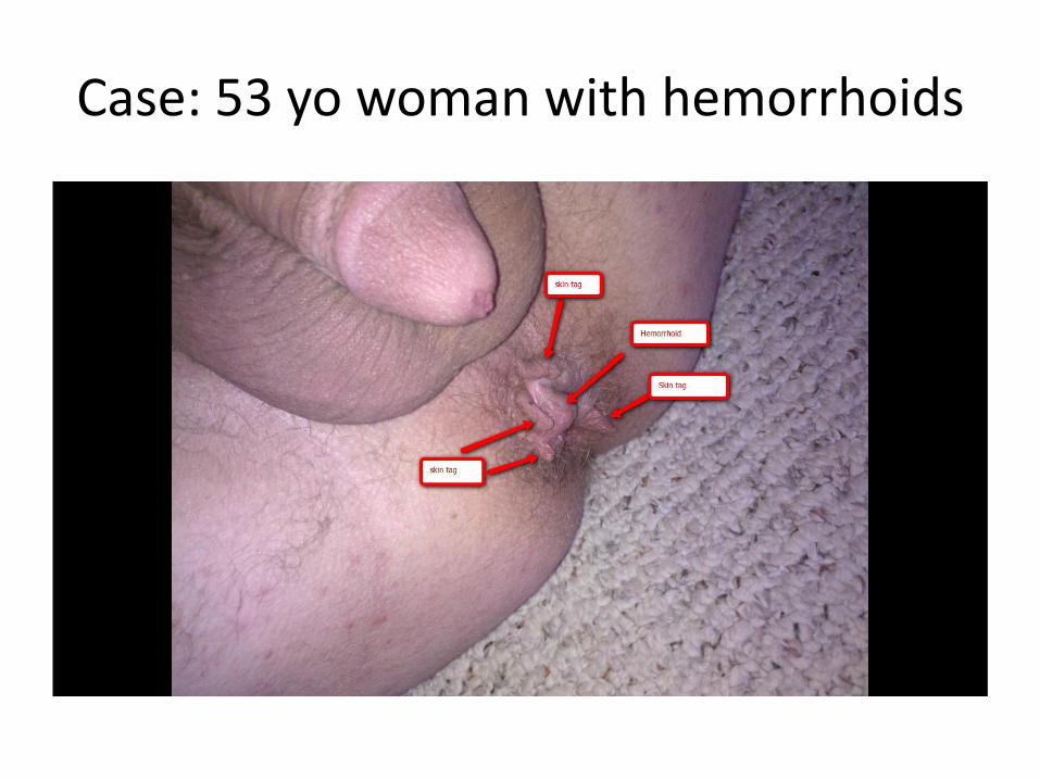
Case: 53 yo woman with hemorrhoids
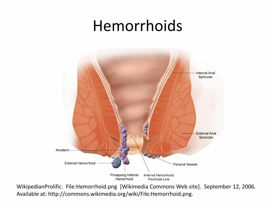
Hemorrhoids
WikipedianProlific. File:Hemorrhoid.png [Wikimedia Commons Web site]. September 12, 2006. Available at: http://commons.wikimedia.org/wiki/File:Hemorrhoid.png.
Volvulus
• Midgut volvulus from malrotation of the gut
• Sigmoid volvulus
Midgut Volvulus: Malrotation of the Gut
• Typical story:
– 1st month of life: bilious vomiting, feeding intolerance, sudden onset of abdominal pain, upper abdominal distention
– Older children: More vague (chronic, unexplained) abdominal pain, irritability, anorexia, nausea/vomiting, failure to thrive
Shalaby MS, Kuti K, Walker G. Intestinal malrotation and volvulus in infants and children BMJ 2013;347:f6949
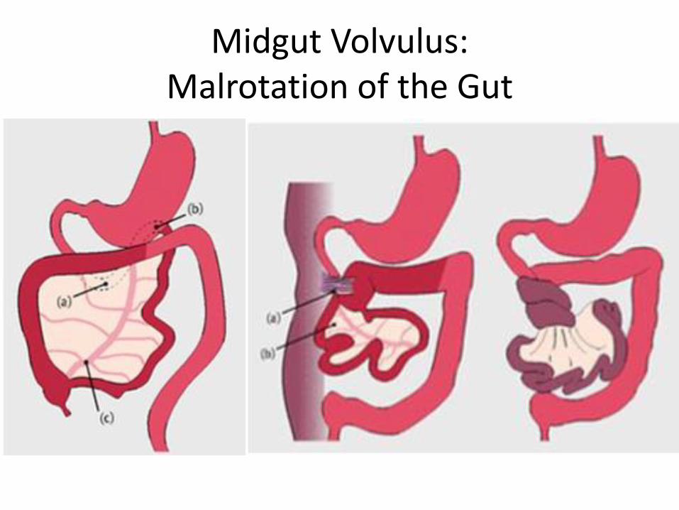
Midgut Volvulus: Malrotation of the Gut
Midgut Volvulus: Malrotation of the Gut
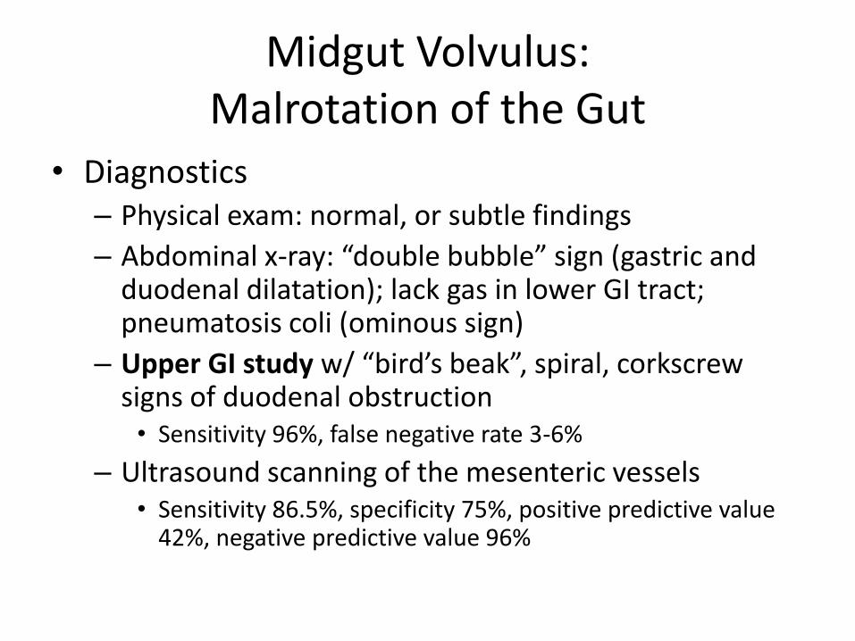
• Diagnostics – Physical exam: normal, or subtle findings
– Abdominal x-ray: “double bubble” sign (gastric and duodenal dilatation); lack gas in lower GI tract; pneumatosis coli (ominous sign)
– Upper GI study w/ “bird’s beak”, spiral, corkscrew signs of duodenal obstruction • Sensitivity 96%, false negative rate 3-6%
– Ultrasound scanning of the mesenteric vessels • Sensitivity 86.5%, specificity 75%, positive predictive value
42%, negative predictive value 96%
Midgut Volvulus: Malrotation of the Gut
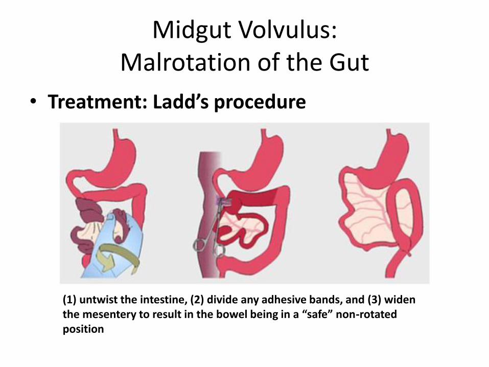
• Treatment: Ladd’s procedure
(1) untwist the intestine, (2) divide any adhesive bands, and (3) widen the mesentery to result in the bowel being in a “safe” non-rotated position
Sigmoid Volvulus
• Older patients
• Typical story – sx of bowel obstruction/ischemia:
– Abdominal pain, distention, inability to pass stool or flatus (obstipation), history of constipation
– Vomiting may be late presenting feature
• Diagnostics: abdominal x-ray shows distended sigmoid colon
• Treatment: sigmoidoscopy/rectal tube placement; resection & primary anastomosis
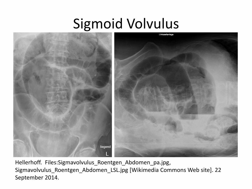
Sigmoid Volvulus
Hellerhoff. Files:Sigmavolvulus_Roentgen_Abdomen_pa.jpg, Sigmavolvulus_Roentgen_Abdomen_LSL.jpg [Wikimedia Commons Web site]. 22 September 2014.
Epigastric Pain
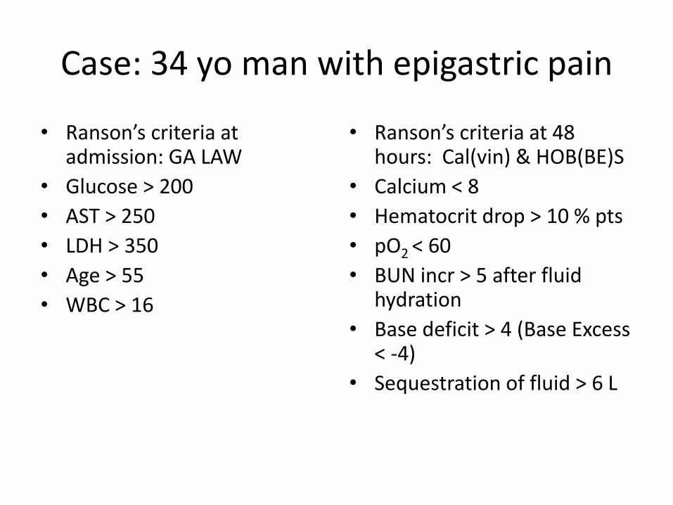
Case: 34 yo man with epigastric pain
• Ranson’s criteria at admission: GA LAW
• Glucose > 200
• AST > 250
• LDH > 350
• Age > 55
• WBC > 16
• Ranson’s criteria at 48 hours: Cal(vin) & HOB(BE)S
• Calcium < 8
• Hematocrit drop > 10 % pts
• pO2 < 60
• BUN incr > 5 after fluid hydration
• Base deficit > 4 (Base Excess < -4)
• Sequestration of fluid > 6 L
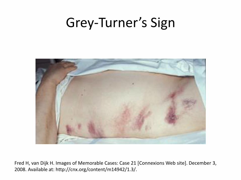
Grey-Turner’s Sign
Fred H, van Dijk H. Images of Memorable Cases: Case 21 [Connexions Web site]. December 3, 2008. Available at: http://cnx.org/content/m14942/1.3/.
Grey Turner’s Sign
The correct eponym for bruising of the flanks caused by acute pancreatitis or other causes is
A. Grey Turner’s Sign
B. Grey-Turner’s Sign
C. Gray Turner’s Sign
D. Gray-Turner’s Sign
E. Turner’s Sign
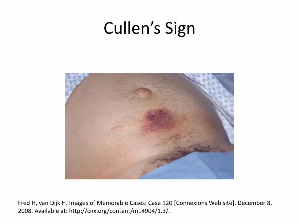
Cullen’s Sign
Fred H, van Dijk H. Images of Memorable Cases: Case 120 [Connexions Web site]. December 8, 2008. Available at: http://cnx.org/content/m14904/1.3/.
Pancreatitis
• Surgery indicated for infected necrosis
– 80% of deaths from acute pancreatitis caused by infection of dead pancreatic tissue
• Pancreatic pseudocysts
– Endoscopic drainage as effective as surgery, both more effective than percutaneous drainage
Johnson MD, Walsh RM, Henderson JM, et al. Surgical versus nonsurgical management of pancreatic pseudocysts. J Clin Gastroenterol 2009 Jul;43(6):586-90.
Peptic Ulcer Disease
• Surgery rarely needed
• Vagotomy
• Gastrectomy
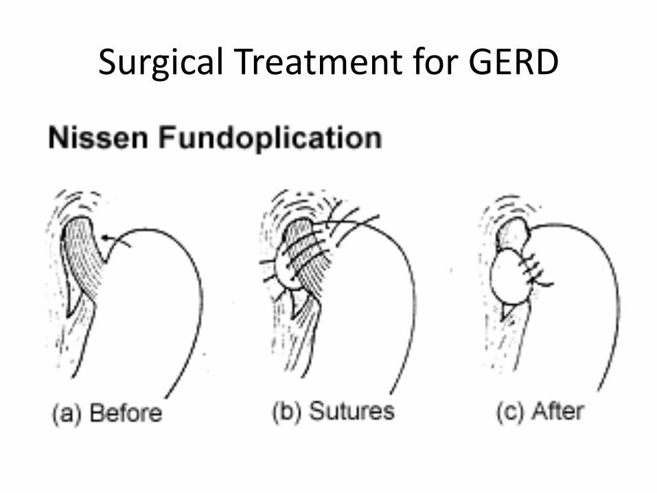
Surgical Treatment for GERD
Surgical Treatment for GERD
• Unresponsive to aggressive antisecretory therapy (proton pump inhibitors)
• After surgery, some patients still require antisecretory therapy
• Potential obstructive complications of Nissen:
– dysphagia
– rectal flatulence
– inability to belch or vomit
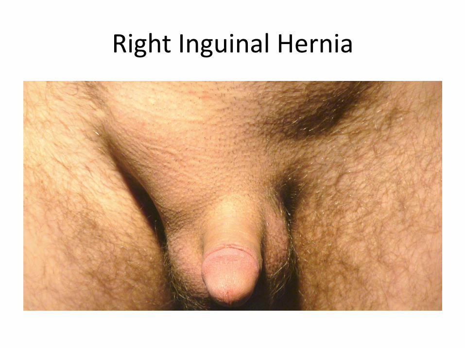
Right Inguinal Hernia
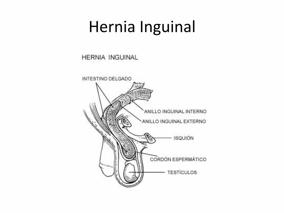
Hernia Inguinal
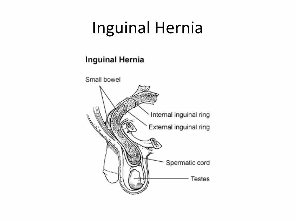
Inguinal Hernia
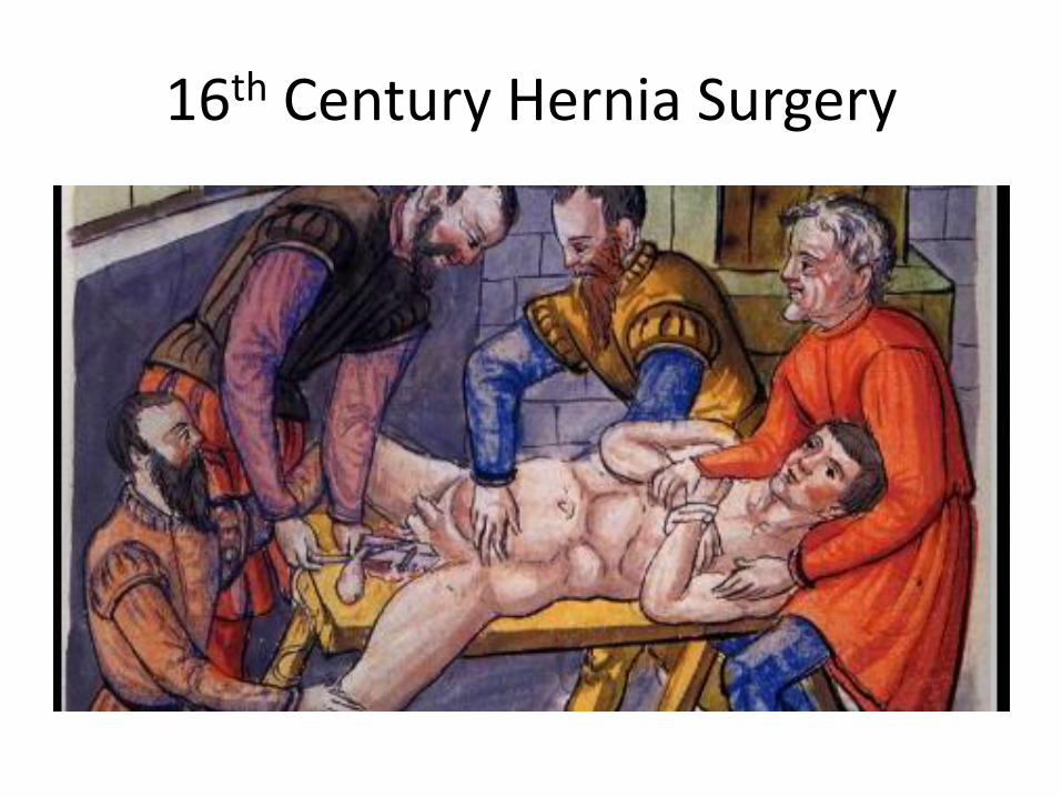
16th Century Hernia Surgery
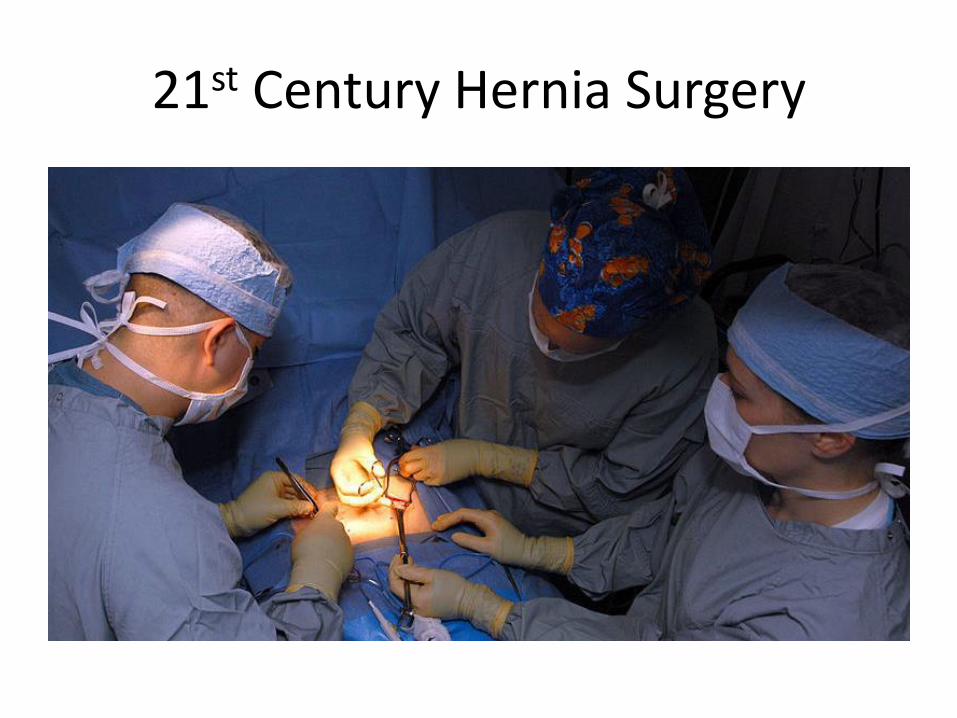
21st Century Hernia Surgery
Hernia Surgery
• Indications for surgery
– Emergent
• Strangulated hernias
–Nonreducible bulge with pain, sometimes after heavy lifting
– Urgent
• Incarcerated hernias
Hernia Surgery
• Indications for surgery
– Elective
• Inguinal hernias – watchful waiting recommended
• Femoral hernias – higher risk of strangulation
• Ventral hernias
• Umbilical
–Normally resolve without intervention by age 5
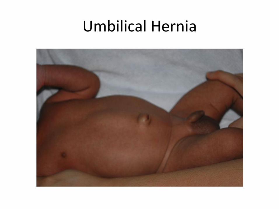
Umbilical Hernia
Hernia Surgery
• What about mesh?
– Fewer recurrences after mesh repair
Scott N, Go PM.N.Y.H, Graham P, McCormack K, Ross SJ, Grant AM. Open Mesh versus non-Mesh for groin hernia repair. Cochrane Database of Systematic Reviews 2002, Issue 4. Art. No.: CD002197. DOI: 10.1002/14651858.CD002197
Case: 6 year old boy with severe abdominal pain in the Peds ED
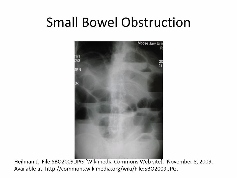
Small Bowel Obstruction
Heilman J. File:SBO2009.JPG [Wikimedia Commons Web site]. November 8, 2009. Available at: http://commons.wikimedia.org/wiki/File:SBO2009.JPG.
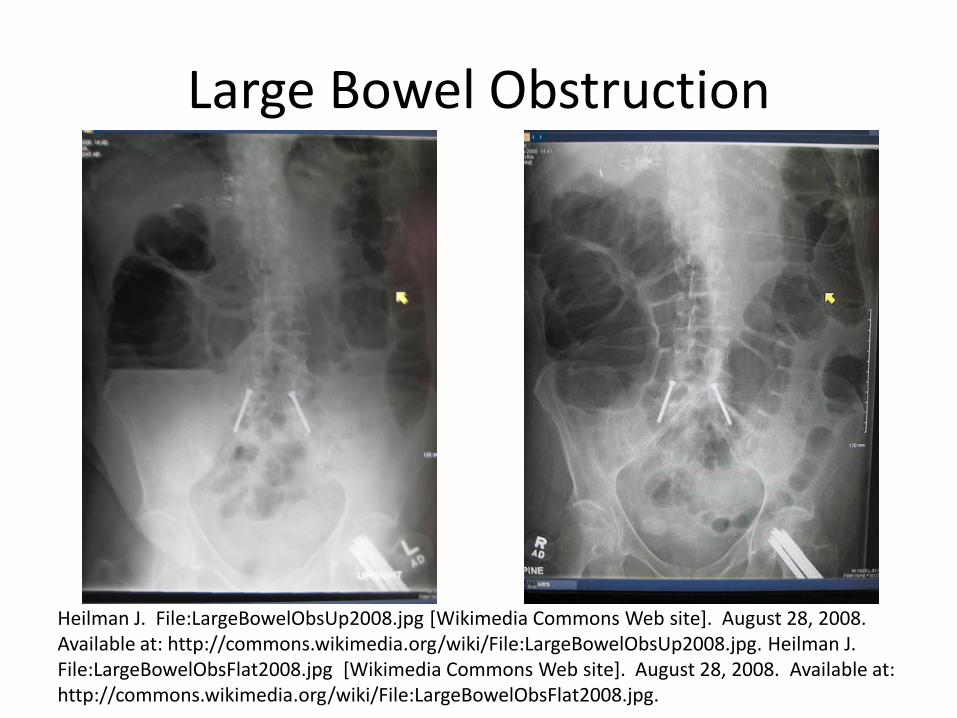
Large Bowel Obstruction
Heilman J. File:LargeBowelObsUp2008.jpg [Wikimedia Commons Web site]. August 28, 2008. Available at: http://commons.wikimedia.org/wiki/File:LargeBowelObsUp2008.jpg. Heilman J. File:LargeBowelObsFlat2008.jpg [Wikimedia Commons Web site]. August 28, 2008. Available at: http://commons.wikimedia.org/wiki/File:LargeBowelObsFlat2008.jpg.
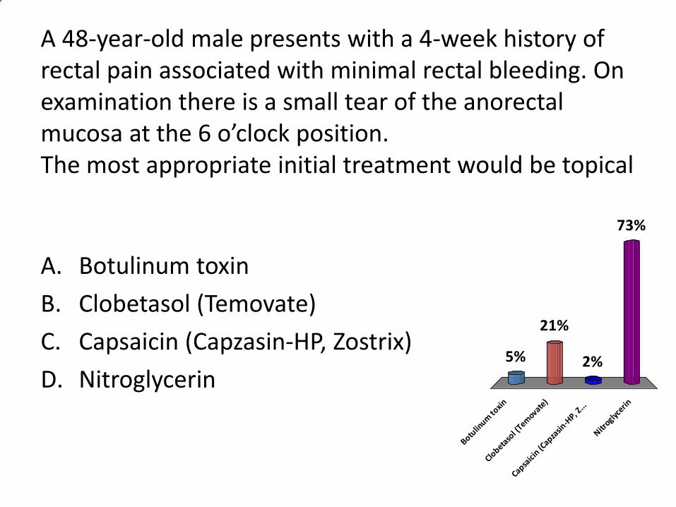
A 48-year-old male presents with a 4-week history of rectal pain associated with minimal rectal bleeding. On examination there is a small tear of the anorectal mucosa at the 6 o’clock position. The most appropriate initial treatment would be topical
A. Botulinum toxin
B. Clobetasol (Temovate)
C. Capsaicin (Capzasin-HP, Zostrix)
D. Nitroglycerin
Botulin
um to
xin
Clobeta
sol (
Temovate
)
Capsaici
n (Capza
sin-H
P, Z...
Nitroglyc
erin
5%
73%
2%
21%
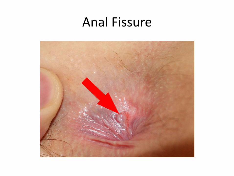
Anal Fissure
Anal Fissure
• Nonsurgical measures that are proven effective in relaxing the sphincter: – Topical nitroglycerin ointment – Diltiazem, nifedipine (topical preparations usually
have to be compounded by a pharmacist) – Botulinum toxin injected into the internal sphincter – Corticosteroid creams may decrease the pain
temporarily • Surgery: internal sphincterotomy Fargo MV, Latimer KM: Evaluation and management of common anorectal conditions. Am Fam Physician 2012;85(6):624-630.
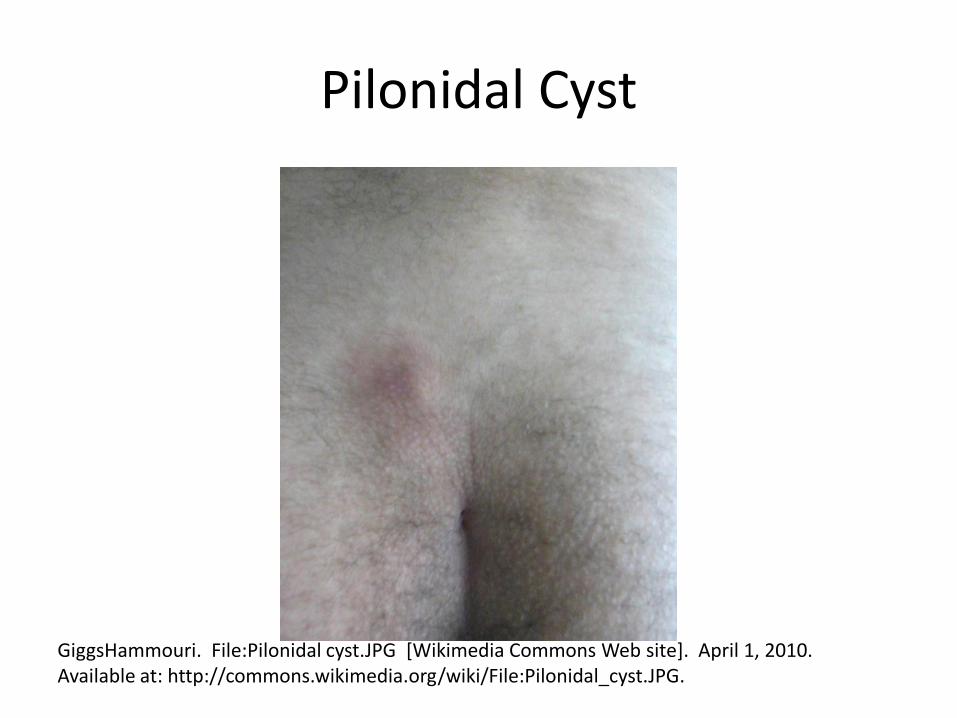
Pilonidal Cyst
GiggsHammouri. File:Pilonidal cyst.JPG [Wikimedia Commons Web site]. April 1, 2010. Available at: http://commons.wikimedia.org/wiki/File:Pilonidal_cyst.JPG.
PREOP/PERIOP/POSTOP CARE WOUNDS INFECTIONS
Preoperative Workup
• Source #1: 2014 ACC/AHA Guidelines on Perioperative Cardiovascular Evaluation and Care for Noncardiac Surgery
Fleisher LA, Fleischmann KE, Auerbach AD, et al. 2014 ACC/AHA guidelines on perioperative cardiovascular evaluation and care for noncardiac surgery: a report of the American College of Cardiology/American Heart Association Task Force on Practice Guidelines (Writing Committee to Revise the 2007 Guidelines on Perioperative Cardiovascular Evaluation for Noncardiac Surgery) Circulation. 2014;130:e278-e333
Preoperative Workup
• Source #2: Feely MA, Collins CS, Daniels PR, et al. Preoperative Testing Before Noncardiac Surgery: Guidelines and Recommendations. Am Fam Physician. 2013 Mar 15;87(6):414-418.
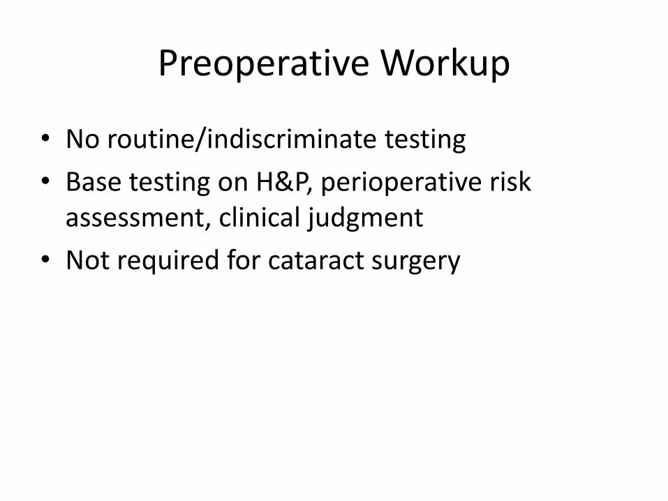
Preoperative Workup
• No routine/indiscriminate testing
• Base testing on H&P, perioperative risk assessment, clinical judgment
• Not required for cataract surgery
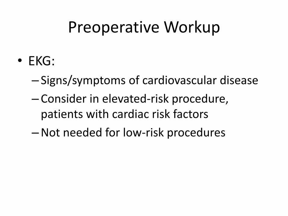
Preoperative Workup
• EKG:
– Signs/symptoms of cardiovascular disease
–Consider in elevated-risk procedure, patients with cardiac risk factors
–Not needed for low-risk procedures
Preoperative Workup
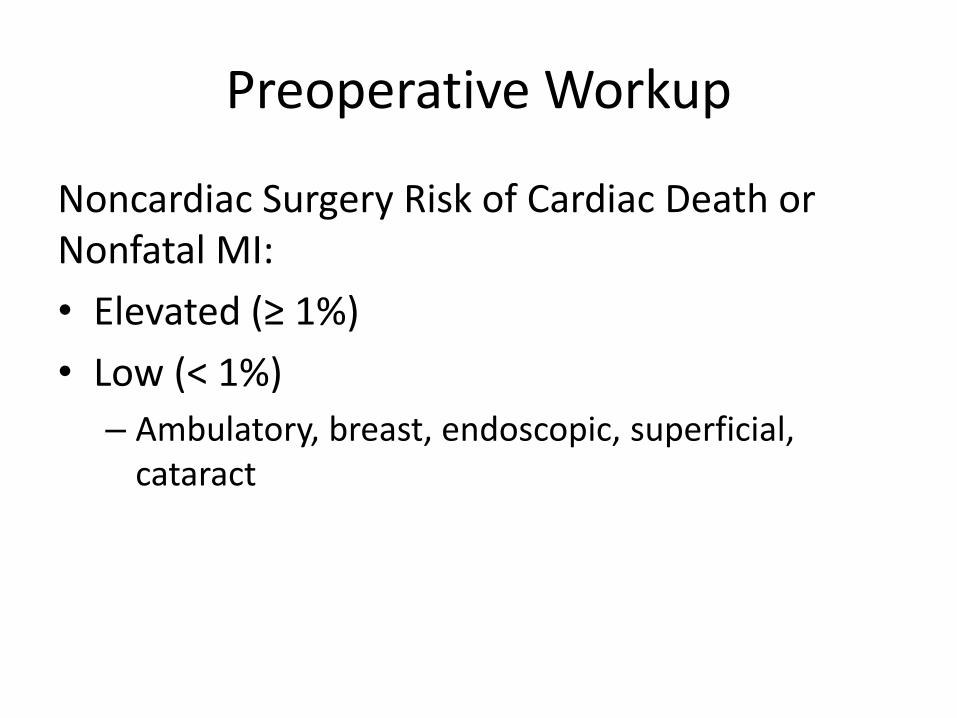
Noncardiac Surgery Risk of Cardiac Death or Nonfatal MI:
• Elevated (≥ 1%)
• Low (< 1%)
– Ambulatory, breast, endoscopic, superficial, cataract
Preoperative Workup
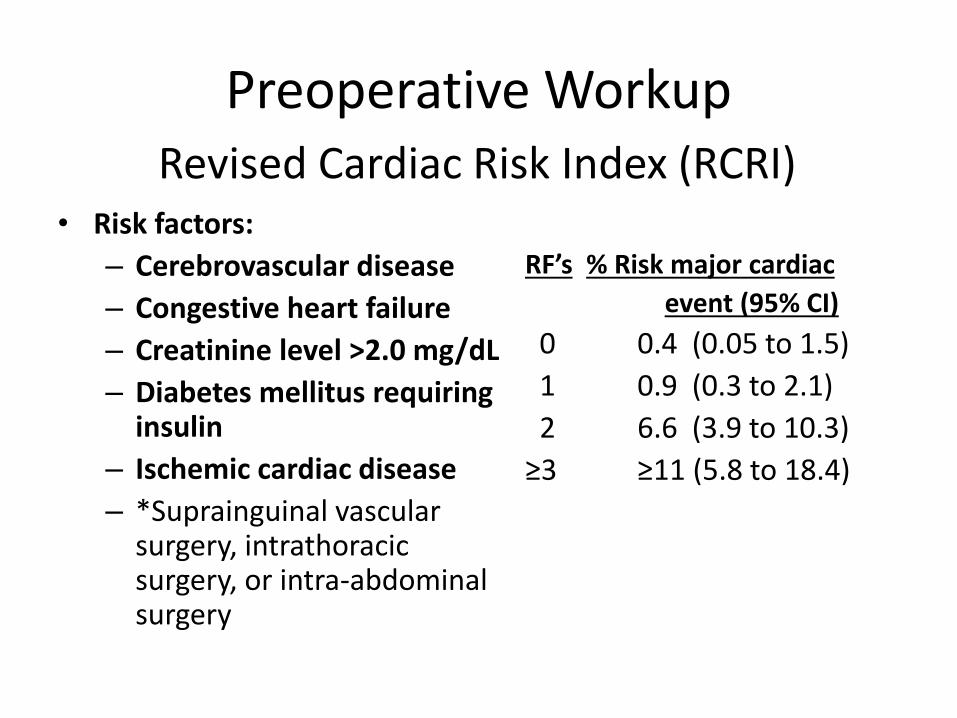
• Risk factors:
– Cerebrovascular disease
– Congestive heart failure
– Creatinine level >2.0 mg/dL
– Diabetes mellitus requiring insulin
– Ischemic cardiac disease
– *Suprainguinal vascular surgery, intrathoracic surgery, or intra-abdominal surgery
RF’s % Risk major cardiac
event (95% CI)
0 0.4 (0.05 to 1.5)
1 0.9 (0.3 to 2.1)
2 6.6 (3.9 to 10.3)
≥3 ≥11 (5.8 to 18.4)
Revised Cardiac Risk Index (RCRI)
Preoperative Workup
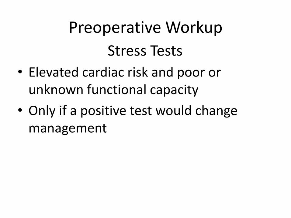
• Elevated cardiac risk and poor or unknown functional capacity
• Only if a positive test would change management
Stress Tests
Preoperative Workup
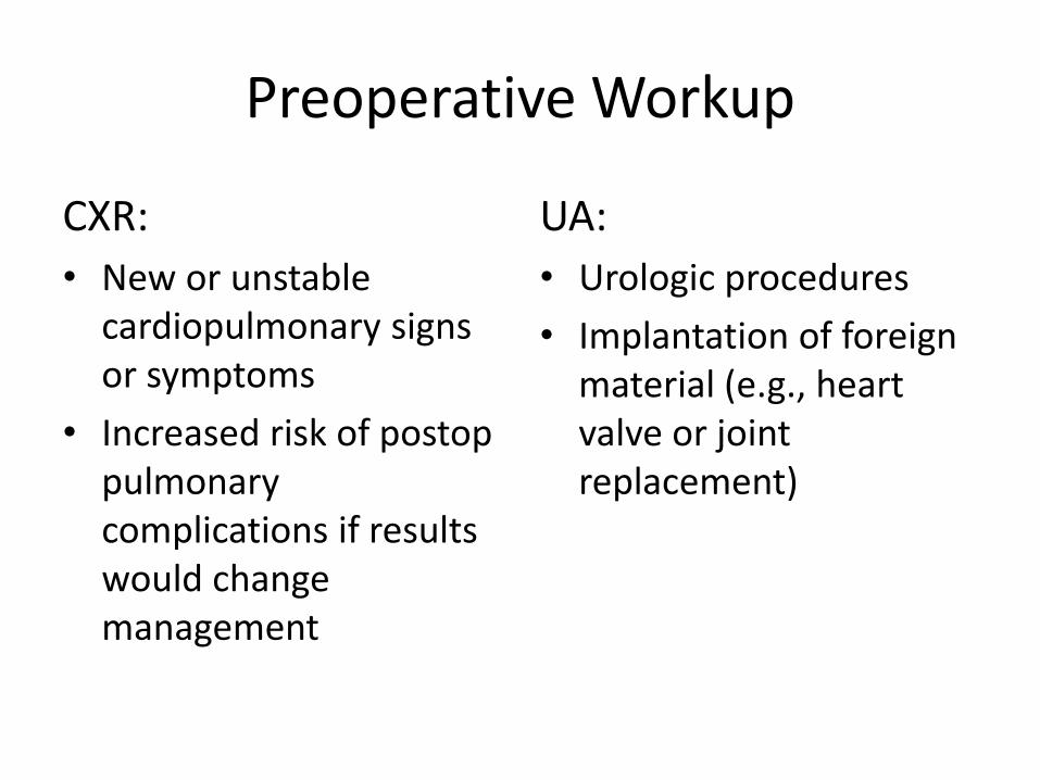
CXR:
• New or unstable cardiopulmonary signs or symptoms
• Increased risk of postop pulmonary complications if results would change management
UA:
• Urologic procedures
• Implantation of foreign material (e.g., heart valve or joint replacement)
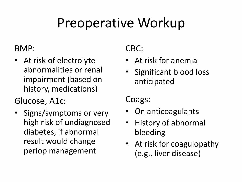
Preoperative Workup
BMP: • At risk of electrolyte
abnormalities or renal impairment (based on history, medications)
Glucose, A1c: • Signs/symptoms or very
high risk of undiagnosed diabetes, if abnormal result would change periop management
CBC: • At risk for anemia
• Significant blood loss anticipated
Coags: • On anticoagulants
• History of abnormal bleeding
• At risk for coagulopathy (e.g., liver disease)
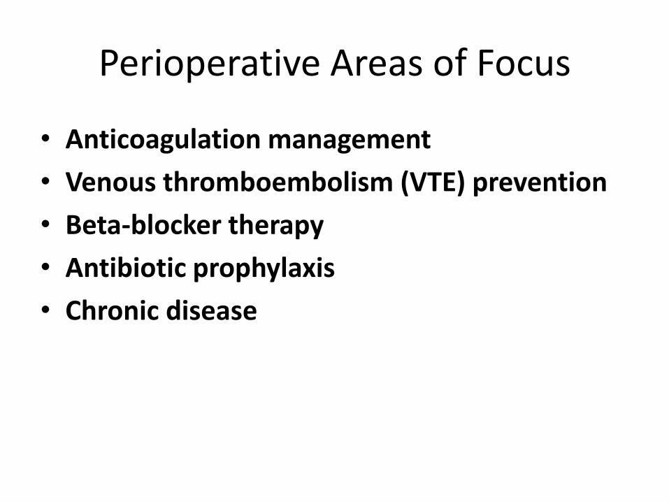
Perioperative Areas of Focus
• Anticoagulation management
• Venous thromboembolism (VTE) prevention
• Beta-blocker therapy
• Antibiotic prophylaxis
• Chronic disease
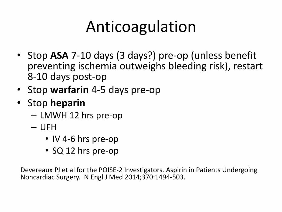
Anticoagulation
• Stop ASA 7-10 days (3 days?) pre-op (unless benefit preventing ischemia outweighs bleeding risk), restart 8-10 days post-op
• Stop warfarin 4-5 days pre-op • Stop heparin
– LMWH 12 hrs pre-op – UFH
• IV 4-6 hrs pre-op • SQ 12 hrs pre-op
Devereaux PJ et al for the POISE-2 Investigators. Aspirin in Patients Undergoing Noncardiac Surgery. N Engl J Med 2014;370:1494-503.
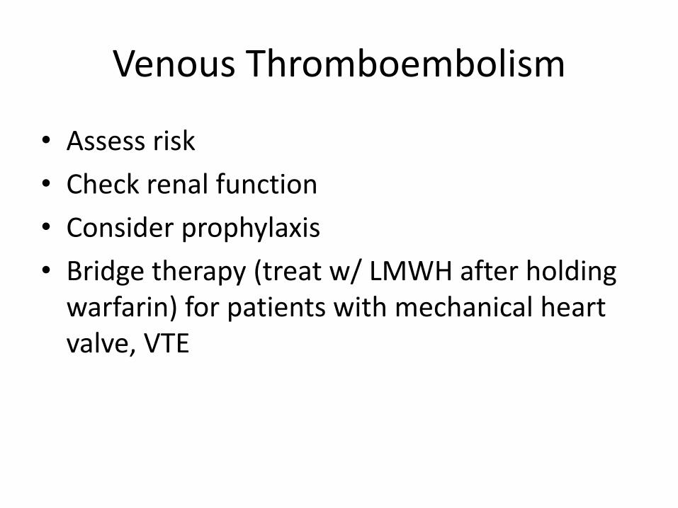
Venous Thromboembolism
• Assess risk
• Check renal function
• Consider prophylaxis
• Bridge therapy (treat w/ LMWH after holding warfarin) for patients with mechanical heart valve, VTE
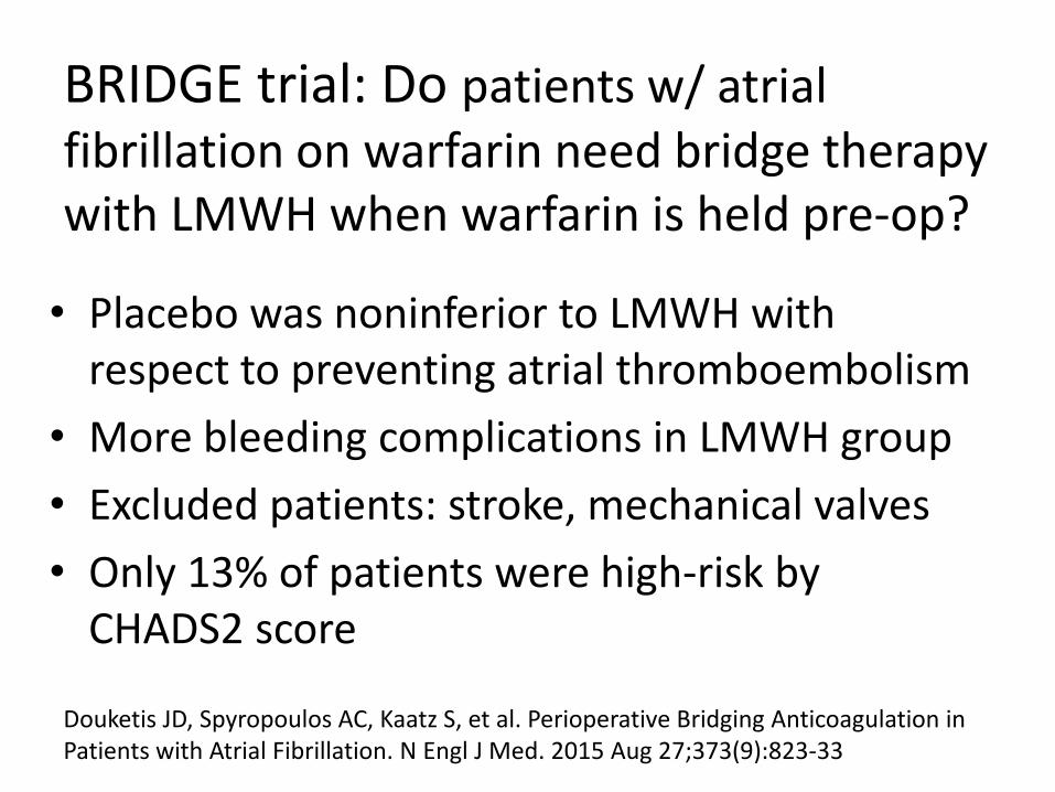
BRIDGE trial: Do patients w/ atrial fibrillation on warfarin need bridge therapy with LMWH when warfarin is held pre-op?
• Placebo was noninferior to LMWH with respect to preventing atrial thromboembolism
• More bleeding complications in LMWH group
• Excluded patients: stroke, mechanical valves
• Only 13% of patients were high-risk by CHADS2 score
Douketis JD, Spyropoulos AC, Kaatz S, et al. Perioperative Bridging Anticoagulation in Patients with Atrial Fibrillation. N Engl J Med. 2015 Aug 27;373(9):823-33
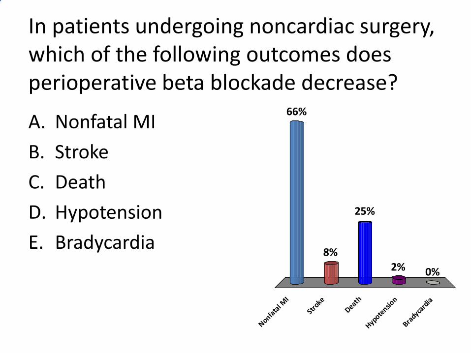
In patients undergoing noncardiac surgery, which of the following outcomes does perioperative beta blockade decrease?
A. Nonfatal MI
B. Stroke
C. Death
D. Hypotension
E. Bradycardia
Nonfata
l MI
Stroke
Death
Hypotensio
n
Bradyca
rdia
66%
8%
0%2%
25%
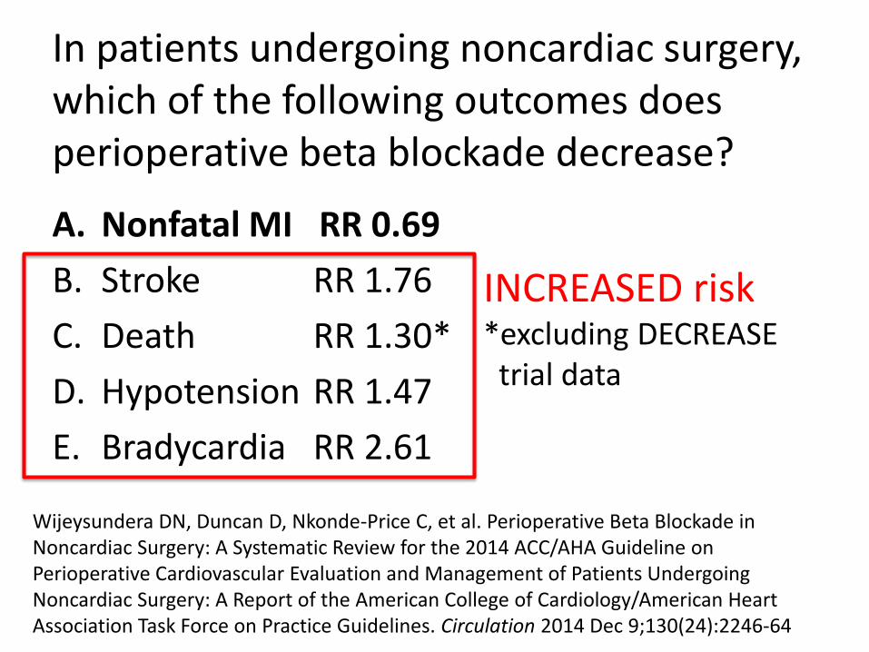
In patients undergoing noncardiac surgery, which of the following outcomes does perioperative beta blockade decrease?
A. Nonfatal MI RR 0.69
B. Stroke RR 1.76
C. Death RR 1.30*
D. Hypotension RR 1.47
E. Bradycardia RR 2.61
INCREASED risk *excluding DECREASE trial data
Wijeysundera DN, Duncan D, Nkonde-Price C, et al. Perioperative Beta Blockade in Noncardiac Surgery: A Systematic Review for the 2014 ACC/AHA Guideline on Perioperative Cardiovascular Evaluation and Management of Patients Undergoing Noncardiac Surgery: A Report of the American College of Cardiology/American Heart Association Task Force on Practice Guidelines. Circulation 2014 Dec 9;130(24):2246-64
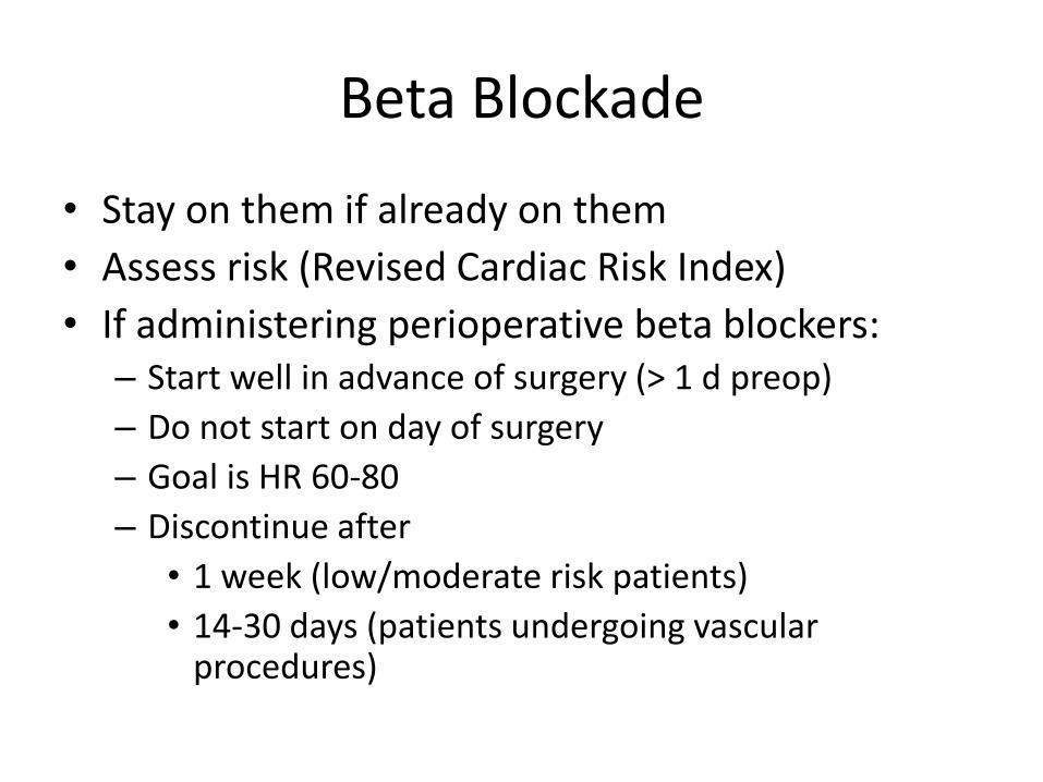
Beta Blockade
• Stay on them if already on them
• Assess risk (Revised Cardiac Risk Index)
• If administering perioperative beta blockers: – Start well in advance of surgery (> 1 d preop)
– Do not start on day of surgery
– Goal is HR 60-80
– Discontinue after
• 1 week (low/moderate risk patients)
• 14-30 days (patients undergoing vascular procedures)
Perioperative Diabetes Management
• Best if A1c < 7
• Tight glycemic control controversial
– 140-180 may be adequate
Statins
• Stay on them if already on them
• Consider initiating in selected high-risk patients
Postoperative Care
• Monitor cardiovascular, pulmonary, fluid status
• Pain management
• Complications
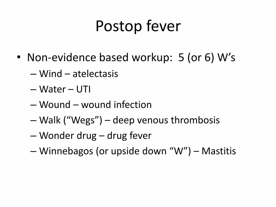
Postop fever
• Non-evidence based workup: 5 (or 6) W’s
– Wind – atelectasis
– Water – UTI
– Wound – wound infection
– Walk (“Wegs”) – deep venous thrombosis
– Wonder drug – drug fever
– Winnebagos (or upside down “W”) – Mastitis
Postop fever
• Recommendations for Evaluation of Fever Within 72 Hours of Surgery
O'Grady NP, Barie PS, Bartlett JG et al., American College of Critical Care Medicine, Infectious Diseases Society of America. Guidelines for evaluation of new fever in critically ill adult patients: 2008 update from the American College of Critical Care Medicine and the Infectious Diseases Society of America. Crit Care Med 2008 Apr;36(4):1330-49.
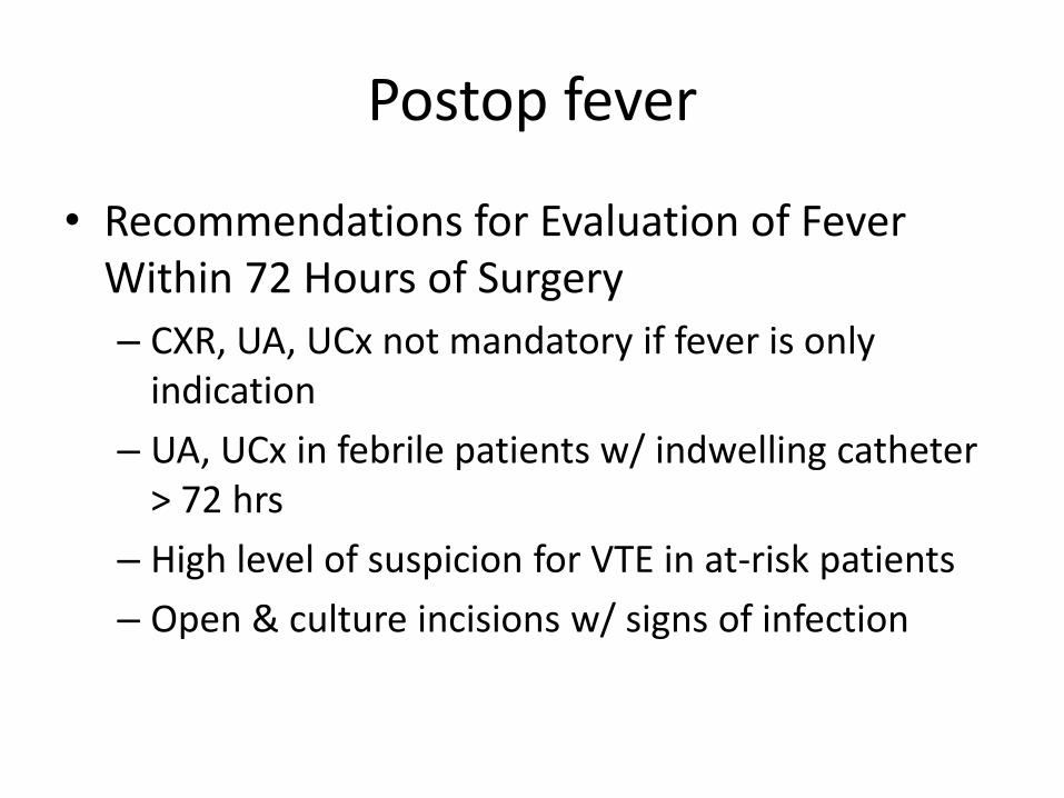
Postop fever
• Recommendations for Evaluation of Fever Within 72 Hours of Surgery
– CXR, UA, UCx not mandatory if fever is only indication
– UA, UCx in febrile patients w/ indwelling catheter > 72 hrs
– High level of suspicion for VTE in at-risk patients
– Open & culture incisions w/ signs of infection
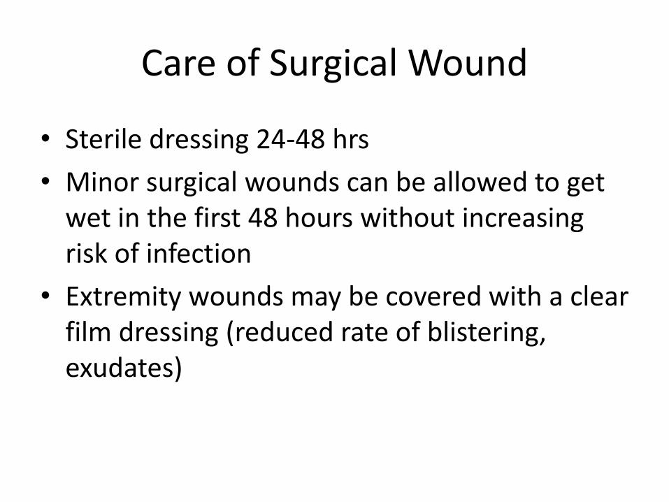
Care of Surgical Wound
• Sterile dressing 24-48 hrs
• Minor surgical wounds can be allowed to get wet in the first 48 hours without increasing risk of infection
• Extremity wounds may be covered with a clear film dressing (reduced rate of blistering, exudates)
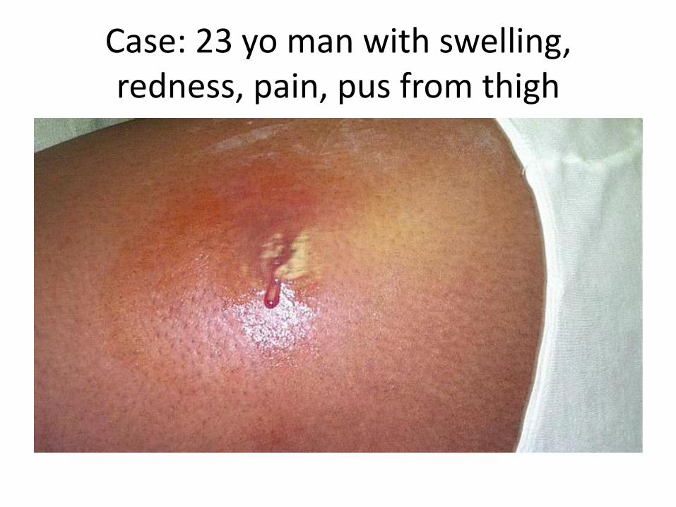
Case: 23 yo man with swelling, redness, pain, pus from thigh
I & D of Skin Abscesses
• Antibiotics after I & D?
– Large abscess > 10 cm, cellulitis, immunocompromised
– Otherwise, I & D alone is sufficient for simple abscesses
Singer HJ, Thode Jr. HC. Systemic antibiotics after incision and drainage of simple abscesses: A meta-analysis. Emerg Med J 2014;31:576-578.
Time Out
OTHER SURGICAL SPECIALTIES: TRAUMA SURGERY VASCULAR SURGERY THORACIC SURGERY OTOLARYNGOLOGY/HEAD AND NECK SURGERY UROLOGY NEUROSURGERY

TRAUMA SURGERY
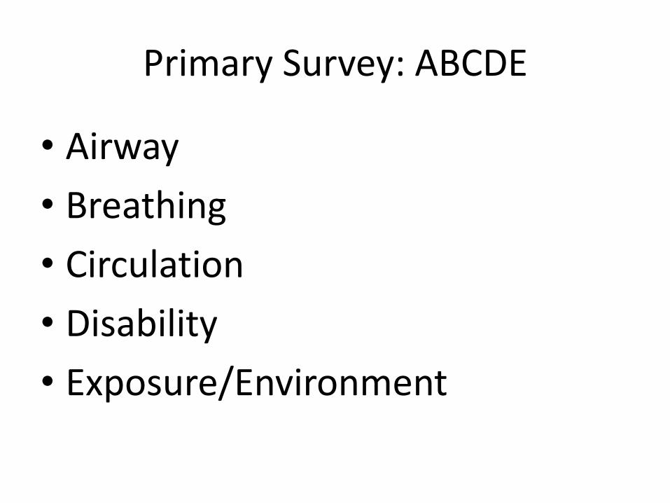
Primary Survey: ABCDE
• Airway
• Breathing
• Circulation
• Disability
• Exposure/Environment
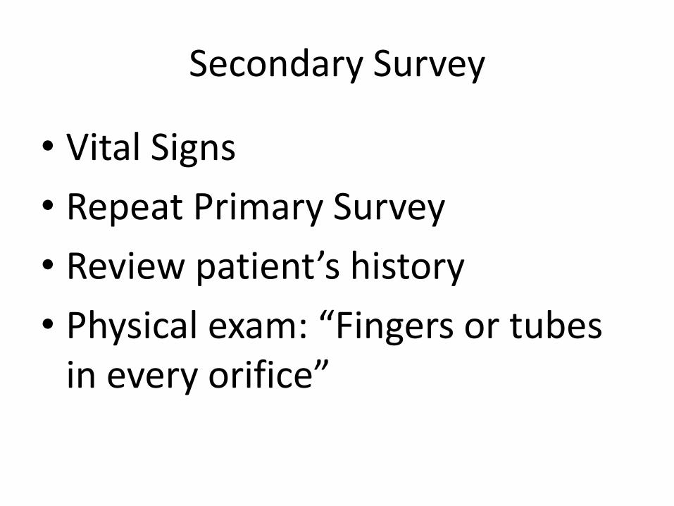
Secondary Survey
• Vital Signs
• Repeat Primary Survey
• Review patient’s history
• Physical exam: “Fingers or tubes in every orifice”

Shock Classification
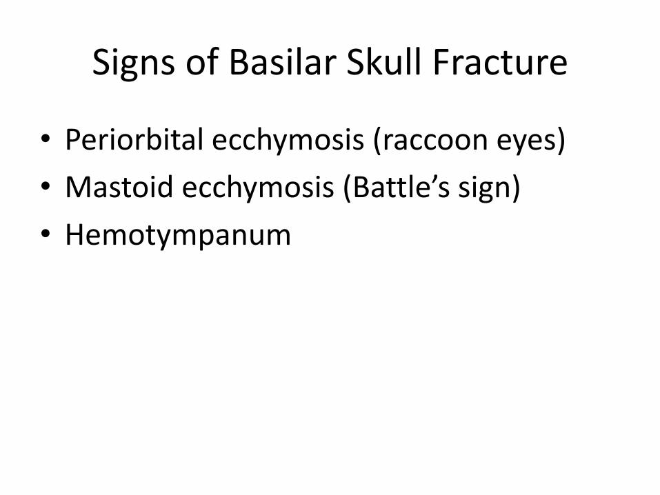
Signs of Basilar Skull Fracture
• Periorbital ecchymosis (raccoon eyes)
• Mastoid ecchymosis (Battle’s sign)
• Hemotympanum
Raccoon Eyes (Periorbital Ecchymoses)
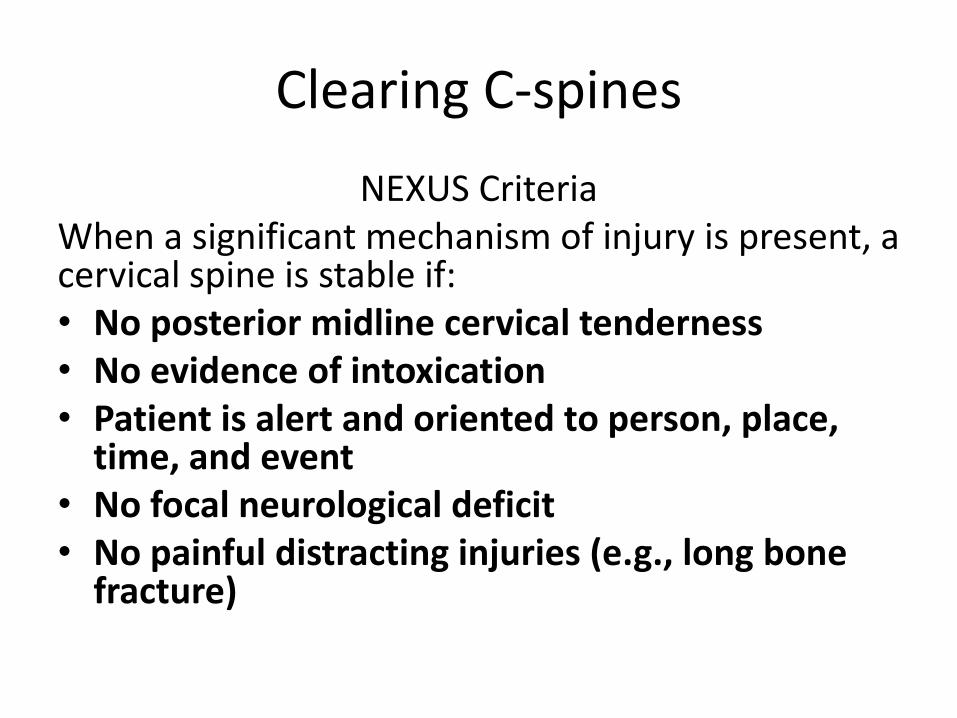
Clearing C-spines
NEXUS Criteria When a significant mechanism of injury is present, a cervical spine is stable if: • No posterior midline cervical tenderness • No evidence of intoxication • Patient is alert and oriented to person, place,
time, and event • No focal neurological deficit • No painful distracting injuries (e.g., long bone
fracture)
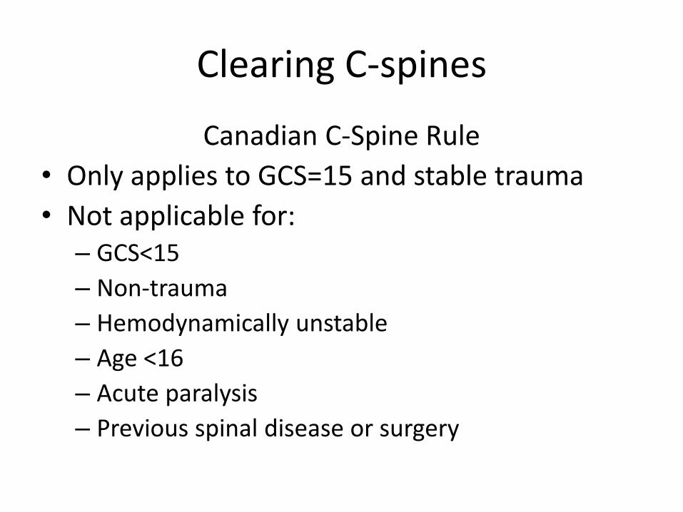
Clearing C-spines
Canadian C-Spine Rule
• Only applies to GCS=15 and stable trauma
• Not applicable for: – GCS<15
– Non-trauma
– Hemodynamically unstable
– Age <16
– Acute paralysis
– Previous spinal disease or surgery
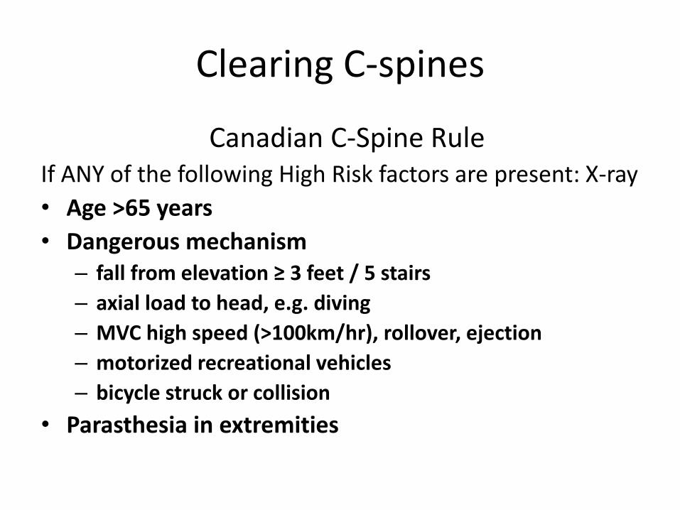
Clearing C-spines
Canadian C-Spine Rule If ANY of the following High Risk factors are present: X-ray
• Age >65 years
• Dangerous mechanism – fall from elevation ≥ 3 feet / 5 stairs
– axial load to head, e.g. diving
– MVC high speed (>100km/hr), rollover, ejection
– motorized recreational vehicles
– bicycle struck or collision
• Parasthesia in extremities
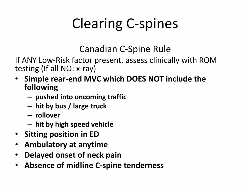
Clearing C-spines
Canadian C-Spine Rule If ANY Low-Risk factor present, assess clinically with ROM testing (If all NO: x-ray) • Simple rear-end MVC which DOES NOT include the
following – pushed into oncoming traffic – hit by bus / large truck – rollover – hit by high speed vehicle
• Sitting position in ED • Ambulatory at anytime • Delayed onset of neck pain • Absence of midline C-spine tenderness
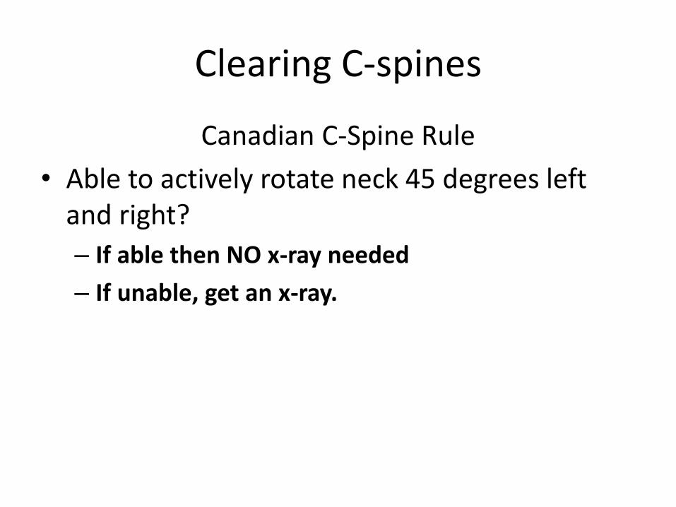
Clearing C-spines
Canadian C-Spine Rule
• Able to actively rotate neck 45 degrees left and right?
– If able then NO x-ray needed
– If unable, get an x-ray.
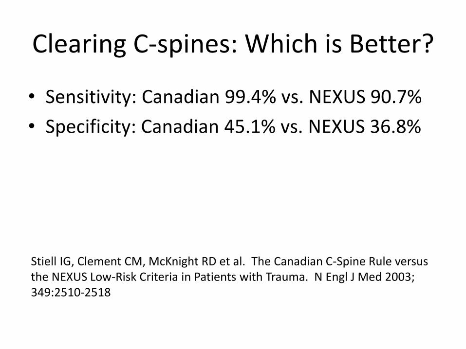
Clearing C-spines: Which is Better?
• Sensitivity: Canadian 99.4% vs. NEXUS 90.7%
• Specificity: Canadian 45.1% vs. NEXUS 36.8%
Stiell IG, Clement CM, McKnight RD et al. The Canadian C-Spine Rule versus the NEXUS Low-Risk Criteria in Patients with Trauma. N Engl J Med 2003; 349:2510-2518
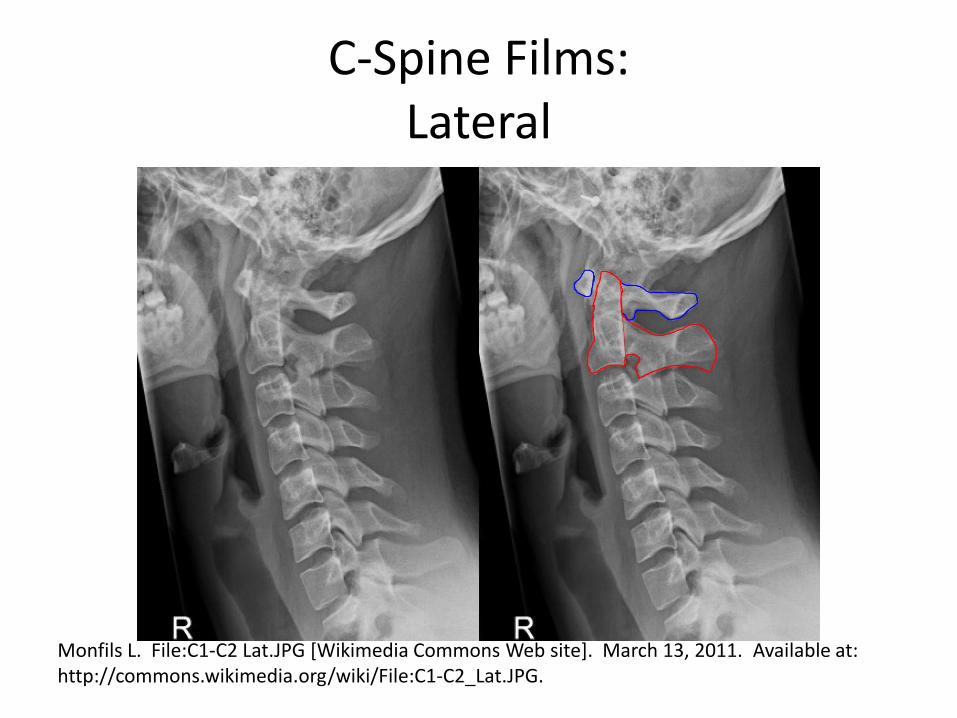
C-Spine Films: Lateral
Monfils L. File:C1-C2 Lat.JPG [Wikimedia Commons Web site]. March 13, 2011. Available at: http://commons.wikimedia.org/wiki/File:C1-C2_Lat.JPG.
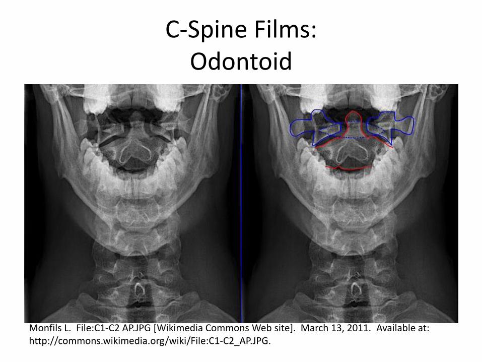
C-Spine Films: Odontoid
Monfils L. File:C1-C2 AP.JPG [Wikimedia Commons Web site]. March 13, 2011. Available at: http://commons.wikimedia.org/wiki/File:C1-C2_AP.JPG.
VASCULAR SURGERY
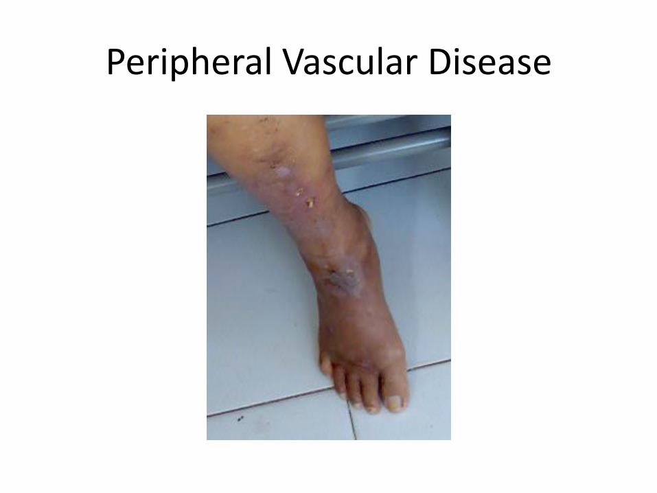
Peripheral Vascular Disease
Peripheral Vascular Disease
• Intermittent claudication (many may not have classic symptoms)
• Late symptoms: rest pain, ulcers, gangrene
• Risk Factors = CAD, esp. smoking
• Diagnosis: ABI, PE – pulses, bruits, hair loss (watering the plants), poor nail growth, dependent rubor, ulcers
Peripheral Vascular Disease
• Treatment: modify risk factors, exercise, meds (ASA, clopidogrel, cilostazol)
• Surgery: not enough evidence to favor bypass surgery over angioplasty (walking distance, disease progression, complications, amputation rate, death)
Fowkes F, Leng GC. Bypass surgery for chronic lower limb ischaemia. Cochrane Database of Systematic Reviews 2008, Issue 2. Art. No.: CD002000. DOI: 10.1002/14651858.CD002000.pub2.
Medical vs. Surgical Management: Asymptomatic Carotid Artery Stenosis • Carotid endarterectomy vs. carotid artery
stenting – no evidence favoring one over the other
• No evidence clearly favoring surgery over medical management
• Low rates of ipsilateral stroke in patients managed medically (1.68% all studies, 1.18% newer studies)
Raman G, Moorthy D, Hadar N, et al. Management Strategies for Asymptomatic Carotid Stenosis: A Systematic Review and Meta-analysis. Ann Intern Med. 2013;158:676-685.
THORACIC SURGERY
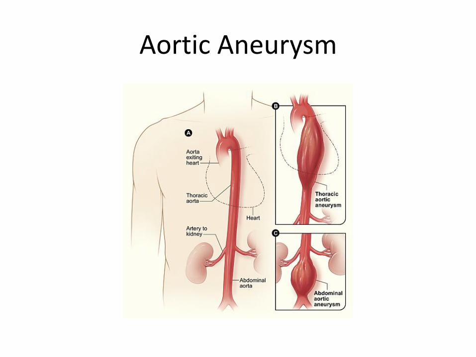
Aortic Aneurysm
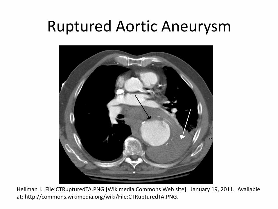
Ruptured Aortic Aneurysm
Heilman J. File:CTRupturedTA.PNG [Wikimedia Commons Web site]. January 19, 2011. Available at: http://commons.wikimedia.org/wiki/File:CTRupturedTA.PNG.
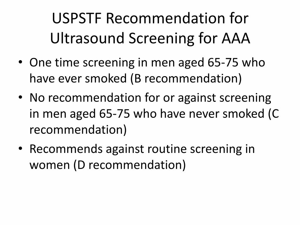
USPSTF Recommendation for Ultrasound Screening for AAA
• One time screening in men aged 65-75 who have ever smoked (B recommendation)
• No recommendation for or against screening in men aged 65-75 who have never smoked (C recommendation)
• Recommends against routine screening in women (D recommendation)
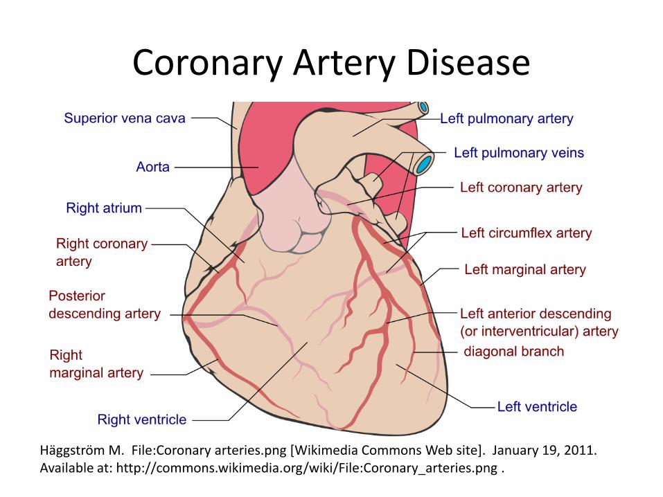
Coronary Artery Disease
Häggström M. File:Coronary arteries.png [Wikimedia Commons Web site]. January 19, 2011. Available at: http://commons.wikimedia.org/wiki/File:Coronary_arteries.png .
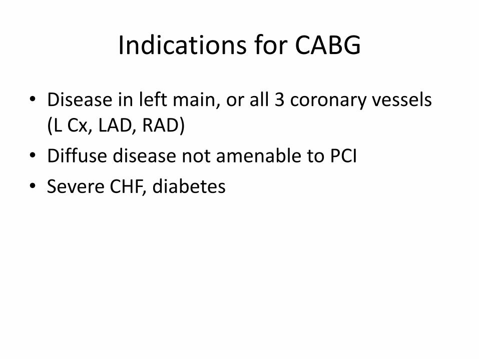
Indications for CABG
• Disease in left main, or all 3 coronary vessels (L Cx, LAD, RAD)
• Diffuse disease not amenable to PCI
• Severe CHF, diabetes
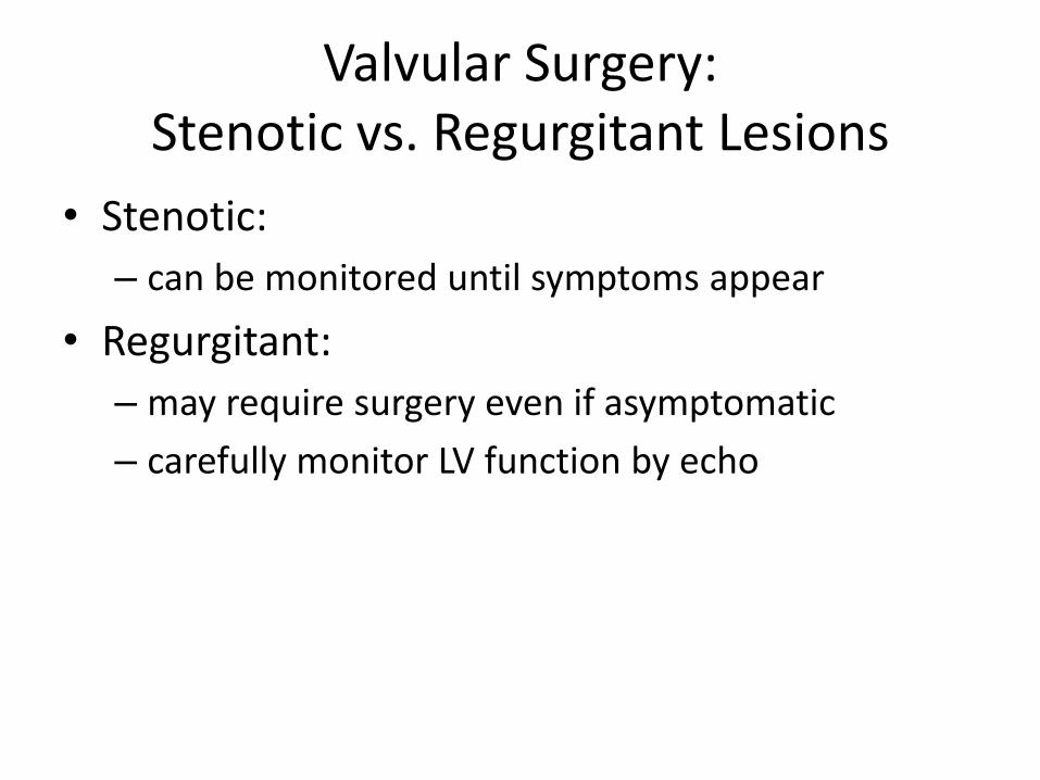
Valvular Surgery: Stenotic vs. Regurgitant Lesions
• Stenotic:
– can be monitored until symptoms appear
• Regurgitant:
– may require surgery even if asymptomatic
– carefully monitor LV function by echo
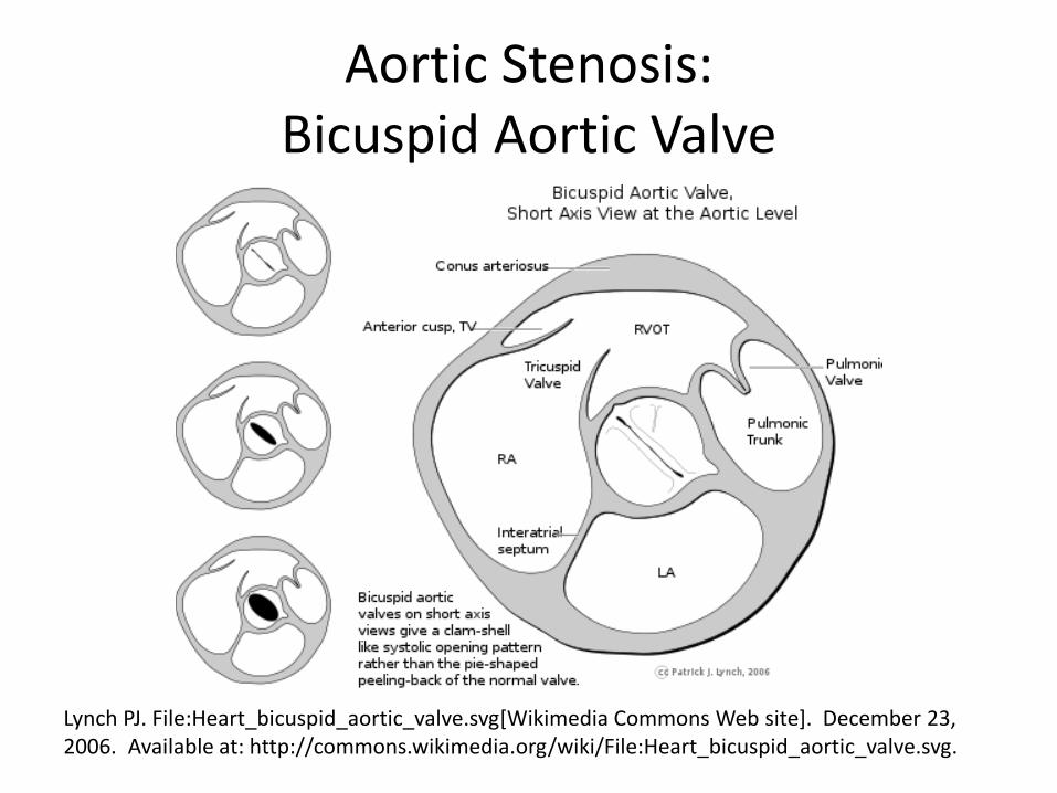
Aortic Stenosis: Bicuspid Aortic Valve
Lynch PJ. File:Heart_bicuspid_aortic_valve.svg[Wikimedia Commons Web site]. December 23, 2006. Available at: http://commons.wikimedia.org/wiki/File:Heart_bicuspid_aortic_valve.svg.
Aortic Stenosis
• Classical presentation: asymptomatic, then angina, exertional syncope, dyspnea
• Average survival after symptoms develop = 2-3 years, 75% die w/in 3 yrs w/out valve replacement
• Critical stenosis: Valve area < 0.8 cm2 or gradient > 50 mm Hg
• Workup – Echocardiogram
• mild/moderate AS – q2-5 yrs • severe AS – annual (more to check LV function)
– CXR, EKG – NO stress testing
Transcatheter vs. Surgical Aortic Valve Replacement
• Severe aortic stenosis, increased surgical risk
• Transcatheter AV replacement (TAVR)
– Death rates at 1 year: TAVR 14%, surgical 19%
– Noninferior and superior to surgical AV replacement
Popma JJ, Adams DH, Reardon MJ et al. Transcatheter aortic valve replacement using a self-expanding bioprosthesis in patients with severe aortic stenosis at extreme risk for surgery. J Am Coll Cardiol. 2014 May 20;63(19):1972-81.
Adams DH, Popma JJ, Reardon MJ et al. Transcatheter Aortic-Valve Replacement with a Self-Expanding Prosthesis. N Engl J Med 2014;370:1790-8.
Mitral Stenosis
• Symptoms mimic CHF
• Atrial fibrillation, pregnancy bring out symptoms
• Treatment:
– Mild disease: diuretics
– Atrial fibrillation: rate control
– Surgery: > mild symptoms, or pulmonary hypertension
• Balloon valvotomy, open commisurotomy, MV reconstruction, MV replacement
Aortic Regurgitation
• Causes: endocarditis, rheumatic fever, collagen vascular disease, aortic dissection, syphilis
• Typical presentation: Initially asymptomatic subtle initial signs (decreased functional capacity or fatigue) sx of L-sided heart failure
• Treatment: – Severe AR + normal LV function: afterload reduction
w/ vasodilators, especially nifedipine, can delay need for surgery
– AV replacement even in asymptomatic patients, before EF < 55 % or end systolic dimension reaches 55 mm
Mitral Regurgitation
• Causes: infectious endocarditis, mitral valve prolapse, rheumatic fever
• Surgery:
– if > mild sx, EF < 60%, or end-systolic dimension approaches 45 mm, even if asymptomatic
– Usually MV repair preferred over replacement
What about Mitral Valve Prolapse?
• Typical symptoms: chest pain, dyspnea, anxiety, palpitations
• Treatment: reassurance – no need for surgery

OTOLARYNGOLOGY HEAD AND NECK SURGERY
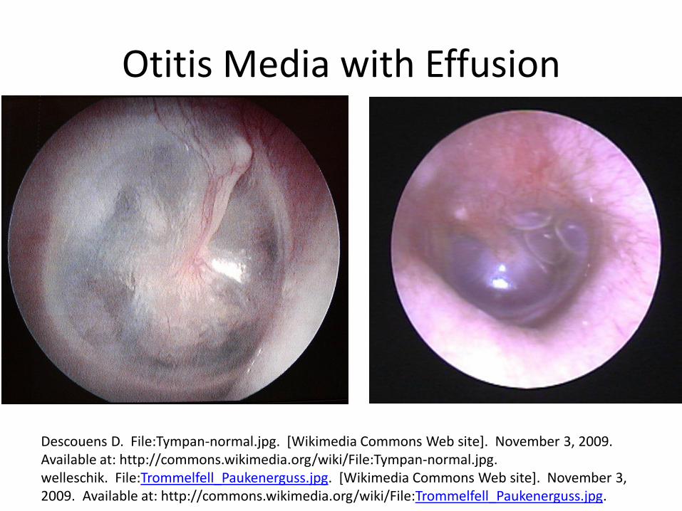
Otitis Media with Effusion
Descouens D. File:Tympan-normal.jpg. [Wikimedia Commons Web site]. November 3, 2009. Available at: http://commons.wikimedia.org/wiki/File:Tympan-normal.jpg. welleschik. File:Trommelfell_Paukenerguss.jpg. [Wikimedia Commons Web site]. November 3, 2009. Available at: http://commons.wikimedia.org/wiki/File:Trommelfell_Paukenerguss.jpg.
Otitis Media with Effusion
• Candidates for surgery – persistent hearing loss or other signs and symptoms – recurrent or persistent OME in at-risk children regardless
of hearing status – structural damage to the tympanic membrane or middle
ear
• Shared decision-making re: surgery • Tympanostomy tube insertion is the preferred initial
procedure (+/- adenoidectomy in children ≥ 4 yo)
Rosenfeld RM, Shin JJ, Schwartz SR et al. Clinical Practice Guideline: Otitis Media with
Effusion Executive Summary (Update). Otolaryngology–Head and Neck Surgery 2016:154(2):201–214
Indications for Functional Endoscopic Sinus Surgery (FESS)
• Failed medical therapy for chronic rhinosinusitis
• Nasal polyps
Luong A, Marple BF. Sinus surgery: indications and techniques. Clin Rev Allergy Immunol. 2006 Jun;30(3):217-22.
Epistaxis
• Pressure
• Silver nitrate cauterization (only 1 side of nasal septum at a time)
• Packing – Anterior: F/U w/ ENT w/in 2-3 days, avoid ASA &
NSAIDs but can continue warfarin
– Posterior: Admit
Management of Acute Epistaxis. Author: Ola Bamimore, MD; Chief Editor: Steven C Dronen, MD, FAAEM http://emedicine.medscape.com/article/764719-overview#showall
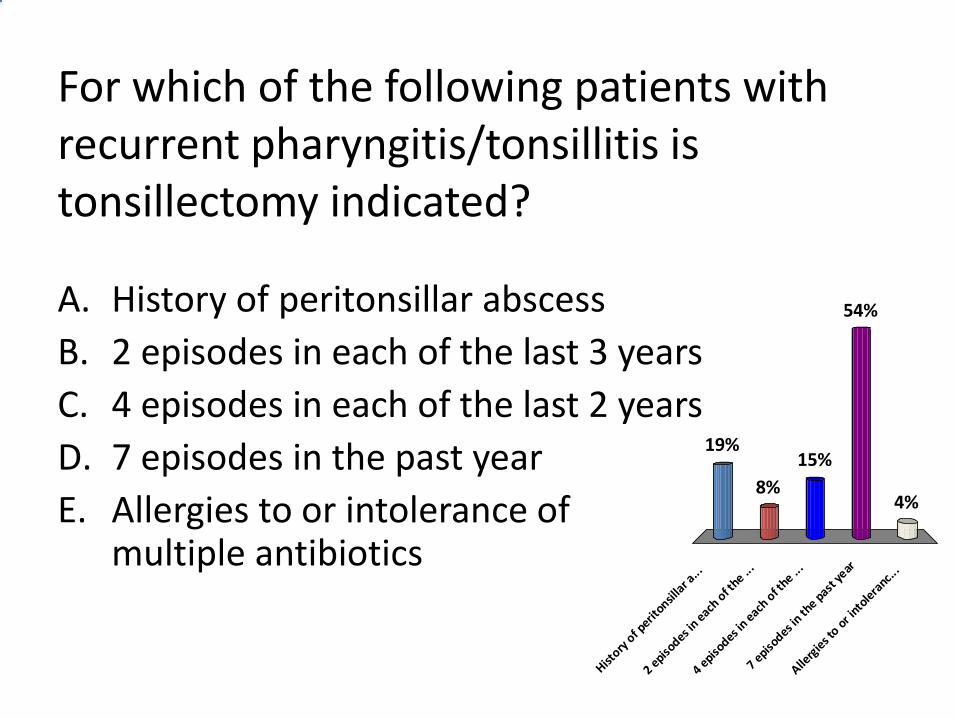
For which of the following patients with recurrent pharyngitis/tonsillitis is tonsillectomy indicated?
A. History of peritonsillar abscess
B. 2 episodes in each of the last 3 years
C. 4 episodes in each of the last 2 years
D. 7 episodes in the past year
E. Allergies to or intolerance of multiple antibiotics
History
of p
eritonsil
lar a
...
2 episo
des in e
ach o
f the ..
.
4 episo
des in e
ach o
f the ..
.
7 episo
des in th
e past
year
Allerg
ies t
o or i
ntole
ranc..
.
19%
8%4%
54%
15%
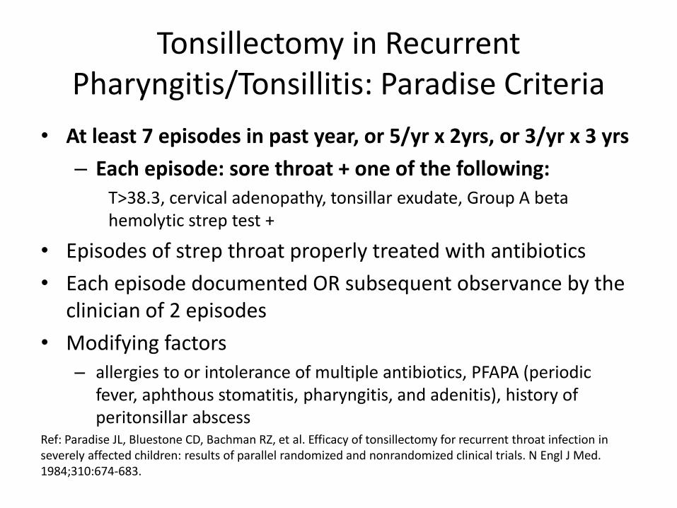
Tonsillectomy in Recurrent Pharyngitis/Tonsillitis: Paradise Criteria
• At least 7 episodes in past year, or 5/yr x 2yrs, or 3/yr x 3 yrs
– Each episode: sore throat + one of the following: T>38.3, cervical adenopathy, tonsillar exudate, Group A beta hemolytic strep test +
• Episodes of strep throat properly treated with antibiotics
• Each episode documented OR subsequent observance by the clinician of 2 episodes
• Modifying factors – allergies to or intolerance of multiple antibiotics, PFAPA (periodic
fever, aphthous stomatitis, pharyngitis, and adenitis), history of peritonsillar abscess
Ref: Paradise JL, Bluestone CD, Bachman RZ, et al. Efficacy of tonsillectomy for recurrent throat infection in severely affected children: results of parallel randomized and nonrandomized clinical trials. N Engl J Med. 1984;310:674-683.
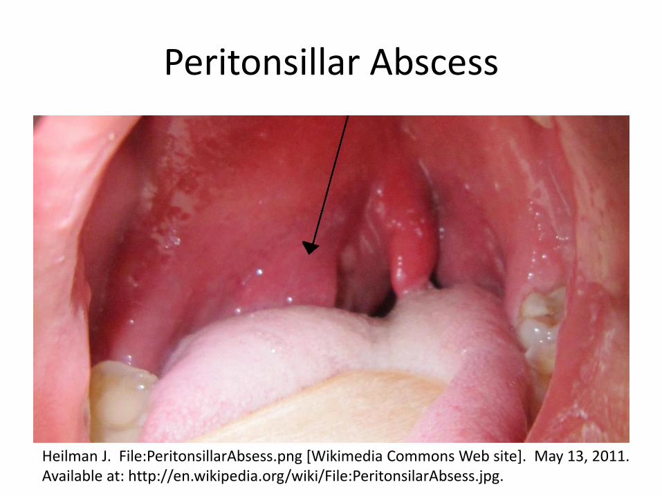
Peritonsillar Abscess
Heilman J. File:PeritonsillarAbsess.png [Wikimedia Commons Web site]. May 13, 2011. Available at: http://en.wikipedia.org/wiki/File:PeritonsilarAbsess.jpg.

UROLOGY
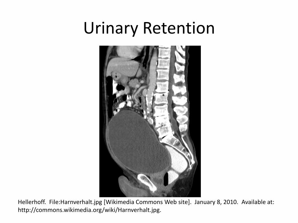
Urinary Retention
Hellerhoff. File:Harnverhalt.jpg [Wikimedia Commons Web site]. January 8, 2010. Available at: http://commons.wikimedia.org/wiki/Harnverhalt.jpg.
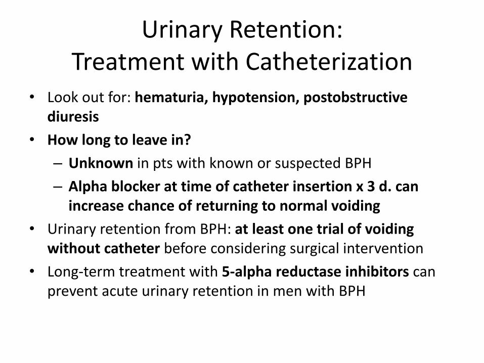
Urinary Retention: Treatment with Catheterization
• Look out for: hematuria, hypotension, postobstructive diuresis
• How long to leave in?
– Unknown in pts with known or suspected BPH
– Alpha blocker at time of catheter insertion x 3 d. can increase chance of returning to normal voiding
• Urinary retention from BPH: at least one trial of voiding without catheter before considering surgical intervention
• Long-term treatment with 5-alpha reductase inhibitors can prevent acute urinary retention in men with BPH
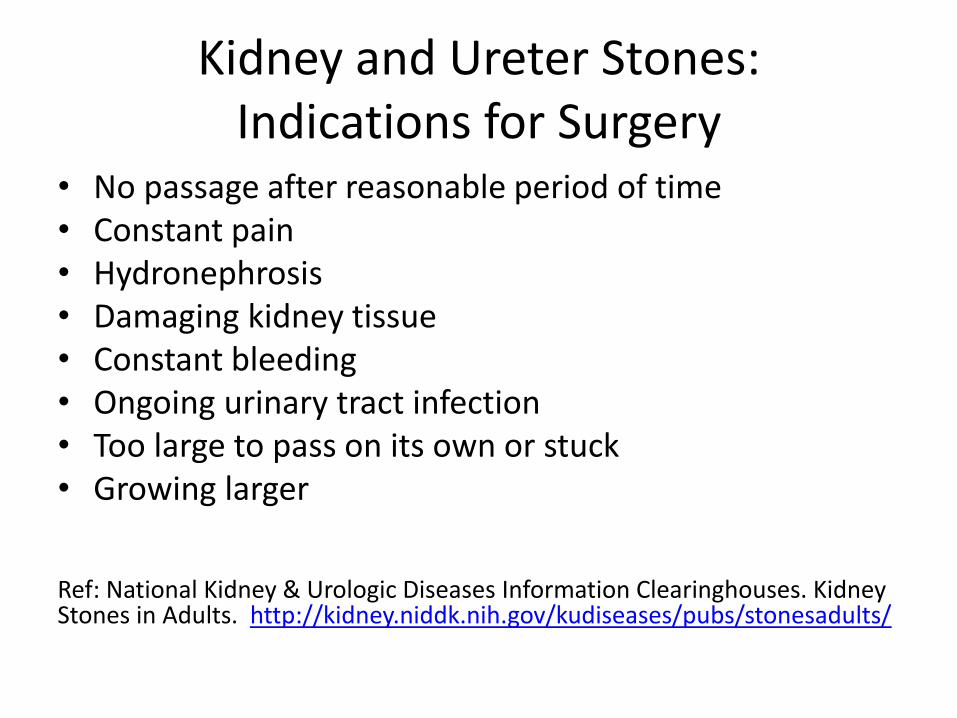
Kidney and Ureter Stones: Indications for Surgery
• No passage after reasonable period of time • Constant pain • Hydronephrosis • Damaging kidney tissue • Constant bleeding • Ongoing urinary tract infection • Too large to pass on its own or stuck • Growing larger Ref: National Kidney & Urologic Diseases Information Clearinghouses. Kidney Stones in Adults. http://kidney.niddk.nih.gov/kudiseases/pubs/stonesadults/
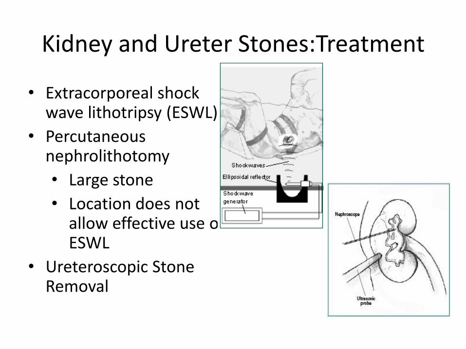
Kidney and Ureter Stones:Treatment
• Extracorporeal shock wave lithotripsy (ESWL)
• Percutaneous nephrolithotomy
• Large stone
• Location does not allow effective use of ESWL
• Ureteroscopic Stone Removal
Case: 53 year old man with gross hematuria
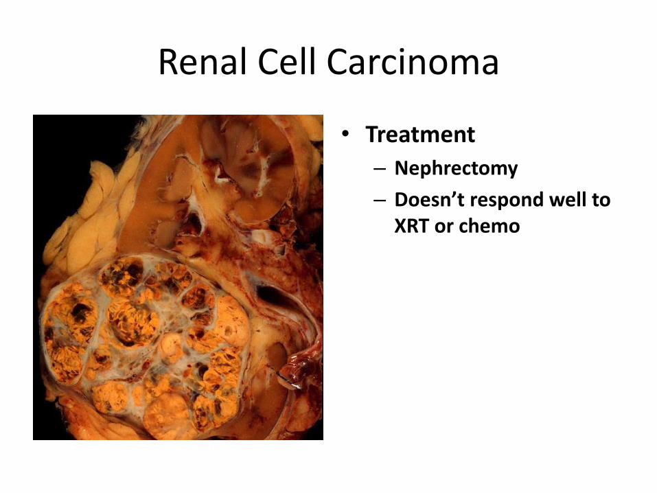
Renal Cell Carcinoma
• Demographics: – Men slightly > women
– African Americans slightly > Caucasians
– Incidence rising
Higgins JC, Fitzgerald JM. Evaluation of Incidental Renal and Adrenal Masses. Am Fam Physician. 2001 Jan 15;63(2):288-295.
• Risk factors: – Exposure to household &
industrial chemicals
– Hypertension
– Family history of RCC
– Occupational exposure to cadmium
– Dialysis patients w/ acquired cystic disease of the kidney (30x)
– Hysterectomy (2x)
Renal Cell Carcinoma
• Diagnosis:
– Classic triad in 10-15%: hematuria, flank pain, abdominal mass
– Often diagnosed incidentally at asymptomatic stage
– Imaging
• Sensitivities: ultrasound 79%, CT 94%
• MRI better than CT at distinguishing benign lesions
Renal Cell Carcinoma
• Treatment
– Nephrectomy
– Doesn’t respond well to XRT or chemo
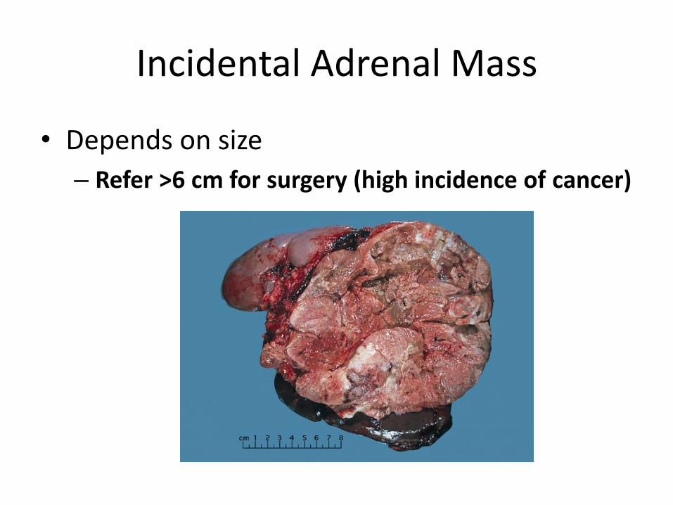
Incidental Adrenal Mass
• Depends on size
– Refer >6 cm for surgery (high incidence of cancer)
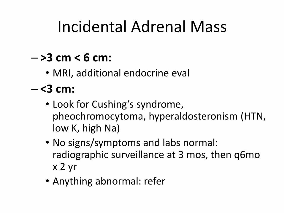
Incidental Adrenal Mass
–>3 cm < 6 cm: • MRI, additional endocrine eval
–<3 cm: • Look for Cushing’s syndrome,
pheochromocytoma, hyperaldosteronism (HTN, low K, high Na)
• No signs/symptoms and labs normal: radiographic surveillance at 3 mos, then q6mo x 2 yr
• Anything abnormal: refer
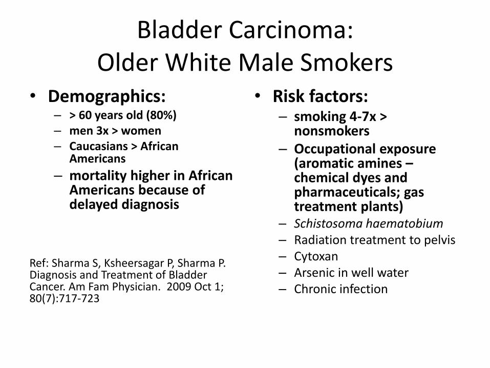
Bladder Carcinoma: Older White Male Smokers
• Demographics: – > 60 years old (80%) – men 3x > women – Caucasians > African
Americans
– mortality higher in African Americans because of delayed diagnosis
Ref: Sharma S, Ksheersagar P, Sharma P. Diagnosis and Treatment of Bladder Cancer. Am Fam Physician. 2009 Oct 1; 80(7):717-723
• Risk factors: – smoking 4-7x >
nonsmokers – Occupational exposure
(aromatic amines – chemical dyes and pharmaceuticals; gas treatment plants)
– Schistosoma haematobium – Radiation treatment to pelvis – Cytoxan – Arsenic in well water – Chronic infection
Bladder Carcinoma
• Typical presentation:
– Painless hematuria
– “Irritative” symptoms (dysuria, frequency)
– Urinary obstructive symptoms
– Symptoms of advanced disease
• lower extremity edema, renal failure, suprapubic palpable mass
Bladder Carcinoma
• Diagnostics –Urine cytology
• 66-79% sensitive, 95-100% specific
– Cystoscopy, bladder wash cytology – Evaluate upper urinary tract – CT preferred –Metastatic workup
• CBC, chemistries (alkaline phosphatase, LFT’s), CXR, CT or MRI, Bone scan if alkaline phosphatase is elevated or other symptoms suggest bone metastases
Bladder Carcinoma
• Treatment:
–Non-muscle invasive: transurethral resection +/- intravesical chemotherapy (mitomycin) or immunotherapy (intravesical BCG)
–Muscle-invasive: radical cystectomy +/- chemotherapy
–Metastatic: chemotherapy

NEUROSURGERY
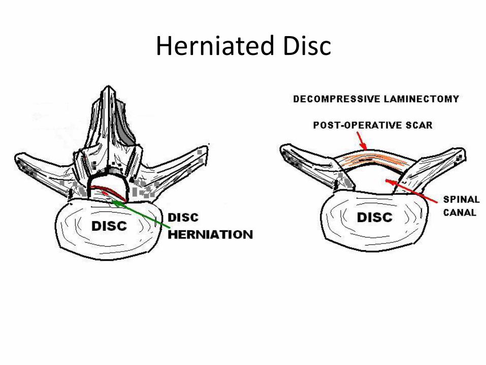
Case: 30 year old man with progressive sciatica
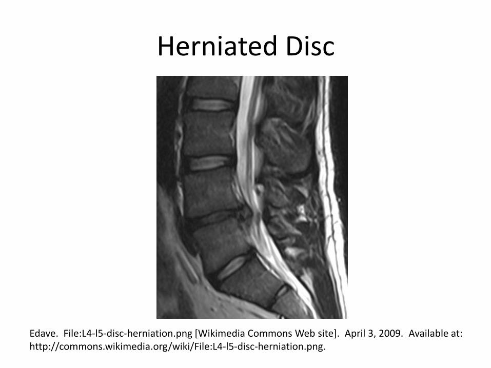
Herniated Disc
Edave. File:L4-l5-disc-herniation.png [Wikimedia Commons Web site]. April 3, 2009. Available at: http://commons.wikimedia.org/wiki/File:L4-l5-disc-herniation.png.
When do patients need surgery for low back pain?
• Severe or progressive neurologic deficits
• Serious underlying conditions are suspected
• Persistent low back pain and signs or symptoms of radiculopathy or spinal stenosis – Only if they are potential candidates for surgery or
epidural steroid injection (for suspected radiculopathy)
• MRI (preferred) or CT
Chou R, Qaseem A, Snow V et al, Clinical Efficacy Assessment Subcommittee of the American College of Physicians, American College of Physicians, American Pain Society Low Back Pain Guidelines Panel. Diagnosis and treatment of low back pain: a joint clinical practice guideline from the American College of Physicians and the American Pain Society. Ann Intern Med 2007 Oct 2;147(7):478-91
Herniated Disc
Which patients need neuroimaging (noncontrast head CT) for headaches? • Emergent:
– headache with new abnormal neurologic findings (e.g., focal deficit, altered mental status, altered cognitive function)
– new sudden-onset severe headache (thunderclap) – Human immunodeficiency virus (HIV)-positive patients
with a new type of headache (consider CT)
• Urgent: – Patients > 50 years old with new type of headache but
normal neurologic examination Ref: Edlow JA, Panagos PD, Godwin SA, Thomas TL, Decker WW, American College of Emergency Physicians. Clinical policy: critical issues in the evaluation and management of adult patients presenting to the emergency department with acute headache. Ann Emerg Med 2008 Oct;52(4):407-36.
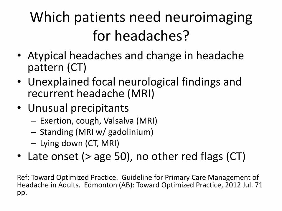
Which patients need neuroimaging for headaches?
• Atypical headaches and change in headache pattern (CT)
• Unexplained focal neurological findings and recurrent headache (MRI)
• Unusual precipitants – Exertion, cough, Valsalva (MRI) – Standing (MRI w/ gadolinium) – Lying down (CT, MRI)
• Late onset (> age 50), no other red flags (CT) Ref: Toward Optimized Practice. Guideline for Primary Care Management of Headache in Adults. Edmonton (AB): Toward Optimized Practice, 2012 Jul. 71 pp.
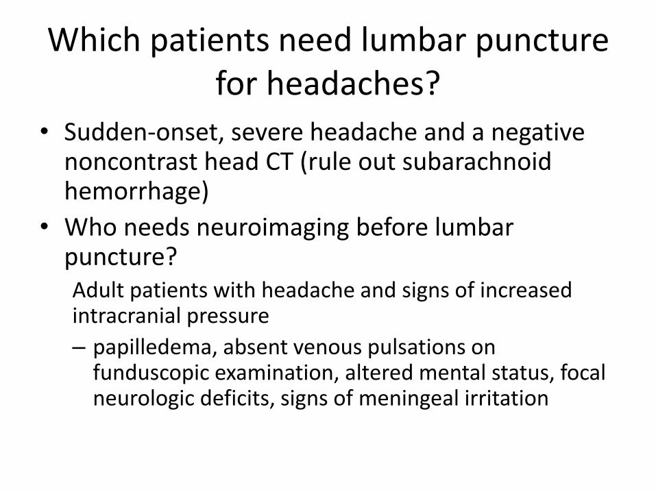
Which patients need lumbar puncture for headaches?
• Sudden-onset, severe headache and a negative noncontrast head CT (rule out subarachnoid hemorrhage)
• Who needs neuroimaging before lumbar puncture? Adult patients with headache and signs of increased intracranial pressure
– papilledema, absent venous pulsations on funduscopic examination, altered mental status, focal neurologic deficits, signs of meningeal irritation
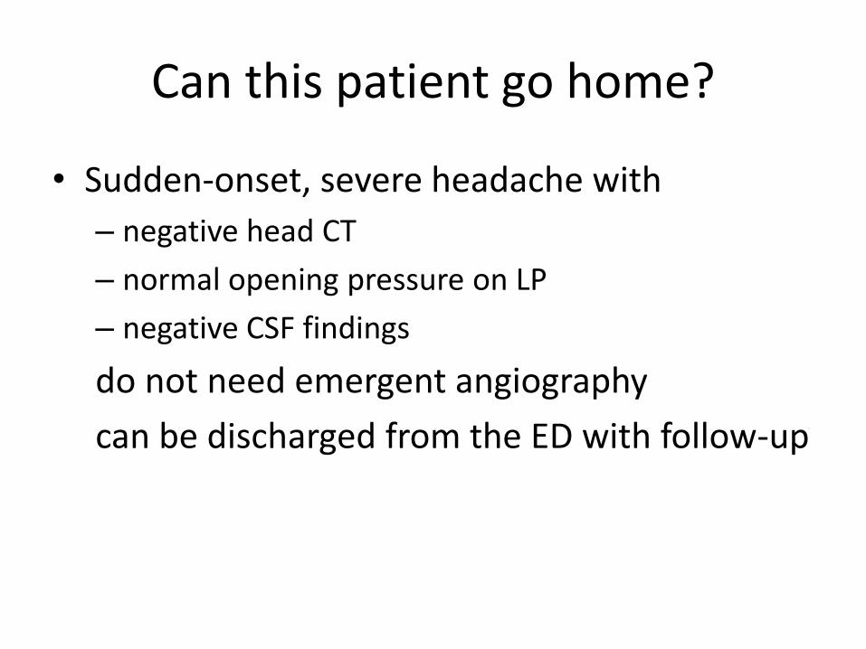
Can this patient go home?
• Sudden-onset, severe headache with
– negative head CT
– normal opening pressure on LP
– negative CSF findings
do not need emergent angiography
can be discharged from the ED with follow-up
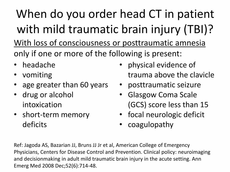
When do you order head CT in patient with mild traumatic brain injury (TBI)?
• headache • vomiting • age greater than 60 years • drug or alcohol
intoxication • short-term memory
deficits
• physical evidence of trauma above the clavicle
• posttraumatic seizure • Glasgow Coma Scale
(GCS) score less than 15 • focal neurologic deficit • coagulopathy
With loss of consciousness or posttraumatic amnesia only if one or more of the following is present:
Ref: Jagoda AS, Bazarian JJ, Bruns JJ Jr et al, American College of Emergency Physicians, Centers for Disease Control and Prevention. Clinical policy: neuroimaging and decisionmaking in adult mild traumatic brain injury in the acute setting. Ann Emerg Med 2008 Dec;52(6):714-48.
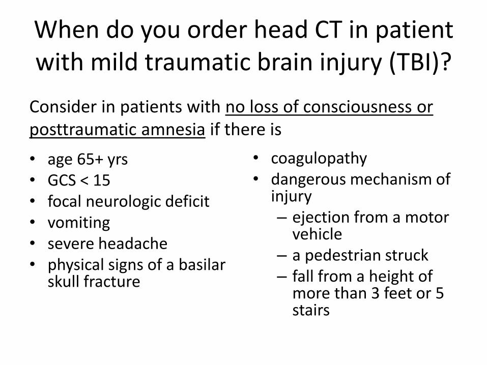
When do you order head CT in patient with mild traumatic brain injury (TBI)?
• age 65+ yrs • GCS < 15 • focal neurologic deficit • vomiting • severe headache • physical signs of a basilar
skull fracture
• coagulopathy • dangerous mechanism of
injury – ejection from a motor
vehicle – a pedestrian struck – fall from a height of
more than 3 feet or 5 stairs
Consider in patients with no loss of consciousness or posttraumatic amnesia if there is
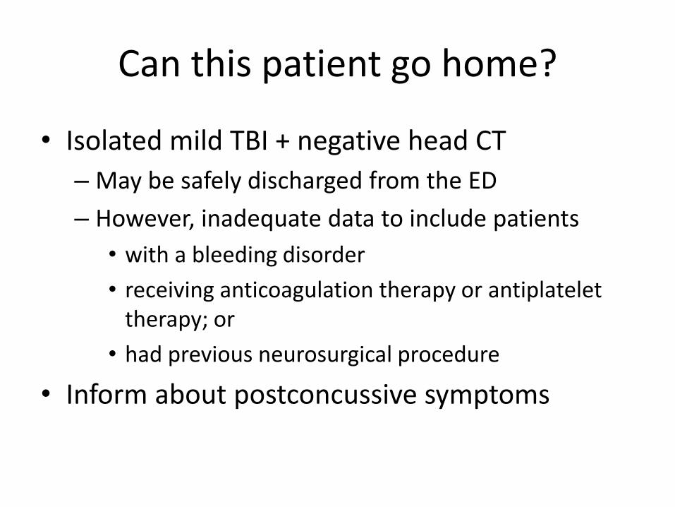
Can this patient go home?
• Isolated mild TBI + negative head CT
– May be safely discharged from the ED
– However, inadequate data to include patients
• with a bleeding disorder
• receiving anticoagulation therapy or antiplatelet therapy; or
• had previous neurosurgical procedure
• Inform about postconcussive symptoms