survival and stimulation of neurite outgrowth in a serum
TRANSCRIPT
Survival and stimulation of neurite outgrowth in a serum-free
culture of spiral ganglion neurons from adult mice
Mauricio Vieira a,b ([email protected]), Barbara L. Christensen a,c ([email protected]),
Bruce C. Wheeler a,b,d ([email protected]), Albert S. Feng a,c ([email protected]), and
Richard Kollmar a,c,* ([email protected])
a Beckman Institute for Advanced Science and Technology, University of Illinois at
Urbana-Champaign, 405 North Mathews Avenue, MC-251, Urbana, IL, USA
b Department of Bioengineering, University of Illinois at Urbana-Champaign, 1304 West
Springfield Avenue, MC-278, Urbana, IL 61801, USA
c Department of Molecular and Integrative Physiology, University of Illinois at Urbana-
Champaign, 407 South Goodwin Avenue, MC-114, Urbana, IL 61801, USA
d Department of Electrical and Computer Engineering, University of Illinois at Urbana-
Champaign, 1406 West Green Street, MC-702, Urbana, IL 61801, USA
* Corresponding Author:
407 South Goodwin Avenue, MC-114
Urbana, IL 61801
USA
Telephone: (217) 333-9861
Facsimile: (217) 333-1133
E-mail: [email protected]
2
Abstract
We have developed a reliable protocol for the serum-free dissociation and culture of
spiral ganglion neurons from adult mice, an important animal model for patients with
post-lingual hearing loss. Pilot experiments indicated that the viability of spiral ganglion
cells in vitro depended critically on the use of Hibernate medium with B27 supplement.
With an optimized protocol, we obtained 2 · 103 neurons immediately after dissociation,
or about one-fifth of those present in the intact spiral ganglion. After four days in culture,
4% of the seeded neurons survived without any exogenous growth factors other than
insulin. This yield was highly reproducible in five independent experiments and enabled
us to measure systematically the numbers and lengths of the regenerating neurites.
Furthermore, the survival rate compared well to the few published protocols for culturing
adult spiral ganglion neurons from other species. Enhanced survival and neurite
outgrowth upon the addition of brain-derived neurotrophic factor and leukemia inhibitory
factor demonstrated that both are potent stimulants for damaged spiral ganglion neurons
in adults. This responsiveness to exogenous growth factors suggested that our culture
protocol will facilitate the screening of molecular compounds as potential treatments for
sensorineural hearing loss.

3
Keywords
Adult
Cell Culture Techniques
Mice
Neurites
Neurons
Spiral Ganglion

4
1. Introduction
The damage caused to spiral ganglion neurons in patients with sensorineural hearing loss
may be halted or reversed by using cochlear implants or gene therapy to supply peptide
growth factors (Holley, 2002). However, our understanding of the signaling events that
underlie the survival of spiral ganglion neurons and the regeneration of their neurites is
incomplete and requires further studies to guide technology development (Bianchi and
Raz, 2004; Gillespie and Shepherd, 2005; Webber and Raz, 2006). The primary culture
of dissociated spiral ganglion neurons from adult mice would be a useful addition to the
experimental armamentarium for such studies. First, initial studies are often easier to
analyze and require fewer animals when conducted in vitro rather than in vivo. Second,
the anatomy, physiology, and pathology of hearing are well-documented for mice and
similar to those of humans (Nadol, 1988; Ohlemiller, 2006; Willott, 2001); unlike other
mammalian animal models, mice offer a full range of molecular-genetic tools, mutant and
transgenic lines, and inbred strains with varying degrees of hearing loss (Zheng et al.,
1999). Finally, adult animals are the most appropriate model for adult patients with
postlingual deafness, who comprise the largest group with sensorineural hearing loss
(Collins, 1997; Gates and Mills, 2005).
No protocol has been published for the primary culture of spiral ganglion neurons
from adult mice. The pioneering work on culturing dissociated spiral ganglion neurons
was carried out more than a decade ago with embryonic rats and chicken (Lefebvre et al.,
1990a; Yamaguchi and Ohmori, 1990). Various protocols have since been established for
culturing spiral ganglion neurons from a range of species and ages, such as embryonic
mice (Rabejac et al., 1994; Vazquez et al., 1994); neonatal mice (Kita et al., 2005; Lin et
al., 1998; Mo and Davis, 1997; Whitlon et al., 2006), rats (Dazert et al., 1998; Hegarty et
al., 1997; Lefebvre et al., 1990b; Malgrange et al., 1996; Marzella et al., 1997; Ripoll and
Rebillard, 1997; Rome et al., 1999; Zheng et al., 1995), and gerbils (Lin, 1997); and adult
rats (Lefebvre et al., 1991), guinea pigs (Anderson et al., 2006; Rask-Andersen et al.,
2005), and humans (Rask-Andersen et al., 2005). Sensory epithelia have also been
cultured from the cochlea of adult guinea pigs (Zhao, 2001); most experiments with spiral

5
ganglion neurons, however, have been conducted with neonatal samples, possibly
because adult neurons are in general more difficult to culture (Banker and Goslin, 1998).
The Hibernate and Neurobasal media and the B27 supplement were developed
specifically for the serum-free isolation and culture of neurons, in part by optimizing the
osmolarity and by including antioxidants (Brewer et al., 1993; Brewer, 1997). In
contrast, almost all protocols cited above use physiological saline for the isolation of
spiral ganglion neurons and generic chemically-defined media, such as Dulbecco
modified Eagle medium, together with the traditional N1 or N2 supplements (Bottenstein
and Sato, 1985) for their culture. Only a single study has described culturing spiral
ganglion neurons in Neurobasal medium with B27 supplement (Anderson et al., 2006),
but the quantitation of survival and neurite outgrowth was not the focus of that work.
Furthermore, most of the cited protocols require fetal bovine serum during the
dissociation or even in the culture media. This may improve neuronal survival, but may
also confound the effects of specific growth factors added as experimental treatments
with the effects of growth factors present in serum.
We developed a protocol for the completely serum-free dissociation and culture of
spiral ganglion neurons from adult mice in Hibernate and Neurobasal media with B27
supplement. Our protocol reproducibly yielded sufficient numbers of neurons for
systematic measurements of cell numbers and neurite lengths. Enhanced survival and
neurite outgrowth upon the addition of neuronal growth factors demonstrated that this
protocol will be useful for studying the regeneration of adult spiral ganglion neurons.

6
2. Materials and methods
2.1. Dissociating spiral ganglion neurons
All animal experiments were approved by the Institutional Animal Care and Use
Committee of the University of Illinois at Urbana-Champaign. Adult mice, strain
CBA/CaJ (The Jackson Laboratory, Bar Harbor, ME), were decapitated between
postnatal days 30 and 72, and both temporal bones were resected. Subsequent steps were
performed in a sterile hood at room temperature, unless stated otherwise. The temporal
bones were washed three times in 1 ml of Hibernate A (Brain Bits, Springfield, IL)
supplemented with 2% (v/v) B27 (Invitrogen, Carlsbad, CA) and kept immersed in the
same medium. The cochlea was separated from the vestibular apparatus, the bony
capsule opened, and the modiolus excised. After the organ of Corti had been peeled
away, the modiolus was split longitudinally to expose the spiral ganglion and transferred
into 1 ml Hibernate A plus B27 in a collection tube at 0°C. Twelve cochleas were
collected for each experiment, with an average dissection time of 20 min per animal.
After all animals had been processed, the dissected modioli were washed together once in
3 ml Hibernate A alone by centrifugation at 200 × g for 1 min and resuspended in the
same volume of Hibernate A containing 5 units/ml dispase (Calbiochem, San Diego, CA)
and 0.5 mg/ml collagenase I (Sigma, Saint Louis, MO). The tissue was incubated for 30
min at 33°C with gentle agitation after every 5 min. The proteases were quenched by
adding bovine serum albumin (BSA; Sigma) to 1 mg/ml, and the modioli were washed
again by centrifugation in 3 ml Hibernate A plus B27. The tissue was resuspended in
1 ml Hibernate A plus B27 and dissociated by aspirating and expelling 30 times through
the 1.4-mm orifice of a 1000-µl polypropylene pipet tip (Fisher Scientific, Hampton,
NH). Tissue clumps were allowed to sediment for 1-2 minutes, and the supernatant was
collected. The dissociation and sedimentation steps were repeated two more times. To
determine the initial yield, the cells in the combined supernatants were counted with a
hemocytometer under differential-interference-contrast illumination (Fig. 1A). Dead
cells were revealed by trypan blue staining, and large cells with a diameter of about

7
20-25 µm were scored as spiral ganglion neurons, whether or not they sported neurite
stubs (Ripoll and Rebillard, 1997). The dissociated cells were pelleted by centrifugation
at 200 × g for 3 min and resuspended at 1.33 · 104 neurons/ml in culture medium
consisting of Neurobasal A (Invitrogen), 0.5 mM glutamine, 25 µM glutamate, 2% (v/v)
B27, 100 U/ml penicillin, and 10 µg/ml streptomycin (both from Sigma).
2.2. Culturing dissociated spiral ganglion neurons
The bottoms of 80-mm2 wells in tissue-culture-treated polystyrene plates (Corning-
Costar, Acton, MA) were coated first in 300 µl of 0.1 mg/ml poly-D-lysine (Sigma) for
16 hours at room temperature, rinsed twice with phosphate-buffered saline, then coated in
300 µl of 10 µg/ml laminin (Sigma) in Neurobasal A for 6 hours at 33°C in a 5% CO2
atmosphere, and again rinsed twice. About one hour before seeding (before washing the
dissected modioli with Hibernate A alone, see above), each well received 150 µl culture
medium containing no additional growth factors (treatment ‘None’), 20 or 40 ng/ml
recombinant human brain-derived neurotrophic factor (BDNF; Promega, Madison, WI),
100 or 200 ng/ml leukemia inhibitory factor (LIF; Sigma), 20 or 40 ng/ml recombinant
human neurotrophin-3 (NTF3 or NT3; Promega), or a combination of the three factors at
these concentrations (treatment ‘All Three’). At the end of the dissociation, 2 · 103 spiral
ganglion neurons in 150 µl culture medium were added to each well, reducing the final
BDNF, LIF, or NTF3 concentrations to one-half of those above. The cultures were kept
in a 5% CO2 atmosphere, first at 33°C for 16 hours and then at 37°C for 80 hours. For
immunofluorescence detection, cultures were seeded at the same density onto chambered
glass slides (Lab-Tek; Nalge Nunc, Rochester, NY).
2.3. Evaluating cultures of spiral ganglion neurons
Cultured spiral ganglion neurons were visualized by immunocytochemistry
(Harlow and Lane, 1999) with a monoclonal mouse antibody (TuJ1; Covance, Denver,
PA) against the neuronal marker β-III tubulin (Hallworth and Luduena, 2000). The
fixative was 4% (w/v) formaldehyde in a buffer containing 60 mM PIPES, 50 mM

8
HEPES, 20 mM EGTA, 4 mM MgCl2 (pH 7.2); the permeabilization buffer was 0.3%
(v/v) triton X-100 in phosphate-buffered saline; the primary antibody and the secondary
horseradish-peroxidase-conjugated goat anti-mouse antibodies (Jackson Immunoresearch
Laboratories, West Grove, PA) were diluted 1:500 and 1:1000, respectively, in saline
containing 1.5% (w/v) BSA; the color reaction with diaminobenzidine was enhanced with
Ni2+ (Vector Laboratories, Burlingame, CA); and the stained samples were stored at
-20°C in a polyvinyl-alcohol-based mounting medium (Fluoromount; Southern Biotech,
Birmingham, AL) until further analysis. For immunofluorescence detection, a polyclonal
rabbit antiserum against bovine heavy-polypeptide neurofilament (neurofilament 200;
Sigma) was included in some experiments, the secondary antibodies were fluorescein-
isothiocyanate-conjugated donkey anti-rabbit and tetramethylrhodamine-5-
isothiocyanate-conjugated donkey anti-mouse (Jackson) diluted 1:200, and nuclei were
labeled with 2 µg/ml nuclear yellow (Invitrogen). In control samples without primary
antibodies, no staining was observed by the secondary antibodies alone at equivalent
durations of the color reaction or camera exposures. Furthermore, the fluorescent anti-
rabbit antibody did not cross-react with the primary mouse antibody and vice versa, and
emission from each fluorophore was detected only in its respective fluorescence channel.
To discriminate between healthy and dying neurons, fragmented DNA was
visualized with a kit for in situ terminal deoxynucleotidyl transferase-mediated
fluorescein-dUTP nick end labeling (TUNEL; Roche Diagnostics, Indianapolis, IN). In
control reactions, no signal was observed after TUNEL reactions without terminal
deoxynucleotidyl transferase, whereas all nuclei were labeled after a partial digest with
30 units/ml DNase I.
To count the neurons and the number of neurites per neuron, all brown-to-black-
stained β-III tubulin-positive cells in a well with large (about 20–25 µm diameter) and
roundish somata were scored (compare Fig. 1B). To measure neurite lengths, only those
neurons were scored whose processes did not overlap or touch the side of the well; this
may have biased our results towards cells with shorter neurites. The neurons were
photographed individually under differential-interference-contrast illumination with a
9
digital camera connected to an inverted microscope. The images were analyzed by
measuring the longest branch of each neurite with the NeuronJ plugin for the ImageJ
software (Meijering et al., 2004). Five independent experiments with duplicate wells for
each treatment were evaluated by using two-way analysis of variance and Bonferroni-
corrected pairwise comparisons. Multicolor fluorescence images were acquired with
epifluorescence optics; each field of view was also photographed under phase contrast
illumination.

10
3. Results
3.1. Dissociation and culture of spiral ganglion neurons from adult mice
To dissociate and culture spiral ganglion neurons from adult mice, we adapted a protocol
that had been developed for hippocampal neurons from adult rats (Brewer et al., 1993;
Brewer, 1997). This protocol is similar to those developed earlier for spiral ganglion
neurons, but incorporates several innovations to maximize neuronal survival (see
INTRODUCTION). A series of pilot experiments (data not shown) suggested that the
viability of our cultures was greatly improved by dissecting and dissociating in
Hibernate A medium supplemented with B27 rather than in physiological saline and by
pairing collagenase with the metalloprotease dispase instead of the sulfhydryl protease
papain during the dissociation. We also found that the proteases could be quenched with
purified BSA instead of complete serum, thus eliminating exposure of the spiral ganglion
neurons to any exogenous growth factors other than the insulin that is contributed by B27
and essential for the survival of neurons in culture. Further improvements were achieved
by using Neurobasal with B27 and 25 µM glutamate (Balazs, 2006; Konur and Ghosh,
2005) as the culture medium and by incubating the cultures initially at 33°C before
transfer to 37°C (Whitlon et al., 2006). On the fourth day of culture without external
growth factors, the neurons appeared healthy, with large, roundish somata and neither
pyknotic nuclei (Fig. 1C, left section) nor DNA strand breaks (Fig. 1C, right section). In
cultures maintained for one week, the number of spiral ganglion neurons decreased by
about 20% between days four and seven. The details of our final protocol are given
under MATERIALS AND METHODS.
To quantify the performance and reproducibility of our protocol, we then
measured the number of neurons, the number of neurites, and the length of the neurites in
five independent experiments. Immediately after dissociation, the yield was
(1.99 ± 0.10) · 103 spiral ganglion neurons per cochlea (mean ± standard error; n = 4), of
which 94% ± 5% were alive. About equal numbers of these neurons bore no or only
short processes; at this stage, we never observed cells with processes longer than a few
11
micrometers. After four days of culture in the absence of exogenous growth factors, each
well contained a number of neurons that corresponded to about 4% of those seeded
initially (Table 1, treatment ‘None’) and a similar number of non-neuronal cells. About
one third of the neurons sported one or more neurites (Fig. 2A, ‘None’). The mean
length of the longest neurite on each neuron was about 50 µm (Fig. 2B, ‘None’), and the
maximum length was 1.3 mm (see Fig. 1B for an example). Labeling with the
monoclonal antibody against β-III tubulin was specific: It was limited to cells with the
large (about 20-25 µm diameter), roundish soma characteristic of spiral ganglion neurons
(Figs. 1B & D, ‘Tubb3’) and coincided with labeling by an antibody against heavy-
polypeptide neurofilament, another well-established neuronal marker (compare ‘Tubb3’
and ‘Nfh’ in Fig. 1E). Even in overdeveloped samples with weakly positive non-
neuronal cells, bipolar non-myelinating Schwann cells could easily be distinguished by
their smaller (less than 10 µm diameter) and spindle-shaped soma (Fig. 1B, right
enlargement). We concluded from these results that our protocol allowed some of the
dissociated spiral ganglion neurons from adult mice to survive and to regenerate neurites
in the absence of exogenous growth factors.
3.2. Responsiveness of cultured spiral ganglion neurons to exogenousgrowth factors
To ascertain that the signaling pathways of the spiral ganglion neurons were not saturated
by endogenous growth factors secreted from glial or other types of cells in our cultures
and that survival and neurite outgrowth could be stimulated further by adding exogenous
growth factors, we added BDNF, LIF, or NTF3 to the medium, each of which is known to
protect cultured spiral ganglion neurons and stimulate neurite outgrowth (Gillespie and
Shepherd, 2005). An initial dose-response analysis suggested optimal concentration
ranges of 10-20 ng/ml for BDNF and NTF3, and 100-200 ng/ml for LIF (data not shown)
that were consistent with previous reports (Lefebvre et al., 1994; Marzella et al., 1997;
Mou et al., 1997; Whitlon et al., 2006; Zheng et al., 1995). Each of the three growth
factors alone significantly improved the survival of spiral ganglion neurons over four
days in culture (Table 1). BDNF or LIF alone, but not NTF3, significantly increased both

12
the number of neurites per neuron (Fig. 2A) and the mean length of the longest neurite
per neuron (Fig. 2B). Within the narrow range applied, growth-factor concentration
affected only neurite length. The effects on neurite outgrowth were additive—both the
neurite number and the longest length were greatest in medium that contained all three
growth factors together. These results demonstrated that some of the cultured adult spiral
ganglion neurons remained responsive to exogenous growth factors.

13
4. Discussion
In this study, we developed a completely serum-free protocol for the dissociation and
culture of spiral ganglion neurons from adult mice. The degree of survival and the extent
of neurite outgrowth were highly reproducible. Furthermore, the cultured spiral ganglion
neurons remained responsive to exogenous growth factors.
The initial yield of spiral ganglion cells after dissociation and their survival in
culture depended critically on the use of both Hibernate medium and B27 supplement
during the isolation. Processing a sufficient number of mice took us several hours, but
cochlear afferents suffer from oxidative stress already after 20 min of ischemia (Pujol et
al., 1992). The combination of Hibernate and B27 has been formulated to extend the
viability of neurons in storage to several days (Brewer and Price, 1996). Moreover, the
antioxidant components of B27–catalase, glutathione, superoxide dismutase, and
vitamin E (Brewer et al., 1993)–have been shown to be essential for the long-term
survival of cultured cortical neurons (Perry et al., 2004). The presence of these
antioxidants may thus account for the improved yield over physiological saline as the
isolation medium.
Our yields were very reproducible and compared well to the few published
protocols for culturing adult spiral ganglia without any exogenous growth factors other
than insulin: We counted 2 · 103 neurons per spiral ganglion immediately after
dissociation, or about 20% of the 0.8-1.2 · 104 neurons found in the intact spiral ganglion
of adult mice (Ehret, 1979; Whitlon et al., 2006); 4% of the seeded neurons, or about 80
per spiral ganglion, survived after four days in culture. By comparison, the yield after
dissociation was 10% for adult rats, and 1% to 6% of the seeded neurons survived
(Lefebvre et al., 1991); 20-30 neurons per spiral ganglion survived for adult guinea pigs
(Anderson et al., 2006). On an absolute scale, all these yields are low, as may be
expected for adult neurons (Banker and Goslin, 1998).
Our culture experiments with exogenous BDNF, LIF, and NTF3 provide new
insights into the responsiveness of adult spiral ganglion neurons to neuronal growth
factors. LIF has been known to enhance survival and neurite outgrowth in cultures

14
derived from neonatal mice and rats (Gillespie et al., 2001; Marzella et al., 1997; Whitlon
et al., 2006), but ours is the first report of a stimulatory effect on cultures derived from
adult animals. BDNF and NTF3 have been shown previously to promote survival and
neurite outgrowth upon ototoxic damage in vivo (Gillespie and Shepherd, 2005); in vitro,
however, with spiral ganglion neurons from adult rats, they have been found to promote
survival only (Lefebvre et al., 1994). In contrast, we observed in our cultures that BDNF
stimulated both survival and neurite outgrowth; this discrepancy may have been due to
the differences in age, species, and culture conditions. Our results thus demonstrate that
LIF and BDNF are both potent growth factors that could be used to treat damaged spiral
ganglion neurons in adults.
Two caveats apply to our work as well as to previous studies with dissociated
spiral ganglia. First, the inevitable presence of other cell types introduces an
uncontrolled variable. The reported ratios of non-neuronal, mostly glial cells to spiral
ganglion neurons range from 1:1 (this study) to 20:1 (Rask-Andersen et al., 2005). Both
glial-conditioned media and purified glial-derived neurotrophic factor (GDNF) promote
the survival and sprouting of spiral ganglion neurons (Gillespie and Shepherd, 2005).
Effects of exogenous growth factors on survival and neurite outgrowth may thus be
mediated or augmented by the secretion of endogenous factors from non-neuronal cells.
The second caveat is that some or all of the neurons observed at the end of the
culture period could be newly-differentiated stem or progenitor cells instead of surviving
and regenerating mature neurons. Earlier culture studies with adult spiral ganglia did not
have to consider this possibility because neural stem cells for the longest time were
thought to exist in just a few locations elsewhere in the adult nervous system. Only
recently have neural stem cells been isolated from spiral ganglia of adult humans and
guinea pigs (Rask-Andersen et al., 2005). On the other hand, neural stem cells have not
been detected in spiral ganglia of mice older than 6-8 weeks (Lopez et al., 2004; Oshima
et al., 2007). Nonetheless, neural stem cells and regenerating mature neurons are equally
important as therapeutic targets in the adult spiral ganglion; the presence of stem cells in
a culture would not diminish its value as an experimental model.

15
In spite of the uncertainties about contributions from non-neuronal cells and
neural stem cells, establishing a reliable culture protocol for spiral ganglion neurons from
adult mice has been an important step towards screening growth factors and other
molecular compounds as potential treatments for sensorineural hearing loss.
16
Note Added in Proof
While this manuscript was under review, a protocol was published for the culture of
spiral ganglion neurons from adult mice that uses fetal bovine serum (Wei, D., Jin, Z.,
Jarlebark, L., Scarfone, E., Ulfendahl, M. 2006. Survival, synaptogenesis, and
regeneration of adult mouse spiral ganglion neurons in vitro. J. Neurobiol. 67, 108–122).

17
Acknowledgements
We thank Holly Fairfield, Milan Mulye, Luke Pro, Samit Shah, and Ben-Paul Umunna
for assistance; Dr. Donna Whitlon for discussions at the outset of this project; and Samit
Shah and the members of the Kollmar lab for comments on the manuscript. This work
was supported by funds from the Beckman Institute for Advanced Science and
Technology, the Campus Research Board, and the Mary Jane Neer Fund at the University
of Illinois at Urbana-Champaign.
18
References
Anderson, M., Bostrom, M., Pfaller, K., Glueckert, R., Schrott-Fischer, A., Gerdin, B.,Rask-Andersen, H., 2006. Structure and locomotion of adult in vitro regeneratedspiral ganglion growth cones - A study using video microscopy and SEM. Hear.Res. 215, 97-107.
Balazs, R., 2006. Trophic effect of glutamate. Curr. Top. Med. Chem. 6, 961-968.Banker, G., Goslin, K., 1998. Culturing Nerve Cells, 2nd ed., MIT Press, Cambridge, MA.Bianchi, L.M., Raz, Y., 2004. Methods for providing therapeutic agents to treat damaged
spiral ganglion neurons. Curr. Drug Targets CNS Neurol. Disord. 3, 195-199.Bottenstein, J.E., Sato, G., 1985. Cell Culture in the Neurosciences, Plenum Press, New
York, NY.Brewer, G.J., Torricelli, J.R., Evege, E.K., Price, P.J., 1993. Optimized survival of
hippocampal neurons in B27-supplemented neurobasalTM, a new serum-freemedium combination. J. Neurosci. Res. 35, 567-576.
Brewer, G.J., Price, P.J., 1996. Viable cultured neurons in ambient carbon dioxide andhibernation storage for a month. Neuroreport 7, 1509-1512.
Brewer, G.J., 1997. Isolation and culture of adult rat hippocampal neurons. J. Neurosci.Methods 71, 143-155.
Collins, J.G., 1997. Prevalence of selected chronic conditions: United States, 1990-1992.Vital and health statistics, 1-89.
Dazert, S., Kim, D., Luo, L., Aletsee, C., Garfunkel, S., Maciag, T., Baird, A., Ryan, A.F.,1998. Focal delivery of fibroblast growth factor-1 by transfected cells inducesspiral ganglion neurite targeting in vitro. J. Cell. Physiol. 177, 123-129.
Ehret, G., 1979. Quantitative analysis of nerve fibre densities in the cochlea of the housemouse (Mus musculus). J. Comp. Neurol. 183, 73-88.
Gates, G.A., Mills, J.H., 2005. Presbycusis. Lancet 366, 1111-1120.Gillespie, L.N., Clark, G.M., Bartlett, P.F., Marzella, P.L., 2001. LIF is more potent than
BDNF in promoting neurite outgrowth of mammalian auditory neurons in vitro.Neuroreport 12, 275-279.
Gillespie, L.N., Shepherd, R.K., 2005. Clinical application of neurotrophic factors: thepotential for primary auditory neuron protection. Eur. J. Neurosci. 22, 2123-2133.
Hallworth, R., Ludueña, R.F., 2000. Differential expression of β tubulin isotypes in theadult gerbil cochlea. Hear. Res. 148, 161-172.
Harlow, E., Lane, D., 1999. Using Antibodies: A Laboratory Manual, Cold Spring HarborLaboratory Press, Cold Spring Harbor, NY.
19
Hegarty, J.L., Kay, A.R., Green, S.H., 1997. Trophic support of cultured spiral ganglionneurons by depolarization exceeds and is additive with that by neurotrophins orcAMP and requires elevation of [Ca2+]i within a set range. J. Neurosci. 17, 1959-1970.
Holley, M.C., 2002. Application of new biological approaches to stimulate sensory repairand protection. Br. Med. Bull. 63, 157-169.
Kita, T., Nakagawa, T., Kim, T.S., Iwai, K., Takebayashi, S., Akaike, A., Ito, J., 2005.Serofendic acid promotes survival of auditory hair cells and neurons of mice.Neuroreport 16, 689-692.
Konur, S., Ghosh, A., 2005. Calcium signaling and the control of dendritic development.Neuron 5, 401-405.
Lefebvre, P.P., Leprince, P., Weber, T., Rigo, J.M., Delree, P., Moonen, G., 1990a.Neuronotrophic effect of developing otic vesicle on cochleo-vestibular neurons:evidence for nerve growth factor involvement. Brain Res. 507, 254-260.
Lefebvre, P.P., Weber, T., Rigo, J.M., Delree, P., Leprince, P., Moonen, G., 1990b.Potassium-induced release of an endogenous toxic activity for outer hair cells andauditory neurons in the cochlea: a new pathophysiological mechanism inMeniere's disease? Hear. Res. 47, 83-93.
Lefebvre, P.P., Van de Water, T.R., Weber, T., Rogister, B., Moonen, G., 1991. Growthfactor interactions in cultures of dissociated adult acoustic ganglia:neuronotrophic effects. Brain Res. 567, 306-312.
Lefebvre, P.P., Malgrange, B., Staecker, H., Moghadass, M., Van de Water, T.R., Moonen,G., 1994. Neurotrophins affect survival and neuritogenesis by adult injuredauditory neurons in vitro. Neuroreport 5, 865-868.
Lin, X., 1997. Action potentials and underlying voltage-dependent currents studied incultured spiral ganglion neurons of the postnatal gerbil. Hear. Res. 108, 157-179.
Lin, X., Chen, S., Tee, D., 1998. Effects of quinine on the excitability and voltage-dependent currents of isolated spiral ganglion neurons in culture. J. Neurophysiol.79, 2503-2512.
Lopez, I.A., Zhao, P.M., Yamaguchi, M., Vellis, J.d., Espinosa-Jeffrey, A., 2004.Stem/progenitor cells in the postnatal inner ear of the GFP-nestin transgenicmouse. Int. J.Dev. Neurosci. 22, 205-213.
Malgrange, B., Lefebvre, P., Van de Water, T.R., Staecker, H., Moonen, G., 1996. Effectsof neurotrophins on early auditory neurones in cell culture. Neuroreport 7, 913-917.
Marzella, P.L., Clark, G.M., Shepherd, R.K., Bartlett, P.F., Kilpatrick, T.J., 1997. LIFpotentiates the NT-3-mediated survival of spiral ganglia neurones in vitro.Neuroreport 8, 1641-1644.
20
Meijering, E., Jacob, M., Sarria, J.C., Steiner, P., Hirling, H., Unser, M., 2004. Designand validation of a tool for neurite tracing and analysis in fluorescencemicroscopy images. Cytometry A 58, 167-176.
Mo, Z.L., Davis, R.L., 1997. Endogenous firing patterns of murine spiral ganglionneurons. J. Neurophysiol. 77, 1294-1305.
Mou, K., Hunsberger, C.L., Cleary, J.M., Davis, R.L., 1997. Synergistic effects of BDNFand NT-3 on postnatal spiral ganglion neurons. J. Comp. Neurol. 386, 529-539.
Nadol, J.B., Jr., 1988. Comparative anatomy of the cochlea and auditory nerve inmammals. Hear. Res. 34, 253-266.
Ohlemiller, K.K., 2006. Contributions of mouse models to understanding of age- andnoise-related hearing loss. Brain Res. 1091, 89-102.
Oshima, K., Grimm, C.M., Corrales, C.E., Senn, P., Martinez Monedero, R., Geleoc,G.S., Edge, A., Holt, J.R., Heller, S., 2007. Differential distribution of stem cellsin the auditory and vestibular organs of the inner ear. J. Assoc. Res. Otolaryngol.8:18-31.
Perry, S.W., Norman, J.P., Litzburg, A., Gelbard, H.A., 2004. Antioxidants are requiredduring the early critical period, but not later, for neuronal survival. J. Neurosci.Res. 78, 485-492.
Pujol, R., Puel, J.L., Eybalin, M., 1992. Implication of non-NMDA and NMDA receptorsin cochlear ischemia. Neuroreport 3, 299-302.
Rabejac, D., Raymond, J., Dechesne, C.J., 1994. Characterization of different neuronpopulations in mouse statoacoustic ganglion cultures. Brain Res. 652, 249-256.
Rask-Andersen, H., Bostrom, M., Gerdin, B., Kinnefors, A., Nyberg, G., Engstrand, T.,Miller, J.M., Lindholm, D., 2005. Regeneration of human auditory nerve. Invitro/in video demonstration of neural progenitor cells in adult human and guineapig spiral ganglion. Hear. Res. 203, 180.
Ripoll, C., Rebillard, G., 1997. A simple technique to efficiently dissociate primaryauditory neurons from 5 day-old rat cochleas. J. Neurosci. Meth. 73, 123-128.
Rome, C., Luo, D., Dulon, D., 1999. Muscarinic receptor-mediated calcium signaling inspiral ganglion neurons of the mammalian cochlea. Brain Res. 846, 196-203.
Vazquez, E., Van de Water, T.R., Del Valle, M., Vega, J.A., Staecker, H., Giraldez, F.,Represa, J., 1994. Pattern of trkB protein-like immunoreactivity in vivo and the invitro effects of brain-derived neurotrophic factor (BDNF) on developing cochlearand vestibular neurons. Anat. Embryol. (Berl.) 189, 157-167.
Webber, A., Raz, Y., 2006. Axon guidance cues in auditory development. Anat. Rec. ADiscov. Mol. Cell. Evol. Biol. 288, 390-396.

21
Whitlon, D.S., Ketels, K.V., Coulson, M.T., Williams, T., Grover, M., Edpao, W., Richter,C.P., 2006. Survival and morphology of auditory neurons in dissociated culturesof newborn mouse spiral ganglion. Neuroscience 138, 653-662.
Willott, J.F., 2001. Handbook of Mouse Auditory Research: From Behavior to MolecularBiology, CRC Press, Boca Raton, FL.
Yamaguchi, K., Ohmori, H., 1990. Voltage-gated and chemically gated ionic channels inthe cultured cochlear ganglion neurone of the chick. J. Physiol. 420, 185-206.
Zhao, H.-B., 2001. Long-term natural culture of cochlear sensory epithelia of guinea pigs.Neurosci. Lett. 315, 73-76.
Zheng, J.L., Stewart, R.R., Gao, W.Q., 1995. Neurotrophin-4/5 enhances survival ofcultured spiral ganglion neurons and protects them from cisplatin neurotoxicity. J.Neurosci. 15, 5079-5087.
Zheng, Q.Y., Johnson, K.R., Erway, L.C., 1999. Assessment of hearing in 80 inbredstrains of mice by ABR threshold analyses. Hear. Res. 130, 94-107.
22
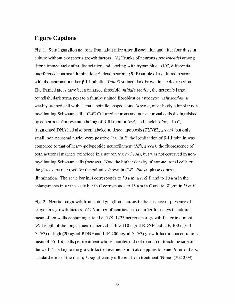
Figure Captions
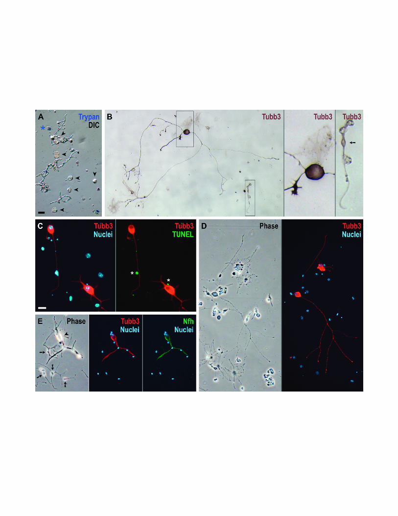
Fig. 1. Spiral ganglion neurons from adult mice after dissociation and after four days in
culture without exogenous growth factors. (A) Trunks of neurons (arrowheads) among
debris immediately after dissociation and labeling with trypan blue. DIC, differential
interference contrast illumination; *, dead neuron. (B) Example of a cultured neuron,
with the neuronal marker β-III tubulin (Tubb3) stained dark brown in a color reaction.
The framed areas have been enlarged threefold: middle section, the neuron’s large,
roundish, dark soma next to a faintly-stained fibroblast or astrocyte; right section, a
weakly-stained cell with a small, spindle-shaped soma (arrow), most likely a bipolar non-
myelinating Schwann cell. (C-E) Cultured neurons and non-neuronal cells distinguished
by concurrent fluorescent labeling of β-III tubulin (red) and nuclei (blue). In C,
fragmented DNA had also been labeled to detect apoptosis (TUNEL, green), but only
small, non-neuronal nuclei were positive (*). In E, the localization of β-III tubulin was
compared to that of heavy-polypeptide neurofilament (Nfh, green); the fluorescence of
both neuronal markers coincided in a neuron (arrowhead), but was not observed in non-
myelinating Schwann cells (arrows). Note the higher density of non-neuronal cells on
the glass substrate used for the cultures shown in C-E. Phase, phase contrast
illumination. The scale bar in A corresponds to 30 µm in A & B and to 10 µm in the
enlargements in B; the scale bar in C corresponds to 15 µm in C and to 30 µm in D & E.
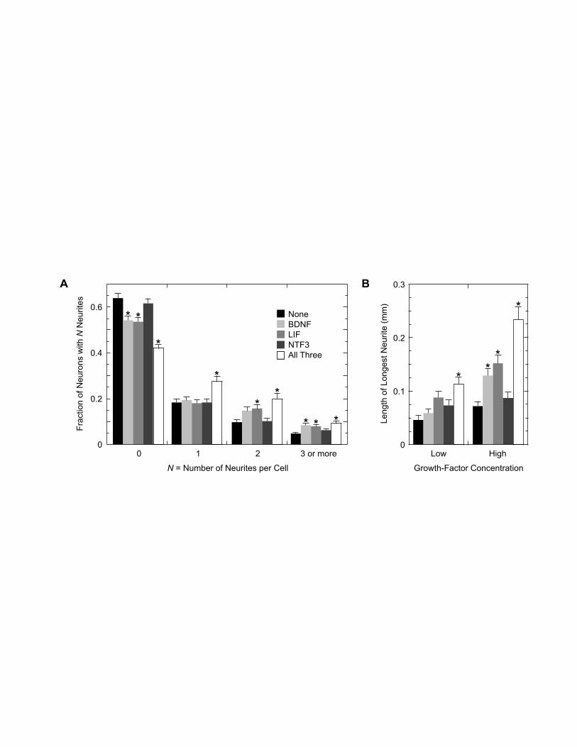
Fig. 2. Neurite outgrowth from spiral ganglion neurons in the absence or presence of
exogenous growth factors. (A) Number of neurites per cell after four days in culture;
mean of ten wells containing a total of 778–1223 neurons per growth-factor treatment.
(B) Length of the longest neurite per cell at low (10 ng/ml BDNF and LIF, 100 ng/ml
NTF3) or high (20 ng/ml BDNF and LIF, 200 ng/ml NTF3) growth-factor concentrations;
mean of 55–156 cells per treatment whose neurites did not overlap or touch the side of
the well. The key to the growth-factor treatments in A also applies to panel B; error bars,
standard error of the mean; *, significantly different from treatment ‘None’ (P ≤ 0.03).
23
Table
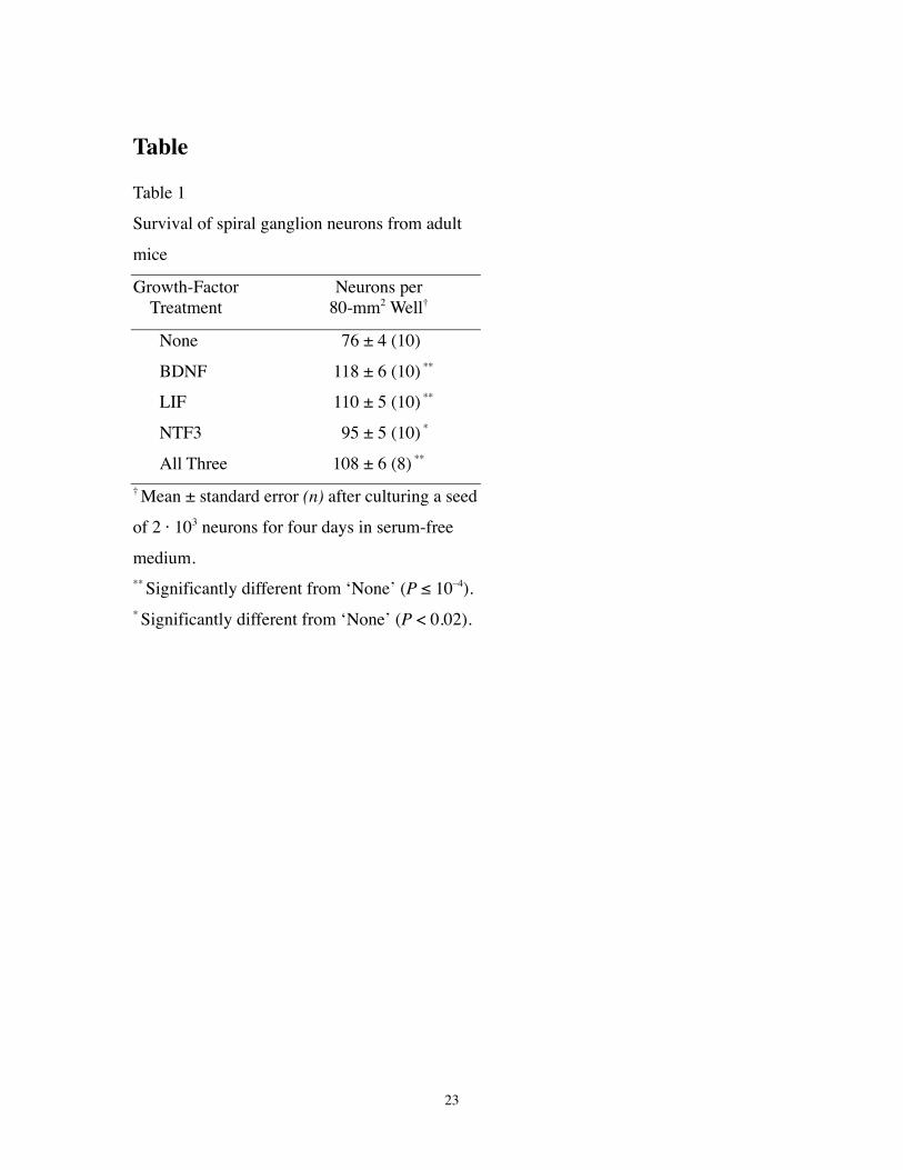
Table 1
Survival of spiral ganglion neurons from adult
mice
Growth-Factor Neurons perTreatment 80-mm2 Well†
None 76 ± 4 (10)
BDNF 118 ± 6 (10) **
LIF 110 ± 5 (10) **
NTF3 95 ± 5 (10) *
All Three 108 ± 6 (8) **
† Mean ± standard error (n) after culturing a seed
of 2 · 103 neurons for four days in serum-free
medium.** Significantly different from ‘None’ (P ≤ 10–4).* Significantly different from ‘None’ (P < 0.02).
0
0.2
0.4
0.6
21 3 or more
Fra
ctio
n of
Neu
rons
with
N N
eurit
es
N = Number of Neurites per Cell
0
0.1
0.2
0.3
Low High0
Leng
th o
f Lon
gest
Neu
rite
(mm
)
Growth-Factor Concentration
A B
NoneBDNFLIFNTF3All Three
**
*
*
* *
***
**
*
*