swedish university of agricultural sciences - · pdf fileswedish university of agricultural...
TRANSCRIPT

Swedish University of Agricultural Sciences Faculty of Veterinary Medicine and Animal Science
Department of Anatomy, Physiology and Biochemistry Section of Biochemistry.
Title: Immunoassays for detection of serum Thymidine
kinase 1 in Dog lymphomas and carcinomas
Jagarlamudi Kiran Kumar
Uppsala
2010
Masters Degree project ( 30 hp)
Section of Biochemistry

2
SLU Swedish University of Agricultural Sciences
Titel : Immunoanalyser för detektion av serum tymidinkinas 1 hund lymfom och karcinom
Jagarlamudi Kiran Kumar
Supervisor : Staffan Eriksson, Department of Anatomy, Physiology and Biochemistry, Section of Biochemistry
Co-supervisor : Henrik von Euler, Department of Clinical Sciences, Division of small animal clinical sciences.
Examinator : Liya Wang, Department of Anatomy, Physiology and Biochemistry, Section of Biochemistry
Master thesis in Animal Sciences, Uppsala 2010 Faculty of Veterinary Medicine and Animal Science
Department of Anatomy, Physiology and Biochemistry
Course code: EX0562 , Advanced E, 30hp
Key words: Immunoassays, Thymidine kinase 1 , dog lymphomas
Online publication of this work: http://epsilon.slu.se
Uppsala 2010

3
INDEX:
Abstract 4
Background of the veterinary medical
problem
5
Introduction to the project 8
Materials & Methods 10
Results 15
Discussion 21
Future prospects 23
Acknowledgements 24
References 25
Appendix 27

4
ABSTRACT:
Serum thymidine kinase 1 (TK1) activity determination is used as marker for tumor
monitoring in both human and veterinary medicine. TK1 is an intracellular enzyme
involved in the salvage pathway of DNA precursor synthesis. TK1 expression is cell
cycle dependent and the activity increases markedly at the end of G1 and reaches a
peak value in S phase and then declines rapidly in G2 ∕M. The pronounced
proliferation in tumor cells result in a higher TK1 activity within cell. So
determination of TK1 activity in tumors provides information regarding prognosis and
effectiveness of treatment. In human medicine, a labeled radioactive substrate is used
for measuring TK1 activity. Some studies have shown that this method can be used
also in veterinary medicine. However, it has limitation for general use in animal
practice since it is expensive and involves handling of radioactive material. In human
medicine, anti TK1 antibodies have been used to determine the concentration of
serum TK1 in patients with carcinomas. This type of measurements has been
successful and would be valuable to translate into veterinary medicine. The purpose of
this study is to make the use of anti-dog TK1 antibodies for detection of serum TK1.
Anti bodies have been produced by immunizing the rabbit with synthetic piece of a
dog TK1 peptide. Suspected band of serum TK1 has been found in the serum from
dogs with tumors but not in the serum from healthy dogs using these antibodies. This
study showed that anti dog TK1 polyclonal antibodies produced by affinity
chromatography binds to serum TK1 with adequate capacity. It has been also shown
that these antibodies can bind with both human and dog cytosolic TK1. These results
are the first step in the development of an ELISA, which may provide clinical
information about prognosis, risk of recurrence and effectiveness of antitumor therapy
in veterinary medicine.
5
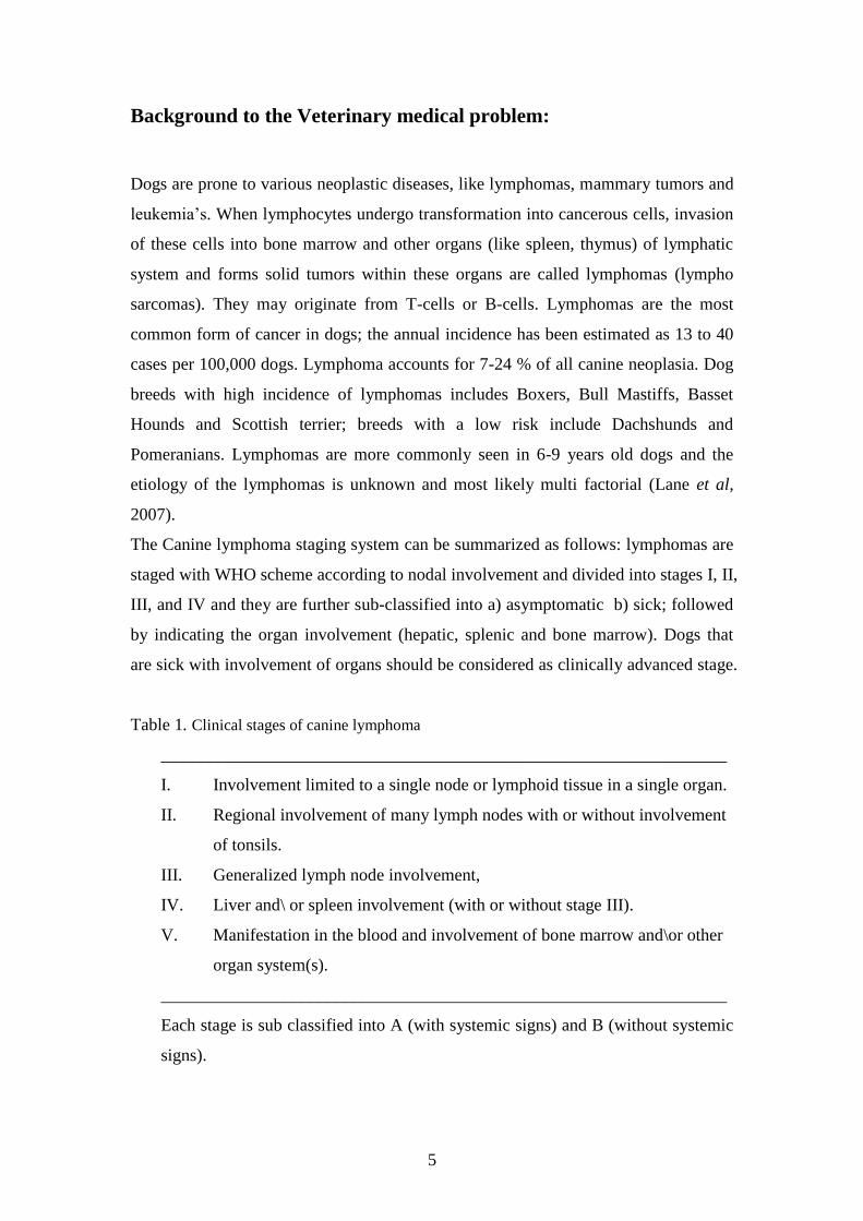
Background to the Veterinary medical problem:
Dogs are prone to various neoplastic diseases, like lymphomas, mammary tumors and
leukemia’s. When lymphocytes undergo transformation into cancerous cells, invasion
of these cells into bone marrow and other organs (like spleen, thymus) of lymphatic
system and forms solid tumors within these organs are called lymphomas (lympho
sarcomas). They may originate from T-cells or B-cells. Lymphomas are the most
common form of cancer in dogs; the annual incidence has been estimated as 13 to 40
cases per 100,000 dogs. Lymphoma accounts for 7-24 % of all canine neoplasia. Dog
breeds with high incidence of lymphomas includes Boxers, Bull Mastiffs, Basset
Hounds and Scottish terrier; breeds with a low risk include Dachshunds and
Pomeranians. Lymphomas are more commonly seen in 6-9 years old dogs and the
etiology of the lymphomas is unknown and most likely multi factorial (Lane et al,
2007).
The Canine lymphoma staging system can be summarized as follows: lymphomas are
staged with WHO scheme according to nodal involvement and divided into stages I, II,
III, and IV and they are further sub-classified into a) asymptomatic b) sick; followed
by indicating the organ involvement (hepatic, splenic and bone marrow). Dogs that
are sick with involvement of organs should be considered as clinically advanced stage.
Table 1. Clinical stages of canine lymphoma
_________________________________________________________________
I. Involvement limited to a single node or lymphoid tissue in a single organ.
II. Regional involvement of many lymph nodes with or without involvement
of tonsils.
III. Generalized lymph node involvement,
IV. Liver and\ or spleen involvement (with or without stage III).
V. Manifestation in the blood and involvement of bone marrow and\or other
organ system(s).
_________________________________________________________________
Each stage is sub classified into A (with systemic signs) and B (without systemic
signs).
6
Clinical observations:
The clinical observations mainly depend on the extent and location of tumor. Multi
centric lymphoma is the most common form with generalized lymphadenopathy,
hepatosplenomegaly and most of the dogs are asymptomatic but nonspecific signs like
anorexia, vomiting, diarrhea, asicites and fever are common. In alimentary lymphoma
gastro-intestinal signs such as vomiting, diarrhea, weight loss, and malabsorption are
common. Mesenteric lymph nodes, spleen and liver may be involved. The mediastinal
form of lymphomas is characterized by enlargement of the craniomediastinal
structures or thymus. Respiratory distress, exercise intolerance, polyuria from
hypocalcaemia are frequently observed. Cutaneous lymphoma is usually generalized
and skin becomes more erythemtous, thickened, ulcerative and exudative. Sometimes
oral involvement also occurs (Lane et al, 2007).
Diagnosis:
Physical examination of lymph nodes, complete blood picture, biopsy and
microscopic examination are required for an accurate diagnosis in the early stages of
lymphomas. Fine needle aspiration and radiographic examination are useful in
different stages of lymphomas. Malignant lymphomas cannot be cured completely,
however, early detection and efficient chemotherapy can control the disease for long
time.
Prognosis:
The prognosis of canine lymphoma depends on various factors like location of tumor,
extent of the disease, presence and absence of clinical signs. Immunophenotyping of
the lymphomas into B- cell and T- cell type may assist in the prognosis. Another
prognostic indicator involves the use of cell proliferation markers in tissue specimens.
Several attempts have been made to use these markers for prognosis and relapse of the
lymphomas in dogs. Serum alpha 1 acid glycoprotein concentration was used for
predicting the relapse of malignant lymphoma. Alpha-fetoprotein can be a good
marker in advanced stages of malignant lymphomas, where the concentration of this
protein is high (von Euler et al, 2004).

7
Biomarkers in Veterinary Medicine:
Cell proliferation markers commonly used in veterinary medicine are Ki-67,
argyrophilic nuclear organizing regions (AgNOR), Mitotic index, proliferation cell
nuclear antigen (PCNA), lactate dehydrogenase (LDH) (Kiupel et al, 1999).
Monoclonal antibody, Ki-67 recognizes a nuclear antigen present in proliferating cells
and permits an immunohistochemical method of assessing tumor cell proliferative
fraction. It is expressed in all phases of active cell cycle (G1, S, G2 and M) and it is
absent in resting cells (G0) (Leong et al, 1995). AgNOR is a good marker for the
prognosis of lymphoma. The quantity of the AgNOR not only reflects the percentage
of cells in cycle but it also increases when the cell cycle is faster and AgNOR counts
correlates well with the tumor grade (Kiupel et al, 1998). Studies on AgNOR
demonstrated significant predictive potential for remission in treated and untreated
cases of canine lymphoma (kiupel et al, 1999). However, histopathological
examination of AgNOR does not help for disease monitoring. AgNOR is associated
with problems in interpretation and is dependent on type of the fixation method,
temperature and staining time. Mitotic index is an indirect measure based on
quantification of the proportion of mitotic cells in a histopathologic specimen and it is
easier to perform than AgNOR and Ki-67 assessment (Romansik et al, 2007). PCNA
is produced in late G1 phase and throughout S-phase, but it can be detected in all
proliferating cells because of long half life, High concentrations of PCNA are detected
in S phase. It has been used to predict prognosis in melanomas in dogs and cats (Roels
et al, 1999). In one study, plasma lactate dehydrogenase (LDH) was used for
evaluation of disease progression in malignant lymphoma of dogs (Nakamura et al,
1997). They found elevated levels of plasma LDH in tumors and in some hemolytic
diseases. In certain serum samples of dogs with tumors, they did not find any increase
in the plasma LDH level. Furthermore, the sensitivity and specificity of LDH is very
low (Nakamura et al, 1997). Thymidine kinase 1 expression both at the RNA and
protein level has been used extensively as a marker for proliferating cells and TK1 is
one of the protein, whose abundance is most closely correlated to the S-phase.
Because of its association with S phase, it is a good proliferative marker for
monitoring and prognosis of tumor diseases.

8
Introduction to the project:
Thymidine kinase (TK) is a cytosolic enzyme involved in the pyrimidine salvage
pathway, which are building blocks of DNA. Thymidine kinase catalyzes the
conversion of deoxythymidine to deoxythymidine monophosphate (dTMP) in the
presence of ATP by transferring the γ- phosphate group. This phosphorylation step is
often a rate-limiting step and deoxythymidine monophosphate is subsequently
phosphorlyated to di and triphosphates before serving as a substrate for DNA
synthesis. TK enzymes in mammalian cells appear in two forms, a cytosolic
thymidine kinase (TK1) and the mitochondrial (TK2) form. Thymidine kinase 1 (TK1)
is present in the cell cytoplasm and is involved in cell proliferation. TK2 is involved
in mitochondrial DNA synthesis, which is independent of the cell cycle (von Euler et
al, 2006).
TK1 plays a primary role in regulation of intracellular thymidine nucleotide pools
during the cell cycle. The level of TK1 increases from late G1 to late S phase and
early G2 phase in both proliferating and tumor cells. Thymidine kinase may also
appear in the body fluids because of cells with disrupted spindle apparatus (von Euler
et al, 2004). TK1 or TK1 like genes seems to be widely distributed in many organisms
from plants to humans, including bacteria and viruses (Welin et al, 2004). The human
TK1 gene is 12.9 kb with seven exons. The 1241bp cDNA with an open reading
frame of 702 bp, encoding a 234 amino acids long protein with a molecular weight of
25.5 kDa and TK1 gene is located on chromosome 17 (Eriksson et al, 2002). To
characterize serum TK1 reducing agent treatment were used and only high
concentration of DTE (Dithioerythritol, 400 mM) showed effects on the molecular
forms of TK1 going from more than 700 kDa to about 100 kDa (Karlström et al,
1990). In human medicine TK1 activity is measured by using a commercially
available assay; the Prolofigen TK-REA (radio enzymatic assay), where a radioactive
substrate analogue is added. It then measures the amount of converted substrate per
unit time, which represents the TK activity in serum.
In one study, they used the same method to determine the TK1 activity in the serum of
dogs with malignant lymphoma and non-hematological or healthy ones (von Euler et
al, 2004). Serum TK1 activity was significantly higher in lymphoma than in normal
dogs. Average TK1 activity after complete remission was not significantly different
compared with healthy dogs. Thus, measuring TK1 activity in dog’s serum serves as a

9
tumor marker providing information regarding prognosis and risk of relapse in dogs
with tumors (von Euler et al, 2004). One publication has described the use of plasma
thymidine kinase activity in diagnosis and monitoring of lymphomas and leukemia in
dogs (Nakamura et al, 1997).
In human medicine, serum TK1 activity has been used for estimating the extent of
tumor disease and information of the prognosis of leukemia’s, multiple myelomas,
Hodgkin’s lymphoma and non-Hodgkin’s lymphomas (Wu et al, 2003). Serum TK1
activity has been determined by using commercially available TK-REA. This assay
provides prognostic information of malignancies and some cases it can aid in the
choice of therapy (Topolcan et al, 2008). A non-radiometric TK enzyme linked
immunosorbent assay (ELISA) was developed by using 3'-azido-2'-deoxythymidine
(AZT) as substrate, which is phosphorylated selectively by TK1 in the serum.
Furthermore, a competitive ELISA measures the amount of AZT monophosphate
using goat polyclonal antibodies (Öhrvik et al, 2004). Recently, a Liaison assay was
developed. It is a non-radiometric assay and technically more accurate, reliable in
measuring the TK1 activity in dogs with lymphomas. In brief, TK1 in the tumor
serum sample catalyzes phosporylation of 3'-azido-thymidine (AZT) to the
corresponding 5'- monophosphate(AZTMP). In the second step, the produced
AZTMP competes with AZTMP conjugated to isoluminol(AZTMP-ABEI) for
specific antibodies bound to magnetic beads. Free AZTMP-ABEI is removed by
washing and the amount of bound AZTMP-ABEI is determined by
chemiluminescence (von Euler et al , 2008).
The Biovica Company Uppsala (Sweden) has developed the very sensitive Divi Tum
assay, which is a non-isotopic method and uses bromo-deoxyuridine as substrate for
TK followed by its incorporation into an oligonucleotide attached to a solid support
(refer www.biovica.com). Even though we have, many assays to determine the serum
TK1 activity in dogs most of them needs special equipment that are expensive. The
dog TK1 gene (amino acid) sequence shows 88.5% similarity with the human. TK1
sequence diversity is 9.1% and most of the differences in sequence is seen in the C-
terminal part (Fig 1)

10
Dog: AYTKRLGSEK EVEVIGGADKYHSVCRLCYFKKASGPPMGLDSERNKENVL 210
HUMAN: AYTKRLGTEKEVEVIGGADKYHSVCRLCYFKKASGQPAGPDNKEN----- 210
DOG: VLVPGKPGEGKEATGVRKLFAPQHVLQCSPAN 243
HUMAN: CPVPGKPGE------ AVAARKLFAPQQILQCSPAN 235
Fig 1: The sequence of Dog and Human TK1 (160-243) . Red color indicates the
differences in the amino acid sequence. The underlined sequence amino acids (196-
223) are used for immunization of rabbit.
The use of TK1 serum tests in humans has increased recently and most of these tests
are based on activity measurement with radio labeled substrates. In veterinary practice,
the commercially available tests are not a convenient alternative because of the need
for a special instrument. Still the result with the Liaison TK assay serves as a good
indicator for the value of this type of information for prognosis and monitoring of
dogs with malignant lymphoma.
In human medicine anti human TK1 anti bodies have been utilized for measuring the
serum TK1 levels in various conditions, e.g. Lung cancer, colorectal and mammary
cancers (He et al, 2000). However, it has not been described by any method that
makes use of dog TK1 antibodies. So it would be interesting to produce the dog TK1
antibodies and evaluate them further whether they can provide a simpler method for
prognosis of tumors in dogs. Three different rabbits Ulrika (U), Dingo (D) and Kurtis
(K) were immunized with synthetic peptide (amino acids 196-223) by Agri Sera AB
(Umea Sweden). Anti sera were already collected from these rabbits after different
periods of immunization before starting project.
The main purpose of this project is to purify these antisera using affinity
chromatography, which was produced by coupling the C-terminal peptide shown in
Fig 1 to a Sepharose 4B matrix. These purified antibodies are used to study serum
TK1 and eventually determine its concentration in various serum samples. Serum
TK1 activity measurements were determined with radioactive thymidine as substrate.
Another objective of this work was to collect blood samples from dogs with tumor
diseases and analyze TK1 levels, trying to find a correlation with disease condition
and serum TK1 levels.

11
Materials and methods:
All serum samples were collected from dogs with tumors at the Department of clinical
medicine, small animal clinic, SLU. Cell cultures from dog MDCK (madin Darby
canine kidney cell lines) and the human T-lymphoblast cell lines like CEM TK+ and
TK− variants were also used.
Purification of rabbit anti TK1 antibodies by Sepharose coupled with C-terminal
TK1 peptide:
CNBR-activated Sepharose 4B (1 gram in final volume of 3.5 ml) was treated with
HCl to remove the preservatives and mixed with dog peptide i.e. (CPMGLDS
RENKENVLVLVPGKPGEGKEANH2) at a concentration of 2 mg ∕ml in 3 ml of coupling
buffer (0.1 M NaHCO3 pH-8.3 containing 0.5 M NaCl). The coupled Sepharose was
washed with five medium volumes of coupling buffer and remaining active groups
were blocked by transferring the medium to 0.1 M Tris-HCl buffer, pH 8.0. The
Sepharose was then treated with three cycles of alternating 0.1 M acetate buffer, pH
4.0 containing 0.5 M NaCl and 0.1 M Tris-HCl, pH 8.0. Finally, the column was
washed with PBS. For purification of anti TK1 antibodies filtered 15 ml of rabbit anti-
serum was diluted in PBS to 50 ml and loaded on the affinity column. It was washed
with 20 ml (50 mM Tris-Cl, pH 8.0; 0.1% Triton X-100; 0.5 M NaCl) followed by (50
mM Tris-Cl, pH 9.0; 0.1% Triton X-100; 0.5 M NaCl), with 50 mM sodium
phosphate, pH 6.3; 0.1% Triton X-100; 0.5 M NaCl. Elution of bound rabbit anti TK1
antibodies was performed with 20 ml 50 mM Glycine –HCl, pH 2.5; 0.1% Triton X-
100; 0.15 M NaCl into tubes containing 4 ml of 1 M Tris-Cl, pH 9.0, to neutralize the
samples.
Determining the protein concentration:
The protein concentrations were determined using the Bio-rad protein assay. The
reference used here was BSA (bovine serum albumin) in PBS.
The Bio-rad reagent was diluted with water and four different concentrations of BSA
(2.7, 5.4, 7.1 and 10.8 µg respectively) has made to set reference. Two samples of
antibody solution were used. Absorbance was measured at 595 nm and a reference
graph was created with the BSA standards. With the help of the graph, the
concentration of antibody protein was determined.

12
Dot blot immuno assays:
The TK1 levels in serum samples were measured using the enhanced
chemiluminscent (ECL) dot blot assay (He et al 2000). In this assay, 3 µl of serum is
applied to the nitrocellulose membrane (Hybond TM-C, Amersham). Recombinant
dog TK1 was used as standard. The membranes were left to dry and then blocked with
10 % non-fat milk in TBS for one hour. The membranes were incubated with primary
antibody (1.25 µg ∕ml) for two hours and washed with TBS-T (0.1% Tween 20) for 15
minutes. Secondary antibodies conjugated with HRP were added (2 ng ∕ml) and
agitated for one hour (He et al, 2000). Then the membranes were washed with TBS-T
for 15 min and ECL substrate applied to membrane for 1 min, and then the membrane
was exposed to X-ray film. The serum TK1 levels were determined by measuring the
intensity of the spot and compared to a TK1 standard.
Depletion of albumin and IgG from the serum:
Proteoprep blue albumin and IgG depletion kit (SIGMA, USA) was used for depletion
of albumin. This kit removes more than 85% albumin and 70% of IgG in the serum
samples. It involves two steps a) column equilibration: medium slurry of 0.4 ml was
transferred to a spin column. Storage solution was removed by centrifugation.
Equilibration buffer (0.4 ml) was added to the spin columns and centrifuged. The
buffer from collection tube was discarded after centrifugation and replaced with new
collection tube. Then this column was used for serum depletion. b) Depletion of
albumin and IgG from the serum samples: Serum samples of 100 µl were added to
spin column and allowed for 10 minutes at room temperature. So that albumin and
IgG in serum are bound to the slurry in the spin column. Centrifugation and reloading
of the elution to the same spin columns removed an additional 5% of the albumin. The
depleted serum in collecting tube was used for further evaluation.
Isolation of serum TK1 using the anti TK1 antibody Sepharose:
Serum samples 100 µl from healthy dogs and dogs with tumors after depletion of
albumin were diluted with 100 µl of TBS and incubated with the anti dog TK1
antibody Sepharose (200 µl). The samples then agitated at 4°C for 3 hours followed
by centrifugation. The sepharose was washed two times with TBS, one time with
TBS-T and one time with TBS. Then 40 µl of sample buffer (containing 0.5 M Tris-
HCl, pH 6.8, glycerol, 10% (w\v) SDS, and 0.1% bromophenol blue) was added to

13
the Sepharose and incubated at room temperature for 30 minutes to elute bound
serum proteins. These samples were heated at 95°C for 5 minutes and analyzed by
SDS-PAGE.
Western blot:
SDS-PAGE and electrophoretic transfer was done as described in Amersham life
sciences western blotting protocols. Samples were loaded on the gel and run for 1
hour at 160 V and 6 µl of marker (Fermentas, USA) for reference protein molecular
weights. Proteins were transferred to PVDF membranes (GE HealthCare) at 90 V for
50 min. Membrane was rinsed 3×5 min with TBS buffer (pH 7.6) and blocked in
TBS-T (5% BSA) buffer. The membrane was incubated with primary antibody (1.25
µg ∕ml) and then incubated with goat-Anti rabbit F (ab)2 fragment and avidin HRP
conjugated (Jackson, USA). The membrane was briefly rinsed with TBST buffer
twice, and then washed once for 15 min and twice for 5 min with TBST buffer. The
membrane was incubated with the ECL reagents (GE HealthCare) for precisely 1 min
at room temperature and then placed in film cassette for 2-5 min exposure (hyper
film-ECL).
Immunoprecipitation:
Immnuoprecipitation with Protein-G sepharose was done as described by (GE
HealthCare). Serum samples of 200 µl along with buffer (containing TBS, pH 7.6, 1
mM befa block, 1% BSA and purified anti dog TK1 antibodies of 2µg ∕ 500µl) to
make final volume of 500µl but in cell extract the protein concentration was 500µg.
These tubes incubated at 4°C overnight. Then the samples were mixed with 100µl of
Protein-G sepharose. These tubes were kept at 4°C with slight agitation for 4 hours.
The Sepharose beads were centrifuged and washed with TBS two times, one time
with TBS-T and then with TBS. Protein-G sepharose was incubated with sample
buffer (0.5M Tris-HCl, pH 6.8, glycerol, 10% (w\v) SDS, 0.1% bromophenol blue at
room temperature for 20 minutes to elute the antibody complexes. The samples were
analyzed by western blotting as described.
Determination of enzyme activity:
Enzyme activity of the serum samples was determined by using radio labeled ³H-
thymidine as a substrate. The reaction was started by adding 20 µl TK assay buffer
(0.1 M Tris-HCl, 0.15 M NaCl, 5 mM MgCl2, 5 mM NaF, 2 mM DTT, and 5 mM
ATP) to 20 µl of serum samples. The substrate was phosporylated by enzyme in
serum with ATP as phosphate donor. The reaction was interrupted at different time

14
points (30, 60, 120 and 240 min) to measure the amount of product formed. A small
fraction (10 µl) of reaction mixture was transferred to DE-81 paper disks. These disks
were washed two times with 1mM ammonium formate for 10 minutes of each. Then
paper disks were incubated in 1 ml of (0.1 M HCl and 0.2M KCl) for 45 minutes to
elute the product which were measured by scintillation counting. The results are
expressed in pmol ∕ min.
FPLC (fast protein liquid chromatography):
FPLC was used for rapid separation of proteins from complex mixtures. Sera and cell
extracts (100 µl) were diluted two fold with buffer (0.01 M Hepes pH -7.6, containing
0.15 M NH4Cl and NaN3) and then filtered. 200 µl samples were fractioned by
sepharose 12 chromatography (GE HealthCare) in a column of 1.0×30 cm. Protein
content was monitored continuously at 280 nm. The column was eluted with the same
buffer and 350 µl of 24 fractions were collected. 200 µl of these fractions are
precipitated with 10% TCA and did western blot as described above. Four proteins;
alpha 2 –macro-globulin, BSA, Ova albumin and Horse myoglobulin with the
molecular weights of 700, 66 ,45 and 17 kDa were used as molecular weight markers.

15
Results:
Sera were collected from rabbits 4 weeks after immunization. Anti serum was loaded
on affinity column made with the C terminal region of dog TK1. The bound anti
TK1 antibodies from rabbit (D) and rabbit (U) were purified in this way. The
concentration of antibodies was determined by the Bio-rad assay. Protein
concentration in case of the rabbit (D) antibodies was 0.8 mg ∕ ml and from rabbit (U)
0.5 mg ∕ ml. The purified antibodies from D (Fig 2) were tested with human
recombinant TK1 (234 aa + his tag) and dog recombinant TK1 ∆12 (230 aa + his tag)
(see appendix)
1 2 3 4 5 6
Fig 2: Western blot analysis with purified rabbit (D) antibody (1.25 µg ∕ml) with
human and dog recombinant TK1. Lane 1-3 are dog recombinant TK1∆12 (2, 1 and
0.5 ng); 4-6 are human recombinant TK1 (2, 1 and 0.5 ng).
Normally TK1 exits as dimer and by activation with ATP ( adenosine triphospate)
it converts into tetramer. The sub unit of TK1 in lower region at 28 kDa in case of
both human and dog (Fig 2) ( see appendix page 29 and 30). In Fig 2, an apparent
dimeric form of TK1 is seen as strong band between the 52 kDa and 72 kDa. To
analyze the upper band of TK1, mass spectrometry of the bands in that region was
performed. The band was conformed as TK1 , which corresponds to molecular weight
of 66 kDa (approx). peptide analysis results were shown in appendix ( page 32).
52 kDa
28 kDa
72 kDa

16
The anti dog TK1 antibodies from D reacts with cytosolic TK1 in crude cell extracts
from dog MDCK cells but also with human TK1 in CEM TK+ cells but no band was
seen in CEM TK− cell extracts (Fig 3).
1 2 3 4 5 3 4 5
Fig 3: Western blot analysis with TK1 antibody from D (1.25 µg ∕ml) the absence (A)
and in presence (B) of an excess of the antigen peptide (0.1 µg ∕ml). Lane 1, 2 are Dog
recombinant TK1 (0.25, 0.05 ng) 3, 4, 5 are extracts from cell lines (CEM TK+,
MDCK, CEM TK−
approx 100 µg protein in each case).
The band at 26 kDa, which was found in both CEM TK+ and MDCK cell extracts
corresponds to cytosolic TK1 and the band was absent in CEM TK− cell extract.
MDCK extract from a cell culture grown to high density cell lines were tested and in
this case a double band was found in the region corresponding to cytosolic TK1.
There were no bands on membrane B in lower TK1 band region demonstrating that an
excess of peptide could block the reactivity with all the polypeptides seen in fig 3A.
The band that appeared at 52 kDa is most likely not related to TK1 since this band
was observed in extracts from TK−
cells and also on the membrane B.
26 kDa
52 kDa
A B

17
Anti dog TK1 antibodies from U and D were used to test the reactivity with human
and dog recombinant TK1 and cell extracts.
1 2 3 4 5 1 2 3 4 5
Fig 4: Western blot analysis with anti dog TK1 antibodies from U (A) and D (B).
Lane 1 is recombinant dog TK1 (0.5 ng) 2, 3 are human TK1 (2 and 1 ng) 4 is
MDCK cell extracts from high density cultures, 5 is MDCK cell extract from
exponentially growing cells and a total of 25 µg protein was tested.
The membrane with antibodies from rabbit U has shown light band with dog
recombinant TK1 (indicated by arrow) and no reactivity with cytosolic TK1 from dog
cell extracts (Fig 4A). Anti bodies from rabbit D showed good reactivity with both
dog and human recombinant TK1 (Fig 4B). the sub unit of cytosolic TK1 (26 kDa)
was appeared as single in MDCK extract from exponentially growing cells. This was
different as double band was appeared in this region in MDCK cell extract from high
dense culture( Fig 3A).
Based on results from Fig 4B anti bodies from rabbit D were used for evaluation of
TK1 from serum samples (serum from healthy dog and dogs with tumor disease). D
anti bodies were coupled to Sepharose 4 B. Albumin was depleted from the serum
samples before incubating with D anti TK1 anti body sepharose.
26
26 kDa
52 kDa
B A
18
1 2 3 4 5 2 3 4 5
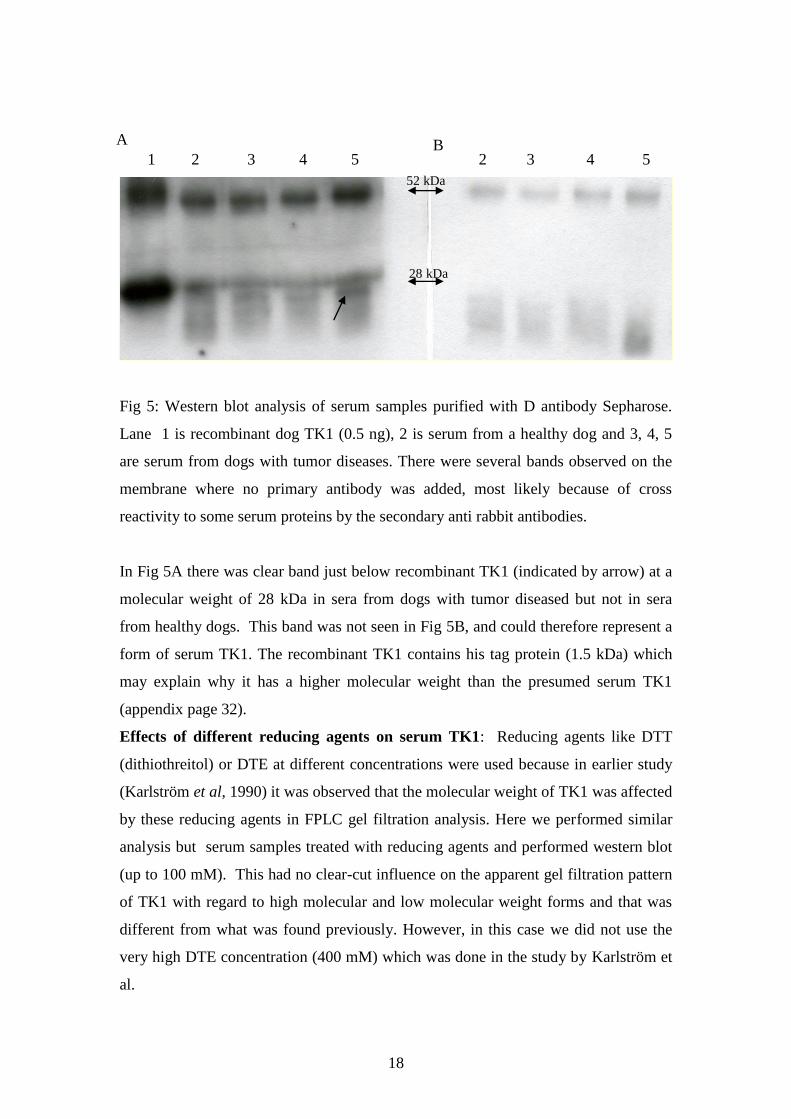
Fig 5: Western blot analysis of serum samples purified with D antibody Sepharose.
Lane 1 is recombinant dog TK1 (0.5 ng), 2 is serum from a healthy dog and 3, 4, 5
are serum from dogs with tumor diseases. There were several bands observed on the
membrane where no primary antibody was added, most likely because of cross
reactivity to some serum proteins by the secondary anti rabbit antibodies.
In Fig 5A there was clear band just below recombinant TK1 (indicated by arrow) at a
molecular weight of 28 kDa in sera from dogs with tumor diseased but not in sera
from healthy dogs. This band was not seen in Fig 5B, and could therefore represent a
form of serum TK1. The recombinant TK1 contains his tag protein (1.5 kDa) which
may explain why it has a higher molecular weight than the presumed serum TK1
(appendix page 32).
Effects of different reducing agents on serum TK1: Reducing agents like DTT
(dithiothreitol) or DTE at different concentrations were used because in earlier study
(Karlström et al, 1990) it was observed that the molecular weight of TK1 was affected
by these reducing agents in FPLC gel filtration analysis. Here we performed similar
analysis but serum samples treated with reducing agents and performed western blot
(up to 100 mM). This had no clear-cut influence on the apparent gel filtration pattern
of TK1 with regard to high molecular and low molecular weight forms and that was
different from what was found previously. However, in this case we did not use the
very high DTE concentration (400 mM) which was done in the study by Karlström et
al.
52 kDa
28 kDa
A B

19
1 2 3 4 1 2 3 4
Fig 6: Western blot analysis of serum samples treated with reducing agents. In the
figure 6, 1 and 2 are sera treated with 200 mM of DTT and 3, 4 with 200 mM of DTE.
1, 3 are sera from dog with tumor disease and 2, 4 are sera from healthy dogs.
In A the membrane is incubated with purified D antibodies and B is without of
primary antibody. Suspected TK1 band appeared below the molecular weight of 28
kDa in sera from dogs with tumor disease (indicated by arrow) but not in sera from
healthy dogs (see Fig 5A). The strong bands in the upper region are mainly due to
cross reactivity of secondary antibodies with some serum proteins. Furthermore these
bands were seen on the membrane without primary antibodies (Fig 6B). Reducing
agents treatment was helpful in reduction of many unspecific proteins that appeared
on membrane with direct serum samples.
28 kDa
52 kDa
A B

20
fr 14 fr 16 fr 18 fr 20 Rec TK1 fr14 fr 16 fr 18 fr 20
Fig 7. Western blot analysis of different FPLC fractions ( fr) .In the Fig 7 , different
FPLC fractions ( 14,16, 18 and 20) of both healthy and tumor diseased dog serum
samples treated with 10% TCA ( trichloroacetic acid) and recombinant dog TK1 of
0.5 ng. Membrane A is with FPLC fractions of healthy dog serum incubated with
antibodies from D. Membrane is with tumor diseased dog serum sample FPLC
fractions incubated with antibodies from D.
In FPLC fractions of healthy dog serum samples (Fig 7A) no bands were observed
around the molecular weight of 28 kDa but in the case of tumor diseased serum
fractions suspected band of TK1 sub unit was detected. In the ( Fig 7B) both dimer
(14 and 16 fractions) and monomer ( fraction 20) forms of serum TK1 were identified.
52 kDa
28 kDa
72 kDa
A B

21
Discussion:
The peptide sequence used to immunize rabbits was from amino acids 196-223,
which is the C-terminal part and most variable region in human and dog TK1
sequence. The possible explanation for differences in reactivity of these two anti TK1
antibodies could be its epitopes. The epitope of D antibodies may present in both the
human and dog amino acid sequence, since it was able to recognize both human and
dog TK1. However, the epitope for the U antibodies is most likely found only in dog
TK1. These U antibodies does not recognize the human recombinant TK1 at low
concentration (eg.1 ng).
In serum samples, there was no clear cut bands observed with the U antibodies and
some un specific bands found in the membrane without the primary antibody due to
cross reactivity of secondary antibodies with serum proteins. Sometimes these
antibodies may detect another compound than TK1 or may cross-react with other
compounds than TK1. Furthermore modifications of serum TK1 will most likely alter
the reactivity of different antibodies. It is known that TK1 found in a stable
multimeric form in serum.
Human recombinant TK1 (28.2 kDa) has almost similar molecular weight as a
truncated form of dog recombinant TK1 ∆12 (27.9 kDa). The dimeric form of dog
recombinant TK1 was identified between 52-72 kDa, which was confirmed by peptide
analysis (see page 32). The suspected band of serum TK1 sub unit was identified at
26 kDa and it had a molecular weight lower than recombinant TK1 due to His tag
protein in recombinant TK1. The suspected TK1 band was found only in serum from
dogs with tumor disease and not in serum from healthy dogs. Reducing agents like
DTT and DTE up to 100 mM concentration did not have clear-cut influence on the
serum TK1 band. However, with 200 mM concentration of DTE and DTT there was
reduction in the other unspecific bands in the serum. FPLC gel filtration of serum
samples on western blot analysis with antibodies from D have identified both dimer
and monomer forms of TK1 in tumor diseased serum but not in serum samples from
healthy dogs.
Some difficulty was found in obtaining dog cell extracts with sufficient TK1 levels to
evaluate the function of these antibodies. In one experiment a double band of TK1
was found in extracts from MDCK cells grown to high cell density. However, in
extracts from exponentially growing MDCK cells only one band with a molecular

22
weight corresponding to the human cellular TK1 was observed. There would be some
degradation or phosporylation of cellular TK1, which may responsible for double
band appearance in dog cell extract. Depletion of albumin from the sera reduced some
unspecific bands and pre-treatment of serum with anti TK1 antibody removed many
unspecific bands. This may permit the identification of the TK1 polypeptide in serum.
Here we identified a protein band of molecular weight at 26 kDa in the serum of dogs
with tumors, which may be TK1 and this is a major finding in this study.
Immunoprecipation of serum samples and cell extracts with purified anti dog TK1
antibodies did not give clear-cut result. There were many unspecific bands in both
normal and sera from dogs with tumors. One general concern about the antibody-
based assays is that they may be less sensitive than assays based on enzyme activity
because there is considerable amplification of the signal during the enzyme reaction.
Therefore, immunologic assays may not be able to determine low concentrations of
TK1 accurately in sera from healthy individuals. An established immunological assay,
which measures low concentrations of TK1 from healthy serum, would be an
alternative to activity measurement. In this project, purification of anti serum to
isolate C-terminal specific anti dog TK1 antibodies and coupling them with Sepharose
gave some promising results. A specific TK1 protein of molecular weight at 26 kDa
was found in sera from dogs with tumors. However, much further work is needed to
translate these results to clinically useful methods.
Tumor markers play an important role in diagnosing cancer in early stages. Even
though we have many tumor markers in veterinary medicine but there are no specific
markers that gives clear indications of early stage tumors, which is very important in
veterinary medicine. In order to produce sensitive, robust and a clinically valuable
diagnostic test we most likely need antibodies with high specificity that are targeted to
different epitopes. Owners or veterinarians may not recognize dogs in early stages of
tumor disease. With the availability of a new immunoassay, it may be possible to
diagnose the tumors earlier and make treatment more effective. An immunoassay for
dog lymphomas may be used as a comparative model for non-Hodgkin’s lymphomas
in humans. The results of this study show that it is possible to produce antibodies that
can detect TK1 in serum samples from dogs with tumors. This together with results
from previous studies may provide a cheap and efficient ELISA for malignant
lymphoma in dogs which may become a reality in the future.

23
Future prospects:
Using the affinity purified antibodies from rabbit D an ELISA could be developed
which measures the concentration of TK1 in serum of dogs with tumors. It may
provide a cheap and convenient test for dogs with tumors. Furthermore, it can help
veterinarians in treatment monitoring and prognosis of tumors in dogs and decide
which companion animals should be treated.

24
Acknowledgements:
I would like to thank Staffan Eriksson and Henrik von Euler for giving me a valuable
opportunity to work on dog tumors for my master thesis. My special thanks to Staffan
Eriksson for his supervision throughout the project and his patience in clarifying my
doubts. I would like to thank Liya Wang, Elena sjuvarson, Louise Egeblad and Ren
Sun for their co-operation and suggestions. Finally, I would like to thank Hanan
Sharif for her help and suggestions during this project work.

25
References:
Eriksson S, Petersen M.B, Johansson K, Eklund H .2002. Structure and function of
cellular deoxyribonucleoside kinases. Cell Mol Life Sci 59, 1327-1346.
Fröberg C.(2006). Karakterisering av hund TK1 för användning som tumörmarkör.
Examensarbete. SLU. Fakulteten för veterinärmedicin och husdjursvetenskap,
Veterinärprogrammet. 2006: 54, ISSN 1652-8697.
He Q., Zou L., Zhang P. A., Lui J. X., Skog S., Fornander T. 2000. The clinical
significance of thymidien kinase1 measurement in serum of breast cancer patients
using anti-TK1 antibody. J Biological Markers 15, 139-146.
Karlström A.R., Neumuller M., Gronowitz J. S., Källander C. F. R . 1990. Molecular
forms in human serum enzymes synthesizing DNA precursors and DNA. Molecular
and Cellular Biochemistry 92, 23-35.
Kiupel M., Bostock D., Bergmann V. 1998. The prognostic significance of AgNOR
counts and PCNA-positive cell counts in canine malignant lymphomas. J Comp
Pathol 119, 407-408.
Kiupel M., Teske E., Bostock D. 1999. Prognostic factors for treated canine malignant
lymphoma. Vet Pathol 36, 292-300.
Lana S E., Rutteman G R., Withrow S J. (2007). Canine lymphomas In: Small animal
clinical oncology: Withrow S J., Vail D M. 4 th edition, Elsevier Saunders 699-728pp.
Leong A S-Y., Vinyuvat S., Milios J. 1995. A comparative study of cell proliferation
markers in breast carcinomas. J.Clin Pathol; Mol Pathol 48, M83-M87.
Nakamura N., Momoi Y., Watari T., Yoshino T., Tsujimoto H., Hasegawa A . 1997.
Plasma thymidine kinase activity in dogs with lymphomas and leukemia. J Vet Med
Sci 17, 133-137.
Romansik E. M., Reilly C. M., Kass P H. 2007. Mitotic Index is predictive for
survival for canine cutaneous Mast cell tumors. J Vet Pathol 44, 335-341.
Roels S ,Tilmant K and Ducatelle R. 1999. PCNA and Ki-67 proliferation markers as
criteria for prediction of clinical behavior of melanocytic tumors in dogs and cats. J
Comp Pathol 121, 13-24.
Topoclan O, Holubec L. 2008. The role of thymidine kinase in cancer diseases. Expert
Opin Med Diagn 2, 129-141.

26
Von Euler.H, Einarsson R., Olsson U, Lagerstedt A-S, Eriksson S. 2004. Serum
thymidine kinase activity in dogs with malignant lymphomas: A potent marker for
prognosis and monitoring the disease. J Vet Int Med 18, 696-702.
Von Euler.H., Öhrivik A.B., Eriksson S.K. 2006. A non radiometric method for
measuring serum thymidine kinase activity in malignant lymphoma in dogs. Res in
Vet Sci 80, 17-24.
Von Euler.H., Rivera P., Aronsson A-C., Bengtsson C., Eriksson S. 2008. Monitoring
therapy in canine malignant lymphoma and leukemia with serum thymidine kinase1
activity-evaluation of a new, fully automated non-radiometric assay. Int Journal
Oncology 34, 505-510.
Welin M., Kosinska U., Cecilia C., Mikkelsen N. E., Zhu C., Wang L., Eriksson S.,
Petersen B. M., Eklund H. 2004. Structures of thymidine kinase 1 of human and
mycoplasmic origin. PNAS 101, 17970-1
Wu C., Yang R-J., Ji Zhou., Bao S., Zou L., Zhang P., Mao Y., Wu J., He Q. 2003.
Production and characterization of a novel chicken IgY antibody raised against C-
terminal peptide from human thymidine kinase 1. J Immun Meth 277, 157-169.
Öhrvik A., Lindh M., Einarsson R., Grassi J., Eriksson S. 2004. Sensitive
nonradiometric method for determining Thymidine kinase 1 activity. Clinical
Chemistry 50 , 9: 1597-1606.

27
Appendix:
1) Amino acid composition of:
a) Dog cytosolic TK1
b) Dog TK1 (wild type)
c) Recombinant TK1 C-12
d) Cytoslic Human TK1
e) Recombinant human TK1
2) Peptide analysis results.
3) Dog and Human TK1 amino acid sequence.

28
a) Predicted amino acid composition of full length cytosolic TK1 from the dog
gene sequenced: (Wang L et al in preparation) original sequence is from Gene
bank.

29
b) Predicted amino acid composition of recombinant full length TK1 from the
dog (Wang L et al in preparation) original sequence is from Gene bank.

30
c) Predicted amino acid composition of recombinant TK1 of dog with His tag
protein ∆ 12 (lacking the 12 C-terminal amino acids) (Wang L et al in
preparation). Original sequence is from Gene bank.

31
d) Predicted amino acid composition of full length human recombinant TK1 with
His tag protein (Wang L et al in preparation) original sequence is from Gene
bank.

32
e) Predicted amino acid composition of full length cytosolic TK1 from human
gene sequenced: (Wang L et al in preparation) original sequence is from Gene
bank.

33
2) Peptide analysis of 60-70 KDa band of∆ 12 Recombinant dog TK1:
(Source: Å. Engstrom et al .., IMBIM, Uppsala University)

34
Dog and Human TK1 amino acid sequence
10 20 30 40 50 60 Dog MSCINLPTVL PGSPSKTRGQ IQVILGPMFS GKSTELMRRV RRFQIAQYKC LVIKYAKDTR
Human MSCINLPTVL PGSPSKTRGQ IQVILGPMFS GKSTELMRRV RRFQIAQYKC LVIKYAKDTR
70 80 90 100 110 120
Dog YSNSFSTHDR VAVIGIDEGQ NTMEALPACL LRDVAQEALG FFPDIVEFSE TMANAGKTVI
Human YSSSFCTHDR NTMEALPACL LRDVAQEALG VAVIGIDEGQ FFPDIVEFCE AMANAGKTVI
130 140 150 160 170 180
Dog VAALDGTFQR KAFGTILNLV PLAESVVKLT AVCMECFREA AYTKRLGSEK EVEVIGGADK
Human VAALDGTFQR KPFGAILNLV PLAESVVKLT AVCMECFREA AYTKRLGTEK EVEVIGGADK
190 200 210 220 230
Dog YHSVCRLCYF KKASGPPMGL DSERNKENVL VLVPGKPGEG KEATGVRKLF
Human YHSVCRLCYF KKASGQPAGP DNKEN..... CPVPGKPGE. ..AVAARKLF
240
Dog APQHVLQCSPAN
Human APQQILQCSPAN .
Red color indicates the difference in the amino acid sequence.