swept-source oct of cortical vitreous & retina stanga.pdf · swept-source oct of cortical...
TRANSCRIPT

NIHR/Wellcome Trust
Clinical Research Facility
At Central Manchester University Hospitals
NHS Foundation Trust
P Stanga ©P Stanga
Swept-Source OCT of
Cortical Vitreous & Retina
P. E. Stanga Consultant Ophthalmologist and Vitreoretinal Surgeon
Professor of Ophthalmology and Retinal Regeneration
Director, Manchester Vision Regeneration (MVR) Lab
Manchester Royal Eye Hospital, NIHR/Wellcome Trust Manchester CRF & University of Manchester

• Allergan Plc. • Bausch & Lomb Inc.
• Bayer AG • ExcelLens Inc.
• Novartis AG • Optos Plc.
• Second Sight Medical Products, Inc. • Thrombogenics Inc.
• Topcon Corp.

OCT: 1998 - Present

P Stanga, S Downes, A Bird et al.
Comparison of optical coherence tomography and fluorescein angiography in
assessing macular edema in retinal dystrophies: preliminary results.
Int Ophthalmol. 2001;23(4-6):321-5
CONCLUSIONS
• OCT is as least as sensitive as FFA for identifying oedema
• OCT is a useful tool in assessing response to oral acetazolamide by comparative measurements of retinal thickness
P Stanga 1 P Stanga

OS IS/OS IS
2007
TOPCON launches the first commercial Fourier Domain OCT
and we can now individualize new tissue landmarks…
5
©P Stanga

Evolution of OCT: 15 years after FD Swept Source
400
27,000
100,000
Sca
n S
pee
d (
A s
can
s/se
con
d)
Time
Domain
Spectral
Domain
Swept
Source

©P Stanga
Advantages by Swept-source
•2x faster imaging speed (100,000 A-line/s)
•Uniform image quality
•Improved vitreous visualization
Advantages by longer wavelength (1,050nm)
•Increased tissue penetration and visibility of the choroid and sclera
•Invisible scanning light reduces eye movement
•Reduced intra-tissular light scattering
Observation & photographying of fundus
•Longer scans: 12mm + 43 picture angle
Swept-Source DRI-OCT 1 Atlantis®: Why is better that the others?

Introduced SS-OCT into Clinical Practice (UK), 2012

Vitreoretinal Assessment SS-OCT in 2014
Improved imaging of the cortical vitreous and the vitreoretinal interface
Achieve better understanding of retinal pathological changes
Assessment of choroidal thickness
Am J Ophthalmol. 2014 Feb;157(2). In vivo imaging of cortical vitreous using 1050-nm swept-source deep range imaging optical coherence
tomography. Paulo E. Stanga, Anna Sala-Puigdollers, Silvestro Caputo, Hojr Jaberansari, Monica Cien, Jane Gray, Yvonne D’Souza, Stephen J Charles,
Susmito Biswas, David B Henson, David McLeod
In vivo imaging of cortical vitreous using 1050-nm swept-source deep range imaging optical coherence tomography.
P. E. Stanga, A. Sala-Puigdollers, S. Caputo, H. Jaberansari, M. Cien, J. Gray, Y. D’Souza, S.J . Charles, S. Biswas, D.B. Henson, D. McLeod. Am J Ophthalmol. 2014 Feb;157(2)

11
SS-OCT vs TD OCT: Vitreoretinal interface

Vitreo-Foveal Traction
©P Stanga
SS-OCT vs TD OCT: Vitreoretinal interface

-Weakly scattering (almost transparent)
-Not stable over the time (movement)
Benefit of swept-Source OCT
Less sensitive to the sample motion
2x faster imaging speed(100kHz)
compared to OCT-2000 (50kHz)
SS-OCT: Superior Cortical Vitreous Visualization?


In-vivo Imaging of Cortical Vitreous using 1,050nm Swept-Source Deep Range Imaging Optical Coherence Tomography (DRI-OCT1 Atlantis®)
Paulo E. Stanga, Anna Sala-Puigdollers, Silvestro Caputo, Hojr Jaberansari, Monica Cien, Jane Gray, Yvonne D’Souza, Stephen J Charles, Susmito Biswas, David B Henson, David McLeod
Am J Ophthalmol. 2014 Feb;157(2)

• To image in-vivo the posterior cortical vitreous, the Bursa Premacularis (BPM) and Space of Martegiani (SM), and to measure the BPM using a new 1,050nm Swept-Source optical coherence tomography (OCT) scanner (Topcon® Deep Range Imaging, DRI-OCT1 Atlantis®)
• Pilot and retrospective study
Purpose

Depth
Width

BPM + SM in 5 years old PVD STAGE 0
PVD STAGES ON DRI-OCT

Bilateral BPM + SM in 100 years old

PVD STAGES ON DRI-OCT
PVD Stage 1: Focal preifoveal PVD, limited to either the temporal or nasal side of the fovea
PVD Stage2: Involving the nasal and temporal side of the fovea with foveal attachmentof the central vitreous

PVD STAGES ON DRI-OCT
PVD Stage 3 : Optic nerve head cortical vitreous attachment only
PVD Stage 4: complete PVD over the macula and optic nerve head

PERCENTAGE OF EYES CLASSIFIED PER GROUP AND SHOWING THE PREVALENCE OF BPM

Conclusions
• Imaging the cortical vitreous employing Swept-Source 100,000 A-line
scans/sec, 1050nm wavelength and 12 mm long scans is feasible
• Our cohort of patients included a wide range of ages: the youngest and oldest patients in whom the cortical vitreous has been assessed in-vivo using OCT technology
• BPM and SM can be imaged in patients from as early as the first to as late as the tenth decade of life

Conclusions
• Swept-Source OCT technology allows for improved and uniform image quality in the same image from the cortical vitreous to the anterior surface of the sclera
• This new OCT technique allows for improved in-vivo anatomical characterisation of the BPM and, for the first time, demonstration of a positive correlation between the presence of BPM and SM
• Further prospective studies are required to understand better the role of the BPM and SM in different eye conditions

With a significant number of therapies being delivered via intravitreal injections, it is becoming increasingly important to being able to image and understand pre and post treatment anatomical changes in-vivo not only at the level of the subretinal space-choroid complex but also at that of the vitreoretinal interface and cortical vitreous

SS-DRI OCT in ERM + Bursa
Premacularis
5-year-old girl ERM formation in conjunction with
Bursa Premacularis
©P Stanga
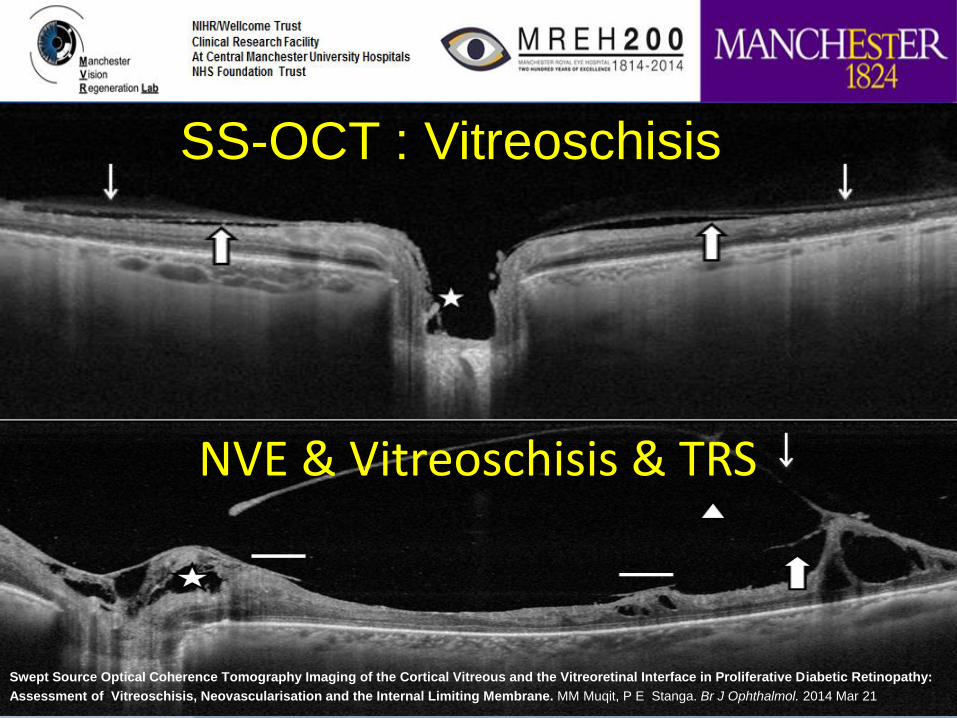
SS-OCT : Vitreoschisis
Swept Source Optical Coherence Tomography Imaging of the Cortical Vitreous and the Vitreoretinal Interface in Proliferative Diabetic Retinopathy:
Assessment of Vitreoschisis, Neovascularisation and the Internal Limiting Membrane. MM Muqit, P E Stanga. Br J Ophthalmol. 2014 Mar 21
NVE & Vitreoschisis & TRS

Vitreoretinal Assessment SS-OCT in 2014
Improved imaging of the cortical vitreous and the vitreoretinal interface
Achieve better understanding of retinal pathological changes
Assessment of choroidal thickness
Am J Ophthalmol. 2014 Feb;157(2). In vivo imaging of cortical vitreous using 1050-nm swept-source deep range imaging optical coherence
tomography. Paulo E. Stanga, Anna Sala-Puigdollers, Silvestro Caputo, Hojr Jaberansari, Monica Cien, Jane Gray, Yvonne D’Souza, Stephen J Charles,
Susmito Biswas, David B Henson, David McLeod

SS-OCT vs TD OCT: Retinal pathology

SS-OCT evaluation of
Retinal Cavernous Haemangioma
No PVD
ERM
Blood
• Good penetration through haemorrhage
• Measure volume of each blood cavern and its wall thickness
• Follow-up
• Assessment of response to therapy


SS-OCT in Optic Disc Pit (ODP)
Maculopathy – helpful in surgery approach
Optic Disc Pit
Glial Tissue
Successful surgical treatment of optic disc pit maculopathy. Ziahosseini K, Sanghvi C, Muzaffar W, Stanga PE. Eye (Lond). 2009 Jun;23(6):1477-9

2 weeks
5 weeks
7 Months
5 Months
Swept Source Optical Coherence Tomography Imaging in Conservative and Surgical Management of Premacular Haemorrhages showing
Inflammatory Response. R Tanawade, M Muqit, D McLeod, P Stanga. Clinical experimental Ophthalmology . 2014 Jun 5. In Press

Vitreoretinal Assessment SS-OCT in 2014
Improved imaging of the cortical vitreous and the vitreoretinal interface
Achieve better understanding of retinal pathological changes
Assessment of choroidal thickness

SS-OCT: Imaging audit tool
Measuring Choroidal Thickness


C Quijano, S Pastor-Idoate, M Gil-Martinez,
S Biswas, P Stanga
Anatomical And Functional Outcomes in X-Linked Retinoschisis Treated With Topical
Carbonic Anhydrase Inhibitor - Dorzolamide
Sikkink SK, Biswas S, Parry NR, Stanga PE, Trump D. X-linked retinoschisis:
an update. J Med Genet. 2007 Apr;44(4):225-32

• Infra-red wavelength (1,050 nm)+high speed scanning (100,000 A-scans/sec) :
allows deeper penetration into choroid and sclera with uniform signal
sensitivity from the vitreous up to the chorioscleral interface
less light scattering improves results in eyes with cataracts
provides uniform sensitivity allowing superior visualization of the
vitreous and choroid and more data to be collected in same scan
• 12mm x 9 mm wide scan captures the macula and disc in the same scan
• Advanced 3D volumetric layer detection algorithms
• Less sensitive to sample motion
Conclusion:
Why do we need SS-OCT ?

• Manchester Vision Reganeration (MVR) Lab at NIHR/Wellcome Trust CRF
Director • Prof Paulo Stanga
Clinical Research Retina Fellows: • Dr Maria Gil Martinez • Dr Salvador Pastor Idoate • Dr Claudia Quijano
• Mr Susmito Biswas • Miss Yvonne D’Souza • Mr George Turner • Mr Steve Charles • Mr Saj Mahmood • Mr Konstantinos Balaskas • Prof David Henson • Prof David McLeod
With thanks to: