symmetry breaking in plants: molecular mechanisms...
TRANSCRIPT
Symmetry Breaking in Plants: MolecularMechanisms Regulating Asymmetric CellDivisions in Arabidopsis
Jalean J. Petricka, Jaimie M. Van Norman, and Philip N. Benfey
Department of Biology and IGSP Center for Systems Biology, Duke University, Durham,North Carolina, 27708
Correspondence: [email protected]
Asymmetric cell division generates cell types with different specialized functions or fates.This type of division is critical to the overall cellular organization and development ofmany multicellular organisms. In plants, regulated asymmetric cell divisions are of particularimportance because cell migration does not occur. The influence of extrinsic cues on asym-metric cell division in plants is well documented. Recently, candidate intrinsic factors havebeen identified and links between intrinsic and extrinsic components are beginning to beelucidated. A novel mechanism in breaking symmetry was revealed that involves the move-ment of typically intrinsic factors between plant cells. As we learn more about the regulationof asymmetric cell divisions in plants, we can begin to reflect on the similarities and differ-ences between the strategies used by plants and animals. Focusing on the underlying mol-ecular mechanisms, this article describes three selected cases of symmetry-breakingevents in the model plant Arabidopsis thaliana. These examples occur in early embryogen-esis, stomatal development, and ground tissue formation in the root.
Plant cells are surrounded by rigid cell wallsthat prevent cell movement. Precisely
oriented cell division is especially importantin the absence of cell migration. When a plantcell divides, a new wall forms between thedaughter cells; this permanently delineatestheir relative positions. As a consequence, theselection of the division plane is crucial forplant tissue organization and overall organarchitecture.
The selection of the division plane is uniquein plants compared with other organisms (for adetailed review of plant cytokinesis see Jurgens
2005). In budding yeast, landmarks at previousbud-sites position the division plane (seeSlaughter et al. 2009), whereas in fission yeastit is positioned by the nucleus. The position ofthe centrosomes and the mitotic spindle deter-mine division plane orientation in animal cells(Balasubramanian et al. 2004; see Munro andBowerman 2009). In plants, the division planeis positioned by a ring of microtubules andF-actin, called the preprophase band thatforms at the cell periphery (Mineyuki 1999).Despite these striking differences, similaritiesare beginning to emerge. For example, in
Editors: Rong Li and Bruce Bowerman
Additional Perspectives on Symmetry Breaking in Biology available at www.cshperspectives.org
Copyright # 2009 Cold Spring Harbor Laboratory Press; all rights reserved.
Advanced Online Article. Cite this article as Cold Spring Harb Perspect Biol doi: 10.1101/cshperspect.a000497
1
on July 27, 2018 - Published by Cold Spring Harbor Laboratory Press http://cshperspectives.cshlp.org/Downloaded from
budding yeast and animals, GTPase activationplays an important role in positioning divisionplanes (Balasubramanian et al. 2004; seeMcCaffrey and Macara 2009; Slaughter et al.2009; Munro and Bowerman 2009; Prehoda2009). In plants, a GTPase was recently foundto be localized at the preprophase band andmay provide spatial information during div-ision (Xu et al. 2008). Therefore, common mol-ecular mechanisms may underlie division planeselection in plants and animals.
In all organisms, asymmetric cell division isa common method to obtain distinct cell typesfrom a single progenitor cell. Before an asym-metric division a cell must first establish aninternal asymmetry; this process is termedbreaking symmetry. Breaking symmetry can begenerally defined as the asymmetric organi-zation of cellular components along one axisleading to cellular polarization as a prerequisiteto division. An asymmetric cell division cangenerate two cells of different morphology(i.e., size and/or shape) or different specializedfunctions or fates. Specifically, the products ofasymmetric cell divisions fall into two majorcategories. In one case, a mother cell candivide asymmetrically to produce a daughtercell to replace it and a daughter cell with a dis-tinct fate, as in the case of stem cell divisions.Alternatively, a mother cell can divide asym-metrically to produce two distinct daughter cellsat the expense of mother cell identity. Followingdivision, there are two basic mechanisms bywhich a mother cell generates daughters thatare distinct from itself. One mechanism isintrinsic to the cell and is defined by segregationof cellular determinants before division. Theother mechanism is extrinsic to the cell andis defined by external cues that direct daughtercell fate (Horvitz and Herskowitz 1992).Intrinsic and extrinsic mechanisms are notnecessarily independent and often work incombination to direct asymmetric divisionand specification of daughter cells.
The first zygotic division of fucoid brownalgae, such as Fucus, is one of the earlieststudied examples of symmetry breaking inplants regulated by both extrinsic and intrinsicmechanisms. Fucus zygotes are an excellent
organism in which to examine the cellularbiology underlying asymmetric cell division(for a recent review see Homble and Leonetti2007). In the presence of extrinsic cues, such aslight, the asymmetric zygotic division isoriented perpendicular to the light gradientso that one cell resides on the “shady” side ofthe gradient (Kropf 1992, 1997; Alessa andKropf 1999). In the absence of environmentalcues, the division is oriented relative to thesperm entry site and therefore appears randomwithin a field of cells (Hable and Kropf 2000).This indicates that Fucus zygotes have an intrin-sic mechanism to break symmetry in the absenceof extrinsic cues. This intrinsic mechanismoccurs rapidly after fertilization, however it canlater be overridden by extrinsic cues. Althoughmuch is known about the role of the cytoskele-ton, membrane depolarization, and ion flux inFucus embryo polarization, the molecular com-ponents regulating these events remainunknown because of a lack of molecular tools.To address the molecular mechanisms regulat-ing asymmetric cell divisions in plants, the useof a more genetically tractable plant is necessary.
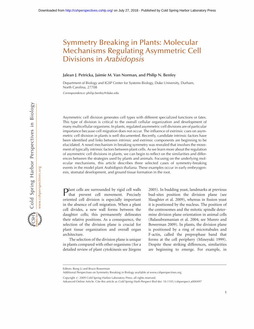
In the model plant Arabidopsis thaliana,many genes that are involved in asymmetriccell division have been identified. Interestingly,many of these genes are thought to functionin both asymmetric division and subsequentcell fate specification suggesting that, in plants,these processes are tightly linked. Despitethese advances, relatively little is known aboutthe cellular processes of breaking symmetry inplants. Because of these two caveats, the termbreaking symmetry must be defined more gen-erally in plants. We use this term to describe aprocess in which a cell divides asymmetricallyto form a daughter cell with a distinct fate.Here we discuss three examples in which themolecular mechanisms that participate insymmetry-breaking events in plants are begin-ning to be elucidated. These examples occurin early embryogenesis, formation of stomata,and root development (Fig. 1). We confine thetopics of this article to Arabidopsis thalianabecause the majority of the experimentalwork, particularly using molecular genetics, isfocused on this species.
J.J. Petricka, J.M. Van Norman, and P.N. Benfey
2 Advanced Online Article. Cite this article as Cold Spring Harb Perspect Biol doi: 10.1101/cshperspect.a000497
on July 27, 2018 - Published by Cold Spring Harbor Laboratory Press http://cshperspectives.cshlp.org/Downloaded from

Root development
Embryo development
Stomatal development
Figure 1. An Arabidopsis plant. Schematic of an adult plant depicting roots, leaves, stems, and flowers. In thisarticle, three symmetry-breaking events are discussed. These examples are taken from different organs of theplant. The first example is taken from embryo development, which occurs after fertilization of the flower inthe silique (seed pod) in Arabidopsis. Next, symmetry breaking in the leaf epidermis during stomataldevelopment is discussed. Finally, the asymmetric cell division that gives rise to the ground tissue in the rootis considered.
Symmetry Breaking in Plants
Advanced Online Article. Cite this article as Cold Spring Harb Perspect Biol doi: 10.1101/cshperspect.a000497 3
on July 27, 2018 - Published by Cold Spring Harbor Laboratory Press http://cshperspectives.cshlp.org/Downloaded from

ASYMMETRIC DIVISION OF THE ZYGOTEAND POLARIZATION OF THEEARLY EMBRYOEmbryogenesis is the process by which azygote undergoes elaborate changes in cellnumber, fate, and morphology to ultimatelyform a mature embryo. An important step inembryogenesis is establishment of asymmetry(or polarity) along the longitudinal bodyaxis. Initial embryo polarity is typically
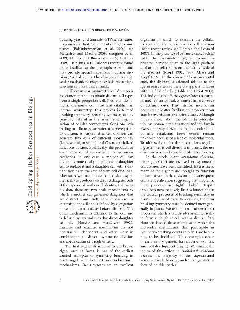
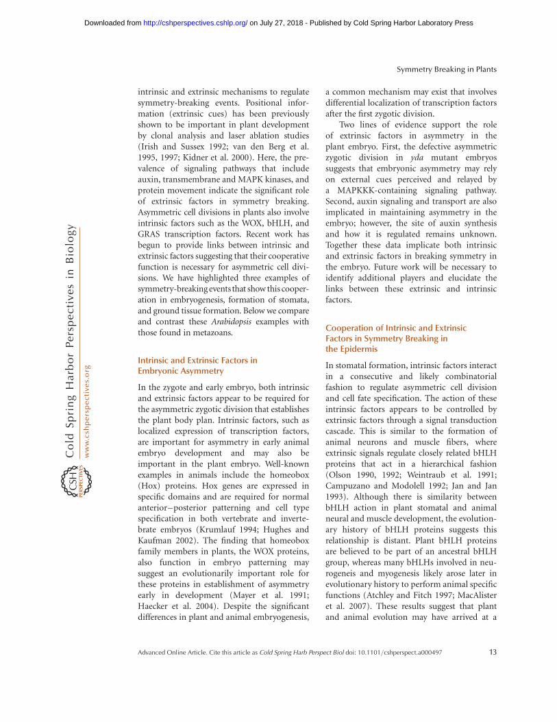
anterior–posterior (head–tail) in animals andapical–basal (shoot–root) in plants. In manymulticellular organisms, breaking zygoticsymmetry is critical for polarity establishmentin the developing embryo because the resultingdaughter cells have two distinct fates. Followingfertilization, the Arabidopsis zygote expandslongitudinally, then divides asymmetrically togenerate a small apical cell and a larger basalcell (Fig. 2A). The invariant cell division
Auxin Maxima
PIN7 Localization
B
1-cell 4-cell 8-cellEgg Zygote
C
Zygote 1-cell 4-cell 8-cellEgg
4-cellEgg Zygote 1-cell
Apicalcell
Basalcell
Mature embryo
CotyledonsHypocotyl
Root
A
8-cell
Hypophysis
Suspensor
Proembryo
WOX2 and WOX8
WOX2
WOX8 and WOX9
WOX9
WOX8
Figure 2. Embryo development and the asymmetric localization of factors in the embryo. (A) Schematic ofembryo development focusing on the events from the egg to the eight-cell stage. After fertilization, thezygote expands and then divides asymmetrically to produce the apical and basal cells. Embryo stages arebased on the number of cells in the apical domain only, thus the first zygotic division results in a one-cellstage embryo. The apical cell undergoes a series of divisions to generate the eight-celled proembryo. Thebasal cell undergoes a series of strictly transverse divisions to generate the suspensor. Later in development,the uppermost cell of the suspensor, the hypophysis, is incorporated into the embryo. Therefore, by theeight-cell stage, four anatomically distinct cell types are present: The upper (green) and lower (yellow-green)tiers of the proembryo, the hypophysis (cream), and the suspensor (white). The suspensor remainsextraembryonic, whereas the remaining cell types give rise to the cotyledons, hypocotyl, and root of themature embryo as depicted by the corresponding color scheme in the eight-cell stage and mature embryo.(B) Schematic of the WOX gene expression patterns. By the eight-cell stage, the expression domains of theWOX genes coincide with the four distinct cell types present. (C) Schematic of the auxin maxima and PIN7localization. PIN7 localization on the upper membrane of basal cells directs the flow of auxin from basal toapical cells. This polarized movement of auxin generates an auxin maximum in the apical domain asdetermined by expression of the DR5 reporter.
J.J. Petricka, J.M. Van Norman, and P.N. Benfey
4 Advanced Online Article. Cite this article as Cold Spring Harb Perspect Biol doi: 10.1101/cshperspect.a000497
on July 27, 2018 - Published by Cold Spring Harbor Laboratory Press http://cshperspectives.cshlp.org/Downloaded from
pattern in Arabidopsis embryogenesis allowslineage analysis of each daughter cell(Mansfield and Briarty 1991). The apical cellundergoes a precise series of divisions generat-ing a spherical eight-celled proembryo, whichwill give rise to the majority of the matureembryo. Apical–basal polarity is alreadyrecognizable in the eight-celled proembryo:The upper tier cells will give rise to the shootand the lower tier cells will form the hypocotyland a portion of the embryonic root (Fig. 2A)(Scheres et al. 1994). The basal cell undergoesrepeated transverse divisions to produce anextraembryonic support structure, the suspen-sor, which connects the embryo to the maternaltissue (Jurgens 2001). The uppermost suspen-sor cell, the hypophysis, is later incorporatedinto the embryo to form a portion of the root(Fig. 2A) (Dolan et al. 1993; Scheres et al.1994). The result of embryogenesis in higherplants is a simple juvenile form, in which mostadult plant organs are absent (compare Fig. 1to mature embryo in Fig. 2A). This is in contrastto embryogenesis in higher animals, whichculminates in essentially a miniature form ofthe adult organism.
The invariant pattern of cell division in earlyArabidopsis embryogenesis indicates a tightregulation of the asymmetric zygotic divisionand subsequent development of the apicaland basal lineages up to the eight-cell stage(see Jenik et al. 2007 for detailed review ofArabidopsis embryogenesis). In this section, wehighlight findings regarding the molecularmechanisms regulating asymmetric division ofthe zygote and cell fate specification of thedaughter cells.
Asymmetric Zygotic Division andSuspensor Cell Fate SpecificationRequire a MAPKKK Signaling Pathway
The mitogen-activated protein kinase kinasekinase (MAPKKK), YODA (YDA), is requiredfor elongation and asymmetric division of thezygote and subsequent development of thebasal cell lineage (Lukowitz et al. 2004). In ydaloss-of-function mutants, zygotes fail toexpand longitudinally and the first zygotic
division produces cells of similar size. Fromthe two-cell to eight-cell stage, yda defects areprimarily observed in the basal cell lineage.Here, divisions appear in random orientationsand suspensor identity markers are notexpressed. Despite this, the apical lineagedevelops normally up to the eight-cell stage,after which it also shows cell division defects.Conversely, in yda gain-of-function mutants,the suspensor proliferates at the apparentexpense of the apical lineage. These resultsindicate that polarity is important for embryodevelopment. Additionally, these data suggestthat a signaling network, including YDA, actsas a switch to activate suspensor cell fate. YDAalso participates in asymmetric cell divisionslater in plant development (see the followingsection). MAPKK kinases function in a varietyof pathways but likely act within signalingmodules downstream of extracellular receptors.Thus, it is tempting to speculate that externalsignals, possibly from maternal tissue, providecues for zygotic asymmetry and specificationof the first embryonic cell fate decision.However, other pathway components and howthey may be differentially distributed before orafter zygotic division remain unknown.
The Role of HomeodomainTranscription Factors in theAsymmetry of the Zygote
Members of the Arabidopsis homeodomaintranscription factor family also have roles inasymmetric cell division and cell fate speci-fication following the first zygotic division.WUS-RELATED HOMEOBOX2 (WOX2) andWOX8 are expressed in both the egg cell andzygote. Following the first zygotic division,WOX2 and WOX8 expression is restricted tothe apical and basal cell lineages, respectively(Fig. 2B) (Haecker et al. 2004). Another WOXgene, WOX9, is also expressed in the basal cell,but its expression is later restricted to the hy-pophysis and then expands into the centraldomain of the proembryo (Fig. 2B) (Haeckeret al. 2004; Wu et al. 2007). The expressionpattern of these three WOX genes coincideswith the four anatomically distinct cell types
Symmetry Breaking in Plants
Advanced Online Article. Cite this article as Cold Spring Harb Perspect Biol doi: 10.1101/cshperspect.a000497 5
on July 27, 2018 - Published by Cold Spring Harbor Laboratory Press http://cshperspectives.cshlp.org/Downloaded from

present at the eight-cell stage: the upper tier,lower tier, hypophysis, and suspensor (Fig. 2).However, the mechanisms regulating expres-sion of WOX2, WOX8, and WOX9 in the earlyembryo are unknown.
Consistent with their asymmetric expres-sion patterns, these WOX genes have criticalroles in the apical and basal lineages. Theembryos of wox2 mutants show cell divisiondefects specifically in the apical domain.However, these defects are mild because ofexpression of related, partially redundantWOX genes in the apical domain (Haeckeret al. 2004; Breuninger et al. 2008). In wox8and wox9 single mutants, embryo developmentis largely normal (Wu et al. 2007; Breuningeret al. 2008). More severe defects are observedin wox8 wox9 double mutants, in which abnor-mal cell divisions occur in both the apicaland basal cell lineages. Several genetic markersof apical cell fate, including WOX2, areundetectable in wox8 wox9 embryos. Theseresults indicate WOX8/WOX9 activity in thebasal lineage is required for formation of theapical lineage, and suggest communicationbetween the lineages is important in the earlyembryo (Breuninger et al. 2008). Additionally,in wox8 wox9 embryos, aspects of apical andbasal lineage development can be restored byectopic WOX2 expression. However, in theseembryos the zygote fails to elongate and thefirst division is more symmetrical, closelyresembling yda embryos. Despite this similar-ity, yda wox8 wox9 triple mutant embryoscompletely arrest development after the firstnearly symmetrical zygotic division. This phe-notype is much more severe than yda or wox8wox9 alone and indicates that these genes donot function in a linear pathway (Breuningeret al. 2008). This also suggests that signalingthrough the YDA pathway and WOX-regulatedtranscription act independently in breakingzygotic symmetry. WOX transcription networksare clearly involved in asymmetric cell divisionand fate specification in the apical and basallineages, however details about how WOXgenes become differentially expressed after thezygotic division are required to understandtheir role in this process.
The Role of Auxin in MaintainingAsymmetry in the Early Embryo
The plant hormone auxin is one of the mostubiquitous plant signaling molecules and itappears to be required for many diverse devel-opmental processes (see Jenik and Barton2005 and Leyser 2006 for recent reviews).Auxin movement occurs directionally fromone cell to another and is mediated by afamily of functionally redundant transmem-brane efflux carriers, called PINs, named aftertheir founding member PINFORMED1(Galweiler et al. 1998; Petrasek et al. 2006).PIN localization on the cell membrane indicatesthe direction of auxin movement and mutationsin PINs result in abnormal auxin transport(reviewed in Paponov et al. 2005). For instance,PIN7 is localized on the apical membrane ofsuspensor cells and expression of a syntheticauxin-dependent transcriptional reporter,DIRECT REPEAT5 (DR5), is expressed only inthe apical lineage from the first zygotic divisionto the eight-cell stage (Fig. 2C) (Friml et al.2003). These data suggest auxin flow is directedfrom basal to apical cells to set up the embryoaxis. In support of this hypothesis, mutationsin multiple PIN genes result in strong embry-onic phenotypes, such as embryos withoutapparent polarity or suspensor-like structuresthat lack a proembryo (Friml et al. 2003).Additionally, mutations in either of two auxinresponse genes, MONOPTEROS (MP) andBODENLOS (BDL), result in abnormal orien-tation of apical cell division planes (Berlethand Jurgens 1993; Hamann et al. 1999). Theseresults suggest that asymmetric auxin transportand response is required for establishment ormaintenance of polarity after the zygoticdivision and specifying embryonic fate in theapical lineage. How the asymmetric dis-tribution of auxin is initially established isunknown.
Putting it all Together inthe Early Embryo
The phenotypic similarity between wox8 wox9embryos and embryos with mutations in
J.J. Petricka, J.M. Van Norman, and P.N. Benfey
6 Advanced Online Article. Cite this article as Cold Spring Harb Perspect Biol doi: 10.1101/cshperspect.a000497
on July 27, 2018 - Published by Cold Spring Harbor Laboratory Press http://cshperspectives.cshlp.org/Downloaded from
multiple PIN genes suggests a link betweenWOX transcriptional regulation and auxinmovement or signaling. WOX8/WOX9 are pre-dicted to regulate a signal, possibly auxin, whichmoves from the basal to the apical lineage andis required for apical development (Breuningeret al. 2008). Indeed, auxin flux (measured byPIN localization) and auxin response (assayedby DR5 expression) are defective in wox8 wox9embryos. However, it is unclear whether WOXtranscription factors directly regulate auxinmovement or response (Breuninger et al.2008). Interestingly, the role for auxin in speci-fication of embryonic fate in the apical lineageappears to be complementary to YDA’s rolein specification of suspensor fate in the basallineage. This suggests that the auxin and YDAsignaling pathways function independentlyof each other (Lukowitz et al. 2004). Togetherthese data indicate that asymmetric division isnot sufficient for development of the apicaland basal lineages, instead it appears that inter-cellular signaling is required to maintain thedistinct cell fate of each lineage after the firstzygotic division.
ASYMMETRIC CELL DIVISIONS INSTOMATAL DEVELOPMENT
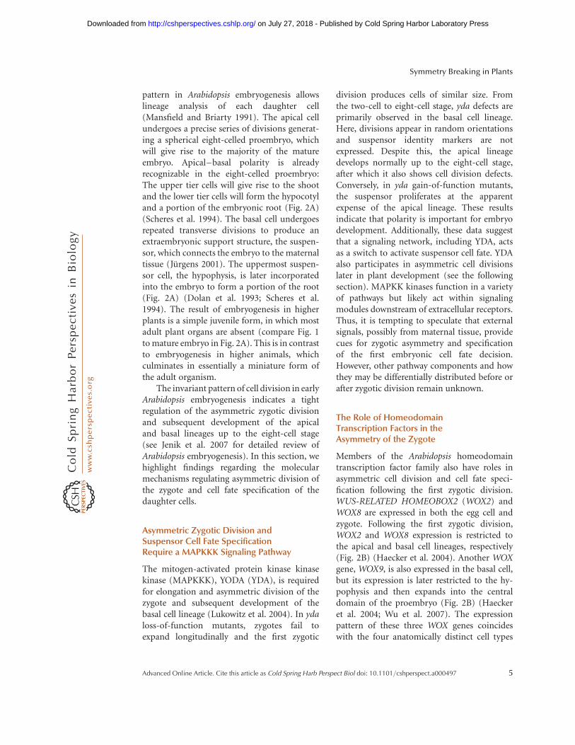
Another example of symmetry breaking inplants can be seen in the asymmetric cell divi-sions of the epidermis that generate stomata.Stomata are pores in the leaf epidermis thatfunction in gas and water exchange. InArabidopsis, formation of stomata is initiatedby a dispersed population of multipotent stemcells, called meristemoid mother cells (MMCs).First, an MMC divides asymmetrically to formtwo cells of unequal size (Fig. 3A). The largercell acts as a spacer between adjacent stomata,whereas the smaller cell, called a meristemoid(M), undergoes several asymmetric cell divi-sions to generate more meristemoids beforebecoming a guard mother cell (GMC)(Bunning 1953; Rasmussen 1981; Geisler et al.2000; Bergmann and Sack 2007). Finally, theGMCs divide symmetrically to produce twoguard cells (GCs), which act as valves surround-ing a pore (Fig. 3A) (Pant and Kidwai 1967;
Geisler et al. 2000; Bergmann and Sack 2007).Thus, stomata are formed progressively througha series of initially asymmetric and a final sym-metric cell division.
In Arabidopsis, these coordinated divisionsresult in stomates that are separated from eachother by an intervening epidermal cell (Sachs1978, 1991; Geisler et al. 2000). This one-cellspacing pattern is derived from an asymmetriccell division, called a spacing division, thatoccurs in a cell that borders an existing M,GMC, or stomate (Fig. 3A) (Geisler et al.2000). For example, when an MMC is flankedby a cell that is already progressing throughstomatal development, a new division plane isoriented such that the resulting meristemoidis separated from existing stoma by the largersister cell (Sachs 1978, 1991; Larkin et al. 1997;Croxdale 2000). Thus, new meristemoid cellsare always physically separated from eachother. The orientation and placement of thedivision plane in the MMC strongly suggeststhe involvement of extrinsic cues from neigh-boring cells rather than from intrinsic cues(Geisler et al. 2000).
Cell–cell Signaling and Breaking ofSymmetry in the Epidermis
Cell–cell signaling was further implicated incoordinating asymmetric cell divisions of thestomatal lineage when the gene correspondingto the too many mouths (tmm) mutation wasfound to encode a leucine-rich repeat receptor-like protein (LRR-RLP). LRR-RLPs are similarto transmembrane receptor kinases but lackan intracellular kinase domain (Geisler et al.2000; Nadeau and Sack 2002). In tmmmutants, the stomata are positioned randomlyrelative to one another because of a failure toproperly orient asymmetric cell divisions. As aconsequence, tmm stomata are frequently indirect contact because the spacing divisionsare not properly executed (Geisler et al. 2000).Based on these data, it was hypothesizedthat TMM transduces extracellular signals in amanner similar to that proposed for otherLRR-RLPs (Jeong et al. 1999; Trotochaudet al. 1999; Nadeau and Sack 2002; Dievart
Symmetry Breaking in Plants
Advanced Online Article. Cite this article as Cold Spring Harb Perspect Biol doi: 10.1101/cshperspect.a000497 7
on July 27, 2018 - Published by Cold Spring Harbor Laboratory Press http://cshperspectives.cshlp.org/Downloaded from

Amplifying division
MeristemoidMother Cell
(MMC)Meristemoid
(M) Guard Mother
Cell (GMC)Guard Cells
(GCs)
SPCH
EPF1
TMM
P
P P P
P
ERf LRR-RLKs
MAPKKK (YODA) MKK4/5 MPK3/6 SPCH
SPCH
Nucleus
Asymmetric cell division
and stomatal
differentiation
Other ligands
SDD1
MUTE FAMA
Spacing division
A
B
Plasma membrane
Figure 3. Asymmetric cell division in stomatal development. (A) Schematic of a cell progressing throughstomatal development. (A, from left to right) An undifferentiated leaf epidermal cell in Arabidopsis acquiresMeristemoid Mother Cell fate (MMC, green) before undergoing an asymmetric cell division to produce aMeristemoid (M, yellow) and a larger sister cell (white). The M cell may then go through a series ofasymmetric cell divisions, called amplifying divisions, before differentiating into a Guard Mother Cell(GMC, orange). The GMC divides symmetrically to produce a pair of Guard Cells (GCs, red) that togethercomprise a stomate. This process may reiterate when the larger sister cell divides asymmetrically in a spacingdivision to produce another M cell that is separated from existing stomata by one cell. The intrinsic factorsSPCH, MUTE, and FAMA act sequentially to regulate asymmetric cell division and stomatal development.(B) Extrinsic factors and the signaling pathway proposed to negatively regulate stomatal development. First,a ligand binds to the leucine-rich repeats (LRR, blue) of a putative heterodimer complex between TMM andone of the ERf LRR-RLKs, which possess an intracellular kinase domain (red). This interaction is thought toinitiate a cascade of phosphorylation events (black circles) involving downstream MAPK signaling proteins(orange), including YODA. Candidate ligands (green) acting to trigger this signaling cascade include smallpeptides such as EPF1 and putative unknown proteins that may be processed by SDD1. Biochemicalevidence for this model is lacking, except for the final phosphorylation of SPCH by MPK3 and MPK6.SPCH phosphorylation ultimately results in repression of asymmetric cell division and stomataldifferentiation. In contrast, when SPCH is unphosphorylated, it promotes asymmetric cell division andmeristemoid fate, as shown in (A).
J.J. Petricka, J.M. Van Norman, and P.N. Benfey
8 Advanced Online Article. Cite this article as Cold Spring Harb Perspect Biol doi: 10.1101/cshperspect.a000497
on July 27, 2018 - Published by Cold Spring Harbor Laboratory Press http://cshperspectives.cshlp.org/Downloaded from
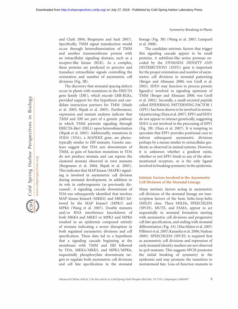
and Clark 2004; Bergmann and Sack 2007).Specifically, TMM signal transduction wouldoccur through heterodimerization of TMMand another transmembrane protein withan intracellular signaling domain, such as areceptor-like kinase (RLK). As a complex,these proteins are predicted to perceive andtransduce extracellular signals controlling theorientation and number of asymmetric celldivisions (Fig. 3B).
The discovery that stomatal spacing defectsoccur in plants with mutations in the ERECTAgene family (ERf ), which encode LRR-RLKs,provided support for this hypothesis and can-didate interaction partners for TMM (Masleet al. 2005; Shpak et al. 2005). Furthermore,expression and mutant analyses indicate thatTMM and ERf are part of a genetic pathwayin which TMM prevents signaling throughERECTA-like1 (ERL1) upon heterodimerization(Shpak et al. 2005). Additionally, mutations inYODA (YDA), a MAPKKK gene, are pheno-typically similar to ERf mutants. Genetic ana-lyses suggest that YDA acts downstream ofTMM, as gain of function mutations in YDAdo not produce stomata and can repress theclustered stomata observed in tmm mutants(Bergmann et al. 2004; Shpak et al. 2005).This indicates that MAP kinase (MAPK) signal-ing is involved in asymmetric cell divisionduring stomatal development, in addition toits role in embryogenesis (as previously dis-cussed). A signaling cascade downstream ofYDA was subsequently identified that involvesMAP kinase kinase4 (MKK4) and MKK5 fol-lowed by the MAP kinase3 (MPK3) andMPK6 (Wang et al. 2007). Double mutantsand/or RNA interference knockdown ofboth MKK4 and MKK5 or MPK3 and MPK6resulted in an epidermis composed entirelyof stomata indicating a severe disruption inboth regulated asymmetric divisions and cellspecification. These data led to a hypothesisthat a signaling cascade beginning at themembrane with TMM and ERf followedby YDA, MKK4/MKK5, and MPK3/MPK6,sequentially phosphorylate downstream tar-gets to regulate both asymmetric cell divisionsand cell fate specification in the stomatal
lineage (Fig. 3B) (Wang et al. 2007; Lampardet al. 2008).
The candidate extrinsic factors that triggerthis signaling cascade appear to be smallproteins. A subtilisin-like serine protease en-coded by the STOMATAL DENSITY ANDDISTRIBUTION1 (SDD1) gene is importantfor the proper orientation and number of asym-metric cell divisions in stomatal patterning(Berger and Altmann 2000; von Groll et al.2002). SDD1 may function to process proteinligand(s) involved in signaling upstream ofTMM (Berger and Altmann 2000; von Grollet al. 2002). Secondly, a small-secreted peptidecalled EPIDERMAL PATTERNING FACTOR 1(EPF1) has been shown to be involved in stoma-tal patterning (Hara et al. 2007). EPF1 and SDD1do not appear to interact genetically, suggestingSDD1 is not involved in the processing of EPF1(Fig. 3B) (Hara et al. 2007). It is tempting tospeculate that EPF1 provides positional cues toinform subsequent asymmetric divisionsperhaps by a means similar to extracellular gra-dients as observed in animal systems. However,it is unknown whether a gradient exists,whether or not EPF1 binds to any of the afore-mentioned receptors, or is the only ligandinvolved in breaking symmetry in the epidermis.
Intrinsic Factors Involved in the AsymmetricCell Divisions of the Stomatal Lineage
Many intrinsic factors acting in asymmetriccell divisions of the stomatal lineage are tran-scription factors of the basic helix-loop-helix(bHLH) class. Three bHLHs, SPEECHLESS(SPCH), MUTE, and FAMA, appear to actsequentially in stomatal formation startingwith asymmetric cell divisions and progressivecell fate specification, and ending with stomataldifferentiation (Fig. 3A) (MacAlister et al. 2007;Pillitteri et al. 2007; Kanaoka et al. 2008; Nadeau2009). SPEECHLESS (SPCH) is required firstas asymmetric cell divisions and expression ofearly stomatal identity markers are not observedin spch mutants. This suggests SPCH promotesthe initial breaking of symmetry in theepidermis and may promote the transition tomeristemoid fate. Loss-of-function mutants in
Symmetry Breaking in Plants
Advanced Online Article. Cite this article as Cold Spring Harb Perspect Biol doi: 10.1101/cshperspect.a000497 9
on July 27, 2018 - Published by Cold Spring Harbor Laboratory Press http://cshperspectives.cshlp.org/Downloaded from

a second transcription factor, MUTE, exhibitexcessive asymmetric cell divisions in theepidermis and the resulting cells fail to differen-tiate into stomata. MUTE expression is notdetected in spch mutants, suggesting that itacts downstream of SPCH to arrest asymmetriccell divisions and allows the transition to guardmother cell fate (MacAlister et al. 2007; Pillitteriet al. 2007). A third bHLH, FAMA, promotesthe transition to guard cell fate. FAMAexpression is not detected in spch and mutemutants, also suggesting that it acts down-stream of SPCH and MUTE (MacAlister et al.2007; Pillitteri et al. 2007). Thus, the sequentialaction of SPCH and MUTE appears to promoteand regulate the breaking of symmetry in theepidermis and together these three bHLHsspecify the resulting daughter cells.
This model is somewhat oversimplifiedas other intrinsic components have been identi-fied, including several that interact with SPCH,MUTE, and FAMA. Four other bHLHs andtwo R2R3 MYB-type transcription factors(TFs) have recently been implicated as intrinsicfactors involved in stomatal patterning andcell specification. The expression pattern andmutant phenotype of FAMA resemble that ofthe R2R3 MYB-type TF FOUR LIPS (FLP)(Lai et al. 2005; Ohashi-Ito and Bergmann2006). Although plant R2R3 MYB-typeTFs have been shown to heterodimerize withbHLHs, FAMA does not heterodimerize withFLP (Lee and Schiefelbein 1999; Zimmermannet al. 2004; Ohashi-Ito and Bergmann 2006).Instead, FAMA interacts genetically, in vitro,and in planta with two additional bHLHs,bHLH071 and bHLH093 (Ohashi-Ito andBergmann 2006). The important role ofbHLH dimerization in symmetry breaking issupported by the recent demonstration thatthe bHLH proteins SCREAM (SCRM)/ICE1or SCREAM2 (SCRM2) heterodimerize invitro and in vivo with SPCH, MUTE, andFAMA. In scrm/ice1 scrm2 double mutants,the epidermis consists only of fully differen-tiated epidermal cells, just like spch mutants(Kanaoka et al. 2008). Taken together, thesedata indicate that the combinatorial andsequential action of intrinsic factors, such as
bHLHs, is integral to the regulation of asym-metric cell division in the stomatal lineage.
A Connection between Cell–cell Signalingand Intrinsic Factors in Stomatal Formation
A link between extrinsic and intrinsic factorsinvolved in breaking symmetry in the stomatallineage is predicted based on genetic experi-ments that show spch is epistatic to yda(MacAlister et al. 2007; Lampard et al. 2008).However, the targeted phosphorylation ofintrinsic factors like SPCH by a MAPK signal-ing cascade, including factors like YDA, wasregarded as dubious because MAPK signalingis common to many pathways in plant develop-ment (Lampard et al. 2008). Despite this, thereceptor/MAPK signaling system was recentlyshown to modulate SPCH in vivo throughphosphorylation that appears to repress SPCHactivity (Fig. 3B) (Lampard et al. 2008). Theregion of SPCH targeted for phosphorylationis not found in any other Arabidopsis protein,but is common to SPCH-related proteins inother plant species. This suggests an evolu-tionarily conserved mechanism for regulationof SPCH activity in a specific cell type, thestomata (Lampard et al. 2008). This shows aconcrete connection between the cell–cellsignaling pathway and the sequential action ofTFs in the symmetry-breaking events of thestomatal lineage.
ASYMMETRIC DIVISION IN THECORTEX-ENDODERMAL CELL LINEAGE
In the Arabidopsis root, an asymmetric celldivision generates the two cell layers of theground tissue. The ground tissue is locatedbetween the vasculature and epidermis and iscomprised of two distinct cell types: the cortexand the endodermis (Fig. 4A). These two celltypes are generated through asymmetric celldivision of a single initial (stem) cell. Thecortex/endodermal initial (CEI) undergoes atransverse asymmetric division to maintainthe CEI and generate a CEI daughter (CEID).The CEID then divides asymmetrically, in alongitudinal orientation, to produce one
J.J. Petricka, J.M. Van Norman, and P.N. Benfey
10 Advanced Online Article. Cite this article as Cold Spring Harb Perspect Biol doi: 10.1101/cshperspect.a000497
on July 27, 2018 - Published by Cold Spring Harbor Laboratory Press http://cshperspectives.cshlp.org/Downloaded from

cell for each ground tissue layer (Fig. 4B).Thus the asymmetric division of a single cellgenerates two morphologically and molecularlydistinct cell types (Benfey et al. 1993; Dolanet al. 1993; Scheres et al. 1994; Di Laurenzioet al. 1996).
The asymmetric division of the CEID isregulated through activity of two tran-scription factors, SHORT-ROOT (SHR) and
SCARECROW (SCR). SHR and SCR aremembers of the GRAS family of transcriptionalregulators and were found in a genetic screento identify molecules with roles in radialpatterning of the root (Benfey et al. 1993;Di Laurenzio et al. 1996; Pysh et al. 1999;Helariutta et al. 2000). Both shr and scrmutants each have only a single layer of groundtissue. In shr mutants, the CEID fails to
SCRmRNA and proteinSHR mRNA
B
C
A
Lateral Root Cap initialLateral Root Cap
Columella
Epidermal initials
Cortex/Endodermal initial (CEI)
CEI daughter (CEID)
Cortex
Endodermis
Quiescent center
Vascular initials
Columella initials
Epidermis
Vasculature
SHR protein
Figure 4. Symmetry breaking in ground tissue formation in the root. (A) Schematic of a longitudinal section ofthe Arabidopsis root tip with each cell type differentially colored. (B) Magnification of (A) focusing on theasymmetric cell divisions that generate the two cell layers of the ground tissue. (B, left) The CEI expands andthen (B, center) divides transversely to regenerate itself and produce the CEID. (B, right) The CEID thendivides longitudinally to generate the cells of the endodermis and cortex. (C) Schematic representation ofthe localization of SHR mRNA, SHR protein and SCR mRNA and protein. Yellow arrows depict themovement of SHR protein from the vasculature into the quiescent center, CEI, and endodermis. Note thatSHR and SCR proteins are colocalized in the nuclei of these cell types. Nuclei are represented by small circleswithin the cells.
Symmetry Breaking in Plants
Advanced Online Article. Cite this article as Cold Spring Harb Perspect Biol doi: 10.1101/cshperspect.a000497 11
on July 27, 2018 - Published by Cold Spring Harbor Laboratory Press http://cshperspectives.cshlp.org/Downloaded from

undergo the longitudinal division resulting in asingle ground tissue layer with only cortex fea-tures. In scr mutants, the CEID also fails todivide longitudinally, however, the resultingsingle layer of ground tissue has features of boththe endodermis and cortex. These data suggestthat SCR is required for the CEID division,whereas SHR is required both for CEID divisionand specification of endodermal cell fate.
SHR Reveals a Novel Mechanism toRegulate Symmetry Breaking
Differences in SHR mRNA and SHR proteinlocalization suggested a novel mechanism forsymmetry breaking in CEI cells. SHR mRNAis restricted to the vasculature (internal to theground tissue), whereas SHR protein is foundboth in the vasculature and the adjacent celllayer (Helariutta et al. 2000; Nakajima et al.2001). This adjacent cell layer includes thequiescent center, CEI, CEID, and endodermis(Fig. 3A,C) (Benfey et al. 1993; Dolan et al.1993). Importantly, SHR protein is specificallyrequired in the adjacent layer for asymmetricCEID division and endodermal specification(Nakajima et al. 2001; Sena et al. 2004; Cuiet al. 2007). This is achieved though the regu-lated movement of SHR protein from the vas-culature into the adjacent cells (Gallagher et al.2004; Gallagher and Benfey 2009). Moreover,ectopic expression of SHR beyond the vascula-ture and endodermis results in supernumeraryendodermal cell layers (Helariutta et al. 2000;Nakajima et al. 2001). The formation of theseextra layers is dependent on SCR expression(Nakajima et al. 2001; Cui et al. 2007). Thesedata indicate that SHR localization, togetherwith SCR, are required for asymmetric celldivision and endodermal specification.
In vivo molecular evidence supportingthis conclusion was recently reported. First,SHR and SCR bind to the SCR promoterin chromatin immunoprecipitation exper-iments (Levesque et al. 2006; Cui et al. 2007).Secondly, SHR and SCR proteins interact asshown by coimmunoprecipitation experiments.Additionally, SHR and SCR have common tran-scriptional targets suggesting that they form a
regulatory complex (Cui et al. 2007). Togetherthese data have led to the following model forasymmetric CEID division and endodermalspecification: SHR moves from the vasculatureinto the adjacent cell layer where it interactswith SCR and is sequestered into the nucleus.In the nucleus, SHR and SCR activate transcrip-tion of SCR and other shared downstreamtargets which function in the asymmetric divi-sion of the CEID. Additionally, nuclear seques-tration confines SHR to the first cell layeradjacent to the vasculature, thereby restrictingendodermal cell fate specification to a singlelayer (Cui et al. 2007).
One prediction of this model is that asufficient amount of SCR is required to blockSHR movement beyond the endodermis. Apositive feedback mechanism by the SHR/SCRcomplex on SCR expression was proposed toachieve sufficient SCR levels to prevent furtherSHR movement. To test this, RNA interferencewas used to reduce SCR expression below athreshold level. Examination of plants withreduced SCR mRNA levels revealed SHR move-ment into additional layers. This resulted inactivation of SCR and consequently additionalasymmetric cell divisions and endodermallayers in these plants (Cui et al. 2007). Thesedata show the importance of a SHR/SCR posi-tive feedback loop in generating sufficient SCRquantities to restrict SHR movement to asingle cell layer. Recent evidence has implicatedadditional players in the SHR/SCR model forregulation of asymmetric cell division, such asthe zinc finger proteins JACKDAW, MAGPIE,and NUTCRACKER (Welch et al. 2007). Thusthe proposed model for regulating symmetrybreaking in the CEID is likely more complicated,involving the action of additional transcrip-tional regulators. In summary, a novel molecularmechanism of intracellular transcription factormovement regulates both the asymmetric div-ision of the CEID and subsequent endodermalcell fate specification.
CONCLUDING REMARKS
These examples of asymmetric cell divisionsin Arabidopsis indicate plant cells use both
J.J. Petricka, J.M. Van Norman, and P.N. Benfey
12 Advanced Online Article. Cite this article as Cold Spring Harb Perspect Biol doi: 10.1101/cshperspect.a000497
on July 27, 2018 - Published by Cold Spring Harbor Laboratory Press http://cshperspectives.cshlp.org/Downloaded from
intrinsic and extrinsic mechanisms to regulatesymmetry-breaking events. Positional infor-mation (extrinsic cues) has been previouslyshown to be important in plant developmentby clonal analysis and laser ablation studies(Irish and Sussex 1992; van den Berg et al.1995, 1997; Kidner et al. 2000). Here, the pre-valence of signaling pathways that includeauxin, transmembrane and MAPK kinases, andprotein movement indicate the significant roleof extrinsic factors in symmetry breaking.Asymmetric cell divisions in plants also involveintrinsic factors such as the WOX, bHLH, andGRAS transcription factors. Recent work hasbegun to provide links between intrinsic andextrinsic factors suggesting that their cooperativefunction is necessary for asymmetric cell divi-sions. We have highlighted three examples ofsymmetry-breaking events that show this cooper-ation in embryogenesis, formation of stomata,and ground tissue formation. Below we compareand contrast these Arabidopsis examples withthose found in metazoans.
Intrinsic and Extrinsic Factors inEmbryonic Asymmetry
In the zygote and early embryo, both intrinsicand extrinsic factors appear to be required forthe asymmetric zygotic division that establishesthe plant body plan. Intrinsic factors, such aslocalized expression of transcription factors,are important for asymmetry in early animalembryo development and may also beimportant in the plant embryo. Well-knownexamples in animals include the homeobox(Hox) proteins. Hox genes are expressed inspecific domains and are required for normalanterior–posterior patterning and cell typespecification in both vertebrate and inverte-brate embryos (Krumlauf 1994; Hughes andKaufman 2002). The finding that homeoboxfamily members in plants, the WOX proteins,also function in embryo patterning maysuggest an evolutionarily important role forthese proteins in establishment of asymmetryearly in development (Mayer et al. 1991;Haecker et al. 2004). Despite the significantdifferences in plant and animal embryogenesis,
a common mechanism may exist that involvesdifferential localization of transcription factorsafter the first zygotic division.
Two lines of evidence support the roleof extrinsic factors in asymmetry in theplant embryo. First, the defective asymmetriczygotic division in yda mutant embryossuggests that embryonic asymmetry may relyon external cues perceived and relayed bya MAPKKK-containing signaling pathway.Second, auxin signaling and transport are alsoimplicated in maintaining asymmetry in theembryo; however, the site of auxin synthesisand how it is regulated remains unknown.Together these data implicate both intrinsicand extrinsic factors in breaking symmetry inthe embryo. Future work will be necessary toidentify additional players and elucidate thelinks between these extrinsic and intrinsicfactors.
Cooperation of Intrinsic and ExtrinsicFactors in Symmetry Breaking inthe Epidermis
In stomatal formation, intrinsic factors interactin a consecutive and likely combinatorialfashion to regulate asymmetric cell divisionand cell fate specification. The action of theseintrinsic factors appears to be controlled byextrinsic factors through a signal transductioncascade. This is similar to the formation ofanimal neurons and muscle fibers, whereextrinsic signals regulate closely related bHLHproteins that act in a hierarchical fashion(Olson 1990, 1992; Weintraub et al. 1991;Campuzano and Modolell 1992; Jan and Jan1993). Although there is similarity betweenbHLH action in plant stomatal and animalneural and muscle development, the evolution-ary history of bHLH proteins suggests thisrelationship is distant. Plant bHLH proteinsare believed to be part of an ancestral bHLHgroup, whereas many bHLHs involved in neu-rogeneis and myogenesis likely arose later inevolutionary history to perform animal specificfunctions (Atchley and Fitch 1997; MacAlisteret al. 2007). These results suggest that plantand animal evolution may have arrived at a
Symmetry Breaking in Plants
Advanced Online Article. Cite this article as Cold Spring Harb Perspect Biol doi: 10.1101/cshperspect.a000497 13
on July 27, 2018 - Published by Cold Spring Harbor Laboratory Press http://cshperspectives.cshlp.org/Downloaded from

similar mechanism to regulate symmetry-breaking events.
The Distinction between Intrinsic andExtrinsic Factors is Blurred in GroundTissue Formation
Asymmetric cell division and subsequentendodermal cell specification due to themovement of the transcription factor SHRconstitutes a novel mechanism for symmetrybreaking. The model for restricting SHRmovement to a single cell layer requiresSCR-mediated sequestration into the nucleus.SCR is downstream of SHR and a positivefeed-forward loop is required to generatesufficient SCR to restrict SHR movement. Inplants, the movement of several other develop-mentally important transcription factorsbeyond their transcriptional domains has beendocumented (Lucas et al. 1995; Perbal et al.1996; Sessions et al. 2000; Wada et al. 2002;Kim et al. 2003). The requirement for move-ment of transcription factors in developmentmay be a plant-specific mechanism thatevolved as a byproduct of restricted cell move-ment. The intercellular movement of proteinscustomarily defined as intrinsic factors stimu-lates the provocative notion of one cell’s intrin-sic factor acting as an extrinsic factor in aneighboring cell.
Perspectives and Future Directions
Plants and animals represent the two mainoutgroups of multicellular eukaryotes; it isintriguing to contemplate the similarities anddifferences in their strategies to accomplish afundamental process such as asymmetric celldivision. As we learn more about the molecularmechanisms regulating symmetry breaking inplants, we can begin to draw interesting parallelsto animal models. For example, partitioningof transcripts before cell division in animals iswell documented, yet how WOX2/WOX8 tran-scripts in plants become differentially localized(or expressed) is unknown. On the other hand,because plants and animals independentlyevolved multicellularity, comparable molecular
mechanisms regulating symmetry breakingmust have also evolved independently. Addi-tionally, there may be more to learn from thedifferences than the similarities. In the case ofa transcription factor moving to regulate anasymmetric cell division, there is not a compa-rable example in animal systems. Future workmay reveal whether this is a universal or plant-specific mechanism to regulate asymmetriccell division. Positioning of the division planeis another area of future interest. In plants, aclear functional link between the cell divisionmachinery and the molecular mechanismsregulating asymmetric cell division has notbeen established. For example, in the mutantsdiscussed in this article it is unclear if thedefects are in asymmetric cell division, theorientation of the axis of symmetry relative tospatial cues, cell fate specification, or a combi-nation of these. Continued work is requiredto distinguish between these possibilities aswell as to determine just how far we canextend the apparent similarities and differencesin regulated asymmetric cell divisions in plantsand animals.
ACKNOWLEDGMENTS
We thank members of the Benfey Laboratoryfor critical reading of the article. Work inPNB’s laboratory on root radial patterning isfunded by grants from the National Instituteof Health (NIH) RO1GM043778 and 1P50-GM081883. J.J.P. and J.M.V.N. contributedequally to this work.
REFERENCES
Alessa L, Kropf DL. 1999. F-actin marks the rhizoid pole inliving Pelvetia compressa zygotes. Development 126:201–209.
Atchley WR, Fitch WM. 1997. A natural classification ofthe basic helix-loop-helix class of transcription factors.Proc Natl Acad Sci 94: 5172–5176.
Balasubramanian MK, Bi E, Glotzer M. 2004. Comparativeanalysis of cytokinesis in budding yeast, fission yeast andanimal cells. Curr Biol 14: R806–818.
Benfey PN, Linstead PJ, Roberts K, Schiefelbein JW, HauserMT, Aeschbacher RA. 1993. Root development inArabidopsis: Four mutants with dramatically alteredroot morphogenesis. Development 119: 57–70.
J.J. Petricka, J.M. Van Norman, and P.N. Benfey
14 Advanced Online Article. Cite this article as Cold Spring Harb Perspect Biol doi: 10.1101/cshperspect.a000497
on July 27, 2018 - Published by Cold Spring Harbor Laboratory Press http://cshperspectives.cshlp.org/Downloaded from
Berger D, Altmann T. 2000. A subtilisin-like serine proteaseinvolved in the regulation of stomatal density and dis-tribution in Arabidopsis thaliana. Genes Dev 14:1119–1131.
Bergmann DC, Sack FD. 2007. Stomatal development. AnnuRev Plant Biol 58: 163–181.
Bergmann DC, Lukowitz W, Somerville CR. 2004. Stomataldevelopment and pattern controlled by a MAPKK kinase.Science 304: 1494–1497.
Berleth T, Jurgens G. 1993. The role of the monopteros genein organising the basal body region of the Arabidopsisembryo. Development 118: 575–587.
Breuninger H, Rikirsch E, Hermann M, Ueda M, Laux T.2008. Differential expression of WOX genes mediatesapical-basal axis formation in the Arabidopsis embryo.Dev Cell 14: 867–876.
Bunning E. 1953. Entwicklungs- und Bewegungphysiologieder Pflanzen. Springer Verlag, Berlin, Germany.
Campuzano S, Modolell J. 1992. Patterning of theDrosophila nervous system: The achaete-scute genecomplex. Trends Genet 8: 202–208.
Croxdale JL. 2000. Stomatal patterning in angiosperms.Amer J Bot 87: 1069–1080.
Cui H, Levesque MP, Vernoux T, Jung JW, Paquette AJ,Gallagher KL, Wang JY, Blilou I, Scheres B, Benfey PN.2007. An evolutionarily conserved mechanism delimit-ing SHR movement defines a single layer of endodermisin plants. Science 316: 421–425.
Dievart A, Clark SE. 2004. LRR-containing receptors regu-lating plant development and defense. Development131: 251–261.
Di Laurenzio L, Wysocka-Diller J, Malamy JE, Pysh L,Helariutta Y, Freshour G, Hahn MG, Feldmann KA,Benfey PN. 1996. The SCARECROW gene regulates anasymmetric cell division that is essential for generatingthe radial organization of the Arabidopsis root. Cell 86:423–433.
Dolan L, Janmaat K, Willemsen V, Linstead P, Poethig S,Roberts K, Scheres B. 1993. Cellular organisation of theArabidopsis thaliana root. Development 119: 71–84.
Friml J, Vieten A, Sauer M, Weijers D, Schwarz H, HamannT, Offringa R, Jurgens G. 2003. Efflux-dependent auxingradients establish the apical-basal axis of Arabidopsis.Nature 426: 147–153.
Gallagher KL, Benfey PN. 2009. Both the conserved GRASdomain and nuclear localization are required forSHORT-ROOT movement. Plant J 57: 785–797.
Gallagher KL, Paquette AJ, Nakajima K, Benfey PN. 2004.Mechanisms regulating SHORT-ROOT intercellularmovement. Curr Biol 14: 1847–1851.
Galweiler L, Guan C, Muller A, Wisman E, Mendgen K,Yephremov A, Palme K. 1998. Regulation of polar auxintransport by AtPIN1 in Arabidopsis vascular tissue.Science 282: 2226–2230.
Geisler M, Nadeau J, Sack FD. 2000. Oriented asymmetricdivisions that generate the stomatal spacing pattern inArabidopsis are disrupted by the too many mouthsmutation. Plant Cell 12: 2075–2086.
Hable WE, Kropf DL. 2000. Sperm entry induces polarity infucoid zygotes. Development 127: 493–501.
Haecker A, Gross-Hardt R, Geiges B, Sarkar A, BreuningerH, Herrmann M, Laux T. 2004. Expression dynamics ofWOX genes mark cell fate decisions during early embryo-nic patterning in Arabidopsis thaliana. Development 131:657–668.
Hamann T, Mayer U, Jurgens G. 1999. The auxin-insensitivebodenlos mutation affects primary root formation andapical-basal patterning in the Arabidopsis embryo.Development 126: 1387–1395.
Hara K, Kajita R, Torii KU, Bergman DC, Kakimoto T. 2007.The secretory peptide gene EPF1 enforces the stomatalone-cell-spacing rule. Genes Dev 21: 1720–1725.
Helariutta Y, Fukaki H, Wysocka-Diller J, Nakajima K, JungJ, Sena G, Hauser MT, Benfey PN. 2000. TheSHORT-ROOT gene controls radial patterning of theArabidopsis root through radial signaling. Cell 101:555–567.
Homble F, Leonetti M. 2007. Emergence of symmetrybreaking in fucoid zygotes. Trends Plant Sci 12: 253–259.
Horvitz HR, Herskowitz I. 1992. Mechanisms of asym-metric cell division: Two Bs or not two Bs, that is thequestion. Cell 68: 237–255.
Hughes CL, Kaufman TC. 2002. Hox genes and theevolution of the arthropod body plan. Evol Dev 4:459–499.
Irish VF, Sussex IM. 1992. A fate map of the Arabidopsisembryonic shoot apical meristem. Development 115:745–753.
Jan YN, Jan LY. 1993. HLH proteins, fly neurogenesis, andvertebrate myogenesis. Cell 75: 827–830.
Jenik PD, Barton MK. 2005. Surge and destroy: The roleof auxin in plant embryogenesis. Development 132:3577–3585.
Jenik PD, Gillmor CS, Lukowitz W. 2007. Embryonic pat-terning in Arabidopsis thaliana. Annu Rev Cell Dev Biol23: 207–236.
Jeong S, Trotochaud AE, Clark SE. 1999. The ArabidopsisCLAVATA2 gene encodes a receptor-like protein requiredfor the stability of the CLAVATA1 receptor-like kinase.Plant Cell 11: 1925–1934.
Jurgens G. 2001. Apical-basal pattern formation inArabidopsis embryogenesis. EMBO J 20: 3609–3616.
Jurgens G. 2005. Cytokinesis in higher plants. Annu Rev ofPlant Biol 56: 281–299.
Kanaoka MM, Pillitteri LJ, Fujii H, Yoshida Y, BogenschutzNL, Takabayashi J, Zhu J-K, Torii KU. 2008. SCREAM/ICE1 and SCREAM2 specify three cell-state transitionalsteps leading to Arabidopsis stomatal differentiation.Plant Cell 20: 1775–1785.
Kidner C, Sundaresan V, Roberts K, Dolan L. 2000.Clonal analysis of the Arabidopsis root confirms thatposition, not lineage, determines cell fate. Planta 211:191–199.
Kim JY, Yuan Z, Jackson D. 2003. Developmental regulationand significance of KNOX protein trafficking inArabidopsis. Development 130: 4351–4362.
Kropf DL. 1992. Establishment and expression of cellularpolarity in fucoid zygotes. Microbiol Rev 56: 316–339.
Kropf DL. 1997. Induction of polarity in fucoid zygotes.Plant Cell 9: 1011–1020.
Symmetry Breaking in Plants
Advanced Online Article. Cite this article as Cold Spring Harb Perspect Biol doi: 10.1101/cshperspect.a000497 15
on July 27, 2018 - Published by Cold Spring Harbor Laboratory Press http://cshperspectives.cshlp.org/Downloaded from
Krumlauf R. 1994. Hox genes in vertebrate development.Cell 78: 191–201.
Lai LB, Nadeau JA, Lucas J, Lee E-K, Nakagawa T, Zhao L,Geisler M, Sack FD. 2005. The Arabidopsis R2R3 MYBproteins FOUR LIPS and MYB88 restrict divisions latein the stomatal cell lineage. Plant Cell 17: 2754–2767.
Lampard GR, MacAlister CA, Bergmann DC. 2008.Arabidopsis stomatal initiation is controlled by MAPK-mediated regulation of the bHLH SPEECHLESS.Science 322: 1113–1116.
Larkin JC, Marks MD, Nadeau J, Sack F. 1997. Epidermal cellfate and patterning in leaves. Plant Cell 9: 1109–1120.
Lee MM, Schiefelbein J. 1999. WEREWOLF, a MYB-relatedprotein in Arabidopsis is a position-dependent regulatorof epidermal cell patterning. Cell 99: 473–483.
Levesque MP, Vernoux T, Busch W, Cui H, Wang JY, Blilou I,Hassan H, Nakajima K, Matsumoto N, LohmannJU, et al. 2006. Whole-genome analysis of the SHORT-ROOT developmental pathway in Arabidopsis. PLoSBiol 4: e143.
Leyser O. 2006. Dynamic integration of auxin transportand signalling. Curr Biol 16: R424–433.
Lucas WJ, Bouche-Pillon S, Jackson DP, Nguyen L, Baker L,Ding B, Hake S. 1995. Selective trafficking of KNOTTED1homeodomain protein and its mRNA through plasmo-desmata. Science 270: 1980–1983.
Lukowitz W, Roeder A, Parmenter D, Somerville C. 2004.A MAPKK kinase gene regulates extra-embryonic cellfate in Arabidopsis. Cell 116: 109–119.
MacAlister CA, Ohashi-Ito K, Bergmann DC. 2007.Transcription factor control of asymmetric divisionsthat establish the stomatal lineage. Nature 445: 537–540.
Mansfield SG, Briarty LG. 1991. Early embryogenesis inArabidopsis thaliana. II. The developing embryo. Can JBot 69: 461–476.
Masle J, Gilmore SR, Farquhar GD. 2005. The ERECTA generegulates plant transcription efficiency in Arabidopsis.Nature 436: 866–870.
Mayer U, Torres Ruiz RA, Berleth T, Misera S, Jurgens G.1991. Mutations affecting body organization in theArabidopsis embryo. Nature 353: 402–407.
McCaffrey LM, Macara IG. 2009. Widely conserved signal-ing pathways in the establishment of cell polarity. ColdSpring Harb Perspect Biol 1: a001370.
Mineyuki Y. 1999. The preprophase band of microtubules:Its function as a cytokinetic apparatus in higher plants.Int Rev of Cytol 187: 1–49.
Munro E, Bowerman B. 2009. Cellular symmetry breakingduring C. elegans development. Cold Spring HarbPerspect Biol 1: a003400.
Nadeau JA. 2009. Stomatal development: New signals andfate determinants. Curr Opin Plant Biol 12: 29–35.
Nadeau JA, Sack FD. 2002. Control of stomatal distributionof the Arabidopsis leaf surface. Science 296: 1697–1700.
Nakajima K, Sena G, Nawy T, Benfey PN. 2001. Intercellularmovement of the putative transcription factor SHR inroot patterning. Nature 413: 307–11.
Ohashi-Ito K, Bergmann DC. 2006. Arabidopsis FAMAcontrols the final proliferation/differentiation switchduring stomatal development. Plant Cell 18: 2493–2505.
Olson EN. 1990. MyoD family: A paradigm for develop-ment? Genes Dev 4: 1454–1461.
Olson EN. 1992. Interplay between proliferation and differ-entiation within the myogenic lineage. Dev Biol 154:261–272.
Pant DD, Kidwai PF. 1967. Development of stomata in someCruciferae. Ann Bot 31: 513–521.
Paponov IA, Teale WD, Trebar M, Blilou I, Palme K.2005. The PIN auxin efflux facilitators: Evolutionaryand functional perspectives. Trends Plant Sci 10:170–177.
Perbal MC, Haughn G, Saedler H, Schwarz-Sommer Z.1996. Non-cell-autonomous function of the Antirrhi-num floral homeotic proteins DEFICIENS andGLOBOSA is exerted by their polar cell-to-cell traffick-ing. Development 122: 3433–3441.
Petrasek J, Mravec J, Bouchard R, Blakeslee JJ, Abas M,Seifertova D, Wisniewska J, Tadele Z, Kubes M,Covanova M, et al. 2006. PIN proteins perform a rate-limiting function in cellular auxin efflux. Science 312:914–918.
Pillitteri LJ, Sloan DB, Bogenschutz NL, Torii KU. 2007.Termination of asymmetric cell division and differen-tiation of stomata. Nature 445: 501–505.
Prehoda KE. 2009. Polarization of Drosophila neuroblastsduring asymmetric division. Cold Spring Harb PerspectBiol 1: a001388.
Pysh LD, Wysocka-Diller JW, Camilleri C, Bouchez D,Benfey PN. 1999. The GRAS gene family in Arabidopsis:Sequence characterization and basic expression analysisof the SCARECROW-LIKE genes. Plant J 18: 111–119.
Rasmussen H. 1981. Terminology and classification ofstomata and stomatal development-A critical survey.Bot J Linn Soc 83: 199–212.
Sachs T. 1978. The development of spacing patterns in theleaf epidermis. In The clonal basis of development,Subtelny S, Sussex IM. eds New York: Academic Press,161–183.
Sachs T. 1991. Pattern formation in plant tissues New York:Cambridge University Press.
Scheres B, Wolkenfelt H, Willemsen V, Terlouw M, LawsonE, Dean C, Weisbeek P. 1994. Embryonic origin of theArabidopsis primary root and root meristem initials.Development 120: 2475–2487.
Sena G, Jung JW, Benfey PN. 2004. A broad competence torespond to SHORT ROOT revealed by tissue-specificectopic expression. Development 131: 2817–2826.
Sessions A, Yanofsky MF, Weigel D. 2000. Cell-cell signalingand movement by the floral transcription factors LEAFYand APETALA1. Science 289: 779–782.
Shpak ED, McAbee JM, Pillitteri LJ, Keiko TU. 2005.Stomatal patterning and differentiation by synergisticinteractions of receptor kinases. Science 309: 290–293.
Slaughter BD, Smith SE, Li R. 2009. Symmetry breaking inthe life cycle of the budding yeast. Cold Spring HarbPerspect Biol 1: a003384.
Trotochaud AE, Hao T, Wu G, Yang Z, Clark SE. 1999. TheCLAVATA1 receptor-like kinase requires CLAVATA3 for itsassembly into a signaling complex that includes KAPPand a Rho-related protein. Plant Cell 11: 393–406.
J.J. Petricka, J.M. Van Norman, and P.N. Benfey
16 Advanced Online Article. Cite this article as Cold Spring Harb Perspect Biol doi: 10.1101/cshperspect.a000497
on July 27, 2018 - Published by Cold Spring Harbor Laboratory Press http://cshperspectives.cshlp.org/Downloaded from

van den Berg C, Willemsen V, Hage W, Weisbeek P, ScheresB. 1995. Cell fate in the Arabidopsis root meristemdetermined by directional signalling. Nature 378:62–65.
van den Berg C, Willemsen V, Hendriks G, Weisbeek P,Scheres B. 1997. Short-range control of cell differen-tiation in the Arabidopsis root meristem. Nature 390:287–289.
von Groll U, Berger D, Altmann T. 2002. The subtilisin-likeserine protease SDD1 mediates cell-to-cell signalingduring Arabidopsis stomatal development. Plant Cell 14:1527–1539.
Wada T, Kurata T, Tominaga R, Koshino-Kimura Y,Tachibana T, Goto K, Marks MD, Shimura Y, Okada K.2002. Role of a positive regulator of root hair develop-ment, CAPRICE, in Arabidopsis root epidermal celldifferentiation. Development 129: 5409–5419.
Wang H, Ngwenyama Liu, Walker JC, Zhang S. 2007.Stomatal development and patterning are regulated byenvironmentally responsive mitogen-activated proteinkinases in Arabidopsis. Plant Cell 19: 63–73.
Weintraub H, Davis R, Tapscott S, Thayer M, Krause M,Benezra R, Blackwell TK, Turner D, Rupp R,Hollenberg S, et al. 1991. The myoD gene family:Nodal point during specification of the muscle celllineage. Science 251: 761–766.
Welch D, Hassan H, Blilou I, Immink R, Heidstra R, ScheresB. 2007. Arabidopsis JACKDAW and MAGPIE zinc fingerproteins delimit asymmetric cell division and stabilizetissue boundaries by restricting SHORT-ROOT action.Genes Dev 21: 2196–2204.
Wu X, Chory J, Weigel D. 2007. Combinations of WOXactivities regulate tissue proliferation during Arabidopsisembryonic development. Dev Biol 309: 306–316.
Xu XM, Zhao Q, Rodrigo-Peiris T, Brkljacic J, He CS, MullerS, Meier I. 2008. RanGAP1 is a continuous marker of theArabidopsis cell division plane. Proc Natl Acad Sci 105:18637–18642.
Zimmermann IM, Heim MA, Weisshaar B, Uhrig JF. 2004.Comprehensive identification of Arabidopsis thalianaMYB transcription factors interacting with R/B-likeBHLH proteins. Plant J 40: 22–34.
Symmetry Breaking in Plants
Advanced Online Article. Cite this article as Cold Spring Harb Perspect Biol doi: 10.1101/cshperspect.a000497 17
on July 27, 2018 - Published by Cold Spring Harbor Laboratory Press http://cshperspectives.cshlp.org/Downloaded from

published online August 26, 2009Cold Spring Harb Perspect Biol Jalean J. Petricka, Jaimie M. Van Norman and Philip N. Benfey
ArabidopsisAsymmetric Cell Divisions in Symmetry Breaking in Plants: Molecular Mechanisms Regulating
Subject Collection Symmetry Breaking in Biology
SymmetryCytoskeletal Mechanisms for Breaking Cellular
R. Dyche MullinsCell Division in Tissue HomeostasisPolarity in Stem Cell Division: Asymmetric Stem
al.Yukiko M. Yamashita, Hebao Yuan, Jun Cheng, et
Symmetry Breaking in BiologyRong Li and Bruce Bowerman Budding Yeast
Symmetry Breaking in the Life Cycle of the
Brian D. Slaughter, Sarah E. Smith and Rong Li
Cell's CompassPlanar Cell Polarity Signaling: The Developing
AxelrodEszter K. Vladar, Dragana Antic and Jeffrey D.
Neuronal PolaritySabina Tahirovic and Frank Bradke
Cellular Polarity in Prokaryotic OrganismsJonathan Dworkin Polarity
Membrane Organization and Dynamics in Cell
Kelly Orlando and Wei Guo
ArabidopsisDivisions in Mechanisms Regulating Asymmetric Cell Symmetry Breaking in Plants: Molecular
Philip N. BenfeyJalean J. Petricka, Jaimie M. Van Norman and
DevelopmentCaenorhabditis elegansCellular Symmetry Breaking during
Edwin Munro and Bruce Bowerman
Polarity and ChemotaxisThe Signaling Mechanisms Underlying Cell
Fei Wang
OogenesisDrosophilaSymmetry Breaking During Siegfried Roth and Jeremy A. Lynch
Asymmetric Division Neuroblasts DuringDrosophilaPolarization of
Kenneth E. PrehodaEstablishment of Cell PolarityWidely Conserved Signaling Pathways in the
Luke Martin McCaffrey and Ian G. MacaraPhysical Model of Cellular Symmetry Breaking
Jasper van der Gucht and Cécile SykesShaping Fission Yeast with Microtubules
Fred Chang and Sophie G. Martin
http://cshperspectives.cshlp.org/cgi/collection/ For additional articles in this collection, see
Copyright © 2009 Cold Spring Harbor Laboratory Press; all rights reserved
on July 27, 2018 - Published by Cold Spring Harbor Laboratory Press http://cshperspectives.cshlp.org/Downloaded from