syncope in a new mother: a case of long-qt syndrome
TRANSCRIPT
26 JOURNAL OF THE ROYAL COLLEGE OF PHYSICIANS OF EDINBURGH VOLUME 49 ISSUE 1 MARCH 2019
J R Coll Physicians Edinb 2019; 49: 26–30 | doi: 10.4997/JRCPE.2019.105 CASE REPORT
ClinicalAbstract
Introduction
Inherited arrhythmia syndromes including long-QT syndrome (LQTS) are a common cause of sudden unexplained death in young adults. These can arise from channelopathies on the myocyte membranes that allow aberrant ion fl ow and conductivity.1
The exact prevalence of each inherited syndrome is unknown owing to variance in disease expression. Data based on nongenotyped infants estimates between 1 : 2,000 and 1 : 700 people could have LQTS; meaning each GP surgery in the UK would be expected to have one patient with the condition, diagnosed or not.2
Patients typically present with a transient loss of consciousness (TLOC), but with a high prevalence of benign syncope in young adults, as well as a wide range of potential aetiologies for TLOC, an underlying arrhythmogenic cause can be overlooked.
In LQTS patients presenting with a fi rst episode of syncope the risk of sudden death over the subsequent 5 years can, however, be as high as 14%.3 Early diagnosis and intervention can reduce this high mortality. The case we present
demonstrates the importance of the electrocardiogram (ECG) in the assessment of all patients with TLOC and how the ECG should always be a central investigation.
Case presentation
An otherwise fi t and healthy 19-year-old female experienced four episodes of TLOC at home in 2013. The history was atypical for seizures with a witness describing loss of awareness followed by brief limb jerking and a fast recovery period. An interictal electroencephalogram (EEG) did not capture an event and antiepileptic drugs (AEDs) were not prescribed.
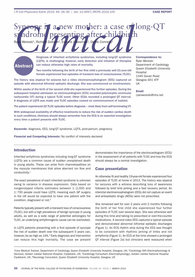
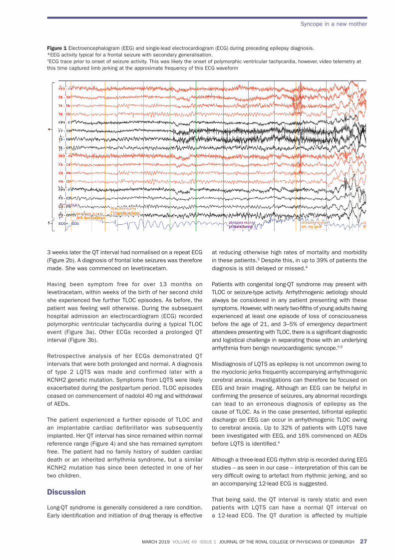
She remained well for over 2 years until 2 months following the birth of her first child she experienced four further episodes of TLOC over several days. She was otherwise well during this time and taking no prescribed or over-the-counter medications. A second video EEG captured a typical episode and demonstrated abnormal bifrontal epileptic discharges (Figure 1). An ECG rhythm strip during the EEG was thought to be consistent with rhythmic jerking of limbs and not arrhythmia (Figure 1). An ECG at the time showed a prolonged QT interval (Figure 2a) but clinicians were reassured when
Diagnosis of inherited arrhythmia syndromes, including long-QT syndrome (LQTS), is challenging; however, early detection and initiation of therapies can reduce otherwise high rates of mortality.
Two months following the birth of her � rst child a previously well 21-year-old female experienced four episodes of transient loss of consciousness (TLOC).
The history was atypical for seizures but a video electroencephalogram (EEG) captured an episode with abnormal bifrontal epileptic discharge. She was commenced on levetiracetam.
Within weeks of the birth of her second child she experienced � ve further episodes. During the subsequent hospital admission an electrocardiogram (ECG) recorded polymorphic ventricular tachycardia (VT) during a typical TLOC event. Other ECGs recorded a prolonged QT interval. A diagnosis of LQTS was made and TLOC episodes ceased on commencement of nadolol.
The patient experienced 22 TLOC episodes before diagnosis – most likely from self-terminating VT.
With widespread availability of effective treatments to reduce the risk of sudden cardiac death in such conditions, clinicians should always remember how the ECG is an essential investigation every time a patient presents with TLOC.
Keywords: diagnosis, EEG, long-QT syndrome, LQTS, post-partum, pregnancy
Financial and Competing Interests: No confl ict of interests declared
1Core Medical Trainee, Department of Cardiology, Queen Elizabeth University Hospital, Glasgow, UK; 2Cardiology StR (Electrophysiology & Devices), Golden Jubilee National Hospital, Clydebank, UK; 3Cardiology Consultant (Electrophysiology), Golden Jubilee National Hospital Clydebank, UK; 4Neurology Consultant, Queen Elizabeth University Hospital, Glasgow, UK
Correspondence to: Ryan WereskiDepartment of CardiologyQueen Elizabeth University Hospital1345 Govan RoadGlasgow G51 4TFUK Email: [email protected]
Syncope in a new mother: a case of long-QT syndrome presenting after childbirthRyan Wereski1, Richard Dobson2, Edward J Newman3, Derek Connelly4
MARCH 2019 VOLUME 49 ISSUE 1 JOURNAL OF THE ROYAL COLLEGE OF PHYSICIANS OF EDINBURGH 27
Syncope in a new mother
3 weeks later the QT interval had normalised on a repeat ECG (Figure 2b). A diagnosis of frontal lobe seizures was therefore made. She was commenced on levetiracetam.
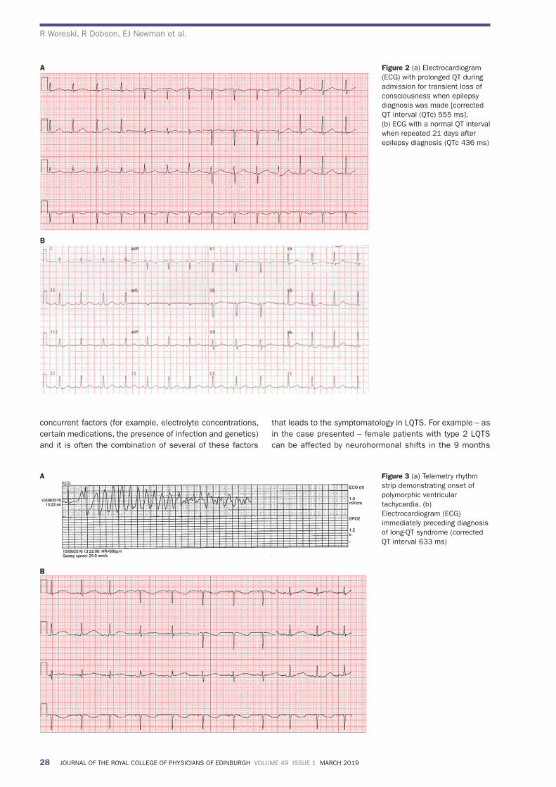
Having been symptom free for over 13 months on levetiracetam, within weeks of the birth of her second child she experienced fi ve further TLOC episodes. As before, the patient was feeling well otherwise. During the subsequent hospital admission an electrocardiogram (ECG) recorded polymorphic ventricular tachycardia during a typical TLOC event (Figure 3a). Other ECGs recorded a prolonged QT interval (Figure 3b).
Retrospective analysis of her ECGs demonstrated QT intervals that were both prolonged and normal. A diagnosis of type 2 LQTS was made and confirmed later with a KCNH2 genetic mutation. Symptoms from LQTS were likely exacerbated during the postpartum period. TLOC episodes ceased on commencement of nadolol 40 mg and withdrawal of AEDs.
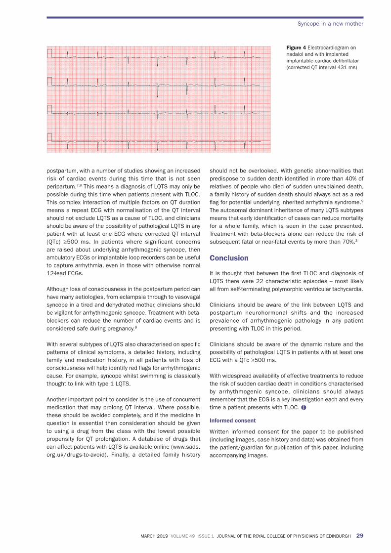
The patient experienced a further episode of TLOC and an implantable cardiac defibrillator was subsequently implanted. Her QT interval has since remained within normal reference range (Figure 4) and she has remained symptom free. The patient had no family history of sudden cardiac death or an inherited arrhythmia syndrome, but a similar KCNH2 mutation has since been detected in one of her two children.
Discussion
Long-QT syndrome is generally considered a rare condition. Early identifi cation and initiation of drug therapy is effective
at reducing otherwise high rates of mortality and morbidity in these patients.3 Despite this, in up to 39% of patients the diagnosis is still delayed or missed.4
Patients with congenital long-QT syndrome may present with TLOC or seizure-type activity. Arrhythmogenic aetiology should always be considered in any patient presenting with these symptoms. However, with nearly two-fi fths of young adults having experienced at least one episode of loss of consciousness before the age of 21, and 3–5% of emergency department attendees presenting with TLOC, there is a signifi cant diagnostic and logistical challenge in separating those with an underlying arrhythmia from benign neurocardiogenic syncope.5,6
Misdiagnosis of LQTS as epilepsy is not uncommon owing to the myoclonic jerks frequently accompanying arrhythmogenic cerebral anoxia. Investigations can therefore be focused on EEG and brain imaging. Although an EEG can be helpful in confi rming the presence of seizures, any abnormal recordings can lead to an erroneous diagnosis of epilepsy as the cause of TLOC. As in the case presented, bifrontal epileptic discharge on EEG can occur in arrhythmogenic TLOC owing to cerebral anoxia. Up to 32% of patients with LQTS have been investigated with EEG, and 16% commenced on AEDs before LQTS is identifi ed.4
Although a three-lead ECG rhythm strip is recorded during EEG studies – as seen in our case – interpretation of this can be very diffi cult owing to artefact from rhythmic jerking, and so an accompanying 12-lead ECG is suggested.
That being said, the QT interval is rarely static and even patients with LQTS can have a normal QT interval on a 12-lead ECG. The QT duration is affected by multiple
Figure 1 Electroencephalogram (EEG) and single-lead electrocardiogram (ECG) during preceding epilepsy diagnosis.*EEG activity typical for a frontal seizure with secondary generalisation.‡ECG trace prior to onset of seizure activity. This was likely the onset of polymorphic ventricular tachycardia, however, video telemetry at this time captured limb jerking at the approximate frequency of this ECG waveform
28 JOURNAL OF THE ROYAL COLLEGE OF PHYSICIANS OF EDINBURGH VOLUME 49 ISSUE 1 MARCH 2019
R Wereski, R Dobson, EJ Newman et al.
concurrent factors (for example, electrolyte concentrations, certain medications, the presence of infection and genetics) and it is often the combination of several of these factors
that leads to the symptomatology in LQTS. For example – as in the case presented – female patients with type 2 LQTS can be affected by neurohormonal shifts in the 9 months
Figure 3 (a) Telemetry rhythm strip demonstrating onset of polymorphic ventricular tachycardia. (b) Electrocardiogram (ECG) immediately preceding diagnosis of long-QT syndrome (corrected QT interval 633 ms)
A
B
Figure 2 (a) Electrocardiogram (ECG) with prolonged QT during admission for transient loss of consciousness when epilepsy diagnosis was made [corrected QT interval (QTc) 555 ms]. (b) ECG with a normal QT interval when repeated 21 days after epilepsy diagnosis (QTc 436 ms)
A
B postpartum, with a number of studies showing an increased risk of cardiac events during this time that is not seen peripartum.7,8 This means a diagnosis of LQTS may only be possible during this time when patients present with TLOC. This complex interaction of multiple factors on QT duration means a repeat ECG with normalisation of the QT interval should not exclude LQTS as a cause of TLOC, and clinicians should be aware of the possibility of pathological LQTS in any patient with at least one ECG where corrected QT interval (QTc) ≥500 ms. In patients where significant concerns are raised about underlying arrhythmogenic syncope, then ambulatory ECGs or implantable loop recorders can be useful to capture arrhythmia, even in those with otherwise normal 12-lead ECGs.
Although loss of consciousness in the postpartum period can have many aetiologies, from eclampsia through to vasovagal syncope in a tired and dehydrated mother, clinicians should be vigilant for arrhythmogenic syncope. Treatment with beta-blockers can reduce the number of cardiac events and is considered safe during pregnancy.9
With several subtypes of LQTS also characterised on specifi c patterns of clinical symptoms, a detailed history, including family and medication history, in all patients with loss of consciousness will help identify red fl ags for arrhythmogenic cause. For example, syncope whilst swimming is classically thought to link with type 1 LQTS.
Another important point to consider is the use of concurrent medication that may prolong QT interval. Where possible, these should be avoided completely, and if the medicine in question is essential then consideration should be given to using a drug from the class with the lowest possible propensity for QT prolongation. A database of drugs that can affect patients with LQTS is available online (www.sads.org.uk/drugs-to-avoid). Finally, a detailed family history
should not be overlooked. With genetic abnormalities that predispose to sudden death identifi ed in more than 40% of relatives of people who died of sudden unexplained death, a family history of sudden death should always act as a red fl ag for potential underlying inherited arrhythmia syndrome.9 The autosomal dominant inheritance of many LQTS subtypes means that early identifi cation of cases can reduce mortality for a whole family, which is seen in the case presented. Treatment with beta-blockers alone can reduce the risk of subsequent fatal or near-fatal events by more than 70%.3
Conclusion
It is thought that between the fi rst TLOC and diagnosis of LQTS there were 22 characteristic episodes – most likely all from self-terminating polymorphic ventricular tachycardia.
Clinicians should be aware of the link between LQTS and postpartum neurohormonal shifts and the increased prevalence of arrhythmogenic pathology in any patient presenting with TLOC in this period.
Clinicians should be aware of the dynamic nature and the possibility of pathological LQTS in patients with at least one ECG with a QTc ≥500 ms.
With widespread availability of effective treatments to reduce the risk of sudden cardiac death in conditions characterised by arrhythmogenic syncope, clinicians should always remember that the ECG is a key investigation each and every time a patient presents with TLOC.
Informed consent
Written informed consent for the paper to be published (including images, case history and data) was obtained from the patient/guardian for publication of this paper, including accompanying images.
Figure 4 Electrocardiogram on nadalol and with implanted implantable cardiac defibrillator (corrected QT interval 431 ms)
MARCH 2019 VOLUME 49 ISSUE 1 JOURNAL OF THE ROYAL COLLEGE OF PHYSICIANS OF EDINBURGH 29
Syncope in a new mother
postpartum, with a number of studies showing an increased risk of cardiac events during this time that is not seen peripartum.7,8 This means a diagnosis of LQTS may only be possible during this time when patients present with TLOC. This complex interaction of multiple factors on QT duration means a repeat ECG with normalisation of the QT interval should not exclude LQTS as a cause of TLOC, and clinicians should be aware of the possibility of pathological LQTS in any patient with at least one ECG where corrected QT interval (QTc) ≥500 ms. In patients where significant concerns are raised about underlying arrhythmogenic syncope, then ambulatory ECGs or implantable loop recorders can be useful to capture arrhythmia, even in those with otherwise normal 12-lead ECGs.
Although loss of consciousness in the postpartum period can have many aetiologies, from eclampsia through to vasovagal syncope in a tired and dehydrated mother, clinicians should be vigilant for arrhythmogenic syncope. Treatment with beta-blockers can reduce the number of cardiac events and is considered safe during pregnancy.9
With several subtypes of LQTS also characterised on specifi c patterns of clinical symptoms, a detailed history, including family and medication history, in all patients with loss of consciousness will help identify red fl ags for arrhythmogenic cause. For example, syncope whilst swimming is classically thought to link with type 1 LQTS.
Another important point to consider is the use of concurrent medication that may prolong QT interval. Where possible, these should be avoided completely, and if the medicine in question is essential then consideration should be given to using a drug from the class with the lowest possible propensity for QT prolongation. A database of drugs that can affect patients with LQTS is available online (www.sads.org.uk/drugs-to-avoid). Finally, a detailed family history
should not be overlooked. With genetic abnormalities that predispose to sudden death identifi ed in more than 40% of relatives of people who died of sudden unexplained death, a family history of sudden death should always act as a red fl ag for potential underlying inherited arrhythmia syndrome.9 The autosomal dominant inheritance of many LQTS subtypes means that early identifi cation of cases can reduce mortality for a whole family, which is seen in the case presented. Treatment with beta-blockers alone can reduce the risk of subsequent fatal or near-fatal events by more than 70%.3
Conclusion
It is thought that between the fi rst TLOC and diagnosis of LQTS there were 22 characteristic episodes – most likely all from self-terminating polymorphic ventricular tachycardia.
Clinicians should be aware of the link between LQTS and postpartum neurohormonal shifts and the increased prevalence of arrhythmogenic pathology in any patient presenting with TLOC in this period.
Clinicians should be aware of the dynamic nature and the possibility of pathological LQTS in patients with at least one ECG with a QTc ≥500 ms.
With widespread availability of effective treatments to reduce the risk of sudden cardiac death in conditions characterised by arrhythmogenic syncope, clinicians should always remember that the ECG is a key investigation each and every time a patient presents with TLOC.
Informed consent
Written informed consent for the paper to be published (including images, case history and data) was obtained from the patient/guardian for publication of this paper, including accompanying images.
Figure 4 Electrocardiogram on nadalol and with implanted implantable cardiac defibrillator (corrected QT interval 431 ms)

30 JOURNAL OF THE ROYAL COLLEGE OF PHYSICIANS OF EDINBURGH VOLUME 49 ISSUE 1 MARCH 2019
R Wereski, R Dobson, EJ Newman et al.
References1 Modell SM, Lehmann MH. The Long-QT syndrome family of
cardiac ion channelopathies: a HuGE review. Genet Med 2006; 8: 143–55.
2 NHS Scotland Information Services Division, GP Workforce and practice list sizes 2007–2017 Report, Published 12/12/17. https://www.isdscotland.org/Health-Topics/General-Practice/Publications/2017-12-12/2017-12-12-GPWorkforce2017-Report.pdf (accessed 29/03/18).
3 Liu JF, Jons C, Moss AJ et al. Risk factors for recurrent syncope and subsequent fatal or near-fatal events in children and adolescents with long-QT syndrome. J Am Coll Cardiol 2011; 57: 941–50.
4 MacCormick JM, McAlister H, Crawford J et al. Misdiagnosis of long-QT syndrome as epilepsy at fi rst presentation. Ann Emerg Med 2009; 54: 26–32.
5 Ganzeboom KS, Colman N, Reitsma JB et al. Prevalence and triggers of syncope in medical students. Am J Cardiol 2003; 91: 1006–8, A8.
6 da Silva RM. Syncope: epidemiology, etiology, and prognosis. Front Physiol 2014; 5: 471.
7 Ishibashi K, Aiba T, Kamiya C et al. Arrhythmia risk and beta-blocker therapy in pregnant women with long QT syndrome. Heart 2017; 103: 1374–9.
8 Seth R, Moss AJ, McNitt S et al. Long QT syndrome and pregnancy. J Am Coll Cardiol 2007; 49: 1092–8.
9 Tan HL, Hofman N, van Langen IM et al. Sudden unexplained death: heritability and diagnostic yield of cardiological and genetic examination in surviving relatives. Circulation 2005; 112: 207–13.