synovial fluid protein adsorption on polymer-based … technologies and analytics, vienna university...
TRANSCRIPT
e u p a o p e n p r o t e o m i c s 4 ( 2 0 1 4 ) 70–80
Available online at www.sciencedirect.com
ScienceDirect
jou rn al hom epage : ht tp : / /www.e lsev ier .com/ locate /euprot
Synovial fluid protein adsorption onpolymer-based artificial hip joint materialinvestigated by MALDI-TOF massspectrometry imaging
Sophie M. Fröhlicha, Victoria Dorrera, Vasiliki-Maria Archodoulakib,Günter Allmaiera, Martina Marchetti-Deschmanna,∗
a Institute of Chemical Technologies and Analytics, Vienna University of Technology, Austriab Institute of Materials Science and Technology, Vienna University of Technology, Austria
a r t i c l e i n f o
Article history:
Received 31 December 2013
Received in revised form
17 April 2014
Accepted 4 May 2014
Available online 13 May 2014
Keywords:
PE-UHMW
a b s t r a c t
UHMW-PE (ultra-high molecular weight polyethylene), most frequently used material in
acetabular cup replacement, is affected by the interaction with its surrounding synovial
fluid. It is assumed that protein layer formation is of high importance for lubrication, how-
ever alters polymer characteristics. This study investigates in vitro protein adsorption on
gamma-irradiated and Vitamin E doped UHMW-PE using synovia as modeling system. SDS-
PAGE and MALDI-TOF mass spectrometry imaging showed adsorption of high abundance
proteins in a mass range between 2 and 200 kDa. Protein layer formation was observed
on planar UHMW-PE material, whereas morphologically modified UHMW-PE regions were
highly affected by protein aggregation.
Protein adsorption
Intact proteins
Mass spectrometry imaging
Synovial fluid
MALDI-TOF
© 2014 The Authors. Published by Elsevier B.V. on behalf of European Proteomics
Association (EuPA). This is an open access article under the CC BY-NC-ND license
(http://creativecommons.org/licenses/by-nc-nd/3.0/).
it has not been determined yet, whether modified PE-UHMW
1. Introduction
Ultra high molecular weight polyethylene (PE-UHMW) is avery frequently used material in artificial joint replacementsystems. It provides easy usability, extreme high mechani-cal wear resistance, high biocompatibility and is to a certain
extent self-lubricating [1]. However, the number of revisionsurgeries is still high, sometimes necessary because of therapid shelf life aging process and in vivo degradation due to∗ Corresponding author. Tel.: +43 1 58801 15162; fax: +43 1 58801 915162E-mail address: [email protected] (M. M
http://dx.doi.org/10.1016/j.euprot.2014.05.0012212-9685/© 2014 The Authors. Published by Elsevier B.V. on behalf ofarticle under the CC BY-NC-ND license (http://creativecommons.org/lic
mechanical wear, oxidation and material modification [2–4].Synovial fluid (SF), the major lubricating system in the jointcompartment, directly interacts with the material surface,leading to biomolecule adsorption and diffusion [1,5] pos-sibly altering the polymer characteristics and stability. Themajor components have already been investigated, however
.archetti-Deschmann).
surface areas lead to enhanced biomolecule adsorption anddiffusion or vice versa. Furthermore, protein layer formationon PE-UHMW has so far not been investigated except for a
European Proteomics Association (EuPA). This is an open accessenses/by-nc-nd/3.0/).

m i c s
faciotes(aseUpa
mgbpgsosotbaofm
aibsdtMiaosfii
ttu[ScpwbPmhoo
e u p a o p e n p r o t e o
ew selected protein and carbohydrate species, e.g. albuminnd hyaluronic acid [6]. It is known that the major abundantomponents of synovial fluid, e.g. hyaluronic acid, albumin ormmunoglobulin, interact with PE-UHMW and tend to adsorbn the surface in model fluid systems. However, the interac-ion of the whole synovial proteome in the presence of lipidstc. with PE-UHMW has never been investigated. Because theynovial fluid composition is very similar to blood plasmaexcept for the high hyaluronic acid content) it is also highlyffected by the patient’s age, sex, life style and pathologicaltatus [7,8]. Therefore it is of high interest, which proteinsxcept albumin and immunoglobulin in fact adsorb on PE-HMW before a deeper understanding of the pathologicalrocess can be obtained and/or material related selectivedsorption effects can be gathered.
Different compositions of PE-UHMW, Vitamin E dopedaterial, highly cross-linked PE-UHMW and materials under-
oing different kinds of sterilization strategies have alreadyeen tested in clinical studies for improving the materials’roperties and reducing protein adsorption. Highly abundantlycoproteins have been shown to adsorb on PE-UHMW inimulating models and adsorption has been investigated toccur unspecific and concentration independent [1]. In theame study model fluids containing only one glycoproteinf interest revealed that the formed protein layer enhanceshe lubrication and friction behavior of PE-UHMW. However,esides the selected glycoproteins also other high abundancend acute phase proteins occurring during inflammation andxidative stress, e.g. during rheumatic diseases, are relevantor adsorption, friction behavior and consequently material
odification.The presented study focuses on the unbiased identification
nd localization of proteins present in synovial fluid adsorb-ng on different varieties of PE-UHMW material. To answeroth questions, protein identity and spatial distribution, masspectrometry imaging (MSI) by means of matrix assisted laseresorption/ionization time-of-flight (MALDI-TOF) examina-ion was correlated to SDS PAGE analysis of adsorbed proteins.SI is a well-established method for the localization and
dentification of analytes of interest within an untargetedpproach [9]. To date MSI has been applied to a huge varietyf biological samples [10–12] and it has to be mentioned thaturface analysis of biomaterials is an exponentially growingeld gaining more importance for the analysis of lubrication
nteraction and material modifications [13,14].One limiting fact of MSI is that protein identification in
he high molecular mass range is only possible based onhe tentative assignment of proteins to measured m/z val-es, respectively the molecular weight of detected molecules
15]. To corroborate protein assignment after MSI analysis,DS PAGE analysis was chosen, providing the possibility toompare synovial protein patterns to patterns of protein com-ounds adsorbed on PE-UHMW of different compositionsith respect to overall detected proteins. In parallel MS-ased protein identification after in-gel digestion is feasible.rotein identification for adsorbed molecules was further-
ore verified by on-tissue (“on polymer”) digestion [16,17]. Theydrophobic surface of PE-UHMW supports the preservationf protein localization during trypsin application in aque-us buffer systems, however, diffusion is a critical point for
4 ( 2 0 1 4 ) 70–80 71
both enzyme and matrix application. To obtain reliable resultshomogeneous layers of trypsin and MALDI matrix solutionwere applied using a piezo printer (Chemical Inkjet Printer,ChIP-1000 [18]) to the protein-carrying PE-UHMW material.
2. Experimental
2.1. Materials
The Institute of Materials Science and Technology, Vienna Uni-versity of Technology, provided gamma-irradiated PE-UHMW(GUR-1050 PE-UHMW) and PE-UHMW samples doped withVitamin E. All chemicals and reagents, unless indicated specif-ically, were from Sigma–Aldrich (USA) with a purity of atleast 99% if not stated otherwise. Ultrapure water (uH2O) wasobtained from a Simplicity system (Millipore, USA) with a spe-cific conductivity of ˝m ≤ 18 S/cm. Human synovial fluid (SF),sampled after joint revision, was provided by the Institute ofMaterials Science and Technology.
2.2. Sample preparation
Virgin gamma irradiated GUR-1050 PE-UHMW and VitaminE doped PE-UHMW samples were used. PE-UHMW was cutinto small blocks of comparable geometry (m = 85 ± 2 mg,V ≈ 125 mm3), which were either used for incubation exper-iments or sliced to 15 �m using cryostat (CM1950, Leica,Germany) and mounted on indium-tin oxide (ITO) coatedglass slides (Sigma–Aldrich) using conductive tape (Shimadzu,Japan). The final polymer slice can exhibit folded regions afterattachment. However, those regions were used to simulateuneven and rough regions and edges, as they actually occurin vivo, caused by mechanical wear and abrasion processes inthe acetabular cup. All PE-UHMW samples were incubated inSF at 37 ◦C for 24 h. After incubation, samples were rinsed withuH2O and vacuum dried.
For MS and MSI experiments standard MALDI matrices,�-cyano-4-hydroxycinnamic acid (HCCA) and sinapinic acid(SA) were dissolved in 70% acetonitrile (ACN), 30% uH2O (v/v)containing 0.1% trifluoroacetic acid (TFA) at a concentrationof 13 (HCCA) and 25 (SA) mg/mL. Automatic matrix deposi-tion was performed using a chemical inkjet printer ChIP-1000(Shimadzu) or a commonly used airbrush device (Conrad,Germany). For the airbrush application of MALDI matrix aworking distance of 10–12 cm was chosen with an approxi-mate angle of 50–60◦ covering the sample in several iterativesteps.
2.3. MS and MSI analysis
MSI experiments were performed on a MALDI-TOF/RTOFinstrument (UltrafleXtreme, Bruker Daltonics, Germany)in positive linear or reflectron mode, equipped with a355 nm SmartBeam laser pulsed at 2000 Hz [19] and on aMALDI-TOF/RTOF instrument (AXIMA TOF2, Shimadzu Kratos
Analytical, UK) equipped with a 20 Hz nitrogen (337 nm) laser.At a spatial resolution of 100–150 �m, mass spectra based on1000 (AXIMA TOF2) or 2000 (UltrafleXtreme) single laser shotswere acquired per position over an average sample size of
o m i c
72 e u p a o p e n p r o t e8000 single imaging positions for polymer samples (approx.8 × 8 mm) if not otherwise stated. For image reconstructionFlexImaging v. 3.0 software (Bruker Daltonics) and Biomap38.04 (Novartis, Switzerland) was used.
2.4. Gel electrophoresis
For protein extraction 200 �L SF, PE-UHMW blocks after incu-bation in synovia and blank material was deposited in1200 �L uH2O before 100 �L trichloroacetic acid (TCA; 20%)were added 5 times and vortexed thoroughly between eachsingle addition. Samples were incubated for 30 min at 4 ◦Cand centrifuged at 14,000 rpm and 4 ◦C for another 30 min.The supernatant and polymer blocks were removed andthe protein pellet re-dissolved in 10 �L lysis buffer (50 mMTris/HCl pH 8.0, 2 mM dithiothreitol, 10% v/v glycerol, 0.5%v/v Tween 20, 10% toluol). For protein precipitation withethanol, 50 �L SF or the PE-UHMW blocks were mixed with450 �L precooled ethanol (4 ◦C) and incubated at −70 ◦C for2 h before centrifugation at 14,000 rpm and room tempera-ture for 30 min. The polymer blocks and the supernatant wereremoved and ethanol residues were evaporated in the vacuumcentrifuge. After drying the pellet was re-dissolved in lysisbuffer. Protein concentration was determined according toBradford [20].
For SDS PAGE analysis protein pellets were dissolved inlithium dodecyl sulfate sample buffer (26.5 mM Tris–HCl,35.25 mM Tris Base, 0.5% lithium dodecyl sulfate, 2.5% glycerol,0.1275 mM EDTA, 0.055 mM SERVA Blue G250, 0.044 mM PhenolRed, pH 8.5, 50 mM dithiothreitol) at 95 ◦C for 5 min and appliedto a precast NuPAGE 4–12% Bis-Tris polyacrylamide gel (Invi-trogen, USA). Electrophoresis was conducted at 125 V (60 mAmax., 12.5 W) in a XcellSurelock Mini Cell electrophoresis sys-tem (Invitrogen, USA). Gels were silver stained [21] for proteindetection.
2.5. Protein identification
Proteins were either identified after SDS PAGE by in-gel orby on-tissue (“on polymer”) digestion. For the first, gel bandswere excised with a clean scalpel and destained using 100 mMNa2S2O3 and 30 mM K4Fe(CN)6·3H2O (1:1, v/v). Gel pieces weretreated with ACN and rehydrated with 100 mM NH4HCO3.After reduction (10 mM DTT in 100 mM NH4HCO3) and alkyl-ation (50 mM iodoacetamide in 100 mM NH4HCO3) the gelpieces were dried in a vacuum centrifuge and rehydrated inapprox. 10 �L 50 mM NH4HCO3 (pH 8.5) containing 5% ACN and125 ng trypsin (porcine, proteomics grade, Roche, Switzerland).Digestion was carried out for 24 h at 37 ◦C. Peptides wereextracted with 50 mM NH4HCO3/ACN (1/1, v/v) and two timeswith uH2O/ACN containing 0.1% TFA (1/1, v/v), for 15 min each.All extracts of one respective spot were pooled and dried in avacuum centrifuge. After reconstitution in 0.1% TFA peptideswere desalted using C18 ZipTips (Millipore, USA) and elutedwith 5 mg/mL HCCA prepared in ACN/0.1%TFA (50/50, v/v) inthe final step. Peptide mass fingerprinting (PMF) and sequence
tag analysis were carried out on a MALDI-TOF/RTOF instru-ment (UltrafleXtreme).For “on polymer” enzymatic treatment, the ChIP-1000 wasused for precise trypsin deposition at a standard concentration
s 4 ( 2 0 1 4 ) 70–80
of 3 ng/�m2 containing 1% Rapigest (Waters, USA) dissolved in50 mM NH4HCO3 (pH 7.5) containing 5% ACN. Trypsin solutionwas applied at a lateral resolution of 100 �m covering the com-plete sample area. For digestion samples were stored at 37 ◦Cfor 24 h in saturated atmosphere (ethanol/water, 50/50, v/v,80% relative humidity). After incubation samples were care-fully washed with uH2O to remove salts, delocalized peptidesnot adsorbed to the polymer and autolytic fragments fromtrypsin. Samples were vacuum dried for 15 min and HCCA wasapplied afterwards as described.
For all enzymatic digestion data, autolytic tryptic products,keratin and blank artifacts were assigned and removed beforedatabase search (SWISSPROT, December 2013) using Mascot[22] with the following parameters: taxonomy Homo sapiens,monoisotopic mass values, peptide mass tolerance of ±0.3 Da(for PMF and PSD experiments), 2 missed cleavages, a fixedmodification carboxyamidomethylation and methionine oxi-dations set as variable modification.
3. Results
3.1. Protein identification on PE-UHMW
Two different PE-UHMW materials were investigated con-cerning protein adsorption: conventionally used GUR-1050PE-UHMW and Vitamin E doped PE-UHMW, which is claimedto be more resistant to oxidative degradation [5]. Afterincubating the two different materials in the same syn-ovia at 37 ◦C for 24 h, adsorbed proteins were extracted.Similar to plasma, 85% of synovia consists of the samewell-described high abundance proteins. However, to iden-tify proteins actually adsorbing in a concerted manner fromsynovia on PE-UHMW in the bio-compartment, we decidedagainst special synovia treatment (e.g. depletion) well awareof the possibility to identify exactly those high abundanceproteins.
For this approach, two different extraction methods wereevaluated: an ethanol and a TCA based extraction methods(details see Section 2.1). Protein concentrations were deter-mined to compare the efficiency of both approaches. Theprotein concentration obtained according to Bradford dif-fered significantly yielding 14.68 ± 4.24 �g/�L for the ethanol(n = 15) based method and 7.74 ± 1.89 �g/�L for the same poly-mer type extracted with TCA (n = 15). However, no qualitativedifferences were observed for the SDS PAGE protein pat-tern and the number of protein lanes was comparable inthe apparent molecular mass range between 3 and 200 kDa.For proceeding experiments TCA precipitation was applied,in order to avoid eventual lipid or glycan contamination(easily extracted with ethanol) and based on the higherreproducibility.
Protein analysis from the two different PE-UHMWspecies revealed an average protein concentration of9.46 �g/�L for GUR-1050 PE-UHMW after extraction withTCA and 13.08 �g/�L for Vitamin E doped samples. Applying
concentration-matched samples to SDS PAGE revealed lighterstained protein bands for the Vitamin E doped samples. Morethan 10 high abundance proteins could be identified afterin-gel digestion (Fig. 1).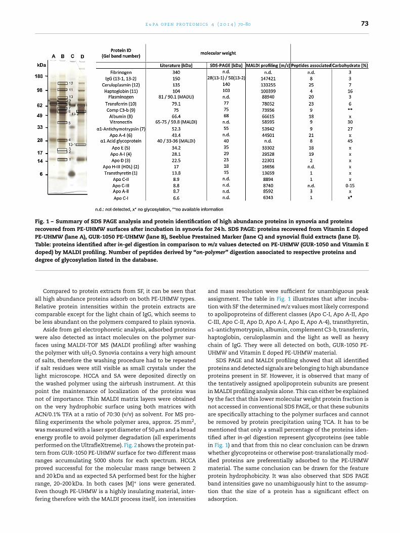
e u p a o p e n p r o t e o m i c s 4 ( 2 0 1 4 ) 70–80 73
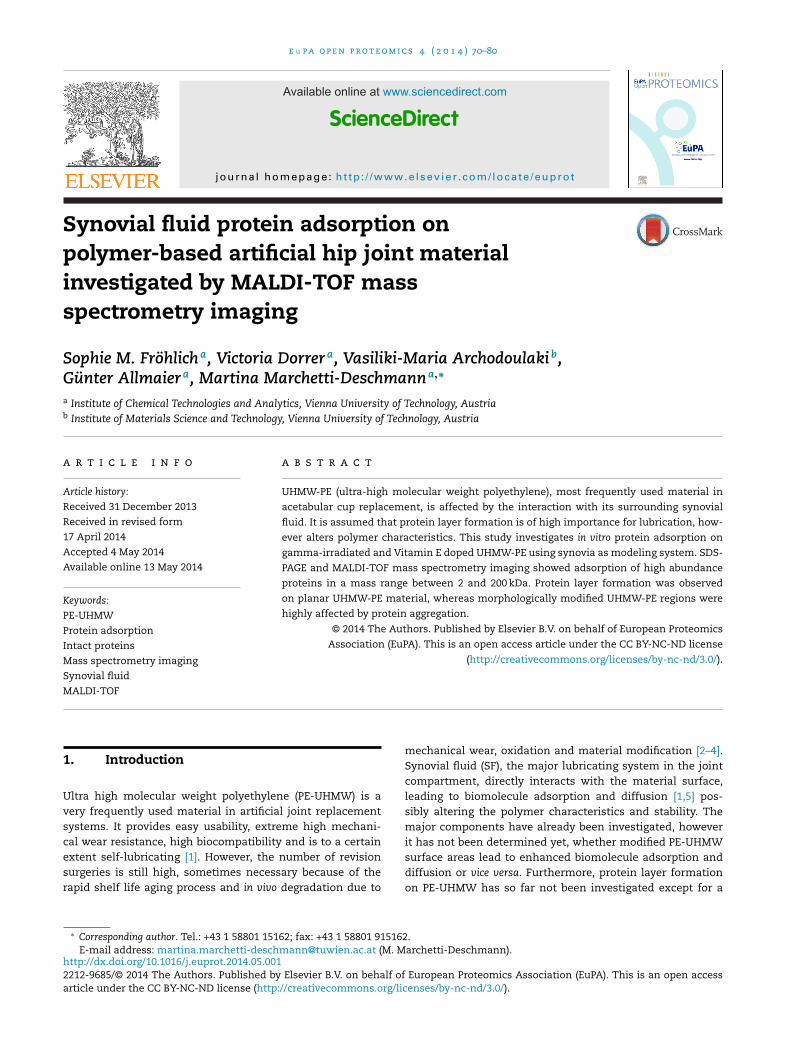
Fig. 1 – Summary of SDS PAGE analysis and protein identification of high abundance proteins in synovia and proteinsrecovered from PE-UHMW surfaces after incubation in synovia for 24 h. SDS PAGE: proteins recovered from Vitamin E dopedPE-UHMW (lane A), GUR-1050 PE-UHMW (lane B), Seeblue Prestained Marker (lane C) and synovial fluid extracts (lane D).Table: proteins identified after in-gel digestion in comparison to m/z values detected on PE-UHMW (GUR-1050 and Vitamin Edoped) by MALDI profiling. Number of peptides derived by “on-polymer” digestion associated to respective proteins anddegree of glycosylation listed in the database.
aRcb
wftoiltpnoAfiweptrparEf
Compared to protein extracts from SF, it can be seen thatll high abundance proteins adsorb on both PE-UHMW types.elative protein intensities within the protein extracts areomparable except for the light chain of IgG, which seems toe less abundant on the polymers compared to plain synovia.
Aside from gel electrophoretic analysis, adsorbed proteinsere also detected as intact molecules on the polymer sur-
aces using MALDI-TOF MS (MALDI profiling) after washinghe polymer with uH2O. Synovia contains a very high amountf salts, therefore the washing procedure had to be repeated
f salt residues were still visible as small crystals under theight microscope. HCCA and SA were deposited directly onhe washed polymer using the airbrush instrument. At thisoint the maintenance of localization of the proteins wasot of importance. Thin MALDI matrix layers were obtainedn the very hydrophobic surface using both matrices withCN/0.1% TFA at a ratio of 70:30 (v/v) as solvent. For MS pro-ling experiments the whole polymer area, approx. 25 mm2,as measured with a laser spot diameter of 50 �m and a broad
nergy profile to avoid polymer degradation (all experimentserformed on the UltrafleXtreme). Fig. 2 shows the protein pat-ern from GUR-1050 PE-UHMW surface for two different massanges accumulating 5000 shots for each spectrum. HCCAroved successful for the molecular mass range between 2
nd 20 kDa and as expected SA performed best for the higherange, 20–200 kDa. In both cases [M]+ ions were generated.ven though PE-UHMW is a highly insulating material, inter-ering therefore with the MALDI process itself, ion intensitiesand mass resolution were sufficient for unambiguous peakassignment. The table in Fig. 1 illustrates that after incuba-tion with SF the determined m/z values most likely correspondto apolipoproteins of different classes (Apo C-I, Apo A-II, ApoC-III, Apo C-II, Apo D, Apo A-I, Apo E, Apo A-4), transthyretin,�1-antichymotrypsin, albumin, complement C3-b, transferrin,haptoglobin, ceruloplasmin and the light as well as heavychain of IgG. They were all detected on both, GUR-1050 PE-UHMW and Vitamin E doped PE-UHMW material.
SDS PAGE and MALDI profiling showed that all identifiedproteins and detected signals are belonging to high abundanceproteins present in SF. However, it is observed that many ofthe tentatively assigned apolipoprotein subunits are presentin MALDI profiling analysis alone. This can either be explainedby the fact that this lower molecular weight protein fraction isnot accessed in conventional SDS PAGE, or that these subunitsare specifically attaching to the polymer surfaces and cannotbe removed by protein precipitation using TCA. It has to bementioned that only a small percentage of the proteins iden-tified after in-gel digestion represent glycoproteins (see tablein Fig. 1) and that from this no clear conclusion can be drawnwhether glycoproteins or otherwise post-translationally mod-ified proteins are preferentially adsorbed to the PE-UHMWmaterial. The same conclusion can be drawn for the feature
protein hydrophobicity. It was also observed that SDS PAGEband intensities gave no unambiguously hint to the assump-tion that the size of a protein has a significant effect onadsorption.
74 e u p a o p e n p r o t e o m i c s 4 ( 2 0 1 4 ) 70–80
m/z
a.i.
6000 9000 12000 15000 18000 0
3000
6000
9000
12000
15000
Apo H-III (HDL)m/z 16656
Transthyretinm/z 13659
Apo C-Im/z 6343
Apo A-IIm/z 8592
Apo C-IIIm/z 8740
Apo C-IIm/z 8894
a)
m/z
a.i.
30000 60000 90000 120000 150000 180000
5000
10000
15000
20000
25000
30000
35000
40000
45000
50000Albumin
m/z 66615
Apo Dm/z 22301
Apo Em/z 33302
Apo A-4m/z 44501
A1ATm/z 53942
Vitronectinm/z 58595
Comp C3-bm/z 73956
Transferrinm/z 78052
Ceruloplasminm/z 133255
IgGm/z 147421
b)
Fig. 2 – Positive ion MALDI mass spectra obtained by MS profiling of GUR-1050 PE-UHMW incubated in synovia for 24 h at37 ◦C. (a) HCCA was used as matrix for the molecular mass range between 2 and 20 kDa and (b) SA was used as matrix for
the molecular mass range between 20 and 200 kDa.3.2. MALDI matrix application on hydrophobicsurfaces for MSI experiments
Investigating protein adsorption on polymer surfaces byMALDI-TOF MS requires thorough optimization of sample
preparation particularly due the low amounts present. Themost critical step is the generation of a homogenous MALDImatrix layer on a large sample surface. The hydrophobic sur-face of the PE-UHMW samples favored the formation of aliquid film if the matrix was not carefully applied dropletby droplet. Especially folded regions, which can occur afterapplying the thin polymer samples to the ITO slides after cryo-microtomy, provide problems regarding matrix crystallization.MALDI matrix solution application with slowly evaporating
solvents leads to a matrix flow leaving no matrix on foldedareas but large crystals on planar regions. This can ofcoursefavor disadvantageous analyte diffusion. Additionally it isknown that protein desorption/ionization during the MALDI
m i c s 4 ( 2 0 1 4 ) 70–80 75
pvootttaabsofib7
ttvtatntpidacwaetmc(sderpm
3P
BfqUs
2aHw1Fa
Fig. 3 – Non-normalized intensity distribution ofLeucine–Enkephalin (peptide solution on glass slide is notobservable, therefore no image provided/transparent)printed with the piezo printer in an “H”-shaped area ontransparent ITO glass before HCCA (ACN/aqueous TFA(50/0.1, v/v)) application with an airbrush. Visualization:FlexImaging. Lateral resolution of the MSI experiment:
e u p a o p e n p r o t e o
rocess is more effective after incorporating analytes in sol-ent systems containing acidic components, as a consequencef efficient protonation. On the polymer surfaces, however,rganic solvent systems were shown in preliminary resultso have better crystallization properties, most likely due toheir much higher volatility [23]. Further studies showed nowhat organic solvent systems revealed very small crystal sizesnd homogenous covering of the polymeric material, whilequeous solvent systems resulted in improved signal qualityut less favorable sweet spot formation in combination withometimes very small areas covered with high concentrationsf matrix. The matrix/solvent system turned out to be per-orming best with respect to signal quality (signal-to-noise,ntensity and mass resolution), surface coverage and applica-ility was a solution of ACN/aqueous 0.1% TFA at a ratio of0/30 for both HCCA and SA.
For matrix application the piezo printer (Chip 1000) andhe airbrush instrument were compared with special respecto analyte diffusion. To prevent the formation of a thin sol-ent layer on top of the polymer due to droplets trickling away,he distance between single matrix droplets (approx. 80 pL)pplied with the piezo printer was set to 100 �m. To coverhe total area of 25 mm2 long printing operation times wereecessary (up to 5 h). For this, analyte diffusion was inves-igated when using an airbrush system instead of the piezorinter for MALDI matrix deposition to reduce overall operat-
ng times. A peptide standard, 500 fM leucine-enkephalin, waseposited on an ITO target with the piezo printer using therea print mode of the instrument, which leads to a completeovering of a pre-defined area. This area was then coveredith HCCA as MALDI matrix using the airbrush. We observed
recognizable liquid layer of solvent on the ITO target, whichvaporated more slowly than the MALDI matrix/solvent sys-ems applied by the piezo printer. Thus after drying, light
icroscopy revealed a thin matrix layer with homogeneousrystal distribution. Moreover MSI experiments demonstratedFig. 3) that the airbrush represents a matrix application deviceufficient for matrix application without significant analyteislocation at a lateral resolution of 100 �m. The very sharpdged area visualized for the peptide ion emphasizes the accu-acy of peptide application by the piezo printer. The appliedeptide was perfectly preserved and detected after MALDIatrix application.
.3. Visualizing intact protein distribution onE-UHMW up to 204 kDa
esides the finding that proteins adsorb on PE-UHMW asiderom their post-translational modifications or hydrophobicuality, it is necessary to localize proteins adsorbing on PE-HMW in order to correlate them to polymer qualities such asurface roughness or oxidation.
For this the 15 �m PE-UHMW slices, incubated in SF for4 h, were investigated concerning the adsorbed protein layernd the localization of certain proteins in a MSI approach.CCA for the low or SA for the high molecular mass range
ere applied using the piezo printer at a lateral resolution of00 �m covering the whole PE-UHMW sample homogeneously.ig. 4a shows an exemplary covered PE-UHMW sample afterpplying HCCA with the piezo printer. A rather thick matrix
100 �m.
layer with fine crystal formation is observed. Similar resultswere obtained for SA. The same proteins as in profiling exper-iments were detected, i.e. all highly abundant species such asalbumin, IgG and the apolipoprotein subclasses.
Profile mass spectra were already presented in Fig. 2aand 2b showing protein adsorption up to 204 kDa. Yetsome proteins exhibited characteristic localization on thepolymer material. Fig. 4b shows the intensity distributionof apolipoprotein D [M+H]+ (m/z 22,301) and Fig. 4e thedistribution of the unknown protein at 204 kDa on GUR-1050 PE-UHMW after total ion current (TIC) normalization.Molecules were almost homogeneously distributed on thepolymer. The analyzed PE-UHMW sample showed a smallfolded area in the center, whereas the majority area wasmounted completely planar to the conductive tape. On per-fectly prepared samples all proteins were detected with asimilar homogeneous lateral distributions. Interestingly thatin the few areas not covered with Apo D especially albuminwas found (Fig. 4c). On both polymer variants, GUR-1050 andVitamin E doped, m/z values correlating to [M+H]+ ions ofhuman serum albumin (m/z 66,615) were detected in the cen-ter region of the polymer slice, with especially high intensitiesin folded area (Fig. 4c and f). The area of high intensity ontop of the sample results from protein adsorption on the con-ductive tape on which the polymer is mounted. Compared tothe conventionally used GUR-1050 PE-UHMW, Vitamin E dopedmaterial (Fig. 4f) showed less signal intensities for all detectedproteins. However, adsorbed proteins, IgG and an uniden-tified compound with m/z 204,000 (Fig. 4d and e), revealedsimilar preferences for folded regions and sharp edges. Toinvestigate the influence of protein degradation due to pro-tease activity, MSI experiments were performed on samples
removed from SF incubation after 15, 30, 60 min and 3 h. It wasfound that homogeneous protein layers are formed alreadyafter 30 min of incubation and no loss of signal intensity or
76 e u p a o p e n p r o t e o m i c s 4 ( 2 0 1 4 ) 70–80
Fig. 4 – MSI experiments of GUR-1050 and Vitamin E doped PE-UHMW incubated in synovia for 24 h at 37 ◦C. (a) Matrixcrystallization on PE-UHMW using HCCA in ACN/0.1% TFA (70/30, v/v). MSI experiments to localize (b) Apo D ([M+H]+, m/z22,301), (c) albumin ([M+H]+, m/z 66,615) (d) IgG ([M+H]+, m/z 147,000) and (e) m/z 204,000 ([M+H]+) on GUR-1050 PE-UHMW(transparent) and (f) albumin [M+H]+ on Vitamin E doped PE-UHMW (transparent) were performed at a lateral resolution of100 �m.
increased signal-to-noise ratio excelling biological variationwas observed (data not shown).
3.4. Identification of high molecular weight proteinsby “on polymer” digestion
To verify tentative protein identifications on PE-UHMW mate-rial, adsorbed proteins have to be enzymatically digestedand the generated peptides have to be sequenced by massspectrometry. To perform enzymatic treatment comparableto “on-tissue” digestion described in literature, the possibil-ity of analyte delocalization after trypsin application wasinvestigated. Using the piezo printer a 20 pM albumin solu-tion was applied at a resolution of 100 �m covering 25 mm2
homogeneously on an ITO surface and dried at room tem-perature. Trypsin was applied in a small rectangular shape(2 × 5 mm, 100 �m resolution) in the central area of the albu-min square with the piezo printer before the sample wasincubated overnight at 37 ◦C in an atmosphere saturatedwith ethanol/water (1:1, v/v). Solvents were removed on thenext day by vacuum drying and MALDI matrix was appliedas described again with the piezo printer. In MSI analysis
the localization of albumin related peptides was investigated(Fig. 5). Albumin was identified by peptide mass fingerprintingand selected peptides further identified by PSD fragmenta-tion. It could be demonstrated that albumin fragments wereprimary located in the area of previous trypsin application ata lateral resolution of 80 �m. The intensity distribution of thenon-normalized data set shown in Fig. 5 reveals several sig-nals outside the defined area, which can be related to artifactsor slight peptide diffusion. Data normalization based on theTIC levels those signals out and reveals them as noise.
After proofing the peptide localization and protein identi-fication process of the MSI approach with albumin, proteinsadsorbed to PE-UHMW were investigated. For enzymatictreatment of PE-UHMW after synovia incubation, trypsinwas applied on the total polymer surface. For this, bufferadjustment turned out to be very critical as PE-UHMW issensitive to basic pH and thus polymer hydrolysis can occuralready at pH 8.5 (a pH usually adjusted for efficient tryp-tic digestion). The addition of Rapigest solution to enhanceproteolytic degradation by denaturing proteins allowed a pHadjustment to 7.5. At this pH value sufficient enzymatic cleav-age of protein was observed while polymer hydrolysis wasprevented.
During the MALDI process PE-UHMW acts as an insulatingmaterial. This effect usually results in peak broadening dueto increased energy transfer for ion desorption/ionization and
therefore increased energy distributions as well as decreasedion extraction efficiencies. While this disadvantageous behav-ior is not critical for intact protein analysis as pointed outbefore, it clearly limits the possibilities of unambiguous
e u p a o p e n p r o t e o m i c s
Fig. 5 – Non-normalized intensity distribution of a trypticpeptide (m/z 1424, [25–36] r.DAHKSEVAHRFK.d) of albuminafter enzymatic digestion of an “I”-shaped area visualizedwith Biomap at a lateral resolution of 60 �m. The intactbovine serum albumin solution (transparent) was printedwith the piezo printer in area mode on transparent ITOglass, trypsin was applied also with the piezo printer at alateral resolution of 80 �m and the matrix (HCCA) wasapplied with the airbrush.
Fig. 6 – Profile mass spectrum of an “on polymer” digestion. Peptheoretical cleavages exemplarily for apolipoprotein E, apolipopr
4 ( 2 0 1 4 ) 70–80 77
protein identification because of low absolute signal inten-sities and partial loss of mass resolution for the generatedprotonated peptides. On average 120 m/z signals were obtainedwithin each mass spectrum generated for each positionof the MSI experiment. Mass spectra were compared tom/z lists derived from in silico digests of already identifiedproteins. It was possible to correlate m/z values with pep-tides belonging to proteins present in synovia. Fig. 6 showsan exemplary profile spectrum of an “on-polymer” digestand the annotation of tryptic peptides for each identifiedprotein.
For unambiguous identification peptides were fragmentedby PSD experiments, however, signal intensities were onlysufficient to generate neutral losses from the precursor ionand fragments related to the first few amino acids from thetermini, which was not sufficient for automated databaseidentification but confirmed protein identity when PSD spec-tra were interpreted manually.
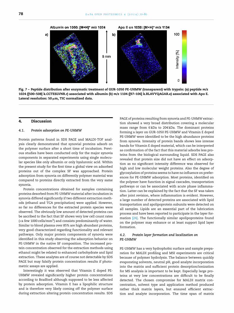
Detected peptide signals were again analyzed for theirdistribution on the polymer samples. Similar localization pre-ferences were found as for the respective proteins. Peptidedistribution was particularly correlated with folded PE-UHMWregions, rough areas and sharp edges. Fig. 7 shows the dis-tribution of selected peptide signals associated with albumin(m/z 1024, [500–508] k.CCTESLVNR.r) and apolipoprotein E (m/z1104, [97–106] k.RLAVYQAGAR.e). The list of associated pep-tide fragments (Fig. 1) includes all high abundance proteins
and the majority of low abundance proteins were also foundin SDS PAGE analysis.tides are separately assigned to proteins according tootein A-I, human serum albumin and plasminogen.
78 e u p a o p e n p r o t e o m i c s 4 ( 2 0 1 4 ) 70–80
Fig. 7 – Peptide distribution after enzymatic treatment of GUR-1050 PE-UHMW (transparent) with trypsin: (a) peptide m/z1024 ([500–508] k.CCTESLVNR.r) associated with albumin (b) m/z 1104 ([97–106] k.RLAVYQAGAR.e) associated with Apo E.Lateral resolution: 50 �m, TIC normalized data.
4. Discussion
4.1. Protein adsorption on PE-UHMW
Protein patterns found in SDS PAGE and MALDI-TOF anal-ysis clearly demonstrated that synovial proteins adsorb onthe polymer surface after a short time of incubation. Previ-ous studies have been conducted only for the major synoviacomponents in separated experiments using single molecu-lar species like only albumin or only hyaluronic acid. Withinthe present study for the first time a global view on adsorbedproteins out of the complex SF was approached. Proteinadsorption from synovia on differently polymer material wascompared to proteins directly extracted from the very samesynovia.
Protein concentrations obtained for samples containingproteins desorbed from PE-UHMW material after incubation insynovia differed significantly if two different extraction meth-ods (ethanol and TCA precipitation) were applied. However,so far no differences for the extracted protein species wereobserved. The obviously low amount of detected proteins canbe ascribed to the fact that SF shows very low cell count rates(<a few 1000 cells/mm3) and consists predominantly of water.Similar to blood plasma over 85% are high abundant proteins,very good characterized regarding functionality and relevantpathways. Only major protein components of synovia wereidentified in this study observing the adsorption behavior onPE-UHMW in the native SF composition. The increased pro-tein concentration observed for the extraction methods usingethanol might be related to enhanced carbohydrate and lipidextraction. These analytes are of course not detectable by SDSPAGE but may falsify protein concentration results if photo-metric assays are applied.
Interestingly it was observed that Vitamin E doped PE-UHMW revealed significantly higher protein concentrations
according to Bradford although supposed to be less affectedby protein adsorption. Vitamin E has a lipophilic structureand is therefore very likely coming off the polymer surfaceduring extraction altering protein concentration results. SDSPAGE of proteins resulting from synovia and PE-UHMW extrac-tion showed a very broad distribution covering a molecularmass range from 6 kDa to 204 kDa. The dominant proteinsforming a layer on GUR-1050 PE-UHMW and Vitamin E dopedPE-UHMW were identified to be the high abundance proteinsfrom synovia. Intensity of protein bands shows less intensebands for Vitamin E doped material, which can be interpretedas confirmation of the fact that this material adsorbs less pro-teins from the biological surrounding liquid. SDS PAGE alsorevealed that protein size did not have an effect on adsorp-tion as no significant intensity difference was observed forhigh and low molecular weight proteins. Also the degree ofglycosylation of proteins seems to have no influence on prefer-ences for PE-UHMW adsorption. Most proteins, identified onthe polymer have function in signal cascades, transportationpathways or can be associated with acute phase inflamma-tion. Latter can be explained by the fact that the SF was takenafter joint revision, where inflammation is evident. However,a large number of detected proteins are associated with lipidtransportation and apolipoprotein subunits were detected onall samples. Lipids are an essential part of the lubricationprocess and have been reported to participate in the layer for-mation [24]. The functionally similar apolipoproteins foundon the polymer may enhance or at least support lipid layerformation.
4.2. Protein layer formation and localization onPE-UHMW
PE-UHMW has a very hydrophobic surface and sample prepa-ration for MALDI profiling and MSI experiments are criticalbecause of polymer hydrolysis. The balance between quicklyevaporating solvents, neutral pH, good analyte incorporationinto the matrix and sufficient protein desorption/ionizationfor MS analysis is important to be kept. Especially large pro-teins at very low concentrations are difficult to be finally
detected. The chosen compromise for MALDI matrix con-centration, solvent type and application method producedrather thick matrix layers, but ensured efficient extrac-tion and analyte incorporation. The time span of matrix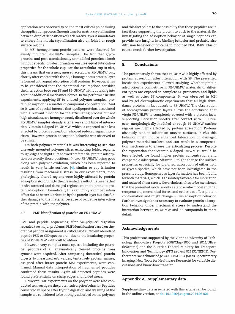
m i c s
atbts
epwptsittaetswhPtasb
urtarrpaiteto
4
Propt
tsdaficf
dcs
Appendix A. Supplementary data
e u p a o p e n p r o t e o
pplication was observed to be the most critical point duringhe application process. Enough time for matrix crystallizationetween droplet depositions of each matrix layer is mandatoryo ensure fine matrix crystallization also on folded or roughurface regions.
In MSI homogeneous protein patterns were observed forvenly mounted PE-UHMW samples. The fact that glyco-roteins and post-translationally unmodified proteins adsorbithout specific cluster formation ensures equal lubricationroperties for the whole cup. For the acetabular cup in vivo,his means that on a new, unused acetabular PE-UHMW cup,hortly after contact with the SF, a homogeneous protein layers formed with equal adsorption of all proteins. However, it haso be considered that the theoretical assumptions considerhe interaction between SF and PE-UHMW without taking intoccount additional mechanical forces. In the performed in vitroxperiments, applying SF to unused polymer samples, pro-ein adsorption is a matter of compound concentration. Ando it was of special interest that apolipoproteins, associatedith a relevant function for the articulating process but notigh abundant, are homogeneously distributed over the wholeE-UHMW samples already after a very short time of interac-ion. Vitamin E doped PE-UHMW, which is expected to be lessffected by protein adsorption, showed reduced signal inten-ities. However, protein adsorption behavior was observed toe similar.
On both polymer materials it was interesting to see thatnevenly mounted polymer slices exhibiting folded regions,ough edges or slight cuts showed preferential protein adsorp-ion on exactly those positions. In vivo PE-UHMW aging goeslong with polymer oxidation, which has been reported toesult in very brittle surfaces [4], similar to cup irritationesulting from mechanical stress. In our experiments, mor-hologically altered regions were highly affected by proteindsorption According to the presented results, this means that
n vivo stressed and damaged regions are more prone to pro-ein adsorption. Theoretically this can imply a compensationffect due to better lubrication by the protein layer but also fur-her damage to the material because of oxidative interactionf the protein with the polymer.
.3. PMF identification of proteins on PE-UHMW
MF and peptide sequencing after “on-polymer” digestionevealed two major problems: PMF identification based on the-retical peptide assignment is critical and sufficient abundanteptide PSD or CID spectra are – due to the insulating proper-ies of PE-UHMW – difficult to obtain.
However, very complex mass spectra including the poten-ial peptides of all enzymatically cleaved proteins fromynovia were acquired. After comparing theoretical proteinigests to measured m/z values, tentatively protein names,ssigned after intact protein MSI experiments, were con-rmed. Manual data interpretation of fragmented peptidesonfirmed those results. Again all detected peptides wereound preferentially on sharp edges and folded areas.
However, PMF experiments on the polymer were also con-ucted to investigate the protein adsorption behavior. Peptidesonserved in space after tryptic digestion and washing of theample are considered to be strongly adsorbed on the polymer
4 ( 2 0 1 4 ) 70–80 79
and this fact points to the possibility that these peptides are infact those supporting the protein to stick to the material. So,investigating the adsorption behavior of single peptides canprovide new insights into binding behavior and probably alsodiffusion behavior of proteins to modified PE-UHMW. This ofcourse needs further investigation.
5. Conclusions
The present study shows that PE-UHMW is highly affected byprotein adsorption after interaction with SF. The presentedincubation experiments allowed studying whether proteinadsorption is competitive if PE-UHMW materials of differ-ent types are exposed to complete SF proteomes and lipidsas well as other SF components. It was observed in MSIand by gel electrophoretic experiments that all high abun-dance proteins in fact adsorb to PE-UHMW. The observationof homogeneous protein layers allows the conclusion thatvirgin PE-UHMW is completely covered with a protein layersupporting lubrication shortly after contact with SF. How-ever, morphologically modified or rough PE-UHMW surfaceregions are highly affected by protein adsorption. Proteinsobviously tend to adsorb on uneven surfaces. In vivo thisbehavior might induce enhanced lubrication on damagedpolymer material surfaces and can result in a compensa-tion mechanism to ensure the articulating process. Despitethe expectation that Vitamin E doped UHWM-PE would beless affected, we found higher protein concentrations andcomparable adsorption. Vitamin E might change the surfaceproperties especially for preferred adsorption of either lipidor glycan species, which have not been investigated in thepresent study. Homogeneous layer formation has been foundfor both materials, which is absolutely favorable for lubricationand reduced shear stress. Nevertheless it has to be mentionedthat the presented model is only a static in vitro model and thattemperature, mechanical forces and cell stress affect proteinconformation and might change in vivo adsorption behavior.Further investigation is necessary to evaluate protein adsorp-tion behavior under mechanical stress to understand theinteraction between PE-UHMW and SF compounds in moredetail.
Acknowledgements
This project was supported by the Vienna University of Tech-nology (Innovative Projects 2009/Chip-1000 and 2011/Ultra-fleXtreme) and the Austrian Federal Ministry for Transport,Innovation and Technology (FFG project 826132/GENIE). Fur-thermore we acknowledge COST BM1104 (Mass SpectrometryImaging: New Tools for Healthcare Research) for valuable dis-cussions and know-how transfer.
Supplementary data associated with this article can be found,in the online version, at doi:10.1016/j.euprot.2014.05.001.

o m i c
r
Effect of lipid absorption on wear and compressive
80 e u p a o p e n p r o t e
e f e r e n c e s
[1] Roba M, Naka M, Gautier E, Spencer ND, Crockett R. Theadsorption and lubrication behavior of synovial fluidproteins and glycoproteins on the bearing-surface materialsof hip replacements. Biomaterials 2009;30(11):2072–8.
[2] Brach Del Prever EM, Bistolfi A, Bracco P, Costa L. UHMWPEfor arthroplasty: past or future? J Orthop Traumatol2009;10(1):1–8.
[3] Fiorito S, Goze C, Adrey J, Magrini L, Goalard C, Bernier P.Increase in free radicals on UHMWPE hip prosthesescomponents due to inflamed synovial cell products. JBiomed Mater Res 2001;57(1):35–40.
[4] Besong AA, Hailey JL, Ingham E, Stone M, Wroblewski BM,Fisher J. A study of the combined effects of shelf ageingfollowing irradiation in air and counterface roughness onthe wear of UHMWPE. Biomed Mater Eng 1997;7(1):59–65.
[5] Bracco P, Oral E. Vitamin E-stabilized UHMWPE for total jointimplants: a review. Clin Orthop Relat Res 2011;469(8):2286–93.
[6] Serro AP, Degiampietro K, Colaco R, Saramago B. Adsorptionof albumin and sodium hyaluronate on UHMWPE: a QCM-Dand AFM study. Colloids Surf B Biointerfaces 2010;78(1):1–7.
[7] Hamada Y, Holmlund AB, Kondoh T, Nakaoka K, Sekiya H,Shiobara N, et al. Severity of arthroscopically observedpathology and levels of inflammatory cytokines in thesynovial fluid before and after visually guidedtemporomandibular joint irrigation correlated with theclinical outcome in patients with chronic closed lock. OralSurg Oral Med Oral Pathol Oral Radiol Endod2008;106(3):343–9.
[8] Teloh HA. Clinical pathology of synovial fluid. Ann Clin LabSci 1975;5(4):282–7.
[9] Amstalden van Hove ER, Smith DF, Heeren RM. A concisereview of mass spectrometry imaging. J Chromatogr A2010;1217(25):3946–54.
[10] MacAleese L, Stauber J, Heeren RM. Perspectives for imagingmass spectrometry in the proteomics landscape. Proteomics2009;9(4):819–34.
[11] Murphy RC, Merrill Jr AH. Lipidomics and imaging massspectrometry. Biochim Biophys Acta 2011;1811(11):635–6.
[12] Miura D, Fujimura Y, Wariishi H. In situ metabolomic mass
spectrometry imaging: recent advances and difficulties. JProteomics 2012;75(16):5052–60.[13] Jin ZM, Dowson D, Fisher J. Analysis of fluid film lubricationin artificial hip joint replacements with surfaces of high
s 4 ( 2 0 1 4 ) 70–80
elastic modulus. Proc Inst Mech Eng H 1997;211(3):247–56.
[14] Kingshott P, Andersson G, McArthur SL, Griesser HJ. Surfacemodification and chemical surface analysis of biomaterials.Curr Opin Chem Biol 2011;15(5):667–76.
[15] Meetani MA, Voorhees KJ. MALDI mass spectrometryanalysis of high molecular weight proteins from wholebacterial cells: pretreatment of samples with surfactants. JAm Soc Mass Spectrom 2005;16(9):1422-1426.
[16] Stauber J, MacAleese L, Franck J, Claude E, Snel M, KaletasBK, et al. On-tissue protein identification and imaging byMALDI-ion mobility mass spectrometry. J Am Soc MassSpectrom 2010;21(3):338–47.
[17] Jiao J, Miao A, Zhang X, Cai Y, Lu Y, Zhang Y, Lu H. Realizationof on-tissue protein identification by highly efficient in situdigestion with graphene-immobilized trypsin for MALDIimaging analysis. Analyst 2013;138(6):1645–8.
[18] Franck JAK, Barnes A, Wisztorski M, Salzet M, Fournier I.Improving tissue preparation for matrix-assisted laserdesorption ionization mass spectrometry imaging. Part 1:using microspotting. Anal Chem 2009;81(19):8193–202.
[19] Holle A, Haase A, Kayser M, Hohndorf J. Optimizing UV laserfocus profiles for improved MALDI performance. J MassSpectrom 2006;41(6):705–16.
[20] Bradford MM. A rapid and sensitive method for thequantitation of microgram quantities of protein utilizing theprinciple of protein-dye binding. Anal Biochem1976;72:248–54.
[21] Shevchenko A, Wilm M, Vorm O, Mann M. Massspectrometric sequencing of proteins silver-stainedpolyacrylamide gels. Anal Chem 1996;68(5):850–8.
[22] Perkins DN, Pappin DJ, Creasy DM, Cottrell JS.Probability-based protein identification by searchingsequence databases using mass spectrometry data.Electrophoresis 1999;20(18):3551–67.
[23] Marchetti-Deschmann M, Gahler J, Archodoulaki V, AllmaierG. Ultra-high molecular weight polyethylene (PE-UHMW)joint implants – choosing the most valuable desorptionprocess (i/vMALDI and DESI) for imaging MS. In: 58th ASMSConference on Mass Spectrometry and Allied Topics. SaltLake City, UT, USA: American Society of Mass Spectrometry;2010.
[24] Greenbaum ES, Burroughs BB, Harris WH, Muratoglu OK.
properties of unirradiated and highly crosslinked UHMWPE:an in vitro experimental model. Biomaterials2004;25(18):4479–84.