synthesis and photobiological activity of ru(ii) dyads...
TRANSCRIPT

Synthesis and Photobiological Activity of Ru(II) Dyads Derived fromPyrrole-2-carboxylate ThionoestersDeborah A. Smithen,† Huimin Yin,‡ Michael H. R. Beh,† Marc Hetu,‡ T. Stanley Cameron,†
Sherri A. McFarland,*,‡,§ and Alison Thompson*,†
†Department of Chemistry, Dalhousie University, P.O. Box 15000, Halifax, Nova Scotia B3H 4R2, Canada‡Department of Chemistry, Acadia University, 6 University Avenue, Wolfville, Nova Scotia B4P 2R6, Canada§Department of Chemistry and Biochemistry, University of North Carolina at Greensboro, 301 McIver Street, Greensboro, NorthCarolina 27402, United States
*S Supporting Information
ABSTRACT: The synthesis and characterization of a series ofheteroleptic ruthenium(II) dyads derived from pyrrole-2-carboxylate thionoesters are reported. Ligands bearing aconjugated thiocarbonyl group were found to be more reactivetoward Ru(II) complexation compared to analogous all-oxygenpyrrole-2-carboxylate esters, and salient features of theresulting complexes were determined using X-ray crystallog-raphy, electronic absorption, and NMR spectroscopy. Selectedcomplexes were evaluated for their potential in photobiologicalapplications, whereupon all compounds demonstrated in vitro photodynamic therapy effects in HL-60 and SK-MEL-28 cells,with low nanomolar activities observed, and exhibited some of the largest photocytotoxicity indices to date (>2000). Importantly,the Ru(II) dyads could be activated by relatively soft doses of visible (100 J cm−2, 29 mW cm−2) or red light (100 J cm−2, 34 mWcm−2), which is compatible with therapeutic applications. Some compounds even demonstrated up to five-fold selectivity formalignant cells over noncancerous cells. These complexes were also shown to photocleave, and in some cases unwind, DNA incell-free experiments. Thus, this new class of Ru(II) dyads has the capacity to interact with and damage biologicalmacromolecules in the cell, making them attractive agents for photodynamic therapy.
■ INTRODUCTION
The biological activity of transition-metal complexes (TMCs)has emerged as a major research focus in recent decades,particularly as metal-based scaffolds can offer significantadvantages over organic compounds with regard to therapeuticand diagnostic applications.1 Investigations concerning TMCsas anticancer agents are increasingly prominent,2 with platinum-and ruthenium-based coordination complexes being the mostwidely studied.3,4 In fact a large body of work exists regardingcytotoxic metal-based anticancer compounds, which is tooexhaustive to cover here.5−7 Certain Ru complexes, namely,NAMI-A, KP1019, and its sodium salt IT-139 (formerly calledNKP-1339), have been investigated in clinical trials as single-agent, cytotoxic, or anti-metastatic alternatives to Pt-deriveddrugs.8 Indeed, while NAMI-A and KP1019 fell short ofexpectations in Phase I studies, IT-139 exhibited a manageablesafety profile alongside promising single-agent anticanceractivity and is currently under development as a multimodalanticancer drug. Unlike their Pt counterparts, Ru complexesfeature three-dimensional chiral cations that can be designed toexhibit desirable aqueous solubility. Their modular architec-tures enable facile chemical modifications through derivatiza-tion of one or more ligands to sample endless molecular andchemical space with electronic properties of biological
relevance. In addition, the expanded octahedral coordinationenvironment of Ru affords access to a larger number ofgeometric isomers and stereoisomers for increased sitediscrimination toward biological targets, as well as to multipleoxidation states for in vivo activation.9,10
Ru compounds have also been extensively investigated aslight-responsive prodrugs for photodynamic therapy(PDT).10−14 Briefly, PDT employs a nontoxic photosensitizer(PS) that is triggered by light to generate cytotoxic reactiveoxygen species (ROS), notably singlet oxygen (1O2).
15,16 Theadvantage of PDT over traditional forms of cancer therapy (i.e.,chemotherapy and radiotherapy), and more recently immuno-therapy, is that it is highly selective, with toxicity confined totissue where PS, light, and oxygen overlap spatiotemporally. Inother words, off-site toxicity can be minimized by judiciouscontrol of the light delivery. Moreover, PDT is also known toinvoke innate and adaptive antitumor immunity in addition todestruction of primary tumors and tumor vasculature.17−20
Despite its potential, PDT is limited by the poor chemicalcharacteristics of the few approved clinical PSs to date.18,19
These PSs are organic structures that require molecular oxygen
Received: January 16, 2017Published: March 16, 2017
Article
pubs.acs.org/IC
© 2017 American Chemical Society 4121 DOI: 10.1021/acs.inorgchem.7b00072Inorg. Chem. 2017, 56, 4121−4132

to exert a cytotoxic effect, thus precluding the treatment ofhypoxic tissue, and cannot be activated by wavelengths of lightthat penetrate tissue best (700−900 nm).Ru complexes have the potential to overcome these
drawbacks. One such compound (TLD1433)14 and itsproprietary light device has entered a Phase 1/2a clinical trialfor treating nonmuscle invasive bladder cancer with PDT(ClinicalTrials.gov Identifier: NCT03053635). This Ru-basedcompound belongs to a class of PSs called metal−organicdyads, which contain π-expansive organic chromophorestethered to neutral 2,2′-bipyridine (bpy), 1,10-phenanthroline(phen), or imidazo[4,5-f ][1,10]phenanthroline (IP) ligands.These Ru dyads are characterized by low-lying tripletintraligand (3IL) excited states with prolonged intrinsiclifetimes (tens to hundreds of microseconds) that are extremelysensitive to trace oxygen and other quenchers, yielding verypotent PDT effects even at low oxygen tension, as well asoxygen-independent excited-state reactivity.14,21−23 These andother Ru-based PSs investigated as PDT agents have mostlyemployed neutral diimines as auxiliary ligands with absorptionmaxima less than 500 nm but can be activated effectively withred light, most likely due to direct triplet−triplet absorptionthat populates these highly effective excited states.There has been some interest in cyclometalated Ru
complexes for PDT, because they exhibit bathochromic shiftsin their longest wavelength absorption maxima by more than100 nm relative to their diimine counterparts.24−26 However,the lower-energy metal-to-ligand charge transfer (MLCT)transitions that are responsible for the longer wavelengthabsorption by cyclometalated Ru complexes also produceexcited-state lifetimes that are much shorter due to the energygap law, which limits the time available for bimolecular excited-state reactions that would be operative in PDT. In addition,some cyclometalated Ru systems were shown to be darkcytotoxic toward various cancer cell lines.27 Presumably, thesetwo factors are responsible for some of the very small in vitroPDT effects reported24 and the perception that such systemsare less useful for PDT. More recently, we demonstrated thatsome cyclometalated Ru C^N complexes derived fromdeprotonated phenylpyridine (phpy−) and π-expansive organicchromophores yield in vitro PDT profiles that are as good as(or better than) some of our best Ru polypyridyl complexes.21
This finding sparked an interest in cyclometalated systemsinvolving the widely studied organometallic C^N motif andother anionic ligands such as the pyrrolide anion.The organometallic chemistry of the pyrrolide anion, though
formally isoelectronic with and geometrically comparable to thewell-established cyclopentadienyl ligand,28,29 is considerablyunderdeveloped.30,31 This lack of interest may stem fromhistorical reports that describe pyrrolide−metal complexes asintrinsically unstable and difficult to handle.32,33 Since then,numerous pyrrolide-containing metal complexes have beenreported: the ligands are often di- and cyclic tetrapyrroles asopposed to the simple pyrrolic system, in which the pyrrolideanion may adopt both σ- and π-bonding modes, thus enhancingthe chemical reactivity of the metal and providing it withsignificant steric and electronic flexibility.34,35 We recentlyreported the synthesis of the first heteroleptic pyrrolide 2,2′-bpy complexes of Ru(II).36 These complexes, formed viachelation of the metal center to the pyrrolide N-atom as well asthe oxygen atom of the carbonyl moiety of 2-formyl, 2-keto,and 2-carboxylato pyrroles, were found to be air- and moisture-stable, and were synthesized in excellent yields in all but the
latter case, whereby attempts garnered success only in the caseof electron-deficient pyrroles, specifically, those bearing halo-substituents about the pyrrole ring. This was thought to be theresult of the electron-withdrawing ester moiety having adestabilizing effect on the Ru−O bond, a trend also observedin a study concerning rhenium complexes of pyrrolideligands.37 Cognizant that thiocarbonyl compounds typicallydisplay greater reactivity than their all-oxygen counterparts,likely due to the larger covalent radius and thus higherpolarizability of the sulfur atom relative to oxygen, wehypothesized that conjugated pyrrolic thionoesters would actas improved ligands for Ru complexation. Herein, we report thesynthesis and characterization of a family of these Ru(II)pyrrolic thionoester dyads and explore the potential of suchcomplexes to act as PSs for PDT.
■ RESULTS AND DISCUSSIONSynthesis and Characterization. Initial efforts sought to
confirm previous findings that 2-carboxylate pyrroles, in theabsence of additional electron-withdrawing substituents, act aspoor ligands for Ru complexation.36 Using slightly modifiedconditions, we thus explored the reaction of a simple trialkyl-substituted pyrrolic ester (1a), obtained via Knorr-typecondensation,38 with Ru(bpy)2Cl2 under microwave irradiation.With the aim of providing stable, highly crystalline Ru(II) dyadsthat were soluble in organic solvents and thus facile tocharacterize, the bis(bpy) Ru(II) chloride salt 2a•Cl, generatedin situ, was converted to the hexafluorophosphate salt upontreatment with NH4PF6. The resulting bis(bpy) Ru(II) salt2a•PF6 was isolated, albeit in low yield, following purificationvia column chromatography (20%, Scheme 1). While low, this
yield was a marked improvement on previous attempts36 tocomplex alkyl 2-carboxylate pyrroles to Ru, an enhancementthat was attributed to the modified isolation procedure thatavoided trituration of the crude complex salt. It is alsointeresting to note that these low yields are in stark contrast tothose obtained for trialkyl-substituted 2-formyl or keto pyrroles,which were shown to be highly effective ligands for Rucomplexation.36
Complexation of the analogous thionoester (3a)39 wassubsequently examined. Vastly superior yields for chelation toRu(II) emerged, as appreciated through comparison ofcomplexation yields for 1a and 3a. Following the proceduredescribed above, 4a•PF6 was isolated in quantitative yield(Scheme 1). To the best of our knowledge, this is the firstreported example of a metal complex featuring a pyrrolideligand chelated through bidentate coordination involving thesulfur moiety of a pyrrole-2-carboxylate thionoester. In additionto the difference in isolated yields attained (Scheme 1), thepyrrolic ester- and thionoester-derived complexes (2a and 4a)also differed in appearance with the former observed to be deep
Scheme 1. Synthesis of Ru(II) Complexes Derived from 2-Carboxylate (1a) and 2-Thiocarboxylate (3a) Pyrroles,Isolated as Their Hexafluorophosphate Salts
Inorganic Chemistry Article
DOI: 10.1021/acs.inorgchem.7b00072Inorg. Chem. 2017, 56, 4121−4132
4122

purple in the solid state and the latter deep red, with thevariation more apparent in solution, whereupon the colors weremore vibrant.Subtle differences were also observed in the NMR spectra of
these two dyads. Formation of heteroleptic [Ru(bpy)2(LL)]2+
complexes produces nonequivalence in all protons, leading tohighly complex 1H and 13C NMR spectra, particularly in thearomatic regions. However, analysis of the alkyl-derived peaksin the 1H NMR spectra revealed that replacing the carbonylgroup of 2a with a thiocarbonyl group (4a) resulted in adeshielding effect upon nearby O-CH2CH3 protons (Table 1,
entries 4 and 5), yet did not significantly affect the environmentof the pyrrolic methyl substituents (Table 1, entries 1−3), atrend also observed for the respective ligands (1a and 3a).Examination of the 13C NMR spectra revealed a similar
deshielding effect on the C = X group when moving from acarbonyl to a thiocarbonyl group (Table 1, entry 6). In the caseof ligands 1a and 3a, the difference in chemical shift was asexpected based on theoretical studies that suggest a linearrelationship between 13CO and their corresponding 13CSvalues, conforming reasonably well to the equation δ(CS) =1.75 δ(CO) − 79.7.41 Employing this equation provides a
calculated value of 203.6 ppm for the CS group of 3a,comparable to the experimental value of 199.5 ppm.Conversely, in the case of Ru(II) complexes 2a and 4a theexperimental value of 194.7 ppm for the CS group of 4a didnot compare well with the calculated value of 222.7 ppm,suggesting that Ru complexation mitigates the deshieldingeffect.
X-ray Structure. An X-ray crystal structure was obtained forthe complex 4a, as the racemate, which confirmed the bindingmode of the ligand and enabled structural analysis. Slowevaporation of a solution of 4a in methanol generated dark redcrystals that were suitable for analysis via X-ray diffraction.Solving the structure revealed that complex 4a crystallizes inthe monoclinic space group C2/c, with the Ru(II) centeradopting a distorted octahedral geometry (Figure 1a).The four Ru−Nbpy bonds are in the range of 2.048(3)−
2.070(2) Å, well within normal limits compared to the parentRu(II) tris(2,2′-bpy) complex42 and similar heteroleptic Ru(II)bis(2,2′-bpy) dipyrrinato43,44 or pyrrolide36 complexes. TheRu−Npyr bond length for 4a is 2.091(2) Å, marginally longerthan those found in similar ruthenium−pyrrole complexesbearing α-formyl (CHO) or ester (C(O)OR) groups in placeof the thionoester (C(S)OR) moiety (Figures 1b, 5, and 6).36
The Ru−Npyr bonds of known pyrrolide and dipyrrinatoderivatives are reported in the range of 2.076(2)−2.087(3) Å.The C5−S bond of complex 4a (1.700(3) Å) is similar in lengthto that of uncoordinated pyrrole 7 (1.650(2) Å),45 but it is∼0.4 Å longer than the carbonyl C5−O bond of ester (5) andformyl (6) derivatives. The Ru−S bond of 4a is also longer(2.365(1) Å) than the Ru−O bond of analogous 2-carboxylatepyrrole complex 5 (2.128(1) Å), which is again longer than thatof the 2-formyl pyrrole−ruthenium complex 6 (2.097(2) Å).The bond length from C1 to C5 in compound 4a, formally aC−C single bond, is 1.396(5) Å, making it slightly shorter thanthat of uncoordinated pyrrole 7 (1.432(2) Å), which wouldsuggest an increase in double-bond character as would betypical for the azafulvenium resonance form of the pyrrolideligand. The Npyr−Ru−S bond angle is 82.64(8)°, which is largerthan the Nbpy−Ru-Nbipy bond angles in complex 4a (78.2(1)°and 79.0(1)°), akin to the bond angles in the ester and formylderivatives (5 and 6). However, the Ru−S−C5 bond angle(98.9(1)°) is smaller than the Ru−O−C5 bond angle of theester and formyl derivatives (5 and 6).Following the success of initial efforts in confirming our
hypothesis that pyrrole-2-thionoesters give higher yields for
Table 1. Comparison of NMR Chemical Shifts of PyrroleSubstituents in Complexes 2a and 4a, and Their RespectiveLigands
NMR chemical shift (ppm)a
liganda Ru(II) complexb
entry group 1a40 3a 2a 4a1c A (CH3) 2.18 2.19 1.14 1.192c B (CH3) 1.91 1.91 1.82 1.783c C (CH3) 2.25 2.25 2.19 2.244c D (CH2) 4.29 4.66 4.3−4.1 4.6−4.45c E (CH3) 1.34 1.46 1.17 1.426d F (CX) 161.9 199.5 172.8 194.7
aSolutions in CDCl3.bSpectra recorded for PF6 salts using CD2Cl2 as
solvent. cChemical shifts (1H). dChemical shifts (13C).
Figure 1. (a) X-ray structure of complex 4a (50% probability ellipsoids) with PF6− counterion. (b) Related literature compounds with published
solved crystal structures.
Inorganic Chemistry Article
DOI: 10.1021/acs.inorgchem.7b00072Inorg. Chem. 2017, 56, 4121−4132
4123

complexation to Ru(II) compared to their all-oxygen analogues,we examined a series of pyrrolic thionoesters in thecomplexation reaction, to assess the substrate scope. Startingwith the alkyl 2-carboxylate pyrroles (1), the correspondingthionocarbonyl pyrroles (3) were prepared in good yield aftertreatment with Lawesson’s reagent at elevated temperature39
(Table 2).The thionoester-bearing pyrroles (3) were then examined as
ligands in the microwave-assisted Ru(II) complexation reactionas per Scheme 1, providing the corresponding PF6 salts inexcellent yields (84%-quant, Table 2). This demonstrates thatthe complexation reaction is tolerant of a wide variety ofstructural features, including both ethyl and benzyl thionoesters(compare Table 2, entries 1 and 2; Table 2, entries 3 and 4;Table 2, entries 5 and 6), α- and β-free pyrroles (Table 2,entries 3, 4 and 7), long-chain alkyl substituents (Table 2,entries 8 and 9), electron-withdrawing substituents (Table 2,entry 10), and aryl substituents (Table 2, entry 11). In the caseof the Ru(II) complex 4k a notably lower yield was obtained(84%, Table 2, entry 11), which can presumably be ascribed toelectronic effects, owing to the incorporation of a conjugatedaryl substituent, which may contribute to a destabilizing effecton the Ru−S bond. All complexes synthesized (4a−4k) werefully characterized and moisture- and air-stable.Electronic Absorption. As expected, the longest wave-
length absorption maxima for these cyclometalated systemswere shifted by up to 100 nm relative to typical Ru(II)complexes derived from neutral diimine-based ligands,46 andtheir extinction coefficients were very similar to that ofPhotofrin47 at wavelengths where clinical PDT is currentlydelivered (∼630 nm). The absorption spectra of the Ru(II)complexes featuring pyrrolide ligands 2-substituted with ester(2a) and thionoester (4a−k) moieties revealed intense bandsin the UV, characteristic of internal π−π* ligand-centeredtransitions, and lower-energy MLCT transitions in the visibleregion.Electronic absorption spectra for the ester-coordinated
complex 2a and the analogous thionoester complexes (4)revealed significant differences in the absorption profiles(Figure 2). Most notably, thionoester-containing complex 4a
exhibits increased molar absorptivity, relative to the corre-sponding ester 2a, in the high-energy MLCT transitionsdesignated RuII(dπ) → LL parentage (320−400 nm);48 in theband centered around 430 nm attributed to the n−π* transitionof the thiocarbonyl group49 and, to a lesser extent, the lower-energy MLCT bands (≥575 nm) that can be assigned tooverlapping Ru(dπ) → bpy(π*) (symmetric) and RuII(dπ) →bpy(π*) (antisymmetric) transitions.48,50,51 However, thebathrochromic shift of ∼25 nm in the longest wavelengthRu(dπ) → bpy(π*) absorption maximum for 2a producedslightly larger extinction coefficients between 550 and 575 nm.Otherwise the local maxima were similar for the ester andthionoester congeners. Comparison of the absorption spectraobtained for Ru(II) complexes featuring ethyl (4a) and benzyl(4b) thionoester pyrrolide ligands showed no significantdifferences between the two. Comparing complexes withpyrrolides bearing β-alkyl (4b) and aryl (4k) substituentsrevealed similar profiles, although generally higher absorptivitywas observed in the latter case (Figure 2).
Photobiology. Six of the Ru(II) complexes featuring 2-thionoester pyrrolide ligands were selected for photobiologicalstudies (4a−c, 4h, 4j,k); these samples represent both ethyland benzyl thionoesters, and bear distinct structural features
Table 2. Synthesis of Pyrroles (3) and Corresponding Ru(II) Complexes (4)
entry pyrrole R1 R2 R3 R4 yield 3 (%)a yield 4 (%)a,b
1 1a Me Me Me Et 72 (3a) quant (4a)2 1b Me Me Me Bn 49 (3b) quant (4b)3 1c Me H Me Et 72 (3c) quant (4c)4 1d Me H Me Bn 50 (3d) quant (4d)5 1e Me Me Et Et 59 (3e) quant (4e)6 1f Me Me Et Bn 47 (3f) 93 (4f)7 1g H Me Me Et 58 (3g) quant (4g)8 1h Me (CH2)4CH3 Me Et 64 (3h) quant (4h)9 1i Me (CH2)2CH3 (CH2)2CH3 Et 68 (3i) quant (4i)10 1j Me CO2Et Me Et 52 (3j)c quant (4j)11 1k Me Ph Me Bn 55 (3k)c 84 (4k)
aIsolated yield. bCompounds isolated as their PF6 salts.cReaction time of 4 h.
Figure 2. UV−vis spectra comparing the absorption profiles of ester(2) and thionoester (4) pyrrolide Ru(II)·PF6 complexes in CH2Cl2,with labeled transitions.
Inorganic Chemistry Article
DOI: 10.1021/acs.inorgchem.7b00072Inorg. Chem. 2017, 56, 4121−4132
4124

including H, alkyl, ester, and aryl substituents. The hexa-fluorophosphate salts of these complexes were converted totheir biologically compatible, water-soluble chloride salts viatreatment with excess tetrabutylammonium chloride, followedby purification using column chromatography (90%-quantyield, characterization details in Supporting Information).Successful counterion exchange was confirmed using electro-spray ionization mass spectrometry (ESI-MS) in negative ionmode, which showed a disappearance of the signal correspond-ing to the PF6
− anion. Four compounds have Me at both R1
and R3, and Et at R4, and differ only at R2: H (4c), Me (4a),−(CH2)4CH3 (4h), or −CO2Et (4j). Two are identical at R1
and R3 (Me) and differ only at R4: Et (4a) or Bn (4b). Anothertwo are also identical at R1 and R3 (Me) and at R4 (Bn),differing only at R2: Me (4b) or Ph (4k). This small subset ofcompounds was chosen to determine the structure−activityeffects with systematic changes to R2 and R4 and to highlightthe utility of this new class of cyclometalated Ru(II) dyads forin vitro PDT (and, in some cases, as traditional but selectiveanticancer compounds).The cytotoxicity and photocytotoxicity of these representa-
tive 2-thionoester pyrrolide Ru(II) dyads was assessed in threehuman cell lines (HL-60 promyelocytic leukemia cells, SK-MEL-28 melanoma cells, and CCD−1064Sk skin fibroblasts)according to an established in-house cellular assay.9,52 Briefly,cells were dosed with metal complex (1 nM-300 μM) andincubated for 16 h at 37 °C prior to a light or sham (dark)treatment. The light treatment was 100 J cm−2 delivered from abroadband visible lamp (34 mW cm−2) or red light-emittingdiodes (LEDs) at 625 nm (29 mW cm−2) over the course of 49and 57 min, respectively. The complexes, as monitored by UV−vis absorption spectroscopy, exhibited no photobleaching uponexposure to 100 J cm−2 of visible or red light delivered at an
irradiance of 34 and 29 mW cm−2, respectively. Alamar Bluewas added at 48 h post treatment, and cell viability wasquantified 16 h later. Cells that were dosed at highconcentrations of PS, where the PS interferes with absorptionand emission of light by the cell viability dye, were alsoobserved manually under a microscope. The effectiveness of theRu(II) complexes as in vitro PDT agents was assessed byquantifying the dark and light cytotoxicity profiles as EC50values (concentration required to reduce cell viability to 50%).Their photocytotoxicity indices (PIs) were then calculated asthe ratio of dark to light EC50 values, reflecting the PDTtherapeutic margin in a given cancer cell line. Dark EC50 valueswere also assessed using noncancerous skin fibroblasts (CCD−1064Sk) to determine any selective dark cytotoxicity towardcancer cells over normal cells. The selectivity factor (SF) isdefined as the dark EC50 determined for CCD−1064Sk skinfibroblasts divided by the dark EC50 determined for a givencancer cell line. For comparison, larger PIs signify larger PDTeffects, and SF values > 1 represent selectivity toward cancercells. Note that, as long as the therapeutic dose is significantlyless than the dark EC50 value of the normal cell line, it is notnecessary to have inherent selectivity for cancer cells overnormal cells. Rather, selectivity is achieved by spatial control ofthe light delivery.As observed for some of the other classes of cyclometalated
Ru(II) complexes, the in vitro cytotoxicity for the Ru(II) dyadsderived from 2-thionoester pyrrolide ligands was high, withEC50 values as low as 280 nM in the absence of a light trigger.This cytotoxicity proved to be very sensitive to the substitutionpattern about the pyrrole ring, and to the type of thionoester, intwo of the three cell lines investigated. Dark EC50 values forHL-60, SK-MEL-28, and CCD−1064Sk cells ranged from 1.1−1.7, 0.28−2.2, and 0.35−6.3 μM, respectively (Tables 3, 4, and
Table 3. Photobiological Activity of Selected 2-Thionoester Pyrrolide Ru(II) Complexes in HL-60 Cells, with PS-to-LightInterval of 16 h
EC50 (μM) EC50 (μM)
compounda dark visibleb PIc redb PIc SFd
4a 1.08 ± 0.03 0.108 ± 0.004 10 0.357 ± 0.014 3 0.44b 1.17 ± 0.07 0.012 ± 0.001 98 0.145 ± 0.003 8 2.04c 1.23 ± 0.40 0.161 ± 0.011 8 0.355 ± 0.008 3 5.14h 1.74 ± 0.07 0.042 ± 0.002 41 0.102 ± 0.016 17 1.44j 1.43 ± 0.06 0.076 ± 0.018 19 0.213 ± 0.005 7 2.94k 1.42 ± 0.07 0.014 ± 0.001 101 0.052 ± 0.003 27 0.2cisplatine 5.9 ± 0.1
aCompounds screened as their chloride salts. bLight 100 J cm−2. cPI = phototherapeutic index. dSF = The ratio of dark EC50 values of CCD-1064Skand HL-60 cells. eCisplatin is not a PS, but it serves as a control.
Table 4. Photobiological Activity of Selected 2-Thionoester Pyrrolide Ru(II) Complexes in SK-MEL-28 Cells, with PS-to-LightInterval of 16 h
EC50 (μM) EC50 (μM)
compounda dark visibleb PIc redb PIc SFe
4a 0.280 ± 0.028 0.004 ± 0.001 70 0.070 ± 0.007 4 1.44b 2.19 ± 0.19 0.001 ± 0.0001 2185 0.036 ± 0.002 61 1.14c 2.01 ± 0.17 0.014 ± 0.0002 144 0.141 ± 0.015 14 3.14h 1.09 ± 0.03 0.011 ± 0.002 99 0.040 ± 0.002 27 2.24j 2.07 ± 0.12 0.004 ± 0.001 518 0.211 ± 0.027 10 2.04k 0.281 ± 0.036 0.010 ± 0.0002 28 0.038 ± 0.004 7 1.2cisplatind 2.8 ± 0.1
aCompounds screened as their chloride salts. bLight 100 J cm−2. cPI = phototherapeutic index. dCisplatin is not a photosensitizer, but it serves as acontrol. eSF = The ratio of dark EC50 values of CCD-1064Sk and SK-MEL-28 cells.
Inorganic Chemistry Article
DOI: 10.1021/acs.inorgchem.7b00072Inorg. Chem. 2017, 56, 4121−4132
4125

S1). In SK-MEL-28 and CCD−1064Sk cells, where there was amuch larger range of activities, 4a and 4k were the most
cytotoxic, and 4c and 4j were the least cytotoxic (by 18-fold inCCD−1064Sk cells and 8-fold in SK-MEL-28 cells). Given that
Figure 3. In vitro cytotoxicity curves for compounds 4a−c, 4h, 4j, and 4k in HL-60, SK-MEL-28, and CCD-1064Sk cells.
Figure 4. In vitro PDT dose−response curves for compounds 4a−c, 4h, 4j, and 4k in HL-60 cells, with visible (blue), red (red), or no (black) lightactivation.
Inorganic Chemistry Article
DOI: 10.1021/acs.inorgchem.7b00072Inorg. Chem. 2017, 56, 4121−4132
4126

4a (R2 = Me) was one of the most cytotoxic compounds and 4c(R2 = H) and 4j (R2 = −CO2Et) were the least dark toxic, thecytotoxicity is very sensitive to changes at R2 when R4 is Et.This is also the case when R4 = Bn, supported by largedifferences between the cytotoxicities of 4b and 4k, with 4kbeing more toxic by almost eight-fold. For the ethylthionoesters, R2 = Me produced potent dark toxicity (EC50 =280 nM), while R2 = H, −(CH2)4CH3, or −CO2Et were lesscytotoxic by ∼10-fold. However, the presence of R2 = Me in thebenzyl thionoesters (EC50 = 2.2 μM) did not lead to nanomolartoxicity, yet R2 = Ph did. The conclusion is that the nature ofthe substituent at R4 influences the cytotoxic effects due tovariances at R2, and vice versa.Notably, 4c was up to 5 times more cytotoxic toward cancer
cells relative to normal cells (SF = 5.1, HL-60), and 4j was alsoselectively cytotoxic toward cancer cells but to a lesser extent(Figure 3). These ethyl thionoester complexes bearing R2 = Hor R2 = −CO2Et were also the least cytotoxic in general. Thisselectivity, in addition to the observation that all of thecompounds exhibit anticancer effects that surpass the goldstandard cisplatin in both cancer cell lines studied (Tables 3and 4), highlights the potential utility of these Ru(II) 2-thionoester pyrrolide dyads as traditional chemotherapeutics.While the six complexes investigated acted as promising
anticancer agents in the absence of a light trigger, thiscytotoxicity was enhanced further with photoactivation usingbroadband visible or red light (Figures 4 and 5). The moreenergetic visible light produced smaller EC50 values for all ofthe complexes across all of the cell lines studied, but even redlight produced PIs as large as 27 (4k) in HL-60 cells andgreater than 60 (4b) in SK-MEL-28 cells (Tables 3 and 4).Both of these PI values are larger than that known for Photofrin
(PI ≈ 10), albeit reported in a different cell line.53 The relativeordering of the light EC50 values measured for the differentcompounds varied between the two cancer cell lines studiedand between the two light conditions used, underscoring theimportance of being cautious with generalizations regarding PSactivity and structure−activity relationships. Nevertheless, sometrends could be discerned.With visible light activation, 4b was exceptionally potent
toward both cancer cell lines, with light EC50 values of 12 nMin HL-60 and 1 nM in SK-MEL-28, giving rise to PIs of 100and greater than 2100, respectively (Tables 3 and 4). Theselarge phototherapeutic margins afford the opportunity todeliver the PS at very low concentration, where it is completelynontoxic without the light trigger. One of the least active PSs inboth cell lines with visible light activation was 4c (EC50 = 161nM, PI 8 in HL-60; EC50 = 14 nM, PI = 144 in SK-MEL-28),demonstrating that the least phototoxic PS in this series is stillup to 300-fold more phototoxic than Photofrin. The trends forlight potencies were as follows: 4b ≈ 4k > 4h > 4j > 4a > 4c(HL-60/visible PDT); 4k > 4h > 4b > 4j > 4c ≈ 4a (HL-60/red PDT); 4b > 4a = 4j > 4k ≈ 4h > 4c (SK-MEL-28/visiblePDT); and 4b ≈ 4k ≈ 4h > 4a > 4c > 4j (SK-MEL-28/redPDT). The trends for the PI values were: 4k ≈ 4b > 4h > 4j >4a > 4c (HL-60/visible PDT); 4k > 4h > 4b ≈ 4j > 4a = 4c(HL-60/red PDT); 4b > 4j > 4c > 4h > 4a > 4k (SK-MEL-28/visible PDT); and 4b > 4h > 4c > 4j > 4k > 4a (SK-MEL-28/red PDT).The benzyl thionoesters (4b and 4k) yielded the most potent
visible light EC50 values and largest PIs in HL-60 cells. Whilebenzyl thionoester 4b also gave the most potent visible lightEC50 values and largest PI in SK-MEL-28 cells, ethylthionoester 4j was also very potent. However, with red light
Figure 5. In vitro PDT dose−response curves for compounds 4a−c, 4h, 4j, and 4k in SK-MEL-28 cells, with visible (blue), red (red), or no (black)light activation.
Inorganic Chemistry Article
DOI: 10.1021/acs.inorgchem.7b00072Inorg. Chem. 2017, 56, 4121−4132
4127
activation in HL60 cells, ethyl thionoester 4h surpassed 4b.Close scrutiny revealed that the light-triggered activities ofthese Ru(II) dyads were sensitive to the cell line employed andthe light treatment delivered. Nevertheless, 4b, 4j, and 4hemerged as good in vitro PDT agents with low nanomolarpotencies and highlight the potential utility of both ethyl andbenzyl thionoester Ru(II) dyads for PDT applications given theability to fine-tune the photocytotoxicity via substituentchanges about the pyrrole ring. Current efforts are underwayto understand the photophysical and photochemical differencesthat may give rise to these differences in photobiologicalactivity, and to explore additional variations at R2 and R4.A possible source of the observed in vitro PDT effects is the
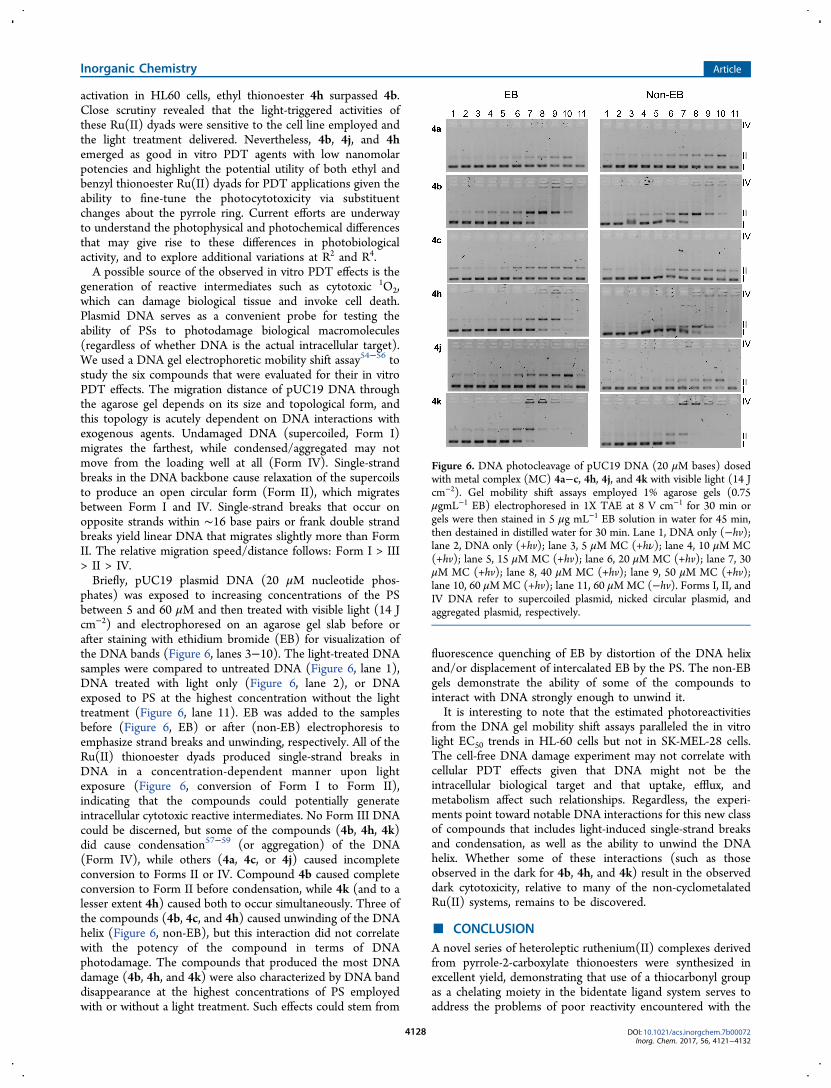
generation of reactive intermediates such as cytotoxic 1O2,which can damage biological tissue and invoke cell death.Plasmid DNA serves as a convenient probe for testing theability of PSs to photodamage biological macromolecules(regardless of whether DNA is the actual intracellular target).We used a DNA gel electrophoretic mobility shift assay54−56 tostudy the six compounds that were evaluated for their in vitroPDT effects. The migration distance of pUC19 DNA throughthe agarose gel depends on its size and topological form, andthis topology is acutely dependent on DNA interactions withexogenous agents. Undamaged DNA (supercoiled, Form I)migrates the farthest, while condensed/aggregated may notmove from the loading well at all (Form IV). Single-strandbreaks in the DNA backbone cause relaxation of the supercoilsto produce an open circular form (Form II), which migratesbetween Form I and IV. Single-strand breaks that occur onopposite strands within ∼16 base pairs or frank double strandbreaks yield linear DNA that migrates slightly more than FormII. The relative migration speed/distance follows: Form I > III> II > IV.Briefly, pUC19 plasmid DNA (20 μM nucleotide phos-
phates) was exposed to increasing concentrations of the PSbetween 5 and 60 μM and then treated with visible light (14 Jcm−2) and electrophoresed on an agarose gel slab before orafter staining with ethidium bromide (EB) for visualization ofthe DNA bands (Figure 6, lanes 3−10). The light-treated DNAsamples were compared to untreated DNA (Figure 6, lane 1),DNA treated with light only (Figure 6, lane 2), or DNAexposed to PS at the highest concentration without the lighttreatment (Figure 6, lane 11). EB was added to the samplesbefore (Figure 6, EB) or after (non-EB) electrophoresis toemphasize strand breaks and unwinding, respectively. All of theRu(II) thionoester dyads produced single-strand breaks inDNA in a concentration-dependent manner upon lightexposure (Figure 6, conversion of Form I to Form II),indicating that the compounds could potentially generateintracellular cytotoxic reactive intermediates. No Form III DNAcould be discerned, but some of the compounds (4b, 4h, 4k)did cause condensation57−59 (or aggregation) of the DNA(Form IV), while others (4a, 4c, or 4j) caused incompleteconversion to Forms II or IV. Compound 4b caused completeconversion to Form II before condensation, while 4k (and to alesser extent 4h) caused both to occur simultaneously. Three ofthe compounds (4b, 4c, and 4h) caused unwinding of the DNAhelix (Figure 6, non-EB), but this interaction did not correlatewith the potency of the compound in terms of DNAphotodamage. The compounds that produced the most DNAdamage (4b, 4h, and 4k) were also characterized by DNA banddisappearance at the highest concentrations of PS employedwith or without a light treatment. Such effects could stem from
fluorescence quenching of EB by distortion of the DNA helixand/or displacement of intercalated EB by the PS. The non-EBgels demonstrate the ability of some of the compounds tointeract with DNA strongly enough to unwind it.It is interesting to note that the estimated photoreactivities
from the DNA gel mobility shift assays paralleled the in vitrolight EC50 trends in HL-60 cells but not in SK-MEL-28 cells.The cell-free DNA damage experiment may not correlate withcellular PDT effects given that DNA might not be theintracellular biological target and that uptake, efflux, andmetabolism affect such relationships. Regardless, the experi-ments point toward notable DNA interactions for this new classof compounds that includes light-induced single-strand breaksand condensation, as well as the ability to unwind the DNAhelix. Whether some of these interactions (such as thoseobserved in the dark for 4b, 4h, and 4k) result in the observeddark cytotoxicity, relative to many of the non-cyclometalatedRu(II) systems, remains to be discovered.
■ CONCLUSIONA novel series of heteroleptic ruthenium(II) complexes derivedfrom pyrrole-2-carboxylate thionoesters were synthesized inexcellent yield, demonstrating that use of a thiocarbonyl groupas a chelating moiety in the bidentate ligand system serves toaddress the problems of poor reactivity encountered with the
Figure 6. DNA photocleavage of pUC19 DNA (20 μM bases) dosedwith metal complex (MC) 4a−c, 4h, 4j, and 4k with visible light (14 Jcm−2). Gel mobility shift assays employed 1% agarose gels (0.75μgmL−1 EB) electrophoresed in 1X TAE at 8 V cm−1 for 30 min orgels were then stained in 5 μg mL−1 EB solution in water for 45 min,then destained in distilled water for 30 min. Lane 1, DNA only (−hv);lane 2, DNA only (+hv); lane 3, 5 μM MC (+hν); lane 4, 10 μM MC(+hv); lane 5, 15 μM MC (+hv); lane 6, 20 μM MC (+hv); lane 7, 30μM MC (+hv); lane 8, 40 μM MC (+hv); lane 9, 50 μM MC (+hv);lane 10, 60 μMMC (+hv); lane 11, 60 μMMC (−hv). Forms I, II, andIV DNA refer to supercoiled plasmid, nicked circular plasmid, andaggregated plasmid, respectively.
Inorganic Chemistry Article
DOI: 10.1021/acs.inorgchem.7b00072Inorg. Chem. 2017, 56, 4121−4132
4128

analogous all-oxygen 2-carboxylate pyrroles. All complexessynthesized were characterized using 1H and 13C NMR andUV/vis spectroscopy, and X-ray crystallography was used toconfirm the binding mode and gain structural information.Photobiological activity of selected complexes was assessed inHL-60 and SK-MEL-28 cells, and dark toxicity was furtherprobed in normal skin fibroblasts. All of the compoundsdemonstrated in vitro PDT effects, and some were among themost potent reported to date. The selectivity exhibited by someof these novel Ru(II) dyads toward cancer cells, alongside theirnanomolar activities, may prove useful in the search forchemotherapeutics with a considerably different mechanism ofaction than cisplatin in an effort to overcome the acquired orinnate resistance to platinum-based cancer therapy. The lownanomolar photo-cytotoxicities and the very large photo-therapeutic margins could potentially offset the dark toxicity ofthe complexes, making them both excellent traditionalanticancer compounds and PDT agents. The compounds alsointeracted strongly with DNA in cell-free experiments,indicating that DNA could be an intracellular target, butmore importantly that all of the compounds are capable ofgenerating potentially cytotoxic reactive intermediates for PDT.Dark and light EC50 values and PIs measured for the differentcompounds varied by cell line and by light treatment condition.The absence of systematic trends across all conditionsprecluded a generalized structure−activity assessment buthighlights the rich diversity yet to be exploited in medicinalinorganic chemistry, as we hereby introduce a new class ofcyclometalated Ru(II) dyads as PSs for in vitro PDT andpossibly other applications. Efforts are underway to explore thecellular uptake profiles with and without a light trigger, thephotophysical properties underlying the observed PDT effects,the PDT mechanism(s), and structure−activity relationshipsfor this interesting compound class.
■ EXPERIMENTAL PROCEDURESFull synthesis and characterization data can be found in the SupportingInformation.Bis(2,2′-bipyridyl)-(O-ethyl-3,4,5-trimethyl-2-carbothiolato-
N-pyrrolato)ruthenium(II) (4a). Ru(II) complex 4a was synthesizedfrom ligand 3a using general procedure 2 (see SupportingInformation) and purified over silica, eluting with 0−4% iso-propylalcohol (IPA)/CH2Cl2 to give complex salt 4a·PF6 as a deep redcrystalline solid (116 mg, 100% yield). mp 172−176 °C; 1H NMR(CD2Cl2, 500 MHz) δ 9.34 (d, 1H, J = 5.5 Hz, ArH). 8.29 (at, 3H, J =8.0 Hz, ArH), 8.18 (d, 1H, J = 8.0 Hz, ArH), 7.95−7.91 (m, 3H, ArH),7.85 (t, 1H, J = 7.8 Hz, ArH), 7.77 (t, 1H, J = 7.8 Hz, ArH), 7.65 (d,1H, J = 5.5 Hz, ArH), 7.53−7.49 (m, 2H, ArH), 7.43 (t, 1H, J = 6.5Hz, ArH), 7.22 (t, 1H, J = 6.5 Hz, ArH), 7.11 (t, 1H, J = 6.5 Hz, ArH),4.64−4.58 (m, 1H, CH2CH3), 4.49−4.43 (m, 1H, CH2CH3), 2.24 (s,3H, CH3), 1.78 (s, 3H, CH3), 1.42 (t, 3H, J = 7.0 Hz, CH2CH3), 1.19(s, 3H, CH3) ppm, 13C NMR (CD2Cl2, 125 MHz, udeft) δ 194.7,158.3, 158.1, 158.0, 157.8, 155.6, 155.0, 152.7, 151.6, 150.9, 139.8,136.3, 136.1, 135.5, 135.3, 131.4, 127.13, 127.06, 126.8, 126.7, 124.4,123.6, 123.4, 123.24, 123.18, 67.4, 14.8, 13.4, 11.8, 9.6 ppm; ESI-MS+:610.1 (M)+; high-resolution mass spectrometry (HRMS): 610.1206Found, 610.1209 Calculated for C30H30N5SORu; ESI-MS−: 145.0(PF6)
−; ε523 nm 9100, ε381 nm 13 200, ε340 nm 19 900, ε295 nm 50 600(CH2Cl2). The corresponding chloride salt (4a·Cl) was obtained usingGP3 as a black solid (30 mg, 100%). mp/dp > 200 °C; ESI-MS+: 610.1(M)+; ESI-MS−: PF6
− ion not observed. Crystal data for compound4a·PF6: C30.50H32F6N5O1.50PRuS, MM = 770.72 g/mol. Dark redpinacoid crystal, dimensions 0.35 × 0.27 × 0.18 mm; monoclinic spacegroup, C2/c; a = 21.9599(9) Å, b = 13.8939(4) Å, c = 24.2344(10) Å,α = 90°, β = 115.9710(15)°, γ = 90°, V = 6647.4(4) Å3, Z = 8, d =1.540 g/cm3, μ(Mo Kα) = 6.510 cm−1, 19 764 reflections (7910
unique, Rint = 0.053), R = 0.0605, wR2 = 0.1642, goodness-of-fit =1.043, R-factor = 5.17%. CCDC deposition number: 1485965.
Metal Compound Solutions. Stock solutions of the chloride saltsof the Ru(II) complexes (4·Cl) were prepared at 5 mM in 10%dimethyl sulfoxide (DMSO) in water and kept at −20 °C prior to use.Working dilutions were prepared through dilution of the aqueousstock with pH 7.4 Dulbecco’s phosphate buffered saline (DPBS).DPBS is a balanced salt solution of 1.47 mM potassium phosphatemonobasic, 8.10 mM sodium phosphate dibasic, 2.68 mM potassiumchloride, and 0.137 M sodium chloride (no Ca2+ or Mg2+). DMSO inthe assay wells was under 0.1% at the highest complex concentration.
HL-60 Cell Culture. HL-60 human promyelocytic leukemia cells(ATCC CCL-240) were cultured at 37 °C under 5% CO2 in RPMI1640 (Mediatech Media MT-10−040-CV) supplemented with 20%fetal bovine serum (FBS; PAA Laboratories, A15−701) and werepassaged three to four times per week according to standard asepticprocedures. Cultures were started at 200 000 cells per milliliter in 25cm2 tissue culture flasks and were subcultured when growth reached800 000 cells mL−1 to avoid senescence associated with prolongedhigh cell density. Complete growth medium was prepared in 200 mLportions as needed by combining RPMI 1640 (160 mL) and FBS (40mL, prealiquoted, and heat inactivated) in a 250 mL Millipore vacuumstericup (0.22 μm) and filtering.
SK-MEL-28 Cell Culture. Adherent SK-MEL-28 malignantmelanoma cells (ATCC HTB-72) were cultured in Eagle’s MinimumEssential Medium (EMEM; Mediatech Media MT-10−009-CV)supplemented with 10% FBS and were incubated at 37 °C under5% CO2 and passaged two to three times per week according tostandard aseptic procedures. SK-MEL-28 cells were started at 200 000cells per milliliter in 75 cm2 tissue culture flasks and were subculturedwhen growth reached 550 000 cells per milliliter by removing oldculture medium and rinsing the cell layer once with Dulbecco’sphosphate-buffered saline (DPBS 1X, Mediatech, 21−031-CV)followed by dissociation of cell monolayer with 1X Trypsin−EDTAsolution (0.25% (w/v Trypsin/0.53 mM EDTA, ATCC 30−2101)).Complete growth medium was added to the cell suspension to allowappropriate aliquots of cells to be transferred to new cell vessels.Complete growth medium was prepared in 150 mL portions as neededby combining EMEM (135 mL) and FBS (15 mL, prealiquoted, andheat inactivated) in a 250 mL Millipore vacuum stericup (0.22 μm)and filtering.
CCD-1064Sk Cell Culture. Adherent CCD-1064Sk normal skinfibroblasts (ATCC CRL-2076) were cultured in Iscove’s ModifiedDulbecco’s Medium (IMDM) supplemented with 10% FBS (PAALaboratories, A15−701), incubated at 37 °C under 5% CO2, and werepassaged two to three times per week according to standard asepticprocedures. CCD-1064Sk cells were started at 200 000 cells permilliliter in 75 cm2 tissue culture flasks and were subcultured whengrowth reached 550 000 cells per milliliter by removing old culturemedium and rinsing the cell monolayer once with Dulbecco’sphosphate-buffered saline (DPBS 1X, Mediatech, 21−031-CV),followed by dissociation of the cell monolayer with trypsin−ethylenediaminetetraacetic acid (EDTA) solution (0.25% w/vTrypsin/0.53 mM EDTA, ATCC 30−2101). Complete growthmedium was added to the cell suspension to allow appropriatealiquots of cells to be transferred to new cell vessels. Complete growthmedium was prepared in 150 mL portions as needed by combiningIMDM (125 mL) and FBS (25 mL, prealiquoted, and heatinactivated) in a 250 mL Millipore vacuum stericup (0.22 μm) andfiltering.
Cytotoxicity and Photocytotoxicity. Cell viability experimentswere performed in triplicate in 96-well ultralow attachment flat bottommicrotiter plates (Corning Costar, Acton, MA), where outer wellsalong the periphery contained 200 μL of DPBS (2.68 mM potassiumchloride, 1.47 mM potassium phosphate monobasic, 0.137 M sodiumchloride, and 8.10 mM sodium phosphate dibasic) to minimizeevaporation from sample wells. Cells growing in log phase (HL-60cells: ∼800 000 cells per milliliter, SK-MEL-28 and CCD-1064Sk cells:∼550 000−600 000 cells per milliliter) with at least 93% viability weretransferred in 50 μL aliquots to inner wells containing warm culture
Inorganic Chemistry Article
DOI: 10.1021/acs.inorgchem.7b00072Inorg. Chem. 2017, 56, 4121−4132
4129

medium (25 μL) and placed in a 37 °C, 5% CO2 water-jacketedincubator (Thermo Electron Corp., FormaSeries II, model 3110,HEPA Class 100) for 3 h to equilibrate (and allow for efficient cellattachment in the case of adherent cells). Ru-based compounds wereserially diluted with DPBS and prewarmed at 37 °C before 25 μLaliquots of the appropriate dilutions were added to cells. PS-treatedmicroplates were incubated at 37 °C under 5% CO2 for 16 h drug-to-light intervals. Control microplates not receiving a light treatment werekept in the dark in an incubator, and light-treated microplates wereirradiated under one of the following conditions: visible light (400−700 nm, 34.2 mW cm−2) using a 190 W BenQ MS 510 overheadprojector or red light (625 nm, 29.1 mW cm−2) from an LED array(PhotoDynamic Inc., Halifax, NS). Irradiation times using these twolight sources were ∼49 and 57 min, respectively, to yield total lightdoses of 100 J cm−2. Both untreated and light-treated microplates wereincubated for another 48 h before 10 μL aliquots of prewarmedAlamar Blue reagent (Life Technologies DAL 1025) were added to allsample wells and subsequently incubated for another 15−16 h. Cellviability was determined on the basis of the ability of the Alamar Blueredox indicator to be metabolically converted to a fluorescent dye byonly live cells. Fluorescence was quantified with a Cytofluor 4000fluorescence microplate reader with the excitation filter set at 530 ± 25nm and emission filter set at 620 ± 40 nm. EC50 values for cytotoxicity(dark) and photo-cytotoxicity (light) were calculated from sigmoidalfits of the dose−response curves using Graph Pad Prism 6.0 accordingto eq 1 (below), where yi and yf are the initial and final fluorescencesignal intensities. For cells growing in log phase and of the samepassage number, EC50 values are generally reproducible to within±25% in the sub-micromolar regime, ±10% below 10 μM, and ±5%above 10 μM. Phototherapeutic indices (PIs), a measure of thetherapeutic window, were calculated from the ratio of dark to lightEC50 values obtained from the dose−response curves.
= +−
+ − ×y y
y y
x1 10 exp[(log EC ) (Hill slope)]ii f
50 (1)
DNA Mobility-Shift Assays. DNA modification by compounds4a−c, 4h, 4j, and 4k was assessed according to a general plasmid DNAgel mobility shift assay with 30 μL total sample volumes in 0.5 mLmicrofuge tubes. Transformed pUC19 plasmid (3 μL, N 95% form I)was added to 15 μL of 5 mM Tris-HCl buffer supplemented with 50mM NaCl (pH 7.5). Serial dilutions of the Ru(II) compounds wereprepared in doubly distilled water (ddH2O) and added in 7.5 μLaliquots to the appropriate tubes to yield final Ru(II) concentrationsranging from 1 to 100 μM. Then, ddH2O (4.5 μL) was added to bringthe final assay volumes to 30 μL. Control samples with no metalcomplex received 12 μL of water. Sample tubes were kept at 37 °C inthe dark or irradiated. Light treatments employed visible light (14 Jcm−2) delivered from a Luzchem LZC-4X photoreactor over thecourse of 30 min. A softer light dose, relative to that used in thecellular assays, was required to see the topological changes to DNAbefore the DNA became too distorted to be imaged with theintercalating dye. After treatment, all samples (dark and light) werequenched by the addition of 6 μL of gel loading buffer (0.025%bromophenol blue, 40% glycerol). Samples (11.8 μL) were loadedonto 1% agarose gels cast with 1X TAE (40 mM Tris-acetate, 1 mMEDTA, pH 8.2) and electrophoresed for 30 min at 80 Vcm−1 in 1XTAE prior to staining for 30 min in an aqueous solution of 2 μg mL−1
EB. The bands were visualized using the Gel Doc-It Imaging system(UVP) with Vision Works software and further processed with theGNU Image Manipulation Program (GIMP).
■ ASSOCIATED CONTENT*S Supporting InformationThe Supporting Information is available free of charge on theACS Publications website at DOI: 10.1021/acs.inorg-chem.7b00072.
Synthetic procedures; characterization, NMR, and UV/vis data and spectra for all Ru(II) complexes; PDT
dose−response curves; phototherapeutic indices for 4a−c, 4h, 4j, and 4k in CCD-1064Sk cells (PDF)X-ray crystallographic data for 4·PF6 (CIF)
■ AUTHOR INFORMATIONCorresponding Authors*E-mail: [email protected]. (A.T.)*E-mail: [email protected]; [email protected].(S.A.M.)
ORCIDAlison Thompson: 0000-0003-4231-3446NotesThe authors declare no competing financial interest.
■ ACKNOWLEDGMENTSThis work was supported by the Natural Sciences andEngineering Research Council of Canada (NSERC).
■ REFERENCES(1) Storr, T.; Thompson, K. H.; Orvig, C. Design of targeting ligandsin medicinal inorganic chemistry. Chem. Soc. Rev. 2006, 35, 534−544.(2) Zaki, M.; Arjmand, F.; Tabassum, S. Current and future potentialof metallo drugs: Revisiting DNA-binding of metal containingmolecules and their diverse mechanism of action. Inorg. Chim. Acta2016, 444, 1−22.(3) Alessio, E. Bioinorganic Medicinal Chemistry; Wiley-VCH VerlagGmbH & Co. KGaA, 2011; p 422.(4) Palermo, G.; Magistrato, A.; Riedel, T.; von Erlach, T.; Davey, C.A.; Dyson, P. J.; Rothlisberger, U. Fighting cancer with transition metalcomplexes: From naked DNA to protein and chromatin targetingstrategies. ChemMedChem 2016, 11, 1199−1210.(5) He, L.; Li, Y.; Tan, C.-P.; Ye, R.-R.; Chen, M.-H.; Cao, J.-J.; Ji, L.-N.; Mao, Z.-W. Cyclometalated iridium(iii) complexes as lysosome-targeted photodynamic anticancer and real-time tracking agents. Chem.Sci. 2015, 6, 5409−5418.(6) Tan, C.-P.; Lu, Y.-Y.; Ji, L.-N.; Mao, Z.-W. Metallomics insightsinto the programmed cell death induced by metal-based anticancercompounds. Metallomics 2014, 6, 978−995.(7) Wang, C.; Lystrom, L.; Yin, H.; Hetu, M.; Kilina, S.; McFarland,S. A.; Sun, W. Increasing the triplet lifetime and extending the ground-state absorption of biscyclometalated Ir(III) complexes for reversesaturable absorption and photodynamic therapy applications. DaltonTrans. 2016, 45, 16366−16378.(8) Trondl, R.; Heffeter, P.; Kowol, C. R.; Jakupec, M. A.; Berger, W.;Keppler, B. K. Nkp-1339, the first ruthenium-based anticancer drug onthe edge to clinical application. Chem. Sci. 2014, 5, 2925−2932.(9) Howerton, B. S.; Heidary, D. K.; Glazer, E. C. Strained rutheniumcomplexes are potent light-activated anticancer agents. J. Am. Chem.Soc. 2012, 134, 8324−8327.(10) Mari, C.; Pierroz, V.; Ferrari, S.; Gasser, G. Combination ofru(ii) complexes and light: New frontiers in cancer therapy. Chem. Sci.2015, 6, 2660−2686.(11) Glazer, E. C. Light-activated metal complexes that covalentlymodify DNA. Isr. J. Chem. 2013, 53, 391−400.(12) Higgins, S. L. H.; Brewer, K. J. Designing red-light-activatedmultifunctional agents for the photodynamic therapy. Angew. Chem.,Int. Ed. 2012, 51, 11420−11422.(13) Knoll, J. D.; Turro, C. Control and utilization of ruthenium andrhodium metal complex excited states for photoactivated cancertherapy. Coord. Chem. Rev. 2015, 282−283, 110−126.(14) Shi, G.; Monro, S.; Hennigar, R.; Colpitts, J.; Fong, J.; Kasimova,K.; Yin, H.; DeCoste, R.; Spencer, C.; Chamberlain, L.; Mandel, A.;Lilge, L.; McFarland, S. A. Ru(ii) dyads derived from. A-oligothiphenes: A new class of potent and versatile, photosensitizersfor pdt. Coord. Chem. Rev. 2015, 282−283, 127−138.
Inorganic Chemistry Article
DOI: 10.1021/acs.inorgchem.7b00072Inorg. Chem. 2017, 56, 4121−4132
4130

(15) Bonnett, R. Chemical Aspects of Photodynamic Therapy; Gordonand Breach Science Publishers, 2000.(16) Hamblin, M. R.; Huang, Y.-Y. Handbook of Photomedicine;Taylor & Francis, 2014.(17) Gollnick, S. O.; Vaughan, L.; Henderson, B. W. Generation ofeffective antitumor vaccines using photodynamic therapy. Cancer Res.2002, 62, 1604−1608.(18) Gollnick, S. O.; Brackett, C. M. Enhancement of anti-tumorimmunity by photodynamic therapy. Immunol. Res. 2010, 46, 216−226.(19) Gollnick, S. O. Photodynamic therapy and antitumor immunity.J. Natl. Compr. Canc. Netw. 2012, 10 (Suppl 2), S40−43.(20) Reginato, E.; Wolf, P.; Hamblin, M. R. Immune response afterphotodynamic therapy increases anti-cancer and anti-bacterial effects.World J. Immunol. 2014, 4, 1−11.(21) Sainuddin, T.; McCain, J.; Pinto, M.; Yin, H.; Gibson, J.; Hetu,M.; McFarland, S. A. Organometallic ru(ii) photosensitizers derivedfrom π-expansive cyclometalating ligands: Surprising theranostic pdteffects. Inorg. Chem. 2016, 55, 83−95.(22) Yin, H.; Stephenson, M.; Gibson, J.; Sampson, E.; Shi, G.;Sainuddin, T.; Monro, S.; McFarland, S. A. In vitro multiwavelengthpdt with 3il states: Teaching old molecules new tricks. Inorg. Chem.2014, 53, 4548−4559.(23) Reichardt, C.; Pinto, M.; Wachtler, M.; Stephenson, M.; Kupfer,S.; Sainuddin, T.; Guthmuller, J.; McFarland, S. A.; Dietzek, B.Photophysics of ru(ii) dyads derived from pyrenyl-substitued imidazo-[4,5-f][1,10]phenanthroline ligands. J. Phys. Chem. A 2015, 119,3986−3994.(24) Pena, B.; David, A.; Pavani, C.; Baptista, M. S.; Pellois, J.-P.;Turro, C.; Dunbar, K. R. Cytotoxicity studies of cyclometallatedruthenium(II) compounds: New applications for ruthenium dyes.Organometallics 2014, 33, 1100−1103.(25) Albani, B. A.; Pena, B.; Dunbar, K. R.; Turro, C. Newcyclometallated Ru(II) complex for potential application in photo-chemotherapy? Photochem. Photobiol. Sci. 2014, 13, 272−280.(26) Noffke, A. L.; Habtemariam, A.; Pizarro, A. M.; Sadler, P.Designing organometallic compounds for catalysis and therapy. Chem.Commun. 2012, 48, 5219−5246.(27) Huang, H.; Zhang, P.; Chen, H.; Ji, L.; Chao, H. Chem. - Eur. J.2015, 21, 715−725.(28) Dias, A. R.; Veiros, L. F. Are cyclopentadienyl complexes morestable than their pyrrolyl analogues? J. Organomet. Chem. 2005, 690,1840−1844.(29) Kotora, M. Metallocene-catalyzed selective reactions. Top.Organomet. Chem. 2004, 8, 57−137.(30) Kowalski, K. Recent developments in the chemistry ofazaferrocenes. Coord. Chem. Rev. 2010, 254, 1895−1917.(31) Dias, A. R.; Ferreira, A. P.; Veiros, L. F. Bonding and fluxionalityin group-4 metal complexes with pyrrolyl ligands. C. R. Chim. 2005, 8,1444−1452.(32) Ferreira da Silva, J. L.; Galvao, A. C.; Ferreira, A. P.; Galvao, A.M.; Dias, A. R.; Gomes, P. T.; Salema, M. S. Effect of ancillary ligandsin the hapticity of the pyrrolyl ligand in [Ti(pyrrolyl) (nme2)xcl3-x] (x= 0, 1, 2, 3) complexes. J. Organomet. Chem. 2010, 695, 1533−1540.(33) King, R. B.; Bisnette, M. B. Organometallic chemistry of thetransition metals. Viii. Π-cyclopentadienyl-π-pyrrolyliron and π-cyclopentadienyl-π-indenyliron. Inorg. Chem. 1964, 3, 796−800.(34) Crewdson, P.; Gambarotta, S.; Yap, G. P. A.; Thompson, L. K.Dinuclear and octanuclear mn(ii) complexes with μ2-c, μ2-n-(pyrrolide), and μ-η1:H5-(pyrrolide) bridges: A structural andmagnetic study. Inorg. Chem. 2003, 42, 8579−8584.(35) Ilango, S.; Vidjayacoumar, B.; Gambarotta, S.; Gorelsky, S. I.Low-valent vanadium complexes of a pyrrolide-based ligand.Electronic structure of a dimeric v(i) complex with a short andweak metal-metal bond. Inorg. Chem. 2008, 47, 3265−3273.(36) Lundrigan, T.; Jackson, C. L. M.; Uddin, M. I.; Tucker, L. A.;Ali, A. A.-S.; Linden, A.; Cameron, T. S.; Thompson, A. Synthesis ofheteroleptic pyrrolide/bipyridyl complexes of ruthenium(II). Can. J.Chem. 2012, 90, 693−700.
(37) Mirebeau, J.-H.; Le Bideau, F.; Marrot, J.; Jaouen, G. Synthesisof rhenium carbonyl complexes bearing substituted pyrrolyl ligands.Organometallics 2008, 27, 2911−2914.(38) Paine, J. B., III; Dolphin, D. Pyrrole chemistry. An improvedsynthesis of ethyl pyrrole-2-carboxylate esters from diethyl amino-malonate. J. Org. Chem. 1985, 50, 5598−604.(39) Groves, B. R.; Smithen, D. A.; Cameron, T. S.; Thompson, A.Thionation reactions of 2-pyrrole carboxylates. RSC Adv. 2016, 6,69691−69697.(40) Takaya, H.; Kojima, S.; Murahashi, S.-I. Rhodium complex-catalyzed reaction of isonitriles with carbonyl compounds: Catalyticsynthesis of pyrroles. Org. Lett. 2001, 3, 421−424.(41) Katritzky, A. R.; Sobiak, S.; Marson, C. M. Comparative study ofthe carbon-13 nuclear magnetic resonance shifts of carbonyl andthiocarbonyl compounds. Magn. Reson. Chem. 1988, 26, 665−70.(42) Biner, M.; Buergi, H. B.; Ludi, A.; Roehr, C. Crystal andmolecular structures of [Ru(bpy)3](pf6)3 and [ru(bpy)3](pf6)2 at105 k. J. Am. Chem. Soc. 1992, 114, 5197−203.(43) Li, G.; Ray, L.; Glass, E. N.; Kovnir, K.; Khoroshutin, A.;Gorelsky, S. I.; Shatruk, M. Synthesis of panchromatic ru(ii) thienyl-dipyrrin complexes and evaluation of their light-harvesting capacity.Inorg. Chem. 2012, 51, 1614−1624.(44) Smalley, S. J.; Waterland, M. R.; Telfer, S. G. Heterolepticdipyrrin/bipyridine complexes of ruthenium(II). Inorg. Chem. 2009,48, 13−15.(45) Kerscher, T.; Kluefers, P.; Kuegel, W. Hydrogen-bond thioacceptors in o-methyl 3,4-dimethylpyrrole-2-thiocarboxylate. ActaCrystallogr., Sect. E: Struct. Rep. Online 2007, 63 (o4217), o4217.(46) Juris, A.; Campagna, S.; Balzani, V.; Gremaud, G.; von Zelewsky,A. Absorption spectra, luminescence properties, and electrochemicalbehavior of tris-heterolepticruthenium(11) polypyridine complexes.Inorg. Chem. 1988, 27, 3652−3655.(47) Matsuoka, M. Infrared Absorbing Dyes; Springer, 1990.(48) Bhattacharyya, D.; Chakraborty, S.; Munshi, P.; Lahiri, G. K.Ruthenium(ii/iii) bipyridine complexes incorporating thiol-basedimine functions: Synthesis, spectroscopic and redox properties.Polyhedron 1999, 18, 2951−2959.(49) Ohno, A.; Koizumi, T.; Ohnishi, Y.; Tsuchihashi, G. Electronicspectra of o-alkyl thiobenzoates and their derivatives. Bull. Chem. Soc.Jpn. 1969, 42, 3556−8.(50) Sullivan, B. P.; Salmon, D. J.; Meyer, T. J. Mixed phosphine 2,2′-bipyridine complexes of ruthenium. Inorg. Chem. 1978, 17, 3334−41.(51) Yu, H.-J.; Huang, S.-M.; Li, L.-Y.; Jia, H.-N.; Chao, H.; Mao, Z.-W.; Liu, J.-Z.; Ji, L.-N. Synthesis, DNA-binding and photocleavagestudies of ruthenium complexes [ru(bpy)2(mitatp)]2+ and [ru(bpy)-2(nitatp)]2+. J. Inorg. Biochem. 2009, 103, 881−890.(52) Lincoln, R.; Kohler, L.; Monro, S.; Yin, H.; Stephenson, M.;Zong, R.; Chouai, A.; Dorsey, C.; Hennigar, R.; Thummel, R. P.;McFarland, S. A. Exploitation of long-lived 3il excited states for metal-organic photodynamic therapy: Verification in a metastatic melanomamodel. J. Am. Chem. Soc. 2013, 135, 17161−17175.(53) Saha, S.; Mallick, D.; Majumdar, R.; Roy, M.; Dighe, R. R.;Jemmis, E. D.; Chakravarty, A. R. Structure-activity relationship ofphotocytotoxic iron(III) complexes of modified dipyridophenazineligands. Inorg. Chem. 2011, 50, 2975−2987.(54) Croke, D. T.; Perrouault, L.; Sari, M. A.; Battioni, J. P.; Mansuy,D.; Helene, C.; Le Doan, T. Structure-activity relationships for DNAphotocleavage by cationic porphyrins. J. Photochem. Photobiol., B 1993,18, 41−50.(55) Praseuth, D.; Gaudemer, A.; Verlhac, J.-B.; Kraljic, I.; Sissoeff, I.;Guille, E. Photocleavage of DNA in the presence of synthetic water-soluble porphyrins. Photochem. Photobiol. 1986, 44, 717−724.(56) Sainuddin, T.; Pinto, M.; Yin, H.; Hetu, M.; Colpitts, J.;McFarland, S. A. Strained ruthenium metal−organic dyads asphotocisplatin agents with dual action. J. Inorg. Biochem. 2016, 158,45−54.(57) Qiu, K.; Yu, B.; Huang, H.; Zhang, P.; Huang, J.; Zou, S.; Chen,Y.; Ji, L.; Chao, H. A dendritic nano-sized hexanuclear ruthenium(II)
Inorganic Chemistry Article
DOI: 10.1021/acs.inorgchem.7b00072Inorg. Chem. 2017, 56, 4121−4132
4131

complex as a one- and two-photon luminescent tracking non-viral genevector. Sci. Rep. 2015, 5, 10707.(58) Shi, Y.; Zhou, L.; Wang, R.; Pang, Y.; Xiao, W.; Li, H.; Su, Y.;Wang, X.; Zhu, B.; Zhu, X.; Yan, D.; Gu, H. In situ preparation ofmagnetic nonviral gene vectors and magnetofection in vitro.Nanotechnology 2010, 21, 115103.(59) Silva, G. A.; Oliveira, A. V.; Bitoque, D. B.; Silva, A. P.; Rosa daCosta, A. M. Transfection efficiency of chitosan and thiolated chitosanin retinal pigment epithelium cells: A comparative study. J. Pharm.BioAllied Sci. 2013, 5, 111−118.
Inorganic Chemistry Article
DOI: 10.1021/acs.inorgchem.7b00072Inorg. Chem. 2017, 56, 4121−4132
4132