synthesis, characterisation and cytotoxic activities of 1, 3
TRANSCRIPT

i
SYNTHESIS, CHARACTERISATION
AND CYTOTOXIC ACTIVITIES OF 1, 3, 6,
8 – TETRAOXYGENATED XANTHONE
DERIVATIVES
By
LIM SHIAN HOI
BACHELOR OF SCIENCE (HONS)
CHEMISTRY
FACULTY OF SCIENCE
UNIVERSITI TUNKU ABDUL RAHMAN
MAY 2011
LIM
SH
IAN
HO
I
B
. SC
. (HO
NS
.) CH
EM
IST
RY
20
11

ii
ABSTRACT
Interest has been shown in the synthesis of xanthonic species because of their
interesting pharmacological activities. In this project, a xanthonic block and
two alkenylated xanthone derivatives have been synthesised and studied for
their cytotoxic activity. The three synthetic xanthones were 1, 3, 6, 8-
tetrahydroxyxanthone, 1, 8-dihydroxy-3, 6-bis(pent-4-enyloxy)-9H-xanthen-9-
one and 1, 3, 8-trihydroxy-6-(pent-4-enyloxy)-9H-xanthen-9-one. 1, 3, 6, 8-
Tetrahydroxyxanthone was the building block for the synthesis of alkenylated
xanthones: 1, 8-dihydroxy-3, 6-bis(pent-4-enyloxy)-9H-xanthen-9-one and 1, 3,
8-trihydroxy-6-(pent-4-enyloxy)-9H-xanthene-9-one. Reaction of 5-bromo-1-
pentene with the xanthonic block in the presence of potassium carbonate in
organic medium was found to promote o-alkenylation to the block.
The structures of the compounds synthesised were established by means of IR,
UV, MS and NMR (1H,
13C, HMQC and HMBC) techniques. The xanthonic
compounds were examined for their cytotoxic activity by using MTT assay and
the inhibitory activities of these compounds toward the in vitro growth of HeLa
(cervical carcinoma) and MDA-MB-231 (human breast adenocarcinoma)
cancer cell lines were studied. All compounds tested were found to be weakly
active against MDA-MB-231 cell. In HeLa cell line test, 1, 3, 6, 8-
tetrahydroxyxanthone gave weak inhibitory activity with IC50 value of more
than 50.0 µg/mL. On the other hand, 1, 8-dihydroxy-3, 6-bis(pent-4-enyloxy)-
9H-xanthen-9-one and 1, 3, 8-trihydroxy-6-(pent-4-enyloxy)-9H-xanthen-9-

iii
one showed moderate activities against the cell line with IC50 values of 7.0
µg/mL and 11.0 µg/mL, respectively.

iv
ABSTRAK
Tumpuan telah diberikan dalam penghasilan spesies xanthone kerana mereka
mempunyai banyak kegiatan farmakologi. Dalam projek ini, satu blok
xanthone dan dua sebatian alkenilisi telah disintesis dan dikaji activity
sitotoksik masing-masing. Sebatian-sebatian yang telah dihasilkan ialah 1, 3, 6,
8-tetrahidroksixanthone, 1, 8-dihidroksi-3, 6-bis(pent-4-eniloksi)-9H-xanthen-
9-one dan 1, 3, 8-trihidroksi-6-(pent-4-eniloksi)-9H-xanthen-9-one. 1, 3, 6, 8-
Tetrahidroksixanthone adalah blok dalam proses alkenilisi untuk mendapat
sebatian alkenilisi: 1, 8-dihidroksi-3, 6-bis(pent-4-eniloksi)-9H-xanthen-9-one
dan 1, 3, 8-trihidroksi-6-(pent-4-eniloksi)-9H-xanthen-9-one. Alkenilisi dengan
menggunakan alkenyl bromide dalam medium organik telah menghasilkan
produk o-alkenilisi.
Sintesis, pengenalan struktur dan activiti sitotoksik ke atas tiga spesis xanthone
yang disintesis telah dilaporkan. Teknik IR, UV, MS and NMR (1H,
13C,
HMQC and HMBC) telah pun digunakan untuk pengenalan struktur ke atas
ketiga-tiga spesis xanthone tersebut. MTT assay telah digunakan untuk
mengkaji aktiviti sitotoksik dan kesan-kesan terhadap pertumbuhan in vitro sel
kanser HeLa dan MDA-MD-231. Keputusan kajian menunjukkan bahawa
semua spesis xanthone yang disintesis tidak mempunyai kesan sitotoksik yang
nyata terhadap sel MDA-MD-231. Manakala, hanya 1, 8-dihidroksi-3, 6-
bis(pent-4-eniloksi)-9H-xanthen-9-one dan 1, 3, 8-trihidroksi-6-(pent-4-
eniloksi)-9H-xanthen-9-one mengenakan aktiviti sitotoksik terhadap sel kanser

v
HeLa. Mereka masing-masing menunjukkan nilai IC50 7.0 µg/mL dan 11.0
µg/mL. 1, 3, 6, 8-Tetrahidroksixanthone menunjukkan nilai IC50 melebihi 50.0
µg/mL terhadap sel kanser HeLa.

vi
ACKNOWLEDGEMENTS
First of all, I would like to thank my supervisor, Dr. Lim Chan Kiang for his
guidance, advice and encouragement throughout the course of this project.
I would also like to thank my seniors, Mr. Lim Cheng Hoe and Ms. Lisa Tho
Lai Yeng for their guidance and advices given to me.
Furthermore, I would like to address my appreciation to my friends, Goh Yi
Fan, Bak Jor Yee and Tan Su Chin for their help and encouragement during the
course of this project.
A special thanks is extended to all the UTAR’s lab assistants for their
cooperation and assistance during this project.
Lastly, I would like to thank my beloved family members for their continuous
support, concern and encouragement in completing this project.

vii
APPROVAL SHEET
I certify that, this project report entitled “SYNTHESIS,
CHARACTERISATION AND CYTOTOXIC ACTIVITIES OF 1, 3, 6, 8-
TETRAOXYGENTED XANTHONE DERIVATIVES” was prepared by
LIM SHIAN HOI and submitted in partial fulfilment of the requirements for
the degree of Bachelor of Science (Hons.) in Chemistry at Universiti Tunku
Abdul Rahman (UTAR).
Approved by
Supervisor
( Dr. Lim Chan Kiang )
Date:

viii
FACULTY OF SCIENCE
UNIVERSITI TUNKU ABDUL RAHMAN
Date:
PERMISSION SHEET
It is hereby certify that LIM SHIAN HOI (ID No: 08 ANB 04599) has
completed this report entitled “SYNTHESIS, CHARACTERISATION AND
CYTOTOXIC ACTIVITIES OF 1, 3, 6, 8-TETRAOXYGENATED
XANTHONE DERIVATIVES” under supervision of DR. LIM CHAN
KIANG from the Department of Chemistry, Faculty of Science.
I hereby give permission to my supervisor to write and prepare manuscript of
these research findings for publishing in any form, if I did not prepare it within
six (6) months time from this date provided that my name is included as one of
the authors for this article. Arrangement of the name depends on my supervisor.

ix
DECLARATION
I hereby declare that the project report is based on my original work except for
quotations and citations which have been duly acknowledged. I also declare
that it has not been previously or concurrently submitted for any other degree at
Universiti Tunku Abdul Rahman (UTAR) or other institutions.
( Lim Shian Hoi )
Date:

x
TABLE OF CONTENTS
Page
ABSTRACT ii
ABSTRAK
iv
ACKNOWLEDGEMENT
vi
APPROVAL SHEET
vii
PERMISSION SHEET
viii
DECLARATION
ix
LIST OF TABLES
xiii
LIST OF FIGURES
xiv
LIST OF ABBREVIATIONS
xviii
CHAPTER
1. INTRODUCTION 1
1.1. General Introduction 1
1.2. Objectives 4
2. LITERATURE REVIEW
6
2.1. Synthesis of Xanthone and Prenylated Xanthone 6
2.1.1. Synthesis of Xanthonic Block 7
2.1.1.1. Grover, Shah and Shah’s Method 7
2.1.1.2. Synthesis via Benzophenone and Diaryl
Ether Intermediate
8
2.1.1.3. Other Synthesis Method 10
2.1.2. Prenylation of Xanthone block 13
2.1.3. Synthesis via Prenylated Starting Materials 16
2.1.4. Improved Synthesis Method 16
2.1.5. Further Synthesis on Prenylated Xanthone 17
2.2. Cytotoxic Activity of Synthetic Xanthone Derivatives 19
2.3. Synthesis of Cytotoxic Xanthone Derivative 22
2.3.1. Bromoalkoxyxanthone 22
2.3.2. Epoxyxanthone 23

xi
2.3.3. Pyranothioxanthone 25
2.3.4. α- Mangostin 28
2.3.5. Psorospermin 32
2.3.6. Xanthonecarboxylic acid 34
3. MATERIALS AND METHODS 35
3.1. Chemicals 35
3.2. Instruments 37
3.2.1. Nuclear Magnectic Resonance Spectrometer (NMR) 37
3.2.2. Infrared Spectrophotometer (IR) 38
3.2.3. Ultraviolet-Visible Spectrophotometer (UV-Vis) 38
3.2.4. Mass Spectrometer (MS) 39
3.2.5. Melting Point Instrument 39
3.3. Methodology 40
3.3.1. Synthesis of 1,3,6,8-Tetrahydroxyxanthone 40
3.3.2. Alkenylation of Xanthonic Block in Organic
Synthesis
42
3.4. Chromatography Methods 44
3.4.1. Thin Layer Chromatography (TLC) 44
3.4.2. Column Chromatography 47
3.5. Bioassay 49
4. RESULTS AND DISCCUSION 51
4.1. Synthesis of 1, 3, 6, 8-Tetrahydroxyxanthone and
Alkenylated Xanthone
51
4.2. Structure Elucidation of Xanthone 52
4.2.1. Structure Elucidation of 1, 3, 6, 8-
Tetrahydroxyxanthone
52
4.2.2. Alkenylation of 1, 3, 6, 8-tetrahydroxyxanthone 60
4.2.2.1. Structure Elucidation of 1, 8-Dihydroxy-3, 6-
bis(pent-4-enyloxy)-9H-xanthen-9-one
61
4.2.2.2. Structure Elucidation of 1, 3, 8-trihydroxy-6-
(pent-4-enyloxy)-9H-xanthen-9one
69

xii
4.3. Mechanism 78
4.3.1. Mechanism of 1, 3, 6, 8-Tetrahydroxyxanthone 78
4.3.2. Mechanism of o-Alkenylation 80
4.4 Bioassay
84
5. CONCLUSIONS 88
5.1. Conclusions 88
5.2. Suggestion for Further Studies 89
REFERENCES 91
APPENDICES 95

xiii
LIST OF TABLES
Table Page
3.1. Chemicals used in synthesising xanthone block 35
3.2. Chemicals used in synthesising the alkenylated xanthone 36
3.3. Chemical used in NMR analysis 36
3.4. Chemical, apparatus and cell used in Bioassay 37
4.1. Summary of assignment of 1H-NMR and
13C-NMR spectra
data of 1, 3, 6, 8-tetrahydroxyxanthone
54
4.2. Summary of the assignment of 1H-NMR ,
13C-NMR and
HMBC spectra data of 1, 8-dihydroxy-3, 6-bis(pent-4-
enyloxy)-9H-xanthen-9-one
64
4.3. Summary of assignment of the 1H-NMR,
13C-NMR,HMBC
spectra data of 1, 3, 8-trihydroxy-6-(pent-4-enyloxy)-9H-
xanthene-9-one
72
4.4 Cytotoxic activities of compounds against HeLa and MDA-
MB-231 cell line
85

xiv
LIST OF FIGURES
Figure Page
1.1. Basic skeleton of xanthone 1
1.2. Structure of psorospermin from Psoroepermum febrifugum 2
1.3. Structure of gambogic acid from Garcinia hamburyi 2
1.4. Structure of α-mangostin (1), β-mangostin (2) and γ-
mangostin (3) from Garcinia mangostana
3
1.5. Structure of 1-hydroxy-2, 3, 5-trimethoxy-xanthone from
Helenia elliptica
3
2.1. Structure of xanthonic block 6
2.2. Synthesis of xanthonic nucleus via benzophenone
intermediate
9
2.3. Synthesis of xanthonic nucleus through diaryl ether
intermediate
9
2.4. Synthesis of xanthone nucleus using cooper as catalyst 11
2.5. Synthesis of O-methyldecussatin 11
2.6. Preparation of 3, 7-dihydroxyxanthone 12
2.7. Preparation of 3, 7-dihydroxyxanthone via Ketimino
intermediate
12
2.8. Preparation of methoxylated xanthones 12
2.9. Synthesis of 1, 3-dihydroxyxanthone 13
2.10. c-Prenylated with 2-methylbut-3-en-2-ol 14
2.11. c-Prenylation with prenyl bromide in the presence of a
strong base
14
2.12. c-Prenylation through claisen rearrangement 15
2.13. Synthesis of c-prenylated 1,1-dimethylally- and 3, 3-
dimethylallyl derivatives of xanthone
15
2.14. Synthesis of xanthone block. Reagents and conditions: (a)
ZnCl2, POCl3, 70 oC, 3 h
17
2.15. Synthesis of prenylated xanthone. Reagents and conditions:
(a)Prenyl bromide,K2CO3, Acetone, reflux, 8 h
18

xv
2.16. Cyclisation of prenylated xanthone. Reagents and
conditions: (a)ZnCl2, o –xylene, 200 oC, 21 h
18
2.17. Cell cycle 20
2.18. Reagents and conditions: (I) zinc chlroride, 200 oC/ 5 min –
180 oC/ 4 h;(II) potassium carbonate, dry DMF, 1, 6-
dibromobutane, room temperature, 24 h
23
2.19. Synthesis pathways for epoxyxanthone derivatives 24
2.20. Thioxanthone bearing epoxy group 25
2.21. (a)Methanesulfonic acid, P2O5, ∆ ; (b) 3-chloro-3-methyl-1-
butyne, CuI, K2CO3, NaI, DMF; (c) N,N-DEA, ∆
25
2.22. (a) (1) NaH, THF; (2) (CH3)2SO4, ∆; (b)(1) OsO4, N-
methylmorpholine N-oxide ;(2)NaHSO3; (c) Ac2O,Py; (d)
NBS, H2O-THF; (e) AIBN, Bu3SnH, toluene, ∆
26
2.23. (a) (1) NaH,THF; (2) (CH3)2SO4, ∆; (b) (1) OsO4, N-
methylmorpholine N-oxide; (2) NaHSO3; (c) Ac2O, Py; (d)
NBS, H2O-THF; (e) AIBN, Bu3SnH, toluene, ∆
27
2.24. Synthesis of fragment 5; (a) BnBr, K2CO3, DMF , room
temperature, 96 % ; (b) mCPBA, CH2Cl2), room
temperature; 6 M HCl, MeOH, room temperature, 95 % in
two steps; (c) Br2, CHCl3, room temperature, 84 %; (d) allyl
bromide, K2CO3,DMF, room temperature, 80 %; (e) 160 oC,
73 %; (f) MeI, K2CO3, DMF, room temperature, 87 %; (g)
OsO4, NaIO4 , Et2O)/ H2O (1/1), room temperature, 95 %;
(h) iPrPh3P+I-, nBuLi, THF , 0
oC, 72 %
29
2.25. Synthesis of fragment 12; (a) NaH, MOMCl, DMF, room
temperature, 96 %; (b) nBuLi; prenyl bromide, THF, 0 oC,
89 %; (c) nBuLi; (EtO)2CO, THF, 0 OC, 95 %; (d) CSA,
MeOH, 60 oC, 100 %; (e) TBSCl,DMAP, Et3N, DMF, room
temperature, 100 %; (f) DIBAL-H, toluene, 78 oC,78 %; (g)
IBX, toluene/DMSO (1/1), room temperature, 76 %; (h)
NaH, MOMCl, CH2Cl2, room temperature, 65 %; (i)
TBAF(tetrabutylammonium floride),THF, 0 oC, 100 % (j)
BnBr, K2CO3, DMF, room temperature 98 %
30
2.26. Synthesis of α-mangostin; (a) sBuLi, THF, 78 oC, 49 %; (b)
IBX, toluene / DMSO (dimethyl sulfoxide) (1/1), room
temperature, 76 %; (c) 10% Pb/C, HCO2NH4, acetone, room
temperature, 63 %; (d) PPh3 (triphenylphosphine), CCl4
(chloroform), THF, room temperature, then silica gel, 43 %;
(e) CSA (camphoresulfonic acid),MeOH, room temperature,
31

xvi
76 %
2.27. Synthesis of psorospermin. Reagents: (a) ZnCl3, POCl3; (b)
allyl bromide, K2CO3; (C) CH3I, K2CO3; (d) mesitylene,
180oC (e) BBr3; (F) benzyl bromide, NaH, DMF; (g) CH3I,
DMF;(h) OSO4, NaIO4; (i) (CF3CH2O)2POCHCH3CO2Me,
KHMDS, 18-CROWN-6; (j) DIBALH/CH2Cl2; (k) t-
BUOOH, (-) DIPT, Ti(i-Opr)4; (i) MsCl, Et3N; (m) raney
nickel/ K2CO3, ethanol
33
2.28. Synthesis of xanthonecarboxylic acid 34
3.1. Flow chart on the synthesis of 1, 3 ,6, 8-
tetrahydroxyxanthone
41
3.2. Synthesis pathway of 1, 3, 6, 8-tetrahydroxyxanthone
42
3.3. Flowchart on the synthesis of o-alkenylated xanthone 43
3.4. Synthesis route for synthesising o-alkenylation 1, 3, 6, 8-
tetrhydeoxyxanthone
44
3.5. Drawing of solvent front and base line 45
3.6. Setup of sintered chromatography column 49
4.1. Reaction scheme of 1, 3, 6, 8-tetrahydroxyxanthone 52
4.2. The structure of 1, 3, 6, 8-tetrahydroxyxanthone 54
4.3. UV-Vis spectrum of 1, 3, 6, 8-tetrahydroxyxanthone 55
4.4. 1H-NMR of 1, 3, 6, 8-tetrahydroxyxanthone (400 MHz,
aceton-d6)
56
4.5 13
C-NMR of 1, 3, 6, 8-tetrahydroxyxanthone (100 MHz,
acetone-d6)
57
4.6 Mass Spectrum of 1, 3, 6, 8-tetrahydroxyxanhtone 58
4.7 IR spectrum of 1, 3, 6, 8-tetrahyhdroxyxanthone 59
4.8 Structure of 1, 8-dihydroxy-3, 6-bis(pent-4-enyloxy)-9H-
xanthen-9-one
61
4.9 UV-Vis spectrum of 1, 8-dihydroxy-3, 6-bis(pent-4-
enyloxy)-9H-xanthen-9-one
65
4.10 1H-NMR spectrum of 1, 8-dihydroxy-3, 6-bis(pent-4-
enyloxy)-9H-xanthen-9-one (400 MHz, acetone-d6)
66
4.11 13
C –NMR of 1, 8-dihydroxy-3, 6-bis(pent-4-enyloxy)-9H-
xanthen-9-one (100 MHz, acetone-d6)
67
4.12 IR spectrum of 1, 8-dihydroxy-3, 6-bis(pent-4-enyloxy)-9H- 68

xvii
xanthen-9-one
4.13 Structure of 1, 3, 8-trihydroxy-6-(pent-4-enyloxy)-9H-
xanthen-9-one
69
4.14 UV-Vis spectrum of 1, 3, 8-trihydroxy-6-(pent-4-enyloxy)-
9H-xanthen-9-one
74
4.15 1H-NMR of 1, 3, 8-trihydroxy-6-(pent-4-enyloxy)-9H-
xanthen-9-one (400 MHz, acetone-d6)
75
4.16 13
C-NMR spectrum of 1, 3, 8-trihydroxy-6-(pent-4-
enyloxy)-9H-xanthen-9-one (100 MHz, acetone-d6)
76
4.17 IR spectrum of 1, 3, 8-trihydroxy-6-(pent-4-enyloxy)-9H-
xanthen-9-one
77
4.18 Synthesis route of 1, 3, 6, 8-tetrahydroxyxanthone 79
4.19 Mechanism of 1, 3, 6, 8-tetrahydroxyxanthone 80
4.20 Reaction of o-alkenylation 82
4.21 Proposed mechanism for synthesis of 1, 3, 8-trihydroxy-6-
(pent-4-enyloxy)-9H-xanthen-9-one
83
4.22 Mechanism of 1, 8-dihydroxy-3, 6-bis(pent-4-enyloxy)-9H-
xanthen-9-one
84
4.23 Graph of cell viability Of Hela cancer cells against
concentration of 1, 8-dihydroxy-3, 6-bis(pent-4-enyloxy)-
9H-xanthen-9-one
85
4.24 Graph of cell viability of HeLa cancer cells against
concentration of 1, 3, 8-trihydroxy-6-(pent-4-enyloxy)-9H-
xanthen-9-one
86

xviii
LIST OF ABBREVIATIONS
AcOH Acetic acid
Ac2O Acetic anhydride
α alpha
AlCl3 Aluminium chloride
NH4Cl (sat) Ammonium chlroride (saturated)
HCO2NH4 Ammonium formate
AIBN Azobisisobutyronitrile
Bn Benzyl
BnBr Benzyl bromide
β Beta
BF3
Boron trifluoride
Br
Bromine atom
CsF
Caesium fluoride
CSA Camphoresulfonic acid
CO2
Carbon dioxide
13C-NMR
Carbon-Nuclear Magnetic Resonance
Cl
Chlorine atom
CCl4 Chloroform
CrO3
Chromium trioxide
Cu
Copper
CuI
Copper Iodide
DNA Deoxyribonucleic acid

xix
CH2Cl2
Dichloromethane
Et2O Diethyl ether
DIBAL-H Diisobutylaluminium hydride
(-) DIPT Diisopropyltryptamine
DMAP 4-Dimethylaminopyridine
(CH3)2SO4 Dimethyl sulphate
DMF Dimethylformamide
(EtO)2CO 2-Ethoxy-1,3-dioxolane
EtOAc Ethyl acetate
(C2H5)2O Ethyl ether
FT-IR Fourier Transform- Infrared
γ gamma
∆ Heat
h Hour
HCl Hydrochloric acid
H Hydrogen atom
HBr Hydrogen bromide
HI Hydrogen Iodide
HO- Hydroxide ion
IR Infrared
IBX 2-Iodoxybenzoic acid
Pb Lead
mCPBA Meta-chloroperoxybenzoic acid
MsCl Methanesulfonyl chloride

xx
CH3SO3H Methanesulphonic acid
MeOH Methanol
MOMCl Methyl chloromethyl ether
CH3 Methyl group
MeI Methyl iodide
MAOS Microwave-Assisted Organic Synthesis
NBS N-bromosuccinimide
n-BuLi n-butyl lithium
N,N-DEA N-nitrosodiethylamine
1D-NMR One Dimensional- Nuclear Magnetic
Resonance
OsO4 Osmium tetroxide
O Oxygen atom
H3PO4 Phosphoric acid
POCl3 Phosphorus chloride
P2O5 Phosphorus pentoxide
PPA Polyphosphoric acid
KI Potassium Iodide
1H- NMR Proton-Nuclear Magnetic Resonance
Py Pyrene
Sat Saturated
NaH Sodium Hydride
NaHSO3 Sodium hydrogen sulfite
NaI Sodium Iodide
NaIO4 Sodium metaperiodate
NaOMe Sodium methoxide
S Sulphur atom

xxi
H2SO4 Sulphuric acid
SC-CO2 Supercritical Fluid- Carbon Dioxide
TBSCl Tert-butyldimethylsilyl chloride
TBAF Tetrabutylammonium fluoride
THF Tetrahydrofuran
t-BuOOH Tert-butyl hydroperoxide
Ti(i-Opr)4 Titanium propoxide
Bu3SnH Tributylstannane
CHCl3 Trichloromethane
Et3N Triethylamine
PPh3 Triphenylphosphine
2D-NMR Two Dimensional- Nuclear Magnetic
Resonance
UV-Vis Ultraviolet- Visible
ZnCl3 Zinc chloride

1
CHAPTER 1
INTRODUCTION
1.1 General Introduction
Xanthone is a compound that possesses a tricyclic scaffold and can also be
called as dibenzo-γ-pyrone or 9-xanthenone. This compound is normally
yellow in colour and has a symmetrical structure. Xanthones were reported to
have anti-tumor, anti-oxidant, anti-inflammatory, anti-allergy, anti-bacterial,
anti-fungal and anti-viral properties [Diderot et al., 2006]. All these biological
properties possessed by xanthone are depending on the position and the type of
substituents that are attached to xanthonic block and this has attracted the
researchers to further their study on xanthone. In this project the main focus is
on cytotoxic property of alkenylated xanthones and the basic skeleton of
xanthone is shown as below:
Figure 1.1: Basic skeleton of xanthone
Xanthones can be obtained through synthesis or via extraction from the natural
resources such as plants from the families of Gentianaceae, Guttiferae,

2
Polygalaceae, Leguminosae, Lythraceae, Moraceae, Loganiaceae, and
Rhamnaceae; fungi and lichens [Diderot et al., 2006]. Generally, natural
xanthones can be classified into five groups, which are simple oxygenated
xanthones, glycoside xanthones, prenylated xanthones, xantholiganoids and
miscellaneous. Figures 1.2, 1.3, 1.4 and 1.5 showed the examples of xanthone
extracted from natural sources.
Figure 1.2: Structure of psorospermin from Psoroepermum febrifugum
Figure 1.3: Structure of gambogic acid from Garcinia hamburyi

3
O
O
H O
O O H
O H O
O
H O
O O H
O
1 2
OH O
O O H
O H
3
H O
Figure 1.4: Structure of α-mangostin (1), β-mangostin (2) and γ-mangostin
(3) from Garcinia mangostana
O
HO O
OCH3
CH3O
CH3O
Figure 1.5: Structure of 1-hydroxy-2, 3, 5-trimethoxyxanthone from
Helenia elliptica
Due to the facts that, xanthone derivatives extracted from the natural sources
were found to be limited in types and positions of the substituent, through
synthesis, various types of xanthone can be produced to allow comprehensive
structural activity relationship study to be carried out for identification of the
cytotoxic functional group present in various xanthone derivatives. So, in this
project, synthesis of alkenylated xanthones is the main focus.

4
From the previous studies, there are six methods being practiced in
synthesising xanthones. These methods included Michael-Kostanecki method,
Friedel-Crafts method, Robinson-Nishikowa method, Ashina-Tanase method,
Tanase method and Ullman method [Diderot et al., 2006]. Characterisation of
the synthetic xanthone were done by means of UV-Vis, 1H-NMR,
13C-NMR,
2D-NMR including HMQC and HMBC, IR and mass analyses. Purification of
xanthones were done by using column chromatography.
1.2 Objectives
The objectives of this project are to:
1. Synthesise xanthonic building block and alkenylated xanthone
derivatives.
2. Purify the synthetic compounds by using column chromatography.
3. Characterise and identify the structure of synthetic compounds by using
FT-IR, 1D- & 2D-NMR, UV-Vis spectroscopic and mass spectrometric
methods.

5
4. To investigate the cytotoxic activity of synthetic xanthones against
cancer cell lines: HeLa and MDA-MB-231.

6
CHAPTER 2
LITERATURE REVIEW
2.1 Synthesis of Xanthone and Its Prenylated Derivatives
Xanthone, having a molecular formula of C13H8O2, with the structure displayed
in Figure 2.1, can have lots of derivatives bearing different substituent and each
of these derivatives has different functions. From the past review, different
derivatives of xanthone have been reported to show several biological
properties as discussed before in the previous chapter. Those biological
properties of xanthone derivatives are depend on the position and the types of
substituent that attached to the xanthonic block.
Figure 2.1: Structure of xanthonic block
Due to the interesting biological properties of xanthone, the main focus of this
project is to synthesis alkenylated xanthones that have cytotoxic effect. From
the past researches, alkenylated xanthone obtained can be classified into o-
alkenylated xanthone or c-alkenylated xanthone. In o-alkenylation, alkenyl

7
bromide is reacted with the –OH group of xanthone through nucleophilic
substitution. While, c-alkenylation involves electrophilic substitution on the
carbon atom of the xanthonic block.
Alkenylated xanthone can be synthesised, either through synthesising the
desired xanthonic nucleus (Section 2.1.1) and followed by alkenylation
(Section 2.1.2) or by using the alkenylated starting materials to synthesised
alkenylated xanthones (Section 2.1.3).
2.1.1 Synthesis of Xanthonic Block
Xanthonic block can be synthesised by using Grover, Shah and Shah’s Method,
via diaryl ether intermediate or other’s methods.
2.1.1.1 Grover, Shah and Shah’s Method
Grover, Shah and Shah’s method is the conventional method used in
synthesising xanthones. This method has been proven to give high yield of
xanthonic nucleus about 90% [Varacha-Lembege et al., 2008].

8
This method utilises Eaton’s reagent as coupling reagent to promote coupling
reaction between phloroglucinol and salicylic acid or substituted salicylic acid.
But in other studies, Eaton’s reagent was reported to be replaced by
polyphosphoric acid (PPA) or ZnCl3/ POCl3 (93-95%) which acts as coupling
agent in the reaction.
2.1.1.2 Synthesis via Benzophenone and Diaryl Ether Intermediate
In other research, another method used in synthesising xanthonic nucleus has
been established by using benzophenone (Figure 2.2) and diaryl ether as
intermediate (Figure 2.3).
Figure 2.2 shows the pathway in synthesising xanthonic nucleus via
benzophenone intermediate. There are two pathways: a and b, that are different
in their starting materials. Path a, involves the synthesis of benzophenone
intermediate through condensation reaction carried out by phosphorus
oxychloride and zinc chloride on ortho-oxygenated benzoic acid and activated
phenol. Whereas in path b, benzophenone intermediate is obtained through
condensation reaction by Friedel-Crafts acylation of the appropriately
substituted benzoyl chlorides with phenolic derivatives. Then, paths a and b
undergo oxidative or dehydrative process to enable cyclisation of 2, 2’-
dioxygenatedbenzophenone to form xanthonic block [Demikiran, 2007].

9
R
COOH
OR
+
HOR
R OR OR
R
O
R
R
O
O
R OR
+
ROR
COOCl
or
ab
Figure 2.2: Synthesis of xanthonic nucleus via benzophenone intermediate
Synthesis of xanthonic nucleus via diaryl ether intermediate is shown in Figure
2.3. This method utilises the condensation reaction of phenol and O-chloro or
bromobenzoic acid. Then, one step reaction is carried out by using lithium
diisopropylamide or acetyl chloride to convert the biphenyl intermediate to
xanthonic block. This method is called Ulmann synthesis [Demikiran, 2007].
R
COOMe
Br
+
HOR
R
O
O
R
COOH
R
HO
O
R
Figure 2.3: Synthesis of xanthonic nucleus through diaryl ether
intermediate

10
2.1.1.3 Other Synthesis Methods
There are still many methods that have been applied to produce the desired
xanthonic block such as by using readily accessible salicylates and saliaryl
triflate. This method makes use of CsF to afford efficient one-step synthesis of
biological interesting xanthone and thioxanthone. This reaction presumably
proceeds by a tandem intermolecular nucleophilic coupling of the benzoate and
aryne with subsequent intramolecular electrophilic cyclisation [Zhao et al.,
2007].
Besides that, the formation of xanthone can also be done by coupling of phenol
and aryl halide in the presence of copper to generate aryl ethers. Cyclisation is
then completed by using o-phenylsalicylic acid derivative with concentrated
H2SO4 heated at 100 oC (Figure 2.4). The drawback of this method is that many
steps are involved in the production of xanthone, at which, this will decrease
the percentage yield.
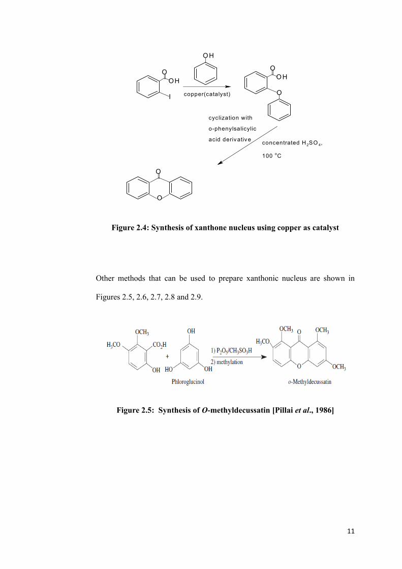
11
OH
I
O
OH
copper(catalyst)
OH
O
O
cyclization with
o-phenylsalicylic
acid derivativeconcentrated H 2SO 4,
100 oC
O
O
Figure 2.4: Synthesis of xanthone nucleus using copper as catalyst
Other methods that can be used to prepare xanthonic nucleus are shown in
Figures 2.5, 2.6, 2.7, 2.8 and 2.9.
Figure 2.5: Synthesis of O-methyldecussatin [Pillai et al., 1986]

12
Figure 2.6: Preparation of 3, 7-dihydroxyxanthone [Lin et al., 1993]
Figure 2.7: Preparation of 3, 7-dihydroxyxanthone via Ketimino
intermediate [Atkinson and Heilbron, 1926]
Figure 2.8: Preparation of methoxylated xanthones [Vitale et al., 1994]

13
Figure 2.9: Synthesis of 1, 3-dihydroxyxanthone [Pillai et al., 1986]
2.1.2 Prenylation of Xanthonic Block
After synthesising the xanthonic block, prenylation is then carried out through
reacting the xanthone block with prenyl bromide in either organic medium or
aqueous medium to produce o-prenylated or c-prenylated xanthones. In organic
medium such as in the presence of acetone with prenyl bromide and K2CO3, o-
prenylated xanthone can be produced and to produce c-prenylated xanthone,
aqueous medium is used. There are several methods shown in Figures 2.10,
2.11, 2.12 and 2.13 to synthesis c-prenylated xanthones.

14
O
O
OH
OH BF3.Et
2O
OHO
O
OH
OH
R1
R2
R1 =Pre; R2 = H
or
R1 = H ; R2 = Pre
or
R1 = R2 = Pre
* Pre = prenyl
Figure 2.10: c-Prenylation with 2-methylbut-3-en-2-ol [Anand and
Jain, 1973]
prenyl bromide
K2CO
3,KI, 15h
O
O OH
O
O O
N,N-dimethylanaline , 4h
O OH
O
+
O
O O
Figure 2.11: c-Prenylation with prenyl bromide in the presence of a
strong base [Anand and Jain, 1973]

15
O OH
O OH
OH
prenyl bromide
NaOMe/ MeOH O OH
O
O OH
R1
R2
R3
R1=R
2=R
3=Pre
or
R1=R
2= Pre, R
3= H
or
R1=R
3= Pre, R
2= H
* Pre = prenyl
Figure 2.12: c-Prenylation through Claisen rearrangement [Patel and
Trivedi, 1988]
O
OHO
prenyl bromide, anhydrous K2CO3, dry
DMF
reflux, 24 h
isoprene, H3PO4 85 %, xylene
31 oC, 30 h
O
OHO
O
OHO
Figure 2.13: Synthesis of c-prenylated 1, 1-dimethylally- and 3, 3-
dimethylallyl derivatives of xanthone [Castanheiro et al.,
2009]

16
2.1.3 Synthesis via Prenylated Starting Materials
This synthesis method is different from Sections 2.1.1 and 2.1.2; it starts with
prenylation of the starting materials before coupling reaction is carried out.
This method involves the use of protecting group such as benzyl group in order
to improve the high selectivity of the reaction (Figures 2.24, 2.25 and 2.26)
[Iikubo et al., 2001].
2.1.4 Improved Synthesis Method
All the synthesis methods mentioned earlier are not environmental friendly. To
make the reaction more environmental friendly, a new approach by using
microwave has been introduced to synthesise prenylated xanthone. This
method is called as microwave-assisted organic synthesis (MAOS)
[Castanheiro et al., 2009].
There are pros and cons in the use of this method. At which, it can have high
yield of prenylated xanthone but can only be applied to certain types of
synthesis. Enhancement has been made by using Mantmorillonite clay as
catalyst. This clay enables the reaction to take place in mild condition,
increasing the reaction selectivity, yield and decreasing the reaction time.
Furthermore, this clay can be easily separated from the reaction mixture,

17
regenerate and purify. This clay also enables the reaction to be carried out in
either solvent free or with solvent condition.
2.1.5 Further Synthesis on Prenylated Xanthone
The reaction does not stop, even though, prenylated xanthone has been
synthesised. Prenylated xanthones can further undergo cyclisation to form
pyranoxanthone (Figure 2.14, 2.15 and 2.16). Normally, this reaction can be
accomplished through refluxing of prenylated xanthone with a catalytic amount
of zinc chloride in dry xylene.
The disadvantage of this reaction is that, the yield from the synthesis was low
and sometimes the cytotoxic activity was found to be weaker than the
prenylated xanthone precursor that was used for cyclisation [Castanheiro et al.,
2009].
COOH
OH
+
OH
HO
R
OH O
O OH
OH
R
R= CH3 OR H R= CH3 OR H
a
Figure 2.14: Synthesis of xanthone block. Reagents and conditions: (a)
ZnCl2, POCl3, 70 0C, 3 h

18
O OH
R
R= CH3 OR H
O OH
O O
R
R= CH3 OR H
O OH
O O
R
R
CH3
O OH
R1
H
R1
H
H
+
a
Figure 2.15: Synthesis of prenylated xanthone. Reagents and conditions: (a)
prenyl bromide, K2CO3, acetone, reflux, 8 h
O O
R
R= CH3 OR H
O OH
aR
O OH
O O
+
O OH
O O
R= CH3 OR H
Figure 2.16: Cyclisation of prenylated xanthone. Reagents and conditions:
(a) ZnCl2, o –xylene, 200 oC, 21 h
Noted that, if there is prenyl group attached to the xanthonic block, the size of
the prenyl group is important and prenyl group is associated with more
selective compound [Castanheiro et al., 2009].

19
2.2 Cytotoxic Activity of Synthetic Xanthone Derivatives
Cyto in greek means cell and toxic is poisonous. Thus, combining the two
words; cytotoxic means toxic to cell or cell killing. Nowadays, cancer has
become a notorious killer around the world and many methods have been
carried out to produce anticancer drug. Cancer is a class of disease or disorder
characterised by the uncontrolled division of cells and the ability of these cells
to invade other tissue or by implantation into distant sites by metastasis
[Suphavenich et al., 2009]. Metastasis is defined as spreading of the cancer cell
to the neighbouring cell. The example of cancer cell was collected and studied
in 1951, called as HeLa cell because this cell was derived from the tumor
removed from a woman named Henrietta Laeks.
Normally, cytotoxic activity of the drugs synthesised is tested by using human
tumor cell lines KB 3.1(oral squamas cell carcinoma), HeLa cell, breast cancer
cell tumor cell line NCI-H460 (non- small lung), MCF-7 (breast
adenocarcinoma), UACC-62 (melanoma) and SF-268 (central nervous system).
To kill the cell, some drugs are used to interfere with the cell cycle
permanently such as to all the cell progress at G1, S or G2/M phase [Ding et al.,
2009]. G1, S and G2/M phases are shown in Figure 2.17.

20
Figure 2.17: Cell cycle
In interphase, including G1 phase, S phase and G2 phase, the cell grows and
copies chromosomes in preparation for cell division. But chromosomes are
only duplicated in S phase. Part of the cell cycle is mitotic phase which can be
divided into mitosis and cytokinesis. Mitosis can be divided into 5 stages:
prophase, prometaphase, metaphase, anaphase and telophase. Cytokinesis is the
last stage in mitotic phase which involved in cleaving the cell’s membrane and
producing individual cell that completed the whole mitotic phase [Cambell, R.
2005].
As conclusion, cell cycle works through cell growth in G1 phase, then continue
to grow, as the cell copies own chromosomes in S phase and then undergo cell
division in G2 phase. Finally, the cell is divided in mitotic phase. This process
repeats to generate new cells. So, by interfering the phases in the cell cycle, this
will prevent the cancer cell from replicating and finally die.

21
In order to interfere the cell cycle of cancer cell, high energy radiation can be
used to destruct the DNA in cancer cell. But there is other alternative by using
chemotherapy that involves the use of chemotherapeutic drugs to interfere the
specific stages in the cell cycle. One of the cytotoxic drugs is xanthone.
Xanthone is cytotoxic because the planar structure of xanthone and xanthone
derivatives has the ability to intercalate to DNA. Xanthone and xanthone
derivatives are then called as DNA intercalator. When DNA alkylating or
binding groups are incorporated in the xanthone structure, the resulting
compounds might show enhanced DNA interacting capacity by synergic effect
from two combined properties; DNA intercalation and DNA alkylation or
groove binding [Na, 2009]. Thus, intercalation with the DNA inhibits the cell
to replicate and finally the cancer cell stop growing. Based on previous
research, the greater the number of prenyl substituents, the higher was the
cytotoxic potency [Han et al., 2008].
Based on the fact that DNA is the target of prenylated xanthones, xanthones
extraction and synthesis have now become a popular study in cytotoxic
research.
The cytotoxic activities of xanthone can be evaluated by several methods.
These include cytotoxic assay, MTT (Method of Transcription and Translation)
assay, SRB (Sulphorhodamine B) assay, WST (Water Soluble Tetrazolium)
assay and clonogenic assay. To qualitatively compare the potency of the tested
compounds, three parameters including GI50, TGI, and LC50 can be used [Han

22
et al., 2008]. GI50 is the concentration of a compound inhibiting 50% of the cell
growth [Han et al., 2008] whereas TGI is the concentration of a compound
completely inhibits the cell growth at 48h and LC50 is the concentration at
which the tested compound kills 50% of the cell at 48h [Han et al., 2008]. The
smaller the GI50, TGI, and LC50 values, the more cytotoxic the xanthones.
Synthesis of xanthone derivatives that have cytotoxic activity are shown in
Section 2.3.
2.3 Synthesis of Cytotoxic Xanthone Derivative
As mentioned in Section 2.2, some of the xanthone derivatives have cytotoxic
activity. While, in this section, the methods to synthesise various type of
cytotoxic xanthones are discussed.
2.3.1 Bromoalkoxyxanthone
In recent research, bromoalkoxyxanthone has been reported to show cytotoxic
activity towards SF-268 (CNS-cancer cell, GI50= 30.2 ± 3.6 µM), NCl-H460
(non-small cell lung cancer, GI50 = 30.2 ± 3.6 µM) and growth inhibitory
activity against MCF-7 (breast adenocarcinoma, estrogen dependent ER(+),
GI50 = 22.7 ± 1.3 µM). All these effects were due the presence of bromine atom

23
that could serve as an anchor for the intercalation with DNA [Sousa et al.,
2009]. The synthesis of bormoalkoxyxanthone is shown as below:
COOH
OH
+
OH
HO
I
O
O OH
+
O OH
OH
II
O
O OO
O
O
+
O
O
O
Br
Figure 2.18: Reagents and conditions: (I) zinc chlroride, 200 oC/ 5 min –
180 oC/ 4 h; (II) potassium carbonate, dry DMF, 1, 6-
dibromobutane, room temperature, 24 h [Sousa et
al., 2009]
2.3.2 Epoxyxanthone
Epoxyxanthone compounds and the open ring halohydrin xanthones have been
tested for their cytotoxic activity. From the experiment carried out, 1, 3-
bisepoxyxanthone was found to be the most active in cell growth inhibition
capacity and 1, 3-bisepoxyxanthone also showed concentration dependent
DNA cross-linking activity. This observation showed that, two-epoxy group
substituted xanthone generated better cytotoxic activity than single epoxy

24
substitution. Even though, the locations of the two epoxypropoxy groups were
different. Two epoxy groups that tethered to 3, 5-position of xanthone can
increase the cytotoxic activity dramatically [Na, 2009].
R
COOH
OH
+
OH
OHHO
R=H, OCH3 Phloroglucinol
ZnCl2
POCl3 O
O OH
OH
R= OCH3
R= H
Epichlorohydrin Cs2CO3,Acetone,
reflux
O
O OH
O
R
RO
R= OCH3
R= H
1M-HCl or HBr
EtOAC
+
O O
R
O O
O
O
R= H
O
O OH
O
R OH
X
R= OCH3, X=Cl
R= OCH3, X= Br
R= H, X= Cl
R= H,X= Br
Figure 2.19: Synthesis pathways for epoxyxanthone derivatives [Woo et al.,
2006]
25
Besides that, from the research carried out, it was shown that thioxanthone with
the presence of epoxy group was found to possess great cytotoxic activity [Na,
2009].
S
O OH
O
O
Figure 2.20: Thioxanthone bearing epoxy group [Na, 2009]
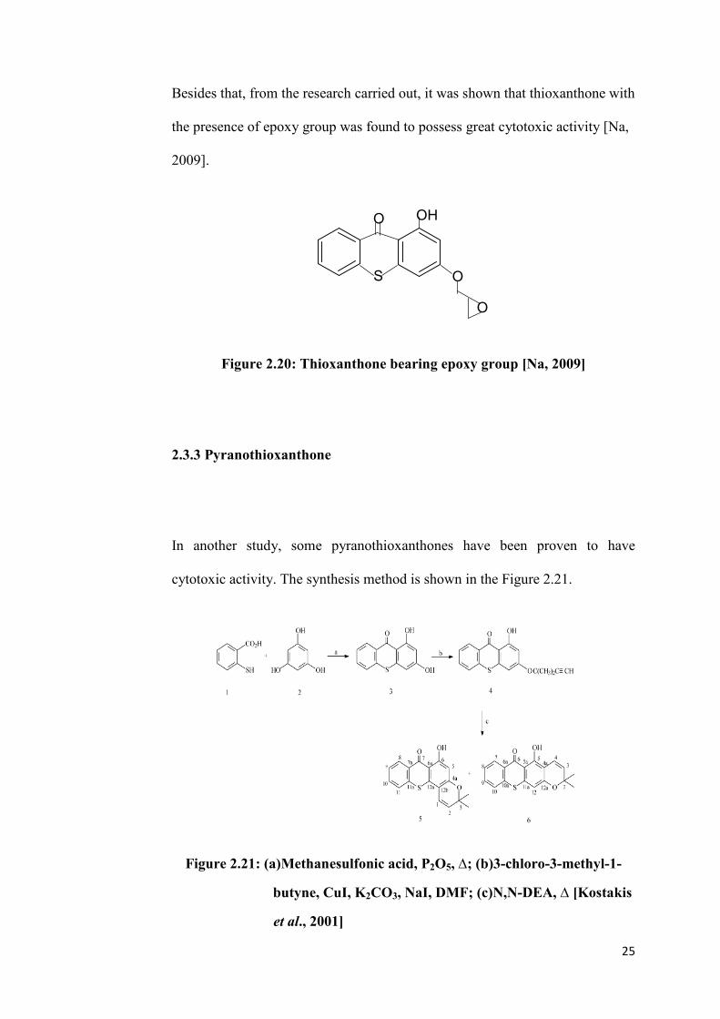
2.3.3 Pyranothioxanthone
In another study, some pyranothioxanthones have been proven to have
cytotoxic activity. The synthesis method is shown in the Figure 2.21.
Figure 2.21: (a)Methanesulfonic acid, P2O5, ∆; (b)3-chloro-3-methyl-1-
butyne, CuI, K2CO3, NaI, DMF; (c)N,N-DEA, ∆ [Kostakis
et al., 2001]

26
Figure 2.22: (a) (1) NaH, THF; (2) (CH3)2SO4, ∆; (b) (1) OsO4,N-
methylmorpholine N-oxide ;(2)NaHSO3; (c) Ac2O,Py;
(d) NBS, H2O-THF; (e) AIBN, Bu3SnH, toluene, ∆
[Kostakis et al., 2001]

27
Figure 2.23: (a) (1) NaH,THF; (2) (CH3)2SO4, ∆; (b) (1) OsO4, N-
methylmorpholine N-oxide; (2) NaHSO3; (c) Ac2O, Py;
(d) NBS, H2O-THF; (e) AIBN, Bu3SnH, toluene, ∆
[Kostakis et al., 2001]
From the research carried out by Ioannis Kostakis et al., pyranothioxanthone 7,
8b, 9a, 10a and 12b were found to possess good cytotoxic property. The
cytotoxic activities of these compounds were evaluated in vitro on the L1210
leukemia cell line, with acronycine as reference compound. Compound 9a
(GI50 =12.1 ±1.3 µM) exhibited interesting cytotoxic activity, being twice more
potent than acronycine (GI50 = 25.0 ± 4.1 µM), while compound 7 (GI50 = 36.9
± 3.6 µM), 8b (GI50 = 36.6 ± 2.9 µM), 10a (GI50 = 27.7 ± 2.8 µM) and 12b
(GI50 = 33.0 ± 3.2 µM), were as potent as acronycine. In general, in terms of
angular isomers, the structure of the compounds that are similar with

28
acronycine will be more potent than the linear analogues. This suggested that
angular orientation of D ring is important for biological activity [Kostakis et al.,
2001].
2.3.4 α-Mangostin
α-Mangostin has been proven to have cytotoxic activity too. α-Mangostin can
inhibit cell growth and human leukemia cell line by introducing cascape-3
dependent apotosis. Further studies revealed that cellular target of α-mangostin
were mitochondria. Recently, research has been carried out in synthesising α-
mangostin. In Figure 2.24, fragment 5 was synthesised by protection of 2, 4-
dihydroxybenzaldehyde 1 with benzyl (Bn) groups, followed by Baeyer-
Villiger oxidation and acid hydrolysis to provide phenol 2. Phenol 2 was
subsequently subjected to bromination and allylation, leading to compound 3 in
high overall yield. Upon heating of compound 3 at 160 oC, Claisen
rearrangement occurred and produced allylbenzene. The resulted phenol was
protected as methyl ether, leading to compound 4. The Lemieux-Johnson
oxidation of compound 4, followed by Wittig reaction provided bromobenzene
4 [Iikubo et al., 2001].

29
OHC
HO OH
a,b HO
BnO OBn
O Br
OBnBnO
Br
OBn
MeO
BnO
Br
OBn
MeO
BnO
c,d
e,f
g,h
1 2 3
45
Figure2.24: Synthesis of fragment 5; (a) BnBr, K2CO3, DMF, room
temperature, 96 % ; (b) mCPBA, CH2Cl2, room
temperature; 6 M HCl, MeOH, room temperature, 95 % in
two steps; (c) Br2, CHCl3, room temperature, 84 %; (d)
allyl bromide K2CO3,DMF, room temperature, 80 %; (e)
160 oC, 73 %; (f) MeI, K2CO3, DMF, room temperature,
87 %; (g) OsO4, NaIO4, Et2O)/ H2O (1/1), room temperature,
95 %; (h) iPrPh3P+I
-, nBuLi, THF, 0
oC, 72 % [Iikubo et
al., 2001]
Figure 2.25 shows the synthesis of fragment 12 through the reaction of 1, 3, 5-
trihydroxybenzene (phloroglucinol) 6. Phloroglucinol was protected with
MOM group (methoxymethyl ether) and followed by prenylation to produce
compound 7 in high overall yield. Introduction of ethoxycarbonyl group
(EtOOC-) to compound 7 gave compound 8 which was then subjected to acid-
catalysed methonolysis, followed by TBS-protection (tetrabutyl silyl), and
finally with DIBAL-reduction (diisobutylaluminium hydride) to give benzyl

30
alcohol 9. Then, IBS (2-iodobenzoic acid) converted compound 9 to aldehyde
compound 10, with the removal of TBS group adjacent to the prenyl group.
Compound 10 was protected as MOM ether for utilisation in regioselective
cyclization. The reaction then followed by exchanging of TBS group with
benzyl group to produce compound 12 [Iikubo et al., 2001].
HO
HO OH
a,b
OMOM
OMOMMOMO
c
OMOM
MOMO OMOM
EtOOC
d-f
TBSO OTBS
OTBS
HOg
TBSO OTBS
OR
OHC
H
10: R = H
11: R = MOM
i,j
BnO OBn
OMOM
OHC
6 7 8
9
12
Figure 2.25: Synthesis of fragment 12; (a) NaH, MOMCl, DMF, room
temperature, 96 %; (b) nBuLi; prenyl bromide, THF, 0 oC,
89 %; (c) nBuLi; (EtO)2CO, THF, 0 oC, 95 %; (d) CSA,
MeOH, 60 oC, 100 %; (e) TBSCl,DMAP, Et3N, DMF, room
temperature, 100 %; (f) DIBAL-H, toluene, 78oC, 78 %; (g)
IBX, toluene/DMSO (1/1), room temperature, 76 %; (h)
NaH, MOMCl, CH2Cl2, room temperature, 65 %; (i)
TBAF (tetrabutylammonium floride, THF, 0 oC, 100 % (j)
BnBr, K2CO3, DMF, room temperature, 98 % [Iikubo
et al., 2001].

31
Figure 2.26 shows the synthesis of α-mangostin with the coupling of the
previously synthesised compounds 5 and 12.
5 +12
HO
OBnOBn
MeO
OBn
OMOM
BnO
OHOHOH
OMOM
MeO
BnO
O
d
OH
OH
MeO
HO
O
O
13 14
15
Figure 2.26: Synthesis of α-mangostin; (a) sBuLi, THF, 78 oC, 49 %; (b)
IBX, toluene/DMSO (dimethyl sulfoxide) (1/1), room
temperature, 76 %; (c) 10 % Pb/C, HCO2NH4, acetone,
room temperature, 63 %; (d) PPh3 (triphenylphosphine),
CCl4 (chloroform), THF, room temperature, then silica gel,
43 %; (e) CSA (camphoresulfonic acid), MeOH, room
temperature, 76 % [Iikubo et al., 2001]

32
2.3.5 Psorospermin
Psorospermin was also found to possess cytotoxic activity. It possessed
excellent anticancer activity against human cell and marine cell. This is
because of intercalation of xanthone group with DNA base pair and alkylation
of epoxide by N7-guanine in the presence of topoisomerase II which is the
crucial enzyme for DNA cycle. From the past, psorospermin was obtained
through extraction from the natural products. But nowadays, as an alternative,
psorospermin can be produced via synthetic approach. The synthesis of
psorospermin, began with the production of xanthonic block using Grover
method. But different from other literature, zinc chloride in POCl3 was heated
to 60 oC for 30 minutes prior to the addition of dimethoxy benzoic acid 5. This
is because zinc chloride was found to be the culprit in decreasing the
percentage yield, due to the insolubility of the glass like fused zinc chloride.
Compound 5 was heated for another 30 minutes before phloroglucinol 6 was
added [Schwaebe et al., 2004]. Then, the processes were continued as shown in
Figure 2.27.

33
Figure 2.27: Synthesis of psorospermin. Reagents: (a) ZnCl3, POCl3; (b)
allyl bromide, K2CO3; (C) CH3I, K2CO3; (d) mesitylene,
180oC (e) BBr3; (F) benzyl bromide, NaH, DMF; (g) CH3I,
DMF; (h) OSO4, NaIO4; (i) (CF3CH2O)2POCHCH3CO2Me,
KHMDS, 18-CROWN-6; (j) DIBALH/CH2Cl2; (k) t-
BUOOH, (-) DIPT, Ti(i-Opr)4; (i) MsCl, Et3N; (m) raney
nickel/ K2CO3, ethanol [Schwaebe et al., 2004]

34
2.3.6 Xanthonecarboxylic Acid
Xanthonecarboxylic acid can be obtained by oxidation of the methyl group of
3a-f to 3i-q, with potassium permanganate in alkaline solution followed by
intramolecular Friedel-Craft acylation with polyphosphoric acid [Pickert et al.,
1998].
COOH
Cl
+
HO
CH3
K2CO
3, Cu, CuI
O
COOH
CH3
KMnO4, KOH
O
COOH
COOH
O
COOH
O
PPA
3a-f
3i-q
Figure 2.28: Synthesis of xanthonecarboxylic acid [Pickert et al.,
1998]
In this project, the xanthone block, was synthesised by using two units of
substituted benzoic acid via coupling reaction in the presence of Eaton’s
reagent. Alkenylation was then carried out to produce alkenylated xanthones.
Under the same reaction conditions as described in the literature which
involved the prenylation of xanthone [Castanheiro et al., 2009]. The difference
was on the substituent that has been used. Substituent used for prenylation was
prenyl bromide while the substituent used for this alkenylation was 5-bromo-1-
pentene.

35
CHAPTER 3
MATERIALS AND METHODS
3.1 Chemicals
The chemicals used in this project are as below:
Table 3.1: Chemicals used in synthesising xanthone block
Chemical name Molecular
formula
Source Molecular
Weight,
Mw (g/mol)
Country
2,4,6-
trihydroxybenzoic
acid monohydrate
C7H6O5 Acros
Organics
170.12 Belgium
Eaton’s reagent P2O5/
MeSO3H
Acros
Organics
- Belgium
Acetone C3H6O QRec 58.08 Malaysia
Ethyl acetate CH3COOC2H5 LAB– SCAN 88.10 Ireland

36
Table 3.2: Chemicals used in synthesising the alkenylated xanthone
Chemical
name
Molecular formula Source Molecular
weight, Mw
(g/mol)
Country
Potassium
Carbonate
K2CO3 John Kollin
Corporation
138.20 -
Acetone CH3COCH3 QReC 58.08 Malaysia
Ethyl acetate CH3COOC2H5 LAB –
SCAN
88.10 Ireland
Hydrochloric
acid (37%)
HCl Fisher
Scientific
36.46 United
Kingdom
5-bromo-1-
pentene
Br(CH2)3CH=CH2 Merck 149.03 German
Table 3.3: Chemical used in NMR analysis
Chemical
name
Molecular
formula
Source Molecular
Weight,Mw
(g/mol)
Country
Acetone-d6 CD3COCD3 Acros
Organics
64.12 Belgium

37
Table 3.4: Chemical and cancer cell lines used in bioassay
Chemical,
apparatus and
cell
Source Country
MTT Sigma - Aldrich United State of
America
DMSO Fisher Scientific United
Kingdom
RPMI 1640
Media
Cellgro Manassas
Fetal Bovine
Serum (FBS)
Hyclone Thermo
Scientific
South America
HeLa Cell ATCC (American Type
Culture Collection)
United State of
America
(USA)
MDA-MB-231 ATCC USA
3.2 Instruments
3.2.1 Nuclear Magnetic Resonance Spectrometer (NMR)
NMR provides spectrum that has the information on the number of
magnetically distinct atom present in the compound studied. In this project, 1H-
& 13
C-NMR, HMBC and HMQC spectra were obtained by using
trimethylsilane (TMS) as internal standard and reference. The frequency used
for 1H-NMR analysis was 400 MHz; while
13C-NMR was performed at 100
MHz. The samples were prepared by dissolving a small amount of sample in

38
sufficient acetone-d6 in order to fill up the NMR tube to a height of 4-5 cm and
the cap was affixed to the tube and wrapped with parafilm to avoid solvent
evaporation
3.2.2 Infrared Spectrophotometer (IR)
Through the IR analysis, IR spectrum was obtained. IR spectrum was used to
indicate the presence of certain functional group in the compound analysed. In
this project, Perkin Elmer 2000-FTIR spectrophotometer was used for sample
analysis and the range used was 4000 cm-1
– 400 cm-1
. The sample was grinded
with KBr powder in the ratio of 1:10. Then, the mixture of the sample with
KBr powder was compressed under a high pressure to form KBr sample pellet.
3.2.3 Ultraviolet-Visible Spectrophotometer (UV-Vis)
The UV-Vis spectrum was used to determine the qualitative information and
the position of the hydroxyl group in xanthone block. From the transition
energy of the highly conjugated organic compound, colour produced by the
compound can be identified. In this project, Perkin-Elmer Lambda (25/35/45)
UV-Vis spectrophotometer was used and the samples were prepared by using
absolute ethanol to dissolve the sample. Then, the sample was measured by
using quartz cuvette in the range of 190 nm to 400 nm.

39
3.2.4 Mass Spectrometer (MS)
Mass spectrometry was used to provide the information on the molecular mass,
molecular formula and the structure of the molecule. This was done by
analysing the molecular ion and fragmentation pattern of the compound. In this
project, the sample was analysed by using Agilent 5975 MSD (nominal mass)
mass spectrometers.
3.2.5 Melting Point Instrument
Melting point instrument was used to provide melting point of the tested
compounds. By obtaining the melting point, the characteristic and the purity of
the tested compound can be determined by comparing the range difference with
the pure compound. In this project, Barnstead Electrothermal 9100 melting
point instrument was used. Heamatocrit capillaries were used to contain the
solid form sample for testing.

40
3.3 Methodology
3.3.1 Synthesis of 1, 3, 6, 8-Tetrahydroxyxanthone
14.1 g (75 mmol) of 2, 4, 6-trihydroxybenzoic acid monohydrate was added
with 75 ml of Eaton’s reagent and warmed in water bath at 80 oC for 10
minutes whilst stirring. The mixture was cooled at room temperature and was
stirred for 20 minutes. The mixture was poured into 500 mL beaker initially
filled with ice and was left to soak in ice bath for 30 minutes. The precipitate
was then filtered via Buchner filtration and the precipitate was placed in oven
at 50 oC overnight, while, the filtrate was extracted with ethyl acetate. After
drying overnight in the oven, the dried precipitate was then extracted with
acetone. Both the extracts were dried under reduced pressure separately.

41
2,4,6-trihydroxybenzoic acid and Eaton's reagent
1. The mixture was warmed in water
bath at 80oC for 10 minutes while
stirring.
2. The mixture was cooled at room temperature and stirred for 20 minutes.
12345 6
78
91 1
0
2345 6
78
911
500 mL beaker filled with ais
3. The mixture was poored into the beakers filled with ais and left to soak for 30 minutes.
hotplate stirrer
4. The precipitate formed was filtered with Buchner filtration and
dried in the oven overight at 50oC.
5. Dried precipitate was extract with acetone.
seperating funnel
6. The filtrate was extraceted with ethyl acetate.
7. Both extracts were dried under reduce pressure seperately.
Figure 3.1: Flow chart on the Synthesis of 1,3,6,8-tetrahydroxyxanthone
Figure 3.1: Flow chart on the synthesis of 1, 3, 6, 8-tetrahydroxyxanthone
Separating
funnel
5. The filtrate was extracted with ethyl
acetate.
6. After overnight drying in the oven, the dried
precipitate was extracted with acetone.
3. The mixture was poured into
the beakers filled with ice and
left to soak for 30 minutes.
7. Both extracts were dried
under reduce pressure
separately.
1. The mixture was warmed in
water bath at 80 oC for 10
minutes while stirring.
2. The mixture was cooled at
room temperature and stirred for
20 minutes.
4. The precipitate formed was
filtered with Buchner filtration
and dried in the oven overnight at
50 oC.

42
COOH
OH
OH
HO
+COOH
OHHO
OH
Eaton's reagent, 80 oC, reflux
10 minutes
O
OH
OH
HO
HO
O
Figure 3.2: Synthesis pathway of 1, 3, 6, 8-tetrahydroxyxanthone
3.3.2 Alkenylation of Xanthonic Block in Organic Solvent
A mixture of 1.04 g (4 mmol) of 1, 3, 6, 8-tetrahydroxyxanthone and 1.656 g
(12 mmol) of K2CO3 was stirred for 5 minutes in 100 mL acetone under room
temperature. 2.384 g (16 mmol) of 5-bromo-1-pentene was added into the
solution. The mixture was refluxed for 6 hours at 65 oC. The solid was filtered
and the filtrate was dried under reduced pressure.

43
Round flat bottom flask filled with the mixture
of 1,3,6,8-tetrahydroxyxanthone and K2CO3 in
100 mL aetone.
12
3
45 6
7
8
9
1 1
0
2
3
45 6
7
8
911
hotplate
strirrer
1. The mixture was stirred for 5 minutes in room temperature.
2. 5-bromo-1-pentene was added into the mixture and the mixture was reflux for 6
hours at 65oC.
3. The solid was filtrated and the filtrate was dried under reduce pressure.
Figure 3.3: Flowchart on the synthesis of o-alkenylated xanthone
2. 5-bromo-1-pentene was added into the mixture
and the mixture was refluxed for 6 hours at 65 oC
3. The solid was filtered and the filtrate
was dried under reduced pressure
1. The mixture was stirred for
5 minutes at room
Flat bottom flask filled with the mixture of
1, 3, 6, 8-tetrahydroxyxanthone and
K2CO3 in 100mL acetone

44
O
OH
OH
HO
HO
O
K2CO3 in 100 mL acetone,
5-bromo-1-pentene, reflux for 6
hours at 65 oC
O
OH
OR2
HO
R1O
O
(1) R1 = R2 = pent-4-en-1-yl
(2) R1= H, R2 = pent-4-en-1-yl
Figure 3.4: Synthetic route for synthesising o-alkenylated 1, 3, 6, 8-
tetrahydroxyxanthone
3.4 Chromatography Method
3.4.1 Thin Layer Chromatography (TLC)
Thin layer chromatography was used in this project to identify the pure
compound, to test for a suitable solvent system for column chromatography

45
and to identify the identical compounds by comparing the Rf value of the
compounds.
Thin layer chromatography consists of two phases which are mobile phase and
stationary phase. Mobile phase used in this project are hexane,
dichloromethane, acetone and ethyl acetate. The stationary phase of the TLC
plate is made of silica gel 60 F254 (Merck 1.05554.0001) coated on aluminium
plate of 4.0 cm x 6.7 cm dimension.
Before spotting the compounds on the TLC plate, the base line and the solvent
front line were drawn on the TLC plate as shown in Figure 3.5.
5.7cm
0.5cm
0.5cm
4.0cm
base
line
solvent front
Figure 3.5: Feature of TLC plate used
Identification of the pure compound, test for a suitable solvent system and to
identify identical compounds can be done by comparing the retention factor, Rf
46
value of the compounds. The compounds must first be dissolved and diluted in
a solvent. The samples dotted on the TLC plate were checked for its thickness.
This was to make sure the compound spotted on the TLC plate was not too
thick to prevent tailing and also not too light to avoid difficulty of detecting the
spots on the TLC developed.
Once all the compounds were spotted on the TLC plate, the TLC plate was
placed into a solvent chamber filled with 4 mL of the desired solvents for
separation. The solvent chamber was sealed with aluminium foil to enable
chamber to be saturated with the solvent’s vapour.
The TLC plate was removed from the solvent chamber when the solvent
reached the solvent front and the developed spot was identified and seen under
ultraviolet light with both short (254 nm) and long (365 nm) wavelenghts.
After that, the spots were circled up by using a pencil.
The retention factor, Rf can be calculated by using the equation as follow:
Rf = D / L
Where,
D = Distance travelled by the compound from the base line (cm)
L = Distance between solvent front and the base line (cm)

47
3.4.2 Column Chromatography
Chemical synthesis afforded a mixture of products. Thus, column
chromatography was needed for purification of crude products. In this study,
normal phase column chromatography with polar stationary phase and
relatively non-polar mobile phase was used. The stationary phase used was
Silicycle silica gel (40-63µm) and the mobile phase used was a mixture of
different solvent such as hexane, acetone, dichloromethane, methanol and ethyl
acetate at different ratio increasing polarity. Thus, gradient elution was applied
to elute and separate different compounds out from the column. Column
chromatography enables the separation of the desired compound out from the
side products based on the principle of differences in the strength of interaction
between various compounds and the stationary phase.
The sample was prepared via dry packing before subjected into the column.
Firstly, the sample solution was introduced drop by drop and mixed with small
amount of silica gel in a beaker. Then, the silica gel was swirled lightly to
enable the remaining silica gel to cover the droplets that formed. Spatula was
used to mix and grind the sample with silica gel until homogenous powdery
solid was formed. The process was repeated until all the sample was coated
onto the silica gel.

48
When packing the column, the column was first filled with hexane up to 1/3 of
the column’s height. A small piece of cotton wool was plugged to the bottom
of column. Then, small amount of sea sand was introduced into the column to
form a thin layer of sand above the plugged column with hexane continuing
running down the column and collected into a conical flask. The conical flask
was sealed with aluminium foil to prevent evaporation of the solvent into the
air.
A mixture of silica gel and hexane was prepared. The slurry of silica gel was
then poured into the column and the stopcock of column was turned on to
allow hexane to run down the column into the conical flask. This was to enable
even distribution of the silica gel in the column and prevent formation of
bubbles. Before introduction of the dry packed sample into the column, the
column must be run for a few times to enable the silica gel to pack firmly in the
column.
Before loading the sample into the column, the hexane level was run and
maintained at 5 cm above the surface of the silica gel. The hexane collected
before loading of sample can be reused by pouring back into the column.
Gradient elution was performed by using a suitable solvent system that was
tested earlier by using TLC. The column was covered with aluminium foil to
prevent solvent evaporation. The fraction collected was then examined by
using TLC.

49
aluminium foil
sea sand
silica gel
dry packed sample in silica gel
solvent
aluminium foil
conical flask
retort stand
sintered
chromatography
column
Figure 3.6: Setup of sintered chromatography column
3.5 Bioassay
The cell viability was evaluated by using the colorimetric 3-(4, 5-
dimethylthiazo-2-yl)-2, 5-diphenyl- tetrazolium bromide (MTT) assay. In this
MTT assay, HeLa and MDA-MB-231 cancer cells were tested. Both of HeLa
cells (0.75 x 10⁵ cells/mL) and MDA-MB-231 cells (3.5 x 10
5 cells/mL) were
cultured in 96-well plates with 0.1 % dimethyl sulfoxide (DMSO) containing
tested compound at 37 oC for 72 hours. Blank cell control and blank medium
control were also prepared and incubated for 72 hours. After treatment, the
cells received 20 µL of 5 mg/mL MTT and were incubated at 37 oC for another
3 hours. After 3 hours, 70 % of supernatant was removed and 150 µL of

50
DMSO was added into each well. DMSO dissolved the formazan crystal to
produce a purple solution. Cell viability was determined by measuring optical
density at 570 nm using a Model 550 micro plate reader (Bio-Rad Laboratories,
Hercules, CA, USA). Cell viability was calculated as follow:
Where
= average absorbance of cell treated with compound
= average absorbance of blank medium
= average absorbance of cell control

51
CHAPTER 4
RESULTS AND DISCUSSION
4.1 Synthesis of 1, 3, 6, 8-Tetrahydroxyxanthone and Alkenylated
Xanthones
The synthesis of 1, 3, 6, 8-tetrahydroxyxanthone involved the reaction of 14.1
g (~75 mmol) of 2, 4, 6-trihydroxybenzoic acid monohydrate with 75 ml of
Eaton’s reagent. The present of Eaton’s reagent enable the coupling reaction of
2, 4, 6-trihydroxybenzoic acid, yielded 4.69 g (43.5 %) of 1, 3, 6, 8-
tetrahydroxyxanthone.
Reaction of (1.04 g) of 1, 3, 6, 8-tetahydroxyxanthone with 5-bromo-1-pentene
in acetone and in the presence of K2CO3 as catalyst, yielded 0.033 g (5.04 %)
of 1, 3, 8-trihydroxy-6-(pent-4-enyloxy)-9H-xanthen-9-one and 0.21 g (26.5 %)
of 1, 8-dihydroxy-3,6-bis(pent-4-enyloxy)-9H-xanthen-9-one.
The structures of 1, 3, 6, 8-tetrahydroxyxanthone, 1, 3, 8-trihydroxy-6-(pent-4-
enyloxy)-9H-xanthen-9-one and 1, 8-dihydroxy-3, 6-bis(pent-4-enyloxy)-9H-
xanthen-9-one were elucidated by using 1D-NMR, 2D-NMR, IR, and UV-Vis
analyses.

52
4.2 Structural Elucidation of Xanthone
4.2.1 Structural Elucidation of 1, 3, 6, 8-Tetrahydroxyxanthone
1, 3, 6, 8-Tetrahydroxyxanthone was synthesised by reacting 2, 4, 6-
trihydroxybenzoic acid monohydrate with Eaton’s reagent. The function of
Eaton’s reagent was to enable the coupling of 2, 4, 6-trihydroxybenzoic acid
with the loss of water molecules to form 1, 3, 6, 8-tertrahydroxyxanthone-2-
carboxylic acid or 1, 3, 6, 8-tertrahydroxyxanthone-4-carboxylic acid. However,
subsequent decarboxylation caused the loss of carboxylic group and produced
1, 3, 6, 8-tetrahydroxyxanthone. The overall reaction was shown in Figure 4.1.
COOH
OH
OH
HO
+COOH
OHHO
OH
Eaton's reagent, 80 oC, reflux
10 minutes
2,4,6-trihydroxybenzoic acid
2,4,6-trihydroxybenzoic acid
O
OH
OH
HO
HO
O
Figure 4.1: Reaction scheme of 1, 3, 6, 8-tetrahydroxyxanthone

53
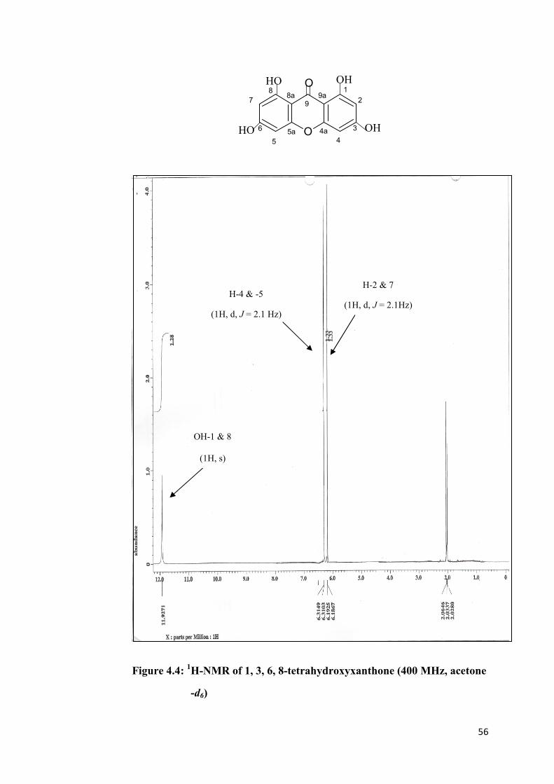
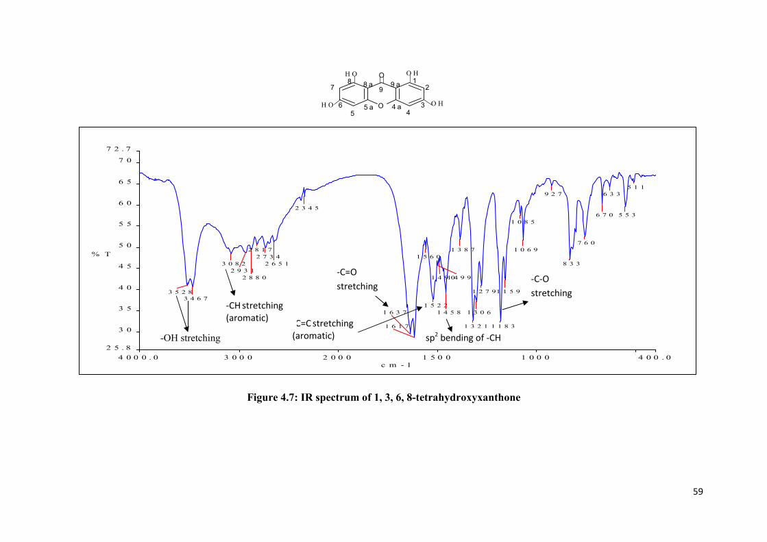
1, 3, 6, 8-Tetrahydroxyxanthone was isolated from the crude as yellow solid,
m.p. 257 to 259 oC. From the thin layer chromatography, this compound gave a
single spot with Rf value 0.35 by using solvent system 50 % hexane and 50 %
ethyl acetate. The UV absorption maxima at 210.0, 234.0, 250.0 and 330.0 nm
were characteristic of hydroxyl xanthone derivative. The IR spectrum (Figure
4.7) showed absorption band at 3528 (O-H), 3467 (O-H), 3082 (aromatic C-H),
1637 (C=O), 1522 (aromatic C=C), 1458 (sp2 C-H bending), and 1183 (C-O)
cm-1
. The mass spectrum (Figure 4.6) showed a molecular ion, M+ at m/z 260.0,
indicating a molecular formula of C13H8O6.
Initial analysis of 1H-NMR spectral data indicated that the molecule consisted
of a symmetrical xanthone skeleton. The 1H-NMR spectrum (Figure 4.4)
exhibited singlet signal for a chelated hydroxyl group at δ 11.93. This chelation
caused deshielding effect to the hydroxyl protons thus a tremendous shift of
signal to the downfield region. The four hydroxyl group at carbons 1, 3, 6, and
8 gave a symmetrical structure. The proton signal at δ 6.31 (1H, d, J = 2.1 Hz)
and δ 6.19 (1H, d, J = 2.1 Hz) were assigned to protons H-4 & -5 and H-2 & -7
respectively. The coupling constant value of 2.1 Hz for the two signals
indicated that H-2 was meta-coupling with H-4 and H-5 was meta-coupling
with H-7.
The 13
C-NMR spectrum (Figure 4.5) showed signals for 7 non-equivalent
carbons in the xanthone skeleton. The chelated carbonyl carbon: C-9 (δ 183.1)
was deshielded to downfield region because of anisotropy effect. The other 6

54
non-equivalent carbons on xanthonic skeleton were C-1 & -8 (δ 163.0), C-3 &
-6 (δ 165.8), C-4a & -5a (δ 157.8), C-8a & -9a (δ 101.1), C-2 & -7 (δ 98.5) and
C-4 & -5 (δ 94.3). The chemical shift of C-1 & -8, C-3 & -6 and C-4a &-5a
were higher than C-8a & -9a, C-2 & -7 and C-4 & -5 because the carbon atom
was directly attached to an electronegative oxygen atom. The spectral data of
1H-NMR and
13C-NMR were summarised in Table 4.1.
O
OH
OH
HO
HO
O1
2
3
4
4a5a
5
6
7
88a
99a
Figure 4.2: The structure of 1, 3, 6, 8-tetrahydroxyxanthone
Table 4.1: Summary of assignment of 1H-NMR and
13C-NMR spectra data
of 1, 3, 6, 8-tetrahydroxyxanthone
Position δ H (ppm) δ C (ppm)
1 & 8 - 163.0
2 & 7 6.19 (1H, d, J = 2.1 Hz) 98.5
3 & 6 - 165.8
4 & 5 6.31 (1H, d, J = 2.1 Hz) 94.3
4a & 5a - 157.8
8a & 9a - 101.1
9 - 183.1
1- & 8- OH 11.93 (1H, s) -

55
O
OH
OH
HO
HO
O1
2
3
4
4a5a
5
6
7
88a
99a
Figure: 4.3: UV-Vis spectrum of 1, 3, 6, 8-tetrahydroxyxanthone
190.0 250 300 350 400.0
-0.02
0.5
1.0
1.5
2.0
2.5
2.66
nm
A
SHB
329.72,1.9680
282.26,0.075996
249.76,2.6582
210.49,2.3173
233.54,2.0666
λmax = 210.0 nm, 234.0 nm, 250.0 nm
and 330.0 nm
56
O
OH
OH
HO
HO
O1
2
3
4
4a5a
5
6
7
88a
99a
Figure 4.4: 1H-NMR of 1, 3, 6, 8-tetrahydroxyxanthone (400 MHz, acetone
-d6)
H-4 & -5
(1H, d, J = 2.1 Hz)
H-2 & 7
(1H, d, J = 2.1Hz)
OH-1 & 8
(1H, s)

57
O
OH
OH
HO
HO
O1
2
3
4
4a5a
5
6
7
88a
99a
Figure 4.5: 13
C-NMR of 1, 3, 6, 8-tetrahydroxyxanthone (100 MHz,
acetone- d6)
C-9
C-3 & -6 C-1 & -8
C-4a & -5a
C-8a & -9a
C-2 & -7
C-4 & -5

58
O
OH
OH
HO
HO
O1
2
3
4
4a5a
5
6
7
88a
99a
(M.W. = 260.0 gmol-1
)
Figure 4.6: Mass spectrum of 1, 3, 6, 8-tetrahydroxyxanthone
M
+ = 260.0
59
O
O H
O H
H O
H O
O1
2
34
4 a5 a5
6
78 8 a
99 a
4 0 0 0 . 0 3 0 0 0 2 0 0 0 1 5 0 0 1 0 0 0 4 0 0 . 0
2 5 . 8
3 0
3 5
4 0
4 5
5 0
5 5
6 0
6 5
7 0
7 2 . 7
c m - 1
% T
3 5 2 8
3 4 6 7
3 0 8 2
2 9 3 1
2 8 8 0
2 8 1 7
2 7 3 4
2 6 5 1
2 3 4 5
1 6 3 7
1 6 1 7
1 5 6 0
1 5 2 2
1 4 9 91 4 9 0
1 4 5 8
1 3 8 7
1 3 2 1
1 3 0 6
1 2 7 9
1 1 8 3
1 1 5 9
1 0 8 5
1 0 6 9
9 2 7
8 3 3
7 6 0
6 7 0
6 3 3
5 5 3
5 1 1
Figure 4.7: IR spectrum of 1, 3, 6, 8-tetrahydroxyxanthone
-OH stretching
-C=C stretching
(aromatic)
-C=O
stretching
-CH stretching
(aromatic)
-C-O
stretching
sp2 bending of -CH

60
4.2.2 Alkenylation of 1, 3, 6, 8-Tetrahydroxyxanthone
1, 3, 6, 8-Tetrahydroxyxanthone was subjected to alkenylation by using 5-
bromo-1-pentene in acetone as organic medium and potassium carbonate as
catalyst. The alkenylation reaction produced two alkenylated xanthones. They
are 1, 8-dihydroxy-3, 6-bis(pent-4-enyloxy)-9H-xanthen-9-one and 1, 3, 8-
trihydroxy-6-(pent-4-enyloxy)-9H-xanthen-9-one.
1, 8-dihydroxy-3, 6-bis(pent-4-enyloxy)-9H-xanthen-9-one and 1, 3, 8-
trihydroxy-6-(pent-4-enyloxy)-9H-xanthen-9-one appeared as white crystals.
Their melting point ranges were 100 to 103 oC and 138
to 140
oC, respectively.
1, 8-Dihydroxy-3,6-bis(pent-4-enyloxy)-9H-xanthen-9-one gave a single spot
of Rf value 0.79 with 50 % hexane and 50 % dichloromethane as the solvent
system of the developed TLC. Whereas 1, 3, 8-trihydroxy-6-(pent-4-enyloxy)-
9H-xanthen-9-one gave a single spot of Rf value 0.44 with 30 % hexane and
70 % dichloromethane as the solvent system of the developed TLC.

61
4.2.2.1 Structure Elucidation of 1, 8-Dihydroxy-3, 6-bis(pent-4-enyloxy)-
9H-xanthen-9-one
O
OH OH
O O
O
Ha
Hb
1 2
3
4
9a
4a
9
5a
8a87
65
1'
2'
3'
4'
5'1
'
2'
3'
4'
5'
Ha
Hb
Figure 4.8: Structure of 1, 8-dihydroxy-3, 6-bis(pent-4-enyloxy)-9H-
xanthen-9-one
Initial analysis of 1H-NMR spectral data indicated that the molecule consisted
of a symmetrical xanthone skeleton. The 1H-NMR spectrum (Figure 4.10)
exhibited singlet signal at δ 11.91 for the presence of chelated hydroxyl group.
This chelation caused deshielding effect to the hydroxyl protons thus a
tremendous shift of signal to the downfield region. The two hydroxyl groups at
C-1 & -8 and the two pent-4-en-1-yl groups at C-3 & -6 gave a symmetrical
plane to the structure. The proton signals at δ 6.32 (2H, d, J = 2.1 Hz) and δ
6.47 (2H, d, J = 2.1 Hz) were assigned to proton H-2 & -7 and H-4 & -5,
respectively. As indicated by the coupling constant 2.1 Hz, of the two signals,
H-2 was found to meta-coupled with H-4 and H-5 was found to meta-coupled
with H-7. These chemical shifts were almost similar to 1, 3, 6, 8-
tetrahydroxyxanthone. The difference was the presence of additional proton
signals which were due to the o-alkenylation at OH- group on carbons C-3 and
C–6 in the xanthonic block.

62
The 13
C-NMR spectrum (Figure 4.11) showed signals for 7 magnetically non-
equivalent carbons in the xanthone skeleton. The chelated carbonyl carbon; C-9
(δ 183.1) was deshielded and appeared in downfield region because of
anisotropy effect. The other 6 pairs of non-equivalent carbons in the xanthonic
skeleton were C-1 & -8 (δ 162.7), C-3 & -6 (δ 166.5), C-4a & -5a (δ 157.5), C-
8a & -9a (δ 101.7), C-2 & -7 (δ 97.6) and C-4 & -5 (δ 93.1). The chemical shift
of C-1 & -8, C-3 & -6 and C-4a & -5a were higher than C-8a & -9a, C-2 & -7
and C-4 & -5 because the carbon atom was directly attached to an
electronegative oxygen atom.
There were a total of 6 characteristic signals appeared in 1H-NMR spectrum
(Figure 4.10) which was due to the presence of pent-4-en-1-yl group. The six
signals were δ 4.15 (4H, t, J = 7.3 Hz), 1.90 (4H, quin, J = 7.3 Hz), 2.24 (4H, q,
J = 7.3 Hz), 5.88 (2H, multiplet), 5.06 (Ha, d, J = 17.2 Hz) and 4.97 (H
b, d, J
=12.2 Hz), at which they were assigned to H-1’, H-2’, H-3’, H-4’, Ha-5’ and
Hb-5’, respectively. Proton H-1’ was deshielded, due to the electron
withdrawing effect imposed by the electronegative oxygen atom which
attached to carbons C-1’. Whereas, H-4’ and Ha-5’ & H
b-5’ were deshielded
due to the anisotropy effect of the C=C in pent-4-en-1-yl group. The two
hydrogen atoms attached at C-5’ are magnetically non-equivalent because they
were restricted from free rotation at C=C bond. Thus, proton Ha-5’ and H
b-5’
gave two different doublet signals at δ 5.06 and δ 4.97, respectively. Proton Ha-
5’ was trans-coupling with proton H-4’ and proton H-b5’ was cis-coupling with
proton H-4’ as indicated by their coupling constant 17.2 Hz and 12.2 Hz,
respectively.

63
The 13
C-NMR spectrum (Figure 4.11) showed signals for 5 magnetically non-
equivalent carbons on the pent-4-en-1-yl group. The 5 magnetically non-
equivalent carbons were C-1’ (δ 68.1), C-2’ (δ 28.1), C-3’ (δ 29.9), C-4’ (δ
137.9) and C-5’ (δ 114.8).
The assignment of the proton and carbon signals to the structure was
accomplished by referring to HMQC and HMBC spectral data. The direct 1J
coupling between carbons and protons were observed in HMQC spectrum
(Appendix 2). HMBC spectrum (Appendix 1) was used for the assignment of
long range 2J and
3J couplings between protons and carbons in the structure.
Based on the long range coupling, it can be used to identify the location where
alkenylation has taken place. The HMBC spectrum showed that H-1’ was 3J
coupling with C-3 and C-3’. The symmetrical structure of this compound also
indicated that H-1’ was 3J coupling with C-6 and C-3’. Thus, the two pent-4-
en-1-yl group, each was attached to the oxygen atoms at C-3 & -6, respectively.
All the spectra data were summarised in Table 4.2

64
Table 4.2 Summary of the assignment of 1H-NMR ,
13C-NMR and HMBC
spectra data of 1, 8-dihydroxy-3, 6-bis(pent-4-enyloxy)-9H-
xanthen-9-one
Position δ H (ppm) δ C
(ppm)
HMBC
1 & 8 - 162.7 -
2 & 7 6.32 (2H, d, J = 2.1 Hz) 97.6 C-1 (2J), C-3 (
2J), C-
4 (3J), C-9a (
3J)
3 & 6 - 166.5 -
4 & 5 6.47 (2H, d, J = 2.1 Hz) 93.1 C-4a (2J), C-3 (
2J),
C-2 (3J), C-9a (
3J)
4a & 5a - 157.5 -
8a & 9a - 101.7 -
9 - 183.1 -
1’ 4.15 (4H, t, J = 7.3 Hz) 68.1 C-3 (3J), C-3’ (
3J),
C-6 (3J)
2’ 1.90 (4H, quin, J = 7.3
Hz)
28.1 C-4’ (3J), C-3’ (
2J)
3’ 2.24 (4H, q, J = 7.3 Hz) 29.9 C-5’ (3J), C-4’ (
2J),
C-2’ (2J), C-1’ (
3J)
4’ 5.88 (2H, multiplet) 137.9 C-3’ (2J)
5’ 5.06 (2Ha, d, J = 17.2
Hz)
114.8 C-3’ (3J), C-4’ (
2J)
5’ 4.97 (2Hb, d, J =12.2
Hz)
114.8 C-3’ (3J), C-4’ (
2J)
1- & 8-OH 11.91 (2H, s) - -

65
The IR spectrum (Figure 4.12), exhibit broad absorption band at 3449 cm-1
indicated the presence of O-H in the compound. The other absorption bands
observed were at 3082 (aromatic C-H), 2949 (sp3 C-H), 1641 (C=O), 1594
(C=C), 1168 (C-O), 1068 (C-O) and 745 cm-1
(aromatic C-H out-of-plane
bending).
The UV (Figure 4.9) absorption maxima at 211, 253 and 328 nm indicated that
this compound was conjugated.
O
OH OH
O O
O
Ha
Hb
1 2
3
4
9a
4a
9
5a
8a87
65
1'
2'
3'
4'
5'1
'
2'
3'
4'
5'
Ha
Hb
190.0 250 300 350 400.0
-0.02
0.5
1.0
1.5
2.0
2.42
nm
A
SHB-1
328.31,1.7394
282.53,0.028362
253.17,2.4181
211.34,1.7206
Figure 4.9: UV-Vis spectrum of 1, 8-dihydroxy-3, 6-bis(pent-4-enyloxy)-
9H-xanthen-9-one
λmax = 211 nm, 253 nm
and 328 nm

66
Figure 4.10: 1H-NMR spectrum of 1, 8-dihydroxy-3, 6-bis(pent-4-enyloxy)-9H-xanthen-9-one (400 MHz, acetone-d6)
1 - & 8-OH
(2H, s)
H-2 &-7
(2H, d, J = 2.4 Hz)
H-4 & -5
(2H, d, J = 2.4 Hz)
H-1’
4H, t, J = 7.3 Hz
H-2’
(4H, quint, J = 7.3 Hz)
H-3’
(4H, q, J = 7.3 Hz)
H-4’
2H, multiplet
Hb -5’
(2H, d, J = 12.2 Hz)
Ha – 5’
(2H ,d, J=17.2Hz)

67
O
OH OH
O O
O
Ha
Hb
1 2
3
4
9a
4a
9
5a
8a87
65
1'
2'
3'
4'
5'1
'
2'
3'
4'
5'
Ha
Hb
Figure 4.11: 13
C –NMR of 1, 8-dihydroxy-3, 6-bis(pent-4-enyloxy)-9H-
xanthen-9-one (100MHz, acetone-d6)
C-9
C-3 & - 6 C-1 & - 8
C-4a & -5a
C-4’
C-5’
C-8a & -9a
C-2 & -7
C-4 & -5
C-1’
C-3’ C-2’

68
O
OH OH
O O
O
Ha
Hb
1 2
3
4
9a
4a
9
5a
8a87
65
1'
2'
3'
4'
5'1
'
2'
3'
4'
5'
Ha
Hb
Figure 4.12: IR spectrum of 1, 8-dihydroxy-3, 6-bis(pent-4-enyloxy)-9H-
xanthen-9-one
-OH
stretching
sp3 –CH stretching
-CH aromatic
stretching
-C=O
stretching
C=C
-CH aromatic out
of plane bending
-C-O stretching

69
4.2.2.2 Structure Elucidation of 1, 3, 8-Trihydroxy-6-(pent-4-enyloxy)-
9H- xanthen-9-one
O
OH OH
HO O
O
Ha
Hb
1 2
3
4
9a
4a
9
5a
8a87
6
5
1'
2'
3'
4'
5'
Figure 4.13: Structure of 1, 3, 8-trihydroxy-6-(pent-4-enyloxy)-
9H-xanthen-9-one
1, 3, 8-Trihydroxy-6-(pent-4-enyloxy)-9H-xanthen-9-one is a mono-substituted
o-alkenylated xanthone which is different from previous 1, 8-dihydroxy-3, 6-
bis(pent-4)-9H-enyloxy-xanthen-9-one as described earlier to have two o-
alkenylated groups.
Initial analysis of 1H-NMR spectral data indicated that the compound has an
asymmetrical xanthone skeleton. The 1H-NMR spectrum (Figure 4.15)
exhibited singlet signals at δ 11.69 and δ 12.06 for two chelated hydroxyl
groups 1-OH and 8-OH, respectively. These chelations caused deshielding
effects to the hydroxyl protons thus a tremendous shift of signals to the
downfield region. The presence of three hydroxyl groups at C-1, C-6 & C-8
and one pent-4-en-1-yl group at C-3 gave an asymmetrical structure. The
proton signal at δ 6.38 (1H, d, J = 2.3 Hz), δ 6.57 (1H, d, J = 2.3 Hz), δ 6.95
(1H, d, J = 2.3 Hz) and δ 6.75 (1H, d, J = 2.3 Hz) were assigned to proton H-2,
H-4, H-5 and H-7 respectively. H-2 was meta-coupling with H-4 and H-5 was

70
meta-coupling with H-7 as indicated by the coupling constant 2.3 Hz displayed
in the four signals.
The 13
C-NMR spectrum (Figure 4.16) showed signal for 13 magnetically non-
equivalent carbons in the xanthone skeleton. The chelated carbonyl carbon: C-9
(δ 183.9) was deshielded to downfield region because of anisotropy effect. The
other 12 non-equivalent carbons in the xanthonic skeleton were C-1 (δ 162.5),
C-2 (δ 98.2), C-3 (δ 167.5), C-4 (δ 93.7), C-4a (δ 157.9), C-5a (δ 156.9), C-5 (δ
101.2), C-6 (δ 155.6), C-7 (δ 104.8), C-8 (δ 162.9), C-8a (δ 106.2) and C-9a (δ
102.2).
There were a total of 6 resonances appeared in 1H-NMR spectrum (Figure 4.14)
which were due to the presence of pent-4-en-1-yl group. The 6 resonances at δ
4.18 (2H, t, J = 6.7 Hz), 1.91 (2H, quin, J = 1.2 Hz), 2.25 (2H, q, J = 6.7 Hz),
5.88 (1H, multiplet), 5.06 (2Ha, d, J = 17.1 Hz) and 4.97 (2H
b, d, J =10.4 Hz),
at which they were assigned to H-1’, H-2’, H-3’, H-4’, Ha-5’ and H
b-5’,
respectively. Proton H-1’ was deshielded, due to the electron withdrawing
effect imposed by the electronegative oxygen atoms directly attached to
carbons C-1’. Whereas, H-4’ and Ha-5’ & H
b-5’ were deshielded, due to the
anisotropy effect of the C=C in the pent-4-en-1-yl group. The two hydrogen
atoms attached at C-5’ are magnetically non-equivalent because they were
restricted from free rotation at C=C bond. Thus, proton Ha-5’ and H
b-5’ gave
two different doublet signals at δ 5.06 and δ 4.97, respectively. Proton Ha-5’
was trans-coupling with proton H-4’ and proton Hb-5’ was cis-coupling with

71
proton H-4’ as indicated by their coupling constant 17.1 Hz and 10.4 Hz,
respectively.
The 13
C-NMR spectrum (Figure 4.15) showed 5 signals for 5 magnetically
non-equivalent carbons present in the pent-4-en-1-yl group. The 5 magnetically
non-equivalent carbons were C-1’ (δ 68.4), C-2’ (δ 28.7), C-3’ (δ 30.3), C-4’ (δ
137.8) and C-5’ (δ 114.9).
The assignment of the proton and carbon signals to the structure was
accomplished by referring to HMQC and HMBC spectral data. The direct 1J
coupling between carbons and protons were observed in HMQC spectrum
(Appendix 4). HMBC spectrum (Appendix 3) was used for the assignment of
long range 2J and
3J coupling between protons and carbons in the structure.
Based on the long range coupling, it can be used to identify the location where
alkenylation has taken place. The HMBC spectrum showed that H-1’ was 3J
coupling with C-3 and C-3’. Thus, the pent-4-en-1-yl group was attached to the
oxygen atoms at C-3. All the spectra data were summarised in Table 4.3

72
Table 4.3 Summary of assignment of the 1H-NMR,
13C-NMR, HMBC
spectra data 1, 3, 8-trihydroxy-6-(pent-4-enyloxy)-9H-xanthen-
9-one
Position δ H (ppm) δ C
(ppm)
HMBC
1 - 162.5 -
2 6.38 (1H, d, J = 2.3 Hz) 98.2 C-1 (2J), C-3 (
2J), C-4 (
3J), C-
9a (3J)
3 - 167.5 -
4 6.57 (1H, d, J = 2.3 Hz) 93.7 C-4a (2J), C-3 (
2J), C-2 (
3J), C-
9a (3J)
4a - 157.9 -
5a - 156.9 -
5 6.95 (1H, d, J = 2.3 Hz) 101.2 C-7 (3J), C-5a (
2J), C-6 (
2J)
6 - 155.6 -
7 6.75 (1H, d, J = 2.3 Hz) 104.8 C-6 (2J), C-8 (
2J), C-5 (
3J), C-
8a (3J)
8 - 162.9 -
8a - 106.2 -
9 - 183.9 -
9a - 102.2 -
1’ 4.18 (2H, t, J = 1.2 Hz) 68.4 C-3’ (3J), C-3 (
3J)
2’ 1.91 (2H, quint, J = 6.7
Hz)
28.7 C-4’ (3J), C-1’ (
2J), C-3’ (
2J)
3’ 2.25 (2H, q, J = 6.7 Hz) 30.3 C-5’ (3J), C-4’ (
2J), C-1’ (
3J),C-
2’ (2J)
4’ 5.88 (1H. multiplet) 137.8 -
5’ 5.06 (2Ha, d, J = 17.1
Hz )
114.9 C-3’ (3J)
5’ 4.97 (2Hb, d, J = 10.4
Hz )
114.9 C-3’ (3J)

73
Position δ H (ppm) δ C
(ppm)
HMBC
1-OH 12.06 (1H, s) - -
8-OH 11.69 (1H,s) - -
The IR spectrum (Figure 4.17) exhibited broad absorption band at 3431 cm-1
indicated the presence of O-H in the compound. The other absorption bands
were 3078 (aromatic C-H), 2952 (sp3 C-H), 2925 (sp
3 C-H), 2855 (sp
3 C-H),
1627 (C=O), 1611 (C=C), 1466 (sp2 C-H), 1196 (C-O), 1172 (C-O), 1118 (C-
O), 1070 (C-O) and 817 (aromatic C-H out-of-plane bending).
The UV (Figure 4.14) absorption maxima at 210, 251, and 329 nm indicated
that this compound was conjugated.

74
O
OH OH
HO O
O
Ha
Hb
1 2
3
4
9a
4a
9
5a
8a87
6
5
1'
2'
3'
4'
5'
190.0 250 300 350 400.0
0.02
0.1
0.2
0.3
0.4
0.5
0.61
nm
A
SHB-2
210.49,0.40656
250.61,0.56521
329.15,0.44994
Figure 4.14: UV-Vis spectrum of 1, 3, 8-trihydroxy-6-(pent-4-enyloxy)-9H-
xanthen-9-one
λmax = 210 nm, 251 nm, and 329 nm

75
Figure 4.15: 1H-NMR of 1, 3, 8-trihydroxy-6-(pent-4-enyloxy)-9H-xanthen-
9-one (400 MHz, acetone-d6)
1-OH (1H, s)
H-2
(1H, d, J = 2.3 Hz)
H-4
(1H, d, J = 2.3 Hz)
H-5
(1H, d, J = 2.3 Hz)
H-7
(1H, d, J = 2.3 Hz)
8-OH (1H, s)
H-1’
(2H, t, J = 6.7 Hz) H-2’
(2H, quint, J = 1.2 Hz) H-3’
(2H, q, J = 6.7 Hz)
H-4’
(1H, multiplet)
Ha -5’
(1H, d, J = 17.1 Hz )
Hb – 5’
(1H, d, J=10.4Hz)

76
Figure 4.16: 13
C-NMR spectrumof 1, 3, 8-trihydroxy-6-(pent-4-enyloxy)-9H-xanthen-
9-one (100 MHz, acetone-d6)
C-9 C-3
C-1 C-4a
C-5a
C-4’ C-5’ C-8
C-8a
C-7
C-9a
C-5
C-2
C-4 C-1’ C-2’ C-3’

77
Figure 4.17: IR spectrum of 1, 3, 8-trihydroxy-6-(pent-4-enyloxy)-9H-xanthen-
9-one
-OH stertching
-CH aromatic
stretching
-CH aromatic
out of plane
bending
sp3
–CH stretching
-C=O
stretching
sp2 –CH
stretching
C=C
stretching
-C-O
stretching

78
4.3 Mechanism
The mechanisms of formation of 1, 3, 6, 8-tetrahydroxyxanthone and o-
alkenylated xanthone were shown in Sections 4.3.1 and 4.3.2, respectively.
4.3.1 Mechanism of 1, 3, 6, 8-Tetrahydroxyxanthone
In synthesising 1, 3, 6, 8-tetrahydroxyxanthone, the coupling of 2, 4, 6-
trihydroxybenzoic acid was catalysed by Eaton’s reagent. Eaton’s reagent was
made up of phosphorus pentoxide in methanesulfonic acid. The function of
phosphorus pentoxide (P4O10) was as a dehydrating agent, while,
methanesulfonic acid (CH3SO3H) was a coupling agent. The mechanism was
shown in Figure 4.19.
COOH
OH
OH
HO
+COOH
OHHO
OH
Eaton's reagent, 80 oC, reflux
10 minutes
O
OH
OH
HO
HO
O
Figure 4.18: Synthesis route of 1, 3, 6, 8- tetrahydroxyxanthone

79
HO OH
OH
C
O
OH
:: H O S
O
O
CH3
HO OH
OH
C
O
OH
: +H
HO OH
OH
C
O
OH
::
COHHO
OH OHHO
OH OH
H O S
O
CH3
O
HO
C
O
OH
::
COH
2HO
OH OHHO
OH OH
HO
OHC
O::+
+
+
S CH3
-O
C
OH OHHO
OH OH
HO
OHC
O::
+
OH O
O
+
HC
OH OHHO
OH
HO
OHC
+
OH
OHO::
S CH3
O
O-O
C
OH OHHO
OH
OHC
O OHO::
HO
O S CH3H
O
O
C
OH2
OHHO
OH
OHC
O OH
HO
O::
+
OH
HO OH
OHC
OHO::
O
CO
H
+
S CH3
O-O
O
OH
HO OH
OHC
OH O::
O
C
O
O S CH3
O
H
O
OH
HO OH
OC
OH O::
O
C
O
+
H
S CH3
O
-O
OHOH
HO OH
OH
O
C
O
Figure 4.19: Proposed mechanism for synthesis of 1, 3, 6, 8 –
tetrahydroxyxanthone

80
Based on the mechanism in Figure 4.19, there were two reactions taken place
which were dehydration and decarboxylation. Thus, methanesulfonic acid
played two different roles in the reaction. It functioned as coupling agent and
also facilitated decarboxylation. This caused the loss of carboxylic group from
the expected xanthonic block : 1, 3, 6, 8-tertrahydroxyxanthone-2-carboxylic
acid or 1, 3, 6, 8-tertrahydroxyxanthone-4-carboxylic acid to give 1, 3, 6, 8-
tetrahydroxyxanthone.
However, this mechanism still needs further identification in terms of its
kinetic and thermodynamic properties.
4.3.2 Mechanism of o-Alkenylation
From the previous discussion in Section 4.1, o-alkenylation of 1, 3, 6, 8-
tetrahydroxyxanthone formed two products. They were 1, 3, 8-trihydroxy-6-
(pent-4-enyloxy)-9H-xanthen-9-one and 1, 8-dihydroxy-3, 6-bis(pent-4-
enyloxy)-9H-xanthen-9-one. The reaction involved was nucleophilic
substitution, at which, 1, 3, 6, 8-tetrahydroxyxanthone was a nucleophile and 5-
bromo-1-pentene was an electrophile.

81
O
OH
OH
HO
HO
O
K2CO3 in 100 mL acetone,
5-bromo-1-pentene, reflux for 6
hours at 65 oC
O
O
OR2
O
R1O
OH H
chelation
(1) R1 = R2 = pent-4-en-1-yl
(2) R1 = H, R2 = pent-4-en-1-yl
Figure 4.20: Reaction of o-alkenylation
Based on Figure 4.20, two free hydroxyl groups in 1, 3, 6, 8-
tetrahydroxyxanthone were able to undergo o-alkenylation. The other two
chelated hydroxyl groups were hardly to undergo o-alkenylation. Thus,
producing one mono-substituted o-alkenylated product, 1, 3, 8-trihydroxy-6-
(pent-4-enyloxy)-9H-xanthen-9-one and one di-substituted o-alkenylated
product, 1, 8-dihydroxy-3, 6-bis(pent-4-enyloxy)-9H-xanthen-9-one.

82
O
O OH
O
OH
HOH
H
HBr
O
O OH
O
OH
HOH
+
Br -
O OHO
OHOOH
HBr+
The organic medium used,
acetone was to prevent the
ionisation of K2CO
3. In order to
promote o-alkenylation, instead
of c-alkenylation
H
H
H
H
Figure 4.21: Proposed mechanism for synthesis of 1, 3, 8-trihydroxy-6-
(pent-4-enyloxy)-9H-xanthen-9-one

83
O
O OH
O
OH
OH
H
HBr
O
O OH
O
OH
OH
+
Br -
O O O
OHOOH
HBr+
The organic medium used,
acetone was to prevent the
ionisation of K2CO3. In order to
promote o-alkenylation, instead
of c-alkenylation
H
H
H
H
H
H
H Br
H
H
H
+
Br -
H
H
+HBr
Figure 4.22: Mechanism of 1, 8-dihydroxy-3, 6-bis(pent-4-enyloxy)-9H-
xanthen-9-one
The mechanisms involved for 1, 3, 8-trihydroxy-6-(pent-4-enyloxy)-9H-
xanthen-9-one and 1, 8-dihydroxy-3, 6-bis(pent-4-enyloxy)-9H-xanthen-9-one
were almost similar. The only difference was 1, 3, 8-trihydroxy-6-(pent-4-
enyloxy)-9H-xanthen-9-one was mono-substituted and 1, 8-dihydroxy-3, 6-
bis(pent-4-enyloxy)-9H-xanthen-9-one was di-substituted with the same
substituent.

84
Although, potassium carbonate, K2CO3 was used as a catalyst in this reaction,
but acetone prevented the ionisation of K2CO3 and promoted o-alkenylation of
1, 3, 6, 8-tetrahydroxyxanthone instead of c-alkenylation of 1, 3, 6, 8-
tetrahydroxyxanthone.
4.4 Bioassay
1, 3, 6, 8-tetrahydroxyxanthone, 1, 8-dihydroxy-3, 6-bis(pent-4-enyloxy)-9H-
xanthen-9-one and 1, 3, 8-trihydroxy-6-(pent-4-enyloxy)-9H-xanthen-9-one
were subjected to cytotoxic assay. All the compounds were tested for their
inhibitory activities toward MDA-MB-231 and HeLa cancer cell lines. The
cytotoxic activities of the compounds tested were presented in IC50 value, (half
maximal inhibitory concentration) or GI50 as mention in Section 2.2. IC50 value
was obtained from the graph of average cell viability versus concentration of
each compound tested (Figure 4.23 and Figure 4.24). The cytotoxic activities
of those compounds are shown in the Table 4.4
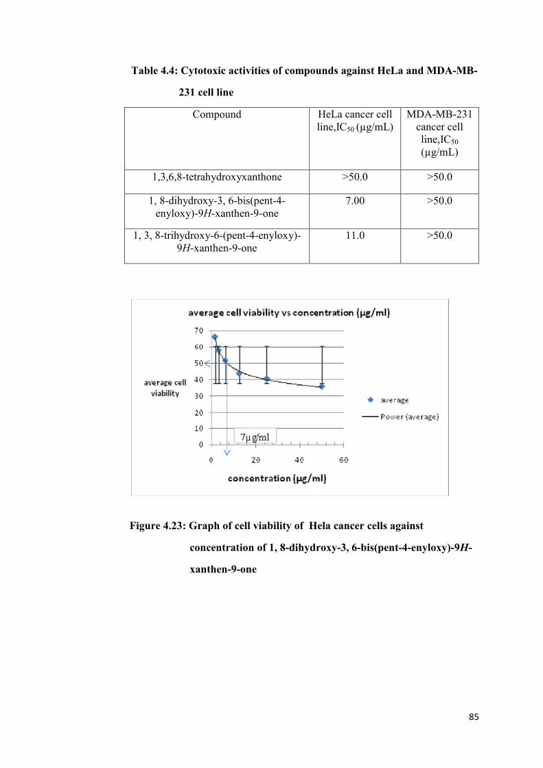
85
Table 4.4: Cytotoxic activities of compounds against HeLa and MDA-MB-
231 cell line
Compound HeLa cancer cell
line,IC50 (µg/mL)
MDA-MB-231
cancer cell
line,IC50
(µg/mL)
1,3,6,8-tetrahydroxyxanthone >50.0 >50.0
1, 8-dihydroxy-3, 6-bis(pent-4-
enyloxy)-9H-xanthen-9-one
7.00 >50.0
1, 3, 8-trihydroxy-6-(pent-4-enyloxy)-
9H-xanthen-9-one
11.0 >50.0
Figure 4.23: Graph of cell viability of Hela cancer cells against
concentration of 1, 8-dihydroxy-3, 6-bis(pent-4-enyloxy)-9H-
xanthen-9-one

86
Figure 4.24: Graph of cell viability of HeLa cancer cells against
concentration of 1, 3, 8-trihydroxy-6-(pent-4-enyloxy)-9H-
xanthen- 9-one
From the results of bioassay, it was found that only 1, 8-dihydroxy-3, 6-
bis(pent-4-enyloxy)-9H-xanthen-9-one and 1, 3, 8-trihydroxy-6-(pent-4-
enyloxy)-9H-xanthen-9-one showed cytotoxic activity toward HeLa cancer cell
line. However, all the compounds tested showed insignificant cytotoxic activity
toward MDA-MB-231 cancer cell line.
The smaller the IC50, the more cytotoxic is the compound towards the cancer
cell line. The compound with IC50 more than 50 µg/mL was considered to be
insignificant in their cytotoxic activity. Thus, 1, 8-dihydroxy-3, 6-bis(pent-4-
enyloxy)-9H-xanthen-9-one with IC50 value of 7.00 µg/mL was found to be the
most cytotoxic toward HeLa cancer cell line as compared to other synthesised

87
compounds. From the results, it also indicated that the more o-substituted
xanthonic block exerted more cytotoxic effect toward the HeLa cancer cell line.
This can be explained when considering the structure of the compound
synthesised.
The xanthonic block, 1, 3, 6, 8-tetrahydroxyxanthone showed insignificant
cytotoxic activity towards HeLa cancer cell lines and the cytotoxic effect
increases when it is mono-substituted with 5-bromo-1-pentene at the –OH
group of C-3. Further increase in the cytotoxic effect when 1, 3, 6, 8-
tetrahydroxyxanthone was di-substituted with 5-bromo-1-pentene at –OH
groups of C-3 and C-6. Thus, the higher the number of o-alkenylated group
attached to xanthone, the more cytotoxic the compound towards HeLa cancer
cell line.

88
CHAPTER 5
CONCLUSIONS
5.1 Conclusions
This project was aimed for the synthesis of xanthonecarboxylic acid but due to
decarboxylation, the carboxyl group has been detached from the xanthonic ring
as carbon dioxide, yielded 1, 3, 6, 8-tetrahydroxyxanthone. There were a total
of three xanthonic compounds being synthesised and isolated in this project.
This included one xanthonic block and two o-alkenylated xanthone. The
xanthonic block was 1, 3, 6, 8-tetrahydroxyxanthone (4.69 g). The two o-
alkenylated xathones were 1, 8-dihydroxy-3, 6-bis(pent-4-enyloxy)-9H-
xanthen-9-one (0.21 g) and 1, 3, 8-trihydroxy-6-(pent-4-enyloxy)-9H-xanthen-
9-one (0.033 g). All these xanthonic compounds had been fully elucidated,
identified and characterised by spectroscopic analyses.
Bioassay had been conducted on HeLa and MDA-MB-231 cancer cell lines by
using 1, 3, 6, 8-tetrahydroxyxanthone, 1, 8-dihydroxy-3, 6-bis(pent-4-
enyloxy)-9H-xanthen-9-one and 1, 3, 8-trihydroxy-6-(pent-4-enyloxy)-9H-
xanthen-9-one as test compounds. All the tested compounds were found to give
insignificant cytotoxic activity toward MDA-MB-231 cancer cell line, having
IC50 of more than 50 µg/mL. In terms of HeLa cancer cell line, only 1, 8-
dihydroxy-3, 6-bis(pent-4-enyloxy)-9H-xanthen-9-one and 1, 3, 8-trihydroxy-

89
6-(pent-4-enyloxy)-9H-xanthen-9-one gave significant cytotoxic activities with
IC50 values of 7.00 µg/mL and 11.00 µg/mL, respectively. However, the
xanthonic block, 1, 3, 6, 8-tetrahydroxyxanthone showed insignificant
cytotoxic activity with IC50 more than 50 µg/mL toward the same cancer cell
line. Thus, it can be concluded that the more o-alkenylation on the xanthonic
compound, the more cytotoxic the compound towards HeLa cancer cell line.
5.2 Suggestion for Further Studies
For further study, different separation methods should be carried out in order to
save the time used for purifying crude products from the synthesis. The other
separation methods suggested to be used are flash column chromatography,
high performance column chromatography or recrystallisation which are found
to give a better separation efficiency.
Besides that, to synthesis xanthonecarboxylic acid, it is suggested a step by
step synthesis should be conducted with the use of protecting group (section
2.3.6) to prevent decarboxylation.
On top of that, it is suggested to carry out c-alkenylation on 1, 3, 6, 8-
tetrahydroxyxanthone to provide insight into the cytotoxic activities of o-
alkenylated xanthones in comparison with c-alkenylated xanthones.

90
Furthermore, extended pharmacological tests should be carried out in order to
find out others pharmacological potential present in the xanthone derivatives.
The pharmacological activities to be studied are anti-inflammatory, anti-
bacterial, anti-fungal, anti-oxidant, anti-allergy and anti-viral activities.
Lastly, further synthesis is suggested to be done on the xanthonic block in
order to produce a wider variety of the xanthonic species for a more
comprehensive structure reactivity relationship (SAR) study to look into
potential structure of xanthone to be developed into drug leads. The further
synthesis might involve cyclisation of the alkenyl group to give additional ring
to the structure or introduce other functional groups to the xanthonic nucleus.

91
REFERENCES
Journal:
Anand, S. M. and Jain A. C. (1973). Claisen Rearrangement of 1-hydroxy-3-
(3-methylbut-2-enyloxy)xanthones. Indian J chem., 11:1237-40
Atkinson, H and Heilbron I. M. (1926). Xanthone as Therapeutic Agents:
Chamistry and pharmacology. J chem Soc,2688-2691
Castanheiro, R. A. P., Pinto M. M. M., Silva A. M. S., Cravo S. M. M., Gales
L., Damas A.M.,Nazareth N., Nascimento M.S.J. and Eaton G.
(2009). Science Direct Bioorganic and Medical Chemistry, 15,
6080-6088.
Demikiran, O. ( 2007). Xanthone in Hypercium: Synthesis and Biological
Activities. Top Heterocycl Chem, 9, 139-178.
Diderot, N. T., Silvere N., Etienne T. (2006). Xanthonea as therapeutic agents:
chemistry and pharmacology. Science direct volume 2, pages 273-298.
Ding, L., Liu B., Qi L. L., Zhou Q. Y., Hou Q., Li J. and Zhang Q.
(2009).Anti-proliferation, Cell Cycle Arrest and Apotosis Induced by
Natural Xanthone from Gentianopsis paludosa MA in Human
Promyelocytic Leukemia Cell line HL-60 cells. Science Direct
Toxicology in vitro, 23, 408-417.

92
Han, Q. B., Yang N. Y., Tian H. L., Qiao C. F., SongJ. Z., Chang D. C., Chen
S. L., Luo K. Q., Xu H. X. ( 2008). Xanthone With Growth Inhibition
Against HeLa Cells from Garcinia xipshuanbannaensis. Science Direct
Phytochemistry, 69, 2187-2192.
Iikubo, K., Yuchi., Noritaka A., Kazuo U. and Shigeru N. (2001).The first
direct synthesis α- mangostin, a Potent Inhibitor of The Acidic
Sphingomyelinase. Pergamon Tetrahedron Letters, 43, 291-293.
Kostakis, I. K., Pouli N., Marakos P., Mikros E., Skaltsounis A. L., Leonce S.,
Atassi G. And Renard P. (2001). Pergamon Bioorganic and Medical
Chemistry, 9, 2793-2802.
Lin, C. N., Liou S.S., Ko F. N. and Teng C. M. (1993). J Pharm Sci, 82:11-6
Na, Y. ( 2009). Recent Cancer Drug Development with Xanthone Structures.
Journal of Pharmacy and Pharmacology, 61,207-712.
Patel G. N. and Trivedi K. N. (1988). J Indian Chem Soc, 65, 192-3.
Pikert, M., and August W. F. (1998). Substituted Xanthone as Antiycobacterial
Agents, Part 1: Synthesis and Assignment of 1H/
13C NMR Chemical
Shift. Willey-VCH Verlag Gmbh,D-69451 Weinheim.
Pillai, R. K. M., Naiksatam P., Johnson F., Rajagopalan R., Watts P. C.,
Cricchio R., Borras S. (1986). J Org Chem, 51, 717-23.
Schwaebe, M. K., Moran T. J. and Whitten J. P. (2004).Total Synthesis of
Psorospermin. Elsevier Tetrahedron Letters, 46, 827-829.

93
Sousa, E., Ana P., Nair N., Luis G., Ana M. D., Maria S. J. N. and Madalena P.
(2009) Bromoalkoxyxanthones as Promising Antitumor Agents:
Synthesis, Crystal Structure and Effect on Human Tumor Cell Lines.
European Journal of Medicinal Chemistry, 44, 3830-3835.
Suphavenich, K., Maitarad P., Hannongbua S., Sudta P., Suksamararn S.,
Tantirungrotechai Y., Limtrakul J. (2009). COMFA and CoMSIA
Studies on a New Series of Xanthone Derivatices Against the
Oral Human Epidermoid Carcinoma (KB) Cancer Cell Line.
Montash Chem, 140, 273-280.
Varacha-Lembege, M., Moreau S., Larrouture S., Montaudon D., Robert J. and
Nuhrich A.(2008). European Journal of Medical Chemistry ,43,
1336-1343.
Vitale, A.A., Romanelli G. P., Autino J.C.,Pomilio A. B.(1994). J Chem Res
(S), 82-83.
Woo, S., Jung J., Lee C.S., Kwon Y. J. and Na Y. (2006). Synthesis of New
Xanthone Analogous and Their Biological Activity Test-cytotoxic,
Topoisomerase II inhibition and DNA Cross- linking Study.
Bioorganic and Medical Chemistry letters,17, 1163-1166.
Zhao, J. and Richard C. L. (2007). Synthesis of Xanthone, Thioxanthone and
Acridones by the Coupling of Arynes and Substituted Benzoates. J Org
Chem, 72(2), 583-588. doi: 10.1021/jo0620718.

94
Book Chapter:
Cambell, R. (2005). International Edition Biology. 7th
Edition. (pp 218 - 233).
Pearson Benjamin Cumming.

95
APPENDICES
APPENDIX 1
HMBC of 1, 8-dihydroxy-3, 6-bis(pent-4-enyloxy)-9H-xanthen-9-one

96
APPENDIX 2
HMQC of 1, 8-dihydroxy-3, 6-bis(pent-4-enyloxy)-9H-xanthen-9-one

97
APPENDIX 3
HMBC of 1, 3, 8-trihydroxy-6-(pent-4-enyloxy)-9H-xanthen-9-one

98
APPENDIX 4
HMQC of 1, 3, 8-trihydroxy-6-(pent-4-enyloxy)-9H-xanthen-9-one

99
APPENDIX 5
Raw Data of MTT assay –HeLa (SHB 1 is 1, 8-dihydroxy-3, 6-bis(pent-4-enyloxy)-9H-xanthen-9-one, SHB 2 is 1, 3, 8-trihydroxy-6-
(pent-4-enyloxy)-9H-xanthen-9-one)
Sample
Conc.
(µg/ml)
Replicate 1 Replicate 2 Replicate 3
1st
2nd
3rd
Control 1st
2nd
3rd
Control 1st
2nd
3rd
Control
SHB1
50 0.724 0.722 0.726 1.610 0.603 0.609 0.605 1.468 0.454 0.459 0.461 1.201
25 0.735 0.739 0.731 1.628 0.681 0.678 0.683 1.408 0.542 0.547 0.549 1.153
12.5 0.756 0.753 0.759 1.580 0.709 0.703 0.701 1.475 0.612 0.619 0.61 1.218
6.25 0.744 0.879 0.892 0.166 0.806 0.802 0.809 0.103 0.723 0.719 0.721 0.125
3.12 0.980 1.037 1.043 0.165 0.851 0.849 0.845 0.124 0.751 0.756 0.759 0.130
1.56 1.201 1.155 1.068 0.174 0.912 0.918 0.915 0.100 0.873 0.879 0.881 0.124
SHB2
50 0.701 0.708 0.809 1.610 0.608 0.610 0.623 1.468 0.432 0.427 0.434 0.979
25 0.741 0.743 0.743 1.628 0.654 0.649 0.630 1.408 0.501 0.508 0.504 0.923
12.5 0.767 0.820 0.742 1.580 0.740 0.749 0.739 1.475 0.596 0.589 0.583 0.896
6.25 0.801 0.940 0.941 0.166 0.801 0.809 0.810 0.103 0.631 0.634 0.638 0.114
3.12 0.971 1.046 0.861 0.165 0.920 0.912 0.917 0.124 0.721 0.718 0.715 0.115
1.56 1.045 1.035 1.089 0.174 1.084 1.088 1.078 0.100 0.835 0.811 0.861 0.100

100
APPENDIX 6
Raw Data of MTT assay- MDA-MB-231 Cancer Cell Line
Sample
Conc.
(µg/ml)
Replicate 1 Replicate 2 Replicate 3
1st
2nd
3rd
Control 1st
2nd
3rd
Control 1st
2nd
3rd
Control
SHB1
50 2.612 2.582 2.890 2.503 2.573 2.09 2.593 2.514 1.024 1.027 1.023 0.953
25 2.210 2.161 2.635 2.643 1.866 1.963 1.624 2.128 1.002 1.004 1.006 1.029
12.5 1.688 2.036 2.726 2.117 1.959 1.773 1.872 2.411 0.990 0.989 1.022 0.920
6.25 1.974 2.549 1.876 0.130 2.060 1.895 1.701 0.106 0.988 0.908 1.037 0.183
3.12 2.201 2.205 2.209 0.129 2.031 2.038 2.029 0.108 0.935 0.942 0.943 0.185
1.56 2.201 2.209 2.206 0.136 2.01 2.015 2.012 0.099 0.903 0.905 0.835 0.190
SHB2
50 2.794 2.143 2.827 2.503 3.053 2.381 2.638 2.514 1.001 1.077 1.079 0.953
25 1.836 1.623 1.751 2.643 2.305 1.939 2.076 2.128 0.998 1.000 0.993 1.029
12.5 1.570 1.575 1.577 2.117 1.976 1.828 1.782 2.411 1.020 0.986 0.989 0.920
6.25 1.569 1.567 1.568 0.130 1.724 1.810 1.989 0.106 0.940 0.949 0.954 0.183
3.12 1.550 1.555 1.557 0.129 1.897 1.895 1.890 0.108 0.939 0.935 0.856 0.185
1.56 1.669 1.675 1.663 0.136 1.908 1.907 1.902 0.099 0.901 0.900 0.890 0.190

101