synthesis, characterization and antibacterial activity of polyaniline/pt–pd nanocomposite
TRANSCRIPT
lable at ScienceDirect
European Journal of Medicinal Chemistry 72 (2014) 18e25
Contents lists avai
European Journal of Medicinal Chemistry
journal homepage: http: / /www.elsevier .com/locate/ejmech
Original article
Synthesis, characterization and antibacterial activity ofpolyaniline/PtePd nanocomposite
Pandi Boomi a, Halliah Gurumallesh Prabu a,*, Jayaraman Mathiyarasu b
aDepartment of Industrial Chemistry, School of Chemical Sciences, Alagappa University, Karaikudi 630 003, Tamilnadu, Indiab Electrodics and Electrocatalysis Division, CSIR-Central Electrochemical Research Institute, Karaikudi 630 006, Tamilnadu, India
a r t i c l e i n f o
Article history:Received 15 May 2013Received in revised form23 September 2013Accepted 28 September 2013Available online 25 November 2013
Keywords:PolyanilinePtePd nanocompositeSynthesisCharacterizationAntibacterial activity
* Corresponding author. Tel.: þ91 4565 228836; faxE-mail addresses: [email protected]
[email protected] (H.G. Prabu).
0223-5234/$ e see front matter � 2013 Elsevier Mashttp://dx.doi.org/10.1016/j.ejmech.2013.09.049
a b s t r a c t
Pt colloid and PtePd colloid, pristine polyaniline, polyaniline/Pt nanocomposite and polyaniline/PtePdnanocomposite were synthesized by simple chemical method. They were characterized by UVeVis, FT-IR,XRD, TGA, SEM, HR-SEM and HR-TEM with EDAX techniques. The results proved that there is a stronginteraction between metal nanoparticles (PtePd) and polyaniline chains. This interaction creates changesin the backbone chain of polyaniline/PtePd nanocomposite when compared to pristine polyaniline. Thesynthesized materials were evaluated for antibacterial activity, minimal inhibitory concentration andminimal bactericidal concentration. The results indicated that the nanocomposites exhibited improvedantibacterial activity when compared to pristine polyaniline and individual metal colloids. This is the firstreport on the chemical synthesis of polyaniline/PtePd nanocomposite, which exhibits antibacterial ac-tivity at micro molar concentration levels.
� 2013 Elsevier Masson SAS. All rights reserved.
1. Introduction
Among the conducting polymers, polyaniline (PANI) has beenextensively investigated due of its ease of synthesis [1], potentialapplication in polymer light-emitting diodes [2], biosensors [3], gassensors [4] and fuel cell catalysts [5]. Antibacterial activity of PANIhas been studied with different bacterial strains for food packagingand medical devices [6]. It has been demonstrated that PANI hassignificant antibacterial activity against E. coli and S. aureus underdark and visible light conditions [7]. PANI was dissolved in dimethylsulfoxide at different concentrations that showed antibacterialproperty against various Gram-positive and Gram-negative bacte-rial strains [8]. PANI incorporated with metal nanoparticles is alsoreported to contain enhanced biological activity. Polyurethanecoated with PANI and PANI/silver nanocomposite has been studiedfor antibacterial activity. It showed that the nanocompositespossessed antibacterial activity and improved biocompatibilitywhen compared to the pristine polyurethane or PANI [9]. Silver-PANI nanocomposite was synthesized by chemical method usingdodecyl benzene sulphonic acid. It exhibited excellent antibacterialactivity when compared to pristine Ag nano powder [10]. The
: þ91 4565 225202., [email protected],
son SAS. All rights reserved.
nanocomposite of PANI-Cu0.05Zn0.95O also reported to contain sig-nificant antibacterial activity against E. coli, S. aureus and C. albicans[11].
Even though, the antibacterial activity of PANI/monometalbased material has been explored, the activity of PANI/PtePdnanoparticle has not been reported. In this study, metal colloidalsolutions of Pt and PtePd were prepared by chemical reductionmethod using reducing and stabilizing agents. Pristine PANI and itscomposites were synthesized by chemical method. The synthesizedmetal colloids, pristine PANI and its composites were characterizedby different techniques. The antibacterial activity, minimal inhibi-tory concentration (MIC) and minimal bactericidal concentration(MBC) behaviour of the synthesized pristine PANI, PANI/Pt andPANI/PtePd nanocomposites were tested and discussed.
2. Experimental
2.1. Materials and methods
Aniline monomer (SigmaeAldrich) was purified by simpledouble distillation under N2(g) atmosphere and stored under N2(g)at 5 �C before use. Palladium (II) chloride was used as received fromAcross. Hexachloro platinic acid and N-methyl-2-pyrrolidone(NMP) were obtained from E-Merck. Ammonium persulphate(Loba-Chemi) and sulphuric acid (sd fine) were used withoutfurther purification. Sodium borohydride and poly(N-vinyl-2-
P. Boomi et al. / European Journal of Medicinal Chemistry 72 (2014) 18e25 19
pyrrolidone) (MW ¼ 40,000) were received from Alfa Aesar. Bac-terial strains of Streptococcus sp (MTCC 890), Staphylococcus sp(MTCC 96), E. coli (MTCC 1671) and Klebsiella sp (MTCC 7407) wereobtained from the Centre for Marine Pharmacology, School ofMarine Sciences, Alagappa University, India.
2.2. Preparation of poly(N-vinyl-2-pyrrolidone) (PVP) stabilized Ptand PtePd colloidal solutions
PVP stabilized Pt colloidal solution was prepared by the chem-ical reduction method. For this, a designed amount (6.6 ml) ofprecursor 3.3 mmol hexachloro platinic acid was dissolved in 20 mlof distilled water and stabilizing agent, 0.15 g of PVP (1.36 mmol ofmonomeric units) was added into this solution. The solutionmixture was stirred vigorously for 30 min until dissolved. 2 mg ofsodium borohydride (20 ml in cold water) was added slowly intothis solution. The colour of the solution was changed quickly fromyellow to dark brown, indicating the formation of Pt particles. Thenthe solution was continuously stirred for 1 h to obtain PVP stabi-lized Pt nanoparticle colloidal solution. Similarly PtePd colloidalsolution was obtained from a mixture of Pt (6.6 ml) and Pd (3.3 ml)precursors.
2.3. Chemical synthesis of pristine PANI, PANI/Pt and PANI/PtePdnanocomposites
0.46 ml of distilled aniline (0.1 M) was added drop-wise into50 ml aqueous solution of sulphuric acid (0.49 ml, 0.1 M) understirring condition. After 10 min of stirring, 10 ml of ammoniumpersulphate (0.228 g, 0.1 M) was added drop-wise. When the so-lution was continuously stirred for 8 h, the colour of the mixturechanged to green and precipitation occurred. The green precipitateobtained was washed with water and methanol several times, andthen dried under vacuum at 60 �C for 12 h to get polyanilinepowder. Similarly, PANI/Pt and PANI/PtePd nanocomposites wereprepared by adding 10 mg Pt and 10 mg PtePd colloidal solutionrespectively into the aniline-sulphuric acid solution.
2.4. Characterization
NMP was used as solvent to prepare pristine PANI, PANI/Pt andPANI/PtePd nanocomposites solution for UVeVis analysis. Theabsorption spectra of metal colloids, pristine PANI and its com-posites were recorded from a UVeVis spectrophotometer (Jasco-V-530) in the wavelength range of 300e900 nm.
Fourier transform infrared (FT-IR) spectra of pristine PANI andits composites were obtained from a FT-IR spectrophotometer(BRUKER Optik GmbH-TENSOR 27) in % transmittance modecovering the wavenumber between 400 and 4000 cm�1. Spec-troscopy grade KBr was used as the windowmaterial. The preparedsamples were mixed with dried KBr, and pelletized.
X-ray diffractometer (X-ray XPERT-PRO) was used to obtain thediffraction patterns and degree of crystallinity of colloids in powderform, pristine PANI and its composite materials. Measurementswere carried out in the continuous mode with a scan speed of 10�
per min covering the angle 2q between 10 and 80�. Cu Kawas usedas the X-ray source with generator setting 30 mA, 40 kV.
Thermogravimetric analysis (Perkin Elmer Diamond) at a heat-ing rate of 20 �C per min in nitrogen atmosphere was used to studythe thermal stability of the powder samples of pristine PANI and itscomposites.
For scanning electron microscopy analysis, the synthesizedmaterial was coated on the carbon tape and images were obtainedusing HITACHI Model S-3000H instrument. High resolution imageswere also obtained using high resolution scanning electron
microscopy (FEI quanta FEG 200 with accelerating voltage at200 kV) with energy dispersive X-ray analysis.
For high resolution transmission electron microscopy analysis,sample was dispersed in isopropyl alcohol using sonicator for15 min. The dispersed sample was coated on carbon coated coppergrid and images were obtained using HR-TEM (JEOL-JEM 2100)with energy dispersive X-ray analysis operating at 200 kV.
2.5. Antibacterial activity test
The synthesized materials were screened for antibacterial ac-tivity by agar well diffusion method [12] against selective Gram-positive (Streptococcus sp and Staphylococcus sp) and Gram-nega-tive (E. coli and Klebsiella sp) bacteria. The pHwasmaintained at 7.3.Sterile molten Mueller Hinton agar (20 ml) was poured into sterilePetri plates and allowed to solidify at room temperature. Six wellswere punched into the agar plate using a sterile well cutter. Over-night culture (108cells/ml) of pathogenic bacteria was swabbed onthe Muller-Hinton agar plates. Further, 100 mg/ml of Pt, PtePdcolloidal solutions, N-methyl-2-pyrrolidone dissolved solution ofpristine PANI, PANI/Pt and PANI/PtePd nanocomposites wereloaded into the each well. Triplicate plates were incubated for 24 hat 37 �C. Then, the zone of inhibition was measured and the resultswere expressed as mm in dia compared to control NMP solution.
2.6. Minimal inhibitory concentration (MIC) analysis
MBC of pristine PANI, PANI/Pt nanocomposite and PANI/PtePdnanocomposite samples dissolved in NMP solution were deter-mined by broth micro-dilution method [13]. Different concentra-tions such as 25, 50, 75, 100, 125 and 150 mL/ml were tested againstStreptococcus sp. In this method, the above sample solution with450 mL of nutrient broth and 50 mL of bacterial culture weretransferred to the tubes and incubated at 37 �C for 24 h and the MICresults were noted.
2.7. Minimal bactericidal concentration (MBC) analysis
For the MBC test, aliquots of different concentrations (25, 50, 75,100, 125 and 150 mL/ml) of pristine PANI, PANI/Pt nanocompositeand PANI/PtePd nanocomposite against Streptococcus sp weredispersed in a Petri plate. The Petri plates were incubated for 24 h at37 �C and the MBC results were noted as the lowest concentrationsafter 24 h [13].
3. Results and discussion
3.1. UVeVis analysis
Fig. S1 shows the absorption spectra of Pt colloidal solution, PtePd colloidal solution, pristine PANI, PANI/Pt nanocomposite andPANI/PtePd nanocomposite. Pt and PtePd colloids did not showany significant absorption peaks, indicating that all the metal ionswere completely reduced into metallic state, resulting in the for-mation of Pt and PtePd nanoparticles [14e16]. The absorptionpeaks observed at around 350 and 620 nm are corresponding to thePANI [17]. The shorter wavelength peak at 350 nm is assigned topep* transition of benzenoid ring, whereas the longer wavelengthpeak at 620 nm corresponds to quinonoid ring of PANI [18]. Thepeak around 620 nm (for PANI/PtePd composite) is broader innature with increased intensity when compared to pristine PANI.This may be due to the incorporation of PtePd nanoparticles intothe PANI.

Fig. 1. TGA of (A) pristine PANI, (B) PANI/Pt nanocomposite and (C) PANI/PtePdnanocomposite.
P. Boomi et al. / European Journal of Medicinal Chemistry 72 (2014) 18e2520
3.2. FT-IR analysis
Fig. S2 shows the vibrational spectra of pristine PANI, PANI/Ptnanocomposite and PANI/PtePd nanocomposite. The character-istic peaks at 1616 cm-1 (curve A) and 1619 cm-1 (curves B and C)are related to the C]C stretching of the quinonoid ring and thepeak at 1418 cm�1 (curves AeC) is attributed to the C]C stretchingof the benzenoid ring of PANI [19]. The peaks at 1229 cm�1 (curveA) and 1277 cm�1 (curves B and C) correspond to the CeNstretching, peaks in the region of 1080e1098 cm�1, are due to ar-omatic in-plane CeH bending modes. In addition, the peaks at3429, 3450 and 3461 cm-1 are assigned as NeH stretching vibra-tion. It is interesting to note that the peak intensity of NeHstretching of the composite enhanced with slight shift in the peakposition than the pristine PANI [20]. It is observed that the Pt andPtePd nanoparticles are incorporated into the polyanilinebackbone.
Fig. 2. SEM with EDAX image of (A) Pt colloidal, (B) PtePd colloidal (C) pristine PANI,nanocomposite.
3.3. X-ray diffraction analysis
XRD patterns of pristine PANI, Pt powder, PtePd powder, PANI/Pt and PANI/PtePd nanocomposites are shown in Fig. S3. PristinePANI (curve A) exhibited a broad peak at 2q 25� that corresponds toperiodicity parallel to the polymer chains of PANI [21]. The peaksobserved at 39.2, 45.6 and 66.5� (curves B and D) are assigned to theface centred cubic (fcc) phase of platinum (111), (200) and (220)planes respectively and are in good agreement with the reporteddata (JCPDS File No. 88-2343) [22]. The 2q positioned of PtePdbimetal and PANI/PtePd nanocomposite (curves C and E) are notsame, indicating the formation of bimetal and PANI/PtePd nano-composite [23e25]. The PANI/Pt and PANI/PtePd nanocompositesare found to havemore crystalline nature than pristine PANI. Thus itis inferred that the nanoparticles of Pt and PtePd are deeplyincorporated into the PANI matrix.
3.4. Thermogravimetric analysis
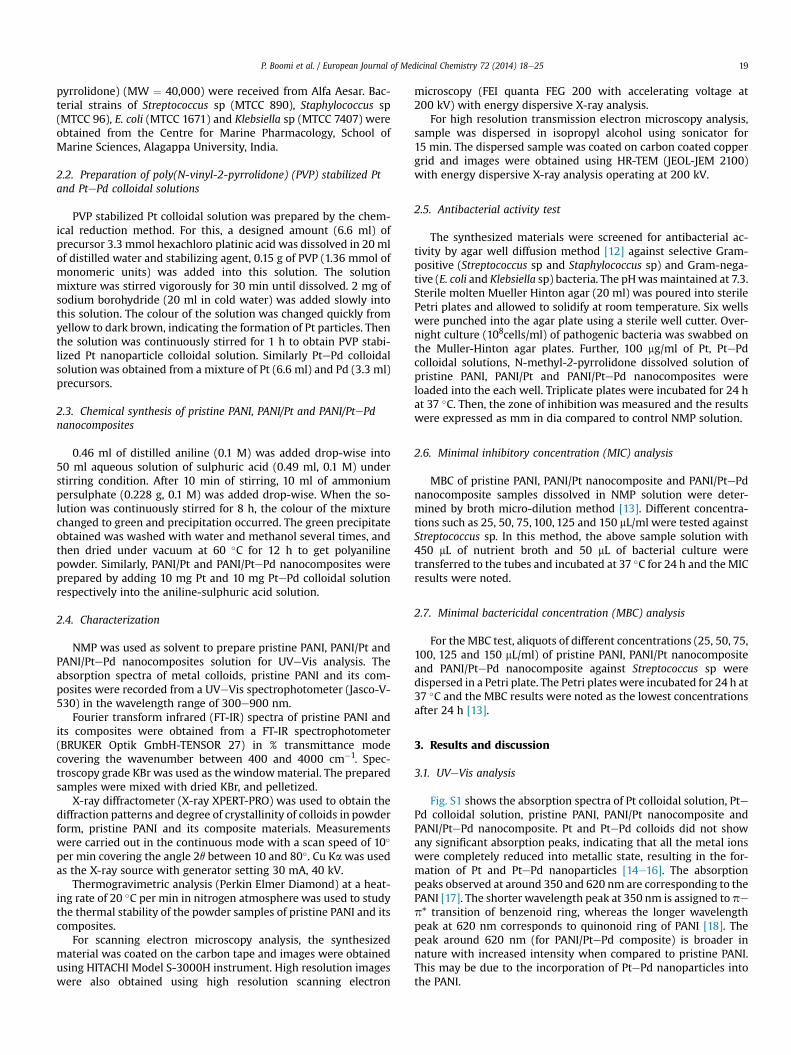
Thermal stability of the pristine PANI and its composites wereanalyzed by thermogravimetric analysis under nitrogen atmo-sphere. Fig. 1 shows TGA curves of pristine PANI, PANI/Pt and PANI/PtePd nanocomposites. The initial mass loss between 45 �C and205 �C is due to the loss of water and unreacted substances. Thesecond weight loss between 205 �C and 540 �C indicated thedecomposition of polymer chains (curves AeC). Pristine PANIdecomposed completely at above 550 �C, whereas, polyaniline/Ptand polyaniline/PtePd nanocomposites did decompose at tem-peratures 610 �C and 690 �C respectively [11,26], which is due to theincreased thermal stabilization of incorporatedmetal nanoparticlesinto the PANI matrix. It is observed that the PANI/PtePd nano-composite has better thermal stability than that of PANI/Pt nano-composite and pristine PANI.
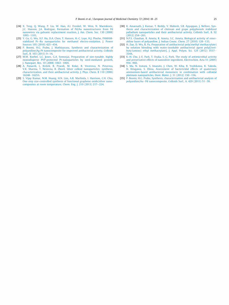
3.5. SEM analysis of colloidal solutions (Pt and PtePd), pristinePANI and nanocomposites (PANI/Pt and PANI/PtePd)
SEM and HR-SEM with EDAX images of colloidal solutions,pristine PANI and PANI metal nanocomposites are shown in Fig. 2.Pt showed spherical as well as needle shapes (image A), whereasPtePd showed only spherical shape (image B) with an average
and HR-SEM with EDAX image of (D) PANI/Pt nanocomposite and (E) PANI/PtePd

P. Boomi et al. / European Journal of Medicinal Chemistry 72 (2014) 18e25 21
diameter of about 100 nm. The SEM of pristine PANI (image C)showed micro porous structure with an average diameter of lessthan 100 nm. It is pertinent to note that incorporation of Pt nano-particles in the PANI matrix has resulted significant changes in thestructure and morphology of the composite. Individual smallspherical (bright spots) shaped Pt and PtePd nanoparticles are welldispersed on PANI surface with a diameter of less than 100 nm(images D and E). The presence of Pt and PtePd were confirmed byenergy dispersive x-ray (EDAX) analysis.
3.6. HR-TEM of Pt colloidal solution and PANI/Pt nanocomposite
Fig. 3 shows the HR-TEM images, histogram, SAED pattern andEDAX analysis of Pt colloidal and PANI/Pt nanocomposite (imagesAeF). Pt nanoparticles showed spherical structure (image A) withan average diameter of 3e10 nm (image B). The highest Pt nano-particle population is found to be 4 nm. The Pt colloidal
Fig. 3. HR-TEM of Pt colloidal solution: (A) image, (B) size histogram, (C) SAED pattern, (D)
nanoparticles are relatively unstable as precipitation occurredwithin a month (data not shown), probably due to the particleaggregation. From the SAED pattern, the planes observed (111),(200) and (220) are corresponding to the Pt nanoparticles (imageC). Presence of Pt is confirmed with EDAX patterns as shown inimage D. Image E revealed that the small spherical Pt particles areuniformly distributed on the surface of the PANI matrix with a fewaggregated finer particles. Presence of different sizes of Pt nano-particles in PANI/Pt nanocomposite is observed from the image F.The average Pt particle size is found to be in the range of 1e11 nmwith highest particle population of 5 nm.
3.7. HR-TEM of PtePd colloidal solution and PANI/PtePdnanocomposite
HR-TEM analysis of PtePd colloidal solution and PANI/PtePdnanocomposite are shown in Fig. 4. Image A showed that the PtePd
EDAX spectrum and HR-TEM of PANI/Pt nanocomposite: (E) image, (F) size histogram.

Fig. 4. HR-TEM of PtePd colloidal solution: (A) image, (B) size histogram, (C) lattice image and (D) SAED pattern and HR-TEM of polyaniline/PtePd: (E) image and (F) size histogramof nanocomposite.
P. Boomi et al. / European Journal of Medicinal Chemistry 72 (2014) 18e2522
nanoparticles have spherical morphology and the particles sizedimension is found to be 1e7 nm as shown in image B. Image Creveals that single crystal PtePd nanoparticles with clear latticefringes of 0.25 nm d-spacing corresponding to the PtePd crystalplane. In addition, (111), (200) and (220) planes of PtePd bimetallicnanoparticles adopt face-centred cubic (fcc) structure that isconfirmed by SAED pattern (image D), which is typically crystallinestructure of PtePd bimetal. Nanocomposite formation of PANI/PtePd is confirmed from the image E. It shows small spherical nano-particles of PtePd that are well dispersed on the surface of the PANIwith few aggregates. Size and shape of Pt nanoparticles is
controlled exclusively by the action of PVP as capping agent [27].These results indicate the importance of PVP for the good disper-sion of PtePd nanoparticles on PANI matrix. PVP promotes theinteraction between the PtePd nanoparticles and PANI.
3.8. Antibacterial activity results
Antibacterial activity was carried out by agar well diffusionmethod and the results were illustrated in Fig. 5. It reveals that, thecolloidal solutions of Pt and PtePd did not show the zone of inhi-bition against the pathogens studied, whereas the zone of

Fig. 5. Antibacterial activity against: (A) E. coli, (B) Klebsiella sp, (C) Streptococcus sp and (D) Staphylococcus sp. [(i) Pt colloidal, (ii) PtePd colloidal, (iii) pristine PANI, (iv) PANI/Ptnanocomposite, (v) PANI/PtePd nanocomposite and (c) control].
P. Boomi et al. / European Journal of Medicinal Chemistry 72 (2014) 18e25 23
inhibition was observed for pristine polyaniline, polyaniline/Pt andpolyaniline/PtePd nanocomposites against E. coli, Klebsiella sp,Streptococcus sp and Staphylococcus sp. The maximum zone of in-hibition was observed for polyaniline/PtePd nanocompositeagainst Staphylococcus sp. (28� 0.70mm dia) followed by Klebsiellasp. (25� 0.85 mm dia), E. coli (22� 0.36 mm dia) and Streptococcussp. (21� 0.30 mm dia). Polyaniline/Pt nanocomposite showed zoneof inhibition against Staphylococcus sp. (23� 0.38mmdia) followedby Klebsiella sp. (20 � 0.36 mm dia), E. coli (19 � 0.40 mm dia) andStreptococcus sp. (18 � 0.41 mm dia). In addition to that the zone ofinhibition of pristine PANI was recorded against Staphylococcus sp.(19 � 0.41 mm dia) followed by Klebsiella sp. (17 � 0.31 mm dia),E. coli (15 � 0.29 mm dia) and Streptococcus sp. (14 � 0.51 mm dia).Thus, the polyaniline/PtePd nanocomposite has showed improvedantibacterial activity against Staphylococcus sp than the other bac-terial strains tested.
Among the bacterial strains tested for antibacterial activity, theStaphylococcus sp showed good response against the materialstested than others species (Fig. 5). Hence, Staphylococcus sp strainwas studied further for MIC and MBC methods. The MIC results ofpristine PANI, PANI/Pt nanocomposite and PANI/PtePd nano-composite are presented in Table 1. It reveals the existence ofantibacterial activity at different concentration levels studied:
Table 1MIC and MBC of pristine PANI, PANI/Pt nanocomposite and PANI/PtePd nanocomposite
Analyte Analyte concentration (mg/ml)
MIC
25 50 75 100 125
Pristine PANI � � þ þ þPANI/Pt nanocomposite � þ þ þ þPANI/PtePd nanocomposite þ þ þ þ þ
(�) Represents no activity; (þ) represents activity.
PANI/PtePd nanocomposite (25e150 mg/ml), PANI/Pt nano-composite (50e150 mg/ml), pristine PANI (75e150 mg/ml). Similarresults have been obtained in MBC test also. From the MIC andMBCresults, it is concluded that the PANI/PtePd nanocomposite hasgood antibacterial activity than PANI/Pt nanocomposite and pris-tine PANI.
From the study by others, it is reported that nanoparticles andnanocomposites are highly active due its large surface and size[28e30]. PANI has significant antibacterial activity due to variousfactors such as surface hydrophilicity [9], electrostatic adsorptionbetween PANI and bacteria [11], higher molecular weight [31] anddirect contact between polymer material and bacterial cells [32]. Inthis present investigation, it is revealed that either platinum orplatinum-palladium bimetal nanoparticle alone does not exhibitany antibacterial activity [33,34]. PANI exhibited a low antibacterialactivity against all range of bacterial species tested. Interestingly,PANI/Pt nanocomposite as well as PANI/PtePd nanocompositeexhibited an improved antibacterial activity when compared to thepristine PANI. Recently, we have reported that the PANI/bimetalnanocomposite has improved antibacterial activity than pristinePANI, due to the interaction between metal nanoparticles and PANImatrix [26] and lower size of the nanoparticles in PANI/metalnanocomposites has improved antibacterial activity [35]. In
against Staphylococcus sp.
MBC
150 25 50 75 100 125 150
þ � � þ þ þ þþ � þ þ þ þ þþ þ þ þ þ þ þ

Scheme 1. Interaction between PANI/PtePd nanocomposite and Bacterial strain.
P. Boomi et al. / European Journal of Medicinal Chemistry 72 (2014) 18e2524
general, polyaniline and its composites may attach to the cell wallmembrane and thus disturb cell wall permeability. They mayalso break inside the cell causing damage by interacting withphosphorus and sulphur containing compounds such as DNA andprotein. Individual damage of bacteria with PANI/PtePd nano-composite is presented in Scheme 1.
Our results revealed that the introduction of metal or bimetalnanoparticles into PANI showed enhanced antibacterial activity. Allthree antibacterial test methods (agar well diffusion, MIC andMBC)confirm improved antibacterial activity for PANI/PtePd nano-composite than PANI/Pt nanocomposite and pristine PANI.
4. Conclusion
Pristine PANI, PANI/Pt and PANI/PtePd nanocomposites weresynthesized. The synthesized materials were characterized by UVeVis, FT-IR, XRD, TGA, SEM, HR-SEM and HR-TEM with EDAX ana-lyses. It is observed that nanocomposite containing PtePd nano-particles and PANI has good antibacterial activity againstStreptococcus sp (MTCC 890), Staphylococcus sp (MTCC 96), E. coli(MTCC 1671) and Klebsiella sp (MTCC 7407). PANI/PtePd has gotimproved antibacterial activity than PANI/Pt nanocomposite. This isthe first report on the antibacterial effect of PANI/PtePd nano-composite and may be considered as potential candidate forapplication in futuristic biomedical fields.
Acknowledgement
This work was financially supported by the UGC-BSR ResearchFellowship in Sciences. The authors thank the School of Physics,Alagappa University, Karaikudi for the provision of XRD analysisand School of Marine Science, Alagappa University, Karaikudi forthe antibacterial analysis.
Appendix A. Supplementary data
Supplementary data related to this article can be found at http://dx.doi.org/10.1016/j.ejmech.2013.09.049.
References
[1] J. Stejskal, R.G. Gilbert, Polyaniline preparation of a conducting polymer, PureAppl. Chem. 74 (2002) 857e867.
[2] H. Bejbouj, L. Vignau, J.L. Miane, T. Olinga, G. Wantz, A. Mouhsen, E.M. Oualim,M. Harmouchi, Influence of the nature of polyaniline-based hole-injectinglayer on polymer light emitting diode performances, Mater. Sci. Eng., B 166(2010) 185e189.
[3] I. Lee, X. Luo, J. Huang, X.T. Cui, M. Yun, Detection of cardiac biomarkers usingsingle polyaniline nanowire-based conductometric biosensors, Biosensors 2(2012) 205e220.
[4] J.L. Wojkiewicz, V.N. Bliznyuk, S. Carquigny, N. Elkamchi, N. Redon, T. Lasri,A.A. Pud, S. Reynaud, Nanostructured polyaniline-based composites for ppbrange ammonia sensing, Sens. Actuators, B 160 (2011) 1394e1403.
[5] Y.F. Huang, C.S. Chang, C.W. Lin, An effective layout of polyaniline nanofibersincorporated in membrane-electrode assembly as methanol transport regu-lator for direct methanol fuel cells, Int. J. Hydrogen Energy 37 (2012) 11975e11983.
[6] M.R. Gizdavic-Nikolaidis, J.R. Bennett, S. Swift, A.J. Easteal, M. Ambrose, Broadspectrum antimicrobial activity of functionalized polyanilines, Acta Biomater.7 (2011) 4204e4209.
[7] N. Shi, X. Guo, H. Jing, J. Gong, C. Sun, K. Yang, Antibacterial effect of theconducting polyaniline, J. Mater. Sci. Technol. 22 (2006) 289e290.
[8] R.G.S.V. Prasad, K.S.V. Chaitanya, M. Tejoram, D. Basavaraju, K.N. Rao,R. Rakesh Kumar, S. Sreenivasan, A.R. Phani, Antibacterial properties ofnanofiber structured conducting polyaniline synthesized by cost effective wetchemical process, J. Pharm. Res. 5 (2012) 370e373.
[9] P.K. Prabhakar, S. Raj, P.R. Anuradha, S.N. Sawant, M. Doble, Biocompatibilitystudies on polyaniline and polyaniline-silver nanoparticle coated poly-urethane composite, Colloids Surf., B. 86 (2011) 146e153.
[10] M.S. Tamboli, M.V. Kulkarni, R.H. Patil, W.N. Gade, S.C. Navale, B.B. Kale,Nanowires of silver-polyaniline nanocomposite synthesized via in situ poly-merization and its novel functionality as an antibacterial agent, Colloids Surf.,B. 92 (2012) 35e41.
[11] X. Liang, M. Sun, L. Li, R. Qiao, K. Chen, Q. Xiao, F. Xu, Preparation and anti-bacterial activities of polyaniline/Cu0.05Zn0.95O nanocomposites, Dalton Trans.41 (2012) 2804e2811.
[12] Shirley, A. Dayanand, B. Sreedhar, Syed G. Dastager, Antimicrobial activity ofsilver nanoparticles synthesized from novel Streptomyces species, Dig. J.Nanomater. Bios. 5 (2010) 447e451.
[13] M. Valodkar, S. Modi, A. Pal, S. Thakore, Synthesis anti-bacterial activity of Cu,Ag and CueAg alloy nanoparticles: a green approach, Mater. Res. Bull. 46(2011) 384e389.
[14] A. Nirmala Grace, K. Pandian, Pt, PtePd and PtePd/Ru nanoparticles entrap-ped polyaniline electrodes-A potent electrocatalyst towards the oxidation ofglycerol, Electrochem. Commun 8 (2006) 1340e1348.
[15] M. Harada, N. Toshima, K. Yoshida, S. Isoda, Aggregated structure analysis ofpolymer-protected platinum/ruthenium colloidal dispersions using EXAFS,HRTEM, and electron diffraction measurements, J. Colloid Interface Sci. 283(2005) 64e78.
[16] H. Hei, H. He, R. Wang, X. Liu, G. Zhang, Controlled synthesis and character-ization of noble metal nanoparticles, Soft Nanosci. Lett. 2 (2012) 34e40.
[17] V. Sridevi, S. Malathi, C.S. Devi, Synthesis and characterization of polyaniline/gold nanocomposites, Chem. Sci. J. 26 (2011) 1e6.
[18] F.L. Lu, F. Wudll, M. Nowak, A.J. Heeger, Phenyl-capped octaaniline (COA): anexcellent model for polyaniline, J. Am. Chem. Soc. 108 (1986) 8311e8313.
[19] M. Trchova, J. Stejskal, Polyaniline: the infrared spectroscopy of conductingpolymer nanotubes, Pure Appl. Chem. 83 (2011) 1803e1817.
[20] Q. Xu, J. Leng, H-b Li, G-j. Lu, Y. Wang, X.-Y. Hu, The preparation of polyaniline/gold nanocomposites by self-assembly and their electrochemical applications,React. Funct. Polym. 70 (2010) 663e668.
[21] S.K. Pillalamarri, F.D. Blum, S.T. Tokuhiro, J.G. Story, M.F. Bertino, Radiolyticsynthesis of polyaniline nanofibers: a new templateless pathway, Chem.Mater 17 (2005) 227e229.
[22] A. Nyczyk, A. Sniechota, A. Adamczyk, A. Bernasik, W. Turek, M. Hasik, In-vestigations of polyaniline-platinum composites prepared by sodium boro-hydride reduction, Eur. Polym. J. 44 (2008) 1594e1602.
[23] G. Chen, D. Xia, Z. Nie, Z. Wang, L. Wang, L. Zhang, J. Zhang, Facile synthesis ofCoePt hollow sphere electrocatalyst, Chem. Mater. 19 (2007) 1840e1844.
P. Boomi et al. / European Journal of Medicinal Chemistry 72 (2014) 18e25 25
[24] X. Teng, Q. Wang, P. Liu, W. Han, A.I. Frenkel, W. Wen, N. Marinkovic,J.C. Hanson, J.A. Rodrigue, Formation of Pd/Au nanostructures from Pdnanowires via galvanic replacement reaction, J. Am. Chem. Soc. 130 (2008)1093e1101.
[25] Y. Gu, G. Wu, X.F. Hu, D.A. Chen, T. Hansen, H.-C. Loye, H.J. Ploehn, PAMAM-stabilized PteRu nanoparticles for methanol electro-oxidation, J. PowerSources 195 (2010) 425e434.
[26] P. Boomi, H.G. Prabu, J. Mathiyarasu, Synthesis and characterization ofpolyaniline/Ag-Pt nanocomposite for improved antibacterial activity, ColloidsSurf., B. 103 (2013) 9e14.
[27] M.M. Koebel, L.C. Jones, G.A. Somorjai, Preparation of size-tunable, highlymonodisperse PVP-protected Pt-nanoparticles by seed-mediated growth,J. Nanopart. Res. 10 (2008) 1063e1069.
[28] A. Panacek, L. Kvitek, R. Prucek, M. Kolar, R. Vecerova, N. Pizurova,V.K. Sharma, T. Nevecna, R. Zboril, Silver colloid nanoparticles: synthesis,characterization, and their antibacterial activity, J. Phys. Chem. B 110 (2006)16248e16253.
[29] S. Vijay Kumar, N.M. Huang, H.N. Lim, A.R. Marlinda, I. Harrison, C.H. Chia,One-step size-controlled synthesis of functional graphene oxide/silver nano-composites at room temperature, Chem. Eng. J. 219 (2013) 217e224.
[30] K. Amarnath, J. Kumar, T. Reddy, V. Mahesh, S.R. Ayyappan, J. Nellore, Syn-thesis and characterization of chitosan and grape polyphenols stabilizedpalladium nanoparticles and their antibacterial activity, Colloids Surf., B. 92(2012) 254e261.
[31] N.P.S. Chauhan, R. Ameta, R. Ameta, S.C. Ameta, Biological activity of emer-aldine bases of polyaniline, J. Indian Counc. Chem. 27 (2010) 128e133.
[32] H. Zuo, D. Wu, R. Fu, Preparation of antibacterial poly(methyl methacrylate)by solution blending with water-insoluble antibacterial agent poly[(tert-buty1amino) ethyl methacrylate], J. Appl. Polym. Sci. 125 (2012) 3537e3544.
[33] K.-H. Cho, J.-E. Park, T. Osaka, S.-G. Park, The study of antimicrobial activityand preservative effects of nanosilver ingredient, Electrochim. Acta 51 (2005)956e960.
[34] S. Ma, N. Izutani, S. Imazato, J. Chen, W. Kiba, R. Yoshikawa, K. Takeda,H. Kitagawa, S. Ebisu, Assessment of bactericidal effects of quaternaryammonium-based antibacterial monomers in combination with colloidalplatinum nanoparticles, Dent. Mater. J. 31 (2012) 150e156.
[35] P. Boomi, H.G. Prabu, Synthesis, characterization and antibacterial analysis ofpolyaniline/AuePd nanocomposite, Colloids Surf., A. 429 (2013) 51e59.