synthesis, characterization and bioactivites of dithiocarbazate schiff
TRANSCRIPT
Synthesis, characterization and bioactivites of
dithiocarbazate Schiff base ligands and their metal
complexes
May Lee Low
To cite this version:
May Lee Low. Synthesis, characterization and bioactivites of dithiocarbazate Schiff base ligandsand their metal complexes. Organic chemistry. Universite Pierre et Marie Curie - Paris VI,2014. English. <NNT : 2014PA066148>. <tel-01175653>
HAL Id: tel-01175653
https://tel.archives-ouvertes.fr/tel-01175653
Submitted on 11 Jul 2015
HAL is a multi-disciplinary open accessarchive for the deposit and dissemination of sci-entific research documents, whether they are pub-lished or not. The documents may come fromteaching and research institutions in France orabroad, or from public or private research centers.
L’archive ouverte pluridisciplinaire HAL, estdestinee au depot et a la diffusion de documentsscientifiques de niveau recherche, publies ou non,emanant des etablissements d’enseignement et derecherche francais ou etrangers, des laboratoirespublics ou prives.
Université Pierre et Marie Curie
Université de cotutelle Ecole doctorale ED406
Laboratoire des BioMolécules
Synthèse, caractérisation et bioactivité de ligands issus de
bases de Schiff dérivées de dithiocarbazate et de leurs
complexes métalliques
May Lee LOW
Thèse de doctorat de Chimie Moléculaire
Dirigée par Karen Crouse et Clotilde Policar
Présentée et soutenue publiquement le 09 Juillet 2014
Devant un jury composé de :
FARINA Yang, Professeur Rapporteur
YUSOF Nor Azah, Professeur Rapporteur
HASENKNOPF Bernold, Professeur Examinateur
CROUSE Karen Anne, Professeur Directeur de thèse
POLICAR Clotilde, Professeur Directeur de thèse
DELSUC Nicolas, Chargé de recherche Invité

ii
All material contained within the thesis, including without limitation text, logos, icons, photographs and all other artwork, is copyright material of Universiti Putra Malaysia unless otherwise stated. Use may be made of any material contained within the thesis for non-commercial purposes from the copyright holder. Commercial use of material may only be made with the express, prior, written permission of Universiti Putra Malaysia.
Copyright © Universiti Putra Malaysia
iii
Abstract of thesis presented to the Senate of Universiti Putra Malaysia and École Doctorale 406 Chimie Moléculaire Université Pierre et Marie Curie in fulfillment of
the requirement for the degree of Doctor of Philosophy
SYNTHESIS, CHARACTERIZATION AND BIOACTIVITIES OF DITHIOCARBAZATE-SCHIFF BASE LIGANDS AND THEIR METAL
COMPLEXES By
LOW MAY LEE 2014
There is an urgent need to discover new drugs with novel mechanisms of action,
higher activity and improved selectivity to address the severe challenge of multi-
drug resistance in treating bacterial infections and cancer. In view of this, Schiff
bases derived from S-substituted dithiocarbazate and their corresponding metal
complexes with a plethora of potentially exciting biological activities and
coordination chemistry are attractive candidates for consideration. Macroacyclic and
open chain metal complexes of tetradentate NNSS and bidentate NS Schiff base
ligands derived from the condensation of S-benzyldithiocarbazate (SBDTC) and S-
methyldithiocarbazate (SMDTC) with 2,5-hexanedione, methyl levulinate, levulinic
acid, 4-carboxybenzaldehyde and 3-acetylcoumarin have been prepared. The
compounds were fully characterized with various physico-chemical and
spectroscopic methods. A total of 11 crystals structure were determined throughout
this work. In order to provide more insight into the behaviour of the complexes in
solution, electron paramagnetic resonance (EPR) and cyclic voltammetry (CV)
experiments were performed. Conjugation of the most promising antimicrobial
compound (Schiff base of SBDTC with 4-carboxybenzaldehyde) to various vectors
(polyarginine, polyethylene glycol (PEG) and phe-arg-!-napthylamide (PA!N) was
iv
achieved using either standard solid phase or solution synthetic methodologies to
prepare improved therapeutic agents. Among the conjugates, the nonaarginine (R9)
derivatives showed the most encouraging synergistic effect upon conjugation and
complexation to copper ion with enhanced water solubility, bacteria cell membrane
permeability and bioactivity. The Cu(II) R9 derivatives possess remarkable
antibacterial activity against a wide spectrum of bacteria and in particular, highly
efficacious against S. aureus with MIC values up to 1-0.5 µM when tested against
nine strains of Gram-positive and Gram-negative bacteria. This appears to be the
pioneer study to show that the conjugation of polyarginine to dithiocarbazate
compounds can greatly influence their therapeutic potential. Cytotoxic assay was
also carried out for selected non-conjugated compounds. All the selected Cu(II)
complexes assayed against breast cancer cells lines (MCF-7 and MDA-MB-231)
exhibited good cytotoxicity with lower IC50 values in comparison to their respective
ligands. This work highlights the relevance of metal complexation strategy to
stabilize the ligands and improve their bioactivity. The structure-activity
relationships of the compounds are discussed.
v
Abstrak tesis yang dikemukakan kepada Senat Universiti Putra Malaysia dan École Doctorale 406 Chimie Moléculaire Université Pierre et Marie Curie
sebagai memenuhi keperluan untuk Ijazah Doktor Falsafah
SINTESIS, PENCIRIAN DAN AKTIVITI BIOLOGI LIGAN
DITIOKARBAZAT-BES SCHIFF DAN KOMPLEKS LOGAM Oleh
LOW MAY LEE 2014
Terdapat keperluan segera untuk menemui ubat-ubatan baru dengan mekanisme
baru, aktiviti yang lebih tinggi dan tindakan yang lebih khusus bagi menangani
cabaran yang serius iaitu rintangan terhadap pelbagai ubat-ubatan dalam rawatan
jangkitan bakteria dan kanser. Memandangkan situasi ini, bes Schiff dan kompleks
logam yang berasal daripada S-gantian ditiokarbazat yang mempunyai pelbagai
potensi aktiviti biologi dan kimia koordinasi menarik merupakan calon-calon yang
baik untuk pertimbangan. Kompleks logam bersistem makro-bukan-kitaran dan
rantaian-terbuka masing-masing dengan ligan tetradentat NNSS dan bidentat NS bes
Schiff yang berasal daripada kondensasi antara S-benzilditiokarbazat (SBDTC) dan
S-metilditiokarbazat (SMDTC) dengan 2,5-heksanadion, metil levulinat, asid
levulinik, 4-carboxibenzaldehid dan 3-asetilcoumarin telah disediakan. Semua
sebatian tersebut telah dicirikan sepenuhnya dengan pelbagai kaedah fiziko-kimia
dan spektroskopi. Sebanyak 11 struktur kristal telah ditentukan sepanjang kajian ini.
Untuk memberi gambaran yang lebih jelas terhadap sifat-sifat kompleks dalam
larutan, eksperimen elektron resonans paramagnet (EPR) dan voltammetri berkitar
(CV) telah dijalankan. Konjugasi sebatian yang paling berpotensi antimikrob (bes
Schiff SBDTC dengan 4-carboxibenzaldehid) dengan pelbagai vektor (poliarginine,
polietilena glikol (PEG) dan phe-arg-!-naptilamida (PA!N)) telah berjaya dicapai

vi
sama ada melalui metodologi sintetik standard peptida fasa pepejal atau larutan bagi
penyediaan agen terapeutik yang lebih baik. Antara sebatian yang dikonjugasi,
nonaarginine (R9) derivatif menunjukkan kesan sinergi yang paling menggalakkan
melalui konjugasi dan juga pengkompleksan dengan ion kuprum yang turut
membawa kepada perningkatan kelarutan dalam air, ketelapan terhadap membran
sel bakteria dan bioaktiviti sebatian. Cu(II) R9 derivatif memiliki aktiviti
antibakteria yang terbaik terhadap spektrum bakteria yang luas dan khususnya,
sangat berkesan terhadap S. aureus dengan nilai-nilai MIC sehingga 1-0.5 "M
apabila diuji terhadap sembilan jenis bakteria Gram-positif dan Gram-negatif. Ini
merupakan kajian perintis yang menunjukkan bahawa konjugasi antara polyarginine
dengan sebatian ditiokarbazat boleh mempengaruhi potensi terapeutik mereka.
Kajian sitotoksik juga dijalankan untuk segelintir sebatian yang tidak dikonjugasi.
Semua Cu(II) kompleks yang diuji terhadap sel-sel kanser payudara (MCF-7 dan
MDA-MB-231) menunjukkan sifat sitotoksik yang baik dengan nilai-nilai IC50 yang
lebih rendah berbanding dengan ligan masing-masing. Ini menunjukkkan
kesesuaian strategi pengkompleksan dengan ion logam untuk menstabilkan ligan dan
meningkatkan bioaktiviti mereka. Perhubungan di antara struktur dan aktiviti
sebatian juga dibincang.
vii
Résumé de thèse présenté au Sénat de Université Putra Malaysia et de l'École Doctorale Chimie Moléculaire 406 Université Pierre et Marie Curie à
l'accomplissement de l'obligation pour le grade de docteur en philosophie
SYNTHÈSE, LA CARACTÉRISATION ET DE BIOACTIVITÉS
DITHIOCARBAZATE - BASE DE SCHIFF LIGANDS ET LEUR MÉTAL
COMPLEXES
Par
LOW MAY LEE
2014
Il y a de nos jours un besoin urgent de découvrir de nouveaux médicaments avec de
nouveaux mécanismes d'action, une activité plus élevée et une meilleure sélectivité
pour relever le défi de la multirésistance dans le traitement des infections
bactériennes et le cancer. Dans cette perspective, des bases de Schiff dérivées de
dithiocarbazates S-substitué et leurs complexes métalliques correspondants sont des
candidats intéressants puisqu’ils peuvent être facilement synthétisés et permettent
une grande diversité de coordination. Dans cette étude, des complexes
macroacycliques tetradentes SSNN et bidente NS dont les ligands sont issus de la
condensation de la S-benzyldithiocarbazate (SBDTC) ou de la S-
methyldithiocarbazate (SMDTC) avec la 2,5-hexanedione, le lévulinate de méthyle,
l'acide lévulinique, le 4-carboxybenzaldéhyde ou le 3-acétylcoumarine ont été
préparés. Les ligands et complexes synthétisés ont été entièrement caractérisés par
différentes méthodes spectroscopiques et physico-chimiques. 11 structures
cristallines ont été obtenues au cours de ce travail et afin d’étudier en détail la
géométrie, la stabilité et les propriétés de ces complexes en solution, des expériences
de résonance paramagnétique électronique (RPE), de titration calorimétrique
isotherme et de voltamétrie cyclique (CV) ont été réalisées. L’activité
viii
antibactérienne de ces complexes a ensuite été étudiée et a permis de sélectionner un
complexe « leader » (plus efficace, s et fonctionnalisable). Ce complexe a alors été
modifié afin d’augmenter sa stabilité en milieux biologique, sa solubilité dans l’eau
ainsi que son activité. Il a été conjugué avec différentes entités des peptides
pénétrants, un polyéthylène glycol (PEG) et un peptide inhibiteur des pompes
d’efflux bactériennes (Phe-Arg-!-napthylamide (PA!N)). Parmi les conjugués
obtenus, ceux comportant un peptide avec 9 arginines (R9) ont montré un effet
synergique lors de la formation des complexes puisque l’activité anti-bactérienne
des complexes s’est avérée meilleure que celle des ligands et du cuivre seuls. Ces
complexes ont montré une remarquable activité antibactérienne sur neuf souches de
bactéries Gram-positives et Gram-négatives et en particulier, ils se sont avérés très
efficaces contre S.aureus avec des valeurs de concentration minimale inhibitrice
(CMI) de 1 à 0,5 µM. L’activité anti-cancéreuse des complexes non-conjugués a
également été étudiée. Tous les complexes de cuivre sélectionnés et testés sur des
cellules de cancer du sein MCF7 et MDA-MB- 231 ont montré une cytotoxicité
élevée avec des valeurs de CI50 plus faibles pour les complexes par rapport à leurs
ligands respectifs. Ceci met à nouveau en évidence la pertinence d’utiliser les
complexes métalliques, pour à la fois stabiliser les ligands et générer des composés
plus actifs. Les relations structure-activité des composés sont discutées.
ix
ACKNOWLEDGEMENTS
I am most grateful to my main supervisors Professor Karen A. Crouse, Professor
Clotilde Policar and Dr. Nicolas Delsuc for their valuable guidance, advice and
support from the very beginning of my PhD journey until the successful completion
of this thesis. They are my role models whom I will always hold at the highest
esteem. To Professor Karen Crouse, thank you for the inspiration. You instilled in
me the love for inorganic chemistry during my undergraduate studies and eventually
introduced me to the beauty of synthetic chemistry of dithiocarbazate for my PhD.
To Professor Clotilde Policar, thank you for accepting me to your group and giving
me the most incredible opportunity to realize this research project. I greatly
appreciate that. To Dr. Nicolas Delsuc, I could not have imagined having a better
mentor than you. Your enthusiasm, optimism and critical opinions keep me going.
Thank you so much.
I would also like to extend my appreciation to my co-supervisors and collaborators
for making this research possible Dr. Pierre Dorlet for his kind assistance and
suggestions with EPR and CV experiments, Dr. Laure Maigre and Professor Jean-
Marie Pagés for antibacterial evaluation, Dr. Régis Guillot and Dr. Mohamed
Ibrahim M. Tahir for single crystal XRD structure determination, Professor Rozita
Rosli and Dr. Abhimanyu Veerakumarasivam for the access to cytotoxic assay as
well as Dr. François Lambert, Dr. Hélène Bertrand and Dr. Thahira Begum for
meaningful discussion.
x
Many thanks to the wonderful present and past members of Laboratoire des
BioMolécules (LBM) / Ecole Normale Supérieure (ENS) Héloïse, Sarah, Vincent,
Cécile, Margharita, Cillian, Anne-Sophie, Sylvain, Jean-Marie, Julian, Benjamin,
Marilyne, Laure, Anais, Nicolas, Mayeul, Geraldine, Rodrique, Laurent, Alex,
Roba, Pierluca, Enrique, Jing, Paul, Akansha, Victor and many more people that I
had the privilege to know. I could not thank them enough for the kindness that I
received throughout my stay in Paris and all the pleasant memories that we shared in
and outside the lab. It means a lot to me. My gratitude also to my lab mates in
Universiti Putra Malaysia (UPM) Georgiana, Shahedh, Ming Yueh, Shatila and Tan.
You are my rock, thanks very much for the friendship.
The financial support for the project from UPM, the Ministry of Higher Education
(Malaysia) and French ANR Blanc 2010, METABACT grant is gratefully
acknowledged. In addition, I am very thankful for the award of an Erasmus Mundus
Maheva Scholarship and a UPM Graduate Research Fellowship (GRF).
And finally, I wish to thank my dearest mom, dad and younger sister for their
endless love and encouragement. Thank you for believing in me and teaching me to
reach for the stars, to work hard and to always strive to be the very best that I can be.
To them I dedicate this work.
Dans la vie, rien n'est à craindre, tout est à comprendre.
Nothing in life is to be feared, it is only to be understood.
- Marie Curie
xi
APPROVAL
I certify that an Examination Committee has met on (date of viva voce) to conduct the final examination of Low May Lee on her Doctor of Philosophy thesis entitled “Synthesis, Characterization and Bioactivities of Dithiocarbazate-Schiff Base Ligands and Their Metal Complexes” in accordance with Universities and University Colleges Act 1971 and the Constitution of the Universiti Putra Malaysia [P.U.(A) 106] 15 March 1998. The Committee recommends that the student be awarded the degree of Doctor of Philosophy. Members of the Thesis Examination Committee were as follows Name of Chairperson, PhD Title (e.g., Professor/Associate Professor/Ir; omit if irrelevant) Name of Faculty Universiti Putra Malaysia (Chairman) Name of Examiner 1, PhD Title (e.g., Professor/Associate Professor/Ir; omit if irrelevant) Name of Faculty Universiti Putra Malaysia (Internal Examiner) Name of Examiner 2, PhD Title (e.g., Professor/Associate Professor/Ir; omit if irrelevant) Name of Faculty Universiti Putra Malaysia (Internal Examiner) Name of External Examiner, PhD Title (e.g., Professor/Associate Professor/Ir; omit if irrelevant) Name of Department and/or Faculty Name of Organisation (University/Institute) Country (External Examiner)
________________________ NORITAH OMAR, PhD Assoc. Prof. and Deputy Dean (Thesis and Publication) School of Graduate Studies Universiti Putra Malaysia Date
xii
APPROVAL
This thesis was submitted to the Senate of Universiti Putra Malaysia and has been accepted as fulfillment of the requirement for the degree of Doctor of Philosophy. The members of the Supervisory Committee are as follows Karen Anne Crouse, PhD Professor Faculty of Science Universiti Putra Malaysia (Chairman) Clotilde Policar, PhD Professor Laboratoire des BioMolécules (UMR 7203) Université Pierre et Marie Curie (Member) Nicolas Delsuc, PhD Chargé de Recherche CNRS Laboratoire des BioMolécules (UMR 7203) Université Pierre et Marie Curie (Member) Mohamed Ibrahim Mohamed Tahir, D.Phil Senior Lecturer Faculty of Science Universiti Putra Malaysia (Member) Thahira B.S.A Ravoof, PhD Senior Lecturer Faculty of Science Universiti Putra Malaysia (Member) Rozita Rosli, PhD Professor Faculty of Medical and Health Science Universiti Putra Malaysia (Member)
________________________ BUJANG BIN KIM HUAT, PhD Professor and Dean School of Graduate Studies Universiti Putra Malaysia Date
xiii
DECLARATION
I hereby confirm that
• this thesis is my original work;
• quotations, illustrations and citations have been duly acknowledged;
• ownership of intellectual property from the thesis is as stipulated in the Memorandum of Agreement (MoA), or as according to the Universiti Putra Malaysia (Research) Rules 2012, in the event where the MoA is absent;
• permission from supervisor and the office of Deputy Vice-Chancellor
(Research and Innovation) are required prior to publishing it (in the form of written, printed or in electronic form) including books, journals, modules, proceedings, popular writings, seminar papers, manuscripts, posters, reports, lecture notes, learning modules or any other materials as stated in the Universiti Putra Malaysia (Research) Rules 2012;
• there is no plagiarism or data falsification/fabrication in the thesis, and
scholarly integrity is upheld as according to the Universiti Putra Malaysia (Graduate Studies) Rules 2003 (Revision 2012-2013) and the Universiti Putra Malaysia (Research) Rules 2012. The thesis has undergone plagiarism detection software.
Signature _________________________ Date __________________ Name and Matric No: Low May Lee (GS26866)
xiv
DECLARATION
This is to confirm that
• the research conducted and the writing of this thesis was under our supervision;
• supervision responsibilities as stated in the Universiti Putra Malaysia
(Graduate Studies) Rules 2003 (Revision 2012-2013) are adhered to. Signature ________________________ Karen Anne Crouse, PhD Professor
Signature ________________________ Clotilde Policar, PhD Professor
Signature ________________________ Mohamed Ibrahim Mohamed Tahir, D.Phil Senior Lecturer
Signature ________________________ Nicolas Delsuc, PhD Chargé de Recherche CNRS
Signature ________________________ Dr. Thahira B.S.A Ravoof, PhD Senior Lecturer
Signature ________________________ Rozita Rosli, PhD Professor
xv
LIST OF TABLES
Table
Page
3.1 Selected bond lengths for SBPY, SMHD, CuSMHD and
CuSBHD. 3.2 Selected bond angles for CuSMHD, CuSBHD, CuATSM
(Blower et al., 2003) and CuAATSM. 3.3 Selected bond lengths for SMML, SBML, SBEL and SBLA. 3.4 Selected bond angles for SMML, SBML, SBEL and SBLA. 3.5 Intermolecular hydrogen bonds for SMML, SBML, SBEL and
SBLA. 3.6 Selected dihedral angles between the two planes for SMML,
SBML, SBEL and SBLA. 3.7 Selected bond lengths for Cu(SMML)2 and Cu(SMLA)2. 3.8 Selected bond angles for Cu(SMML)2 and Cu(SMLA)2.
3.9 Main bond lengths (Å) and angles (°) in the coordination sphere
around the rhenium atom. 3.10 Bond lengths (Å) and angles (°) in the dithiocarbazate fragments. 3.11 EPR parameters measured from the spectra of the copper(II)
complexes. 3.12 Electrochemical data for CuSMHD and CuSBHD versus
Ag/AgCl. 3.13 Electrochemical data for the Cu(II) complexes vs Ag/AgCl at
0.1V. 3.14 Electrochemical data for the Cu(SMML)2 vs Ag/AgCl at various
scan rate (V/s). 4.1 Selected ligands and their Cu(II) complexes that were
synthesized and studied. 4.2 LC-ES-MS data for all Cu(II) dithiocarbazate Schiff base ligand-
conjugates and R1-SB4CB for comparison.
59
59
65
65
66
66
69
69
74
74
85
90
92
94
105
136
xvi
4.3 Thermodynamic parameters of conjugated ligand complexation with copper determined by ITC at 25 °C.
4.4 EPR parameters measured from the spectra of the copper(II)
complexes. 4.5 Electrochemical data vs Ag/AgCl. 5.1 Bacteria strains. 5.2 Antimicrobial activity of the tetradentate series. 5.3 Antimicrobial activity of non-conjugated bidentate series. 5.4 Antimicrobial activity of bioconjugate series. 5.5 Final antimicrobial evaluation against 9 strains of bacteria.
5.6 Cytotoxic assay results.
138
141
143
157
162 - 163
168 - 169
176 - 177
181 - 182
187
xvii
LIST OF FIGURES
Figure Page
1.1 (a) Decade-wise approval of new antibiotics and (b) prevalence of MRSA.
2.1 Various S-substituents at position R1 in dithiocarbazates. 2.2 Examples of different series of carbonyl compounds that have
been used for the preparation of dithiocarbazate ligands. 2.3 Examples of different dithiocarbazate derivatives (a) with sugars,
amino acid and calixarene (b) with modifications at N1 atom. 2.4 Different conformations of dithiocarbazate. 2.5 (a) Thione-thiol tautomerism (b) C=S and S=C conformers. 2.6 Compounds with antimigratory activity. 2.7 (a) Schiff bases of SBDTC with 2-acetylpyridine, 2-
benzoylpyridine and 6-methyl-2-formylpyridine, respectively in thione form (b) saccarinate anion.
2.8 Mixed ligand Pt(II) and Pd(II) complexes with Schiff base
(derived from SBDTC and 2-hydroxyacetophenone) and phosphine ligands.
2.9 Ternary structures of VO complexes and the phenanthroline
bases used. 3.1 RP-HPLC chromatogram of SBHD at 220 nm (top) and 280 nm
(bottom). 3.2 RP-HPLC chromatogram of CuSBHD. 3.3 RP-HPLC chromatogram of SMML. 3.4 RP-HPLC chromatogram of Cu(SMML)2. 3.5 RP-HPLC chromatogram of SM4CB. 3.6 FT-IR spectra recorded for ligand SBHD and complex CuSBHD.
3.7 FT-IR spectra recorded for ligand SMML and complex Cu(SMML)2.
2
5
6
7
8
9
11
14
19
20
53
53
53
54
54
57
57
xviii
3.8 ORTEP drawing of (a) SMHD (b) SBPY (c) CuSMHD and d) CuSBHD. Ellipsoids are drawn at the 50% probability level.
3.9 ORTEP diagrams and intermolecular hydrogen bonds (shown as
dotted line) of (a) SMML (b) SBLA (c) SBML (d) SBEL. Ellipsoids are drawn at the 50% probability level.
3.10 ORTEP diagrams of (a) Cu(SMML)2 and (b) Cu(SMLA)2 with
intermolecular hydrogen bonds (shown as dotted lines) in (c). Ellipsoids are drawn at the 50% probability level.
3.11 ORTEP diagram and intermolecular interactions (shown as
dotted red line) for Re2(SBCM)2. Ellipsoids are drawn at the 50 % probability level.
3.12 1H NMR and 13C NMR spectra of SMML. 3.13 1H NMR and 13C NMR spectra of SB4CB.
3.14 (a) UV-vis spectra recorded at 25 "M in DMSO using a cell length of 1 cm. The insert shows the d-d band of the two complexes at concentration of 1 mM. (b) UV-Vis spectra recorded for SMML and Cu(SMML)2 at 25 "M. Insert shows d-d band of the complex at 1 mM.
3.15 UV-Vis spectra obtained by addition of Cu(OAc)2.H2O at 25°C
to a solution of SM4CB (ca. 2.5 x 10⁻⁵ M) at a) in DMSO solution as well as in 0.1M acetate buffer, pH6.
3.16 The EPR spectrum of CuSMHD, CuSBHD, Cu(SMML)2 and
Cu(SM4CB)2 at 1 mM in frozen DMF. Microwave frequency 9.50 GHz, microwave power 0.25 mW, modulation amplitude 0.2 mT, modulation frequency 100 kHz, time constant 164 ms, T=50 K.
3.17 Transoid and cisoid ligands conformation. 3.18 The EPR spectrum of Cu(R1-SB4CB)2 at 1 mM in various
solvents. #: major species, *: minor species. 3.19 The EPR spectrum of Cu(R9-SB4CB)2 at 1 mM in various
solvents. #: major species, *: minor species. 3.20 The different possible species of the Cu(II) complexes that could
exist in solution.
58
62 - 63
68
72 - 73
77
78
81
82
84
85
87
88
88
xix
3.21 Cyclic voltammograms of the Cu complexes at 1.7 mM in anhydrous deoxygenated DMF containing 0.1 M tetrabutylammonium hexafluorophosphate as the supporting electrolyte. Working electrode glassy carbon; counter electrode Pt wire; reference electrode Ag/AgCl, scan rate 100 mV/s. All sweeps were initiated in the direction of the arrow.
3.22 Cyclic voltammograms of the Cu(SMML)2 at 1.7 mM in
anhydrous deoxygenated DMF containing 0.1 M tetrabutylammonium perchlorate as the supporting electrolyte. Working electrode glassy carbon; counter electrode Pt wire; reference electrode Ag/AgCl. (a) and (b) Scan rate= 0.1 V/s. (c) Various scan rates= 0.02, 0.05, 0.1, 0.2, 0.5 V/s. All sweeps were initiated in the direction of the arrow.
3.23 Plot of the anodic (Ipa) and cathodic (Ipc) current with the square
root of scan rate for Cu(SMML)2. 4.1 Schematic diagram of a functionalized bis(thiosemicarbazone)
conjugated to a biologically active molecule (BAM). 4.2 Applications of cell-penetrating peptides as molecular delivery
vehicles. 4.3 CPP loading and targeting strategies. 4.4 Chromatograms of PEG-SB4CB synthesized via Fmoc strategy.
(a) Crude (b) Purified. Detection at ! = 220 nm (top) and 280 nm (bottom). The peak at 17.5 min corresponds to the expected product.
4.5 RP-HPLC chromatograms obtained on a C8 column. Samples
were eluted using a gradient of acetonitrile from 5 to 95% in water over 30 min with 1 mL min-1 flow rate at room temperature (both solvents contain 0.1% TFA). Detection: ! = 220 nm (top) and 280 nm (bottom).
4.6 Chromatograms of PA!N-SB4CB synthesized in solution. (a)
Crude (b) Purified. The peak at 21.5 min corresponds to the expected product.
4.7 1H and 13C spectra of the Schiff base-conjugate (R1-SB4CB).
4.8 MALDI spectra of R9-SB4CB. The hydrolyzed fragment (cleavage of C=N bond) is noticeable during MALDI characterization.
90
93
94
99
101
102
121
124
126
129
130
xx
4.9 UV-Vis titration of various ligands (concentration set at ca. 2.5 x 10⁻⁵ M) with Cu(OAc)2.H2O (concentration set at ca. 5 x 10⁻⁴ M) at 25°C. a) Titration of R1-SB4CB in methanol and its corresponding titration curve monitored at 340 nm. b) Titration of SM4CB in acetate buffer pH 6 and its corresponding titration curve monitored at 340 nm. c) Titration of R9-SM4CB in acetate buffer pH 6 and its corresponding titration curve monitored at 340 nm.
4.10 UV-Vis spectra obtained by addition of Cu(OAc)2.H2O at 25°C
to a solution of R9-SB4CB (ca. 2.5 x 10⁻⁵ M) at a) pH 4 (0.1 M acetate buffer), b) pH 7.4 (0.01 M PBS buffer) and at c) pH 9 (0.1 M borate buffer).
4.11 LC chromatogram of Cu(R1-SB4CB)2 (top) and R1-SB4CB
(bottom) showing the two isomeric peaks with similar molecular mass. A linear gradient elution developed from holding time of 5 min at 100% (0.1% formic acid in water ) and then from 0-60% (0.1% formic acid in acetonitrile) in 30 min. Experiments were carried out at a flow rate of 10 µL min-1 at room temperature with peaks detection at 220 nm and 280 nm.
4.12 ITC titration of Cu(R9-SB4CB)2, Cu(OAc)2 (concentration at ca. 5 x 10⁻⁵ M) was added every 300 s to the ligand R9-SB4CB solution (concentration at ca. 1 x 10⁻⁵ M) in 0.1M acetate buffer at pH 6.. The top curve represents the corrected heat flow with time. The bottom curve represents the heat of reaction (measured by peak integration) as a function of Cu/ligand ratio. The solid line is the best theoretical fit to the experimental data. The three first points were removed for the fitting.
4.13 The EPR spectra of both parent and conjugated compounds (1 mM) in frozen DMF were indicative of the same species being formed with approximate calculated g! and g! values of ~2.05 and ~2.15, respectively. Microwave frequency 9.50 GHz, microwave power 0.25 mW, modulation amplitude 0.2 mT, modulation frequency 100 kHz, time constant 164 ms, T = 50 K.
4.14 EPR spectra of 1mM Cu(R9-SB4CB)2 and Cu(OAc)2 in frozen acetate buffer pH 6 (0.1 M) are different from one another. Microwave frequency 9.50 GHz, microwave power 0.2 mW, modulation amplitude 0.2 mT, modulation frequency 100 kHz, time constant 164 ms, T = 50 K.
4.15 Cyclic voltammograms of Cu(R1-SB4CB)2 and Cu(SB4CB)2, 1.7 mM in anhydrous deoxygenated DMF with 0.1 M tetrabutylammonium perchlorate as the supporting electrolyte. Working electrode glassy carbon; counter electrode Pt wire; reference electrode Ag/AgCl, scan rate 100 mV/s. All sweeps were initiated in the direction of the arrow.
132 - 133
134 - 135
137
139
140
141
142
xxi
5.1 Structural features of the cell wall that distinguishes the Gram-positive from the Gram-negative bacteria.
5.2 Mechanism of antimicrobial agents.
5.3 The membrane target of antimicrobial peptides of multicellular
organisms and the basis of specificity.
5.4 (a) Diagrammatic comparison of the five families of efflux pumps (b) Targeting the efflux pump. Illustrations of various targets in the efflux pump complex of RND family.
5.5 Example of a metallodrug consists of hydroxamic acid and an
ancillary ligand. 5.6 Influence of DMSO on the growth of bacteria strains over time.
(a) AG100 and (b) EA289. 5.7 Effect of complexation on the non-conjugated bidentate series of
molecules against the different strains of E. coli (AG100 T and AG100A acrAB-) and E. aerogenes (EA289 acrAB- and EA298 tolC-). The ratio MIC (free ligand) / MIC(complexed ligand) has been calculated with the MIC (in presence of PMBN) reported according to the stoichiometry of the complex.
5.8 Effect of complexation on the conjugated bidentate series of
molecules against the different strains of E. coli (AG100 T and AG100A acrAB-) and E. aerogenes (EA289 acrAB- and EA298 tolC-).
5.9 Effect of complexation on Cu(SB4CB)2, Cu(SM4CB)2 Cu(R9-
SB4CB)2, Cu(RW9-SB4CB)2, Cu(R9-SM4CB)2, Cu(RW9-SM4CB)2 against the different strains without the presence of PMBN.
5.10 Effect of complexation on Cu(SB4CB)2, Cu(SM4CB)2 Cu(R9-
SB4CB)2, Cu(RW9-SB4CB)2, Cu(R9-SM4CB)2, Cu(RW9-SM4CB)2 against the different strains in the presence of PMBN.
146
148
150
152
155
161
171
180
185
186
xxii
LIST OF SCHEMES
Scheme Page
3.1 Synthesis of the non-conjugated parent compounds. 4.1 Strategies to prepare metal complex conjugated with peptides. 4.2 Synthetic pathway for the synthesis of the functionalized copper
complexes. 4.3 Synthesis of the PEGylated copper complex by Fmoc strategy. 4.4 Synthesis of ligand-peptide conjugates by Boc-strategy. 4.5 Solution synthesis of PA!N-SB4CB.
49
104
105
120
123
125
xxiii
LIST OF APPENDICES
Figure Page
A1 1H NMR spectrum of SBHD A2 13C NMR spectrum of SBHD A3 1H NMR spectrum of SMHD A4 13C NMR spectrum of SMHD A5 1H NMR spectrum of SBPY A6 13C NMR spectrum of SBPY A7 1H NMR spectrum of SMLA A8 13C NMR spectrum of SMLA A9 1H NMR spectrum of SBML A10 13C NMR spectrum of SBML A11 1H NMR spectrum of SBLA A12 13C NMR spectrum of SBLA A13 1H NMR spectrum of SBEL A14 13C NMR spectrum of SBEL A15 1H NMR spectrum of SM4CB A16 13C NMR spectrum of SM4CB A17 1H NMR spectrum of SBCM A18 1H NMR spectrum of Zn(SBCM)2 A19 1H NMR spectrum of Re2(SBCM)2 A20 1H NMR spectrum of PEG-SB4CB A21 13C NMR spectrum of PEG-SB4CB A22 1H NMR spectrum of PEGAC
227
227
228
228
229
229
230
230
231
231
232
232
233
233
234
234
235
235
236
237
237
238
xxiv
A23 13C NMR spectrum of PEGAC A24 1H NMR spectrum of R1AC A25 13C NMR spectrum of R1AC A26 1H NMR spectrum of R4-SB4CB A27 1H NMR spectrum of R4-AC A28 1H NMR spectrum of R9-SB4CB A29 1H NMR spectrum of R9-SM4CB A30 1H NMR spectrum of R9AC A31 1H NMR spectrum of RW9-SB4CB A32 1H NMR spectrum of RW9-SM4CB A33 1H NMR spectrum of RW9AC A34 1H NMR spectrum of PA!N-SB4CB B1 ESI-MS spectrum of CuSBHD B2 ESI-MS spectrum of CuSMHD B3 ESI-MS spectrum of SBPY B4 HR-MS spectrum of SMML B5 HR-MS spectrum of SMLA B6 HR-MS spectrum of SM4CB B7 HR-MS spectrum of SBML B8 HR-MS spectrum of SBEL B9 HR-MS spectrum of SBLA B10 HR-MS spectrum of SB4CB B11 ESI-MS spectrum of Cu(SMML)2
B12 ESI-MS spectrum of Cu(SMLA)2 B13 ESI-MS spectrum of Cu(SM4CB)2
238
239
239
240
240
241
241
242
242
243
243
244
245
245
246
247
247
248
248
249
249
250
250
251
251
xxv
B14 ESI-MS spectrum of Cu(SBML)2
B15 ESI-MS spectrum of Cu(SBLA)2
B16 ESI-MS spectrum of Cu(SB4CB)2
B17 HR-MS spectrum of SBCM B18 ESI-MS spectrum of Cu(SBCM)2 B19 ESI-MS spectrum of Zn(SBCM)2
B20 ESI-MS spectrum of Re2(SBCM)2
B21 HR-MS spectrum of PEG-SB4CB B22 HR-MS spectrum of PEGAC B23 HR-MS spectrum of R1AC B24 MALDI-TOF-MS full spectrum of R4-SB4CB B25 MALDI-TOF-MS enlarged spectrum of R4-SB4CB B26 MALDI-TOF-MS full spectrum of PA!N-SB4CB B27 MALDI-TOF-MS enlarged spectrum of PA!N-SB4CB B28 MALDI-TOF-MS full spectrum of R9-SB4CB B29 MALDI-TOF-MS enlarged spectrum of R9-SB4CB B30 MALDI-TOF-MS full spectrum of RW9-SB4CB B31 MALDI-TOF-MS enlarged spectrum of RW9-SB4CB B32 MALDI-TOF-MS full spectrum of R9-SM4CB B33 MALDI-TOF-MS enlarged spectrum of R9-SM4CB B34 MALDI-TOF-MS full spectrum of RW9-SM4CB B35 MALDI-TOF-MS enlarged spectrum of RW9-SM4CB B36 MALDI-TOF-MS full spectrum of R4AC B37 MALDI-TOF-MS enlarged spectrum of R4AC B38 MALDI-TOF-MS full spectrum of R9AC
252
252
253
253
254
254
255
255
256
256
257
258
259
260
261
262
263
264
265
266
267
268
269
270
271
xxvi
B39 MALDI-TOF-MS enlarged spectrum of R9AC B40 MALDI-TOF-MS full spectrum of RW9AC B41 MALDI-TOF-MS enlarged spectrum of RW9AC B42 LC-MS (EMS) spectrum of R1-SB4CB at 17.6 min B43 LC-MS (EPI) spectrum of R1-SB4CB at 17.6 min B44 LC-MS (EMS) spectrum of R1-SB4CB at 15.3 min B45 LC-MS (EPI) spectrum of R1-SB4CB at 15.3 min B46 TIC (EMS) chromatogram of R1-SB4CB B47 TIC (EPI) chromatogram of R1-SB4CB B48 ESI-MS spectrum of Cu(R1-SB4CB)2 B49 LC-MS [EMS and ER (inset)] spectra of Cu(R1-SB4CB)2 at 17.6
min B50 LC-MS (EPI) spectrum of Cu(R1-SB4CB)2 at 17.6 min B51 LC-MS [EMS and ER (inset)] spectra of Cu(R1-SB4CB)2 at 15.3
min B52 LC-MS (EPI) spectrum of Cu(R1-SB4CB)2 at 15.3 min B53 TIC (EMS) chromatogram of Cu(R1-SB4CB)2 B54 TIC (EPI) chromatogram of Cu(R1-SB4CB)2 B55 ESI-MS spectrum of Cu(PEG-SB4CB)2 B56 LC-MS [EMS and ER (inset)] spectra of Cu(PEG-SB4CB)2 at
16.1 min B57 LC-MS (EPI) spectrum of Cu(PEG-SB4CB)2 at 16.1 min B58 LC-MS [EMS and ER (inset)] spectra of Cu(PEG-SB4CB)2 at
15.2 min B59 LC-MS (EPI) spectrum of Cu(PEG-SB4CB)2 at 15.2 min B60 TIC chromatogram of Cu(PEG-SB4CB)2 B61 ESI-MS spectrum of Cu(PA!N-SB4CB)2
272
273
274
275
275
276
276
277
277
278
279
279
280
280
281
281
282
283
283
284
284
285
286
xxvii
B62 TIC (EMS) chromatogram of Cu(PA!N-SB4CB)2 B63 TIC (EPI) chromatogram of Cu(PA!N-SB4CB)2 B64 LC-MS [EMS and ER (inset)] spectra of Cu(PA!N-SB4CB)2 B65 LC-MS (EPI) spectrum of Cu(PA!N-SB4CB)2 B66 LC-MS [EMS and ER (inset)] spectra of Cu(R4-SB4CB)2 at
14.0 min B67 LC-MS (EPI) spectrum of Cu(R4-SB4CB)2 at 14.0 min B68 LC-MS [EMS and ER (inset)] spectra of Cu(R4-SB4CB)2 at 17.3
min B69 LC-MS (EPI) spectrum of Cu(R4-SB4CB)2 at 17.3 min B70 TIC (EMS) chromatogram of Cu(R4-SB4CB)2
B71 TIC (EPI) chromatogram of Cu(R4-SB4CB)2 C1 UV-Vis spectrum of SMLA at 25 "M C2 UV-Vis spectrum of Cu(SMLA)2 at 25 "M and 1 Mm C3 UV-Vis spectrum of SBML at 25 "M C4 UV-Vis spectrum of Cu(SBML)2 at 25 "M and 1 mM C5 UV-Vis spectrum of SBLA at 25 "M C6 UV-Vis spectrum of Cu(SBLA)2 at 25 "M and 1 mM C7 UV-Vis spectrum of SBEL at 25 "M C8 UV-Vis spectrum of SM4CB at 25 "M C9 UV-Vis spectrum of Cu(SM4CB)2 at 25 "M C10 UV-Vis spectrum of SB4CB at 25 "M C11 UV-Vis spectrum of Cu(SB4CB)2 at 25 "M C12 UV-Vis titration of Cu-R4SB4CB C13 Plot of absorbance against equivalent of Cu for UV-Vis titration
of Cu-R4SB4CB
287
287
288
288
289
289
290
290
291
291
292
292
292
293
293
293
294
294
294
295
295
295
296
xxviii
C14 UV-Vis titration of Cu-PA!NSB4CB C15 Plot of absorbance against equivalent of Cu for UV-Vis titration
of Cu-PA!NSB4CB C16 UV-Vis titration of Cu-PEGSB4CB C17 Plot of absorbance against equivalent of Cu for UV-Vis titration
of Cu-PEGSB4CB C18 UV-Vis titration of Cu-RW9SM4CB C19 Plot of absorbance against equivalent of Cu for UV-Vis titration
of Cu-RW9SM4CB C20 UV-Vis titration of Cu- RW9SB4CB C21 Plot of absorbance against equivalent of Cu for UV-Vis titration
of Cu-RW9SB4CB C22 UV-Vis titration of Cu-R9SB4CB C23 Plot of absorbance against equivalent of Cu for UV-Vis titration
of Cu-R9SB4CB C24 UV-vis spectra of SBCM, Cu(SBCM)2, Zn(SBCM)2 and
Re2(SBCM)2 recorded at 25 "M in DMSO using a cell length of 1 cm. The insert shows the d-d band of the complex Cu(SBCM)2 at concentration of 1 mM
D1 FT-IR spectrum of SMHD D2 FT-IR spectrum of CuSMHD D3 FT-IR spectrum of SBPY D4 FT-IR spectrum of SMLA D5 FT-IR spectrum of Cu(SMLA)2
D6 FT-IR spectrum of SBML D7 FT-IR spectrum of Cu(SBML)2 D8 FT-IR spectrum of SBLA D9 FT-IR spectrum of Cu(SBLA)2
D10 FT-IR spectrum of SBEL
296
296
297
297
297
298
298
298
299
299
300
300
300
300
301
301
301
302
302
302
303
xxix
D11 FT-IR spectrum of SM4CB D12 FT-IR spectrum of Cu(SMH4CB)2 D13 FT-IR spectrum of SB4CB D14 FT-IR spectrum of CuS(SB4CB)2
D15 FT-IR spectrum of Re2(SBCM)2 and SBCM D16 FT-IR spectrum of Cu(SBCM)2
D17 FT-IR spectrum of Zn(SBCM)2 F1 RP-HPLC chromatogram of SMHD F2 RP-HPLC chromatogram of CuSMHD F3 RP-HPLC chromatogram of SBPY F4 RP-HPLC chromatogram of SMDTC F5 RP-HPLC chromatogram of SBDTC F6 RP-HPLC chromatogram of SMLA F7 RP-HPLC chromatogram of Cu(SMLA)2 F8 RP-HPLC chromatogram of SBML F9 RP-HPLC chromatogram of Cu(SBML)2 F10 RP-HPLC chromatogram of SBLA F11 RP-HPLC chromatogram of Cu(SBLA)2
F12 RP-HPLC chromatogram of SBEL F13 RP-HPLC chromatogram of CuSM4CB F14 RP-HPLC chromatogram of SB4CB F15 RP-HPLC chromatogram of CuSB4CB F16 RP-HPLC chromatogram of SBCM F17 RP-HPLC chromatogram of Zn(SBCM)2
F18 RP-HPLC chromatogram of Re2(SBCM)2
303
303
304
304
304
305
305
311
311
311
312
312
312
313
313
313
314
314
314
315
315
315
316
316
316
xxx
F19 RP-HPLC chromatogram of Cu(SBCM)2
F20 RP-HPLC chromatogram of R1-SB4CB (crude) F21 RP-HPLC chromatogram of R1-SB4CB (purified) F22 RP-HPLC chromatogram of R4-SB4CB (crude) F23 RP-HPLC chromatogram of R4-SB4CB (purified) F24 RP-HPLC chromatogram of R9-SB4CB (crude) F25 RP-HPLC chromatogram of R9-SB4CB (purified) F26 RP-HPLC chromatogram of RW9-SB4CB (crude) F27 RP-HPLC chromatogram of RW9-SB4CB (purified) F28 RP-HPLC chromatogram of RW9-SM4CB (crude) F29 RP-HPLC chromatogram of RW9-SM4CB (purified) F30 RP-HPLC chromatogram of R1-Ac (crude) F31 RP-HPLC chromatogram of R1-Ac (purified) F32 RP-HPLC chromatogram of R4-Ac (crude) F33 RP-HPLC chromatogram of R4-Ac (purified) F34 RP-HPLC chromatogram of R9-Ac (crude) F35 RP-HPLC chromatogram of R9-Ac (purified) F36 RP-HPLC chromatogram of RW9-Ac (crude) F37 RP-HPLC chromatogram of RW9-Ac (purified) F38 RP-HPLC chromatogram obtained from the SPPS Fmoc
synthesis of the aliphatic ligand, SMLA-R9 conjugate. None of the major peaks correspond to the desired product as observed by MALDI-TOF-MS. The coupling and deprotection were difficult and the Schiff base was hydrolysed resulting in the product R9-ketone (m/z: 1521, RT = 6.5 min) and R9-Fmoc (m/z: 1645, RT = 10.5 min)
G1 Cyclic voltammogram of ferrocene G2 Cyclic voltammograms of the Cu(SMLA)2
317
317
317
318
318
318
319
319
319
320
320
320
321
321
321
322
322
322
323
323
324
325
xxxi
G3 Cyclic voltammograms of the Cu(SM4CB)2 G4 Cyclic voltammograms of the Cu(SBML)2 G5 Cyclic voltammograms of the Cu(SBLA)2 G6 Cyclic voltammograms of the Cu(SB4CB)2 G7 Cyclic voltammograms of the Cu(SBCM)2 G8 Plot of the anodic (Ipa) and cathodic (Ipc) current with the square
root of scan rate for Cu(SBCM)2 and (above) cyclic voltammograms of Cu(SBCM)2 at 0.1 V/s in the range of -1.5 V to 1.5 V
H1 ITC titration of Cu(R1-SB4CB)2 H2 ITC titration of Cu(RW9-SB4CB)2 H3 ITC titration of Cu(R9-SM4CB)2
H4 ITC titration of Cu(RW9-SM4CB)2 H5 ITC titration of Cu(SB4CB)2
I1 The EPR spectrum of Cu(SMLA)2 at 1 mM I2 The EPR spectrum of Cu(SBML)2 at 1 mM I3 The EPR spectrum of Cu(SBLA)2 at 1 mM I4 The EPR spectrum of Cu(SB4CB)2 at 1 mM I5 The EPR spectrum of Cu(R9-SM4CB)2 at 1 mM I6 The EPR spectrum of Cu(R1-SB4CB)2 at 1 mM I7 The EPR spectrum of Cu(PEG-SB4CB)2 at 1 mM I8 The EPR spectrum of Cu(PA!N-SB4CB)2 at 1 mM I9 The EPR spectrum of Cu(R4-SB4CB)2 at 1 mM I10 The EPR spectrum of Cu(R9-SM4CB)2 at 1 mM I11 The EPR spectrum of Cu(RW9-SB4CB)2 at 1 mM I12 The EPR spectrum of Cu(RW9-SM4CB)2 at 1 mM I13 The EPR spectrum of Cu(SBCM)2 at 1 mM
326
327
328
329
330
331
332
332
333
333
334
335
335
335
336
336
336
337
337
337
338
338
338
339
xxxii
Table E1 Crystallographic data and structure refinement details for
compounds SBPY, SMHD, CuSMHD and CuSBHD E2 Crystallographic data and structure refinement details for
compounds for compounds SMML, SBML and SBEL E3 Crystallographic data and structure refinement details for
compounds for compounds SBLA, Cu(SMML)2 and Cu(SMLA)2
E4 Crystallographic data and structure refinement details for
compounds for compounds Re2( SBCM)2 J1 CHNS data
Page
306-307
308
309
310
340-341
xxxiii
LIST OF ABBREVIATIONS
A. baumannii Acinetobacter baumannii A. fumigates Aspergillus fumigatus A. niger Aspergillus niger A. ochraceous Aspergillus ochraceus ABC ATP binding cassette Abs. Absorbance ACN Acetonitrile #H Enthalpy of the reactions AMPs Antimicrobial peptides ABC ATP binding cassette Arg Arginine a.u. Arbitrary unit B. cereus Bacillus cereus B. subtilis Bacillus subtilis BAM Biologically active molecule BBN Bombesin BHT Buthylatedhydroxytoluene Boc Tert-butyloxycarbonyl C. lusitaniae Candida lusitaniae C. albicans Candida albicans C. lypolytica Candida lypolytica Caov-3 Human ovarian cancer Cb4PDTC 4-carboxybenzaldehyde
xxxiv
CD3OD Deuterated methanol CEM-SS T-lymphoblastic leukemia CHCA alpha-cyano-4-hydroxycinnamic acid CHNS Carbon, hydrogen, nitrogen, sulphur CH3CN Acetonitrile CH3OH Methanol CI Chemical ionization CPPs Cell penetrating peptides Cu(OAc)2·H2O Copper(II) acetate monohydrate CV Cyclic voltammetry DCM Dichloromethane DFO Desferrioxamine B DFT Density functional theory DIEA N,N-Diisopropylethylamine DiSC3(5) 3,3$-Dipropylthiadicarbocyanine iodide DMEM Dulbecco's modified Eagle's medium DMF Dimethylformamide DMSO Dimethyl sulfoxide DMSO-d6 Deuterated dimethyl sulfoxide DNA Deoxyribonucleic acid dpq Dipyrido[3,2-d:2$,3$-f]quinoxaline dppz Dipyrido[3,2-a:2$,3$-c] phenazine DTC Dithiocarbazate E. aerogenes Enterobacter aerogenes E. coli Escherichia coli
xxxv
E. histolytica Entamoeba histolytica Ep Peak potentials E1/2 Half-wave potentials EPIs Efflux pump inhibitors EPR Electron paramagnetic resonance ER Estrogen receptor ESI-MS Electrospray ionization-mass spectroscopy EtOH Ethanol F Phenylalanine F. oxysporum Fusarium oxysporum FAB Fast atom bombardment FBS Fetal bovine serum FDA Food and Drug Administration Fmoc Fluorenlymethyloxycarbonyl Fmoc-AEEA-OH [2-[2-(Fmoc-amino)ethoxy]ethoxy]acetic acid FT-IR Fourier transform infrared spectroscopy FTSC 2-formylpyridine thiosemicarbazone GRP gastrin-releasing peptide HATU 1-[Bis(dimethylamino)methylene]-1H-1,2,3-triazolo[4,5-
b]pyridinium 3-oxid hexafluorophosphate HBTU N,N,N$,N$-Tetramethyl-O-(1H-benzotriazol-1-
yl)uraniumhexafluorophosphate, O-(Benzotriazol-1-yl)-N,N,N$,N$-tetramethyluronium hexafluorophosphate
HELA Cervical cancer cells HEPES 2-[4-(2-hydroxyethyl)piperazin-1-yl]ethanesulfonic acid HF Hydrofluoric acid
xxxvi
HIV-TAT Human immunodeficiency virus - trans-activator of transcription
HL-60 Human promyelocytic leukemia cells HOAt 1-hydroxy-7-azabenzotriazole HOBt Hydroxybenzotriazole HR-MS High resolution mass spectroscopy HT-29 Colon cancer cells HTS High throughput screening IC50 Inhibition concentration at 50% INT Iodonitrotetrazolium Ipa Anodic current Ipc Cathodic current ITC Isothermal titration calorimetry K562 Human acute myelocytic leukemia cell line KANR Resistance to kanamycin Kass Association constant K. pneumonia Klebsiella pneumonia LC-MS Liquid chromatography–mass spectrometry LMCT Ligand-to-metal charge-transfer LPS Lipopolysaccharide M. tuberculosis Mycobacterium tuberculosis MATE Multidrug and toxic compound extrusion MALDI-TOF-MS Matrix-assisted laser desorption/ionization-time-of-flight-
mass spectroscopy MBHA 4-Methylbenzhydrylamine MCF-7 Human breast carcinoma cells expressing nuclear estrogen
receptors
xxxvii
MDA-MB-231 Human breast carcinoma cells not expressing nuclear estrogen
receptors MDCK Madin-Darby canine kidney MDR Multi-drug resistance MeOH Methanol Me2S Dimethysulfide MFS Major facilitator superfamily MHB Mueller-Hinton broth MIC Minimum inhibitory concentration m/z Mass-to-charge ratio MOPS 3-(N-morpholino)propanesulfonic acid MPA 3-mercaptopropionic acid MRSA Methicillin-resistant Staphylococcus aureus MTT 3-(4,5-dimethylthiazol-2-yl)-2,5-diphenyltetrazolium bromide n Stoichiometry NHE Normal hydrogen electrode NMP N-Methyl-2-pyrrolidone NMR Nuclear magnetic resonance NS Nitrogen-sulphur OPNG ortho-nitrophenyl-!-D-galactopyranoside ORTEP Oak Ridge thermal ellipsoid plot 1O2 Singlet oxygen P. aeruginosa Pseudomonas aeruginosa PA!N Phenylalanine-arginine-!-napthylamide PBS Phosphate buffered saline
xxxviii
Pc4PDTC S4PDTC with pyridine-2-carboxaldehyde PEG Polyethylene glycol phen 1,10-phenanthroline PMB Polymyxin B PMBN Polymyxin B nonapeptide PNAs Polynucleic acids Pro Proline pyta 4-(2-pyridyl)-1,2,3-triazole QSAR Quantitative structure-activity relationship R Arginine RND Resistance-nodulation-division RP-HPLC Reversed phase-high performance liquid chromatography RPM Revolutions per minute r.t. Room temperature RT Retention time S. aureus Staphylococcus aureus S. ceciricaee Saccaromyces ceciricaee sac Saccharinate anion S2PDTC S-2-picolyldithiocarbazate S4PDTC S-4-picolyldithiocarbazate SB2ATP SBDTC-2-acetylthiophene SB3ATP SBDTC-3-acetylthiophene SB4CB 4-(Benzylsulfanylthiocarbonyl-hydrazonomethyl)-benzoic
acid SBCM N'-[1-(2-Oxo-2H-chromen-3-yl)-ethylidene]-
hydrazinecarbodithioic acid benzyl ester
xxxix
SBDTC S-benzyldithiocarbazate SBEL 4-(Benzylsulfanylthiocarbonyl-hydrazono)-pentanoic acid
ethyl ester SBHD N'-[4-(Benzylsulfanylthiocarbonyl-hydrazono)-1-methyl-
pentylidene]-hydrazinecarbodithioic acid benzyl ester SBLA 4-(Benzylsulfanylthiocarbonyl-hydrazono)-pentanoic acid SBML 4-(Benzylsulfanylthiocarbonyl-hydrazono)-pentanoic acid
methyl ester SBPY (2,5-Dimethyl-pyrrol-1-yl)-dithiocarbamic acid benzyl ester SCE Saturated calomel electrode SCXRD Single crystal X-ray diffraction S. enterica Salmonella enterica SM4CB 4-(Methylsulfanylthiocarbonyl-hydrazonomethyl)-benzoic
acid SMDB S-methyl-!-N-(2-acetylfuran) dithiocarbazate SMDTC S-methyldithiocarbazate SMHD N'-[1-Methyl-4-(methylsulfanylthiocarbonyl-hydrazono)-
pentylidene]-hydrazinecarbodithioic acid methyl ester SMISA S-methyldithiocarbazate with isatin SMLA 4-(Methylsulfanylthiocarbonyl-hydrazono)-pentanoic acid SMML 4-(Methylsulfanylthiocarbonyl-hydrazono)-pentanoic acid
methyl ester SMR Small multidrug resistance SOD Superoxide dismutase SPPS Solid-phase peptide synthesis STSC Salicylaldehyde thiosemicarbazone TFA Trifluoroacetic acid TIS Triisopropylsilane
xl
TRIS 2-Amino-2-hydroxymethyl-propane-1,3-diol Trp Tryptophan %T Percentage of transmission UV-Vis Ultraviolet–visible W Tryptophan WT Wild type XO Xanthine oxidase ! Extinction coefficient %2 molecular orbital coefficient %2
A!, g!, g! EPR parameters
xli
TABLE OF CONTENTS Page
ABSTRACT ABSTRAK RÉSUMÉ ACKNOWLEDGEMENTS APPROVAL DECLARATION LIST OF TABLES LIST OF FIGURES LIST OF SCHEMES LIST OF APPENDICES LIST OF ABBREVIATIONS CHAPTER
1 INTRODUCTION
2 LITERATURE REVIEW 2.1 S-substituted dithiocarbazate 2.2 Schiff bases and metal complexes 2.3 Biological activity
2.3.1 Anticancer activity 2.3.2 Antibacterial and antifungal activity 2.3.3 Iron chelators 2.3.4 Antituberculosis activity 2.3.5 Antiamoebic activity
2.3.6 Other biological properties 2.4 Objectives
3 NON-CONJUGATED PARENTS COMPOUNDS
3.1 Introduction 3.1.1 Types of ligands systems
3.1.1.1 Tetradentate NNSS 3.1.1.2 Potentially bidentate NS or tridentate
ONS ligands with an acid or ester functionality
3.1.1.3 Potentially bidentate NS or tridentate ONS ligands with natural potent aldehyde or ketones moieties
3.1.2 Choice of metals 3.1.2.1 Copper 3.1.2.2 Zinc
iii - iv v - vi
vii - viii ix - x xi-xii
xiii - xiv xv - xvi
xvii - xxi xxii
xxiii - xxxii xxxiii - xl
1-3
4 5 - 9
9 - 15
15 - 17 17 - 18
18 18 - 19 20 - 22
23
24 - 26 26 - 27
28
29 - 30 30
xlii
3.1.2.3 Rhenium 3.2 Methodology
3.2.1 Materials 3.2.2 Instrumentation 3.2.3 Synthesis
3.2.3.1 Macroacyclic Cu(II) system with tetradentate NNSS ligands
3.2.3.2 Open chain Cu(II) system with bidentate NS ligands with acid or ester functionality
3.2.3.3 Open chain metal system with bidentate NS ligands with natural ketone moiety
3.3 Results and Discussion 3.3.1 Synthesis 3.3.2 Characterization of metal complexes in solid
state 3.3.2.1 FT-IR 3.3.2.2 Single crystal XRD description
3.3.3 Characterization of metal complexes in solution 3.3.3.1 NMR 3.3.3.2 UV-VIS 3.3.3.3 EPR 3.3.3.4 Electrochemistry
3.4 Conclusion
4 FUNCTIONALIZED COMPOUNDS 4.1 Introduction
4.1.1 Key drawbacks of metallodrugs 4.1.2 Conjugated metal complexes
4.1.2.1 Schiff base conjugates 4.1.2.2 PEGylation 4.1.2.3 Cell penetrating peptide as cell delivery
vectors 4.1.2.4 Design of metal complex-conjugates 4.2 Methodology
4.2.1 Materials 4.2.2 Instrumentation 4.2.3 Synthesis
4.3 Results and Discussion 4.3.1 Synthesis
4.3.2 Characterization of ligand conjugates 4.3.2.1 NMR 4.3.2.2 MALDI-TOF-MS/ESI-MS
31 - 32
32 32 - 35
35 - 38
38 - 45
45 - 47
48 - 55
55 - 57 58 - 74
75 - 78 79 - 82 82 - 88 88 - 94 94 - 95
96 - 98
98 - 99 100
100 - 103
103 - 108
108 109 - 110 110 - 119
119 - 127
127 - 129
130
xliii
4.3.3 Characterization of metal-complexes conjugates 4.3.3.1 UV-VIS 4.3.3.2 LC-MS 4.3.3.3 ITC 4.3.3.4 EPR 4.3.3.5 Electrochemistry
4.4 Conclusion
5 BIOLOGICAL ACTIVITIES 5.1 Introduction
5.1.1 Mechanism of actions of antimicrobial agents and multi-drug resistance
5.1.2 Antimicrobial peptides 5.1.3 Efflux pumps and inhibitors 5.1.4 Contribution of metal complexes to the
improvement of antimicrobial agents 5.2 Methodology 5.2.1 Antimicrobial testing (MIC determination)
5.2.1.1 Bacterial strains, culture media and chemicals
5.2.1.2 Determination of bacterial susceptibility 5.2.2 In vitro cytotoxicity testing 5.3 Results and Discussion 5.3.1 Antimicrobial evaluation
5.3.1.1 Macroacyclic Cu(II) system with tetradentate NNSS ligands
5.3.1.2 Open chain Cu(II) system with bidentate NS ligands with acid or ester functionality
5.3.1.3 Functionalized compounds 5.3.2 Cytotoxicity
5.4 Conclusion
6 SUMMARY AND RECOMMENDATION
REFERENCES
APPENDICES LIST OF PUBLICATIONS AND CONFERENCES ATTENDED
BIODATA OF STUDENT
131 - 135 135 - 137 137 - 139 139 - 141 142 - 143 143 - 144
145 - 149
149 - 150 151 - 152 153 - 155
156 - 157
157 - 158 158 - 159
159 -167
167 - 176
176 - 186 187 - 189 189 - 190
191 - 193
194 - 222 223 - 341 342 - 343
344
1
CHAPTER 1
INTRODUCTION
The use of novel, exotic original compounds from nature’s chest to treat diseases has
been a quest of mankind since ancient time (Li and Vederas, 2009). Although
natural products have historically been a rich source of lead therapeutic molecules,
Harvey (2008, p. 894) pointed out that “the difficulties in access and supply,
complexities of natural product chemistry and inherent slowness of working with
them” have contributed to the de-emphasis of natural products programs in industry
over the years. It is foreseeable that developments in the field of synthesis will only
continue as synthetic compounds hold the upper hand in meeting the demand of the
highly competitive pharmaceutical industry to adapt to the current state-of-the-art
advancement in science and technology (Ferguson, 1975; Li and Vederas, 2009;
Strohl, 2000).
In term of metal-containing drugs, the platinum drug cisplatin introduced clinically
in 1971 and approved by Food and Drug Administration (FDA) in late 1978, has
been the most e!ective metal-based anticancer drug in the market (Hoeschele, 2009;
Swarts et al., 2008). The resounding therapeutic success of cisplatin and its
analogues has triggered tremendous effort in search of alternative metal-based
chemotherapeutic agents in the past few decades (Ronconi and Fregona, 2009;
Jakupec et al. 2008). The rationale for these studies is that metal centers other than
platinum might open up new avenues in the development of clinically useful drug
(Ronconi et al., 2006). Furthermore, there is an urgency to discover and characterize
new drugs with enhanced activity, selectivity, bioavailability and fewer side-effects
2
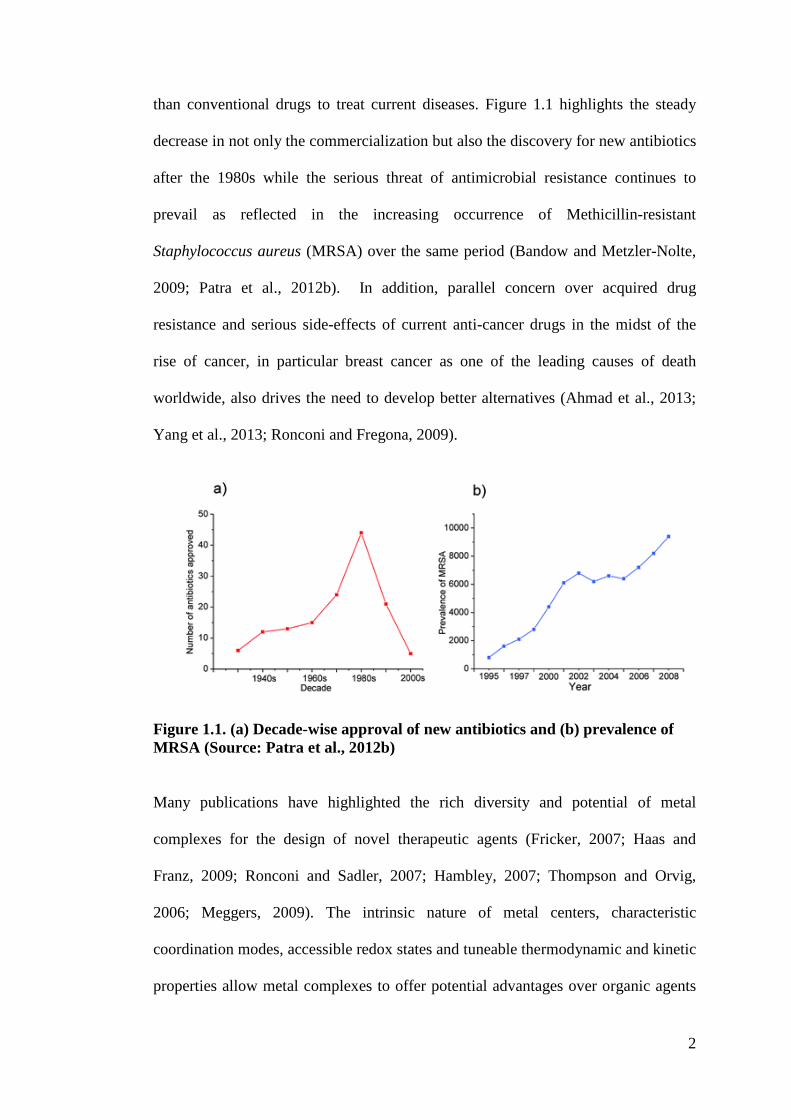
than conventional drugs to treat current diseases. Figure 1.1 highlights the steady
decrease in not only the commercialization but also the discovery for new antibiotics
after the 1980s while the serious threat of antimicrobial resistance continues to
prevail as reflected in the increasing occurrence of Methicillin-resistant
Staphylococcus aureus (MRSA) over the same period (Bandow and Metzler-Nolte,
2009; Patra et al., 2012b). In addition, parallel concern over acquired drug
resistance and serious side-effects of current anti-cancer drugs in the midst of the
rise of cancer, in particular breast cancer as one of the leading causes of death
worldwide, also drives the need to develop better alternatives (Ahmad et al., 2013;
Yang et al., 2013; Ronconi and Fregona, 2009).
Figure 1.1. (a) Decade-wise approval of new antibiotics and (b) prevalence of MRSA (Source: Patra et al., 2012b)
Many publications have highlighted the rich diversity and potential of metal
complexes for the design of novel therapeutic agents (Fricker, 2007; Haas and
Franz, 2009; Ronconi and Sadler, 2007; Hambley, 2007; Thompson and Orvig,
2006; Meggers, 2009). The intrinsic nature of metal centers, characteristic
coordination modes, accessible redox states and tuneable thermodynamic and kinetic
properties allow metal complexes to offer potential advantages over organic agents
3
alone (Rijt and Sadler, 2009). In addition, Sadler (2009, p. 10647) stated that “the
ligands not only control the reactivity of the metal but also play critical roles in
determining the nature of interactions involved in the recognition of biological target
sites such as deoxyribonucleic acid (DNA), enzymes and protein receptors” (p.
10647). The great expansion of research in the coordination chemistry of nitrogen-
and sulphur-containing ligands such as Schiff bases derived from
thiosemicarbazones and dithiocarbazates has taken place during recent years (Pelosi,
2010; Beraldo and Gambinob, 2004; Ali and Livingstone, 1974). Schi! base metal
complexes have played a prominent role in the development of coordination
chemistry. This area of research has a wide spectrum, ranging from synthesis to
application in many diverse fields. Schiff bases are condensation products of
primary amines and aldehydes or ketones (e.g. RCH=NR’, where R and R’ may
represents alkyl and/or aryl substituents) that have often been used as chelating
ligands for preparation of complex compounds which are useful as catalysts, in
various biological systems, polymers and dyes besides some uses as antifertility and
enzymatic agents (Kumar et al., 2009; Soliman and Linert, 2007). Since this class of
ligands possess both hard nitrogen and soft sulphur donor atoms, they are capable to
act as good chelating agents for various metal ions (Mohamed et al., 2009). The
flexibility and bioactivity of nitrogen and sulphur containing Schiff bases have also
been associated with the presence of both imino (-N=CH-) and thioamino (-(C=S)-
NH-) moieties in their structures (Tarafder et al., 2008). Coordination of such
compounds with metal ions often enhances their activities (Lobana et al., 2009). The
low cost as well as the relatively easy preparation of Schiff base derivatives also
provide a major attraction in creating novel leads that can be synthesized in a
practical and step-economical fashion.
4
CHAPTER 2
LITERATURE REVIEW
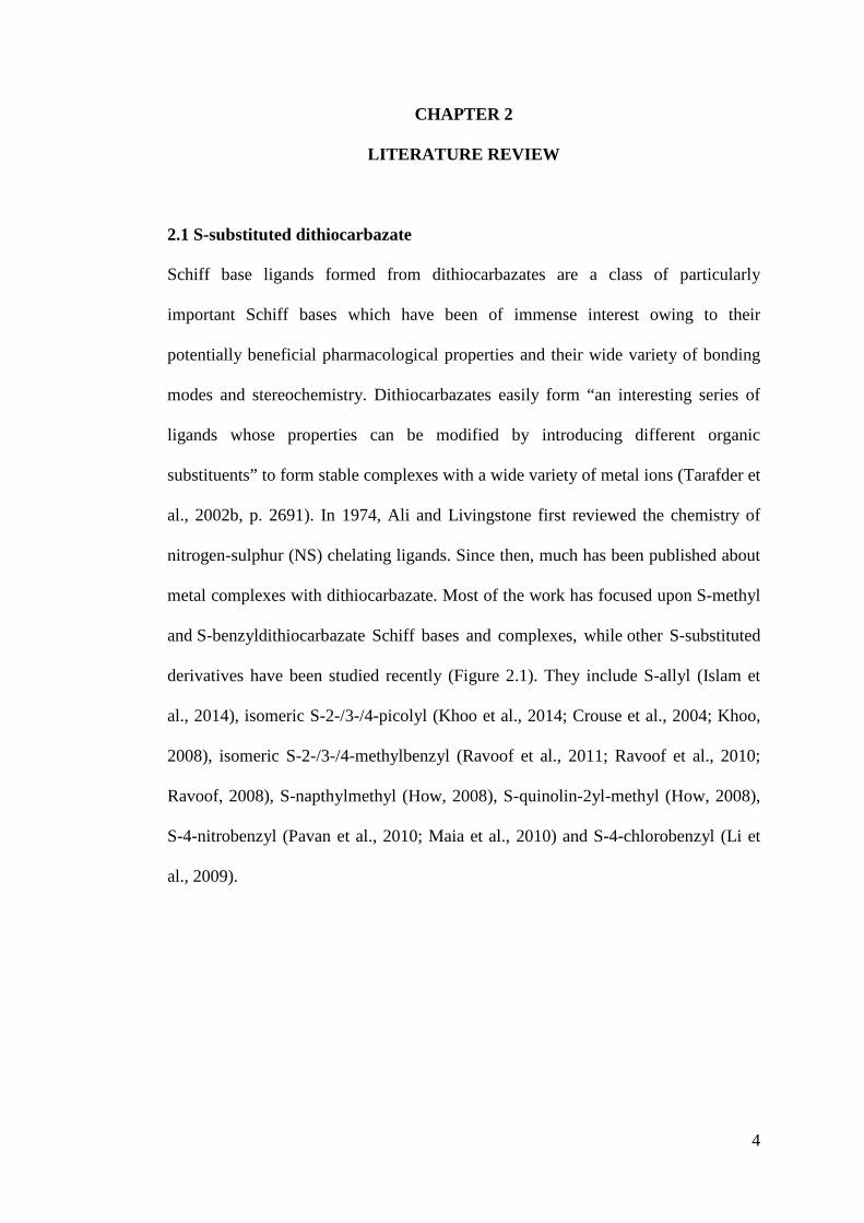
2.1 S-substituted dithiocarbazate
Schiff base ligands formed from dithiocarbazates are a class of particularly
important Schiff bases which have been of immense interest owing to their
potentially beneficial pharmacological properties and their wide variety of bonding
modes and stereochemistry. Dithiocarbazates easily form “an interesting series of
ligands whose properties can be modified by introducing different organic
substituents” to form stable complexes with a wide variety of metal ions (Tarafder et
al., 2002b, p. 2691). In 1974, Ali and Livingstone first reviewed the chemistry of
nitrogen-sulphur (NS) chelating ligands. Since then, much has been published about
metal complexes with dithiocarbazate. Most of the work has focused upon S-methyl
and S-benzyldithiocarbazate Schiff bases and complexes, while other S-substituted
derivatives have been studied recently (Figure 2.1). They include S-allyl (Islam et
al., 2014), isomeric S-2-/3-/4-picolyl (Khoo et al., 2014; Crouse et al., 2004; Khoo,
2008), isomeric S-2-/3-/4-methylbenzyl (Ravoof et al., 2011; Ravoof et al., 2010;
Ravoof, 2008), S-napthylmethyl (How, 2008), S-quinolin-2yl-methyl (How, 2008),
S-4-nitrobenzyl (Pavan et al., 2010; Maia et al., 2010) and S-4-chlorobenzyl (Li et
al., 2009).
5
Figure 2.1. Various S-substituents at position R1 in dithiocarbazates.
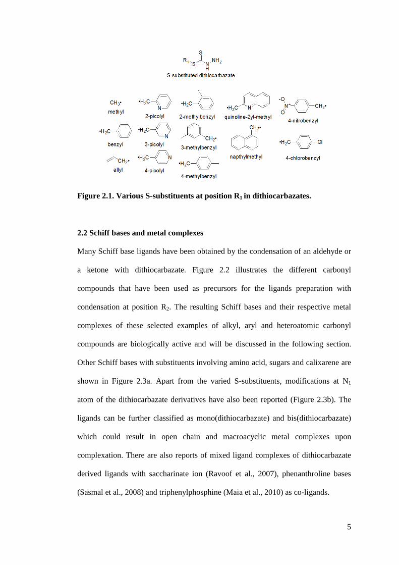
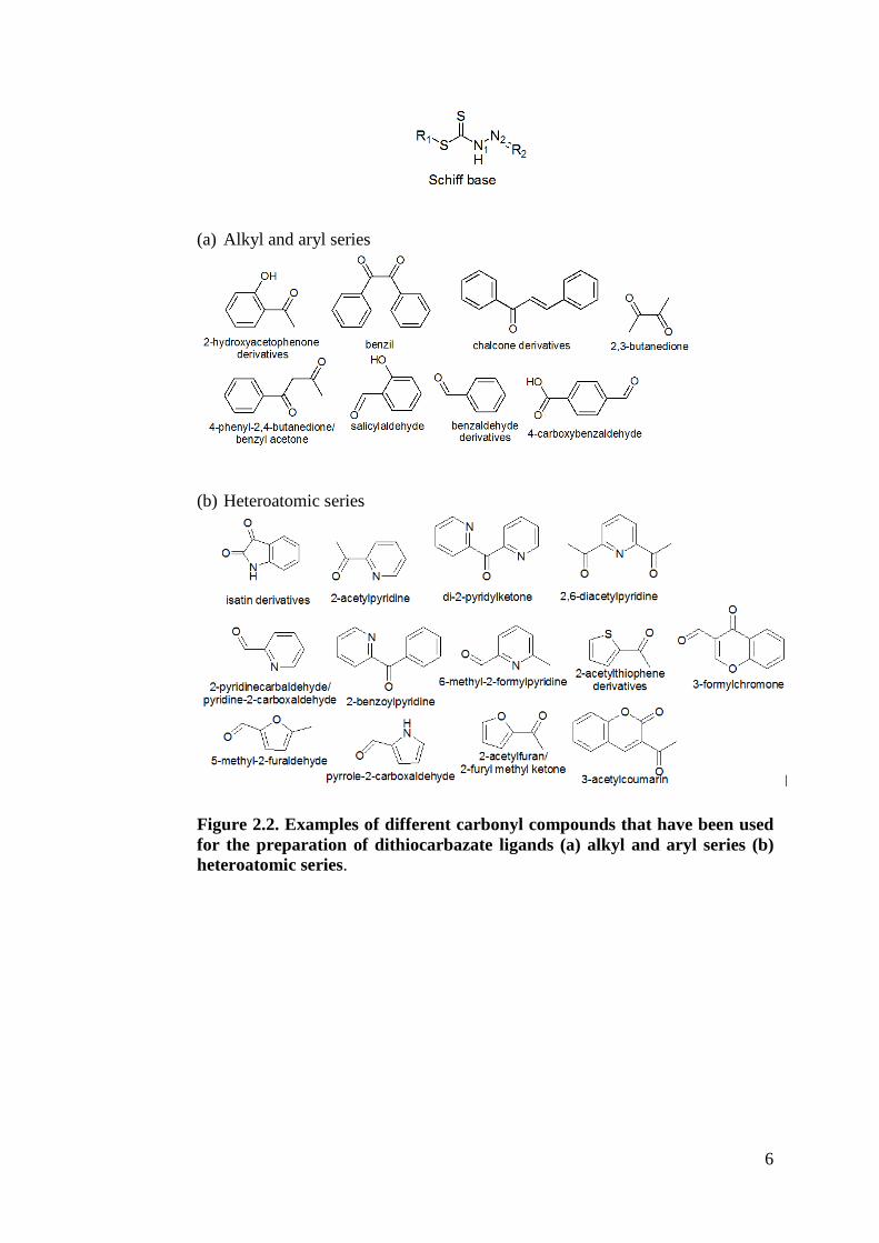
2.2 Schiff bases and metal complexes
Many Schiff base ligands have been obtained by the condensation of an aldehyde or
a ketone with dithiocarbazate. Figure 2.2 illustrates the different carbonyl
compounds that have been used as precursors for the ligands preparation with
condensation at position R2. The resulting Schiff bases and their respective metal
complexes of these selected examples of alkyl, aryl and heteroatomic carbonyl
compounds are biologically active and will be discussed in the following section.
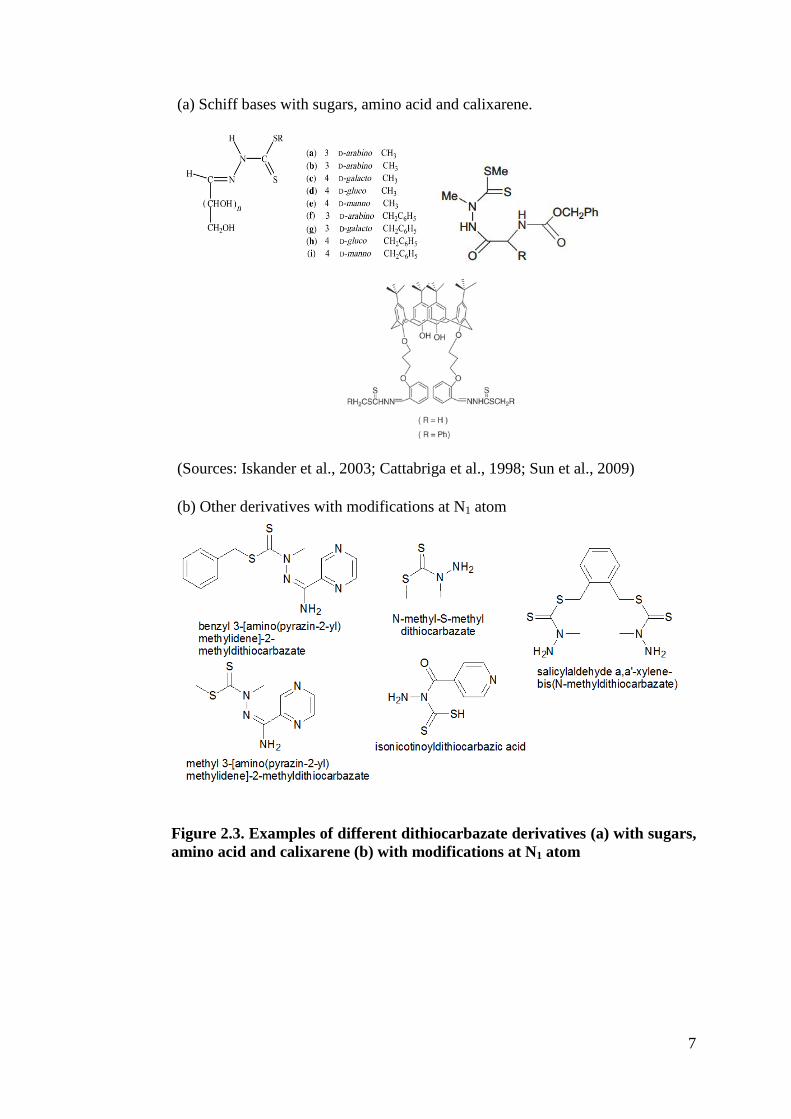
Other Schiff bases with substituents involving amino acid, sugars and calixarene are
shown in Figure 2.3a. Apart from the varied S-substituents, modifications at N1
atom of the dithiocarbazate derivatives have also been reported (Figure 2.3b). The
ligands can be further classified as mono(dithiocarbazate) and bis(dithiocarbazate)
which could result in open chain and macroacyclic metal complexes upon
complexation. There are also reports of mixed ligand complexes of dithiocarbazate
derived ligands with saccharinate ion (Ravoof et al., 2007), phenanthroline bases
(Sasmal et al., 2008) and triphenylphosphine (Maia et al., 2010) as co-ligands.
6
(a) Alkyl and aryl series
(b) Heteroatomic series
Figure 2.2. Examples of different carbonyl compounds that have been used for the preparation of dithiocarbazate ligands (a) alkyl and aryl series (b) heteroatomic series.
7
(a) Schiff bases with sugars, amino acid and calixarene.
(Sources: Iskander et al., 2003; Cattabriga et al., 1998; Sun et al., 2009) (b) Other derivatives with modifications at N1 atom
Figure 2.3. Examples of different dithiocarbazate derivatives (a) with sugars, amino acid and calixarene (b) with modifications at N1 atom
8
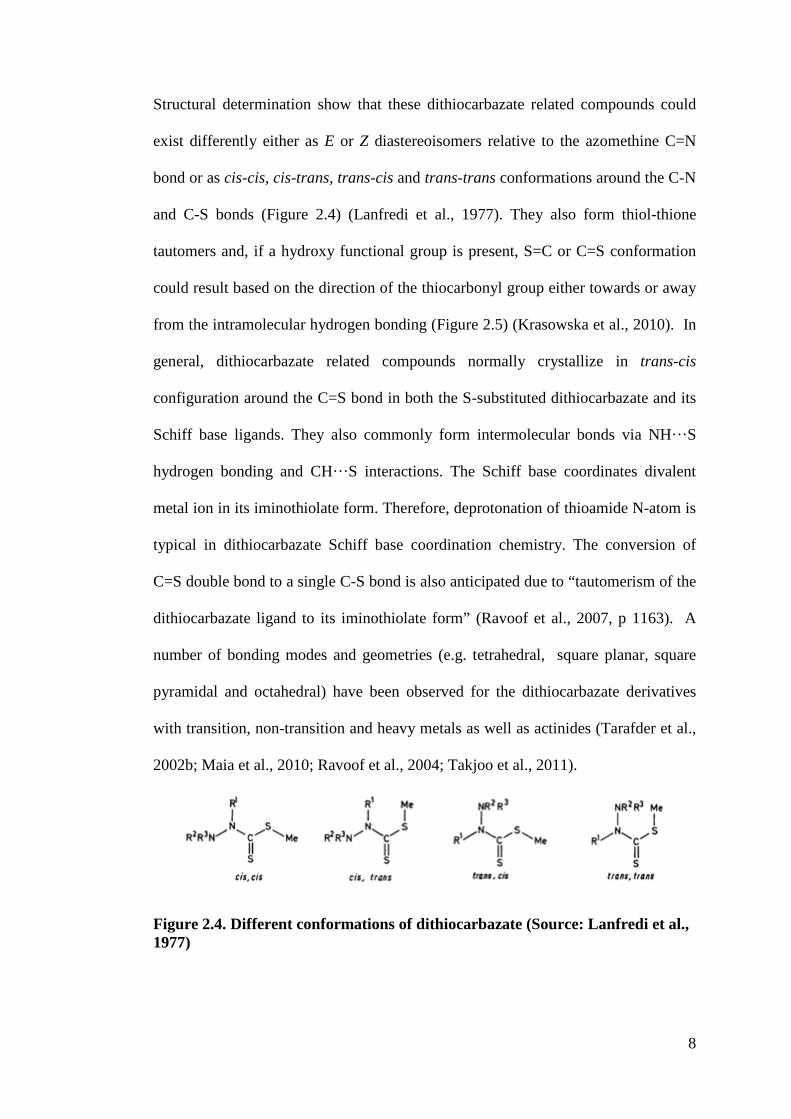
Structural determination show that these dithiocarbazate related compounds could
exist differently either as E or Z diastereoisomers relative to the azomethine C=N
bond or as cis-cis, cis-trans, trans-cis and trans-trans conformations around the C-N
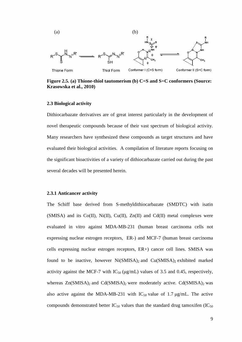
and C-S bonds (Figure 2.4) (Lanfredi et al., 1977). They also form thiol-thione
tautomers and, if a hydroxy functional group is present, S=C or C=S conformation
could result based on the direction of the thiocarbonyl group either towards or away
from the intramolecular hydrogen bonding (Figure 2.5) (Krasowska et al., 2010). In
general, dithiocarbazate related compounds normally crystallize in trans-cis
configuration around the C=S bond in both the S-substituted dithiocarbazate and its
Schiff base ligands. They also commonly form intermolecular bonds via NH···S
hydrogen bonding and CH···S interactions. The Schiff base coordinates divalent
metal ion in its iminothiolate form. Therefore, deprotonation of thioamide N-atom is
typical in dithiocarbazate Schiff base coordination chemistry. The conversion of
C=S double bond to a single C-S bond is also anticipated due to “tautomerism of the
dithiocarbazate ligand to its iminothiolate form” (Ravoof et al., 2007, p 1163). A
number of bonding modes and geometries (e.g. tetrahedral, square planar, square
pyramidal and octahedral) have been observed for the dithiocarbazate derivatives
with transition, non-transition and heavy metals as well as actinides (Tarafder et al.,
2002b; Maia et al., 2010; Ravoof et al., 2004; Takjoo et al., 2011). !
Figure 2.4. Different conformations of dithiocarbazate (Source: Lanfredi et al., 1977)
9
(a) (b)
Figure 2.5. (a) Thione-thiol tautomerism (b) C=S and S=C conformers (Source: Krasowska et al., 2010)
2.3 Biological activity
Dithiocarbazate derivatives are of great interest particularly in the development of
novel therapeutic compounds because of their vast spectrum of biological activity.
Many researchers have synthesized these compounds as target structures and have
evaluated their biological activities. A compilation of literature reports focusing on
the significant bioactivities of a variety of dithiocarbazate carried out during the past
several decades will be presented herein.
2.3.1 Anticancer activity
The Schiff base derived from S-methyldithiocarbazate (SMDTC) with isatin
(SMISA) and its Co(II), Ni(II), Cu(II), Zn(II) and Cd(II) metal complexes were
evaluated in vitro against MDA-MB-231 (human breast carcinoma cells not
expressing nuclear estrogen receptors, ER-) and MCF-7 (human breast carcinoma
cells expressing nuclear estrogen receptors, ER+) cancer cell lines. SMISA was
found to be inactive, however Ni(SMISA)2 and Cu(SMISA)2 exhibited marked
activity against the MCF-7 with IC50 (!g/mL) values of 3.5 and 0.45, respectively,
whereas Zn(SMISA)2 and Cd(SMISA)2 were moderately active. Cd(SMISA)2 was
also active against the MDA-MB-231 with IC50 value of 1.7 !g/mL. The active
compounds demonstrated better IC50 values than the standard drug tamoxifen (IC50
10
against MCF-7= 5.0 !g/mL; IC50 against MDA-MB-231= 8.0 !g/mL) (Manan et al.,
2011b). Another closely related study involving S-benzyldithiocarbazate (SBDTC)
Schiff bases derived from 5-fluoroisatin, 5-chloroisatin, 5-bromoisatin showed that
the cytotoxic activity of the halo substituted isatins against the MCF-7 breast cancer
cell lines tested was in the order of Br (2.6 !g/mL) > F (3.2 !g/mL) > Cl
(14.0 !g/mL) (Manan et al., 2011a). The ONS Schiff base of SBDTC with
salicylaldehyde and its Zn(II) and Sb(III) complexes were also strongly active
against human cell T-lymphoblastic leukemia CEM-SS (IC50= 2.3 to 4.3 !g/mL)
while the Cu(II), U(VI) and Th(IV) complexes were moderately active. The Ni(II),
Zr(IV) and Cr(III) complexes were found to be inactive. Complexation seems to
reduce the cytotoxicity of this ligand (Tarafder et al., 2000a).
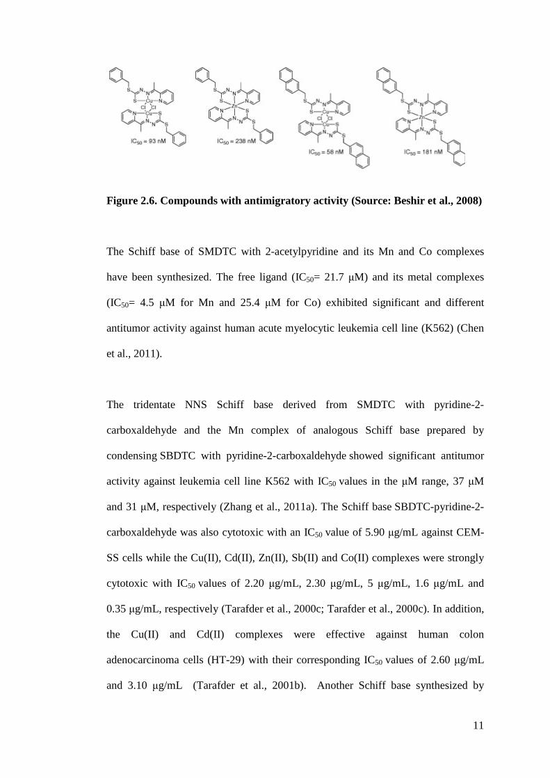
A bridged dimeric Cu(II) complex of the Schiff base product of condensation
of SBDTC and 2-acetylpyridine, Cu2Cl2(L)2 (Figure 2.6) was identified as the best
inhibitor of cell motility with nanomolar potency from the screening performed by
Beshir et al (2008). The compound appeared to be selective for certain cell lines as it
was most active towards Madin-Darby canine kidney (MDCK) cells followed by
human breast carcinoma T47D cells, less in human breast carcinoma BT20 cells and
show a weaker activity in human colorectal carcinoma HCT116 cells. From the
structure-activity relationship investigation, Beshir et al. (2008) concluded that a
two-ligand structure with bulky nonpolar S-substituents in a transoid conformation
is important for the antimigratory activity of these metal-ligand complexes.
11
Figure 2.6. Compounds with antimigratory activity (Source: Beshir et al., 2008) The Schiff base of SMDTC with 2-acetylpyridine and its Mn and Co complexes
have been synthesized. The free ligand (IC50= 21.7 !M) and its metal complexes
(IC50= 4.5 !M for Mn and 25.4 !M for Co) exhibited significant and different
antitumor activity against human acute myelocytic leukemia cell line (K562) (Chen
et al., 2011).
The tridentate NNS Schiff base derived from SMDTC with pyridine-2-
carboxaldehyde and the Mn complex of analogous Schiff base prepared by
condensing SBDTC with pyridine-2-carboxaldehyde showed significant antitumor
activity against leukemia cell line K562 with IC50 values in the !M range, 37 !M
and 31 !M, respectively (Zhang et al., 2011a). The Schiff base SBDTC-pyridine-2-
carboxaldehyde was also cytotoxic with an IC50 value of 5.90 !g/mL against CEM-
SS cells while the Cu(II), Cd(II), Zn(II), Sb(II) and Co(II) complexes were strongly
cytotoxic with IC50 values of 2.20 !g/mL, 2.30 !g/mL, 5 !g/mL, 1.6 !g/mL and
0.35 !g/mL, respectively (Tarafder et al., 2000c; Tarafder et al., 2000c). In addition,
the Cu(II) and Cd(II) complexes were effective against human colon
adenocarcinoma cells (HT-29) with their corresponding IC50 values of 2.60 !g/mL
and 3.10 !g/mL (Tarafder et al., 2001b). Another Schiff base synthesized by
12
reacting S-4-picolyldithiocarbazate (S4PDTC) with pyridine-2-carboxaldehyde
(Pc4PDTC) showed moderate cytotoxicity against human myeloid leukemia cells
(HL-60) with IC50 value of 9 !g/mL while the Schiff base synthesized by reacting
S4PDTC with 4-carboxybenzaldehyde (Cb4PDTC) was inactive. Complexing
Pc4PDTC with Cd(II) and Cu(II) enhanced its cytotoxicity from moderately to
highly active (IC50 value of 1.20-1.70 !g/mL). Pc4PDTC containing two pyridine
rings and its Cd(II) and Cu(II) complexes were also highly active against colon
cancer cells HT-29 with IC50 value " 1.0 !g/mL (Khoo et al., 2014). S-2-
picolyldithiocarbazate (S2PDTC) proved moderately active against HT-29 and
weakly active toward CEM-SS with IC50 values of 9.5 and 24.0 !g/mL, respectively,
while among its Schiff bases reported herein, only the NNS Schiff base with
pyridine-2-carboxaldehyde showed strong activity toward CEM-SS and HT-29 with
IC50 values of 2.3 !g/mL. All of the Ni(II) complexes were inactive against CEM-SS
cancer cells (Crouse et al., 2004).
SBDTC-2-acetylthiophene (SB2ATP), IC50 = 13 !g/mL and Cd(SB3ATP)2, IC50 = 9
!g/mL showed significant bioactivity towards human promyelocytic leukemia cells
(HL-60). SB2ATP, SBDTC-3-acetylthiophene (SB3ATP), Co(SB2ATP)2,
Cu(SB2ATP)2, Cu(SB3ATP)2, Zn(SB2ATP)2 and Cd(SB2ATP)2 were also
selective with significant chemotherapeutic activity against MCF-7 with IC50 = 1.4–
4.2 !g/mL. The Schiff bases however displayed higher cytotoxic activity compared
to their metal complexes except for Cu(SB3ATP)2 (Chan et al., 2008). The Zn
complex of the Schiff base, SBDTC-5-methyl-2-furaldehyde was also found to be
highly active against CEM-SS leukemia cell line with IC50 value of 2.0 !g/mL,
while the Cd complex was slightly less active than that of Zn with IC50 value of 4.95
13
!g/mL (Tarafder et al., 2002a). The Cd(II) complexes of SMDTC with 2-furyl-
methylketone and 5-methyl-2-furaldehyde and Co(II) complex of SMDTC-2-furyl-
methylketone were found to be very active against CEM-SS and cervical cancer
cells (HELA) with IC50 values between 1.8 and 3.6 !g/mL (Chew et al., 2004). The
Pb(II) complex of SBDTC with 5-methyl-2-furaldehyde was highly cytotoxic
against leukemic cells CEM-SS with IC50 of 3.25 !g/mL (Tarafder et al., 2002b).
The Cu (II), Ni (II) and Zn (II) complexes of SMDTC with 2-furylmethylketone
showed very good activity against CEM-SS cells with IC50 values of 1.6, 2.1 and 3.0
!g/mL, respectively. The Cu(II) and Zn(II) complexes were also highly active
against HELA cells with IC50 values of 1.5 and 2.1 !g/mL (Tarafder et al., 2002c).
The comparison of cytotoxic activity of SMDTC-2-benzolpyridine, SBDTC-2-
benzolpyridine and their metal complexes indicated that the presence of bulky
nonpolar S-substituents on dithiocarbazate moiety and complexation with metals can
enhance the cytotoxic activities. In particular, the Zn(II) complex of the S-benzyl
derivative effectively inhibited K562 leukemia cell line at a concentration more than
61-fold lower than the Schiff base ligand and the IC50 values of both Zn(II)
complexes were also higher against the normal hepatocyte QSG7701 cell line,
demonstrating that the compounds were able to distinguish the tumor cells from
normal cells (Li et al., 2012). The pentadentate Schiff base of 2,6-diacetylpyridine
with SBDTC exhibited marked cytotoxicity against CEM-SS giving IC50 value of
4.3 !g/mL, but its Ni(II) complex was inactive (Ali et al., 2001a). The Schiff bases
of both SMDTC- and SBDTC-6-methyl-2-formylpyridine exhibited strong
cytotoxicity against human ovarian cancer (Caov-3) cell lines with the S-methyl
derivative (IC50 = 1.0 !g/mL) being twice as active as the S-benzyl derivative. The
14
Pt complex of the S-methyl derivative was moderately active but the Pd(II) complex
was only weakly active against this cancer. None of the complexes of S-benzyl
derivatives are active against the ovarian cancer cell line (Caov-3) (Ali et al., 2006).
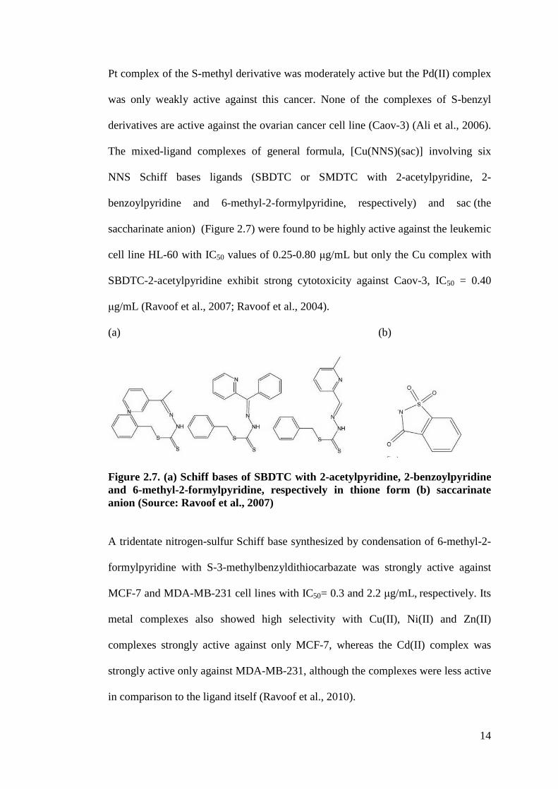
The mixed-ligand complexes of general formula, [Cu(NNS)(sac)] involving six
NNS Schiff bases ligands (SBDTC or SMDTC with 2-acetylpyridine, 2-
benzoylpyridine and 6-methyl-2-formylpyridine, respectively) and sac (the
saccharinate anion) (Figure 2.7) were found to be highly active against the leukemic
cell line HL-60 with IC50 values of 0.25-0.80 !g/mL but only the Cu complex with
SBDTC-2-acetylpyridine exhibit strong cytotoxicity against Caov-3, IC50 = 0.40
!g/mL (Ravoof et al., 2007; Ravoof et al., 2004).
(a) (b)
Figure 2.7. (a) Schiff bases of SBDTC with 2-acetylpyridine, 2-benzoylpyridine and 6-methyl-2-formylpyridine, respectively in thione form (b) saccarinate anion (Source: Ravoof et al., 2007)
A tridentate nitrogen-sulfur Schiff base synthesized by condensation of 6-methyl-2-
formylpyridine with S-3-methylbenzyldithiocarbazate was strongly active against
MCF-7 and MDA-MB-231 cell lines with IC50= 0.3 and 2.2 !g/mL, respectively. Its
metal complexes also showed high selectivity with Cu(II), Ni(II) and Zn(II)
complexes strongly active against only MCF-7, whereas the Cd(II) complex was
strongly active only against MDA-MB-231, although the complexes were less active
in comparison to the ligand itself (Ravoof et al., 2010).

15
The OS donor ligand derived from the reaction of SBDTC with benzoyl chloride and
its Cu(II), Ni(II) and Pb(II) complexes displayed marked cytotoxicity against HL-60
leukemia cells with IC50" 5.0 !g/mL while Cd(II) and Co(II) complexes were only
moderately cytotoxic. In this case, the ligand was more potent compared to its metal
complexes (How et al., 2008). SBDTC and its Sn(II) complex were very effective
against renal carcinoma Tk10 kidney cancer cells and leukemia TK6 cell line. The
IC50 values were in the range 1.0-4.0 !g/mL with SBDTC being the most active
compound. The SNNS quadridentate Schiff base of SBDTC with benzil and its
Sn(II) complex were also effective against skin cancer cells (UACC melanoma) with
IC50 of 5.2 and 2.7 !g/mL, respectively (Tarafder et al., 2000b). The NS Schiff base
prepared by condensing SBDTC with 2,3-butanedione (1:1 mole ratio) was strongly
active against leukemic cells CEM-SS with IC50 value of 2.05 !g/mL (Tarafder et
al., 2001a).
2.3.2 Antibacterial and antifungal activity
The Schiff base formed from pyridine-2-carboxaldehyde SMDTC and its Zn
complex showed marked and broad antimicrobial and antifungal activities compared
to the S-benzyl derivatives with MIC values as low as 12.5 !g/mL (Zhang et al.,
2011a). The antibacterial activity of the Schiff bases of SBDTC with ferrocene-
based chalcones containing a F or Cl substituent in the para position or a pyridine
ring were the most active in the series and their activity against Gram-negative
bacterial (Escherichia coli (E. coli) and Pseudomonas aeruginosa (P. aeruginosa))
strains was found to be higher than that for the drugs ketoconazole, kanamycin and
penicillin (Liu et al., 2012). In another closely related investigation, the Zn(II) and
Cu(II) complexes of SBDTC with ferrocene-based chalcone Schiff base ligand
16
containing a para-Cl substituent and the Zn(II) complex of SBDTC with ferrocene-
based chalcone having a methyl group in the aromatic ring were the most active in
the series with MIC values in the range of 1.319 ! 10"8 M to 3.750 ! 10"7 M against
bacteria and fungi tested (Staphylococcus aureus (S. aureus), Bacillus cereus (B.
cereus), E. coli, P. aeruginosa, Aspergillus niger (A. niger), Aspergillus fumigatus
A. fumigates) (Liu et al., 2013). SMDTC-2-benzolpyridine and its Cu(II)
complex showed excellent activity against Gram positive bacteria (Bacillus subtilis
(B. subtilis), S. aureus) and yeast (Candida lusitaniae (C. lusitaniae)) with MIC
values of 1-5 !g/mL. It was found that the SMDTC derived ligand was more potent
than the SBDTC derivative towards the tested microorganisms and complexation
with metals also had a synergetic effect resulting in enhanced antimicrobial activity
(Li et al., 2012). Both the Cu(II) complex of the Schiff base S4PDTC with pyridine-
2-carboxaldehyde and the Cd(II) complex of S4PDTC 4-carboxybenzaldehyde
(Cb4PDTC) showed good antifungal activity against Candida albicans (C. albicans)
with MIC values lower than Nystatin (Khoo et al., 2014) . The Schiff base derived
from SBDTC with pyrrole-2-carboxaldehyde was a stronger antifungal agent than
Nystatin against Saccaromyces ceciricaee (S. ceciricaee) and Candida lypolytica (
C. lypolytica) (Tarafder et al., 2002a). The NSS Schiff bases of S2PDTC with 2-
acetylfuran showed better activity than Nystatin toward against the fungus, C.
lypolytica while its metal complexes were not active (Crouse et al., 2004). Co(II)
complex of SMDTC with 2-furyl-methylketone and Cd (II) complex of SMDTC
with 5-methyl-2-furaldehyde gave the most effective activity against fungi tested (C.
lypolytica and Aspergillus ochraceus (A. ochraceous) (Chew et al., 2004) while the
Cu(II) complex of SMDTC with 2-furylmethylketone showed clear activity against
C. lypolytica with better activity than Nystatin (Tarafder et al., 2002c). Bi(III) and
17
As(III) metal complexes of SBDTC-3-acetylcoumarin (L) with the formula
[ClBi(L)2] and [PhAs(L)2] showed low MIC values (10#!g/mL for bacterial strain B.
subtilis and 16#!g/mL for fungal strain Fusarium oxysporum (F. oxysporum)). The
metal complexes were more active against fungal strains compared to bacterial
strains and had better activity than the free ligands (Dawara et al., 2012). In the
series with salicylaldehyde Schiff base of isonicotinoyldithiocarbazic acid, the best
activity was shown by the Ni(II) complex (MIC = 75 !g/mL) against the gram-
negative pathogenic strain of E. coli (Kalia et al., 2012). The Cd complex of the
SNNS Schiff base SBDTC-benzil was active against bacteria P. aeroginosa and B.
cereus with the MIC values better or comparable to kanamycin (Tarafder et al.,
2000b). The Cu(II) complex of SBDTC-salicylaldehyde proved to be the best in the
series against B. cereus (MIC=79.6 !g/mL) (Tarafder et al., 2000a).
2.3.3 Iron chelators
All dithiocarbazate ligands derived from either SBDTC or SMDTC with 2-
acetylpyridine, di-2-pyridylketone and 2-pyridinecarbaldehyde were more effective
than standard desferrioxamine B (DFO) at releasing intracellular Fe and SMDTC-2-
pyridinecarbaldehyde was the most active of all compounds tested. Furthermore, the
three SMDTC derivatives were more effective at mobilising intracellular 59Fe than
their corresponding SBDTC analogues. 2-pyridinecarbaldehyde derivatives with
both SMDTC and SBDTC exhibit no apparent cytotoxicity (>10 !M). This property,
in combination with their high activity in sequestering intracellular Fe present ideal
properties for a chelator in the treatment of Fe overload. The other four
dithiocarbazates (with 2-acetylpyridine and di-2-pyridyl ketone) showed moderate to
potent anti-proliferative activity which may be problematic for treatment of Fe
18
overload. On the other hand, the anti-proliferative activity of these compounds can
be advantageous in the treatment of cancer (Basha et al., 2012).
2.3.4 Antituberculosis activity
Ni, Co and Zn complexes of a non-Schiff base isonicotinoyldithiocarbazic acid
ligand synthesized from isoniazid with carbon disulphide showed MIC values of 2, 2
and 50 !g/mL against Mycobacterium tuberculosis (M. tuberculosis) H37Rv, and 10,
100 and 50 !g/mL against a multi-drug-resistant strain of M. tuberculosis. They had
little cytotoxic effect on the transformed human rhabdomyosarcoma cell line RD
cells making them potentially useful to treat multi-drug resistant tuberculosis
infections (Kanwar et al., 2008). Others dithiocarbazate derivatives such as 2-/3-/4-
pyridinecarbonimidoyldithiocarbazic acid esters, methyl 3-[amino(pyrazin-2-
yl)methylidene]-2-methyldithiocarbazate and benzyl 3-[amino(pyrazin-2-
yl)methylidene]-2-methyldithio carbazate have been studied and were among the
promising classes of compounds showing action against tuberculosis (Olczak et al.,
2010). In another study by Pavan et al. (2010), dithiocarbazate compounds derived
from benzoylacetone showed poor activity. The low activity was associated with the
difference in the molecular structures from the potent analogues which contained
pyridine rings further affirming that the aromatic heteroatom N moiety played an
important role in the anti-tuberculosis activity of the compounds (Pavan et al.,
2010).
2.3.5 Antiamoebic activity
Mixed ligand Pt(II) and Pd(II) complexes prepared with ONS Schiff bases derived
from SBDTC and 2-hydroxyacetophenone (Figure 2.8) or SBDTC and 4-phenyl-
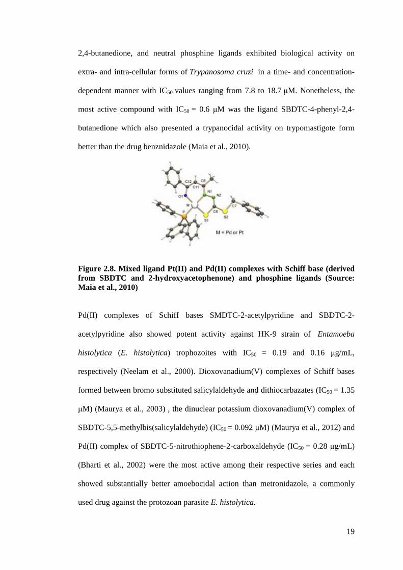
19
2,4-butanedione, and neutral phosphine ligands exhibited biological activity on
extra- and intra-cellular forms of Trypanosoma cruzi in a time- and concentration-
dependent manner with IC50 values ranging from 7.8 to 18.7 !M. Nonetheless, the
most active compound with IC50 = 0.6 !M was the ligand SBDTC-4-phenyl-2,4-
butanedione which also presented a trypanocidal activity on trypomastigote form
better than the drug benznidazole (Maia et al., 2010).
Figure 2.8. Mixed ligand Pt(II) and Pd(II) complexes with Schiff base (derived from SBDTC and 2-hydroxyacetophenone) and phosphine ligands (Source: Maia et al., 2010)
Pd(II) complexes of Schiff bases SMDTC-2-acetylpyridine and SBDTC-2-
acetylpyridine also showed potent activity against HK-9 strain of Entamoeba
histolytica (E. histolytica) trophozoites with IC50 = 0.19 and 0.16 !g/mL,
respectively (Neelam et al., 2000). Dioxovanadium(V) complexes of Schiff bases
formed between bromo substituted salicylaldehyde and dithiocarbazates (IC50 = 1.35
!M) (Maurya et al., 2003) , the dinuclear potassium dioxovanadium(V) complex of
SBDTC-5,5-methylbis(salicylaldehyde) (IC50 = 0.092 !M) (Maurya et al., 2012) and
Pd(II) complex of SBDTC-5-nitrothiophene-2-carboxaldehyde (IC50 = 0.28 !g/mL)
(Bharti et al., 2002) were the most active among their respective series and each
showed substantially better amoebocidal action than metronidazole, a commonly
used drug against the protozoan parasite E. histolytica.
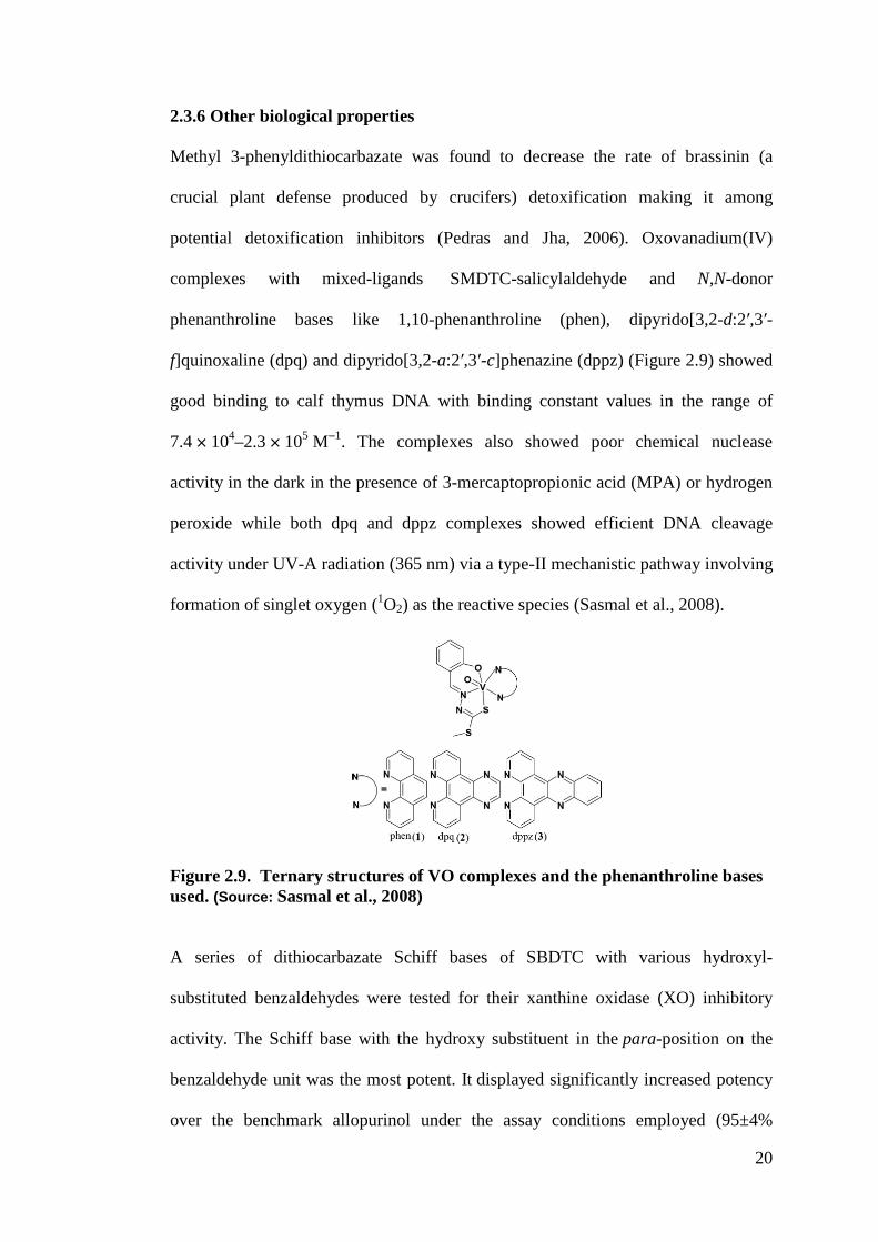
20
2.3.6 Other biological properties
Methyl 3-phenyldithiocarbazate was found to decrease the rate of brassinin (a
crucial plant defense produced by crucifers) detoxification making it among
potential detoxification inhibitors (Pedras and Jha, 2006). Oxovanadium(IV)
complexes with mixed-ligands SMDTC-salicylaldehyde and N,N-donor
phenanthroline bases like 1,10-phenanthroline (phen), dipyrido[3,2-d:2$,3$-
f]quinoxaline (dpq) and dipyrido[3,2-a:2$,3$-c]phenazine (dppz) (Figure 2.9) showed
good binding to calf thymus DNA with binding constant values in the range of
7.4 ! 104–2.3 ! 105 M"1. The complexes also showed poor chemical nuclease
activity in the dark in the presence of 3-mercaptopropionic acid (MPA) or hydrogen
peroxide while both dpq and dppz complexes showed efficient DNA cleavage
activity under UV-A radiation (365 nm) via a type-II mechanistic pathway involving
formation of singlet oxygen (1O2) as the reactive species (Sasmal et al., 2008).
Figure 2.9. Ternary structures of VO complexes and the phenanthroline bases used. (Source: Sasmal et al., 2008)
A series of dithiocarbazate Schiff bases of SBDTC with various hydroxyl-
substituted benzaldehydes were tested for their xanthine oxidase (XO) inhibitory
activity. The Schiff base with the hydroxy substituent in the para-position on the
benzaldehyde unit was the most potent. It displayed significantly increased potency
over the benchmark allopurinol under the assay conditions employed (95±4%
21
inhibition at 50 !M and IC50 = 0.7±0.1 !M) (Leigh et al., 2011). SBDTC (Tarafder et
al., 2001b) and the Ni and Cu complexes of SBDTC-2,3-butanedione (Tarafder et
al., 2001a) were found to be stronger antioxidants than Vitamin E. Antioxidant
properties of SBDTC and two other Schiff bases synthesized by reacting S4PDTC
with pyridine-2-carboxaldehyde (Pc4PDTC) and 4-carboxybenzaldehyde
(Cb4PDTC) were also found to be comparable to the commercially available
synthetic antioxidant buthylatedhydroxytoluene (BHT) (Tarafder et al., 2001b; Khoo
et al., 2014). Alkyl pyridine-carbonyldithiocarbazates were shown to be uncouplers
of oxidative phosphorylation in mitochondria. Greater activity was observed with
increasing alkyl chain length, the optimum being C9, indicating the influence of
hydrophobicity towards the activity (Terada et al., 1978; Kubota et al., 1978).
Technetium-nitrido complexes of the Schiff base S-methyl 3-(2-hydroxyphenyl
methylene) dithiocarbazate had been investigated as potential Tc-99m nitrido tumor
imaging disposition in mice (Borel et al., 1992). N-methyl-SMDTC had also been
effectively used for high-yield preparation of nitrido Tc-99m and Re-188
radiopharmaceuticals (Uccelli et al., 2011; Boschi et al., 2010). The anti-fertility
data indicated a highly significant decline in the motility of sperm as well as in
sperm count in the treated animals in the case of dimethyltin(IV) complex derived
from SBDTC-4-nitrobenzanilide (Singh et al., 2009). The results suggested that the
ligand of SBDTC-3-acetylcoumarin was most effective in reducing fertility and
complexation with bismuth and arsenic enhanced its activity (Dawara et al., 2012).
The pescticide activity and DNA cleavage activity of Ge(IV) complex with SBDTC-
3-fomylchromone Schiff base were found to be better in comparison to the ligand
itself (Dawara et al., 2011).
22
At this stage, it is impossible to draw any meaningful correlations from this limited
and diverse group. A library of compounds must be systematically designed and
their activities determined in order to achieve that. The above reports on
dithiocarbazate, its Schiff bases and metal complexes highlight that even subtle
change in the structures lead to dramatic alterations of the biological properties of
this class of compounds. The pharmacological activities of these metal complexes
depend on the type of metal ion, its ligands and the structure of the compounds.
These factors play important roles in the recognition of target sites. In conjunction
with the continuous effort to develop new derivatives that possess potent biological
activities, the major aim of the present work is to expand the synthesis involving
multidentate dithiocarbazate derivatives to include bioconjugation and to explore the
biological potential of the compounds synthesized to determine their cytotoxicity
and their potencies against selected bacterial strains expressing a multi-drug
resistance phenotype. Although many new dithiocarbazate derivatives have been
added to the family in recent years, there is still no clear structure-activity
relationship to explain their activity. The actual mechanism of action is still
unknown. Electron paramagnetic resonance (EPR) and electrochemistry have been
carried out in this work to connect the biological activities to their structures and
redox properties in order to identify features that promote better activity. It is
expected that this structure-activity analysis will serve to orient further synthetic
efforts towards determining the optimum features essential to promote higher
bioactivities and ultimately to guide future work designed to reveal their mode of
action.
23
2.4 Objectives
1. To synthesize bioactive Schiff bases (i.e. potentially bi-, tri- or tetradentate
ligands) and their open chain or macroacyclic metal complex systems
derived from S-substituted dithiocarbazates.
2. To incorporate grafting work with vectors (i.e. polyarginine, polyethylene
glycol (PEG) and phe-arg-%-napthylamide (PA%N)) in order to prepare
improved therapeutic agents.
3. To characterize the synthesized compounds using various physico-chemical
and spectroscopic techniques.
4. To study the biological activity of the compounds prepared and to attempt to
elucidate their structure-activity relationships.
24
CHAPTER 3
NON-CONJUGATED PARENTS COMPOUNDS
In this work, three series of parent compounds: (i) tetradentate NNSS ligands (ii)
potentially bidentate NS or tridentate ONS ligands with acid or ester functionalities
and (iii) potentially bidentate NS or tridentate ONS ligands with natural ketone
moiety were prepared by condensation of S-substituted dithiocarbazates with 2,5-
hexanedione, methyl levulinate, levulinic acid, 4-carboxybenzaldehyde and 3-
acetylcoumarin. All were complexed with copper to synthesize their respective
metal complexes.
3.1 Introduction
3.1.1 Ligand systems
3.1.1.1 Tetradentate NNSS
Macrocyclic and macroacyclic Schiff bases have been widely studied (Vigato and
Tamburini, 2004). They show various coordination abilities and potential
applications in biology which range from therapeutic drug candidates to diagnostic
agents (Holland et al., 2008) and they provide synthetic models for the metal
containing sites in metalloproteins and metalloenzymes (Gennari et al., 2012). It is
therefore worthwhile to explore these interesting properties by investigating the
synthesis and characterization of new Cu(II) bis(dithiocarbazate) in this work. The
compounds are analogues of the Cu(II) bis(thiosemicarbazone) that have garnered
much attention resulting in biological breakthroughs (Paterson and Donnelly, 2011)
particularly as radiopharmaceuticals (Donnelly, 2011).
25
It is anticipated that the replacement of nitrogen atom with sulphur may provide
interesting results warranting further exploration into dithiocarbazate compounds.
Moreover, to form the Schiff bases, the 2,5-hexanedione has been chosen to expand
the ligand flexibility by introducing backbones containing more than two carbons.
This enhanced flexibility may facilitate increased tetrahedral distortion leading to
incorporation of metal cations that prefer non-square planar geometries such as
Cu(I) ion. Previous studies have shown that physico-chemical properties such as
redox potential as well as biological activity have been related to the geometry at the
metal site (Durot et al., 2005; Drew et al., 1995; Rorabacher, 2004; Basha et al.,
2012; Jansson et al., 2010). These were fine examples demonstrating the marked
influence of ligand environment towards the redox potential of their respective
Cu(II)/Cu(I), Fe(III)/Fe(II) and Mn(III/II) metal systems. While choice of the metal
ions and substituent functional groups of the ligands have been carefully chosen to
affect the geometry of metal complexes (Ostermeier et al., 2010; Jones and
McCleverty, 1970; Cowley et al., 2004; Stefani et al., 2012), the studies by Diaz et
al. (1998;1999) that highlighted the differences in coordination geometry identified
using EPR by comparing the open chain and cyclic metal complex system which
subsequently affect their biological activity was found to be particularly attractive
since this analytical tool enabled a structural view of the complexes in solution. The
group noted that the Cu(II) complexes of the open chain mono(thiosemicarbazone)
with a higher degree of tetrahedral distortion should be further explored as
potentially better SOD-like mimics than the macroacyclic bis(thiosemicarbazone)
complexes. Although the reports were primarily focused on superoxide dismutase
(SOD) mimics, it would also be meaningful to carry out such comparison in this
26
work as the open chain system will be envisaged to offer interesting diversity and
aid towards the understanding of the structure-bioactivity relationship.
3.1.1.2 Potentially bidentate NS or tridentate ONS ligands with an acid or ester
functionality
The open chain series in this work consisting ligands of methyl levulinate and
levulinic acid with SBDTC and SMDTC as well as their corresponding Cu(II)
complexes will be a worthy comparison to their macroacyclic Cu(II) tetradentate
system with 2,5-hexanedione bis(dithiocarbazate). Furthermore, the Schiff base
derivatives containing the acid or ester functional group have proved to be attractive
from both biological and physico-chemical aspects. For instance, recent attention
was dedicated to metal complexes of &-ketoglutaric acid (Baldini et al., 2004) and
pyruvic acid thiosemicarbazone (Diaz et al., 1994; Wiecek et al., 2009). These
aliphatic ligands with a variety of potential donor atoms and many possible
conformations provided a versatile chelating behavior. In addition, the metal
complexes were found to be potent against the selected human leukemia and cancer
cell lines tested and thus may be regarded as potentially significant antitumor agent
(Baldini et al., 2004; Diaz et al., 1994; Wiecek et al., 2009). Similiarly, the
analogous Schiff base keto-ester methylpyruvate with SMDTC has been screened by
the National Cancer Institute, Bethesda, Maryland and has been found to exhibit
promising activity against leukemia cells as mentioned by Ali et al. (2001b). The
authors also stated that the “ligand coordinated to the metal(II) ion as a
uninegatively charged tridentate chelating agent via the carbonylic oxygen atom, the
azomethine nitrogen atom and the thiolato sulfur atom” but resulted in varied
conformation geometry with different metal (Ali et al., 2001b p. 1037). The Cu(II)
27
complexes have the general formula, CuLX (L= Schiff base; X=Cl", Br") with a
distorted square-planar structure whereas the Zn(II), Cd (II) and Ni(II) complexes of
empirical formula, ML2 supported a six-coordinate distorted octahedral structure for
these complexes as confirmed by the X-ray crystallographic structural analysis (Ali
et al., 2001b; Ali et al., 1999; Ali et al., 2004). To date, metal complexes of methyl
levulinate or levulinic acid dithiocarbazate have not been reported although
semicarbazone derivatives of levulinic acid have been identified as potent
anticonvulsant agents showing broad spectrum of activity with low neurotoxicity
(Navneet and Pradeep, 2005).
Since the carbonylic oxygen atom and the azomethine nitrogen atom are further
apart with three carbons in between them, these ligands could adapt NS bidentate
chelation ability different from the ONS chelating ligands with &-ketoglutaric acid,
pyruvic acid or methyl pyruvate. In addition, 4-carboxybenzaldehyde was utilized to
form the Schiff bases in order to provide insight on the influence of an aromatic
acid. As most sulphur-nitrogen chelating ligands and their complexes are highly
hydrophobic and their low solubility in water imposes experimental limitations in
biological studies, the introduction of a hydrophilic group such as -COOH in
systems should permit increased solubility in water (Pogni et al., 2000). Besides
that, the presence of a carboxylic functional group would allow for further ligand
optimization via bioconjugation to improve the properties of the compounds and
their applications.
28
3.1.1.3 Potentially bidentate NS or tridentate ONS ligands with natural potent
aldehyde or ketones moieties.
In order to expand the synthesis and exploration of dithiocarbazate derivatives,
attention was also directed herein to aromatic dithiocarbazate derived from natural
aldehydes or ketones that had well established medicinal properties. The rationale
behind this attempt was based on the prospect of the synergistic effects developed
from integration of the promising bioactivity of individual components (i.e. metal
center and ligand comprising the carbonyl group and substituted dithiocarbazate
moiety). A number of publications had highlighted the potential of utilizing such
natural aromatic compounds like chromone (Barve et al., 2006; Khan et al., 2009),
chalcone (Zhang et al., 2011b) and curcumin (Padhye et al., 2009). This prompted
the preparation of new metal complexes with Schiff base formed from the
condensation of SBDTC and 3-acetylcoumarin in this work. Coumarin derivatives
are attractive because of their wide variety of biological activities including
antioxidant, antibacterial, antifungal and cytotoxic (Datta et al., 2011; Bagihalli et
al., 2008; Phaniband et al., 2011; Kulkarni et al., 2009; Creaven et al., 2009).
Moreover, the reported coumarin Schiff bases and their metal complexes were
shown to exhibit outstanding luminescence properties which may provide advantage
for application of these compounds as probes (Datta et al., 2011). Further findings
reveal that the coumarin derivatives interact strongly with DNA and can cause DNA
cleavage (Phaniband et al., 2011; Kulkarni et al., 2009).
29
3.1.2 Choice of metals
3.1.2.1 Copper
Copper complexes represent a class of compounds that have been subjected to
intensive research because of their potential therapeutic applications (Duncan and
White, 2012) in particular as effective antitumor (Chakraborty et al., 2010: Afrasiabi
et al., 2003) and antibacterial agents (Li et al., 2000; Joseph et al., 2012). A number
of related thiosemicarbazone copper complexes have been found to be active in cell
destruction, as well as in the inhibition of DNA synthesis (Ferrari et al., 2002a;
Ferrari et al., 2002b; Bisceglie et al., 2012; Ferrari et al., 2004) Cancer cells have
also been shown to take up greater amounts of copper than normal cells (Jansson et
al., 2010). The altered metabolism of cancer cells and the differential response
between normal and cancer cells to copper are the basis for the development of
copper complexes endowed with anticancer characteristics (Gandin et al., 2013).
Recently, copper(II) complexes of thiosemicarbazone NNSS ligands have been
explored extensively as radiopharmaceuticals for the specific targeting of hypoxic
tissue (Donnelly, 2011). They are also known to be stable (Kass = 1018), neutral and
can easily cross cellular membranes (Paterson and Donnelly, 2011; Donnelly, 2011;
Ngarivhume et al., 2005). Copper complexes with such multidentate Schiff base
ligands are attractive for study because of their rich spectroscopic and magnetic
properties that often change during the course of enzyme catalysis (Ferrari et al.,
2002b). Copper also exhibits different oxidation states affirming its important role as
a model to provide better understanding of biological metalloenzymes and
metalloprotein systems (Balamurugan et al., 2006; Knoblauch et al., 1999;
'ura(ková et al., 1999). For instance, the CuN2S2 chromophore is present in blue
copper proteins such as plastocyanine (Donnelly, 2011; Sarkar et al., 2009). With
30
these examples in mind and following the interest concerning the electrochemical
investigation into the Cu(I)-Cu(II) redox couple, it is only logical for copper ion to
serve as an excellent choice in the continuing search for new and effective
metallodrugs.
3.1.2.2 Zinc
In addition to Cu(II) complexes, another two metal complexes, zinc(II) and
rhenium(I) will be presented as well. Both are important as their complexes are
diamagnetic and this facilitates complementary methods of detection to EPR
spectroscopy for the Cu(II) complex through the use of nuclear magnetic resonance
(NMR) spectroscopy for Zn(II) and Re(I) species (Kirin et al., 2005). In addition,
one of the most promising previous findings by Awidat (2005) was that bis (S-
methyl-%-N-(2-acetylfuran)dithiocarbazate) (SMDB) complexed with zinc was
found to shown remarkable selectivity towards brain cancer cells with a toxicity
towards normal cells that was relatively much lower than tamoxifen, the current
drug of choice in the treatment of brain cancer. The IC50 values for SMDB-Zn on
glioma cell lines A172, U87MG, T98G and normal brain cell line HCN-2 were at
3.7 !g/mL, 1.76 !g/mL, 2.7 !g/mL and 7 !g/mL, respectively (Awidat, 2005).
Zn(II) complexes of the analogous bis(thiosemicarbazone) were also found to be
weakly fluorescent owing to intraligand excitation and this fluorescence has been
used to track the uptake and intracellular distribution of the Zn(II) complexes in
diverse cancer cell lines (Lim et al., 2010). The understanding of intracellular
distribution of these complexes is important to design compounds that are selective
towards specific tissues and organelles (Holland et al., 2007).
31
3.1.2.3 Rhenium
Although different dithiocarbazate metal complexes have been investigated, few
works deal with Re complexes (Mevellec et al., 2002; Boschi et al., 2010) and none
has yet to report on a Re(I) tricarbonyl core. Much significant progress in targeted
rhenium (Re-188 and Re-186) radiopharmaceuticals recently has focused on the use
of the Re carbonyl core (Donnelly, 2011). The use of radionuclides has increased the
demand for new complexes containing carrier-free radionuclides having reasonable
half lives and proper energy of radiation (Fuks et al., 2010). The carbonyl approach
exploits the stability of the metal tricarbonyl core whilst allowing a variety of bi-
and tridentate ligands to react with it to further functionalize these with
biomolecules or targeting vectors (Donnelly, 2011; Amoroso et al., 2007; Santos et
al., 2004; Clède et al., 2013). Because of the small size and charge of the fac-
Re(CO)3+ group, it does not significantly influence biological properties of even the
smallest biomolecules used in the synthesis of radiopharmaceuticals making it an
attractive candidate not just for medicinal therapeutic application but also as
diagnostic agents (Fuks et al., 2010; François et al., 2014; Clède et al., 2012) For
instance, an innovative approach to prepare a rhenium tricarbonyl complex with a 4-
(2-pyridyl)-1,2,3-triazole (pyta) ancillary ligand endowed with luminescent and
infrared properties allowed a relevant bio-imaging correlative study using both
infrared (IR) and luminescence modalities (Clède et al., 2012). Vibrational
spectroscopies are attractive for bio-imaging as in the case of vibrational excitations
in the IR region where no photo-bleaching is induced in contrast to what is observed
with organic fluorophores in the visible or UV-range. Furthermore, IR-probes also
show advantages in their stability in biological environments and intense absorption
32
in the 1800-2200 cm"1 range which is the transparent IR window of biological media
(Clède et al., 2013; Clède et al., 2012; Policar et al., 2011).
3.2 Methodology
3.2.1 Materials
All chemicals and solvents were of analytical grade and were used as received
without further purification. Common solvents for solution synthesis were obtained
from either Carlo Ebra or VMR. Anhydrous DMF was from Sigma Aldrich or Acros
Organics and DMSO of HPLC grade was from Alfa Aesar. Chemicals: Benzyl
chloride (Aldrich), iodomethane (Merck), potassium hydroxide (Merck),
hydrazinium hydroxide (Merck), carbon disulfide (Merck), 2,5-hexanedione
(Merck), methyl levulinate (Alfa Aesar), levulinic acid (Janssen), 4-
carboxylbenzaldehyde (Acros Organics), 3-acetylcoumarin (Aldrich), copper (II)
acetate monohydrate (HmbG chemicals), zinc (II) acetate (Touzart), rhenium (I)
pentacarbonyl chloride (Strem chemicals), tetrabutylammonium perchlorate (Fluka),
tetrabutylammonium hexafluorophosphate (Fluka). SBDTC and SMDTC were
prepared as previously reported (Chan et al., 2008; Chew et al., 2004).
3.2.2 Instrumentation
The IR spectra were recorded in the range of 550–4000 cm"1 on a Perkin-Elmer 100
series FT-IR spectrophotometer in ATR mode. Microanalyses were carried out using
either a Leco CHNS-932 analyzer, a LECO TruSpec CHN/CHNS instrument or
performed at the CNRS (Gif-sur-Yvette and Vernaison, France). The UV–Vis
spectra were recorded on a Cary 300 bio spectrophotometer (200-800 nm) or Perkin
Elmer Lambda 45 with a 1 cm optical path quartz cuvette. 1H NMR and 13C NMR
33
spectra were recorded with Bruker DRX300 spectrometers. The chemical shifts
(!/ppm) were calibrated relative to residual solvent signals. Electrospray-ionization
(ESI) mass spectra were recorded with a Finnigan Mat 95S in the BE configuration
at low resolution. High resolution (HR) mass spectra were obtained on a JEOL MS
700 spectrometer (CI or FAB) or on a Bruker hybride APEX spectrometer. EPR
spectra were recorded on an X-band Bruker Elexsys 500 spectrometer equipped with
a continuous flow helium cryostat (Oxford Instruments) and a temperature control
system. The field modulation frequency was 100!kHz. The spectra were all recorded
under non-saturating conditions. Cyclic voltametry (CV) measurements were
recorded under argon using a 620C electrochemical analyzer (CH Instruments, Inc).
The working electrode was a glassy carbon disk, a Pt wire was used as counter-
electrode and the reference electrode was an Ag/AgCl electrode (0.223!V versus
NHE). Immediately before the measurement of each voltammogram, the working
electrode was carefully polished with alumina suspensions (1, 0.3 and 0.05 !m,
successively), sonicated in an ethanol bath and then washed carefully with ethanol.
The solutions were made up of 100 µL of the complexes in anhydrous deoxygenated
DMF (0.01 M) with 0.5 mL of tetrabutylammonium perchlorate or
tetrabutylammonium hexafluorophosphate (0.1 M) as the supporting electrolyte.
Peak potentials, Ep, and half-wave potentials, E1/2, were referenced to the
ferrocene/ferrocinium couple, 0.54 V in DMF versus SCE. The
ferrocene/ferrocinium half-wave potential under the conditions used was 0.07 V.
RP-HPLC analysis was carried out using Waters HPLC system connected to Breeze
software that consisted of combination of a dual wavelength UV-Vis absorbance
detector (Waters 2487) and a binary pump (Waters 1525) equipped with an
analytical cell for reaction monitoring or purity checking. The analytical
measurements were performed using either a ACE C8 or C18 column (250x4.5mm)
packed with spherical 5 µm particles of 300 Å pore size. Experiments were carried
out at a flow rate of 1 mL min
Sample concentration
for single crystal X-ray diffraction (SCXRD) determination:
For CuSMHD, CuSBHD and SMHD
perfluoropolyether oil and cooled rapidly to 1
analyzed using an Oxford Cryosystems C
using an Agilent Xcaliber Eos Gemini diffractometer (graphite
K& radiation, " = 0.71073 Å). Intensity data were processed with
software suite (Pro, 2011
program SIR92 (Altomore et al., 1994)
Subsequent full-matrix least
Program Suite (Betteridge et al., 2003)
parameters of all non
difference map. Those attached to carbon atoms were repositioned geometrically.
The H atoms were initially refined with soft restraints on the bond lengths and
angles to regularize their geometry, after which the positions were refined
riding constraints.
For SBPY, SMML, SBML, SBEL, SBML, Cu(SMLA)
Re2(SBCM)2: X-ray diffraction data was collected by using a Kappa X8 APPEX II
Bruker diffractometer with graphite
Å). The crystal was mounted on a CryoLoop (Hampton Research) with Paratone
(Hampton Research) as cryoprotectant and then flash frozen in a nitrogen
at 100 K. The temperature of the crystal was maintained at the selected value (100K)
measurements were performed using either a ACE C8 or C18 column (250x4.5mm)
packed with spherical 5 µm particles of 300 Å pore size. Experiments were carried
flow rate of 1 mL min-1 at room temperature. Injection volume was 50
ample concentration was approximately 1 mg/mL. Two methods were employed
ray diffraction (SCXRD) determination:
For CuSMHD, CuSBHD and SMHD: A crystal mounted on a glass fiber using
perfluoropolyether oil and cooled rapidly to 150 K in a stream of cold N
analyzed using an Oxford Cryosystems Cobra unit. Diffraction data were measured
using an Agilent Xcaliber Eos Gemini diffractometer (graphite-monochromated Mo
= 0.71073 Å). Intensity data were processed with
Pro, 2011). The structures were solved using the direct
Altomore et al., 1994) which located all non
matrix least-squares refinement was carried out using CRYSTALS
Betteridge et al., 2003). Coordinates and anisotropic thermal
parameters of all non-hydrogen atoms were refined. All H atoms were located in a
difference map. Those attached to carbon atoms were repositioned geometrically.
The H atoms were initially refined with soft restraints on the bond lengths and
angles to regularize their geometry, after which the positions were refined
For SBPY, SMML, SBML, SBEL, SBML, Cu(SMLA)2, Cu(SMML)
ray diffraction data was collected by using a Kappa X8 APPEX II
Bruker diffractometer with graphite-monochromated MoKa radiation (
was mounted on a CryoLoop (Hampton Research) with Paratone
(Hampton Research) as cryoprotectant and then flash frozen in a nitrogen
at 100 K. The temperature of the crystal was maintained at the selected value (100K)
34
measurements were performed using either a ACE C8 or C18 column (250x4.5mm)
packed with spherical 5 µm particles of 300 Å pore size. Experiments were carried
at room temperature. Injection volume was 50 µL.
approximately 1 mg/mL. Two methods were employed
A crystal mounted on a glass fiber using
50 K in a stream of cold N2 was
unit. Diffraction data were measured
monochromated Mo
= 0.71073 Å). Intensity data were processed with the CrysAlis Pro
. The structures were solved using the direct-methods
which located all non-hydrogen atoms.
squares refinement was carried out using CRYSTALS
. Coordinates and anisotropic thermal
s were located in a
difference map. Those attached to carbon atoms were repositioned geometrically.
The H atoms were initially refined with soft restraints on the bond lengths and
angles to regularize their geometry, after which the positions were refined with
, Cu(SMML)2 and
ray diffraction data was collected by using a Kappa X8 APPEX II
radiation ( = 0.71073
was mounted on a CryoLoop (Hampton Research) with Paratone-N
(Hampton Research) as cryoprotectant and then flash frozen in a nitrogen-gas stream
at 100 K. The temperature of the crystal was maintained at the selected value (100K)
35
within an accuracy of ±1 K by means of a 700 series Cryostream cooling device.
The data were corrected for Lorentz polarization, and absorption effects. The
structure was solved by direct methods using SHELXS-97 (Sheldrick, 1997a) and
refined against F2 by full-matrix least-squares techniques using SHELXL-97
(Sheldrick, 1997b) with anisotropic displacement parameters for all non-hydrogen
atoms. Hydrogen atoms were located on a difference Fourier map and introduced
into the calculations as a riding model with isotropic thermal parameters. All
calculations were performed by using the crystal structure crystallographic software
package WINGX (Farrugia, 1999).
3.2.3 Synthesis
3.2.3.1 Macroacyclic Cu(II) system with tetradentate NNSS ligands
SBHD The title compound was synthesized with some modification of the method
described by Ali et al. (1987). 2,5-hexanedione (0.587 mL, 0.005 mol, 1 equiv) was
added to a hot solution of SBDTC (1.983 g, 0.01 mol, 2 equiv) in absolute ethanol
(150 mL) and the mixture was further heated for 5 min. A white precipitate was
formed and was immediately filtered off, washed with cold ethanol and dried in
vacuo over silica gel to yield the expected Schiff base (0.997 g, Yield 42%).
Elemental analysis for C22H26N4S4: Calcd. C 55.66, H 5.52, N 11.80; Found C
54.79, H 5.59, N 11.75. 1H NMR (300 MHz, DMSO-d6) ) 12.18 (s, 2H), 7.39 -7.20
(m, 10H), 4.40 (s, 4H), 1.96 (s, 6H). 13C NMR (75 MHz, DMSO-d6) ) 197.16,
158.26, 137.15, 129.15, 128.41, 127.05, 37.56, 34.05, 17.74. IR: # (cm-1) = 3147 (m,
b), 1640 (w), 1054 (s), 981 (m), 828 (m). UV-Vis in DMSO: "max nm (log ! ) = 276
(4.32), 308 (4.41), *360 (3.32, sh). RP-HPLC: RT (min) = 15.3, 18.3, 22.4.
36
SMHD SMDTC (1.222 g, 0.01 mol, 2 equiv) was dissolved in hot ethanol (150 mL)
and 2,5-hexanedione (0.587 mL, 0.005 mol, 1 equiv) was added to this solution. The
mixture was heated while being stirred to reduce the volume to 1/3 of the original
volume. The mixture was kept at 4°C overnight and white precipitate was formed.
The product was filtered off, washed with cold ethanol and dried in vacuo over silica
gel to afford 1.129 g of SMHD (Yield 70%). The compound was further
recrystallized from methanol and crystals suitable for X-ray diffraction analysis
were obtained from the same solvent through slow evaporation at room temperature.
Elemental analysis for C10H18N4S4: Calcd. C 37.24, H 5.63, N 17.37; Found C
37.86, H 4.87, N 17.84. 1H NMR (300 MHz, DMSO-d6) ) 12.13 (s, 2H), 2.57 (s,
4H), 2.43 (s, 6H), 2.00 (s, 6H). 13C NMR (75 MHz, DMSO-d6) ) 198.95, 157.63,
33.97, 17.77, 16.94. IR: # (cm–1) = 3111 (m, b), 1628 (m), 1046 (s), 988 (m), 827
(m). UV-Vis in DMSO: "max nm (log ! ) = 276 (4.25), 305 (4.37), *360 (2.75, sh).
RP-HPLC: RT (min) = 6.4, 11.1, 18.7.
CuSBHD The copper complex was prepared by adding copper (II) acetate
monohydrate, Cu(OAc)2.H2O (0.020 g, 0.0001 mol, 1 equiv) in acetonitrile (20 mL)
to a solution of SBHD (0.047 g, 0.0001 mol, 1 equiv) in acetonitrile (150 mL) at
room temperature. The solution was stirred for an hour and then concentrated to
reduce volume before being placed at 4°C overnight. The product was filtered off to
yield 0.039 g (Yield 73%). The compound was recrystallized from acetonitrile and
black crystals of diffraction quality were obtained from the same solvent after
several days through slow evaporation at 4°C. Elemental analysis for C22H25CuN4S4:
Calcd. C 49.27, H 4.51, N 11.85; Found C 49.40, H 4.63, N 10.46. ESI-MS: m/z =
[M+H]+ Calcd. 536.04, Found 536.02; [M+Na]+ Calcd. 558.02, Found 558.01;
37
[M+K]+ Calcd. 573.99, Found 573.98; [2M+3H]+ Calcd. 1073.08, Found 1073.04.
IR: # (cm–1) = 1629 (m), 1606 (w), 992 (s), 955 (s), 857 (m). UV-Vis in DMSO: "max
nm (log !) = 275 (4.37), *294 (4.26, sh), *340 (4.01, sh), *400 (3.55, sh), *600
(2.45, sh). RP-HPLC: RT (min) = 28.5.
CuSMHD The copper complex was prepared by adding Cu(OAc)2.H2O (0.200 g,
0.001 mol, 1 equiv) in methanol (20 mL) to a hot solution of the above SMHD
(0.322 g, 0.001 mol, 1 equiv) in methanol (100 mL). The reaction was heated until
the volume reduced to 1/3 of the original volume and then placed at 4°C overnight.
The product which formed was filtered off to afford 0.296 g of CuSMHD (Yield
77%). The compound was further recrystallized from acetonitrile and black crystals
of diffraction quality were obtained from the same solvent after several weeks
through slow evaporation at room temperature. Elemental analysis for:
C10H17CuN4S4: Calcd. C 31.27, H 4.20, N 14.59; Found C 31.35, H 4.24, N 14.64.
ESI-MS: m/z = [M + H]+ Calcd. 383.97, Found 383.96; [M+Na]+ Calcd. 405.96,
Found 405.94; [M+K]+ Calcd. 421.93, Found 421.92. IR: # (cm–1) = 1628 (m),
1611(w), 1000 (s), 964 (s), 821 (m). UV-Vis in DMSO: "max nm (log !) = 273 (4.34),
*294 (4.24, sh), *340 (3.99, sh), *400 (3.49, sh) *600 (2.43, sh). RP-HPLC: RT
(min) = 23.3.
SBPY SBPY was a side product from the initial attempt to synthesize SBHD.
Prolonged heating and purification via column chromatography caused the desired
compound to undergo cyclization forming a pyrrole. Single crystals of diffraction
quality were obtained from DMSO and analyzed by single crystal X-ray diffraction.
ESI-MS: m/z = [M + H]+ Calcd. 277.08, Found 277.08; [M + Na]+ Calcd. 299.07,
38
Found 299.06. 1H NMR (300 MHz, DMSO-d6): ! (ppm) = 12.29 (s, 1H), 7.45 –
7.20 (m, 5H), 5.69 (s, 2H), 4.45 (s, 2H), 2.00 (s, 6H). 13C NMR (75 MHz, DMSO-
d6): ! (ppm) = 204.09, 136.30, 129.02, 128.55, 127.43, 126.50, 104.34, 38.17,
10.99. IR: # (cm–1) = 3264 (m), 2917 (w), 1055 (s), 972 (w), 828 (w). UV-Vis in
DMSO: "max nm (log ! ) = 282 (4.02). RP-HPLC: RT (min) = 22.3.
3.2.3.2 Open chain Cu(II) system with bidentate NS ligands with acid or ester
functionality
Preparation of ligands
The general procedure used to prepare the ligands can be summarized as follows: to
a solution (the solvent differs depending on the compound; each is reported below)
of S-substituted dithiocarbazate, an equimolar amount of levulinic acid/ methyl
levulinate/4-carboxybenzaldehyde (dissolved in the same solvent) was added
dropwise. The mixture was heated to reduce the volume by about 1/3 of the original
volume and then placed in the refrigerator overnight. The products formed were
filtered, washed with diethyl ether and dried in vacuo over silica gel. Each
compound was recrystallized from the solvent used for its synthesis and crystals
suitable for X-ray diffraction analysis were obtained from the same solvent through
slow evaporation at room temperature. The structures of four ligands were
successfully solved by SCXRD analysis. The purity and stability of the products
dissolved in the minimum quantity of CH3OH or DMSO-water mixture were
checked by RP-HPLC.
SMML 1.222 g (0.01 mol, 1 equiv) of SMDTC was solubilized in 100 mL of hot
ethanol. An equimolar amount (1.22 mL, 0.01 mol, 1 equiv) of methyl levulinate
was added dropwise to the solution of the dithiocarbazate, heated and stirred for one
39
to two hours until the volume was reduced to yield 1.062 g of product (Yield 45 %).
HR-MS: m/z = [M+Na]+ Calcd. 257.03889, Found 257.03934. 1H NMR (300 MHz,
CD3OD) ) 3.66 (s, 3H), 2.65 (s, 4H), 2.51 (s,3H), 1.99 (s, 3H). 13C NMR (75 MHz,
CD3OD) ) 202.68, 175.37, 156.36, 52.27, 34.23, 30.81, 30.61, 17.79, 17.31. IR: #
(cm–1) = 3224 (m) 1717 (s) 1640 (m) 1025 (s) 990 (s) 834 (m). UV-Vis in DMSO:
"max nm (log ! ) = 274 (3.97), 304 (4.29). RP-HPLC: RT (min) = 6.4 (SMDTC) and
15.3 (SMML).
SMLA 0.611 g (0.005 mol, 1 equiv) of SMDTC was solubilized in 50 mL of hot
acetonitrile. An equimolar amount (0.50 mL, 0.005 mol, 1 equiv) of levulinic acid
was added dropwise to the solution of the dithiocarbazate, heated and stirred for one
to two hours until the volume was reduced to yield 0.357 g of product (Yield 33 %).
HR-MS: m/z = [M+Na]+ Calcd. 243.02324, Found 243.02334. 1H NMR (300 MHz,
CD3OD) ) 2.63 (s, 4H), 2.52 (s, 3H), 1.99 (s, 3H). 13C NMR (75 MHz, CD3OD) )
202.70, 176.84, 156.56, 34.39, 30.69, 17.81, 17.38. IR: # (cm–1) = 3163 (w, b) 1715
(m) 1638 (m) 1067 (s) 923 (w) 819 (m). UV-Vis in DMSO: "max nm (log ! ) = 273
(4.05), 304 (4.32). RP-HPLC: RT (min) = 6.3 (SMDTC) and 11.7 (SMLA).
SBML 1.000 g (0.005 mol, 1 equiv) amount of SBDTC was solubilized in 50 mL of
hot ethanol. An equimolar amount (0.61 mL, 0.005 mol, 1 equiv) of methyl
levulinate was added dropwise to the solution of the dithiocarbazate, heated and
stirred for one to two hours until the volume was reduced to yield 0.911 g of product
(Yield 59 %). HR-MS: m/z = [M+Na]+ Calcd. 333.07019, Found 333.07034. 1H
NMR (300 MHz, CD3OD) ) 7.40 – 7.20 (m, 5H), 4.44 (s, 2H), 3.47 (s, 3H), 2.61 (s,
4H), 1.98 (s, 3H). 13C NMR (75 MHz, CD3OD) ) 200.87, 175.39, 156.61, 138.50,
40
130.53, 129.65, 128.40, 52.12, 40.01, 34.15, 30.47, 17.38. IR: # (cm–1) = 3201 (w)
1717 (s) 1642 (w) 1029 (s) 984 (m) 782 (m). UV-Vis in DMSO: "max nm (log ! ) =
275 (4.15), 306 (4.33). RP-HPLC: RT (min) = 15.3 (SBDTC) and 20.0 (SBML).
SBLA SBDTC (2.000 g, 0.01 mol, 1 equiv) was dissolved in 100 mL of hot
acetonitrile. An equimolar amount (1 mL, 0.01 mol, 1 equiv) of levulinic acid was
added dropwise to the solution of the dithiocarbazate, heated and stirred for one to
two hours until the volume was reduced to yield 1.595 g of product (Yield 54 %).
HR-MS: m/z = [M+Na]+ Calcd. 319.05454, Found 319.05468. 1H NMR (300 MHz,
CD3OD) ) 7.40 – 7.17 (m, 5H), 2.60 (s, 4H), 4.47 (s, 2H), 1.98 (s, 3H).13C NMR
(300 MHz, CD3OD) ) 200.88, 176.77, 156.91, 138.70, 130.47, 129.59, 128.30,
39.81, 34.38, 30.64, 17.44. IR: # (cm–1) = 3116 (w, b) 1698 (s) 1652 (w) 1047 (s)
925 (s) 778 (m). UV-Vis in DMSO: "max nm (log ! ) = 275 (4.10), 306 (4.28). RP-
HPLC: RT (min) = 15.3 (SBDTC) and 17.5 (SBML).
SBEL 1.000 g (0.005 mol, 1 equiv) of SBDTC was solubilized in 50 mL of hot
ethanol. An equimolar amount (0.61 mL, 0.005 mol, 1 equiv) of levulinic acid was
added dropwise to the solution of the dithiocarbazate, heated and stirred for one to
two hours until the volume was reduced to yield 0.327 g of product. (Yield 44%).
HR-MS: m/z = [M+Na]+ Calcd. 347.08584, Found 347.08601. 1H NMR (300 MHz,
CD3OD) ) 7.39 – 7.20 (m, 5H), 4.44 (s,2H), 3.93 (q, J = 6, 2H), 2.59 (s, 3H), 1.98
(s, 4H), 1.02 (t, J = 6, 3H). 13C NMR (75 MHz, CD3OD) ) 200.83, 174.98, 156.62,
138.43, 130.57, 129.69, 128.43, 61.68, 40.09, 34.09, 30.64, 17.43, 14.50. IR: # (cm–
1) = 3217 (w) 1717 (s) 1642 (w) 1029 (s) 983 (m) 782 (m). UV-Vis in DMSO: "max
41
nm (log ! ) = 275 (4.11), 306 (4.30). RP-HPLC: RT (min) = 15.3 (SBDTC) and 21.1
(SBEL).
SM4CB 0.611 g (0.005 mol, 1 equiv) amount of SMDTC was solubilized in 40 mL
of hot acetonitrile. An equimolar amount (0.751 g, 0.005 mol, 1 equiv) of 4-
carboxybenzaldehyde was dissolved in 200 mL of hot acetonitrile, added dropwise
to the solution of the dithiocarbazate, heated and stirred for 2 hours until the volume
was reduced to yield 0.817 g of product. (Yield 73%). HR-MS: m/z = [M+H]+ Calcd.
255.02565, Found 255.02574. 1H NMR (300 MHz, DMSO-d6) ) 13.44 (s, 1H),
13.16 (s, 1H), 8.29 (s, 1H), 7.92 (dd, J = 55.5, 9, 4H), 2.54 (s, 3H). 13C NMR (75
MHz, DMSO-d6) ) 199.01, 166.83, 145.14, 137.40, 132.25, 129.90, 127.43, 16.82.
IR: # (cm–1) = 3108 (w, b) 1688 (s) 1612 (w) 1046 (s) 927 (s) 795 (m). UV-Vis in
DMSO: "max nm (log ! ) = 348 (4.54), *360 (4.48,sh). RP-HPLC: RT (min) = 13.3.
SB4CB 0.496 g (0.0025 mol, 1 equiv) amount of SBDTC was dissolved in 20 mL of
hot acetonitrile. An equimolar amount (0.375 g) of 4-carboxybenzaldehyde was
dissolved in 150 mL of hot acetonitrile, added dropwise to the solution of the
dithiocarbazate, heated and stirred for 2 hours until the volume was reduced to yield
0.462 g of product. (Yield 56 %). HR-MS: m/z = [M+H]+ Calcd. 331.05695, Found
331.05700. 1H NMR (300 MHz, DMSO-d6) ) 13.48 (s, 1H), 13.15 (s, 1H), 8.29 (s,
1H), 7.89 (dd, J = 57, 9, 4H), 7.46-7.24 (m, 5H), 4.49 (s, 2H). 13C NMR (75 MHz,
DMSO-d6) ) 197.13, 166.81, 145.47, 137.26, 136.63, 132.30, 129.90, 129.31,
128.54, 127.50, 127.31, 37.69. IR: # (cm–1) = 3089 (w, b) 1688 (s) 1611 (w) 1036 (s)
929 (m) 797 (s). UV-Vis in DMSO: "max nm (log ! ) = 349 (4.51), * 364 (4.44,sh).
RP-HPLC: RT (min) = 16.8.
42
Preparation of Cu(II) complexes
To a solution of the ligand (the solvent differs depending on the compound and is
reported below), a solution containing an half-molar amount of Cu(OAc)2·H2O
dissolved in methanol was added dropwise. The resulting mixture was stirred
overnight at room temperature, concentrated and then let to stand at room
temperature. Only for obtaining complexes with the ligands SM4CB and SB4CB,
was the solution heated to reduce the volume by about 1/3 of the original volume
and then let to cool to room temperature or placed in the refrigerator overnight. The
black or brown products formed were filtered, washed with pentane and dried in
vacuo over silica gel. The structures of two Cu(II) complexes were successfully
solved by single crystal X-ray analysis. The purity and stability of the products
dissolved in the minimum quantity of CH3OH/DMSO-water mixture were analyzed
by RP-HPLC.
Cu(SMML)2 0.234g (0.001 mol, 1 equiv) of SMML was solubilized in 50 mL of
methanol. 0.100 g (0.0005 mol, 0.5 equiv) of Cu(OAc)2·H2O was then dissolved in
10 mL of methanol, added to the ligand solution, and stirred overnight. Black solid
was precipitated. The solid was collected by filtration, washed with pentane and
dried to give Cu(SMML)2 (0.419 g, 79 %). Black crystals of diffraction quality were
obtained from ethanol solution after several days through slow evaporation at room
temperature. Elemental analysis for C16H26CuN4S4O4: Calcd. C 36.24, H 4.94, N
10.64; Found C 36.41, H 5.03, N 10.64. ESI-MS: m/z = [M+H]+ Calcd. 530.03,
Found 530.02; [M+Na]+ Calcd. 552.01, Found 552.00. IR: # (cm–1) = 1726 (s), 1610
(m), 1009 (s) 983 (s) 860 (m). UV-Vis in DMSO: "max nm (log !) = 273 (4.45), 428
(3.32), *607 (3.08,sh). RP-HPLC: RT (min) = 14.1 and 21.4.
43
Cu(SMLA)2 0.131 g (0.0006 mol, 1 equiv) of SMLA was solubilized in 25 mL of
methanol. 0.060 g (0.0003 mol, 0.5 equiv) of Cu(OAc)2·H2O was then dissolved in
10 mL of methanol, added to the ligand solution, and stirred overnight. The solution
was concentrated to reduce volume, triturated with acetonitrile and allow to stand.
Black solid was precipitated. The solid was collected by filtration, washed with
pentane and dried to give Cu(SMLA)2 (0.065 g, 43 %). Black crystals of diffraction
quality were obtained from methanol after several weeks through slow evaporation
at 4°C. Elemental analysis for C14H26CuN4S4O6: Calcd. C 31.24, H 4.87, N 10.41;
Found C 31.58, H 4.31, N 10.48. ESI-MS: m/z = [M+H]+ Calcd. 502.00, Found
501.99; [M+Na]+ Calcd. 523.98, Found 523.97. IR: # (cm–1) = 3660- 2160 (b) 1701
(s) 1608 (m) 948 (s) 914 (m) 853 (m). UV-Vis in DMSO: "max nm (log !) = 270
(4.46), 307 (4.14,sh), 428 (3.10), *488(3.00,sh) * 608 (2.85,sh). RP-HPLC: RT
(min) = 16.6.
Cu(SBML)2 0.186 g (0.0006 mol, 1 equiv) of SBML was solubilized in 30 mL of
toluene. 0.060 g (0.0003 mol, 0.5 equiv) of Cu(OAc)2·H2O was then dissolved in 10
mL of methanol, added to the ligand solution, and stirred overnight. The solution
was concentrated to reduce volume, triturated with pentane and allow to stand.
Black solid was precipitated. The solid was collected by filtration, washed with
pentane and dried to give Cu(SBML)2 (0.131 g, 64%). Elemental analysis for
C28H34CuN4S4O4: Calcd. C 49.28, H 5.02, N 8.21; Found C 50.07, H 5.09, N 8.31.
ESI-MS: m/z = [M+Na]+ Calcd. 704.08, Found 704.06. IR: # (cm–1) = 1726 (s) 1614
(m) 1009 (s) 981 (s) 764 (m). UV-Vis in DMSO: "max nm (log !) = 273 (4.51), 433
(3.28), * 496 (3.18,sh), *608 (3.05,sh). RP-HPLC: RT (min) = 19.0 and 25.0.
44
Cu(SBLA)2 0.178 g (0.0006 mol, 1 equiv) of SBLA was solubilized in 30 mL of
toluene. 0.060 g (0.0003 mol, 0.5 equiv) of Cu(OAc)2·H2O was then dissolved in 10
mL of ethanol, added to the ligand solution, and stirred overnight. The solution was
concentrated to reduce volume, triturated with pentane and allow to stand. Black
solid was precipitated. The solid was collected by filtration, washed with pentane
and dried to give Cu(SBLA)2 (0.125 g, 64%). Elemental analysis for
C28H30CuN4S4O4: Calcd. C 47.72, H 4.62, N 8.56; Found C 47.15, H 4.64, N 8.51.
ESI-MS: m/z = [M+Na]+ Calcd. 676.04, Found 676.03. IR: # (cm–1) = 3330-2300 (b)
1698 (s) 1613 (w) 1600 (w) 948 (s) 919 (s) 763 (w). UV-Vis in DMSO: "max nm (log
!) = 272 (4.48), 431 (3.30), *471(3.24, sh), *608 (3.05, sh). RP-HPLC: RT (min) =
16.8 and 21.2.
Cu(SM4CB)2 0.203 g (0.0008 mol, 1 equiv) of SM4CB was solubilized in 150 mL
of hot acetonitrile. 0.080 g (0.0004 mol, 0.5 equiv) of Cu(OAc)2·H2O was then
dissolved in 20 mL of acetonitrile, added to the ligand solution, and heated at 82°C
(reflux temperature) with stirring for one-two hour until the volume was reduced.
The solution was allowed to stand at 4°C overnight. Brown solid was precipitated.
The solid was collected by filtration, washed with pentane and dried to give
Cu(SM4CB)2 (0.167 g, 73 %). Elemental analysis for C20H18CuN4S4O4: Calc C
42.13, H 3.18, N 9.83; Found C 41.84, H 3.39, N 9.65. ESI-MS: m/z = [M+3Na-
2H]+ Calcd. 635.91, Found 635.90; [M+2Na-H]+ Calcd. 613.93, Found 613.92;
[M+Na]+ Calcd. 591.95, Found 591.94. IR: # (cm–1) = 3280-2320 (b) 1687 (s) 1607
(m) 964 (m) 939 (m) 819 (m). UV-Vis in DMSO: "max nm (log !) = 322 (4.45). RP-
HPLC: RT (min) = 13.3.
45
Cu(SB4CB)2 0.1322 g (0.0004 mol, 1 equiv) of SB4CB was solubilized in 75 mL of
acetonitrile. 0.040 g (0.0002 mol, 0.5 equiv) of Cu(OAc)2·H2O was then dissolved
in 10 mL of acetonitrile, added to the ligand solution, and heated at 82°C (reflux
temperature) with stirring for one-two hour until the volume was reduced. The
solution was allowed to stand at 4°C overnight. Brown solid was precipitated. The
solid was collected by filtration, washed with pentane and dried to give Cu(SB4CB)2
(0.106 g, 73 %). Elemental analysis for C32H26CuN4S4O4: Calcd. C 53.21, H 3.63, N
7.76; Found C 53.39, H 3.81, N 7.65. ESI-MS: m/z = [M+3Na-2H]+ Calcd. 787.98,
Found 787.96, [M+2Na-1H]+ Calcd. 765.99, Found 765.98, [M+Na]+ Calcd. 744.01,
Found 744.00. IR: # (cm–1) = 3280-2350 (b) 1687 (s) 1605 (m) 958 (m) 915 (m) 816
(m). UV-Vis in DMSO: "max nm (log !) = 322 (4.48). RP-HPLC: RT (min) = 16.8.
3.2.3.3 Open chain Cu(II) system with bidentate NS ligands with natural ketone
moiety
SBCM This compound was prepared by a slight modification of a previously
reported procedure (Dawara et al., 2012). SBDTC (0.397 g, 0.002 mol) was
dissolved in hot absolute ethanol (40 mL). An equimolar amount of 3-
acetylcoumarin also dissolved in hot absolute ethanol (0.378 g, 60 mL) was added
dropwise to the solution of the dithiocarbazate. The mixture was heated over 2-3
hours until finally the volume was 1/3 of the original volume to produce orange
precipitate. The solution was allowed to stand for a few hours at 4°C. The product
was filtered off, washed with pentane and dried in vacuo over silica gel to afford
0.619 g of SBCM (Yield 84 %). HR-MS: m/z = [M+Na]+ Calcd. 391.05454, Found
391.05492. 1H NMR (300 MHz, DMSO-d6) ) 12.56 (s, 1H), 12.50 (s, 0H), 8.19 (s,
1H), 8.10 (s, 0H), 7.85 (dd, J = 7.8, 1.4, 1H), 7.77 – 7.60 (m, 2H), 7.50 – 7.21 (m,
46
12H), 4.48 (s, 2H), 4.42 (s, 1H), 2.34 (s, 3H), 2.20 (s, 1H). IR: # (cm–1) = 3025 (w)
2872 (w) 1700 (s) 1559 (m) 1042 (m) 999 (s) 841 (m). UV-Vis in DMSO: "max nm
(log ! ) = 286 (4.22), *342 (4.12, sh). RP-HPLC: RT (min) = 23.7.
Cu(SBCM)2 The ligand (0.0368g, 0.0001 mol, 1 equiv) was dissolved in hot
solution of ethanol:acetonitrile (2:1 ratio, 100 mL). Cu(OAc)2·H2O (0.010 g,
0.00005 mol, 0.5 equiv) dissolved in 10 ml of ethanol was added and the reaction
mixture was stirred at room temperature overnight. The solution was concentrated to
reduce volume and allow to stand. Dark brown solid was precipitated. The solid was
collected by filtration, washed with pentane and dried under vacuum to afford the
expected complexes Cu(SBCM)2. (0.031 g, 78 %) Elemental analysis for
C38H30CuN4S4O4: Calcd. C57.16 H 3.79 N 7.02; Found C 56.53, H 3.80, N 7.02.
ESI-MS: m/z = [M+K]+ Calcd. 836.02, Found 836.01; [M+Na]+ Calcd. 820.04,
Found 820.03; [M+H]+ Calcd. 798.06, Found 798.05. IR: # (cm–1) = 1726 (s) 1565
(m) 986 (s) 965 (s) 853 (w) 839 (w). UV-Vis in DMSO: "max nm (log !) = 285
(4.48), *341 (4.42, sh), 606 (2.95). RP-HPLC: RT (min) = 23.5 and 28.0.
Zn(SBCM)2 The ligand (0.0368g, 0.0001 mol, 1 equiv) was dissolved in hot
solution of ethanol:acetonitrile (2:1 ratio, 100 mL). Zn(OAc)2 (0.009 g, 0.00005 mol,
0.5 equiv) dissolved in 10 ml of ethanol was added and the reaction mixture was
stirred at room temperature overnight. The solution was concentrated to reduce
volume allow to stand. White solid was precipitated. The solid was collected by
filtration, washed with pentane and under vacuum to afford the expected complexes
Zn(SBCM)2. (0.033 g, 83%) Elemental analysis for C38H30ZnN4S4O4: Calcd. C
57.03, H 3.78, N 7.00; Found C 56.49, H 3.84, N 7.10. ESI-MS: m/z = [M+H]+
47
Calcd. 799.1, Found 799.5. 1H NMR (300 MHz, DMSO-d6) ) 8.22 (s, 2H), 7.76 (t, J
= 7.1, 4H), 7.52 (dd, J = 16.7, 8.4, 4H), 7.36 – 7.20 (m, 10H), 3.87 (s, 4H), 2.53 (s,
6H). IR: # (cm–1) = 1706 (s) 1567 (m) 984 (s) 952 (m) 843 (w). UV-Vis in DMSO:
"max nm (log !) = 292 (4.50), *344 (4.41, sh). RP-HPLC: RT (min) = 23.4.
Re(SBCM)2
The ligand (0.074 g, 0.0002 mol, 1 equiv) was dissolved in toluene (30 mL) at 110
°C. Re(CO)5Cl (0.072 g, 0.0002 mol, 1 equiv) was added and the reaction mixture
was refluxed overnight. A yellow precipitate appeared. The reaction was cooled
down to room temperature, filtered and the solid was washed with toluene. The
solid dried under vacuum to afford the expected complexes (0.0441 g, 33 %).
Yellow-orange crystals of diffraction quality were obtained from toluene after
several weeks through slow evaporation at room temperature. Elemental analysis
for C44H30ReN4S4O10: Calcd. C 41.44, H 2.37, N 4.39; Found C 40.04, H 2.41, N
4.43. ESI-MS: m/z = [M+H]+ Calcd. 1275.00, Found 1275.00, [M+Na]+ Calcd.
1296.98, Found 1296.98. 1H NMR (300 MHz, DMSO-d6) ) 1H NMR (300 MHz,
DMSO) ) 8.30 (s, 2H), 7.73 – 7.65 (m, 4H), 7.50 – 7.40 (m, 8H), 7.38 – 7.24 (m,
7H), 4.45 (s, 4H), 2.52 (s, 4H). IR: # (cm–1) = 2017 (s) 1914 (s) 1892 (s) 1724 (s)
1566 (m) 1022 (m) 1004(m) 846 (w) UV-Vis in DMSO: "max nm (log !) = 2.86
(4.53), *325 (4.44,sh). RP-HPLC: RT (min) = 25.7.
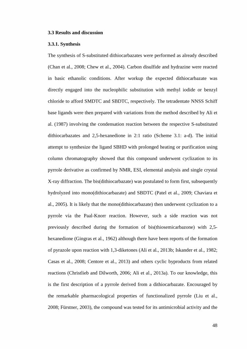
48
3.3 Results and discussion
3.3.1. Synthesis
The synthesis of S-substituted dithiocarbazates were performed as already described
(Chan et al., 2008; Chew et al., 2004). Carbon disulfide and hydrazine were reacted
in basic ethanolic conditions. After workup the expected dithiocarbazate was
directly engaged into the nucleophilic substitution with methyl iodide or benzyl
chloride to afford SMDTC and SBDTC, respectively. The tetradentate NNSS Schiff
base ligands were then prepared with variations from the method described by Ali et
al. (1987) involving the condensation reaction between the respective S-substituted
dithiocarbazates and 2,5-hexanedione in 2:1 ratio (Scheme 3.1: a-d). The initial
attempt to synthesize the ligand SBHD with prolonged heating or purification using
column chromatography showed that this compound underwent cyclization to its
pyrrole derivative as confirmed by NMR, ESI, elemental analysis and single crystal
X-ray diffraction. The bis(dithiocarbazate) was postulated to form first, subsequently
hydrolyzed into mono(dithiocarbazate) and SBDTC (Patel et al., 2009; Chaviara et
al., 2005). It is likely that the mono(dithiocarbazate) then underwent cyclization to a
pyrrole via the Paal-Knorr reaction. However, such a side reaction was not
previously described during the formation of bis(thiosemicarbazone) with 2,5-
hexanedione (Gingras et al., 1962) although there have been reports of the formation
of pyrazole upon reaction with 1,3-diketones (Ali et al., 2013b; Iskander et al., 1982;
Casas et al., 2008; Centore et al., 2013) and others cyclic byproducts from related
reactions (Christlieb and Dilworth, 2006; Ali et al., 2013a). To our knowledge, this
is the first description of a pyrrole derived from a dithiocarbazate. Encouraged by
the remarkable pharmacological properties of functionalized pyrrole (Liu et al.,
2008; Fürstner, 2003), the compound was tested for its antimicrobial activity and the
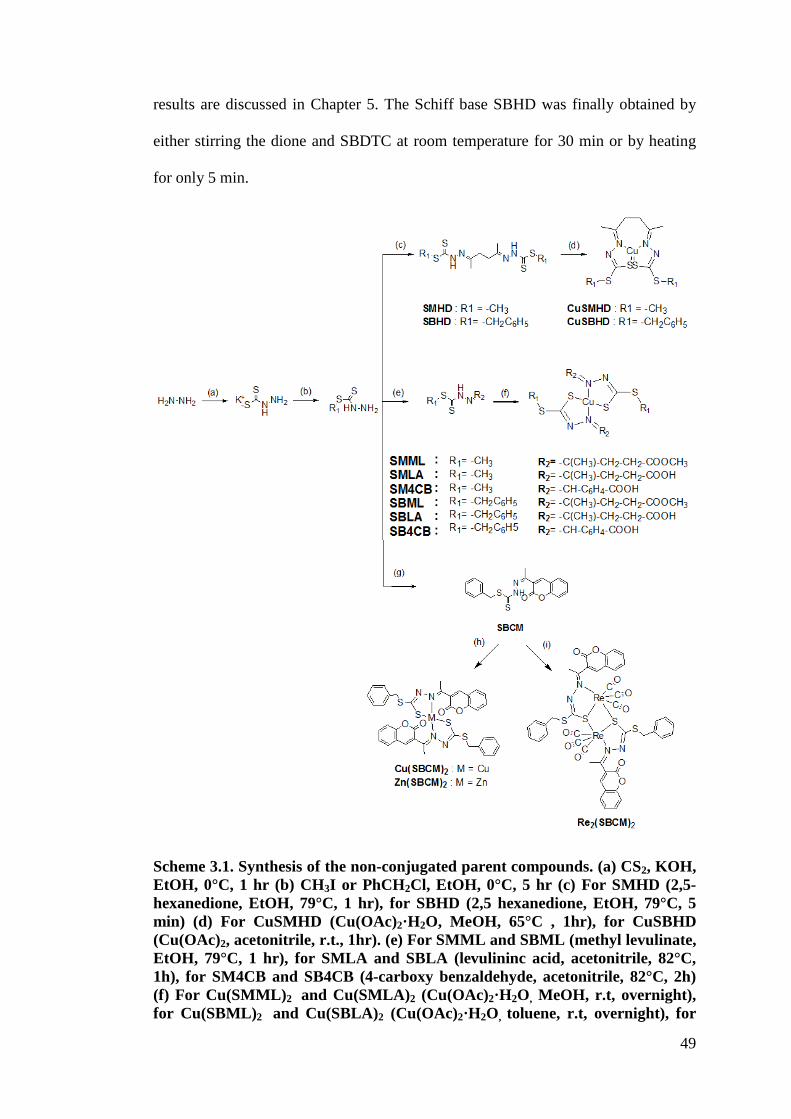
49
results are discussed in Chapter 5. The Schiff base SBHD was finally obtained by
either stirring the dione and SBDTC at room temperature for 30 min or by heating
for only 5 min.
Scheme 3.1. Synthesis of the non-conjugated parent compounds. (a) CS2, KOH, EtOH, 0°C, 1 hr (b) CH3I or PhCH2Cl, EtOH, 0°C, 5 hr (c) For SMHD (2,5- hexanedione, EtOH, 79°C, 1 hr), for SBHD (2,5 hexanedione, EtOH, 79°C, 5 min) (d) For CuSMHD (Cu(OAc)2·H2O, MeOH, 65°C , 1hr), for CuSBHD (Cu(OAc)2, acetonitrile, r.t., 1hr). (e) For SMML and SBML (methyl levulinate, EtOH, 79°C, 1 hr), for SMLA and SBLA (levulininc acid, acetonitrile, 82°C, 1h), for SM4CB and SB4CB (4-carboxy benzaldehyde, acetonitrile, 82°C, 2h) (f) For Cu(SMML)2 and Cu(SMLA)2 (Cu(OAc)2·H2O, MeOH, r.t, overnight), for Cu(SBML)2 and Cu(SBLA)2 (Cu(OAc)2·H2O, toluene, r.t, overnight), for
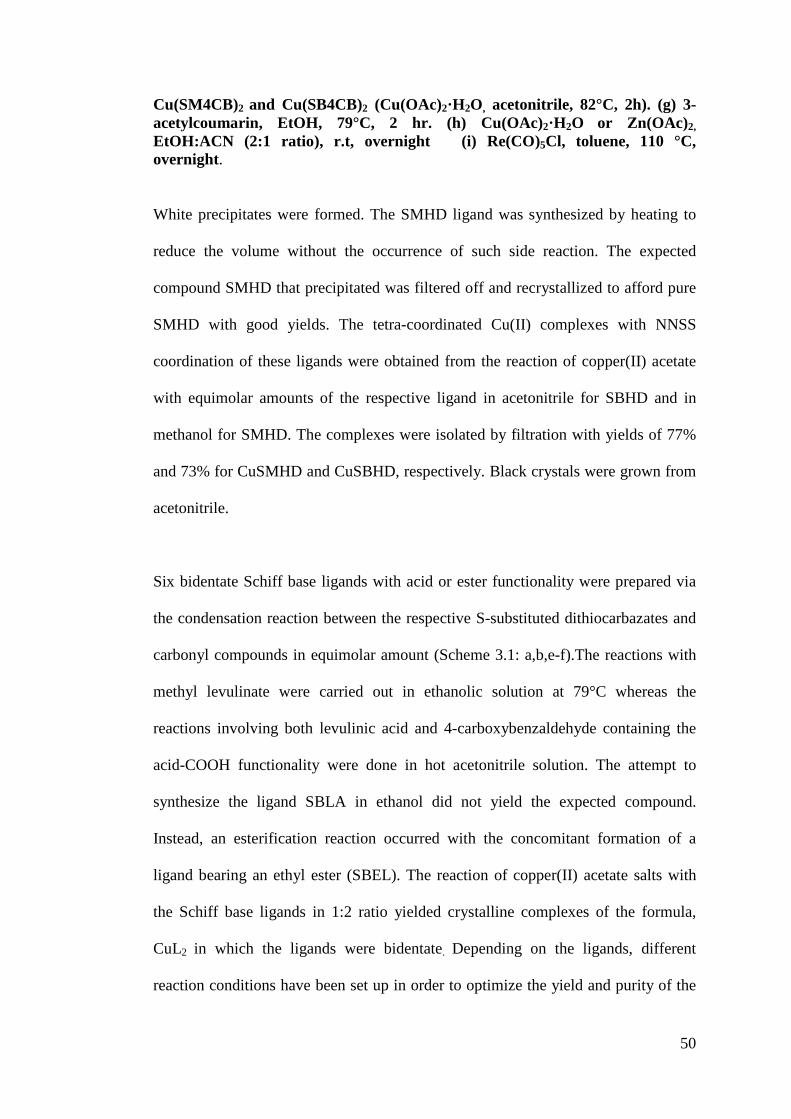
50
Cu(SM4CB)2 and Cu(SB4CB)2 (Cu(OAc)2·H2O, acetonitrile, 82°C, 2h). (g) 3-acetylcoumarin, EtOH, 79°C, 2 hr. (h) Cu(OAc)2·H2O or Zn(OAc)2, EtOH:ACN (2:1 ratio), r.t, overnight (i) Re(CO)5Cl, toluene, 110 °C, overnight.
White precipitates were formed. The SMHD ligand was synthesized by heating to
reduce the volume without the occurrence of such side reaction. The expected
compound SMHD that precipitated was filtered off and recrystallized to afford pure
SMHD with good yields. The tetra-coordinated Cu(II) complexes with NNSS
coordination of these ligands were obtained from the reaction of copper(II) acetate
with equimolar amounts of the respective ligand in acetonitrile for SBHD and in
methanol for SMHD. The complexes were isolated by filtration with yields of 77%
and 73% for CuSMHD and CuSBHD, respectively. Black crystals were grown from
acetonitrile.
Six bidentate Schiff base ligands with acid or ester functionality were prepared via
the condensation reaction between the respective S-substituted dithiocarbazates and
carbonyl compounds in equimolar amount (Scheme 3.1: a,b,e-f).The reactions with
methyl levulinate were carried out in ethanolic solution at 79°C whereas the
reactions involving both levulinic acid and 4-carboxybenzaldehyde containing the
acid-COOH functionality were done in hot acetonitrile solution. The attempt to
synthesize the ligand SBLA in ethanol did not yield the expected compound.
Instead, an esterification reaction occurred with the concomitant formation of a
ligand bearing an ethyl ester (SBEL). The reaction of copper(II) acetate salts with
the Schiff base ligands in 1:2 ratio yielded crystalline complexes of the formula,
CuL2 in which the ligands were bidentate. Depending on the ligands, different
reaction conditions have been set up in order to optimize the yield and purity of the
51
complexes. Early trials at high temperature and and in polar alcoholic solution often
resulted in oily products and further efforts to crystallize the complexes were futile.
The structures of four Schiff bases (SMML, SBML, SBEL, SBLA) and two Cu (II)
complexes, Cu(SMML)2 and Cu(SMLA)2 were confirmed by single-crystal X-ray
analyses.
The synthesis of the bidentate NS Schiff base with natural ketone moiety, SBCM
and its proposed structures of metal complexes are schematically represented in
Scheme 3.1 (a,b,g-i). The complexes Cu(SBCM)2 and Zn(SBCM)2 were prepared by
reacting SBCM with metal(II) acetate in ethanol:acetonitrile (2:1 ratio) solution
mixture at room temperature whereas the adduct, Re2(SBCM)2, was obtained by
refluxing the free ligand with ReCl(CO)5 in toluene. The single crystal of the Re(I)
complex was also grown from toluene.
The purity and stability of the ligands and their corresponding complexes at
physiological pH are important prerequisites for the evaluation of their biological
activity. Therefore RP-HPLC experiments have been performed to gain insights into
their stability in aqueous solutions. The tetradentate ligands and their respective
Cu(II) complexes were eluted on a C18-column with an increasing amount of
acetonitrile (CH3CN) in H2O (from 5% to 100% of CH3CN over 30 minutes),
containing 0.1 % trifluoroacetic acid (TFA) to maintain the pH. The compounds
were detected using a UV detector at 220 nm and 280 nm. The chromatograms of
the pure ligands of the tetradentate series showed 3 peaks that could correspond
respectively to the hydrolyzed hydrazone, the expected ligand and the pyrrole
byproduct (Figure 3.1) whereas the complexes showed only a single peak
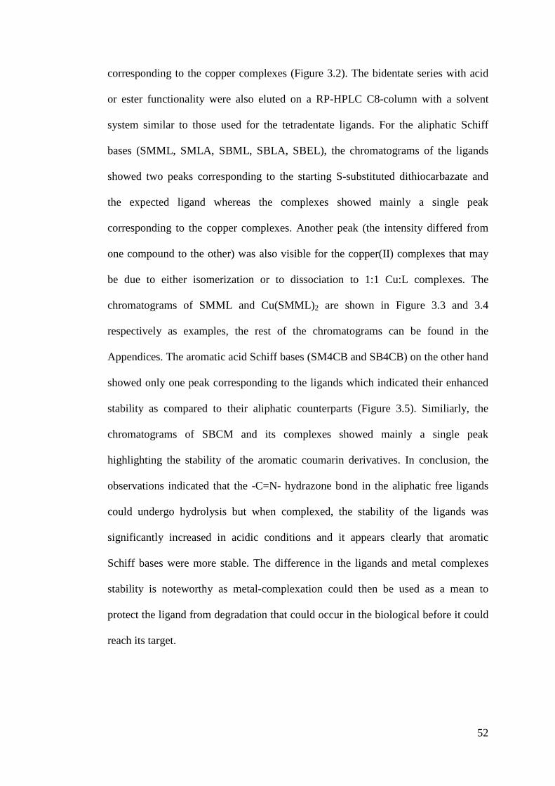
52
corresponding to the copper complexes (Figure 3.2). The bidentate series with acid
or ester functionality were also eluted on a RP-HPLC C8-column with a solvent
system similar to those used for the tetradentate ligands. For the aliphatic Schiff
bases (SMML, SMLA, SBML, SBLA, SBEL), the chromatograms of the ligands
showed two peaks corresponding to the starting S-substituted dithiocarbazate and
the expected ligand whereas the complexes showed mainly a single peak
corresponding to the copper complexes. Another peak (the intensity differed from
one compound to the other) was also visible for the copper(II) complexes that may
be due to either isomerization or to dissociation to 1:1 Cu:L complexes. The
chromatograms of SMML and Cu(SMML)2 are shown in Figure 3.3 and 3.4
respectively as examples, the rest of the chromatograms can be found in the
Appendices. The aromatic acid Schiff bases (SM4CB and SB4CB) on the other hand
showed only one peak corresponding to the ligands which indicated their enhanced
stability as compared to their aliphatic counterparts (Figure 3.5). Similiarly, the
chromatograms of SBCM and its complexes showed mainly a single peak
highlighting the stability of the aromatic coumarin derivatives. In conclusion, the
observations indicated that the -C=N- hydrazone bond in the aliphatic free ligands
could undergo hydrolysis but when complexed, the stability of the ligands was
significantly increased in acidic conditions and it appears clearly that aromatic
Schiff bases were more stable. The difference in the ligands and metal complexes
stability is noteworthy as metal-complexation could then be used as a mean to
protect the ligand from degradation that could occur in the biological before it could
reach its target.

53
Figure 3.1. RP-HPLC chromatogram of SBHD at 220 nm (top) and 280 nm (bottom).
Figure 3.2. RP-HPLC chromatogram of CuSBHD.
Figure 3.3. RP-HPLC chromatogram of SMML.
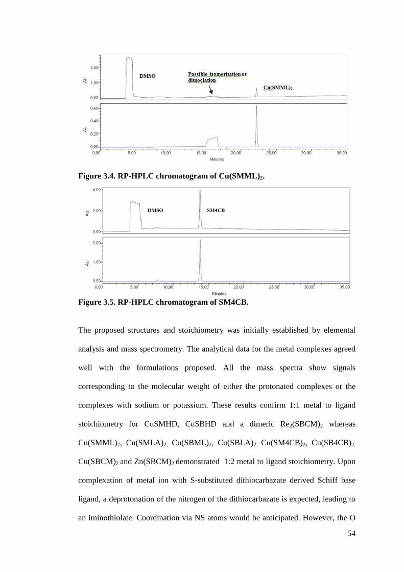
54
Figure 3.4. RP-HPLC chromatogram of Cu(SMML)2.
Figure 3.5. RP-HPLC chromatogram of SM4CB.
The proposed structures and stoichiometry was initially established by elemental
analysis and mass spectrometry. The analytical data for the metal complexes agreed
well with the formulations proposed. All the mass spectra show signals
corresponding to the molecular weight of either the protonated complexes or the
complexes with sodium or potassium. These results confirm 1:1 metal to ligand
stoichiometry for CuSMHD, CuSBHD and a dimeric Re2(SBCM)2 whereas
Cu(SMML)2, Cu(SMLA)2, Cu(SBML)2, Cu(SBLA)2, Cu(SM4CB)2, Cu(SB4CB)2,
Cu(SBCM)2 and Zn(SBCM)2 demonstrated 1:2 metal to ligand stoichiometry. Upon
complexation of metal ion with S-substituted dithiocarbazate derived Schiff base
ligand, a deprotonation of the nitrogen of the dithiocarbazate is expected, leading to
an iminothiolate. Coordination via NS atoms would be anticipated. However, the O
55
atom present in the ligands containing acid, ester or coumarin moiety could also
potentially participate in metal coordination. In order to confirm the formation of the
metal complexes and to characterize them, several other techniques were employed.
3.3.2 Characterization of metal complexes in solid state
3.3.2.1 FT-IR
Two examples of the FT-IR spectra used to monitor complexation, one from
tetradentate series and the other from bidentate series are shown in Figure 3.6 and
3.7, respectively. The IR spectra for the rest of the compounds can be found in the
Appendices. The IR spectra of the tetradentate ligands and bidentate SMML and
SBML ligands exhibit characteristic bands +NH at ca. 3130 and 3200 cm-1,
respectively. Strong broad structured bands spanning from around 3300–2300
cm"1 appeared in the spectra of the ligands SMLA, SBLA, SM4CB and SB4CB
which contained the acid functionality and their respective Cu(II) complexes due
to +OH, +NH and +CH overlapping stretching vibrations. The IR spectrum of
coumarin-derived SBCM however showed only a weak band at
3056 cm"1 assignable to #(NH) stretching. Another band attributed to #(SH) was
noticeable at 2872 cm"1. These observations demonstrated that while most of the
ligands existed in thione form, SBCM existed in both thione and thiolate forms in
the solid state. In addition, all the ligands possess bands +(C=N) at 1652-1559 cm-1,
+(C=S) at 1067-1025 cm-1, +(CSS) at 990-923 cm-1and v(NN) at 841-778 cm-1.
Upon formation of the complexes, the band corresponding to the +(NH) stretching of
the ligands disappeared. The +(C=N) attributed to azomethine bond was observed to
experience a downward shift of 5-40 cm-1 in most of the spectra with the exception
of metal complexes of SBCM that shifted to higher wavenumbers (~1566 cm-1). A
56
second band due to +(N=C) in complexes containing anionic dithiocarbazate
moieties was also resolved (Rapheal et al., 2007) for CuSMHD, CuSBHD and
Cu(SBLA)2. The hydrazinic +(N-N) band also shifted to either higher and lower
wavenumbers upon complexation. All these observations indicates the deprotonation
of the Schiff base ligands and coordination via the azomethine nitrogen atom during
complexation with metal ion which resulted in the adoption of a more stable
structure where the conjugated system was elongated in the complexes as compared
to the free ligands. The disappearance of +(C=S) and the splitting of the asymmetric
+(CSS) band into two peaks in the spectra of the all metal complexes were strong
evidence of coordination via the thiolate sulfur atoms (Crouse et al., 2004; Akbar Ali
and Tarafdar, 1977). In the bidentate series, the strong band at 1717-1687 cm"1 of
the ligands can be assigned to the carbonyl +(C=O). The band remained unchanged
upon complexation in the spectra of Cu(SM4CB)2, Cu(SB4CB)2 and Cu(SBLA)2 but
slightly shifted for Cu(SMML)2, Cu(SBML)2, Cu(SMLA)2, Cu(SBCM)2,
Zn(SBCM)2 and Re2(SBCM)2. Despite the shift in wavenumbers, the X-ray
structures solved for Cu(SMML)2, Cu(SMLA)2 and Re2(SBCM)2 revealed that the
metal ions were coordinated by the bidentate ligands via only the azomethine
nitrogen and thiolate sulfur atoms. No coordination via the carbonylic oxygen was
observed. Such a shift can be interpreted as being caused by the vibrational coupling
of the C=O in the solid state H-bonded structure (Ferrari et al., 2000). Single crystal
X-ray analyses of the ligands and complexes indicated the formation of
intermolecular hydrogen bonds involving the carbonylic oxygen and the presence of
other short contacts. Previous publications had also reported comparable shifting
that did not correspond to coordinated O atoms (Creaven et al., 2009). Fac-geometry
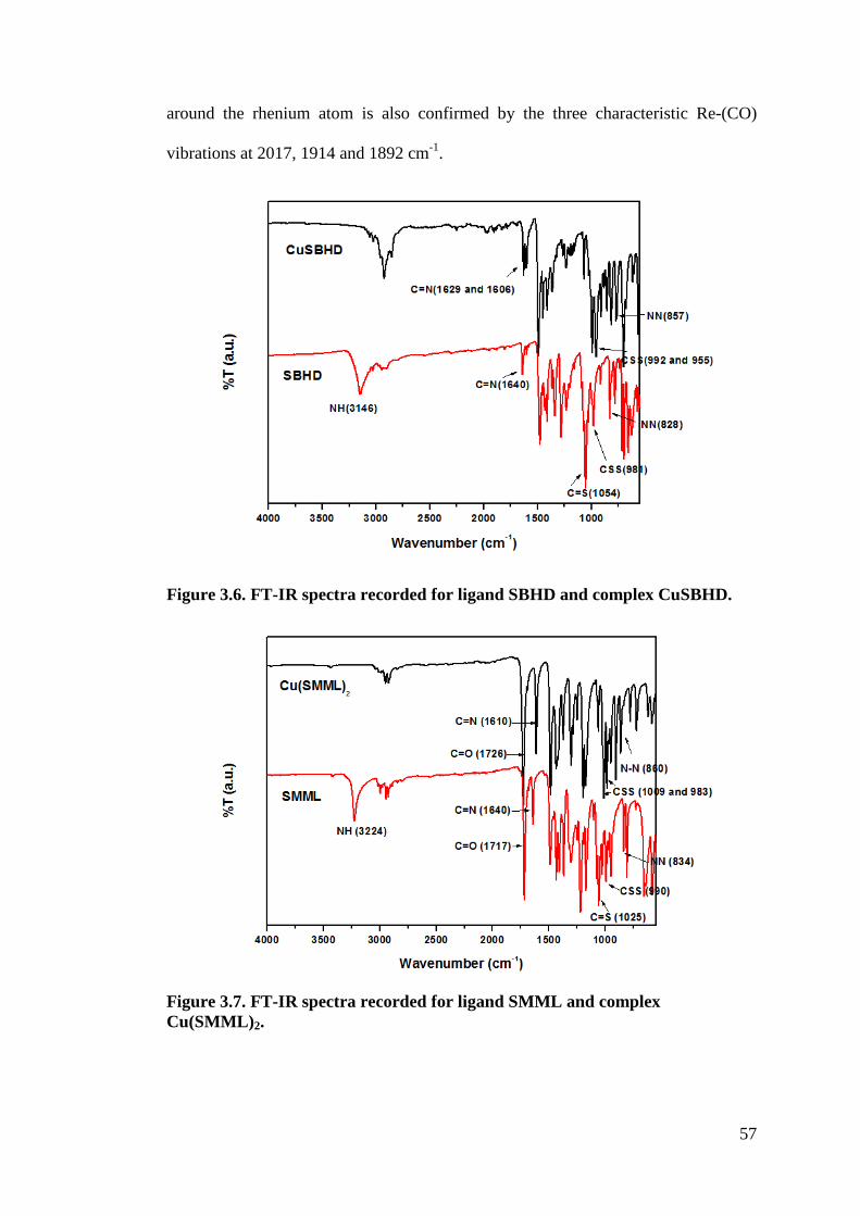
57
around the rhenium atom is also confirmed by the three characteristic Re-(CO)
vibrations at 2017, 1914 and 1892 cm-1.
Figure 3.6. FT-IR spectra recorded for ligand SBHD and complex CuSBHD.
Figure 3.7. FT-IR spectra recorded for ligand SMML and complex Cu(SMML)2.
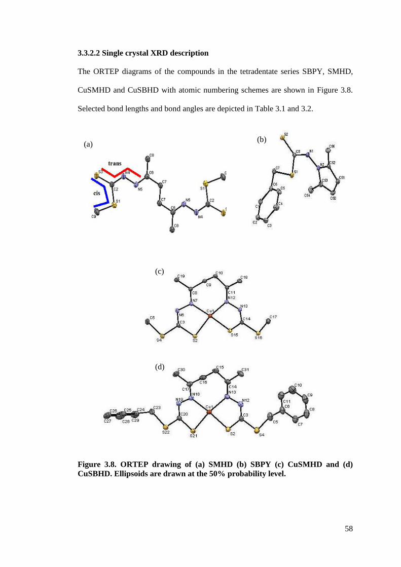
58
3.3.2.2 Single crystal XRD description
The ORTEP diagrams of the compounds in the tetradentate series SBPY, SMHD,
CuSMHD and CuSBHD with atomic numbering schemes are shown in Figure 3.8.
Selected bond lengths and bond angles are depicted in Table 3.1 and 3.2.
Figure 3.8. ORTEP drawing of (a) SMHD (b) SBPY (c) CuSMHD and (d) CuSBHD. Ellipsoids are drawn at the 50% probability level.
(a) (b)
(d)
(c)
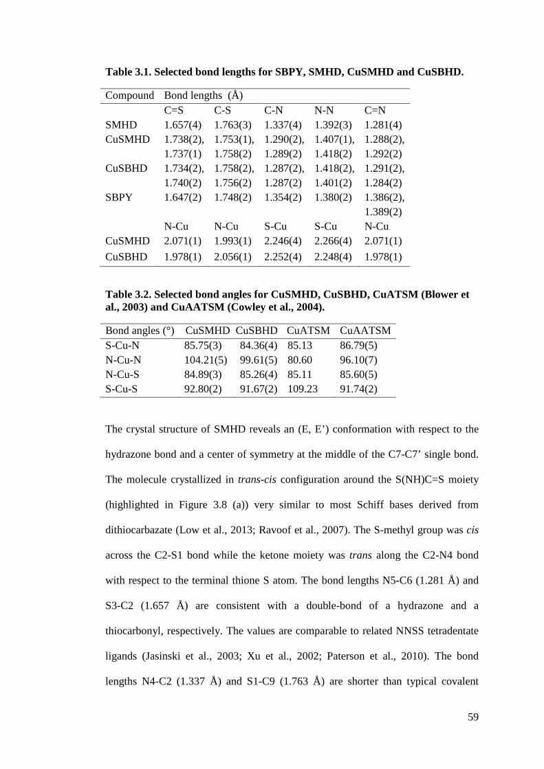
59
Table 3.1. Selected bond lengths for SBPY, SMHD, CuSMHD and CuSBHD. Compound Bond lengths (Å) C=S C-S C-N N-N C=N SMHD 1.657(4) 1.763(3) 1.337(4) 1.392(3) 1.281(4) CuSMHD 1.738(2),
1.737(1) 1.753(1), 1.758(2)
1.290(2), 1.289(2)
1.407(1), 1.418(2)
1.288(2), 1.292(2)
CuSBHD
1.734(2), 1.740(2)
1.758(2), 1.756(2)
1.287(2), 1.287(2)
1.418(2), 1.401(2)
1.291(2), 1.284(2)
SBPY 1.647(2) 1.748(2) 1.354(2) 1.380(2) 1.386(2), 1.389(2)
N-Cu N-Cu S-Cu S-Cu N-Cu CuSMHD 2.071(1) 1.993(1) 2.246(4) 2.266(4) 2.071(1) CuSBHD 1.978(1) 2.056(1) 2.252(4) 2.248(4) 1.978(1)
Table 3.2. Selected bond angles for CuSMHD, CuSBHD, CuATSM (Blower et al., 2003) and CuAATSM (Cowley et al., 2004). Bond angles (°) CuSMHD CuSBHD CuATSM CuAATSM S-Cu-N 85.75(3) 84.36(4) 85.13 86.79(5) N-Cu-N 104.21(5) 99.61(5) 80.60 96.10(7) N-Cu-S 84.89(3) 85.26(4) 85.11 85.60(5) S-Cu-S 92.80(2) 91.67(2) 109.23 91.74(2)
The crystal structure of SMHD reveals an (E, E’) conformation with respect to the
hydrazone bond and a center of symmetry at the middle of the C7-C7’ single bond.
The molecule crystallized in trans-cis configuration around the S(NH)C=S moiety
(highlighted in Figure 3.8 (a)) very similar to most Schiff bases derived from
dithiocarbazate (Low et al., 2013; Ravoof et al., 2007). The S-methyl group was cis
across the C2-S1 bond while the ketone moiety was trans along the C2-N4 bond
with respect to the terminal thione S atom. The bond lengths N5-C6 (1.281 Å) and
S3-C2 (1.657 Å) are consistent with a double-bond of a hydrazone and a
thiocarbonyl, respectively. The values are comparable to related NNSS tetradentate
ligands (Jasinski et al., 2003; Xu et al., 2002; Paterson et al., 2010). The bond
lengths N4-C2 (1.337 Å) and S1-C9 (1.763 Å) are shorter than typical covalent
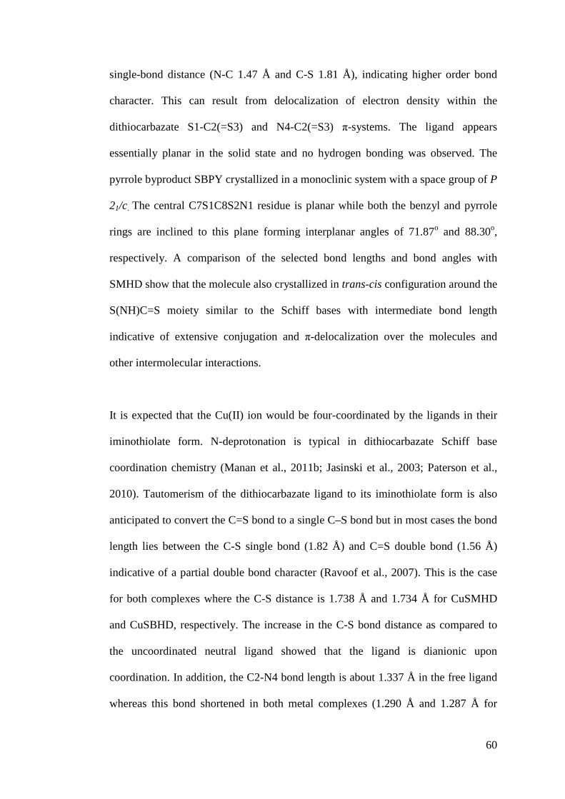
60
single-bond distance (N-C 1.47 Å and C-S 1.81 Å), indicating higher order bond
character. This can result from delocalization of electron density within the
dithiocarbazate S1-C2(=S3) and N4-C2(=S3) ,-systems. The ligand appears
essentially planar in the solid state and no hydrogen bonding was observed. The
pyrrole byproduct SBPY crystallized in a monoclinic system with a space group of P
21/c. The central C7S1C8S2N1 residue is planar while both the benzyl and pyrrole
rings are inclined to this plane forming interplanar angles of 71.87o and 88.30o,
respectively. A comparison of the selected bond lengths and bond angles with
SMHD show that the molecule also crystallized in trans-cis configuration around the
S(NH)C=S moiety similar to the Schiff bases with intermediate bond length
indicative of extensive conjugation and ,-delocalization over the molecules and
other intermolecular interactions.
It is expected that the Cu(II) ion would be four-coordinated by the ligands in their
iminothiolate form. N-deprotonation is typical in dithiocarbazate Schiff base
coordination chemistry (Manan et al., 2011b; Jasinski et al., 2003; Paterson et al.,
2010). Tautomerism of the dithiocarbazate ligand to its iminothiolate form is also
anticipated to convert the C=S bond to a single C–S bond but in most cases the bond
length lies between the C-S single bond (1.82 Å) and C=S double bond (1.56 Å)
indicative of a partial double bond character (Ravoof et al., 2007). This is the case
for both complexes where the C-S distance is 1.738 Å and 1.734 Å for CuSMHD
and CuSBHD, respectively. The increase in the C-S bond distance as compared to
the uncoordinated neutral ligand showed that the ligand is dianionic upon
coordination. In addition, the C2-N4 bond length is about 1.337 Å in the free ligand
whereas this bond shortened in both metal complexes (1.290 Å and 1.287 Å for
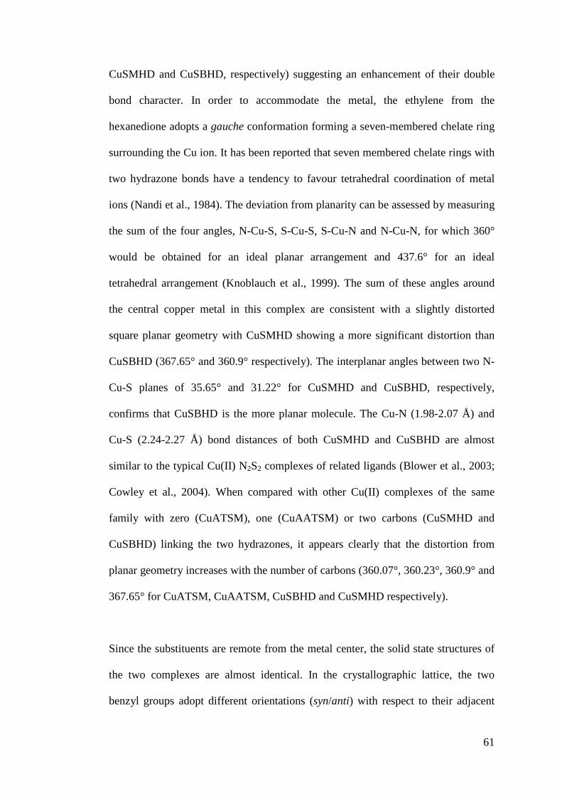
61
CuSMHD and CuSBHD, respectively) suggesting an enhancement of their double
bond character. In order to accommodate the metal, the ethylene from the
hexanedione adopts a gauche conformation forming a seven-membered chelate ring
surrounding the Cu ion. It has been reported that seven membered chelate rings with
two hydrazone bonds have a tendency to favour tetrahedral coordination of metal
ions (Nandi et al., 1984). The deviation from planarity can be assessed by measuring
the sum of the four angles, N-Cu-S, S-Cu-S, S-Cu-N and N-Cu-N, for which 360°
would be obtained for an ideal planar arrangement and 437.6° for an ideal
tetrahedral arrangement (Knoblauch et al., 1999). The sum of these angles around
the central copper metal in this complex are consistent with a slightly distorted
square planar geometry with CuSMHD showing a more significant distortion than
CuSBHD (367.65° and 360.9° respectively). The interplanar angles between two N-
Cu-S planes of 35.65° and 31.22° for CuSMHD and CuSBHD, respectively,
confirms that CuSBHD is the more planar molecule. The Cu-N (1.98-2.07 Å) and
Cu-S (2.24-2.27 Å) bond distances of both CuSMHD and CuSBHD are almost
similar to the typical Cu(II) N2S2 complexes of related ligands (Blower et al., 2003;
Cowley et al., 2004). When compared with other Cu(II) complexes of the same
family with zero (CuATSM), one (CuAATSM) or two carbons (CuSMHD and
CuSBHD) linking the two hydrazones, it appears clearly that the distortion from
planar geometry increases with the number of carbons (360.07°, 360.23°, 360.9° and
367.65° for CuATSM, CuAATSM, CuSBHD and CuSMHD respectively).
Since the substituents are remote from the metal center, the solid state structures of
the two complexes are almost identical. In the crystallographic lattice, the two
benzyl groups adopt different orientations (syn/anti) with respect to their adjacent
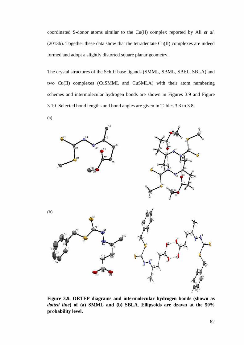
62
coordinated S-donor atoms similar to the Cu(II) complex reported by Ali et al.
(2013b). Together these data show that the tetradentate Cu(II) complexes are indeed
formed and adopt a slightly distorted square planar geometry.
The crystal structures of the Schiff base ligands (SMML, SBML, SBEL, SBLA) and
two Cu(II) complexes (CuSMML and CuSMLA) with their atom numbering
schemes and intermolecular hydrogen bonds are shown in Figures 3.9 and Figure
3.10. Selected bond lengths and bond angles are given in Tables 3.3 to 3.8.
(a)
(b)
Figure 3.9. ORTEP diagrams and intermolecular hydrogen bonds (shown as dotted line) of (a) SMML and (b) SBLA Ellipsoids are drawn at the 50% probability level.
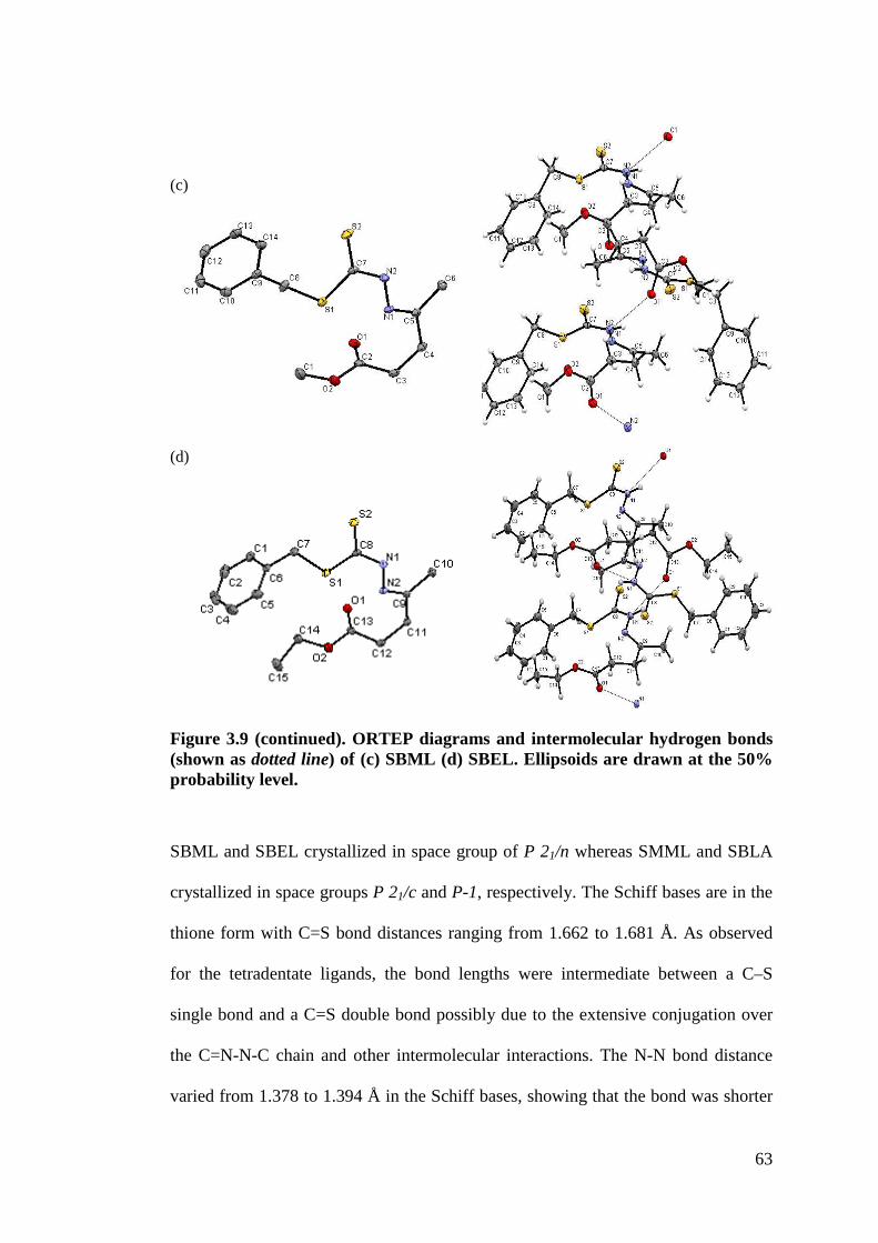
63
(c)
(d)
Figure 3.9 (continued). ORTEP diagrams and intermolecular hydrogen bonds (shown as dotted line) of (c) SBML (d) SBEL. Ellipsoids are drawn at the 50% probability level.
SBML and SBEL crystallized in space group of P 21/n whereas SMML and SBLA
crystallized in space groups P 21/c and P-1, respectively. The Schiff bases are in the
thione form with C=S bond distances ranging from 1.662 to 1.681 Å. As observed
for the tetradentate ligands, the bond lengths were intermediate between a C–S
single bond and a C=S double bond possibly due to the extensive conjugation over
the C=N-N-C chain and other intermolecular interactions. The N-N bond distance
varied from 1.378 to 1.394 Å in the Schiff bases, showing that the bond was shorter
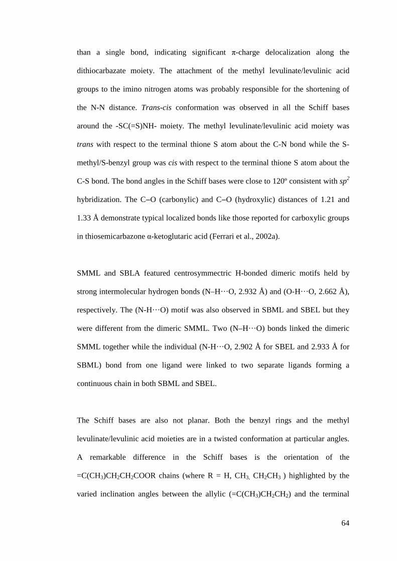
64
than a single bond, indicating significant ,-charge delocalization along the
dithiocarbazate moiety. The attachment of the methyl levulinate/levulinic acid
groups to the imino nitrogen atoms was probably responsible for the shortening of
the N-N distance. Trans-cis conformation was observed in all the Schiff bases
around the -SC(=S)NH- moiety. The methyl levulinate/levulinic acid moiety was
trans with respect to the terminal thione S atom about the C-N bond while the S-
methyl/S-benzyl group was cis with respect to the terminal thione S atom about the
C-S bond. The bond angles in the Schiff bases were close to 120º consistent with sp2
hybridization. The C"O (carbonylic) and C"O (hydroxylic) distances of 1.21 and
1.33 Å demonstrate typical localized bonds like those reported for carboxylic groups
in thiosemicarbazone &-ketoglutaric acid (Ferrari et al., 2002a).
SMML and SBLA featured centrosymmectric H-bonded dimeric motifs held by
strong intermolecular hydrogen bonds (N–H···O, 2.932 Å) and (O-H·· ·O, 2.662 Å),
respectively. The (N-H·· ·O) motif was also observed in SBML and SBEL but they
were different from the dimeric SMML. Two (N–H···O) bonds linked the dimeric
SMML together while the individual (N-H···O, 2.902 Å for SBEL and 2.933 Å for
SBML) bond from one ligand were linked to two separate ligands forming a
continuous chain in both SBML and SBEL.
The Schiff bases are also not planar. Both the benzyl rings and the methyl
levulinate/levulinic acid moieties are in a twisted conformation at particular angles.
A remarkable difference in the Schiff bases is the orientation of the
=C(CH3)CH2CH2COOR chains (where R = H, CH3, CH2CH3 ) highlighted by the
varied inclination angles between the allylic (=C(CH3)CH2CH2) and the terminal
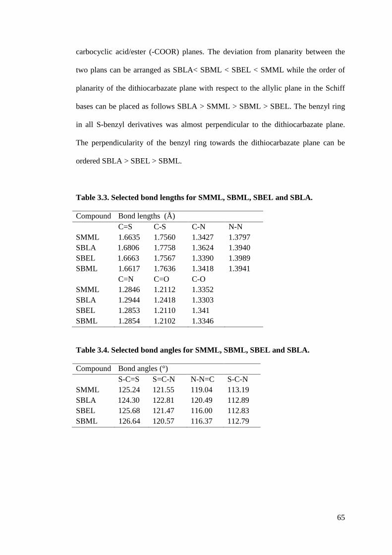
65
carbocyclic acid/ester (-COOR) planes. The deviation from planarity between the
two plans can be arranged as SBLA< SBML < SBEL < SMML while the order of
planarity of the dithiocarbazate plane with respect to the allylic plane in the Schiff
bases can be placed as follows SBLA > SMML > SBML > SBEL. The benzyl ring
in all S-benzyl derivatives was almost perpendicular to the dithiocarbazate plane.
The perpendicularity of the benzyl ring towards the dithiocarbazate plane can be
ordered SBLA > SBEL > SBML.
Table 3.3. Selected bond lengths for SMML, SBML, SBEL and SBLA. Compound Bond lengths (Å) C=S C-S C-N N-N SMML 1.6635 1.7560 1.3427 1.3797 SBLA 1.6806 1.7758 1.3624 1.3940 SBEL 1.6663 1.7567 1.3390 1.3989 SBML 1.6617 1.7636 1.3418 1.3941 C=N C=O C-O SMML 1.2846 1.2112 1.3352 SBLA 1.2944 1.2418 1.3303 SBEL 1.2853 1.2110 1.341 SBML 1.2854 1.2102 1.3346
Table 3.4. Selected bond angles for SMML, SBML, SBEL and SBLA. Compound Bond angles (°) S-C=S S=C-N N-N=C S-C-N SMML 125.24 121.55 119.04 113.19 SBLA 124.30 122.81 120.49 112.89 SBEL 125.68 121.47 116.00 112.83 SBML 126.64 120.57 116.37 112.79
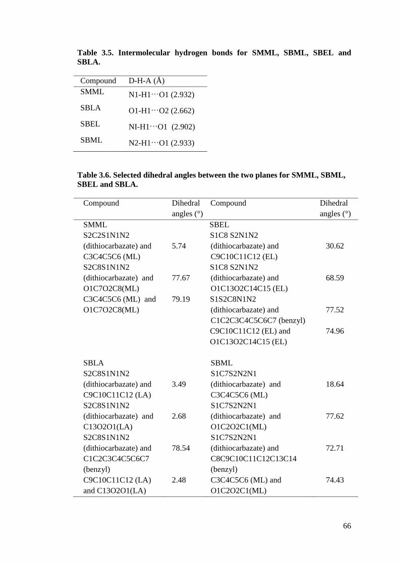
66
Table 3.5. Intermolecular hydrogen bonds for SMML, SBML, SBEL and SBLA. Compound D-H-A (Å) SMML N1-H1 O1 (2.932) SBLA O1-H1 O2 (2.662) SBEL NI-H1 O1 (2.902) SBML N2-H1 O1 (2.933)
Table 3.6. Selected dihedral angles between the two planes for SMML, SBML, SBEL and SBLA.
Compound Dihedral angles (°)
Compound Dihedral angles (°)
SMML SBEL S2C2S1N1N2 (dithiocarbazate) and C3C4C5C6 (ML) S2C8S1N1N2 (dithiocarbazate) and O1C7O2C8(ML) C3C4C5C6 (ML) and O1C7O2C8(ML)
5.74 77.67 79.19
S1C8 S2N1N2 (dithiocarbazate) and C9C10C11C12 (EL) S1C8 S2N1N2 (dithiocarbazate) and O1C13O2C14C15 (EL) S1S2C8N1N2 (dithiocarbazate) and C1C2C3C4C5C6C7 (benzyl) C9C10C11C12 (EL) and O1C13O2C14C15 (EL)
30.62 68.59 77.52 74.96
SBLA
SBML
S2C8S1N1N2 (dithiocarbazate) and C9C10C11C12 (LA) S2C8S1N1N2 (dithiocarbazate) and C13O2O1(LA) S2C8S1N1N2 (dithiocarbazate) and C1C2C3C4C5C6C7 (benzyl) C9C10C11C12 (LA) and C13O2O1(LA)
3.49 2.68 78.54 2.48
S1C7S2N2N1 (dithiocarbazate) and C3C4C5C6 (ML) S1C7S2N2N1 (dithiocarbazate) and O1C2O2C1(ML) S1C7S2N2N1 (dithiocarbazate) and C8C9C10C11C12C13C14 (benzyl) C3C4C5C6 (ML) and O1C2O2C1(ML)
18.64 77.62 72.71 74.43
67
Cu(SMML)2 crystallized in the C 2/c space group as a centrosymmetric complex in
which the two ligands were symmetrically related to each other and have the same
bond angles and distances. The central copper atom was bis-chelated by the
uninegatively charged bidentate ligand through the azomethine nitrogen atoms (N2)
and thiolate sulfur atoms (S1). Both nitrogen and sulfur atoms in the two ligands are
coordinated at the same position relative to each other. The sum of the four angles,
N-Cu-S, S-Cu-S, S-Cu-N, and N-Cu-N, in the complex was 374.33° confirming
significant deviation from the square-planar geometry about the copper ions. Neither
the carbonylic nor hydroxylic oxygen of the methyl levulinate moiety participated in
complexation. The conjugation system of the moieties was influenced by
coordination with the metal as shown by slight lengthening of the N–N bond
distance (1.407 Å). The bond lengths in the dithiocarbazate moiety that are expected
to be most affected by coordination are azomethine C–N and C–S. The C1–S1 and
C1-N1 bond lengths in the complex were 1.737 and 1.296 Å respectively. The C1–
S1 distance was longer than that observed in the Schiff base, SMML, indicating
single bond character as expected when complexation involves the ligand in its thiol
form. The C1–N1 bond distances in the complex were also typical for double bonds
in Schiff bases compounds clearly indicating that complexation involves
deprotonation at N1.
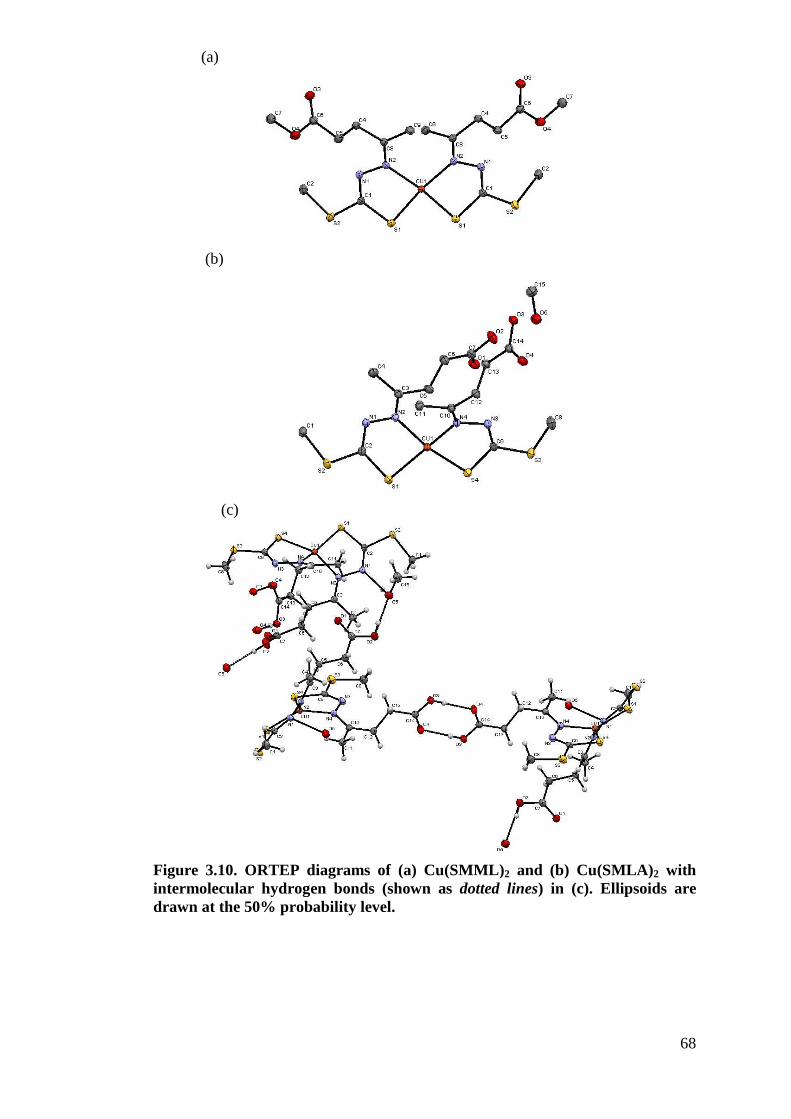
68
Figure 3.10. ORTEP diagrams of (a) Cu(SMML)2 and (b) Cu(SMLA)2 with intermolecular hydrogen bonds (shown as dotted lines) in (c). Ellipsoids are drawn at the 50% probability level.
(a)
(b)
(c)
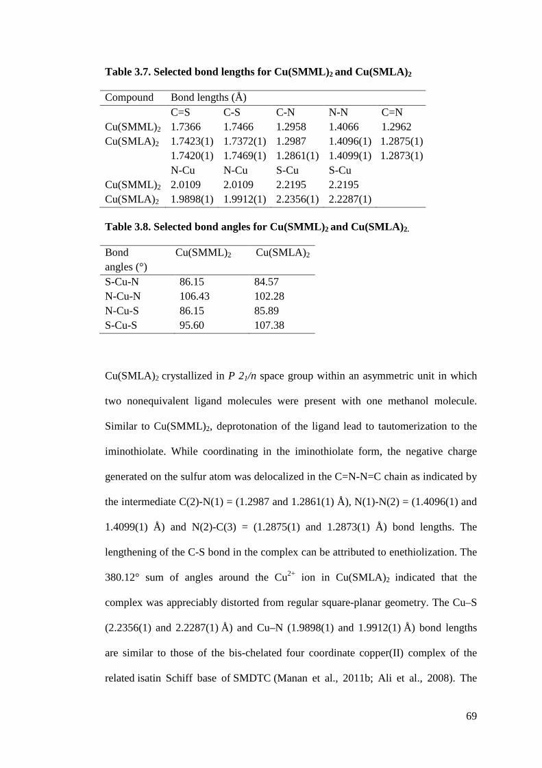
69
Table 3.7. Selected bond lengths for Cu(SMML)2 and Cu(SMLA)2 Compound Bond lengths (Å) C=S C-S C-N N-N C=N Cu(SMML)2 1.7366 1.7466 1.2958 1.4066 1.2962 Cu(SMLA)2 1.7423(1)
1.7420(1) 1.7372(1) 1.7469(1)
1.2987 1.2861(1)
1.4096(1) 1.4099(1)
1.2875(1) 1.2873(1)
N-Cu N-Cu S-Cu S-Cu Cu(SMML)2 2.0109 2.0109 2.2195 2.2195 Cu(SMLA)2 1.9898(1) 1.9912(1) 2.2356(1) 2.2287(1) Table 3.8. Selected bond angles for Cu(SMML)2 and Cu(SMLA)2. Bond angles (°)
Cu(SMML)2
Cu(SMLA)2
S-Cu-N 86.15 84.57 N-Cu-N 106.43 102.28 N-Cu-S 86.15 85.89 S-Cu-S 95.60 107.38
Cu(SMLA)2 crystallized in P 21/n space group within an asymmetric unit in which
two nonequivalent ligand molecules were present with one methanol molecule.
Similar to Cu(SMML)2, deprotonation of the ligand lead to tautomerization to the
iminothiolate. While coordinating in the iminothiolate form, the negative charge
generated on the sulfur atom was delocalized in the C=N-N=C chain as indicated by
the intermediate C(2)-N(1) = (1.2987 and 1.2861(1) Å), N(1)-N(2) = (1.4096(1) and
1.4099(1) Å) and N(2)-C(3) = (1.2875(1) and 1.2873(1) Å) bond lengths. The
lengthening of the C-S bond in the complex can be attributed to enethiolization. The
380.12° sum of angles around the Cu2+ ion in Cu(SMLA)2 indicated that the
complex was appreciably distorted from regular square-planar geometry. The Cu–S
(2.2356(1) and 2.2287(1) Å) and Cu–N (1.9898(1) and 1.9912(1) Å) bond lengths
are similar to those of the bis-chelated four coordinate copper(II) complex of the
related isatin Schiff base of SMDTC (Manan et al., 2011b; Ali et al., 2008). The
70
difference between the bond lengths in the same complex can be ascribed to the
constraints imposed by chelation. The packing for Cu(SMLA)2 was also determined.
In addition to the interactions between the (O3-H·· ·O4) bond involving two
neighbouring independent molecules similar to its free ligand SBLA, an extended
network of hydrogen bonds involving the crystallized methanol molecule and the
other ligand in Cu(SMLA)2 that was closed to the solvent molecule was also
observed. The linkage involved the hydroxylic oxygen atom (O2) of one complex
molecule, the alcoholic oxygen atom (O3) of methanol and the uncoordinated
azomethine nitrogen atom (N1) from another complex molecule. The continuous
hydrogen bond networks developed were (O2–H2···O5, 2.655 Å) and (O5-H5···N1,
2.854 Å). As a consequence of these H-bond systems, the two ligands were not
planar in the portion involved in metal coordination and the terminal chains with the
carboxylic group which bridged the adjacent molecule were fairly distorted as can
be seen from the dihedral angles involving the levulinic acid moiety (84.78° for C3–
C4–C5 and C6-C7-O1-O2, 89.31° for C10–C11–C12 and C13-C14-O4-O3). The
presence of methanol in the lattice highlights the spatial effect in which the terminal
chains with the carboxylic group were forced to align in parallel whereas in
Cu(SMML)2 the ester terminal chains were almost 180° from one another
minimizing steric constraints. This explains the observation that Cu(SMLA)2 was
more distorted from square planar than Cu(SMML)2.
The ORTEP diagram of Re2(SBCM)2 complex with atomic numbering scheme is
shown in Figure 3.11. Selected bond lengths and bond angles are given in Tables 3.9
and 3.10. The complex crystallized in P 21/c space group with unit cell parameters
a=14.7072(4), b=12.6588(3), c=12.1386(3) and &=90o, %=99.374(1)o, -=90o. A
71
number of thiosemicarbazone complexes of Re(I) carbonyl compounds have been
studied by X-ray diffraction (Carballo et al., 2002; Santos et al., 2004). However, to
date, this is the first Re(I) tricarbonyl complex with a dithiocarbazate Schiff base
ligand to have been characterized structurally by X-ray diffractometry.
Crystallographic analysis of the complex Re2(SBCM)2 in this work showed that the
rhenium atom was octahedrally coordinated to (i) three carbonyl carbon atoms in
fac arrangement, (ii) the N and S atoms of the deprotonated bidentate
dithiocarbazate Schiff base ligand with 3-acetylcoumarin in which the metal forms a
five-membered chelate ring and (iii) the sulfur atom of a neighbouring molecule that
had replaced the chloride atom resulting in a centrosymmetric dimer. The Re-S-
Re bridge (Re-S = 2.4669 (1) and 2.5565 (1) Å) was more asymmetric than Re
complex (a) (data adopted from Carbolla et al., 2002). Like Re complex (a), the
planar Re2S2 diamond for Re2(SBCM)2 in which the Re-Re distance was too long
for any significant bonding interaction, had bond angles close to 90°. The three Re-
C distances were close to one another with an average bond distance of 1.916 Å in
the range shown by numerous other Re(I) complexes containing the
Re(CO)3+ core (Czerwieniec et al., 2005; Fuks et al., 2010). The Re-N (2.2002(1)
Å), Re-S (2.4669(1) Å) and Re-S’ (2.5565(1) Å) distances do not differ significantly
from those determined for Re complex (a). Therefore, it can be concluded that in the
dimeric Re2(SBCM)2 complex the geometry around each Re center was slightly
distorted octahedral. The C12-S1 was shorter (1.7755 Å) in this complex in
comparison to (a), but it was longer than in most free dithiocarbazate Schiff bases
(ca 1.6 Å) suggesting a predominantly thiol character upon complexation. This
conclusion was reinforced by the observed shortening of N2-C12 (ca. 1.34 Å from
the previous series in this work and 1.2638 Å in Re2(SBCM)2). The configuration
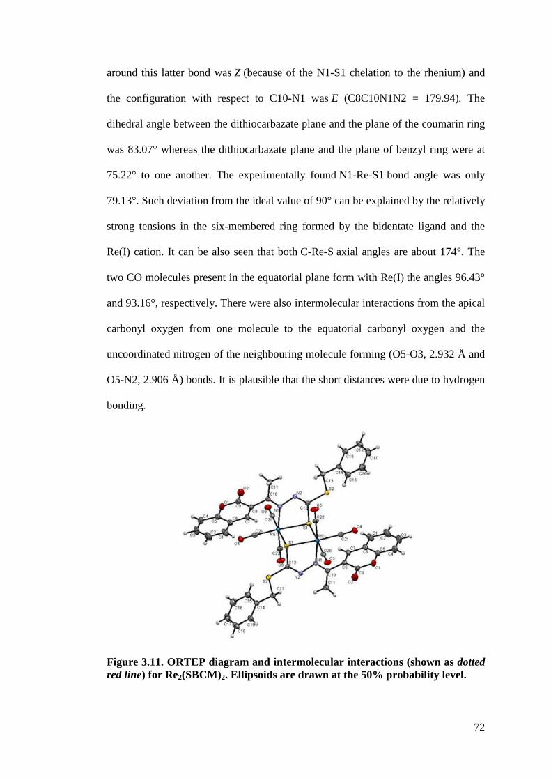
72
around this latter bond was Z (because of the N1-S1 chelation to the rhenium) and
the configuration with respect to C10-N1 was E (C8C10N1N2 = 179.94). The
dihedral angle between the dithiocarbazate plane and the plane of the coumarin ring
was 83.07° whereas the dithiocarbazate plane and the plane of benzyl ring were at
75.22° to one another. The experimentally found N1-Re-S1 bond angle was only
79.13°. Such deviation from the ideal value of 90° can be explained by the relatively
strong tensions in the six-membered ring formed by the bidentate ligand and the
Re(I) cation. It can be also seen that both C-Re-S axial angles are about 174°. The
two CO molecules present in the equatorial plane form with Re(I) the angles 96.43°
and 93.16°, respectively. There were also intermolecular interactions from the apical
carbonyl oxygen from one molecule to the equatorial carbonyl oxygen and the
uncoordinated nitrogen of the neighbouring molecule forming (O5-O3, 2.932 Å and
O5-N2, 2.906 Å) bonds. It is plausible that the short distances were due to hydrogen
bonding.
Figure 3.11. ORTEP diagram and intermolecular interactions (shown as dotted red line) for Re2(SBCM)2. Ellipsoids are drawn at the 50% probability level.
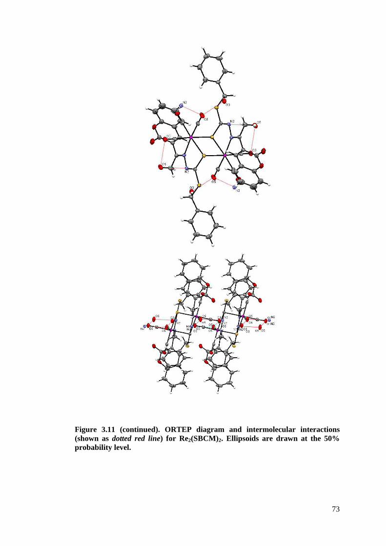
73
Figure 3.11 (continued). ORTEP diagram and intermolecular interactions (shown as dotted red line) for Re2(SBCM)2. Ellipsoids are drawn at the 50% probability level.
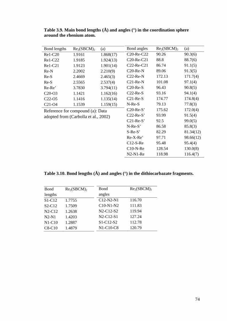
74
Table 3.9. Main bond lengths (Å) and angles (°) in the coordination sphere around the rhenium atom.
Reference for compound (a): Data adopted from (Carbolla et al., 2002)
Table 3.10. Bond lengths (Å) and angles (°) in the dithiocarbazate fragments.
Bond angles Re2(SBCM)2 (a) C20-Re-C22 90.26 90.3(6) C20-Re-C21 88.8 88.7(6) C22-Re-C21 86.74 91.1(5) C20-Re-N 89.06 91.3(5) C22-Re-N 172.13 171.7(4) C21-Re-N 101.08 97.1(4) C20-Re-S 96.43 90.8(5) C22-Re-S 93.16 94.1(4) C21-Re-S 174.77 174.8(4) N-Re-S 79.13 77.8(3) C20-Re-S’ 175.62 172.0(4) C22-Re-S’ 93.99 91.5(4) C21-Re-S’ 92.5 99.0(5) N-Re-S’ 86.58 85.8(3) S-Re-S’ 82.29 81.34(12) Re-X-Re’ 97.71 98.66(12) C12-S-Re 95.48 95.4(4) C10-N-Re 128.54 130.0(8) N2-N1-Re 118.98 116.4(7)
Bond lengths Re2(SBCM)2 (a) Re1-C20 1.9161 1.868(17) Re1-C22 1.9185 1.924(13) Re1-C21 1.9123 1.901(14) Re-N 2.2002 2.210(9) Re-S 2.4669 2.465(3) Re-S 2.5565 2.537(4) Re-Re’ 3.7830 3.794(11) C20-O3 1.1421 1.162(16) C22-O5 1.1416 1.135(14) C21-O4 1.1539 1.159(15)
Bond lengths
Re2(SBCM)2
S1-C12 1.7755 S2-C12 1.7509 N2-C12 1.2638 N2-N1 1.4203 N1-C10 1.2887 C8-C10 1.4879
Bond angles
Re2(SBCM)2
C12-N2-N1 116.70 C10-N1-N2 111.83 N2-C12-S2 119.94 N2-C12-S1 127.24 S1-C12-S2 112.78 N1-C10-C8 120.79
75
3.3.3 Characterization of metal complexes in solution
3.3.3.1 NMR
The NMR spectra of ligands SMML (Figure 3.12), SMLA, SBML, SBLA and
SBEL were recorded in CD3OD whereas SM4CB, SB4CB (Figure 3.13), SBCM,
SMHD, SBHD, Zn(SBCM)2 and Re2(SBCM)2 were carried out in DMSO-d6 due to
their low solubility in CD3OD at room temperature. The NH signal was not observed
in any of the 1H NMR spectra in CD3OD due to fast exchange with solvent.
However, the spectra illustrated the expected hydrogens for each compound. The S-
methyl derivatives (SMML and SMLA) showed characteristics signals at ca. 2.52
ppm attributed to S-CH3 while the S-benzyl derivatives (SBML, SBLA and SBEL)
displayed distinct multiplets of their five aromatic protons and singlets of their S-
CH2 protons at ca. 7.40-7.17 ppm and 4.45 ppm, respectively. The proton signals for
the two CH2 groups of both levulinic acid and methyl levulinate were found to be
identical at ca. 2.62 ppm whereas the -CH3 group was at ca. 1.98 ppm. Other
distinctive signals arising from the protons of the ester Schiff bases were observed at
3.66 ppm (-OCH3 of SMML), 3.47 ppm (-OCH3 of SBML) and 3.93 ppm in
addition to 1.02 ppm (-OCH2CH3 of SBEL). The spectra of aromatic acid ligands
SM4CB and SB4CB in DMSO-d6 demonstrated signals at 8.29 ppm and 8.01-7.80
ppm attributed to -CH=N and aromatic hydrogens of 4-carboxybenzaldehyde
moiety, respectively. Both ligands also showed characteristic signals of their S-
substituted dithiocarbazate similar to those discussed above. The -NH
(dithiocarbazate) signal of both SM4CB and SB4CB occurred at ca. 13 ppm whereas
for SMHD and SBHD occurred at ca.12.15 indicating the presence of thioimine NH.
This signal and the absence of the C-SH signal at ca. 4 ppm indicated that in
solution the thione tautomer remains as the predominant species (Roy et al., 2007).
76
The NH signals above 12 ppm indicate that the Z-configurational isomer is
predominant (Rebolledo et al., 2005). The presence of -SC(=S)NH- at ca. 200 ppm
in the 13C NMR spectra of all the Schiff bases indicated that the thione form
predominates in solution. This signal was shifted downfield due to the deshielding
effect of the neighbouring amine nitrogen and electronegative sulphur. The C=N
signal appeared at ca. 156 ppm of the result of hydrazone bond formation when
carbonyl compounds react with the S-substituted dithiocarbazate. The -S-CH2 peak
occurred upfield at ca. 40 ppm while the S-CH3 peak was found at ca. 17 ppm. The
aromatic carbons were observed at ca. 138 - 127 ppm as expected. The 1H NMR
spectrum of SBCM in DMSO-d6 however showed double set of signals due to
existence of SBCM in two isomeric forms. Such observation has been reported by
Hunoor et al. (2010) with 3-acetylcoumarin-isonicotinoylhydrazone. The relative
percentage of the two isomers was found to be 60% and 40%. The singlets at 12.56,
4.48 and 2.34 are assigned to -NH, -CH2-S-, -CH3 (3-acetylcoumarin), respectively.
Overlapping signals from the coumarin and benzyl rings were observed in the
aromatic region at 7.85-7.21 ppm. Upon complexation of SBCM with Zn(II) and
Re(I), the hydrazone proton signal disappeared, which clearly indicates
deprotonation of NH by enolisation. The downfield shift of methyl protons for both
complexes further supports the coordination through azomethine nitrogen. The
others protons in the 1H NMR spectra of the complexes have not shown considerable
change. Due the low solubility of the coumarin derivatives, it was impossible to
record good 13C-NMR spectra.
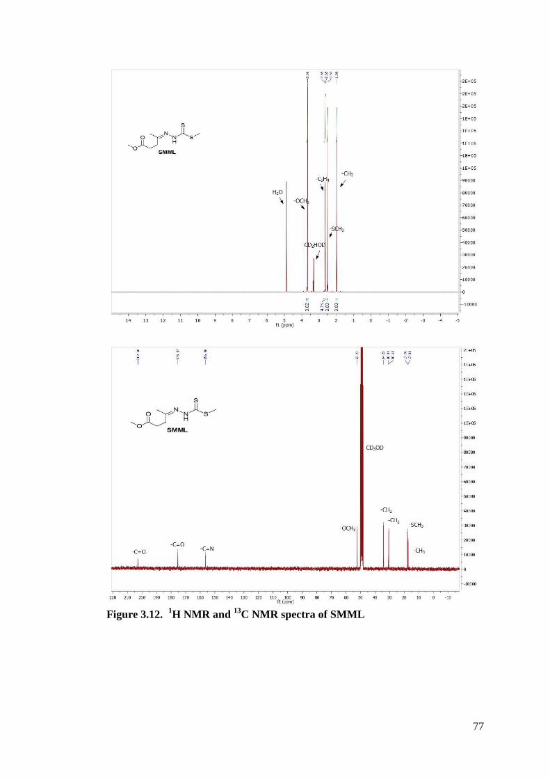
77
Figure 3.12. 1H NMR and 13C NMR spectra of SMML
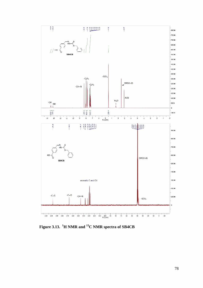
78
Figure 3.13. 1H NMR and 13C NMR spectra of SB4CB
79
3.3.3.2 UV-VIS
Electronic spectra of all the metal complexes in DMSO were scanned in the region
200–800 nm at concentrations between 25 µM and 1 mM. The electronic spectra of
the Schiff bases showed two bands, from ca. 273 nm to 364 nm arising from ,.,*
and n.,* transitions. For all the metal complexes, the first band corresponding to
the ,.,* (256-300 nm) transition is always observed, whereas the second
intraligand band at higher wavelength (300-400 nm) ascribed to n.,* band showed
either a blue shift with a reduction of intensity or disappeared. This is due to
donation of the lone pair of electrons to the metal and hence the coordination of the
azomethine group (Latheef and Kurup, 2008). Most Cu(II) complexes with the
exception of Cu(SM4CB)2 and Cu(SB4CB)2 showed the presence of ligand-to-metal
charge transfer (LMCT) band (400-450 nm) arising from S.M(II) interaction.
Some spectra of the d9 Cu(II) complexes also revealed the presence of a broad d-d
band at ca. 604 nm attributed to 2B1g.2A1g for a distorted square planar
environment around the copper(II) ion due to Jahn-Teller distortion (West et al.,
1993; Nair and Joseyphus, 2008). There was a slight hypsochromic shift at 325 nm
in the spectrum of the Re2(SBCM)2 complex whereas in the Zn(SBCM)2 complex a
slight batochromic shift was seen at 292 nm. Two examples of the UV-Vis spectra
each for tetradentate series and bidentate series are shown in Figure 3.14.
Cu(SM4CB)2 and Cu(SB4CB)2, however, showed neither the sulfur-to-copper
LMCT band at 400 nm nor the d-d band. The absence of these bands even at high
concentration (1 mM) suggests that the ligand does not have a suitable low-lying
antibonding ,* orbital (Ali et al., 2001b). These two complexes showed a broad
band spanning 300-400 nm with / max at ca. 322 nm. The spectra of the complexes
80
were different from those of the free ligands that showed intra-ligand transitions at
ca. 349 nm and 362 nm. The changes observed for the complexes were in agreement
with the results of titration experiment carried out in both DMSO and 0.1M acetate
buffer solution in order to ascertain the formation of the Cu(II) complexes with the
proposed 1:2 metal to ligand stoichiometry. In these titration experiments, the
changes in the UV-Vis spectra were obvious with an absorption band at 300-325 nm
arising when the complex formed while the intensity of ligand band at "max * 345 nm
decreased upon addition of Cu(OAc)2 Complexation proceeded with a sharp end-
point at 0.5 equivalents with clear isosbestic points indicative of a single
complexation event (Figure 3.15).
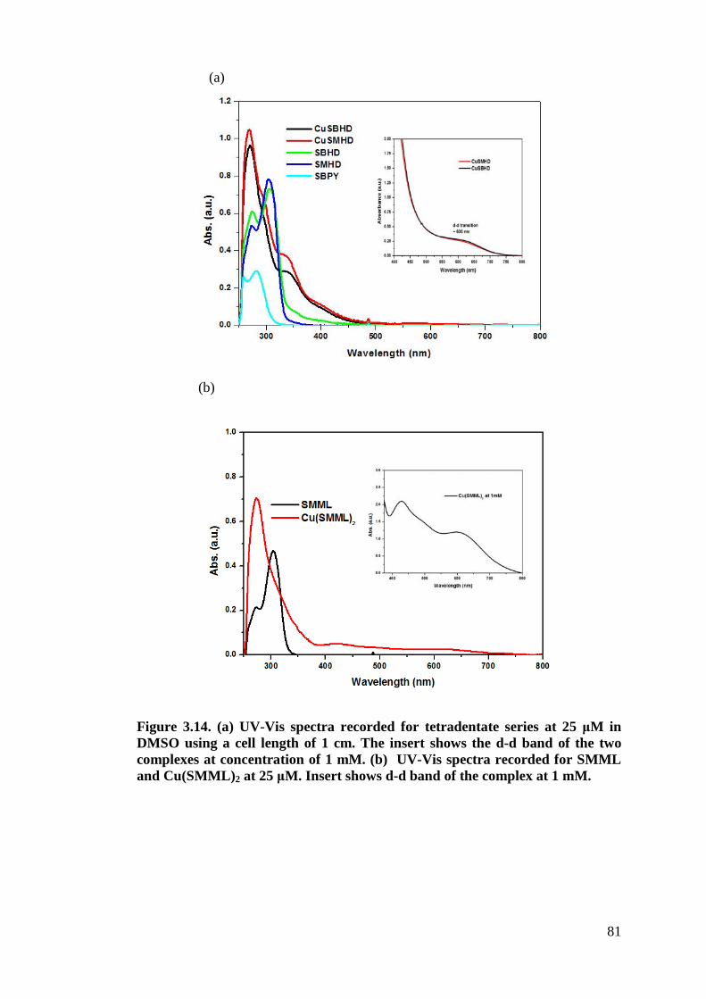
81
Figure 3.14. (a) UV-Vis spectra recorded for tetradentate series at 25 !M in DMSO using a cell length of 1 cm. The insert shows the d-d band of the two complexes at concentration of 1 mM. (b) UV-Vis spectra recorded for SMML and Cu(SMML)2 at 25 !M. Insert shows d-d band of the complex at 1 mM.
(b)
(a)
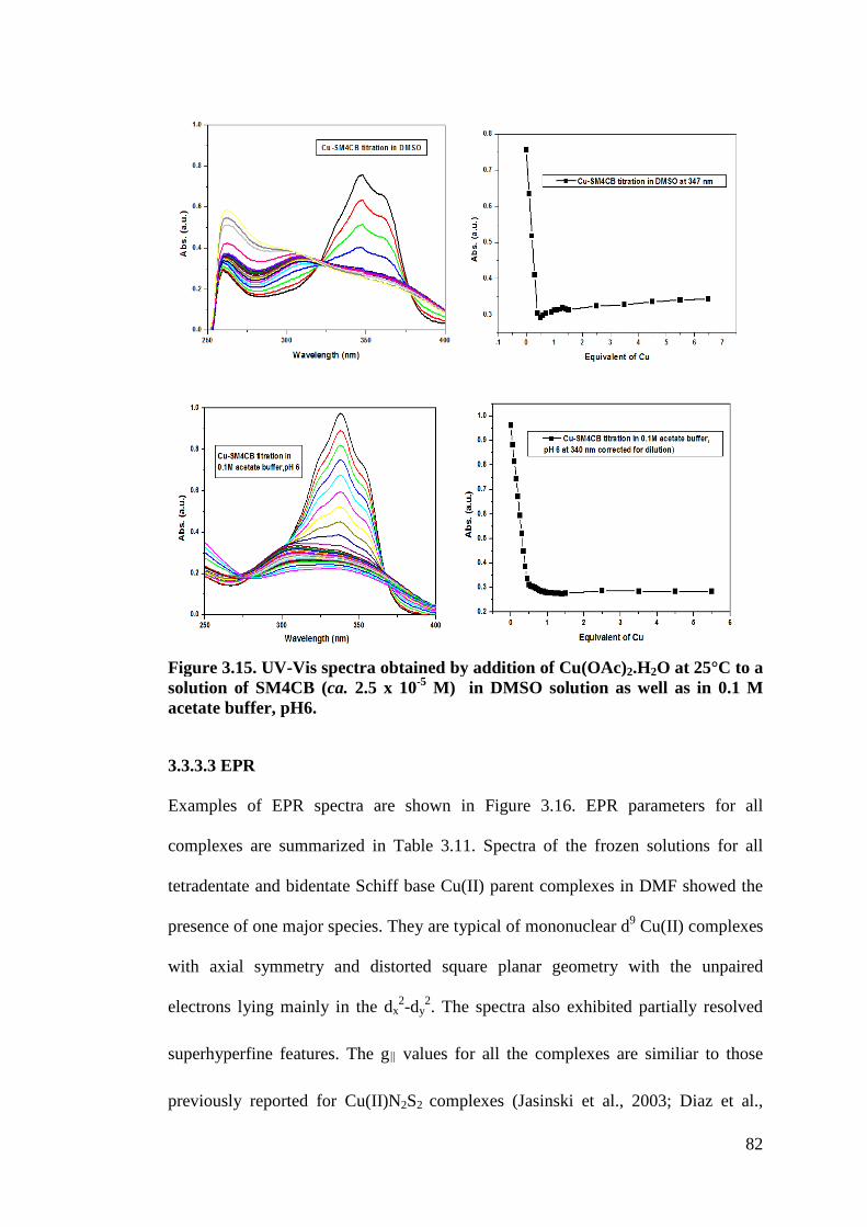
82
Figure 3.15. UV-Vis spectra obtained by addition of Cu(OAc)2.H2O at 25°C to a solution of SM4CB (ca. 2.5 x 10⁻⁵ M) in DMSO solution as well as in 0.1 M acetate buffer, pH6.
3.3.3.3 EPR
Examples of EPR spectra are shown in Figure 3.16. EPR parameters for all
complexes are summarized in Table 3.11. Spectra of the frozen solutions for all
tetradentate and bidentate Schiff base Cu(II) parent complexes in DMF showed the
presence of one major species. They are typical of mononuclear d9 Cu(II) complexes
with axial symmetry and distorted square planar geometry with the unpaired
electrons lying mainly in the dx2-dy
2. The spectra also exhibited partially resolved
superhyperfine features. The g! values for all the complexes are similiar to those
previously reported for Cu(II)N2S2 complexes (Jasinski et al., 2003; Diaz et al.,
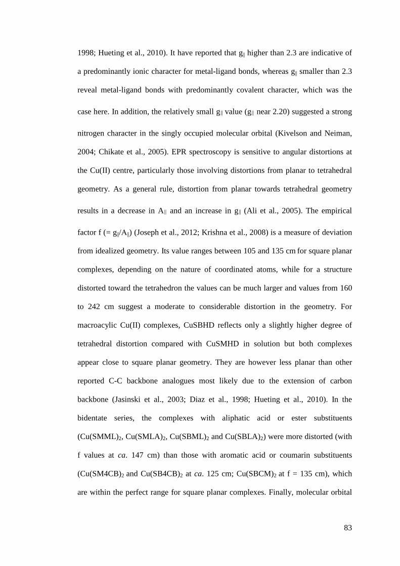
83
1998; Hueting et al., 2010). It have reported that g|| higher than 2.3 are indicative of
a predominantly ionic character for metal-ligand bonds, whereas g|| smaller than 2.3
reveal metal-ligand bonds with predominantly covalent character, which was the
case here. In addition, the relatively small g! value (g! near 2.20) suggested a strong
nitrogen character in the singly occupied molecular orbital (Kivelson and Neiman,
2004; Chikate et al., 2005). EPR spectroscopy is sensitive to angular distortions at
the Cu(II) centre, particularly those involving distortions from planar to tetrahedral
geometry. As a general rule, distortion from planar towards tetrahedral geometry
results in a decrease in A! and an increase in g! (Ali et al., 2005). The empirical
factor f (= g||/0||) (Joseph et al., 2012; Krishna et al., 2008) is a measure of deviation
from idealized geometry. Its value ranges between 105 and 135 cm for square planar
complexes, depending on the nature of coordinated atoms, while for a structure
distorted toward the tetrahedron the values can be much larger and values from 160
to 242 cm suggest a moderate to considerable distortion in the geometry. For
macroacylic Cu(II) complexes, CuSBHD reflects only a slightly higher degree of
tetrahedral distortion compared with CuSMHD in solution but both complexes
appear close to square planar geometry. They are however less planar than other
reported C-C backbone analogues most likely due to the extension of carbon
backbone (Jasinski et al., 2003; Diaz et al., 1998; Hueting et al., 2010). In the
bidentate series, the complexes with aliphatic acid or ester substituents
(Cu(SMML)2, Cu(SMLA)2, Cu(SBML)2 and Cu(SBLA)2) were more distorted (with
f values at ca. 147 cm) than those with aromatic acid or coumarin substituents
(Cu(SM4CB)2 and Cu(SB4CB)2 at ca. 125 cm; Cu(SBCM)2 at f = 135 cm), which
are within the perfect range for square planar complexes. Finally, molecular orbital
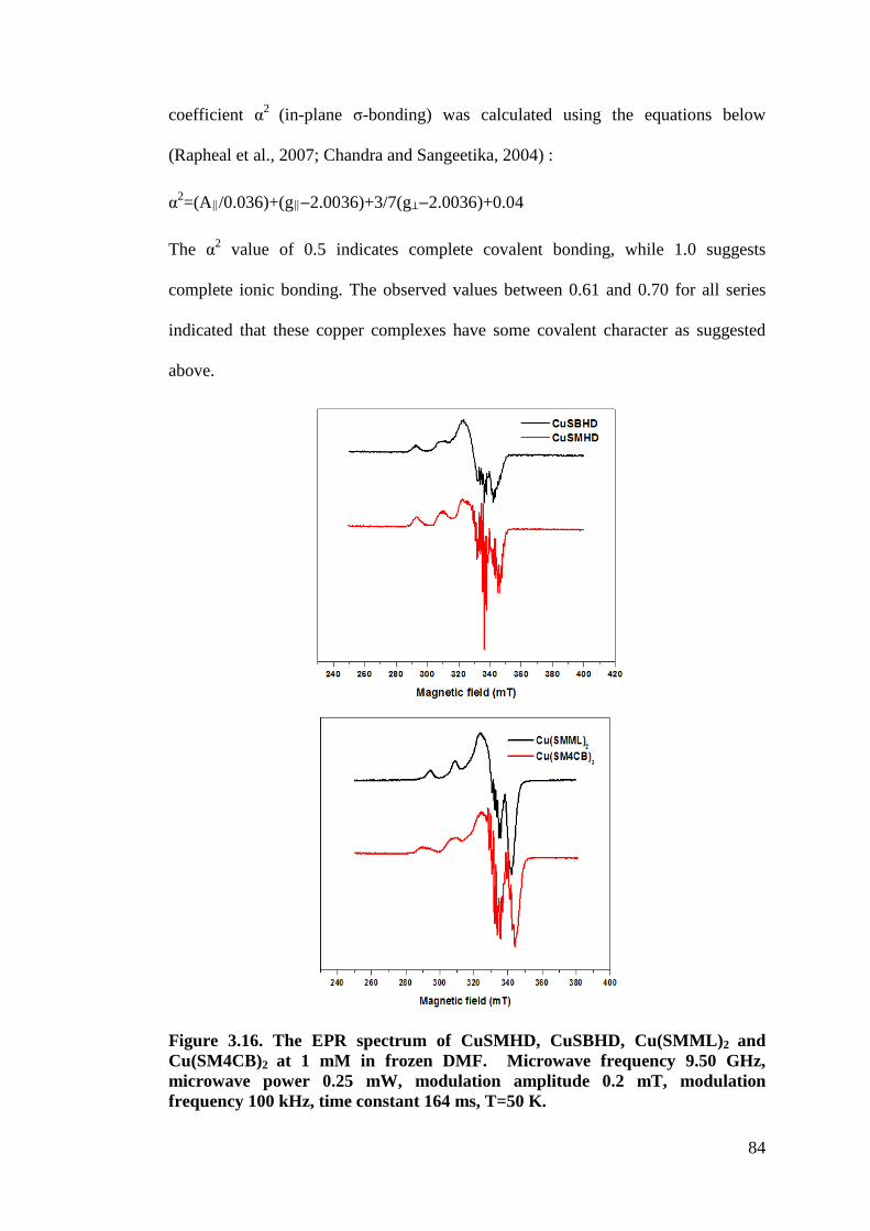
84
coefficient &2 (in-plane 1-bonding) was calculated using the equations below
(Rapheal et al., 2007; Chandra and Sangeetika, 2004) :
&2=(A!/0.036)+(g!"2.0036)+3/7(g""2.0036)+0.04
The &2 value of 0.5 indicates complete covalent bonding, while 1.0 suggests
complete ionic bonding. The observed values between 0.61 and 0.70 for all series
indicated that these copper complexes have some covalent character as suggested
above.
Figure 3.16. The EPR spectrum of CuSMHD, CuSBHD, Cu(SMML)2 and Cu(SM4CB)2 at 1 mM in frozen DMF. Microwave frequency 9.50 GHz, microwave power 0.25 mW, modulation amplitude 0.2 mT, modulation frequency 100 kHz, time constant 164 ms, T=50 K.
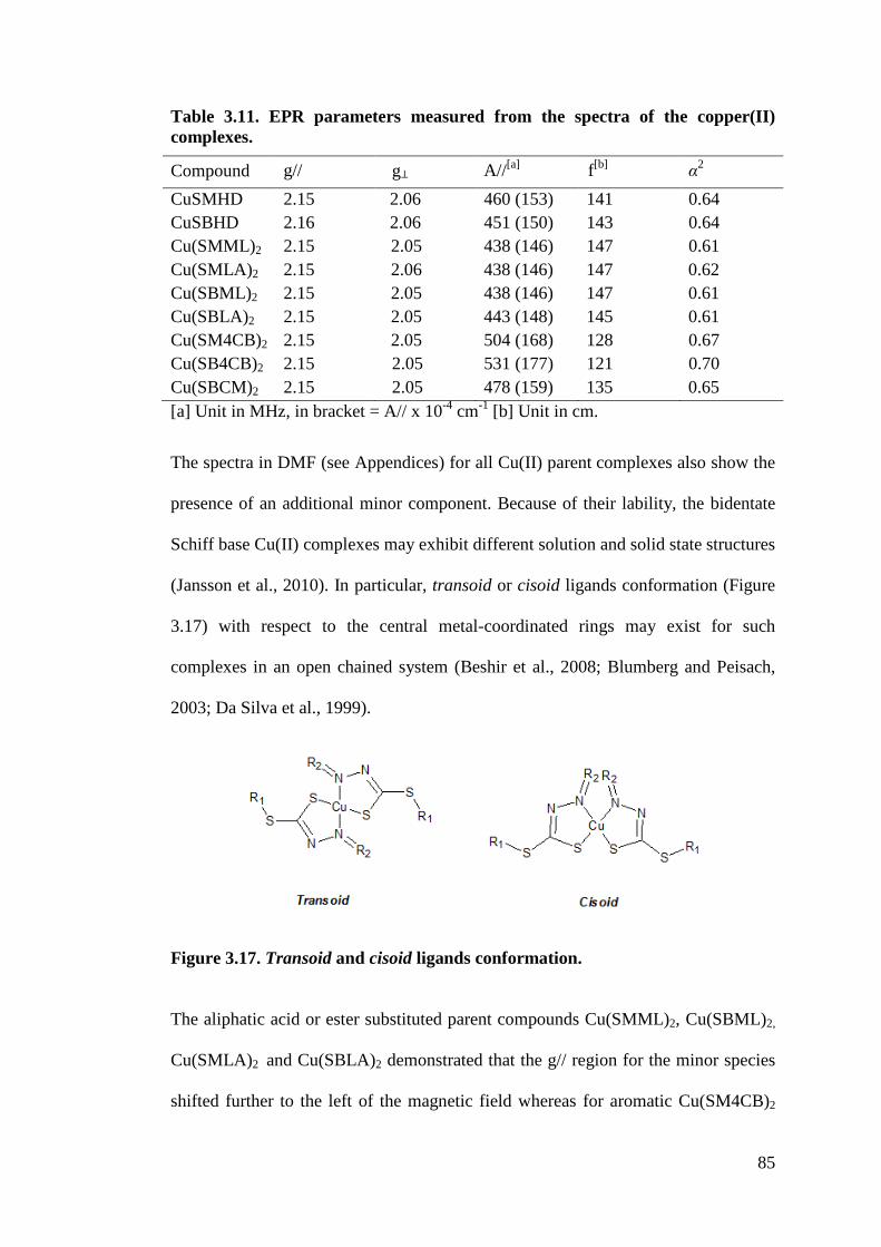
85
Table 3.11. EPR parameters measured from the spectra of the copper(II) complexes.
Compound g// g" A//[a] f[b] $2 CuSMHD 2.15 2.06 460 (153) 141 0.64 CuSBHD 2.16 2.06 451 (150) 143 0.64 Cu(SMML)2 2.15 2.05 438 (146) 147 0.61 Cu(SMLA)2 2.15 2.06 438 (146) 147 0.62 Cu(SBML)2 2.15 2.05 438 (146) 147 0.61 Cu(SBLA)2 2.15 2.05 443 (148) 145 0.61 Cu(SM4CB)2 2.15 2.05 504 (168) 128 0.67 Cu(SB4CB)2 2.15 2.05 531 (177) 121 0.70 Cu(SBCM)2 2.15 2.05 478 (159) 135 0.65 [a] Unit in MHz, in bracket = A// x 10-4 cm-1 [b] Unit in cm.
The spectra in DMF (see Appendices) for all Cu(II) parent complexes also show the
presence of an additional minor component. Because of their lability, the bidentate
Schiff base Cu(II) complexes may exhibit different solution and solid state structures
(Jansson et al., 2010). In particular, transoid or cisoid ligands conformation (Figure
3.17) with respect to the central metal-coordinated rings may exist for such
complexes in an open chained system (Beshir et al., 2008; Blumberg and Peisach,
2003; Da Silva et al., 1999).
Figure 3.17. Transoid and cisoid ligands conformation.
The aliphatic acid or ester substituted parent compounds Cu(SMML)2, Cu(SBML)2,
Cu(SMLA)2 and Cu(SBLA)2 demonstrated that the g// region for the minor species
shifted further to the left of the magnetic field whereas for aromatic Cu(SM4CB)2
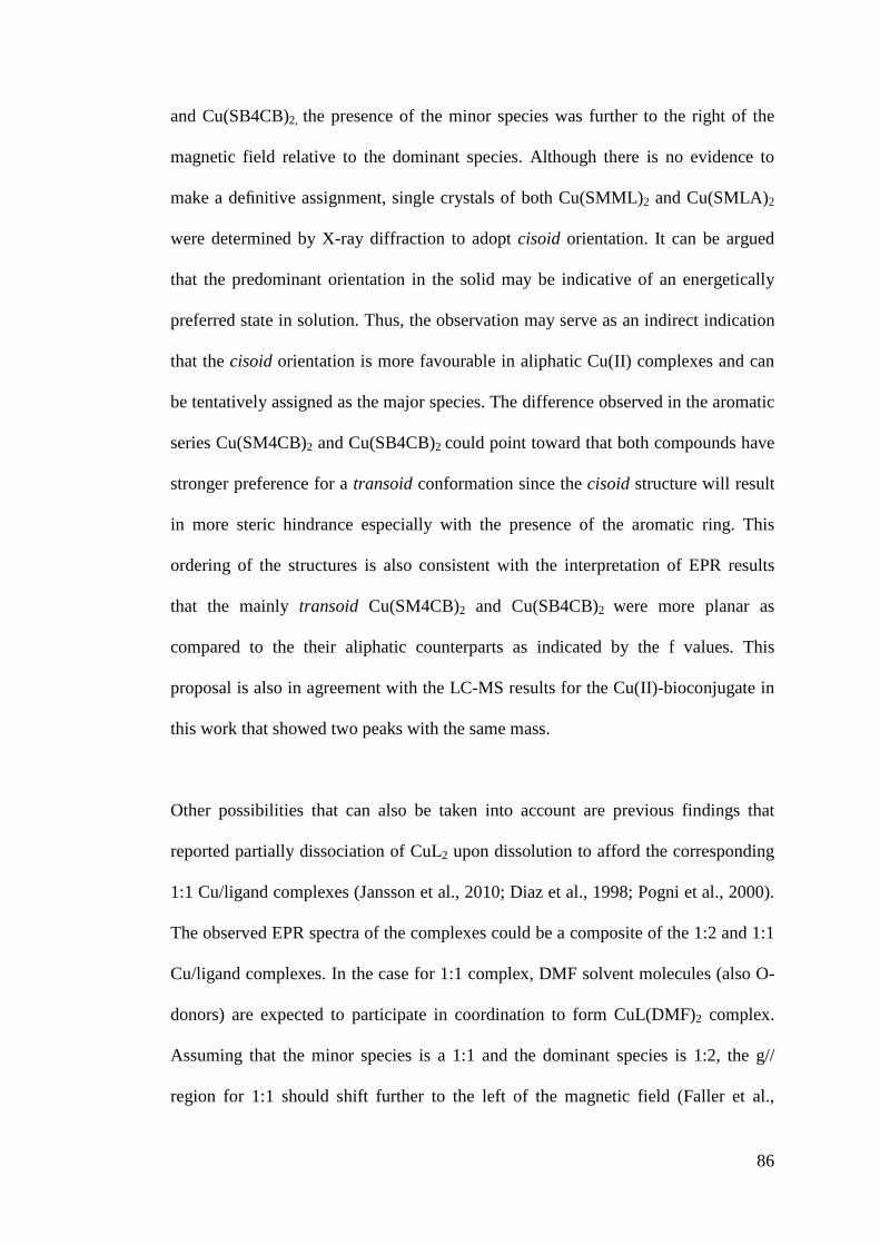
86
and Cu(SB4CB)2, the presence of the minor species was further to the right of the
magnetic field relative to the dominant species. Although there is no evidence to
make a definitive assignment, single crystals of both Cu(SMML)2 and Cu(SMLA)2
were determined by X-ray diffraction to adopt cisoid orientation. It can be argued
that the predominant orientation in the solid may be indicative of an energetically
preferred state in solution. Thus, the observation may serve as an indirect indication
that the cisoid orientation is more favourable in aliphatic Cu(II) complexes and can
be tentatively assigned as the major species. The difference observed in the aromatic
series Cu(SM4CB)2 and Cu(SB4CB)2 could point toward that both compounds have
stronger preference for a transoid conformation since the cisoid structure will result
in more steric hindrance especially with the presence of the aromatic ring. This
ordering of the structures is also consistent with the interpretation of EPR results
that the mainly transoid Cu(SM4CB)2 and Cu(SB4CB)2 were more planar as
compared to the their aliphatic counterparts as indicated by the f values. This
proposal is also in agreement with the LC-MS results for the Cu(II)-bioconjugate in
this work that showed two peaks with the same mass.
Other possibilities that can also be taken into account are previous findings that
reported partially dissociation of CuL2 upon dissolution to afford the corresponding
1:1 Cu/ligand complexes (Jansson et al., 2010; Diaz et al., 1998; Pogni et al., 2000).
The observed EPR spectra of the complexes could be a composite of the 1:2 and 1:1
Cu/ligand complexes. In the case for 1:1 complex, DMF solvent molecules (also O-
donors) are expected to participate in coordination to form CuL(DMF)2 complex.
Assuming that the minor species is a 1:1 and the dominant species is 1:2, the g//
region for 1:1 should shift further to the left of the magnetic field (Faller et al.,
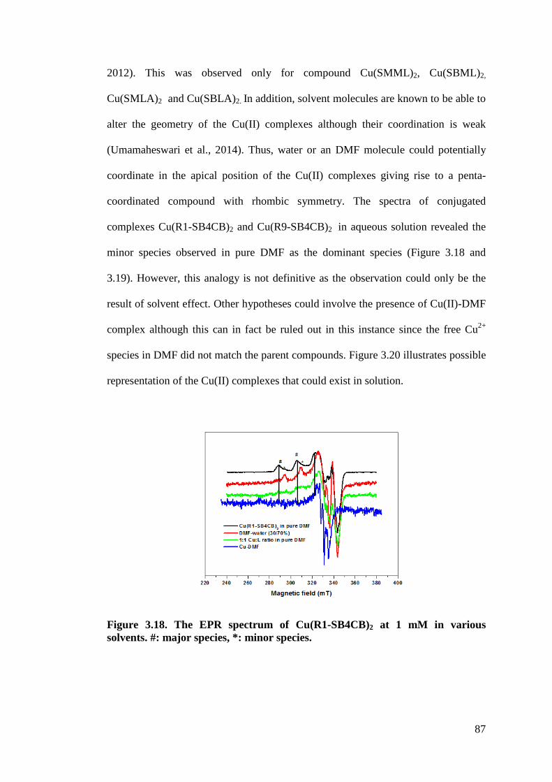
87
2012). This was observed only for compound Cu(SMML)2, Cu(SBML)2,
Cu(SMLA)2 and Cu(SBLA)2. In addition, solvent molecules are known to be able to
alter the geometry of the Cu(II) complexes although their coordination is weak
(Umamaheswari et al., 2014). Thus, water or an DMF molecule could potentially
coordinate in the apical position of the Cu(II) complexes giving rise to a penta-
coordinated compound with rhombic symmetry. The spectra of conjugated
complexes Cu(R1-SB4CB)2 and Cu(R9-SB4CB)2 in aqueous solution revealed the
minor species observed in pure DMF as the dominant species (Figure 3.18 and
3.19). However, this analogy is not definitive as the observation could only be the
result of solvent effect. Other hypotheses could involve the presence of Cu(II)-DMF
complex although this can in fact be ruled out in this instance since the free Cu2+
species in DMF did not match the parent compounds. Figure 3.20 illustrates possible
representation of the Cu(II) complexes that could exist in solution.
Figure 3.18. The EPR spectrum of Cu(R1-SB4CB)2 at 1 mM in various solvents. #: major species, *: minor species.
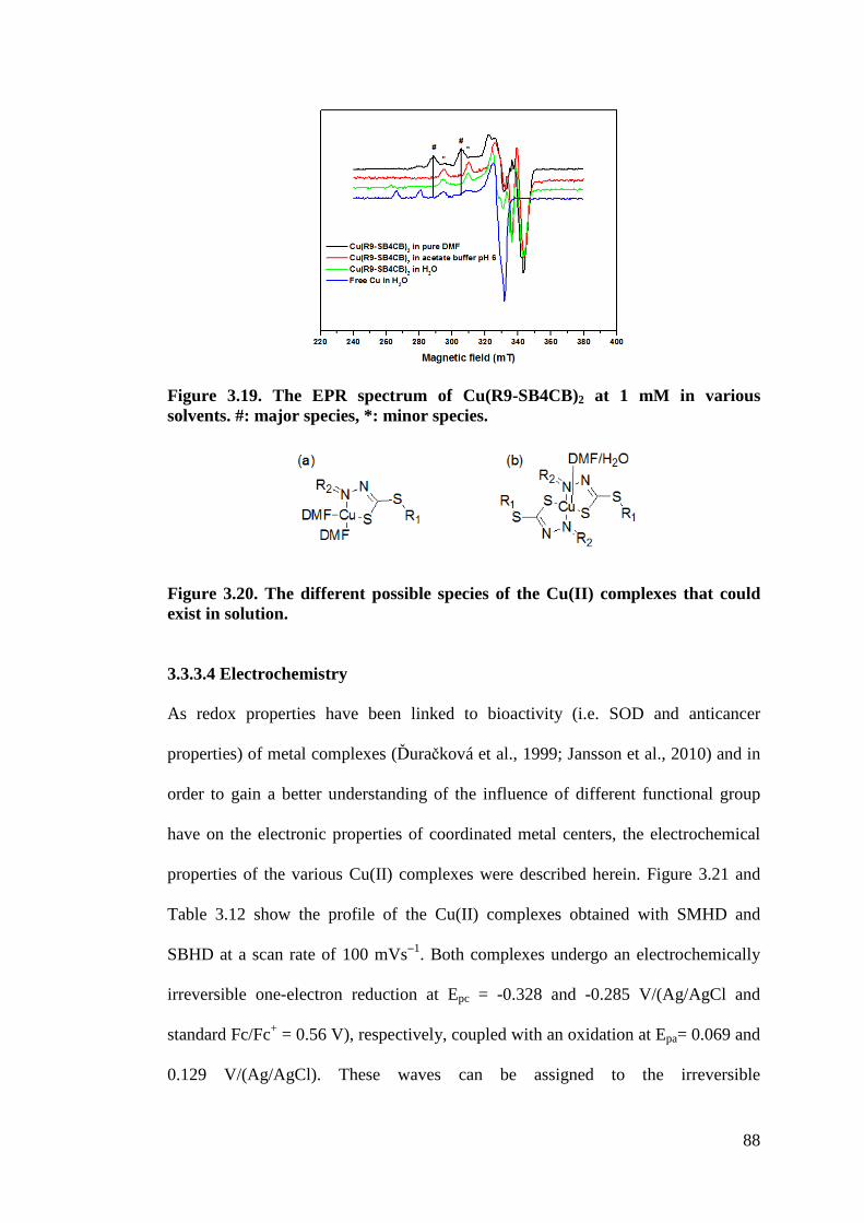
88
Figure 3.19. The EPR spectrum of Cu(R9-SB4CB)2 at 1 mM in various solvents. #: major species, *: minor species.
Figure 3.20. The different possible species of the Cu(II) complexes that could exist in solution.
3.3.3.4 Electrochemistry
As redox properties have been linked to bioactivity (i.e. SOD and anticancer
properties) of metal complexes ('ura(ková et al., 1999; Jansson et al., 2010) and in
order to gain a better understanding of the influence of different functional group
have on the electronic properties of coordinated metal centers, the electrochemical
properties of the various Cu(II) complexes were described herein. Figure 3.21 and
Table 3.12 show the profile of the Cu(II) complexes obtained with SMHD and
SBHD at a scan rate of 100 mVs"1. Both complexes undergo an electrochemically
irreversible one-electron reduction at Epc = -0.328 and -0.285 V/(Ag/AgCl and
standard Fc/Fc+ = 0.56 V), respectively, coupled with an oxidation at Epa= 0.069 and
0.129 V/(Ag/AgCl). These waves can be assigned to the irreversible
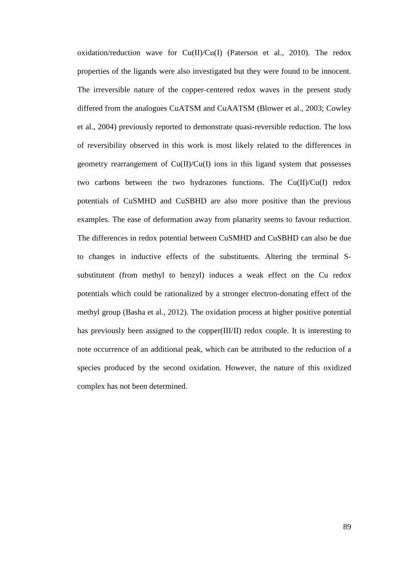
89
oxidation/reduction wave for Cu(II)/Cu(I) (Paterson et al., 2010). The redox
properties of the ligands were also investigated but they were found to be innocent.
The irreversible nature of the copper-centered redox waves in the present study
differed from the analogues CuATSM and CuAATSM (Blower et al., 2003; Cowley
et al., 2004) previously reported to demonstrate quasi-reversible reduction. The loss
of reversibility observed in this work is most likely related to the differences in
geometry rearrangement of Cu(II)/Cu(I) ions in this ligand system that possesses
two carbons between the two hydrazones functions. The Cu(II)/Cu(I) redox
potentials of CuSMHD and CuSBHD are also more positive than the previous
examples. The ease of deformation away from planarity seems to favour reduction.
The differences in redox potential between CuSMHD and CuSBHD can also be due
to changes in inductive effects of the substituents. Altering the terminal S-
substitutent (from methyl to benzyl) induces a weak effect on the Cu redox
potentials which could be rationalized by a stronger electron-donating effect of the
methyl group (Basha et al., 2012). The oxidation process at higher positive potential
has previously been assigned to the copper(III/II) redox couple. It is interesting to
note occurrence of an additional peak, which can be attributed to the reduction of a
species produced by the second oxidation. However, the nature of this oxidized
complex has not been determined.
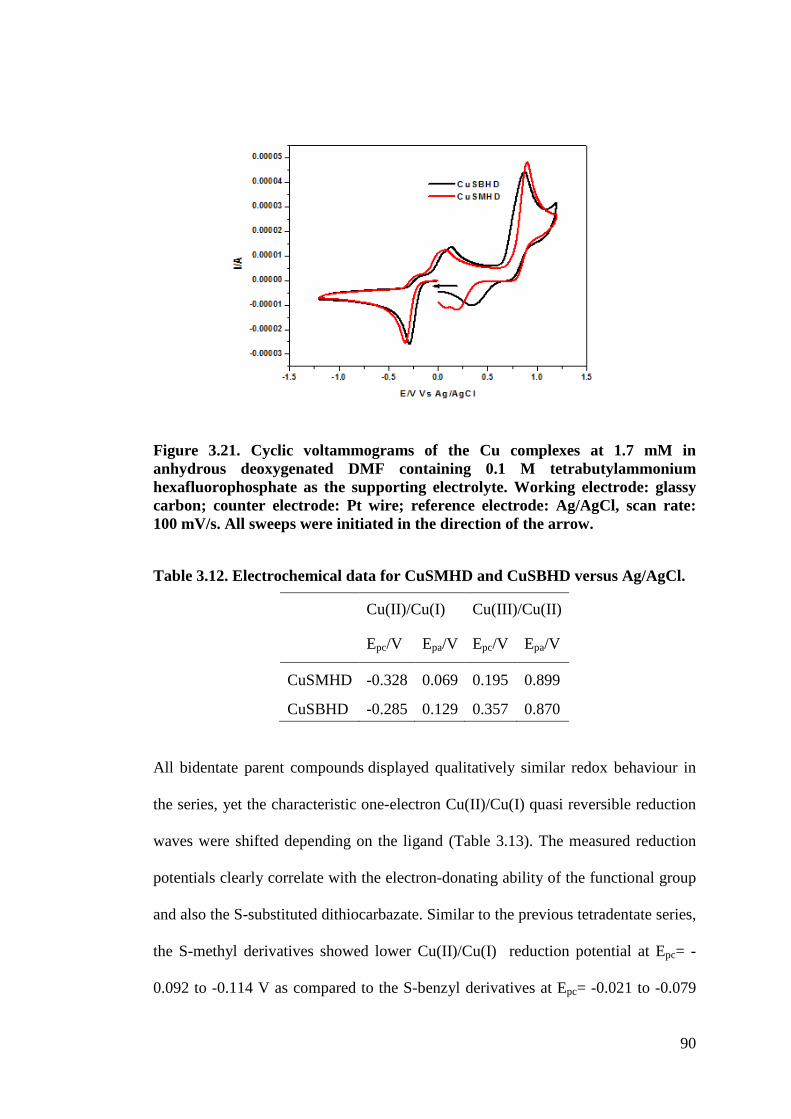
90
Figure 3.21. Cyclic voltammograms of the Cu complexes at 1.7 mM in anhydrous deoxygenated DMF containing 0.1 M tetrabutylammonium hexafluorophosphate as the supporting electrolyte. Working electrode: glassy carbon; counter electrode: Pt wire; reference electrode: Ag/AgCl, scan rate: 100 mV/s. All sweeps were initiated in the direction of the arrow.
Table 3.12. Electrochemical data for CuSMHD and CuSBHD versus Ag/AgCl.
Cu(II)/Cu(I) Cu(III)/Cu(II)
Epc/V Epa/V Epc/V Epa/V
CuSMHD -0.328 0.069 0.195 0.899
CuSBHD -0.285 0.129 0.357 0.870
All bidentate parent compounds displayed qualitatively similar redox behaviour in
the series, yet the characteristic one-electron Cu(II)/Cu(I) quasi reversible reduction
waves were shifted depending on the ligand (Table 3.13). The measured reduction
potentials clearly correlate with the electron-donating ability of the functional group
and also the S-substituted dithiocarbazate. Similar to the previous tetradentate series,
the S-methyl derivatives showed lower Cu(II)/Cu(I) reduction potential at Epc= -
0.092 to -0.114 V as compared to the S-benzyl derivatives at Epc= -0.021 to -0.079
91
V. Among the functional groups the reduction potential towards the more negative
can be arranged in the following order Cu(SMML)2 <Cu(SMLA)2 <Cu(SM4CB)2.
Cyclic voltammograms were also recorded at different scan rates between 0.4 V and
-0.4 V vs. Ag/AgCl from 0.02 V/s to 0.5V/s for all the compounds which showed
similar trend. Taking Cu(SMML)2 as an example (Figure 3.22), the peak currents
were still found to be proportional to scan rates with the peak currents ratio of the
anodic signal and the cathodic signal (ipa/ipc) remained close to 1 (Table 3.14)
independent of the sweep rate used indicating the reversibility and stability of the
electrochemically generated product (Evans et al., 1983). Furthermore, when
plotting the peak current as a function of the square root of the scan rate for
reversible electron transfer, a linear correlation was found although not particularly
perfect suggesting that the redox process is confined to the surface (Figure 3.23).
However, another indicator of reversible electron transfer called the current
function, whose values were given by (ip / +1/2) were not entirely constant anymore
for all scan rates and the most obvious indication that the process was not
completely reversible was the separation of anodic and cathodic peak potential.
Inspection of the voltammetric data shows that at increasing scan rates, the reduction
(Epc) and oxidation (Epa) peaks are shifted to more negative and positive values
respectively. The separation between them, 2Ep, exceeds the Nernstian requirement
of 59 mV expected for a reversible one-electron process. For Cu(SMML)2, this
value increases from 2Ep = 78 mV at 0.05 V/s to 2Ep = 135 mV at 5 V/s indicating a
kinetic inhibition of the electron transfer process (dos Santos-Claro, 2005). This
behavior cannot be attributed to some uncompensated solution resistance, as the
internal standard Fc/Fc+ couple, that shows rapid heterogeneous electron transfer in
most of the solvents, gave a 2Ep value of 51 mV (measured at v = 0.1 V/s). Thus,
92
any possible uncompensated resistance is sufficiently small so that the resulting
voltage drops are negligible compared to the 2Ep values attributable to kinetic
effects (Parajón-Costa et al., 2004). Therefore the reaction of higher scan rates can
be considered to be quasi-reversible and this quasi-reversibility associated with the
reduction process probably arises as a consequence of a geometry change towards a
distorted tetrahedral environment around the Cu(I) species
.
Table 3.13. Electrochemical data for the Cu(II) complexes vs Ag/AgCl at 0.1V Cu(II)/Cu(I) Cu(II)
/Cu(III) Epa[V] Epc[V] 2Ep=Epa-
Epc[mV] 2E1/2=0.5 (Epa+Epc) [V]
ipa/ipc Epa[V]
Cu(SMML)2 0.002 -0.092 94 -0.045 1.00 1.000 Cu(SMLA)2 -0.014 -0.095 81 -0.055 0.60 0.998 Cu(SM4CB)2 -0.011 -0.114 103 -0.063 1.11 1.006 Cu(SBML)2 0.021 -0.066 87 -0.023 1.01 1.001 Cu(SBLA)2 0.008 -0.078 86 -0.035 0.84 0.985 Cu(SB4CB)2 0.015 -0.079 94 -0.032 0.88 1.029 Cu(SBCM)2 0.077 -0.021 98 0.028 1.19 0.973
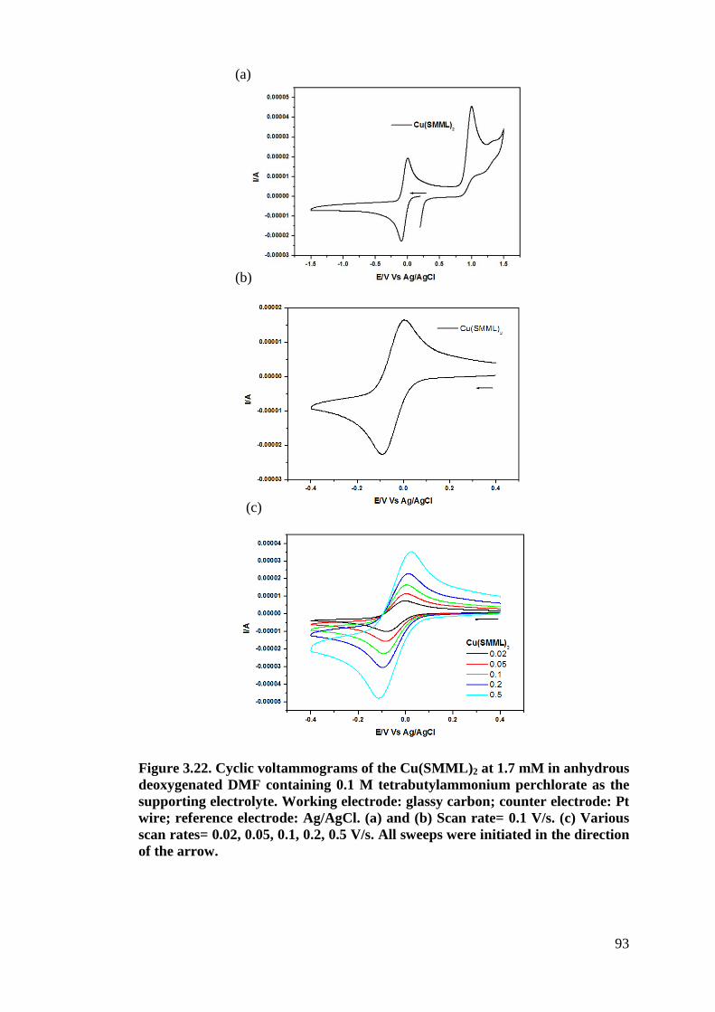
93
Figure 3.22. Cyclic voltammograms of the Cu(SMML)2 at 1.7 mM in anhydrous deoxygenated DMF containing 0.1 M tetrabutylammonium perchlorate as the supporting electrolyte. Working electrode: glassy carbon; counter electrode: Pt wire; reference electrode: Ag/AgCl. (a) and (b) Scan rate= 0.1 V/s. (c) Various scan rates= 0.02, 0.05, 0.1, 0.2, 0.5 V/s. All sweeps were initiated in the direction of the arrow.
(a)
(c)
(b)
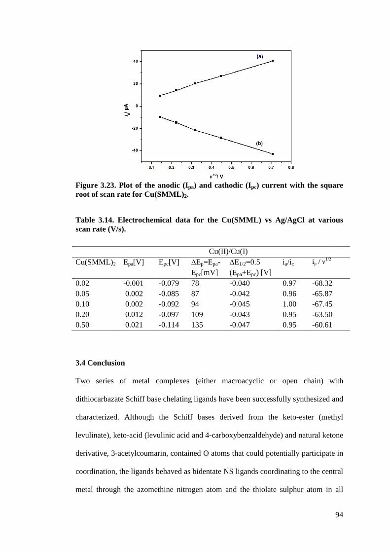
94
Figure 3.23. Plot of the anodic (Ipa) and cathodic (Ipc) current with the square root of scan rate for Cu(SMML)2.
Table 3.14. Electrochemical data for the Cu(SMML) vs Ag/AgCl at various scan rate (V/s).
Cu(II)/Cu(I) Cu(SMML)2 Epa[V] Epc[V] 2Ep=Epa-
Epc[mV] 2E1/2=0.5 (Epa+Epc) [V]
ia/ic ip / +1/2
0.02 -0.001 -0.079 78 -0.040 0.97 -68.32 0.05 0.002 -0.085 87 -0.042 0.96 -65.87 0.10 0.002 -0.092 94 -0.045 1.00 -67.45 0.20 0.012 -0.097 109 -0.043 0.95 -63.50 0.50 0.021 -0.114 135 -0.047 0.95 -60.61
3.4 Conclusion
Two series of metal complexes (either macroacyclic or open chain) with
dithiocarbazate Schiff base chelating ligands have been successfully synthesized and
characterized. Although the Schiff bases derived from the keto-ester (methyl
levulinate), keto-acid (levulinic acid and 4-carboxybenzaldehyde) and natural ketone
derivative, 3-acetylcoumarin, contained O atoms that could potentially participate in
coordination, the ligands behaved as bidentate NS ligands coordinating to the central
metal through the azomethine nitrogen atom and the thiolate sulphur atom in all
95
complexes. Eleven structures of the compounds have been determined by single
crystal X-ray crystallographic analysis. The 1H, 13C NMR and FTIR spectra of the
Schiff bases indicated that the ligands retained the thione form in both solid state
and solution. Only the ligand SBCM existed in both thione and thiolate form in solid
state. RP-HPLC analysis of the free ligands showed that the aromatic ligands were
more stable than the aliphatic ligands. Their stability was further improved upon
complexation. Electronic and EPR spectra showed that most of the paramagnetic
Cu(II) complexes had distorted square planar geometries with the exception of
Cu(SB4CB)2, Cu(SM4CB)2 and Cu(SBCM)2 as demonstrated from their empirical
factor f (= g||/0||). Cu(II) complexes with aliphatic acid and ester bidentate ligands
(145-147 cm) confirm slightly higher degree of tetrahedral distortion than those with
tetradentate ligands (141 and 143 cm) in solution. However, Cu(SM4CB)2,
Cu(SB4CB)2 and Cu(SBCM)2 that possess aromatic acid or coumarin substituents
(Cu(SM4CB)2 and Cu(SB4CB)2 at ca. 125 cm; Cu(SBCM)2 at f = 135 cm) are in the
square planar range. The Re(I) and Zn(II) complexes were diamagnetic. There were
also differences between the macroacyclic and open chain Cu(II) complexes with
regard to their electrochemistry. The latter series showed Cu(II)/Cu(I) quasi
reversibility at more positive potential whereas the former series was not reversible
suggesting that the open chain Cu(II) complexes can accommodate Cu(I) more
easily as a result of their flexibility. To sum up, this chapter demonstrated that by
using a convergent synthesis strategy, a library of complexes with tune stabilities,
geometries and electrochemical properties can be generated. In addition, the
introduction of carboxylic acid functions opens the possibility to conjugate these
complexes with vectors. The biological activities of all these compounds were
evaluated and are discussed in Chapter 5.
96
CHAPTER 4
FUNCTIONALIZED COMPOUNDS
4.1 Introduction
4.1.1 Key drawbacks of metallodrugs
In the past years, much e!ort has been devoted to synthesis and testing of metal
complexes in search of new metal metallodrugs for biological application. However,
only a fraction of the thousands of tested metal compounds has entered clinical trials
or has been approved worldwide with many challenging issues in terms of favorable
selectivity, solubility, and stability as well as knowledge about their mode of action
remaining unsolved (Fricker, 2007; Timerbaev et al., 2006).
The poor water solubility of many metal complexes is one of the key drawbacks “in
the course of their analysis or application” (Miklán et al., 2007, p. 108). Studies in
aqueous and buffer solutions are of utmost importance for an understanding of the
mechanism of action of bioactive molecules and the design of stronger chelators
(Milunovic et al., 2012). Such is the case for thiosemicarbazone derived Schiff base
metal complexes (Enyedy et al., 2010; Raja et al., 2011). There is relatively few
information that is available in the literature for these families of compounds as such
investigations are often hampered by the low aqueous solubility of the compounds.
There are also cases in which a high amount of DMSO is used to pre-dissolve the
metal complexes in order to overcome their poor solubility in aqueous media during
biological studies. However, the use of DMSO affects the reliability of the results
obtained. DMSO has been shown to considerably retard the growth of fungi and
cancerous cells and lead to bacteria and cell death at above certain concentrations
97
(Dolan et al., 2013; Ng et al., 2013). Therefore, compounds that are highly water-
soluble are preferred for biological applications as they enable preclinical
development of a drug candidate without the use of solubilizers (Bacher et al.,
2013).
Apart from that, cell uptake and metal complex accumulation are also challenges for
medicinal and bioinorganic chemistry. The efficient and rapid passage of metal
complexes into cells remains a major hurdle in the design of potential therapeutic
agents. A significant number of drug candidates and probe molecules fail because of
insufficient cell uptake, requiring high quantities of drug administration, which often
leads to undesirable side effects (Rijt et al., 2011) Only molecules in a narrow range
of molecular weight and polarity are able to directly cross the plasma membrane by
passive diffusion (Brunner and Barton, 2006). It is therefore critical to optimize the
properties of their cellular uptake in the effort to develop potent and selective metal
complexes as chemotherapeutic or diagnostic agents.
The capability to design and synthesize new molecules with tuned functions offers a
powerful and efficacious way to overcome these shortcomings. Recently, new
strategies have been developed in an attempt to maximize the impact of drugs on
cancer cells and minimize the problem of adverse side effects through effective
delivery of complexes with tumour-targeting properties and biologically-active
ligands (Hambley, 2007; Storr et al., 2006; Thompson and Orvig, 2006; Thompson
and Orvig, 2003). The use of multifunctional ligands that adequately bind metal ions
and also include specific targeting features is an attractive choice which is gaining
popularity today with applications at the forefront of all areas of medicinal inorganic
98
chemistry research. The field has benefited greatly from advances in targeted ligand
design, which lead to the development of improved therapeutic agents (Ronconi and
Sadler, 2007).
4.1.2 Conjugated metal complexes
By combining ligands or metal complexes with vectors, a large number of new
bioconjugates with interesting biological properties can be prepared. An excellent
review by Nils Metzler-Nolte (2010, p. 195) gives an overview of the use of
peptides “as targeting vectors for the directed delivery of metal-based drugs or
probes for biomedical investigations”. The numerous peptides that have been
utilized “include neuropeptides (enkephalin, neuropeptide Y, neurotensin), uptake
peptides (TAT and poly-Arg) and intracellular localization sequences”. These
bioconjugates display biomedical applications in radiopharmaceutical, as anticancer
and antibacterial drugs or targeted CO-releasing molecules as well as in biosensor
applications.
4.1.2.1 Schiff base conjugate
As this work involves dithiocarbazate derivatives, the development of Schiff base
metal complex bioconjugates from the family of nitrogen-sulphur donor ligands will
be specifically highlighted (Donnelly, 2011; Paterson and Donnelly, 2011). To date,
the concept of bifunctional ligands has been most successfully applied to
radioimaging with Cu(II) bis(thiosemicarbazone) (Figure 4.1). Many new
tetradentate bifunctional bis(thiosemicarbazone) chelators have been designed and
conjugated to various vectors such as the tumor targeting bombesin derivative
BBN(7-14)-NH2 that has high affinity for gastrin-releasing peptide (GRP) receptors
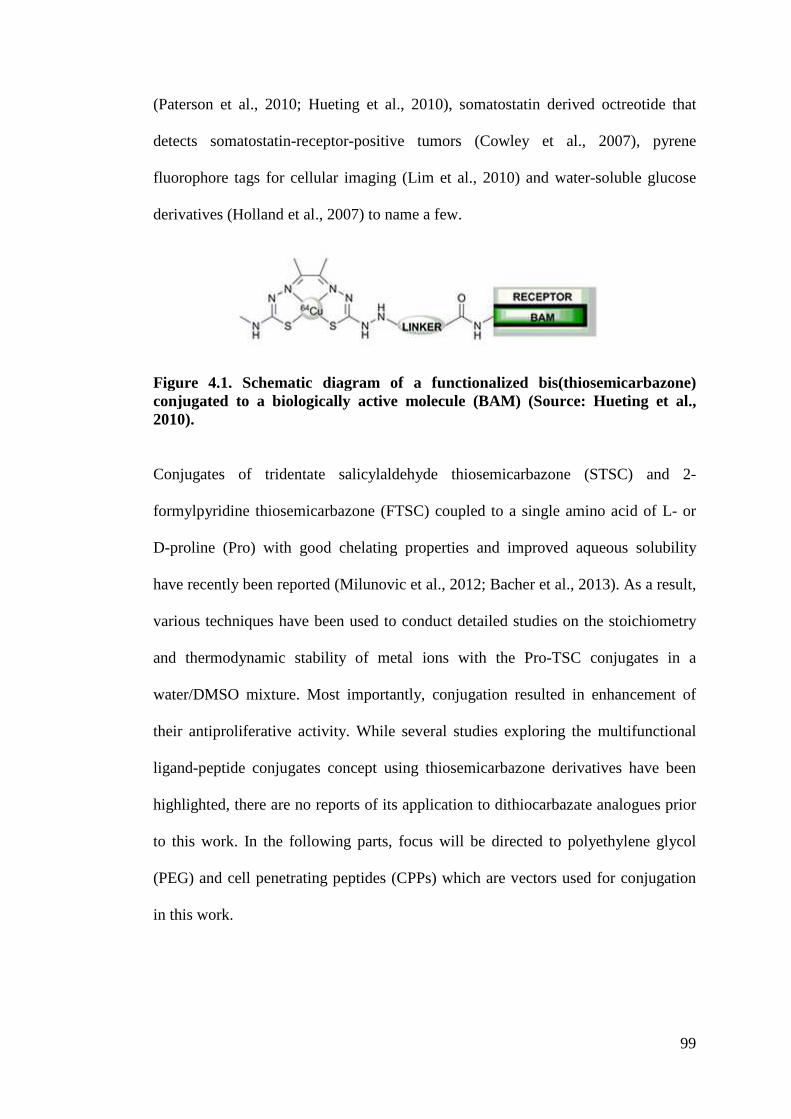
99
(Paterson et al., 2010; Hueting et al., 2010), somatostatin derived octreotide that
detects somatostatin-receptor-positive tumors (Cowley et al., 2007), pyrene
fluorophore tags for cellular imaging (Lim et al., 2010) and water-soluble glucose
derivatives (Holland et al., 2007) to name a few.
Figure 4.1. Schematic diagram of a functionalized bis(thiosemicarbazone) conjugated to a biologically active molecule (BAM) (Source: Hueting et al., 2010).
Conjugates of tridentate salicylaldehyde thiosemicarbazone (STSC) and 2-
formylpyridine thiosemicarbazone (FTSC) coupled to a single amino acid of L- or
D-proline (Pro) with good chelating properties and improved aqueous solubility
have recently been reported (Milunovic et al., 2012; Bacher et al., 2013). As a result,
various techniques have been used to conduct detailed studies on the stoichiometry
and thermodynamic stability of metal ions with the Pro-TSC conjugates in a
water/DMSO mixture. Most importantly, conjugation resulted in enhancement of
their antiproliferative activity. While several studies exploring the multifunctional
ligand-peptide conjugates concept using thiosemicarbazone derivatives have been
highlighted, there are no reports of its application to dithiocarbazate analogues prior
to this work. In the following parts, focus will be directed to polyethylene glycol
(PEG) and cell penetrating peptides (CPPs) which are vectors used for conjugation
in this work.
100
4.1.2.2 PEGylation
PEGylation describes the covalent modification of proteins, peptides, antibody
fragments or non-peptide small drug molecules by attachment to one or more
polyethylene glycol (PEG) chains. PEG is a non-toxic, non-immunogenic, non-
antigenic polymer that is highly water soluble and has been approved by FDA.
PEGylation is used as a well-recognized approach for drug delivery (Harris and
Chess, 2003; Riley and Riggs-Sauthier, 2008; Veronese and Pasut, 2005). The
introduction of PEG into pharmaceuticals improves their pharmacokinetics with “a
prolonged residence in body, a decreased degradation by metabolic enzymes and a
reduction or elimination of protein immunogenicity” (Veronese and Pasut, 2005, p
1451). There is an emerging interest in the use of metal-PEG complexes in
biological studies (Stephan et al., 2005; Heldt et al., 2004) including work on the
modification of cisplatin with PEG (Dhar et al., 2011; Ohya et al., 2001) as well as
PEGylated iridium cyclometalated complexes that showed improved aqueous
solubility and cell permeability (Li et al., 2010).
4.1.2.3 Cell penetrating peptide as cell delivery vectors
Crossing the plasma membrane is a challenge for some molecules with many
potential drugs not displaying the required balance between lipophilicity and
positive charge allowing for membrane permeability while supporting aqueous
solubility. These limitations greatly prevent them from reaching their desired target
in cellulo and results in the reduction of their therapeutics efficacy (Stewart et al.,
2008). Others like polynucleic acids (PNAs) would not be able to penetrate into cells
due to the presence of their high negative charge (Walrant et al., 2011). Over the
past two decades, CPPs, also called “Trojan horse peptides”, have attracted
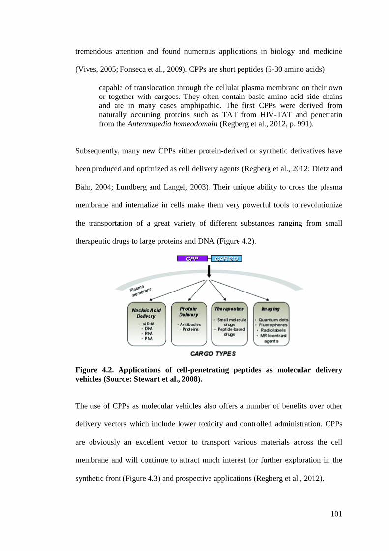
101
tremendous attention and found numerous applications in biology and medicine
(Vives, 2005; Fonseca et al., 2009). CPPs are short peptides (5-30 amino acids)
capable of translocation through the cellular plasma membrane on their own or together with cargoes. They often contain basic amino acid side chains and are in many cases amphipathic. The first CPPs were derived from naturally occurring proteins such as TAT from HIV-TAT and penetratin from the Antennapedia homeodomain (Regberg et al., 2012, p. 991).
Subsequently, many new CPPs either protein-derived or synthetic derivatives have
been produced and optimized as cell delivery agents (Regberg et al., 2012; Dietz and
Bähr, 2004; Lundberg and Langel, 2003). Their unique ability to cross the plasma
membrane and internalize in cells make them very powerful tools to revolutionize
the transportation of a great variety of different substances ranging from small
therapeutic drugs to large proteins and DNA (Figure 4.2).
Figure 4.2. Applications of cell-penetrating peptides as molecular delivery vehicles (Source: Stewart et al., 2008).
The use of CPPs as molecular vehicles also offers a number of benefits over other
delivery vectors which include lower toxicity and controlled administration. CPPs
are obviously an excellent vector to transport various materials across the cell
membrane and will continue to attract much interest for further exploration in the
synthetic front (Figure 4.3) and prospective applications (Regberg et al., 2012).
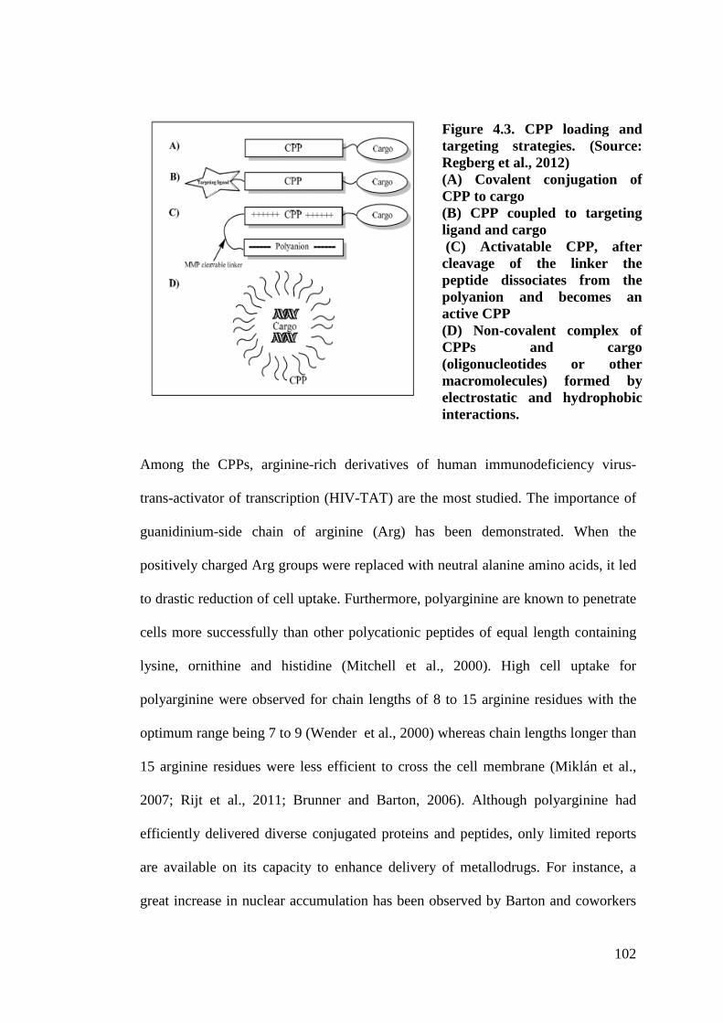
102
Figure 4.3. CPP loading and targeting strategies. (Source: Regberg et al., 2012) (A) Covalent conjugation of CPP to cargo (B) CPP coupled to targeting ligand and cargo (C) Activatable CPP, after cleavage of the linker the peptide dissociates from the polyanion and becomes an active CPP (D) Non-covalent complex of CPPs and cargo (oligonucleotides or other macromolecules) formed by electrostatic and hydrophobic interactions.
Among the CPPs, arginine-rich derivatives of human immunodeficiency virus-
trans-activator of transcription (HIV-TAT) are the most studied. The importance of
guanidinium-side chain of arginine (Arg) has been demonstrated. When the
positively charged Arg groups were replaced with neutral alanine amino acids, it led
to drastic reduction of cell uptake. Furthermore, polyarginine are known to penetrate
cells more successfully than other polycationic peptides of equal length containing
lysine, ornithine and histidine (Mitchell et al., 2000). High cell uptake for
polyarginine were observed for chain lengths of 8 to 15 arginine residues with the
optimum range being 7 to 9 (Wender et al., 2000) whereas chain lengths longer than
15 arginine residues were less efficient to cross the cell membrane (Miklán et al.,
2007; Rijt et al., 2011; Brunner and Barton, 2006). Although polyarginine had
efficiently delivered diverse conjugated proteins and peptides, only limited reports
are available on its capacity to enhance delivery of metallodrugs. For instance, a
great increase in nuclear accumulation has been observed by Barton and coworkers
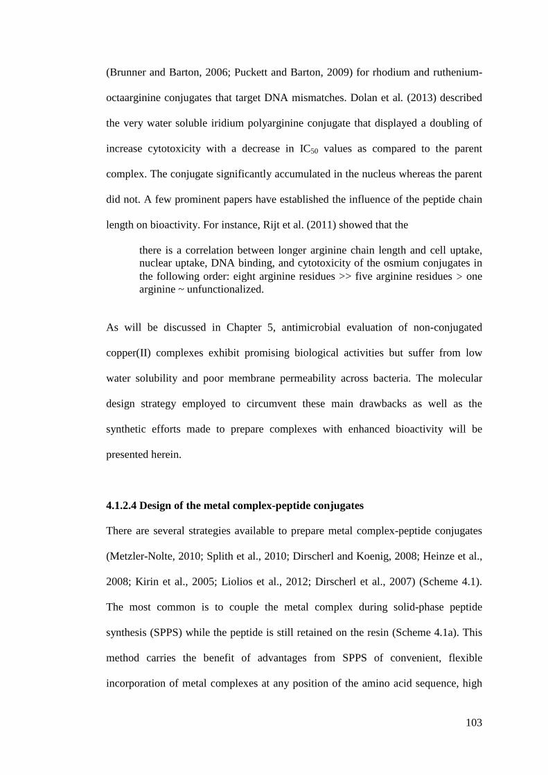
103
(Brunner and Barton, 2006; Puckett and Barton, 2009) for rhodium and ruthenium-
octaarginine conjugates that target DNA mismatches. Dolan et al. (2013) described
the very water soluble iridium polyarginine conjugate that displayed a doubling of
increase cytotoxicity with a decrease in IC50 values as compared to the parent
complex. The conjugate significantly accumulated in the nucleus whereas the parent
did not. A few prominent papers have established the influence of the peptide chain
length on bioactivity. For instance, Rijt et al. (2011) showed that the
there is a correlation between longer arginine chain length and cell uptake, nuclear uptake, DNA binding, and cytotoxicity of the osmium conjugates in the following order: eight arginine residues >> five arginine residues ! one arginine ~ unfunctionalized.
As will be discussed in Chapter 5, antimicrobial evaluation of non-conjugated
copper(II) complexes exhibit promising biological activities but suffer from low
water solubility and poor membrane permeability across bacteria. The molecular
design strategy employed to circumvent these main drawbacks as well as the
synthetic efforts made to prepare complexes with enhanced bioactivity will be
presented herein.
4.1.2.4 Design of the metal complex-peptide conjugates
There are several strategies available to prepare metal complex-peptide conjugates
(Metzler-Nolte, 2010; Splith et al., 2010; Dirscherl and Koenig, 2008; Heinze et al.,
2008; Kirin et al., 2005; Liolios et al., 2012; Dirscherl et al., 2007) (Scheme 4.1).
The most common is to couple the metal complex during solid-phase peptide
synthesis (SPPS) while the peptide is still retained on the resin (Scheme 4.1a). This
method carries the benefit of advantages from SPPS of convenient, flexible
incorporation of metal complexes at any position of the amino acid sequence, high
yield and high purity of the conjugates providing the compound is stable enough to
survive all SPPS manipulations. Alternatively, the peptide
first via SPPS followed by in situ sol
4.1b). The other is to attach the metal complexes in solution after the peptide
synthesis has been completed on the resin, cleaved and purified (Scheme 4.1c). The
post-labeling second and third options are definit
(option b) or ligands (option c) that are sensitive towards SPPS conditions
two strategies prevent demetallation and decomposition of the metal complexes.
Scheme 4.1. Strategies to prepare metal comple
To generate the copper complexes in this work, the strategy based on
with bidentate ligands was employed as shown below (Scheme 4.2). To allow the
conjugation of the complexes with various vectors (PEG, peptides),
a reactive function in the ligand structure is required. Among the orthogonal reactive
functions, carboxylic acid has been chosen since it is an easy
can react with the terminal amine function of the peptides and the PE
Table 4.1 summarized the conjugates that were synthesized and characterized in this
work.
yield and high purity of the conjugates providing the compound is stable enough to
survive all SPPS manipulations. Alternatively, the peptide-ligand can be synthesized
first via SPPS followed by in situ solution complexation with metal ions (Scheme
4.1b). The other is to attach the metal complexes in solution after the peptide
synthesis has been completed on the resin, cleaved and purified (Scheme 4.1c). The
labeling second and third options are definitely preferable for metal
(option b) or ligands (option c) that are sensitive towards SPPS conditions
two strategies prevent demetallation and decomposition of the metal complexes.
Scheme 4.1. Strategies to prepare metal complex conjugated with peptides.
To generate the copper complexes in this work, the strategy based on
with bidentate ligands was employed as shown below (Scheme 4.2). To allow the
conjugation of the complexes with various vectors (PEG, peptides),
a reactive function in the ligand structure is required. Among the orthogonal reactive
functions, carboxylic acid has been chosen since it is an easy-to
can react with the terminal amine function of the peptides and the PE
Table 4.1 summarized the conjugates that were synthesized and characterized in this
104
yield and high purity of the conjugates providing the compound is stable enough to
ligand can be synthesized
ution complexation with metal ions (Scheme
4.1b). The other is to attach the metal complexes in solution after the peptide
synthesis has been completed on the resin, cleaved and purified (Scheme 4.1c). The
ely preferable for metal-complexes
(option b) or ligands (option c) that are sensitive towards SPPS conditions. These
two strategies prevent demetallation and decomposition of the metal complexes.
x conjugated with peptides.
To generate the copper complexes in this work, the strategy based on Scheme 4.1b
with bidentate ligands was employed as shown below (Scheme 4.2). To allow the
conjugation of the complexes with various vectors (PEG, peptides), the presence of
a reactive function in the ligand structure is required. Among the orthogonal reactive
to-handle group that
can react with the terminal amine function of the peptides and the PEG derivative.
Table 4.1 summarized the conjugates that were synthesized and characterized in this
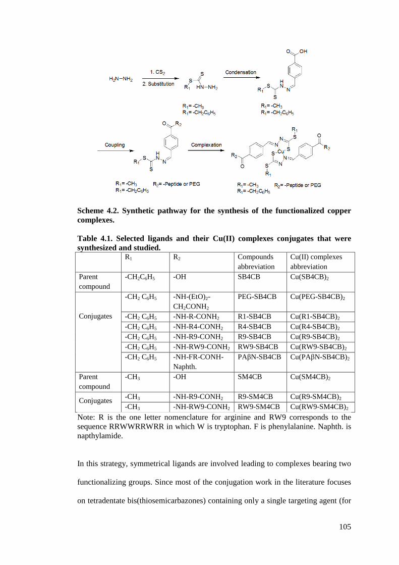
105
Scheme 4.2. Synthetic pathway for the synthesis of the functionalized copper complexes. Table 4.1. Selected ligands and their Cu(II) complexes conjugates that were synthesized and studied. R1 R2 Compounds
abbreviation Cu(II) complexes abbreviation
Parent compound
-CH2C6H5 -OH SB4CB Cu(SB4CB)2
Conjugates
-CH2 C6H5 -NH-(EtO)2-CH2CONH2
PEG-SB4CB Cu(PEG-SB4CB)2
-CH2 C6H5 -NH-R-CONH2 R1-SB4CB Cu(R1-SB4CB)2 -CH2 C6H5 -NH-R4-CONH2 R4-SB4CB Cu(R4-SB4CB)2 -CH2 C6H5 -NH-R9-CONH2 R9-SB4CB Cu(R9-SB4CB)2 -CH2 C6H5 -NH-RW9-CONH2 RW9-SB4CB Cu(RW9-SB4CB)2 -CH2 C6H5 -NH-FR-CONH-
Naphth. PA%N-SB4CB Cu(PA%N-SB4CB)2
Parent compound
-CH3 -OH SM4CB Cu(SM4CB)2
Conjugates -CH3 -NH-R9-CONH2 R9-SM4CB Cu(R9-SM4CB)2 -CH3 -NH-RW9-CONH2 RW9-SM4CB Cu(RW9-SM4CB)2
Note: R is the one letter nomenclature for arginine and RW9 corresponds to the sequence RRWWRRWRR in which W is tryptophan. F is phenylalanine. Naphth. is napthylamide.
In this strategy, symmetrical ligands are involved leading to complexes bearing two
functionalizing groups. Since most of the conjugation work in the literature focuses
on tetradentate bis(thiosemicarbazones) containing only a single targeting agent (for
106
radiopharmaceutical purposes), having two functional groups may or may not
produce the desired results. In fact, in some instances, it could be counterproductive
as this can further compromise the pharmacokinetics. On the other hand, this
bivalent approach could also impart higher affinity and selectivity. Ma et al. (2011)
described the preparation of ligands with two carboxylate functional groups that bear
two tumor-targeting peptides. The presence of dimeric CPPs in ligands (Hoyer et al.,
2012) and Pt complexes (Abramkin et al., 2012) have been reported to dramatically
improve their capacity to enter various cells increasing their cytotoxicity potential.
A complex having a PEG was first designed to increase the water solubility. PEG is
a neutral water solubilizing agent that does not show any antimicrobial activity.
Therefore it was to be anticipated that this conjugation would highlight only the
bioactivity of the complex itself, while enhancing its water solubility. In addition,
PEG functionalized with carboxylic acid and amine is commercially available. To
eventually modulate the bacteria uptake of the complexes, the ligands were also
conjugated to a CPP R9. As mentioned before, cell uptake is optimum for peptides
having from 8 to 15 arginine residues. Since the complexes preparation involved two
equivalents of ligand in which two peptides were to be introduced leading to a total
of 18 arginine residues, thus in addition to R9, different peptide chain with lengths
of one (R1) or four arginines (R4) were introduced to determine their potential
influence on the antimicrobial activity of the conjugates. In addition to that, some
CPPs and antimicrobial peptides (AMPs) are known to share several common
characteristics such as high density in basic residues and an amphipathic secondary
structure (a hydrophobic side and a cationic side) in membrane environments (Strøm
et al., 2003). Recent reports have shown that conjugation of metal binding ligands
107
with AMPs significantly enhances the permeability and activity of the compounds
against bacteria that have developed cross-resistance against conventional antibiotics
(Chantson et al., 200534 Chantson et al., 200634 Pagès et al., 2013). Since these
bioactive peptide sequences consist mainly of arginine and tryptophan, it would also
be desirable to utilize RW9 (a CPP derived from penetratin) as a vector for
bioconjugation in search of new improved antibacterial agent (Walrant et al., 2011).
And not least, a peptide known as an inhibitor of bacteria efflux pumps was grafted
also. The challenge of multi-drug resistance has become a pressing issue in treating
bacterial infections and one resistance pathway of multi-drug resistant bacteria
involves the “over-expression of efflux pumps which expel structurally unrelated
antibiotics thus decreasing their intracellular concentration” (Nikaido and Pagès,
2012, p. 340; Pagès and Amaral, 2009). Recent attention has been directed to the
discovery of small molecules that inhibit the efflux pumps of the “resistance-
nodulation-division (RND) family in gram-negative bacteria” (Okandeji et al., 2011,
p. 7679). These compounds “act by competitively binding the substrate interacting
sites of RND-family efflux” (Santos et al., 2012, p. 292) and for that reason, the
efflux pumps inhibitors (EPIs) are often used as adjuvants to restore susceptibility of
MDR bacterial pathogens to drugs. “The prototypical inhibitor of RND-family
efflux pumps in gram-negative bacteria is” a C-capped dipeptide, MC-207,110 (Phe-
Arg-%-naphthylamide, PA%N) (Okandeji et al., 2011, p. 7679). In this work, the
adjuvant PA%N was directly linked to the antibacterial agent, namely the Cu(II)
complex in order to eventually reveal any synergistic effect. At this moment, there is
no knowledge of the mechanism of antimicrobial activity of dithiocarbazate derived
Schiff base compounds and therefore efflux pumps give us a potential starting target.
It was hoped that during the pursuit of this target, light would be shed on the
108
problem and show the path to improving the activity and pharmacological properties
of our lead compounds.
4.2 Methodology
4.2.1 Materials
All chemicals and solvents used were similar to those mentioned in Chapter 3 unless
stated otherwise herein. Peptide synthesis: 4-Methylbenzhydrylamine (MBHA) resin
(0.54 mmol/g, Iris Biotech GmbH), fluorenlymethyloxycarbonyl(Fmoc)-rink amide
resin MBHA resin (0.52 mmol/g, Iris Biotech GmbH), tert-butoxycarbonyl (Boc)-L-
Arg (Tos)-OH (Iris Biotech GmbH), Boc-L-Trp (CHO)-OH (Neosystem
Laboratoire), Fmoc-AEEA-OH (Iris Biotech GmbH), PA%N (Bachem),
hydroxybenzotriazole, HOBt (Molekula), N,N,N$,N$-Tetramethyl-O-(1H-
benzotriazol-1-yl)uraniumhexafluorophosphate, O-(Benzotriazol-1-yl)-N,N,N$,N$-
tetramethyluronium hexafluorophosphate, HBTU (Novabiochem), 1-
[bis(dimethylamino)methylene]-1H-1,2,3-triazolo[4,5-b]pyridinium3-oxid hexa-
fluorophosphate, HATU (Iris Biotech GmbH), 1-hydroxy-7-azabenzotriazole,
HOAt (GL BioChem (Shanghai) Ltd/ Applied Biosystems), N,N-
diisopropylethylamine, DIEA (Alfa Aesar), piperidine (Carlo Ebra RS), TFA (Carlo
Ebra RS), hydrofluoric acid, HF (Merck), triisopropylsilane, TIS (Merck), N-
methyl-2-pyrrolidone, NMP (Merck), acetic acid (AnalaR Normapur,VMR
chemicals). Buffers: Acetate buffer pH 4 and 6, 0.1 M (Na acetate-Janssen Chimica,
acetic acid-AnalaR Normapur, VMR chemicals), PBS pH 7.4, 0.01M (Sigma
Aldrich tablet) and borate buffer ph 9 (Na tetraborate-Acros Organics).
109
4.2.2 Instrumentation
All instrumentations were similar to those mentioned in Chapter 3 unless stated
otherwise herein. Peptides and conjugates were characterized by MALDI-TOF-MS
in the positive ion reflector mode on an ABI Voyager DE-Pro MALDI-TOF mass
spectrometer (Applied Biosystems) using as matrix a saturated solution of CHCA in
CH3CN/H2O/TFA (50:50:0.1 v:v:v). LC-MS was carried out using an Applied
Biosystems QTRAP LC-MS/MS System with a linear gradient elution developed
from holding time of 5 min at 100% A and then from 0-60% B in 30 min. Eluent A
was 0.1% formic acid in water, while eluent B was 0.1% formic acid in acetonitrile.
Experiments were carried out at a flow rate of 10 µL min-1 at room temperature with
a microLC C18 column1 mm in diameter with UV detector. Peaks were detected at
220 nm and 280 nm. ITC titrations were performed and analysed using the Nano
ITC standard volume calorimetry (TA Instruments) apparatus. Aliquots (10 !L) of
the Cu(II)(OAc)2 solution were added to the solution of the ligand in 0.1M acetate
buffer at pH 6. An equilibration time of 300 s was allowed between each addition.
Measurements were conducted at 25 °C in a 983 µL cell volume with a 250 µL
syringe with stirring rate 300 RPM. The concentrations of the metal salts and of the
ligands were adjusted to yield total conversion and a good signal/noise ratio for each
experiment. All enthalphy of reactions (2H) values were corrected against the heat
of dilution. ITC results were analyzed using the Nano Analyze software supplied
(version 2.1.6), utilizing a one binding site model. The crude products (conjugated
ligands) were analyzed and purified by RP-HPLC using three Waters HPLC systems
connected to Breeze software:
System A was similar to Chapter 3.
110
System B consisted of combination of a dual wavelength UV-Vis absorbance
detector (Waters 2487) and a binary pump (Waters 1525) equipped with and a semi-
preparative cell for purification purposes. Purification of crude products was
achieved with C8 semi preparative column (250!10mm, 5 µm particles of 300 Å
pore size). Experiments were carried out at a flow rate of 4 mL min-1 at room
temperature. Injection volume was 1.5 mL. Sample concentration was
approximately 10 mg/mL.
System C consisted of a dual wavelength UV-Vis absorbance detector (Waters
2487) and a Waters 600 preparative pump. Purification of crude products was
achieved with a Waters X-bridge C18 preparative column (19!50mm, 5 µm
particles of 300 Å pore size). Experiments were carried out at a flow rate of 10 mL
min-1 at room temperature. Injection volume was 1.5 mL. Sample concentration was
approximately 10 mg/mL.
All the peaks were detected at 220 nm and 280 nm. All the HPLC experiments were
performed using water containing 0.1% of TFA as eluent A and acetonitrile
containing 0.1% of TFA as eluent B. Several elution gradients were developed for
the different compounds as detailed in the synthetic section.
4.2.3 Synthesis
General procedure A
The PEG derivative was synthesized on solid support by Fmoc strategy using Fmoc
protected MBHA Rink amide resin (loading 0.52 mmol/g). The resin was swollen in
DCM one hour prior to use. Fmoc removal was performed by using 20% (v:v)
piperidine in NMP once for 1 min and then once for 15 min. The resin was washed
five times with NMP. Then, a solution of Fmoc-AEEA-OH (3 equiv) was
111
preactivated with HBTU/HOBt (3 equiv/3 equiv) and DIEA (6 equiv) in NMP (1
mL for 0.1 g of resin) and added to the resin in a reaction vessel (polypropylene
syringe). After completion (monitored by Kaiser test for primary amines, in case of
positive Kaiser test, the same amino acid was condensed again until a negative test is
obtained) the Fmoc group of the PEG derivative was removed as previously
described. A solution of Schiff base (SB4CB) (3 equiv) was preactivated with
HATU/HOAt (3 equiv/3 equiv) and DIEA (6 equiv) in NMP (1 mL for 0.1 g of
resin) and added to the resin. The mixture was allowed to react on the automatic
shaker for 2 hours. Cleavage from the resin was performed by shaking for 3 h with a
solution of 5 mL of TIS (2.5%), deionised water (2.5%), and TFA (95%). The
mixture was then filtered and the volume of the solution was reduced by
evaporation. Cold diethyl ether was then added to precipitate the compound, which
was then centrifuged for 5 min at 8000 RPM (three times). The compound was
solubilized in deionised water and lyophilized. The crude product was then purified
with preparative RP-HPLC. For N-terminus acetylated PEG, instead of the coupling
with the ligand, the acetylation was performed with 10% acetic anhydride in DCM
for 1 hour at room temperature. The compound was cleaved as described but no
precipitation occurred upon addition of diethyl ether. The crude was purified by
silica-gel column chromatography.
PEG-SB4CB
To PEG-NH2 on Fmoc-MBHA Rink amide resin (0.25 g, 0.13 mmol) was added a
solution of SB4CB (0.129 g, 0.39 mmol, 3 equiv), HATU (0.148 g, 0.39 mmol, 3
equiv), HOAt (0.053 g, 0.39 mmol, 3 equiv) and DIPEA (0.136 mL, 0.78 mmol, 6
equiv) in DMF (2.5 mL). The crude peptide material was purified with System C:
112
gradient elution of 5 to 100% of B in A for 10 min. The peak eluted at 7.2 min. The
fraction was freeze-dried to give PEG-SB4CB as a cream powder. Analytical RP-
HPLC (System A: gradient elution of 5 to 100% of B in A for 30 min): RT = 17.5
min. HR-MS: m/z = [M+Na]+ Calcd. 497.12877, Found 497.12892. 1H NMR (300
MHz, CD3OD) ! 8.57 (s, 1H), 8.08 (s, 1H), 7.82 (dd, J = 19.5, 9, 4H), 7.45 – 7.21
(m, 5H), 4.52 (s, 2H), 3.96 (s, 2H), 3.69 (s, 6H), 3.59 (d, J =6, 2H). 13C NMR (75
MHz, CD3OD) ! (ppm) 200.29, 175.93, 169.77, 145.41, 138.40, 138.36, 137.28,
130.54, 129.71, 129.02, 128.71, 128.49, 72.11, 71.38, 71.22, 70.71, 41.05, 39.91.
AcPEG-NH2 (PEGAC)
PEG-NH2 on Fmoc-MBHA Rink amide resin (0.784 g, 0.42 mmol) was acetylated
with 10% acetic anhydride in DCM for 1 hour at room temperature. The crude was
purified by silica-gel column chromatography eluted with a gradient of MeOH in
DCM from 4/96 to 10/90 (v:v) MeOH/DCM to yield the expected compound. The
expected compound fraction was evaporated to give AcPEG-NH2 as a colourless
oily solid (88 mg, 98%). HR-MS: m/z = [M+Na]+ Calcd. 227.10023, Found
227.10031. 1H NMR (300 MHz, CD3OD) ) 3.99 (s, 2H), 3.72-3.64 (m, 4H), 3.56 (t,
J = 6, 2H), 3.40 – 3.34 (m, 2H), 1.95 (s, 3H). 13C NMR (75 MHz, CD3OD) ) 175.95,
173.71, 72.06, 71.38, 71.22, 70.89, 40.54, 22.65.
General procedure B
Side-chain protected polyarginine (R1, R4, R9 and RW9) were assembled on
MBHA resin (loading 0.54 mmol/g) either manually or using the peptide synthesizer
(Applied Biosystem 433A) with HBTU/HOBt as coupling agents. All amino acids
were coupled as Boc derivatives. The side chain Arg residue was protected by a
113
tosyl group and the tryptophan side chain was protected by a formyl group. For
RW9, deformylation was performed with 20% piperidine in NMP for approximately
2 hours (sequential time length addition 1 min, 3 min, 5 min, 7 min, 15 min, 30 min
and finally 60 min) prior to Boc removal and coupling to the Schiff base. Boc
removal was perfomed by using TFA for 1 min (twice) and washed with 10% DIEA
in DCM. Coupling was the same as for Fmoc synthesis. Cleavage from the resin and
tosyl protecting group removal was performed using HF in the presence of dimethyl
sulfide for 2 hours at 0°C. After HF removal, cold diethyl ether was added to
precipitate the peptide. The precipitate was collected, redissolved using 10% acetic
acid in water and lyophilized. For N-terminus acetylated peptides, the acetylation
was performed with 10% acetic anhydride in DCM for 1 hour at room temperature.
R1-SB4CB
To R1-NH2 on MBHA resin (0.25 g, 0.135 mmol) was added a solution of SB4CB
(0.135 g, 0.405 mmol, 3 equiv), HATU (0. 154 g, 0.405 mmol, 3 equiv), HOAt
(0.055 g, 0.405 mmol, 3 equiv) and DIPEA (0.140 mL, 0.810 mmol, 6 equiv) in
DMF (2.5 mL). The crude peptide material was purified with System C: gradient
elution of 5 to 100% B in A for 10 min. The peak eluted at 6.06 min. The fraction
was freeze-dried to give R1-SB4CB as a cream powder. Analytical RP-HPLC
(System A: gradient elution of 5 to 100% of B in A for 30 min): RT = 16.1 min. LC-
MS: m/z = [M+H]+ Calcd. 486.17, Found 486.10. 1H NMR (300 MHz, CD3OD) )
8.10 (s, 1H), 7.86 (dd, J = 27, 9, 4H), 7.44 – 7.22 (m, 5H), 4.61 (dd, J = 9, 3, 1H),
4.53 (s, 2H), 3.24 (td, J = 7.5, 3, 2H), 2.06 – 1.94 (m, 1H), 1.90 – 1.63 (m, 3H). 13C
NMR (75 MHz, CD3OD) ! = 200.36, 176.66, 169.66, 158.79, 145.29, 138.74,
114
138.35, 136.70, 130.53, 129.71, 129.26, 128.70, 128.50, 54.55, 42.12, 39.93, 30.44,
26.65
R4-SB4CB
To R4-NH2 on MBHA resin (0.25 g, 0.135 mmol) was added a solution of SB4CB
(0.135 g, 0.405 mmol, 3 equiv), HATU (0. 154 g, 0.405 mmol, 3 equiv), HOAt
(0.055 g, 0.405 mmol, 3 equiv) and DIPEA (0.140 mL, 0.810 mmol, 6 equiv) in
DMF (2.5 mL). The crude peptide material was purified with System C: gradient
elution of 30 to 50% B in A for 10 mins. The peak eluted at 3.34 min. The fraction
was freeze-dried to give R4-SB4CB as a cream powder. Analytical RP-HPLC
(System A: gradient elution of 5 to 100% of B in A for 30 min): RT = 14.0 min.
MALDI-TOF-MS: m/z = [M+H]+ Calcd. 954.48, Found 954.49. 1H NMR (300
MHz, CD3OD) ) 8.10 (s, 1H), 7.87 (dd, J = 27, 6, 4H), 7.44 – 7.23 (m, 5H), 4.55 –
4.47 (m, 3H), 4.43 – 4.31 (m, 3H), 3.28 – 3.13 (m, 8H), 2.00 – 1.61 (m, 16H).
R9-SB4CB
To R9-NH2 on MBHA resin (0.25 g, 0.135 mmol) was added a solution of SB4CB
(0.135 g, 0.405 mmol, 3 equiv), HATU (0. 154 g, 0.405 mmol, 3 equiv), HOAt
(0.055 g, 0.405 mmol, 3 equiv) and DIPEA (0.140 mL, 0.810 mmol, 6 equiv) in
DMF (2.5 mL). The crude peptide material was purified with System C: gradient
elution of 5 to 100% B in A for 10 min. The peak eluted at 4.86 min. The fraction
was freeze-dried to give R9-SB4CB as a cream powder. Analytical RP-HPLC
(System A: gradient elution of 5 to 100% of B in A for 30 min): RT = 13.3 min.
MALDI-TOF-MS: m/z = [M]+ Calcd. 1733.98, Found 1733.93. 1H NMR (300 MHz,
115
CD3OD) ) 8.11 (s, 1H), 7.89 (dd, J = 39, 9, 4H), 7.44 – 7.23 (m, 5H), 4.53 (s, 2H),
4.40 – 4.17 (m, 9H), 3.30 – 3.09 (m, 18H), 2.02 – 1.52 (m, 36H).
RW9-SB4CB
To RW9-NH2 on MBHA resin (0.25 g, 0.135 mmol) was added a solution of
SB4CB (0.135 g, 0.405 mmol, 3 equiv), HATU (0. 154 g, 0.405 mmol, 3 equiv),
HOAt (0.055 g, 0.405 mmol, 3 equiv) and DIPEA (0.140 mL, 0.810 mmol, 6 equiv)
in DMF (2.5 mL). The crude peptide material was purified with System C: gradient
elution of 5 to 100% B in A for 10 min. The peak eluted at 5.69 min. The fraction
was freeze-dried to give RW9-SB4CB as a cream powder. Analytical RP-HPLC
(System A: gradient elution of 5 to 100% of B in A for 30 min): RT = 16.5 min.
MALDI-TOF-MS: m/z = [M]+ Calcd. 1823.91, Found 1823.27. 1H NMR (300 MHz,
CD3OD) ) 8.06 (s, 1H), 7.79 (dd, J = 45, 9, 4H), 7.55 (d, J = 6, 1H), 7.46 – 7.22 (m,
10H), 7.17 – 6.84 (m, 9H), 4.61 (dd, J = 9, 6, 1H), 4.54 (s, 2H), 4.48 (td, J = 4.5, 3,
2H), 4.33 – 4.21 (m, 3H), 4.14 – 4.00 (m, 3H), 3.35 (s, 5H), 3.27 – 2.95 (m, 15H),
1.97 – 1.37 (m, 24H).
R9-SM4CB
To R9-NH2 on MBHA resin (0.25 g, 0.135 mmol) was added a solution of SM4CB
(0.089 g, 0.405 mmol, 3 equiv), HATU (0. 154 g, 0.405 mmol, 3 equiv), HOAt
(0.055 g, 0.405 mmol, 3 equiv) and DIPEA (0.140 mL, 0.810 mmol, 6 equiv) in
DMF (2.5 mL). After cleavage, the crude peptide material was purified with System
C: gradient elution of 5 to 100% B in A for 10 min. The peak eluted at 4.32 min.
The fraction was freeze-dried to give R9-SM4CB as a cream powder. Analytical RP-
HPLC (System A: gradient elution of 5 to 100% of B in A for 30 min): RT = 11.8
116
min. MALDI-TOF-MS: m/z = [M+H]+ Calcd. 1658.95, Found 1658.78. 1H NMR
(300 MHz, CD3OD) ) 8.11 (s, 1H), 7.91 (dd, J = 39, 9, 4H), 4.42 – 4.20 (m, 9H),
3.30 – 3.11 (m, 18H), 2.60 (s, 3H), 2.04 – 1.55 (m, 36H).
RW9-SM4CB
To RW9-NH2 on MBHA resin (0.25 g, 0.135 mmol) was added a solution of
SM4CB (0.089 g, 0.405 mmol, 3 equiv), HATU (0. 154 g, 0.405 mmol, 3 equiv),
HOAt (0.055 g, 0.405 mmol, 3 equiv) and DIPEA (0.140 mL, 0.810 mmol, 6 equiv)
in DMF (2.5 mL). The crude peptide material was purified with System C: gradient
elution of 20 to 50% B in A for 10 min. The peak eluted at 6.11 min. The fraction
was freeze-dried to give RW9-SM4CB as a cream powder. Analytical RP-HPLC
(System A: gradient elution of 5 to 100% of B in A for 30 min): RT = 14.0 min.
MALDI-TOF-MS: m/z = [M+H]+ Calcd. 1748.89, Found 1748.45. 1H NMR (300
MHz, CD3OD) ) 8.07 (s, 1H), 7.95 (d, J = 9, 2H), 7.77 (d, J = 6, 2H), 7.55 (d, J = 6,
1H), 7.46 – 7.28 (m, 5H), 7.19 – 6.84 (m, 9H), 4.57 (dd, J = 15, 9, 1H), 4.49 – 4.36
(m, 2H), 4.33 – 4.17 (m, 3H), 4.14 – 3.96 (m, 3H), 3.28 – 2.96 (m, 15H), 2.61 (s,
3H), 1.94 – 1.27 (m, 24H).
AcR1-NH2 (R1AC)
To R1-NH2 on MBHA resin (0.25 g, 0.135 mmol), the acetylation was performed
with 10% acetic anhydride in DCM for 1 hour at room temperature. The crude
peptide material was purified with System C: gradient elution of 0 to 30% B in A for
10 mins. The peak eluted at 3.26 min. The fraction was freeze-dried to give R4AC
as a cream powder. Analytical RP-HPLC (System A: gradient elution of 0 to 30% of
B in A for 30 min): RT = 4.0 min. HR-MS: m/z = [M+H]+ Calcd. 216.14550,
117
Found 216.14556. 1H NMR (300 MHz, CD3OD) ) 4.35 (dd, J = 9, 6, 1H), 3.28 –
3.17 (m, 2H), 2.01 (s, 3H), 1.96 – 1.83 (m, 1H), 1.75 – 1.59 (m, 3H). 13C NMR (75
MHz, CD3OD) ) 176.79, 173.61, 54.00, 42.09, 30.38, 26.46, 22.66.
AcR4-NH2 (R4AC)
To R4-NH2 on MBHA resin (0.25 g, 0.135 mmol), the acetylation was performed
with 10% acetic anhydride in DCM for 1 hour at room temperature. The crude
peptide material was purified with System C: gradient elution of 0 to 30% B in A for
10 min. The peak eluted at 4.36 min. The fraction was freeze-dried to give R4AC as
a cream powder. Analytical RP-HPLC (System A: gradient elution of 0 to 30% of B
in A for 30 min): RT = 6.8 min. MALDI-TOF-MS: [M+K]+ Calcd. 836.02, Found
722.33; [M+Na]+ Calcd. 820.04, Found 706.37; [M+H]+ Calcd. 684.45, Found
684.38. 1H NMR (300 MHz, CD3OD) ) 4.38 – 4.24 (m, 4H), 3.21 (t, J = 6.6, 8H),
2.02 (s, 3H), 1.98 – 1.56 (m, 16H).
AcR9-NH2 (R9AC)
To R9-NH2 on MBHA resin (0.25 g, 0.135 mmol), the acetylation was performed
with 10% acetic anhydride in DCM for 1 hour at room temperature. The crude
peptide material was purified with System B: gradient elution of 0 to 30% B in A for
30 min. The peak eluted at 9 min. The fraction was freeze-dried to give R9AC as a
cream powder. Analytical RP-HPLC (System A: gradient elution of 5 to 100% of B
in A for 30 min): RT = 6.0 min. MALDI-TOF-MS: [M+K]+ Calcd. 836.02, Found
1486.90; [M+Na]+ Calcd. 1502.87, Found 1486.90; [M+H]+ Calcd. 1464.95, Found
1464.93. 1H NMR (300 MHz, CD3OD) ) 4.35 – 4.12 (m, 9H), 3.28 – 3.16 (m, 18H),
2.04 (s, 3H), 1.96 – 1.63 (m, 36H).
118
AcRW9-NH2 (RW9AC)
To RW9-NH2 on MBHA resin (0.25 g, 0.135 mmol), the acetylation was performed
with 10% acetic anhydride in DCM for 1 hour at room temperature. The crude
peptide material was purified with System B: gradient elution of 5 to 100% B in A
for 30 mins. The peak eluted at 12 min. The fraction was freeze-dried to give
RW9AC as a cream powder. Analytical RP-HPLC (System A: gradient elution of 5
to 100% of B in A for 30 min): RT = 10.6 min. MALDI-TOF-MS: [M+K]+ Calcd.
836.02, Found 1593.72; [M+Na]+ Calcd. 820.04, Found 1577.75; [M+H]+ Calcd.
1554.89, Found 1554.78. 1H NMR (300 MHz, CD3OD) ) 7.56 (d, J = 9, 1H), 7.48 –
7.30 (m, 5H), 7.14 – 6.89 (m, 9H), 4.60 (dd, J = 9, 6, 1H), 4.44 (dd, J = 15, 9, 2H),
4.34 – 4.21 (m, 2H), 4.15 – 3.98 (m, 4H), 3.36 (d, J = 5.7, 1H), 3.29 – 2.94 (m,
16H), 2.05 (s, 3H), 1.89 – 1.29 (m, 24H).
Procedure C (Solution synthesis)
PA"N-SB4CB
The ligand SB4CB (10 mg, 0.061 mmol, 1 equiv), PA%N.2HCl (23.5 mg, 0.091
mmol, 1.5 equiv), HATU (17.3 mg, 0.091 mmol, 1.5 equiv) and HOAt (6.2mg,
0.091 mmol, 1.5 equiv) were placed in a round-bottom flask under argon. Dried
DMF (1 mL) was added followed by DIEA (0.026 mL, 0.303 mmol, 5 equiv). The
reaction mixture was stirred overnight at room temperature. Analytical HPLC
showed conversion of starting material into a less polar product >50%. After
dilution with water, the crude peptide material was purified with System C: gradient
elution of 5 to 100% B in A for 10 min. The peak eluted at 8.57 min. The fraction
was freeze-dried to give PA%N-SB4CB as a yellowish solid. Analytical HPLC: RT =
21.5 min. MALDI-TOF-MS: m/z = [M+H]+ Calcd. 759.29, Found 759.24. 1H NMR
119
(300 MHz, CD3OD) ) 8.19 (d, J = 3, 1H), 8.06 (s, 1H), 7.86 – 7.73 (m, 7H), 7.58 –
7.05 (m, 14H), 4.83 (dd, J = 9, 6, 1H), 4.58 (dd, J = 15, 6, 1H), 4.53 (s, 2H), 3.29 –
3.07 (m, 4H), 2.04 – 1.61 (m, 4H).
Preparation of copper (II) complexes, CuL2
To the bioconjugate L (mmol, 2 equiv) in methanol was added a solution of
Cu(OAc)2·H2O (mmol, 1 equiv), the solution mixture was stirred overnight and then
evaporated to dryness to give brown oily solid.
4.3 Results and discussion
4.3.1 Synthesis
The synthesis of the bidentate dithiocarbazate Schiff base ligand bioconjugates with
PEG and peptides are reported here for the first time as well as the synthesis of their
Cu(II) complexes. The preparation of eight new ligand-vector conjugates and their
Cu complexes containing Schiff base ligands derived from the condensation of
SMDTC/SBDTC with 4-carboxybenzaldehyde and various vectors comprising
CPPs, PEG and EPI are detailed in this work. In addition, five acetylated vectors
were also prepared for meaningful comparison during the antimicrobial evaluation.
The compounds were synthesized using either solid phase peptide synthesis
techniques or solution phase coupling conditions. The presence of metal ion and/or
Schiff base in the system can interfere with standard conjugation synthetic
procedures. Therefore, the procedure needs to be optimized accordingly for each
compound to prevent hydrolysis, decomposition and demetallation.
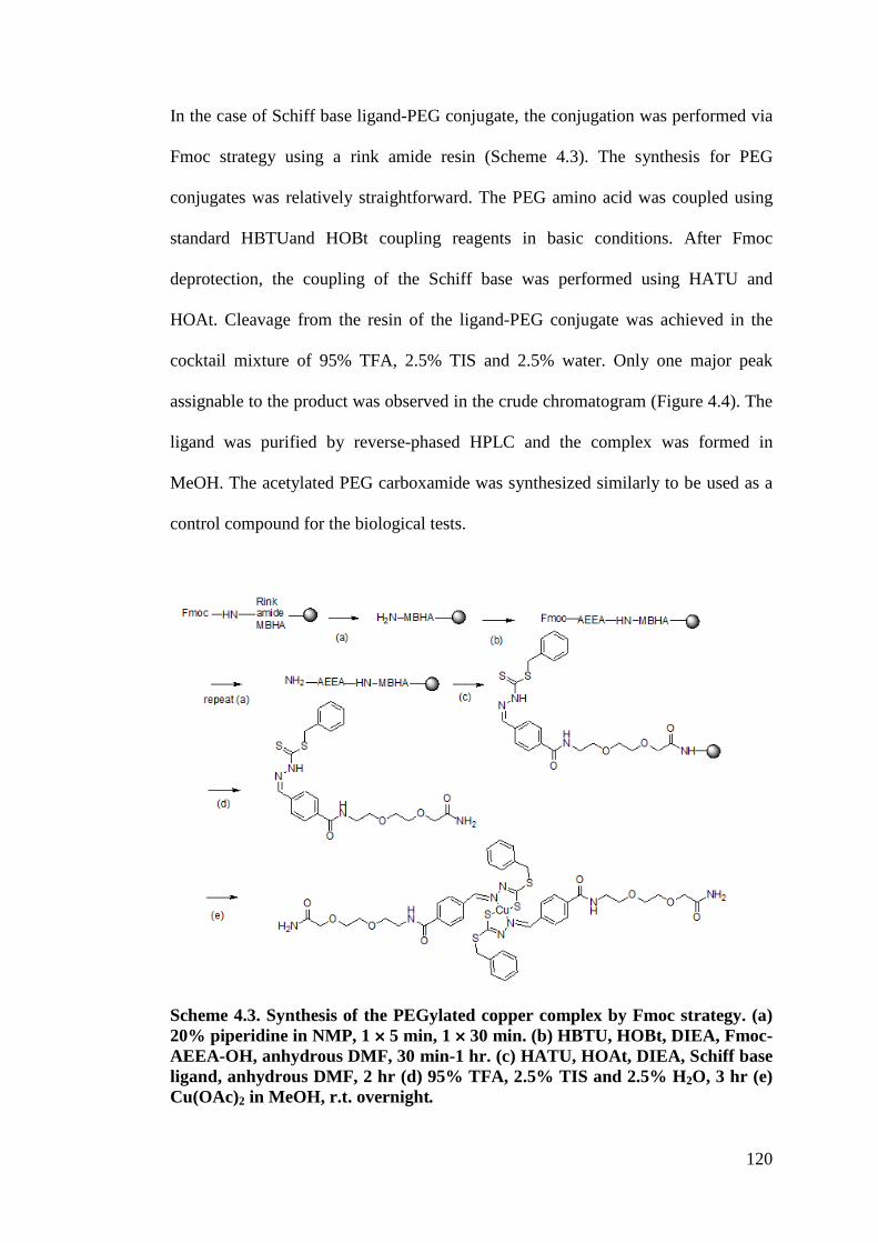
120
In the case of Schiff base ligand-PEG conjugate, the conjugation was performed via
Fmoc strategy using a rink amide resin (Scheme 4.3). The synthesis for PEG
conjugates was relatively straightforward. The PEG amino acid was coupled using
standard HBTUand HOBt coupling reagents in basic conditions. After Fmoc
deprotection, the coupling of the Schiff base was performed using HATU and
HOAt. Cleavage from the resin of the ligand-PEG conjugate was achieved in the
cocktail mixture of 95% TFA, 2.5% TIS and 2.5% water. Only one major peak
assignable to the product was observed in the crude chromatogram (Figure 4.4). The
ligand was purified by reverse-phased HPLC and the complex was formed in
MeOH. The acetylated PEG carboxamide was synthesized similarly to be used as a
control compound for the biological tests.
Scheme 4.3. Synthesis of the PEGylated copper complex by Fmoc strategy. (a) 20% piperidine in NMP, 1 ! 5 min, 1 ! 30 min. (b) HBTU, HOBt, DIEA, Fmoc-AEEA-OH, anhydrous DMF, 30 min-1 hr. (c) HATU, HOAt, DIEA, Schiff base ligand, anhydrous DMF, 2 hr (d) 95% TFA, 2.5% TIS and 2.5% H2O, 3 hr (e) Cu(OAc)2 in MeOH, r.t. overnight.
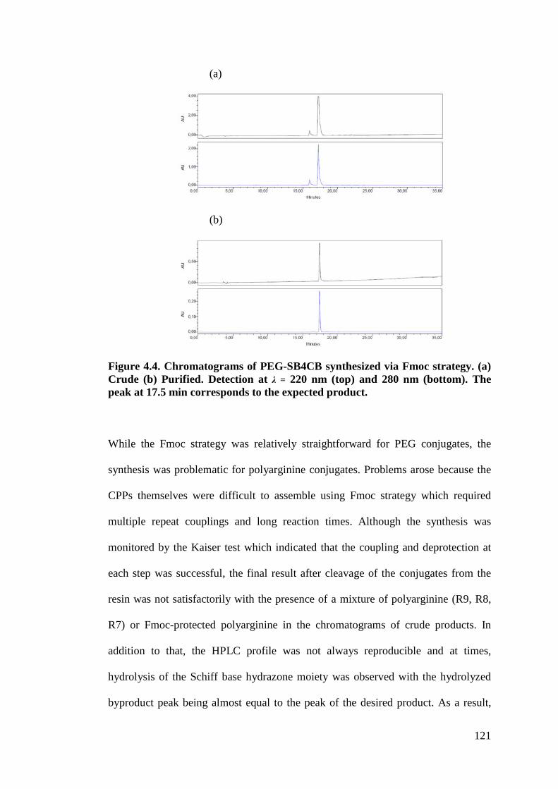
121
(a)
(b)
Figure 4.4. Chromatograms of PEG-SB4CB synthesized via Fmoc strategy. (a) Crude (b) Purified. Detection at ! = 220 nm (top) and 280 nm (bottom). The peak at 17.5 min corresponds to the expected product.
While the Fmoc strategy was relatively straightforward for PEG conjugates, the
synthesis was problematic for polyarginine conjugates. Problems arose because the
CPPs themselves were difficult to assemble using Fmoc strategy which required
multiple repeat couplings and long reaction times. Although the synthesis was
monitored by the Kaiser test which indicated that the coupling and deprotection at
each step was successful, the final result after cleavage of the conjugates from the
resin was not satisfactorily with the presence of a mixture of polyarginine (R9, R8,
R7) or Fmoc-protected polyarginine in the chromatograms of crude products. In
addition to that, the HPLC profile was not always reproducible and at times,
hydrolysis of the Schiff base hydrazone moiety was observed with the hydrolyzed
byproduct peak being almost equal to the peak of the desired product. As a result,
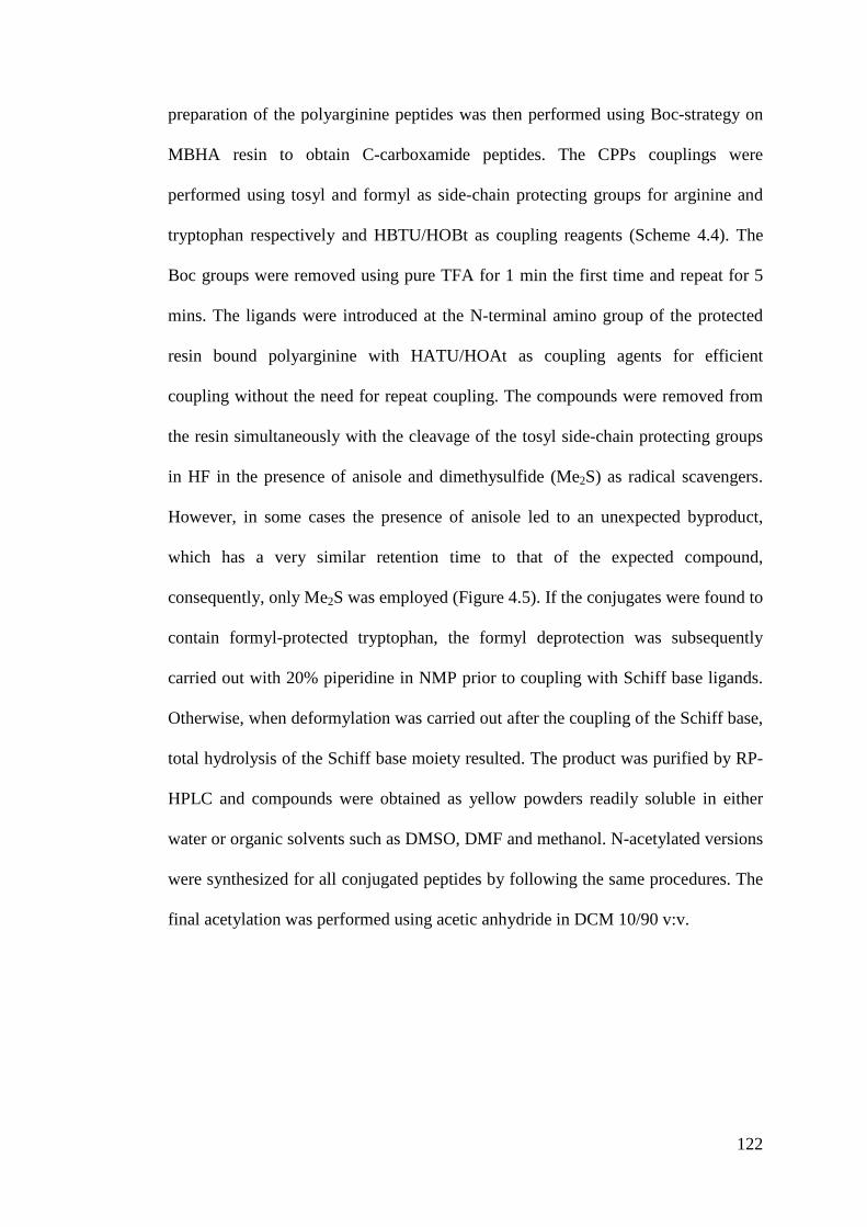
122
preparation of the polyarginine peptides was then performed using Boc-strategy on
MBHA resin to obtain C-carboxamide peptides. The CPPs couplings were
performed using tosyl and formyl as side-chain protecting groups for arginine and
tryptophan respectively and HBTU/HOBt as coupling reagents (Scheme 4.4). The
Boc groups were removed using pure TFA for 1 min the first time and repeat for 5
mins. The ligands were introduced at the N-terminal amino group of the protected
resin bound polyarginine with HATU/HOAt as coupling agents for efficient
coupling without the need for repeat coupling. The compounds were removed from
the resin simultaneously with the cleavage of the tosyl side-chain protecting groups
in HF in the presence of anisole and dimethysulfide (Me2S) as radical scavengers.
However, in some cases the presence of anisole led to an unexpected byproduct,
which has a very similar retention time to that of the expected compound,
consequently, only Me2S was employed (Figure 4.5). If the conjugates were found to
contain formyl-protected tryptophan, the formyl deprotection was subsequently
carried out with 20% piperidine in NMP prior to coupling with Schiff base ligands.
Otherwise, when deformylation was carried out after the coupling of the Schiff base,
total hydrolysis of the Schiff base moiety resulted. The product was purified by RP-
HPLC and compounds were obtained as yellow powders readily soluble in either
water or organic solvents such as DMSO, DMF and methanol. N-acetylated versions
were synthesized for all conjugated peptides by following the same procedures. The
final acetylation was performed using acetic anhydride in DCM 10/90 v:v.
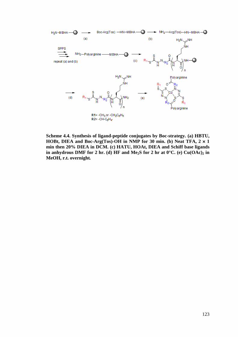
123
Scheme 4.4. Synthesis of ligand-peptide conjugates by Boc-strategy. (a) HBTU, HOBt, DIEA and Boc-Arg(Tos)-OH in NMP for 30 min. (b) Neat TFA, 2 ! 1 min then 20% DIEA in DCM. (c) HATU, HOAt, DIEA and Schiff base ligands in anhydrous DMF for 2 hr. (d) HF and Me2S for 2 hr at 0°C. (e) Cu(OAc)2 in MeOH, r.t. overnight.

124
(a)
(b)
(c)
(d)
Figure 4.5. RP-HPLC chromatograms obtained on a C8 column. Samples were eluted using a gradient of acetonitrile from 5 to 95% in water over 30 min with 1mL min-1 flow rate at room temperature (both solvents contain 0.1% TFA). Detection: ! = 220 nm (top) and 280 nm (bottom) (a) HPLC profile of the crude R9-SM4CB conjugate from Boc-strategy with the addition of anisole during HF-cleavage. The peak at ~6 min is the hydrolyzed compound, the peak at 11.8 min is the desired product while the peak at ~14.5 min is the byproduct caused by anisole. (b) RP-HPLC profile of the crude R9-SM4CB conjugate from Boc-strategy without the addition of anisole during HF-cleavage. The byproduct observed in (a) caused by anisole was no longer visible. (c) RP-HPLC elution profile of the crude R9-SM4CB conjugate from Fmoc-strategy. The profile was less well resolved as compared to Boc-strategy and not always reproducible. (d) RP-HPLC elution profile of the purified R9-SM4CB.
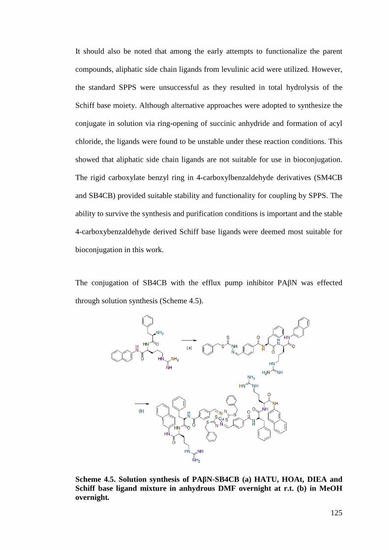
125
It should also be noted that among the early attempts to functionalize the parent
compounds, aliphatic side chain ligands from levulinic acid were utilized. However,
the standard SPPS were unsuccessful as they resulted in total hydrolysis of the
Schiff base moiety. Although alternative approaches were adopted to synthesize the
conjugate in solution via ring-opening of succinic anhydride and formation of acyl
chloride, the ligands were found to be unstable under these reaction conditions. This
showed that aliphatic side chain ligands are not suitable for use in bioconjugation.
The rigid carboxylate benzyl ring in 4-carboxylbenzaldehyde derivatives (SM4CB
and SB4CB) provided suitable stability and functionality for coupling by SPPS. The
ability to survive the synthesis and purification conditions is important and the stable
4-carboxybenzaldehyde derived Schiff base ligands were deemed most suitable for
bioconjugation in this work.
The conjugation of SB4CB with the efflux pump inhibitor PA%N was effected
through solution synthesis (Scheme 4.5).
Scheme 4.5. Solution synthesis of PA"N-SB4CB (a) HATU, HOAt, DIEA and Schiff base ligand mixture in anhydrous DMF overnight at r.t. (b) in MeOH overnight.!
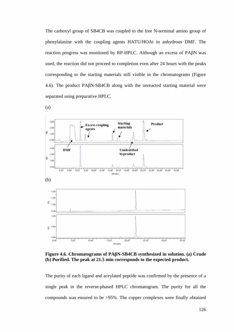
126
The carboxyl group of SB4CB was coupled to the free N-terminal amino group of
phenylalanine with the coupling agents HATU/HOAt in anhydrous DMF. The
reaction progress was monitored by RP-HPLC. Although an excess of PA%N was
used, the reaction did not proceed to completion even after 24 hours with the peaks
corresponding to the starting materials still visible in the chromatograms (Figure
4.6). The product PA%N-SB4CB along with the unreacted starting material were
separated using preparative HPLC.
(a)
(b)
Figure 4.6. Chromatograms of PA"N-SB4CB synthesized in solution. (a) Crude (b) Purified. The peak at 21.5 min corresponds to the expected product. The purity of each ligand and aceylated peptide was confirmed by the presence of a
single peak in the reverse-phased HPLC chromatogram. The purity for all the
compounds was ensured to be >95%. The copper complexes were finally obtained
127
by mixing a 0.5 equivalent of Cu(OAc)2 to the ligand in MeOH. All ligand
conjugates and their respective complexes were comprehensively characterized
spectroscopically.
4.3.2 Characterization of ligands conjugates
4.3.2.1 NMR
All ligands were analyzed by NMR spectroscopy in order to confirm the integrity of
the Schiff base hydrazone moiety. For illustration, the example of R1-SB4CB will
be discussed here while the rest of the spectra are available in the Appendices. 1H
and 13C spectra of R1-SB4CB in CD3OD are shown in Figure 4.7. The presence of
the Schiff base is immediately apparent from the NMR spectra of the bioconjugates.
R1-SB4CB possesses peaks that corresponds to the SB4CB moiety: characteristic
resonance signals at ) 8.10 ppm and 7.90-7.80 ppm attributed to -CH=N and the
aromatic ring of 4-carboxybenzaldehyde moiety, the 5 aromatic protons of SBDTC
in the region of 7.43-7.23 ppm and the presence of a singlet of the S-CH2 protons
observed at 4.53 ppm. These chemicals shifts of SB4CB moiety are consistent in
almost all the NMR spectra of the bioconjugates with exception of RW9-SB4CB
and PA%N- SB4CB in which the SB4CB aromatic region multiplets overlapped with
the peptides tryptophan, phenylalanine and napthylamide protons. For SM4CB
derived conjugates, the aromatic ring in the region of 7.43-7.23 ppm and S-CH2
protons at 4.53 ppm were absent while the distinctive methyl signal for SMDTC was
clearly visible at 2.61 ppm. Signals arising from the protons of arginine were
observed at ca. 4.85-4.59 ppm, 3.26-3.21 ppm and 2.06-1.62 ppm corresponding to
&-CH, )-CH2 and %, --CH2 respectively. The &-CH2 peaks shifted highfield with
increasing number of arginine with the signals observed at ca. 4.40-4.17 ppm for
128
R9-SB4CB. In RW9 derivatives, the tryptophan %-CH2 signal partially overlapping
with the arginine )-CH2 protons as well as the CD3OD solvent peak while the &-CH
of tryptophan can be easily located between ca. 4.60-4.45 ppm. Such overlappings
were also observed for PA%N-SB4CB in which the protons of &-CH and %-CH2 each
overlapped with the CD3OD solvent peaks at 4.78 ppm and 3.31 ppm. For PEG-
SB4CB, the 10 protons of PEG can be found in the region ca. 4.00-3.50 ppm. In
principle, dithiocarbazate derivatives can exhibit thione-thiol tautomerism due to the
presence of thioamide function –NH-C(S)SR. In the 13C NMR spectra of R1-
SB4CB, the presence of -NH-C(=S)S at ) 200.36 ppm indicates that the Schiff bases
predominate as the thione form in solution (if thiol form is present, a signal at ca.
158 ppm will be observed) (How, 2008). This signal was shifted downfield due to
the deshielding effect of the neighbouring amine nitrogen and electronegative
sulphur. The CH=N signal of the Schiff base occurred at 145.29 ppm indicative of
the hydrazone bond which was formed when 4-carboxybenzaldehyde reacted with
the SBDTC. The -S-CH2 peak occurred relatively upfield at 39.93 ppm. The
aromatic carbons were observed around 138-128 ppm as expected for carbons in
aromatic rings. In addition, the spectrum also showed the expected signals with
carbon atoms of arginine giving four signals at ) 26.65, 30.44, 42.12 and 54.55 ppm
while the two amide and one amine carbon atoms gave signals at ) 176.66, 169.66
and 158.79 ppm. Overall, the NMR spectra of all the bioconjugates were comparable
and in agreement with the expected structure, which confirm their formation.
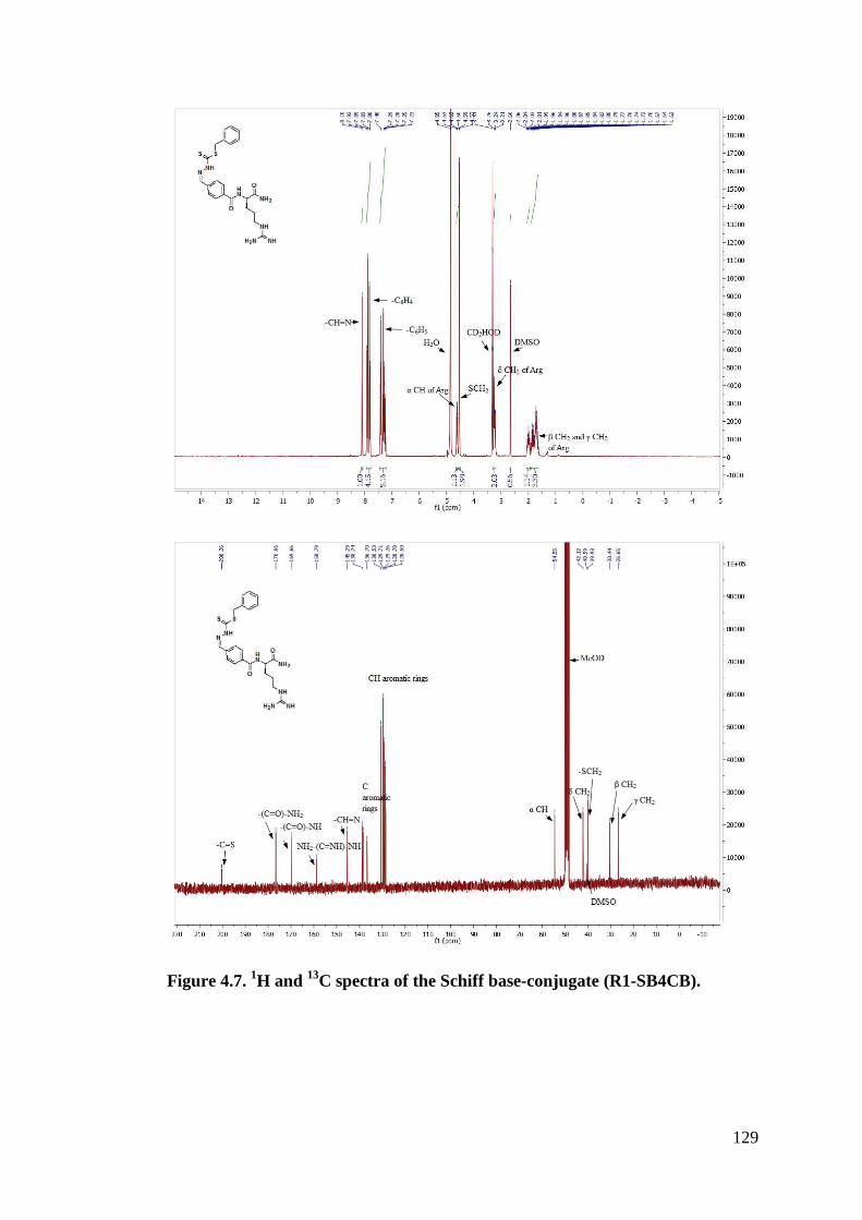
129
Figure 4.7. 1H and 13C spectra of the Schiff base-conjugate (R1-SB4CB).
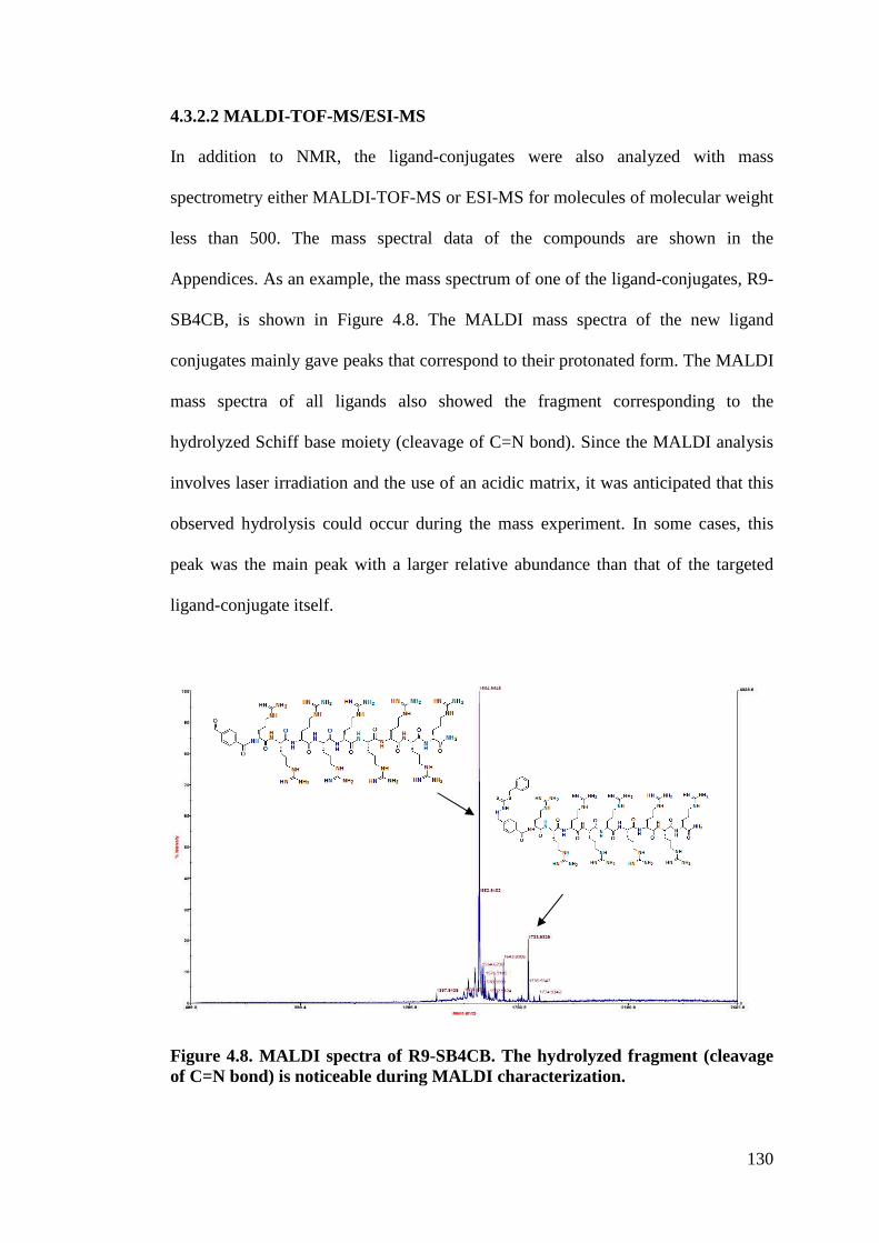
130
4.3.2.2 MALDI-TOF-MS/ESI-MS
In addition to NMR, the ligand-conjugates were also analyzed with mass
spectrometry either MALDI-TOF-MS or ESI-MS for molecules of molecular weight
less than 500. The mass spectral data of the compounds are shown in the
Appendices. As an example, the mass spectrum of one of the ligand-conjugates, R9-
SB4CB, is shown in Figure 4.8. The MALDI mass spectra of the new ligand
conjugates mainly gave peaks that correspond to their protonated form. The MALDI
mass spectra of all ligands also showed the fragment corresponding to the
hydrolyzed Schiff base moiety (cleavage of C=N bond). Since the MALDI analysis
involves laser irradiation and the use of an acidic matrix, it was anticipated that this
observed hydrolysis could occur during the mass experiment. In some cases, this
peak was the main peak with a larger relative abundance than that of the targeted
ligand-conjugate itself.
Figure 4.8. MALDI spectra of R9-SB4CB. The hydrolyzed fragment (cleavage of C=N bond) is noticeable during MALDI characterization.
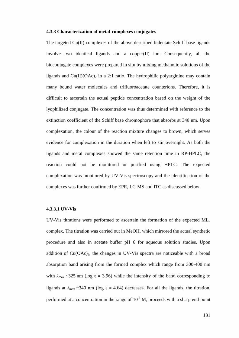
131
4.3.3 Characterization of metal-complexes conjugates
The targeted Cu(II) complexes of the above described bidentate Schiff base ligands
involve two identical ligands and a copper(II) ion. Consequently, all the
bioconjugate complexes were prepared in situ by mixing methanolic solutions of the
ligands and Cu(II)(OAc)2 in a 2:1 ratio. The hydrophilic polyarginine may contain
many bound water molecules and trifluoroacetate counterions. Therefore, it is
difficult to ascertain the actual peptide concentration based on the weight of the
lyophilized conjugate. The concentration was thus determined with reference to the
extinction coefficient of the Schiff base chromophore that absorbs at 340 nm. Upon
complexation, the colour of the reaction mixture changes to brown, which serves
evidence for complexation in the duration when left to stir overnight. As both the
ligands and metal complexes showed the same retention time in RP-HPLC, the
reaction could not be monitored or purified using HPLC. The expected
complexation was monitored by UV-Vis spectroscopy and the identification of the
complexes was further confirmed by EPR, LC-MS and ITC as discussed below.
4.3.3.1 UV-Vis
UV-Vis titrations were performed to ascertain the formation of the expected ML2
complex. The titration was carried out in MeOH, which mirrored the actual synthetic
procedure and also in acetate buffer pH 6 for aqueous solution studies. Upon
addition of Cu(OAc)2, the changes in UV-Vis spectra are noticeable with a broad
absorption band arising from the formed complex which range from 300-400 nm
with "max ~325!nm (log !! " 3.96) while the intensity of the band corresponding to
ligands at "max ~340 nm (log !! "!4.64) decreases. For all the ligands, the titration,
performed at a concentration in the range of 10-5 M, proceeds with a sharp end-point
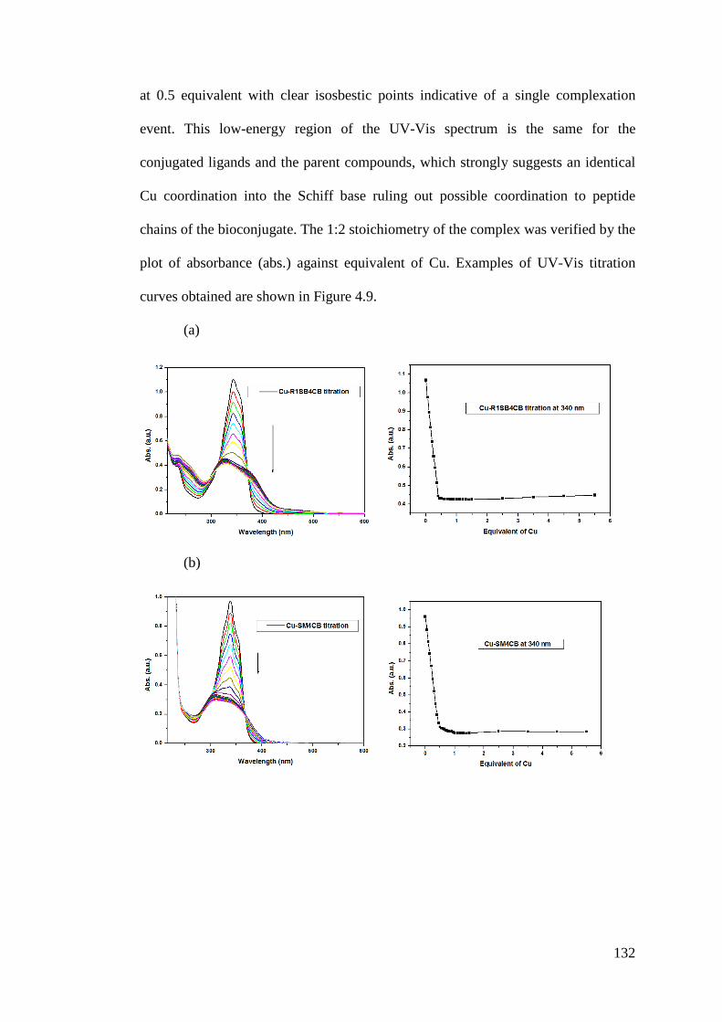
132
at 0.5 equivalent with clear isosbestic points indicative of a single complexation
event. This low-energy region of the UV-Vis spectrum is the same for the
conjugated ligands and the parent compounds, which strongly suggests an identical
Cu coordination into the Schiff base ruling out possible coordination to peptide
chains of the bioconjugate. The 1:2 stoichiometry of the complex was verified by the
plot of absorbance (abs.) against equivalent of Cu. Examples of UV-Vis titration
curves obtained are shown in Figure 4.9.
(a)
(b)
133
(c)
Figure 4.9. UV-Vis titration of various ligands (concentration set at ca. 2.5 x 10⁻⁵ M) with Cu(OAc)2.H2O (concentration set at ca. 5 x 10⁻⁴ M) at 25°C. a) Titration of R1-SB4CB in methanol and its corresponding titration curve monitored at 340 nm. b) Titration of SM4CB in acetate buffer pH 6 and its corresponding titration curve monitored at 340 nm for comparison with conjugated compounds. c) Titration of R9-SM4CB in acetate buffer pH 6 and its corresponding titration curve monitored at 340 nm.
Understanding the behavior of these compounds in aqueous and buffer systems
under physiological conditions (pH range 6.0-7.4) is important for the study of
biological activity. Many Schiff bases derived from S-substituted dithiocarbazate
prepared to date and their metal complexes suffer from poor aqueous solubility that
hinders investigation of their properties. The tremendous increase in solubility of
their bioconjugate derivatives allows for some investigations to be carried out as
well as their complexation in aqueous solution. The complexation of Cu(II) was
confirmed to be pH dependent. After screening a range of aqueous buffer solutions,
acetate buffer (pH 6) was utilized as it does not interfere with the UV absorption and
the complexation proceeded well. Attempts were unsuccessful with water (no
complexation) and buffers such as HEPES (no complexation), TRIS (no
complexation), MOPS (changes in the UV-Vis signature too small for analysis),
PBS (precipitation of the ligands) and borate buffer (reduction of the Cu(II) ion /
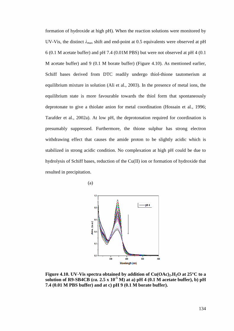
134
formation of hydroxide at high pH). When the reaction solutions were monitored by
UV-Vis, the distinct "max shift and end-point at 0.5 equivalents were observed at pH
6 (0.1 M acetate buffer) and pH 7.4 (0.01M PBS) but were not observed at pH 4 (0.1
M acetate buffer) and 9 (0.1 M borate buffer) (Figure 4.10). As mentioned earlier,
Schiff bases derived from DTC readily undergo thiol-thione tautomerism at
equilibrium mixture in solution (Ali et al., 2003). In the presence of metal ions, the
equilibrium state is more favourable towards the thiol form that spontaneously
deprotonate to give a thiolate anion for metal coordination (Hossain et al., 1996;
Tarafder et al., 2002a). At low pH, the deprotonation required for coordination is
presumably suppressed. Furthermore, the thione sulphur has strong electron
withdrawing effect that causes the amide proton to be slightly acidic which is
stabilized in strong acidic condition. No complexation at high pH could be due to
hydrolysis of Schiff bases, reduction of the Cu(II) ion or formation of hydroxide that
resulted in precipitation.
(a)
Figure 4.10. UV-Vis spectra obtained by addition of Cu(OAc)2.H2O at 25°C to a solution of R9-SB4CB (ca. 2.5 x 10⁻⁵ M) at a) pH 4 (0.1 M acetate buffer), b) pH 7.4 (0.01 M PBS buffer) and at c) pH 9 (0.1 M borate buffer).
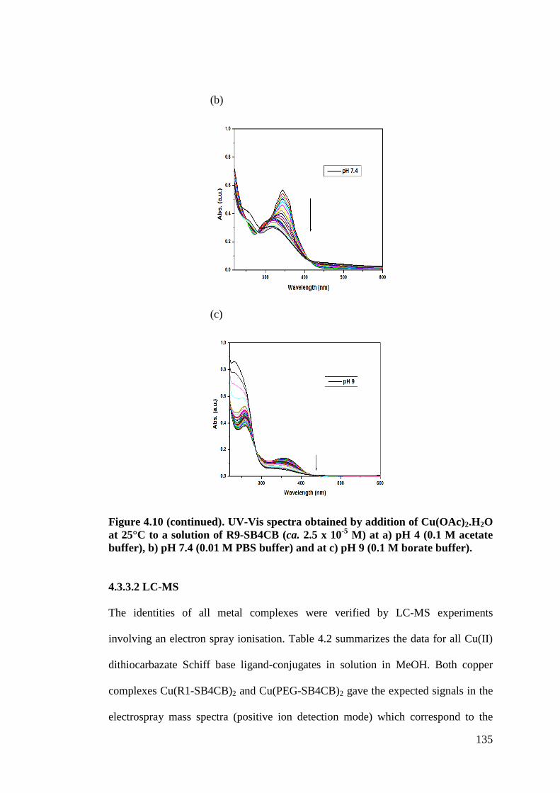
135
(b)
(c)
Figure 4.10 (continued). UV-Vis spectra obtained by addition of Cu(OAc)2.H2O at 25°C to a solution of R9-SB4CB (ca. 2.5 x 10⁻⁵ M) at a) pH 4 (0.1 M acetate buffer), b) pH 7.4 (0.01 M PBS buffer) and at c) pH 9 (0.1 M borate buffer).
4.3.3.2 LC-MS
The identities of all metal complexes were verified by LC-MS experiments
involving an electron spray ionisation. Table 4.2 summarizes the data for all Cu(II)
dithiocarbazate Schiff base ligand-conjugates in solution in MeOH. Both copper
complexes Cu(R1-SB4CB)2 and Cu(PEG-SB4CB)2 gave the expected signals in the
electrospray mass spectra (positive ion detection mode) which correspond to the
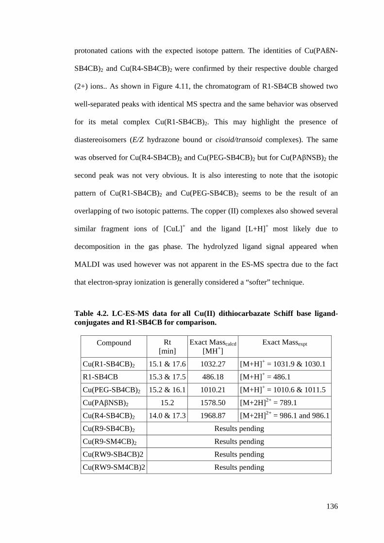
136
protonated cations with the expected isotope pattern. The identities of Cu(PAßN-
SB4CB)2 and Cu(R4-SB4CB)2 were confirmed by their respective double charged
(2+) ions.. As shown in Figure 4.11, the chromatogram of R1-SB4CB showed two
well-separated peaks with identical MS spectra and the same behavior was observed
for its metal complex Cu(R1-SB4CB)2. This may highlight the presence of
diastereoisomers (E/Z hydrazone bound or cisoid/transoid complexes). The same
was observed for Cu(R4-SB4CB)2 and Cu(PEG-SB4CB)2 but for Cu(PA%NSB)2 the
second peak was not very obvious. It is also interesting to note that the isotopic
pattern of Cu(R1-SB4CB)2 and Cu(PEG-SB4CB)2 seems to be the result of an
overlapping of two isotopic patterns. The copper (II) complexes also showed several
similar fragment ions of [CuL]+ and the ligand [L+H]+ most likely due to
decomposition in the gas phase. The hydrolyzed ligand signal appeared when
MALDI was used however was not apparent in the ES-MS spectra due to the fact
that electron-spray ionization is generally considered a “softer” technique.
Table 4.2. LC-ES-MS data for all Cu(II) dithiocarbazate Schiff base ligand-conjugates and R1-SB4CB for comparison.
Compound Rt [min]
Exact Masscalcd [MH+]
Exact Massexpt
Cu(R1-SB4CB)2 15.1 & 17.6 1032.27 [M+H]+ = 1031.9 & 1030.1 R1-SB4CB 15.3 & 17.5 486.18 [M+H]+ = 486.1 Cu(PEG-SB4CB)2 15.2 & 16.1 1010.21 [M+H]+ = 1010.6 & 1011.5 Cu(PA%NSB)2 15.2 1578.50 [M+2H]2+ = 789.1 Cu(R4-SB4CB)2 14.0 & 17.3 1968.87 [M+2H]2+ = 986.1 and 986.1 Cu(R9-SB4CB)2 Results pending Cu(R9-SM4CB)2 Results pending Cu(RW9-SB4CB)2 Results pending Cu(RW9-SM4CB)2 Results pending
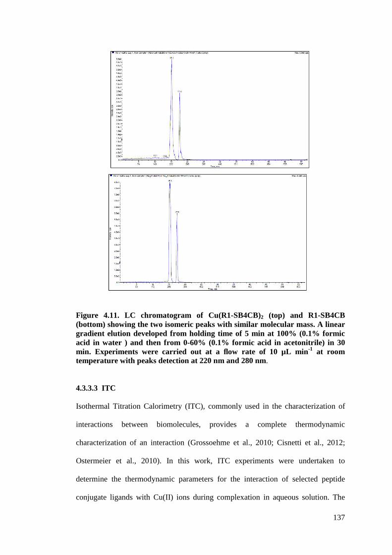
137
Figure 4.11. LC chromatogram of Cu(R1-SB4CB)2 (top) and R1-SB4CB (bottom) showing the two isomeric peaks with similar molecular mass. A linear gradient elution developed from holding time of 5 min at 100% (0.1% formic acid in water ) and then from 0-60% (0.1% formic acid in acetonitrile) in 30 min. Experiments were carried out at a flow rate of 10 µL min-1 at room temperature with peaks detection at 220 nm and 280 nm.
4.3.3.3 ITC
Isothermal Titration Calorimetry (ITC), commonly used in the characterization of
interactions between biomolecules, provides a complete thermodynamic
characterization of an interaction (Grossoehme et al., 2010; Cisnetti et al., 2012;
Ostermeier et al., 2010). In this work, ITC experiments were undertaken to
determine the thermodynamic parameters for the interaction of selected peptide
conjugate ligands with Cu(II) ions during complexation in aqueous solution. The
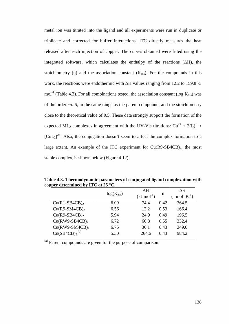
138
metal ion was titrated into the ligand and all experiments were run in duplicate or
triplicate and corrected for buffer interactions. ITC directly measures the heat
released after each injection of copper. The curves obtained were fitted using the
integrated software, which calculates the enthalpy of the reactions (5H), the
stoichiometry (n) and the association constant (Kass). For the compounds in this
work, the reactions were endothermic with 5H values ranging from 12.2 to 159.8 kJ
mol-1 (Table 4.3). For all combinations tested, the association constant (log Kass) was
of the order ca. 6, in the same range as the parent compound, and the stoichiometry
close to the theoretical value of 0.5. These data strongly support the formation of the
expected ML2 complexes in agreement with the UV-Vis titrations: Cu2+ + 2(L) .
[CuL2]2+. Also, the conjugation doesn’t seem to affect the complex formation to a
large extent. An example of the ITC experiment for Cu(R9-SB4CB)2, the most
stable complex, is shown below (Figure 4.12).
Table 4.3. Thermodynamic parameters of conjugated ligand complexation with copper determined by ITC at 25 °C.
log(Kass) 5H
(kJ mol-1) n 5S (J mol-1K-1)
Cu(R1-SB4CB)2 6.00 74.4 0.42 364.5 Cu(R9-SM4CB)2 6.56 12.2 0.53 166.4 Cu(R9-SB4CB)2 5.94 24.9 0.49 196.5 Cu(RW9-SB4CB)2 6.72 60.8 0.55 332.4 Cu(RW9-SM4CB)2 6.75 36.1 0.43 249.0 Cu(SB4CB)2
[a] 5.30 264.6 0.43 984.2 [a] Parent compounds are given for the purpose of comparison.
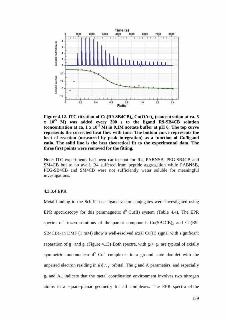
139
Figure 4.12. ITC titration of Cu(R9-SB4CB)2, Cu(OAc)2 (concentration at ca. 5 x 10⁻⁵ M) was added every 300 s to the ligand R9-SB4CB solution (concentration at ca. 1 x 10⁻⁵ M) in 0.1M acetate buffer at pH 6.. The top curve represents the corrected heat flow with time. The bottom curve represents the heat of reaction (measured by peak integration) as a function of Cu/ligand ratio. The solid line is the best theoretical fit to the experimental data. The three first points were removed for the fitting. Note: ITC experiments had been carried out for R4, PABNSB, PEG-SB4CB and SM4CB but to no avail. R4 suffered from peptide aggregation while PABNSB, PEG-SB4CB and SM4CB were not sufficiently water soluble for meaningful investigations.
4.3.3.4 EPR
Metal binding to the Schiff base ligand-vector conjugates were investigated using
EPR spectroscopy for this paramagnetic d9 Cu(II) system (Table 4.4). The EPR
spectra of frozen solutions of the parent compounds Cu(SB4CB)2 and Cu(R9-
SB4CB)2 in DMF (1 mM) show a well-resolved axial Cu(II) signal with significant
separation of g" and g! (Figure 4.13). Both spectra, with g!"> g" are typical of axially
symmetric mononuclear d9 CuII complexes in a ground state doublet with the
unpaired electron residing in a dx2- y2 orbital. The g and A parameters, and especially
g! and A!, indicate that the metal coordination environment involves two nitrogen
atoms in a square-planar geometry for all complexes. The EPR spectra of the
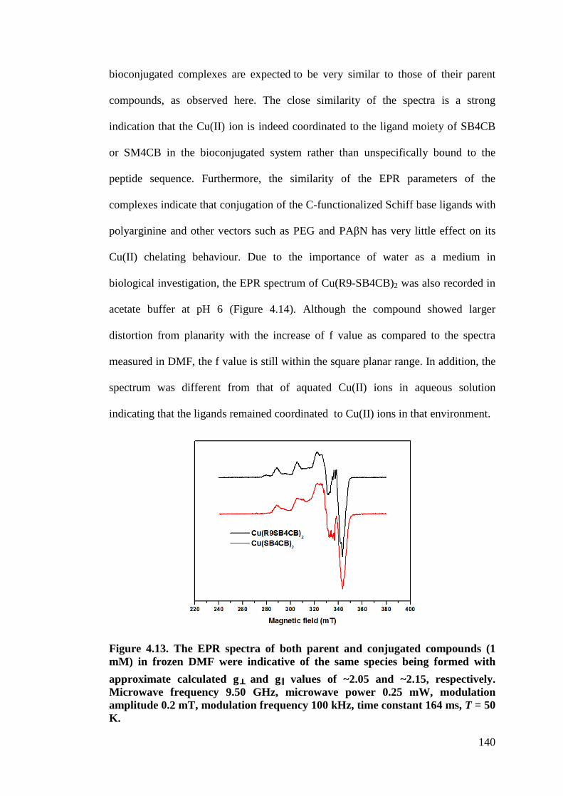
140
bioconjugated complexes are expected to be very similar to those of their parent
compounds, as observed here. The close similarity of the spectra is a strong
indication that the Cu(II) ion is indeed coordinated to the ligand moiety of SB4CB
or SM4CB in the bioconjugated system rather than unspecifically bound to the
peptide sequence. Furthermore, the similarity of the EPR parameters of the
complexes indicate that conjugation of the C-functionalized Schiff base ligands with
polyarginine and other vectors such as PEG and PA%N has very little effect on its
Cu(II) chelating behaviour. Due to the importance of water as a medium in
biological investigation, the EPR spectrum of Cu(R9-SB4CB)2 was also recorded in
acetate buffer at pH 6 (Figure 4.14). Although the compound showed larger
distortion from planarity with the increase of f value as compared to the spectra
measured in DMF, the f value is still within the square planar range. In addition, the
spectrum was different from that of aquated Cu(II) ions in aqueous solution
indicating that the ligands remained coordinated to Cu(II) ions in that environment.
Figure 4.13. The EPR spectra of both parent and conjugated compounds (1 mM) in frozen DMF were indicative of the same species being formed with approximate calculated g and g!!!! values of ~2.05 and ~2.15, respectively. Microwave frequency 9.50 GHz, microwave power 0.25 mW, modulation amplitude 0.2 mT, modulation frequency 100 kHz, time constant 164 ms, T = 50 K.
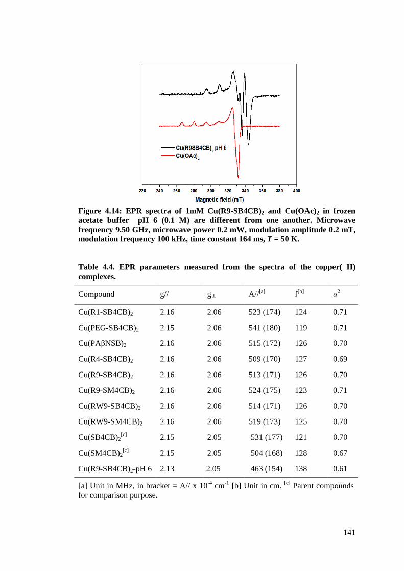
141
Figure 4.14: EPR spectra of 1mM Cu(R9-SB4CB)2 and Cu(OAc)2 in frozen acetate buffer pH 6 (0.1 M) are different from one another. Microwave frequency 9.50 GHz, microwave power 0.2 mW, modulation amplitude 0.2 mT, modulation frequency 100 kHz, time constant 164 ms, T = 50 K. Table 4.4. EPR parameters measured from the spectra of the copper( II) complexes.
Compound g// g A//[a] f[b] $2
Cu(R1-SB4CB)2 2.16 2.06 523 (174) 124 0.71
Cu(PEG-SB4CB)2 2.15 2.06 541 (180) 119 0.71
Cu(PA%NSB)2 2.16 2.06 515 (172) 126 0.70
Cu(R4-SB4CB)2 2.16 2.06 509 (170) 127 0.69
Cu(R9-SB4CB)2 2.16 2.06 513 (171) 126 0.70
Cu(R9-SM4CB)2 2.16 2.06 524 (175) 123 0.71
Cu(RW9-SB4CB)2 2.16 2.06 514 (171) 126 0.70
Cu(RW9-SM4CB)2 2.16 2.06 519 (173) 125 0.70
Cu(SB4CB)2[c] 2.15 2.05 531 (177) 121 0.70
Cu(SM4CB)2[c] 2.15 2.05 504 (168) 128 0.67
Cu(R9-SB4CB)2-pH 6 2.13 2.05 463 (154) 138 0.61
[a] Unit in MHz, in bracket = A// x 10-4 cm-1 [b] Unit in cm. [c] Parent compounds for comparison purpose.
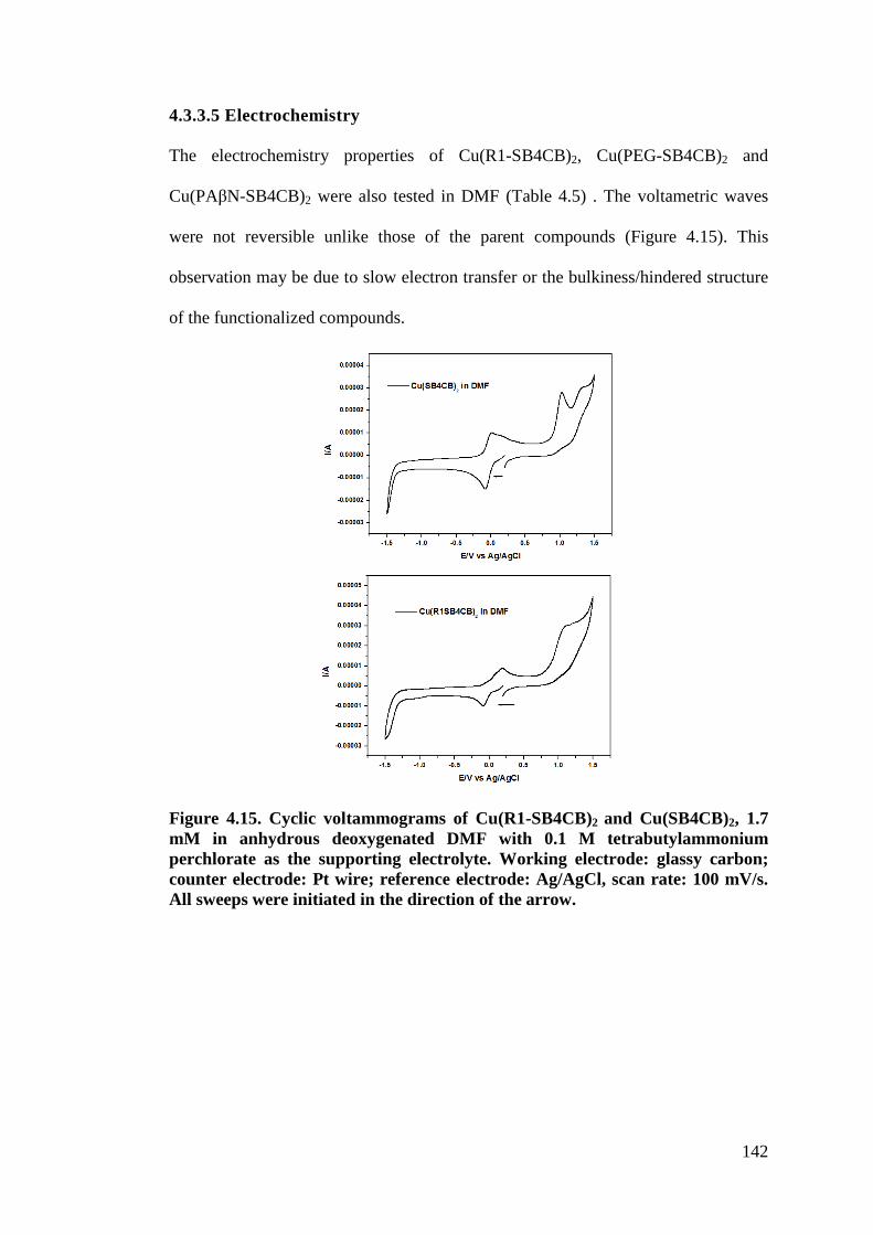
142
4.3.3.5 Electrochemistry
The electrochemistry properties of Cu(R1-SB4CB)2, Cu(PEG-SB4CB)2 and
Cu(PA%N-SB4CB)2 were also tested in DMF (Table 4.5) . The voltametric waves
were not reversible unlike those of the parent compounds (Figure 4.15). This
observation may be due to slow electron transfer or the bulkiness/hindered structure
of the functionalized compounds.
Figure 4.15. Cyclic voltammograms of Cu(R1-SB4CB)2 and Cu(SB4CB)2, 1.7 mM in anhydrous deoxygenated DMF with 0.1 M tetrabutylammonium perchlorate as the supporting electrolyte. Working electrode: glassy carbon; counter electrode: Pt wire; reference electrode: Ag/AgCl, scan rate: 100 mV/s. All sweeps were initiated in the direction of the arrow.
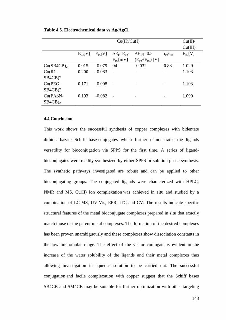
143
Table 4.5. Electrochemical data vs Ag/AgCl.
4.4 Conclusion
This work shows the successful synthesis of copper complexes with bidentate
dithiocarbazate Schiff base-conjugates which further demonstrates the ligands
versatility for bioconjugation via SPPS for the first time. A series of ligand-
bioconjugates were readily synthesized by either SPPS or solution phase synthesis.
The synthetic pathways investigated are robust and can be applied to other
bioconjugating groups. The conjugated ligands were characterized with HPLC,
NMR and MS. Cu(II) ion complexation was achieved in situ and studied by a
combination of LC-MS, UV-Vis, EPR, ITC and CV. The results indicate specific
structural features of the metal bioconjugate complexes prepared in situ that exactly
match those of the parent metal complexes. The formation of the desired complexes
has been proven unambiguously and these complexes show dissociation constants in
the low micromolar range. The effect of the vector conjugate is evident in the
increase of the water solubility of the ligands and their metal complexes thus
allowing investigation in aqueous solution to be carried out. The successful
conjugation and facile complexation with copper suggest that the Schiff bases
SB4CB and SM4CB may be suitable for further optimization with other targeting
Cu(II)/Cu(I) Cu(II)/ Cu(III)
Epa[V] Epc[V] 2Ep=Epa-Epc[mV]
2E1/2=0.5 (Epa+Epc) [V]
ipa/ipc Epa[V]
Cu(SB4CB)2 0.015 -0.079 94 -0.032 0.88 1.029 Cu(R1-SB4CB)2
0.200 -0.083 - - - 1.103
Cu(PEG-SB4CB)2
0.171 -0.098 - - - 1.103
Cu(PA%N-SB4CB)2
0.193 -0.082 - - - 1.090
144
groups. Such optimization is anticipated to result in enhanced biological properties
as well as offering the possibility of being utilized for radioimaging. In addition,
only two different substituents on the S-dithiocarbazate moiety (a methyl and a
benzyl) were investigated in this work but many others can be introduced in the
same way, which would potentially give access to a larger library of complexes. All
the compounds presented here were further evaluated for their potential
antimicrobial activity which will be discussed in the next chapter.

145
CHAPTER 5
BIOLOGICAL ACTIVITIES
5.1 Introduction
5.1.1 Mechanism of action of antimicrobial agents and multi-drug resistance
Bacteria could be broadly classified into Gram-positive and Gram-negative groups
based on the outcome of Gram staining. The major difference between both groups
is the structural organization outside the plasma membrane as shown in Figure 5.1
(Lolis and Bucala, 2003). According to Salton and Kim (1996), “most Gram-
positive bacteria have a thick (20 to 80 nm) and continuous cell wall which is
composed largely of peptidoglycan”. The peptidoglycan is linked covalently to
“other cell wall polymers such as the teichoic acids, polysaccharides, and
peptidoglycolipids”. On the contrary, Gram-negative bacteria have a thin (5 nm to
10 nm) peptidoglycan layer and another outer membrane structure outside the layer.
In addition, lipoprotein molecules can be found in between the membrane structure
and peptidoglycan layer while the lipopolysaccharides (LPS) are on the exterior
surface of the outer membrane structure.
During an infection, “bacterial cells grow and divide, replicating repeatedly to reach
large numbers”. In order to do so, bacteria “must synthesize or take up many types
of biomolecules” causing cell deaths and disruption of cell function of the host
organisms (Neu and Gootz, 1996). Bacteria could also promote infection and disease
by producing toxins that can exert their effects directly on a target cell or by
disabling the immune system (Deisingh and Thompson, 2002).
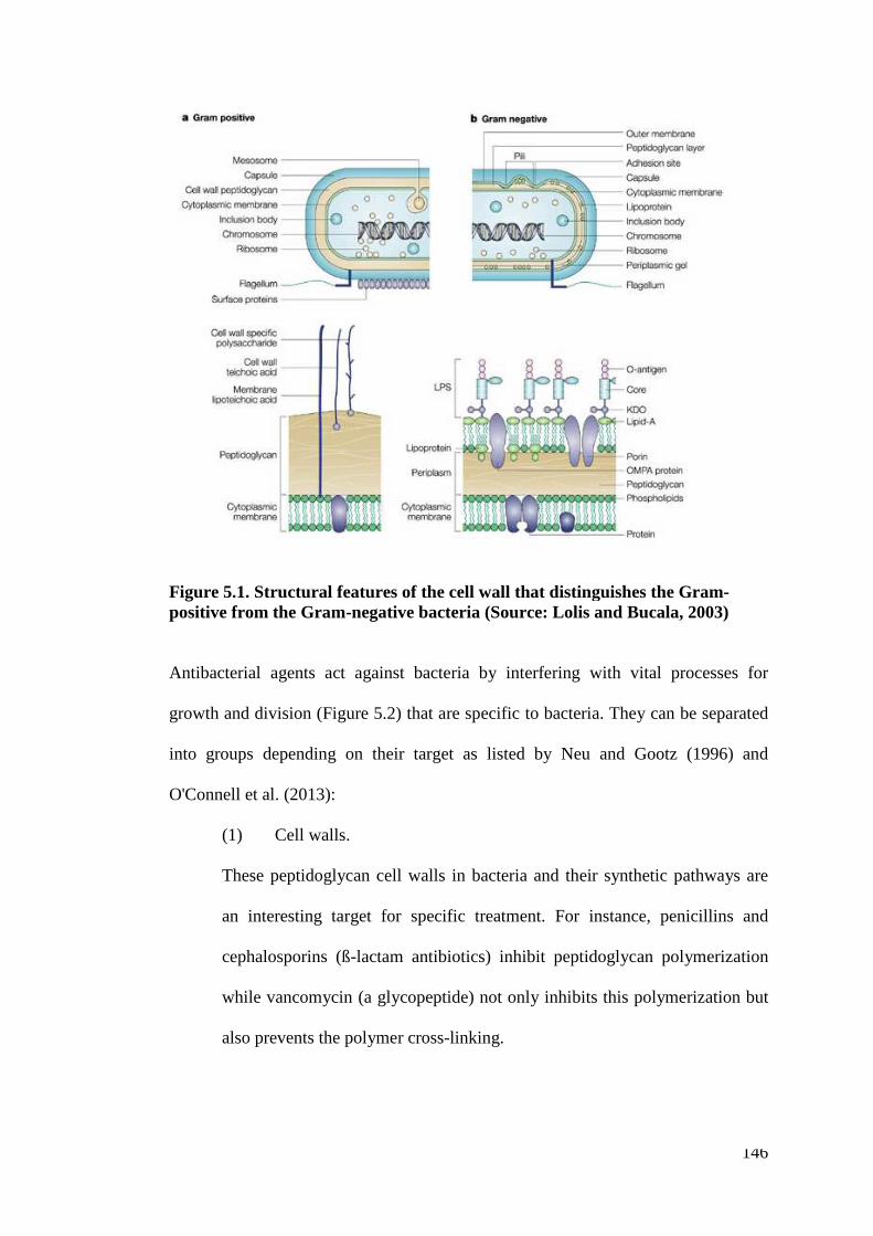
146
Figure 5.1. Structural features of the cell wall that distinguishes the Gram-positive from the Gram-negative bacteria (Source: Lolis and Bucala, 2003)
Antibacterial agents act against bacteria by interfering with vital processes for
growth and division (Figure 5.2) that are specific to bacteria. They can be separated
into groups depending on their target as listed by Neu and Gootz (1996) and
O'Connell et al. (2013):
(1) Cell walls.
These peptidoglycan cell walls in bacteria and their synthetic pathways are
an interesting target for specific treatment. For instance, penicillins and
cephalosporins (ß-lactam antibiotics) inhibit peptidoglycan polymerization
while vancomycin (a glycopeptide) not only inhibits this polymerization but
also prevents the polymer cross-linking.
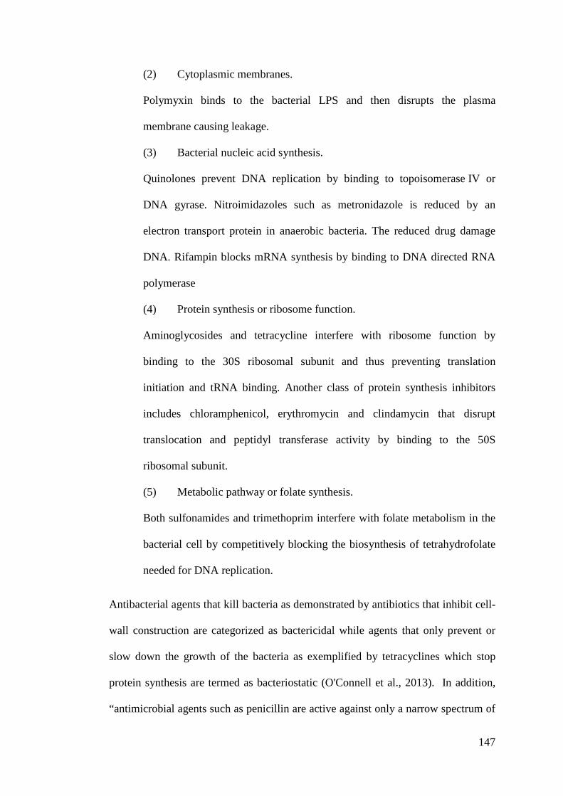
147
(2) Cytoplasmic membranes.
Polymyxin binds to the bacterial LPS and then disrupts the plasma
membrane causing leakage.
(3) Bacterial nucleic acid synthesis.
Quinolones prevent DNA replication by binding to topoisomerase!IV or
DNA gyrase. Nitroimidazoles such as metronidazole is reduced by an
electron transport protein in anaerobic bacteria. The reduced drug damage
DNA. Rifampin blocks mRNA synthesis by binding to DNA directed RNA
polymerase
(4) Protein synthesis or ribosome function.
Aminoglycosides and tetracycline interfere with ribosome function by
binding to the 30S ribosomal subunit and thus preventing translation
initiation and tRNA binding. Another class of protein synthesis inhibitors
includes chloramphenicol, erythromycin and clindamycin that disrupt
translocation and peptidyl transferase activity by binding to the 50S
ribosomal subunit.
(5) Metabolic pathway or folate synthesis.
Both sulfonamides and trimethoprim interfere with folate metabolism in the
bacterial cell by competitively blocking the biosynthesis of tetrahydrofolate
needed for DNA replication.
Antibacterial agents that kill bacteria as demonstrated by antibiotics that inhibit cell-
wall construction are categorized as bactericidal while agents that only prevent or
slow down the growth of the bacteria as exemplified by tetracyclines which stop
protein synthesis are termed as bacteriostatic (O'Connell et al., 2013). In addition,
“antimicrobial agents such as penicillin are active against only a narrow spectrum of
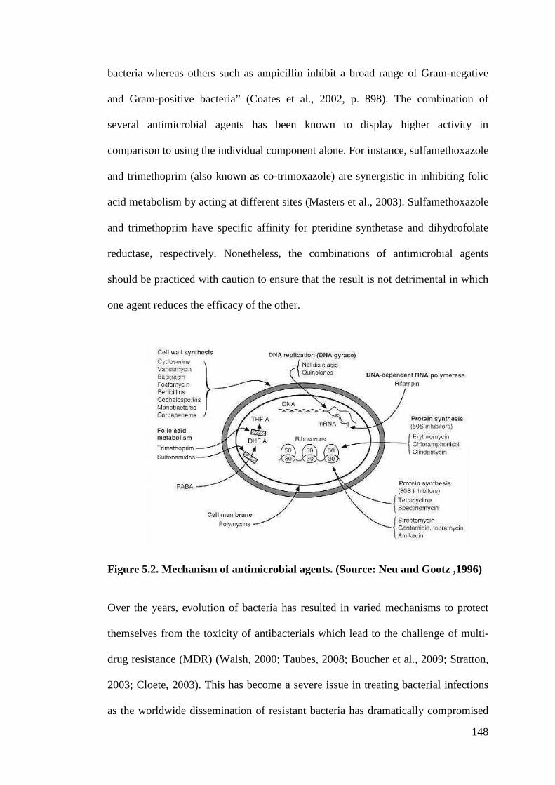
148
bacteria whereas others such as ampicillin inhibit a broad range of Gram-negative
and Gram-positive bacteria” (Coates et al., 2002, p. 898). The combination of
several antimicrobial agents has been known to display higher activity in
comparison to using the individual component alone. For instance, sulfamethoxazole
and trimethoprim (also known as co-trimoxazole) are synergistic in inhibiting folic
acid metabolism by acting at different sites (Masters et al., 2003). Sulfamethoxazole
and trimethoprim have specific affinity for pteridine synthetase and dihydrofolate
reductase, respectively. Nonetheless, the combinations of antimicrobial agents
should be practiced with caution to ensure that the result is not detrimental in which
one agent reduces the efficacy of the other.
Figure 5.2. Mechanism of antimicrobial agents. (Source: Neu and Gootz ,1996)
Over the years, evolution of bacteria has resulted in varied mechanisms to protect
themselves from the toxicity of antibacterials which lead to the challenge of multi-
drug resistance (MDR) (Walsh, 2000; Taubes, 2008; Boucher et al., 2009; Stratton,
2003; Cloete, 2003). This has become a severe issue in treating bacterial infections
as the worldwide dissemination of resistant bacteria has dramatically compromised
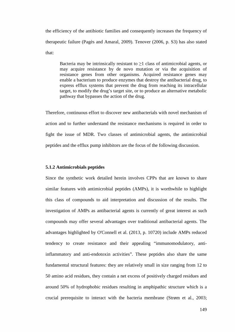
149
the efficiency of the antibiotic families and consequently increases the frequency of
therapeutic failure (Pagès and Amaral, 2009). Tenover (2006, p. S3) has also stated
that:
Bacteria may be intrinsically resistant to 61 class of antimicrobial agents, or may acquire resistance by de novo mutation or via the acquisition of resistance genes from other organisms. Acquired resistance genes may enable a bacterium to produce enzymes that destroy the antibacterial drug, to express efflux systems that prevent the drug from reaching its intracellular target, to modify the drug’s target site, or to produce an alternative metabolic pathway that bypasses the action of the drug.
Therefore, continuous effort to discover new antibacterials with novel mechanism of
action and to further understand the resistance mechanisms is required in order to
fight the issue of MDR. Two classes of antimicrobial agents, the antimicrobial
peptides and the efflux pump inhibitors are the focus of the following discussion.
5.1.2 Antimicrobials peptides
Since the synthetic work detailed herein involves CPPs that are known to share
similar features with antimicrobial peptides (AMPs), it is worthwhile to highlight
this class of compounds to aid interpretation and discussion of the results. The
investigation of AMPs as antibacterial agents is currently of great interest as such
compounds may offer several advantages over traditional antibacterial agents. The
advantages highlighted by O'Connell et al. (2013, p. 10720) include AMPs reduced
tendency to create resistance and their appealing “immunomodulatory, anti-
inflammatory and anti-endotoxin activities”. These peptides also share the same
fundamental structural features: they are relatively small in size ranging from 12 to
50 amino acid residues, they contain a net excess of positively charged residues and
around 50% of hydrophobic residues resulting in amphipathic structure which is a
crucial prerequisite to interact with the bacteria membrane (Strøm et al., 2003;
150
O'Connell et al., 2013). The phospholipid membrane of bacterial cells is the main
target for AMPs. As shown in Figure 5.3 (Zasloff, 2002), bacterial membranes differ
from the membranes of plants and mammalians in which the outer leaflet of the
bilayer that is exposed to the extracellular environment is composed mainly of
negatively charged lipids in bacteria whereas the leaflet in multicellular animal is
made of neutral lipids. Many hypotheses have therefore been presented for the
mechanism of action of AMPs (Brogden, 2005; Hancock and Lehrer, 1998)
including
fatal depolarization of the normally energized bacterial membrane, the creation of physical holes that cause cellular contents to leak out, the activation of deadly processes such as induction of hydrolases that degrade the cell wall, the scrambling of the usual distribution of lipids between the leaflets of the bilayer, resulting in disturbance of membrane functions, and the damaging of critical intracellular targets after internalization of the peptide (Zasloff, 2002, p. 390).
Figure 5.3. The membrane target of antimicrobial peptides of multicellular organisms and the basis of specificity (Source: Zasloff, 2002)
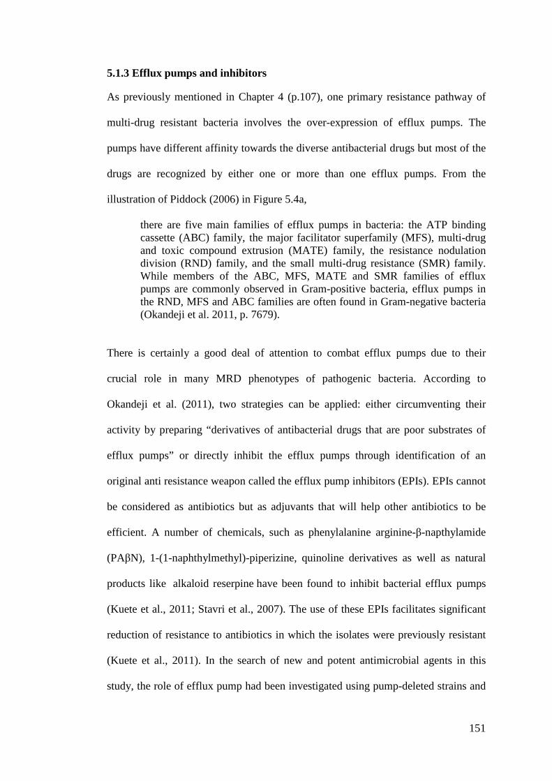
151
5.1.3 Efflux pumps and inhibitors
As previously mentioned in Chapter 4 (p.107), one primary resistance pathway of
multi-drug resistant bacteria involves the over-expression of efflux pumps. The
pumps have different affinity towards the diverse antibacterial drugs but most of the
drugs are recognized by either one or more than one efflux pumps. From the
illustration of Piddock (2006) in Figure 5.4a,
there are five main families of efflux pumps in bacteria: the ATP binding cassette (ABC) family, the major facilitator superfamily (MFS), multi-drug and toxic compound extrusion (MATE) family, the resistance nodulation division (RND) family, and the small multi-drug resistance (SMR) family. While members of the ABC, MFS, MATE and SMR families of efflux pumps are commonly observed in Gram-positive bacteria, efflux pumps in the RND, MFS and ABC families are often found in Gram-negative bacteria (Okandeji et al. 2011, p. 7679).
There is certainly a good deal of attention to combat efflux pumps due to their
crucial role in many MRD phenotypes of pathogenic bacteria. According to
Okandeji et al. (2011), two strategies can be applied: either circumventing their
activity by preparing “derivatives of antibacterial drugs that are poor substrates of
efflux pumps” or directly inhibit the efflux pumps through identification of an
original anti resistance weapon called the efflux pump inhibitors (EPIs). EPIs cannot
be considered as antibiotics but as adjuvants that will help other antibiotics to be
efficient. A number of chemicals, such as phenylalanine arginine-%-napthylamide
(PA%N), 1-(1-naphthylmethyl)-piperizine, quinoline derivatives as well as natural
products like alkaloid reserpine have been found to inhibit bacterial efflux pumps
(Kuete et al., 2011; Stavri et al., 2007). The use of these EPIs facilitates significant
reduction of resistance to antibiotics in which the isolates were previously resistant
(Kuete et al., 2011). In the search of new and potent antimicrobial agents in this
study, the role of efflux pump had been investigated using pump-deleted strains and
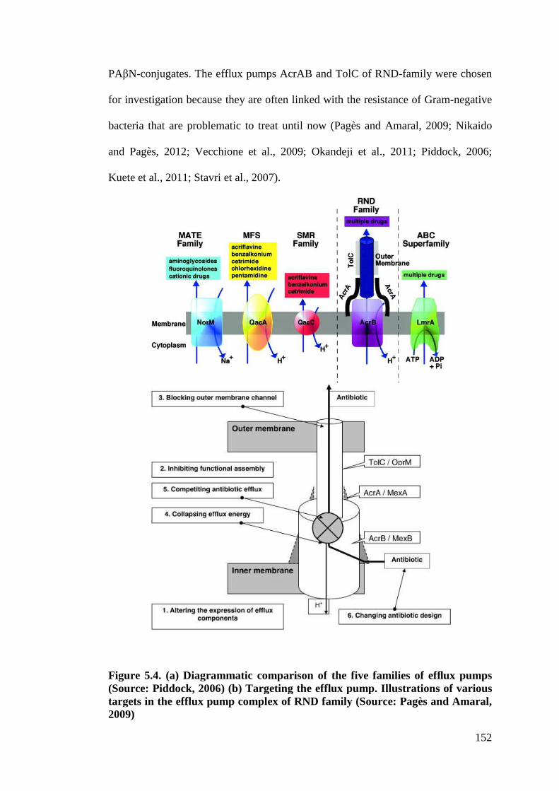
152
PA%N-conjugates. The efflux pumps AcrAB and TolC of RND-family were chosen
for investigation because they are often linked with the resistance of Gram-negative
bacteria that are problematic to treat until now (Pagès and Amaral, 2009; Nikaido
and Pagès, 2012; Vecchione et al., 2009; Okandeji et al., 2011; Piddock, 2006;
Kuete et al., 2011; Stavri et al., 2007).
Figure 5.4. (a) Diagrammatic comparison of the five families of efflux pumps (Source: Piddock, 2006) (b) Targeting the efflux pump. Illustrations of various targets in the efflux pump complex of RND family (Source: Pagès and Amaral, 2009)
153
5.1.4 Contribution of metal complexes to the improvement of antimicrobial
agents
The rich coordination geometry of metals offers an exciting platform for the
discovery of new and potent metallodrugs with novel mechanisms of action (Sadler:
N. J. Farrer and P. J. Sadler, 2011). These attributes may allow antibacterial metal
complexes to be less likely to induce resistance in bacteria or at least to delay
development of resistance. The geometrical arrangement of ligands in metal
complexes, which vary according to the number and types of ligands bound to the
metal centre, and to the coordination preference of the metal, affect their bioactivity
(Ng et al., 2013). In the past few decades, we have witnessed the development and
evolution of potential antibacterial agents derived from metal-based compounds
although the mechanisms of their action are still not well understood. Patra et al.
(2012b) reviewed work that explored organometallic derivatization of existing drugs
as an attractive approach to overcome the resistance issue. For instance, organo
ruthenium derivatives of quinolones (topoisomerase II enzyme inhibitor which
unwinds DNA and interferes with DNA replication) showed good activity and were
proven to have direct interaction with double-stranded DNA (Turel et al., 2010,
Chen et al., 2004). It is interesting to note that organometallic derivatives of
platensimycin (bacterial fatty acid biosynthesis inhibitor) appeared to completely
change the mechanism of action. The compound no longer inhibited FabF enzyme
but exhibited multi-causal death effect (Patra et al., 2009; Patra et al., 2012a). The
review also discussed metal-specific mode of action of potential metallodrugs
derived from arsenic, silver and gold ions. In some cases, it has been shown that the
activity of a metal-derivatized drug could be the sum of the activity of the original
drug and the inherent toxicity of the metal ion (Patra et al., 2012b). In reviews by
154
Beraldo and Gambinob (2004), and Pelosi (2010) the significant bioactivities of
Schiff base ligands derived from semicarbazones and thiosemicarbazones and their
metal complexes were demonstrated. Enhancement of activity upon complexation
was shown to be related to changes in either lipophilicity or redox effects with
possible mechanisms of action involving production of free radicals, metal complex
DNA interaction or inhibition of metalloenzymes. In very recent years, modification
of metallocenes with peptides has been shown to not only enhance activity but also
to alter the specificity of the compounds towards either Gram-positive or Gram-
negative bacteria. (Metzler-Nolte, 2010; Chantson et al., 2005; Chantson et al.,
2006). This strategy was also utilized by Jean-Marie Pages, Isabelle Artaud and
coworkers (Pagès et al., 2013) in preparing a multicomponent metal chelating group-
short antimicrobial peptide-fluorescent tag conjugate. The compound proved to be
active against multi-drug resistant clinical isolates while the imaging investigation
allowed determination of the localization and accumulation of the active compound
inside bacteria (Pagès et al., 2013). Previously, the team had developed highly
efficient inhibitors of bacterial metalloenzymes involved in the N-formyl-
methionine excision pathway of nascent polypeptides (Petit et al., 2009; Huguet et
al., 2012; Mamelli et al., 2009). They adopted the strategy of delivering the drug
inhibitor via a metal-chaperone (Alimi et al., 2012). Metallodrugs consist of two
parts: the drug, usually a hydroxamic acid, and an ancillary ligand as shown in
Figure 5.5 below. The mechanism of action involves dissociation of the
metallodrugs inside the bacteria promoting the delivery of the drug to its target
(Artaud et al., 2014).
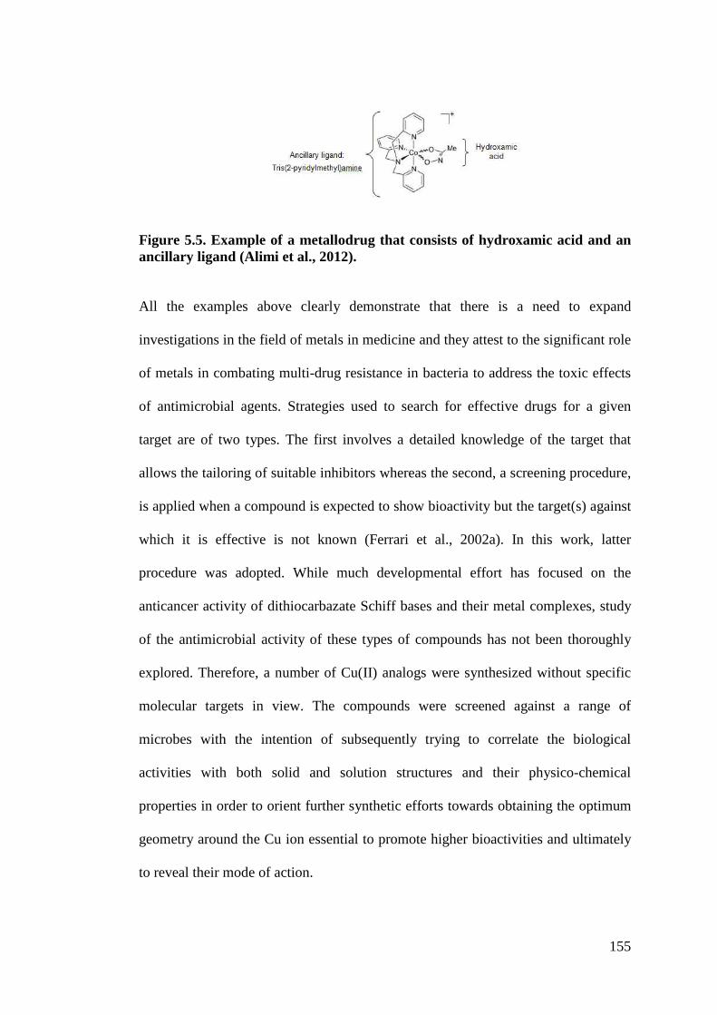
155
Figure 5.5. Example of a metallodrug that consists of hydroxamic acid and an ancillary ligand (Alimi et al., 2012).
All the examples above clearly demonstrate that there is a need to expand
investigations in the field of metals in medicine and they attest to the significant role
of metals in combating multi-drug resistance in bacteria to address the toxic effects
of antimicrobial agents. Strategies used to search for effective drugs for a given
target are of two types. The first involves a detailed knowledge of the target that
allows the tailoring of suitable inhibitors whereas the second, a screening procedure,
is applied when a compound is expected to show bioactivity but the target(s) against
which it is effective is not known (Ferrari et al., 2002a). In this work, latter
procedure was adopted. While much developmental effort has focused on the
anticancer activity of dithiocarbazate Schiff bases and their metal complexes, study
of the antimicrobial activity of these types of compounds has not been thoroughly
explored. Therefore, a number of Cu(II) analogs were synthesized without specific
molecular targets in view. The compounds were screened against a range of
microbes with the intention of subsequently trying to correlate the biological
activities with both solid and solution structures and their physico-chemical
properties in order to orient further synthetic efforts towards obtaining the optimum
geometry around the Cu ion essential to promote higher bioactivities and ultimately
to reveal their mode of action.
156
5.2 Methodology
5.2.1 Antimicrobial testing (MIC determination)
5.2.1.1 Bacterial strains, culture media and chemicals
The bacteria chosen for this study are listed in the Table 5.1 below. The
microorganisms studied included reference (from the American Type Culture
Collection) and clinical (Laboratory collection) strains of gram negative bacteria E.
coli, E. aerogenes, A. baumannii, K. pneumoniae, P. aeruginosa and Salmonella
enterica (S. enterica) serotype Typhimurium as well as gram positive S. aureus.
EA289 is an E. aerogenes KANS (sensitive to kanamycin, MDR isolate that exhibits
active efflux of norfloxacin and efflux pump overproduction), EA298 constructed
from EA289 is deleted of TolC (Pradel and Pagès, 2002). AG100 is an E. coli Wild
Type (WT) and AG100A is its KANR (resistant to kanamycin) derivative, deleted of
AcrAB and hypersensitive to chloramphenicol, tetracycline, ampicillin and nalidixic
acid (Viveiros et al., 2005). Strains were grown at 37°C on Mueller-Hinton medium
24 h prior to any assay. Mueller-Hinton broth (MHB) was used for the susceptibility
test. Chemicals polymyxin!B nonapeptide (PMBN) were obtained from Sigma-
Aldrich and the culture medium was purchased from Becton Dickinson.
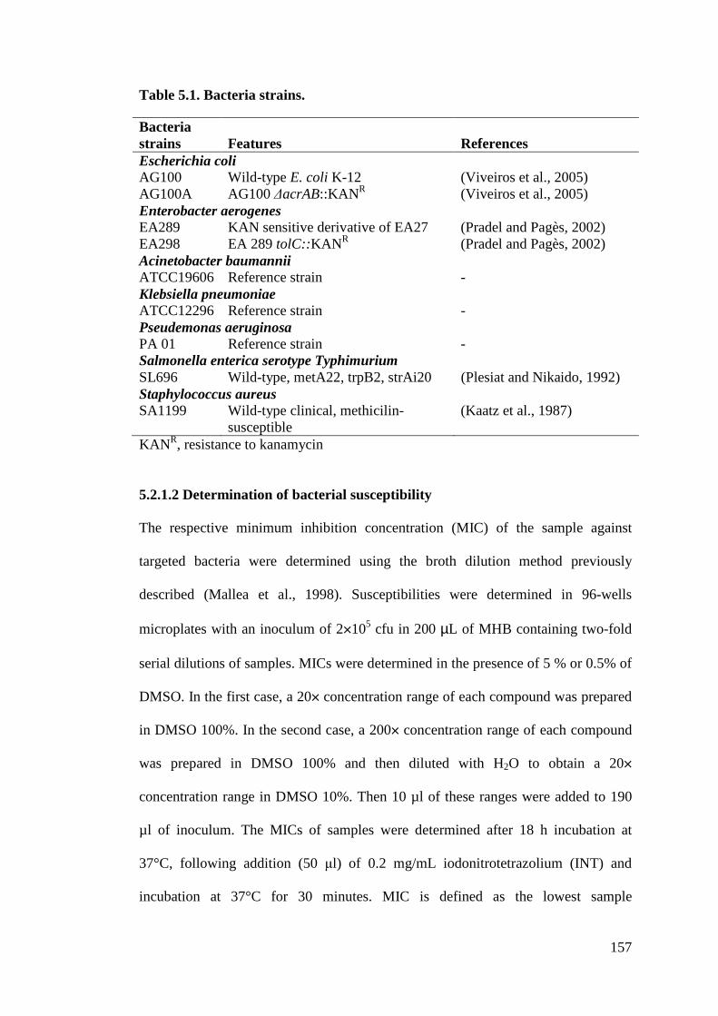
157
Table 5.1. Bacteria strains.
KANR, resistance to kanamycin
5.2.1.2 Determination of bacterial susceptibility
The respective minimum inhibition concentration (MIC) of the sample against
targeted bacteria were determined using the broth dilution method previously
described (Mallea et al., 1998). Susceptibilities were determined in 96-wells
microplates with an inoculum of 2!105 cfu in 200 µL of MHB containing two-fold
serial dilutions of samples. MICs were determined in the presence of 5 % or 0.5% of
DMSO. In the first case, a 20! concentration range of each compound was prepared
in DMSO 100%. In the second case, a 200! concentration range of each compound
was prepared in DMSO 100% and then diluted with H2O to obtain a 20!
concentration range in DMSO 10%. Then 10 µl of these ranges were added to 190
µl of inoculum. The MICs of samples were determined after 18 h incubation at
37°C, following addition (50 !l) of 0.2 mg/mL iodonitrotetrazolium (INT) and
incubation at 37°C for 30 minutes. MIC is defined as the lowest sample
Bacteria strains Features References Escherichia coli AG100 Wild-type E. coli K-12 (Viveiros et al., 2005) AG100A AG100 %acrAB::KANR (Viveiros et al., 2005) Enterobacter aerogenes EA289 KAN sensitive derivative of EA27 (Pradel and Pagès, 2002) EA298 EA 289 tolC::KANR (Pradel and Pagès, 2002) Acinetobacter baumannii ATCC19606 Reference strain - Klebsiella pneumoniae ATCC12296 Reference strain - Pseudemonas aeruginosa PA 01 Reference strain - Salmonella enterica serotype Typhimurium SL696 Wild-type, metA22, trpB2, strAi20 (Plesiat and Nikaido, 1992) Staphylococcus aureus SA1199 Wild-type clinical, methicilin-
susceptible (Kaatz et al., 1987)
158
concentration that prevented the color change of the medium and exhibited complete
inhibition of microbial growth. The range of samples concentration used for MIC
determination is universally accepted to be in doubling dilution steps up and down
from 1 mg/L as required (Andrews, 2001). Therefore, the sample dilution range was
from 0-128 µM. Samples were tested alone or in the presence of PMBN at 51.2
mg/L final concentration (1/5 of its direct MIC). All assays were performed in
duplicate or triplicate. Ciprofloxafin was used as standard antibiobic reference.
5.2.2 In vitro cytotoxicity testing
The cell lines used for testing, MCF-7 (human breast cancer cells possessing nuclear
esstrogen receptor) and MDA-MB-231 (human breast cancer cells without nuclear
estrogen receptor) were obtained from the National Cancer Institute, U.S.A. Both
cell lines were cultured in RPMI-1640 / DMEM (High glucose) (Sigma) medium
supplemented with 10% fetal calf serum. The cells were plated into 96-well plate at
cell density 6000 cells/well and incubated for 24 hours. After 24 hours, the media
(5% serum) were discarded and cells rinsed with PBS solution. 200 µL of a series of
concentrations (50.0, 25.0, 10.0, 5.0, 1.0 and 0.5 µM) for each sample prepared was
added to each well. The 96-well plate was incubated for another 72 hr, after which
the well plate was removed from incubator. Cytotoxicity was determined using the
microtitration of 3-(4,5-dimethylthiazol-2-yl)-2,5-diphenyltetrazolium bromide
(MTT) assay (Sigma, USA) as reported by Mosmann (1983). 20 µL of MTT
solution (5 mg/mL) was added to each well. The plate was wrapped with aluminium
foil and incubated for 4 hours. After 4 hours, 200 µL of sample containing MTT
solution was discarded from the well. 200 µL of DMSO was added to each well to
dissolve the formazan crystals formed. The effect of the compound on cell line
159
viability was measured on an automated spectrophotometric plate reader (model
MRX II microplate Elisa reader) at a test wavelength of 570 nm. Cytotoxicity was
expressed as IC50, i.e. the concentration that reduced the absorbance of treated cells
by 50% with reference to the control (untreated cells). The IC50 values were
determined from the plotted absorbance data for the dose-response curves. Controls
that contained only cells were included for each sample. Tamoxifen was used as the
cytotoxic standard.
5.3 Results and discussion
The free Schiff bases and their Cu(II) complexes as well as their bioconjugates
described in the previous chapters were tested in collaboration with Dr. Laure
Maigre and Professor Jean-Marie Pagès of Facultés de Médecine (UMR-MD1),
Université de la Méditerranée, Marseille, France for the compounds ability to inhibit
the growth of nine strains of both gram-negative and gram-positive bacteria. The
effects of membrane permeabilizing agent (PMBN) and efflux pumps were
investigated in an attempt to link the activity of these compounds with the bacteria
penetration and the detoxification mechanisms of bacteria. Such investigations
constitute the pioneering effort with these dithiocarbazate derivatives.
5.3.1 Antimicrobial evaluation
5.3.1.1 Macroacyclic Cu(II) system with tetradentate NNSS ligands
The initial synthesis and MIC evaluation involved tetradentate bis(dithiocarbazate)
ligands and their respective Cu(II) complexes as shown in Table 5.2. As noted in
previous chapters, a major drawback of these dithiocarbazate-Schiff bases is their
poor solubility in aqueous solution. To ensure complete dissolution of the tested
160
compounds, up to 5% (v:v) of DMSO was used. Indeed, it is not ideal since DMSO
could be toxic to bacteria at such a high concentration and/or could modify the
membrane permeability (Yu and Quinn, 1994), thus affecting the apparent MIC
values.
DMSO effect
It has been shown that solutions from 1% to 10% DMSO considerably affect the
growth of fungi and cancerous cells, and with a 15% solution, growth of certain
bacteria is effectively eliminated (Notman et al., 2006; Yu and Quinn, 1998; Ng et
al., 2013). This is also the case for bacteria strains A. Baumannii and P. Aeroginosa
at 5% of DMSO thus preventing the determination of the MIC under this condition.
DMSO has also been reported to enhance permeability of the lipid membrane and
also to cause the cell membrane to become less rigid facilitating membrane diffusion
of exogenous species (Randhawa, 2006; Ghajar and Harmon, 1968; Anselet al.,
1969; Dolan et al., 2013). As shown below (Figure 5.6), in comparison to 5%
DMSO, 0.5% DMSO would not significantly affect the growth of the bacteria as
compared to 5% DMSO. Therefore, it is important to keep in mind that DMSO is
not necessarily innocent.
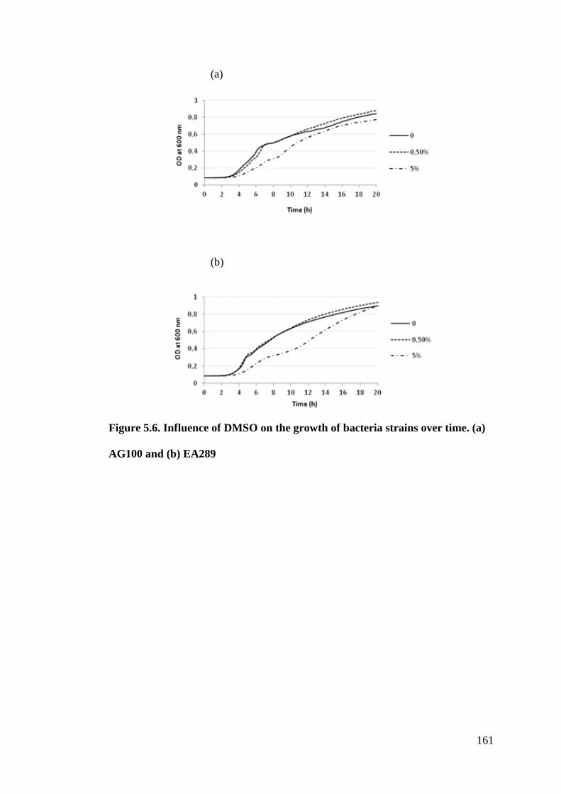
161
(a)
(b)
Figure 5.6. Influence of DMSO on the growth of bacteria strains over time. (a)
AG100 and (b) EA289
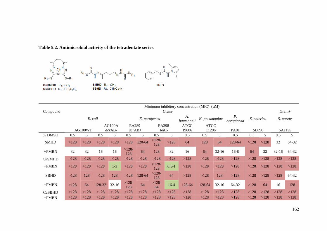
162
Table 5.2. Antimicrobial activity of the tetradentate series.
Minimum inhibitory concentration (MIC) (µM) Compound Gram- Gram+
E. coli E. aerogenes A. baumannii K. pneumoniae P.
aeruginosa S. enterica S. aureus
AG100WT AG100A acrAB-
EA289 acrAB+
EA298 tolC-
ATCC 19606
ATCC 11296 PA01 SL696 SA1199
% DMSO 0.5 5 0.5 5 0.5 5 0.5 5 0.5 0.5 5 0.5 0.5 5 0.5 5
SMHD >128 >128 >128 >128 >128 128-64 >128-128 >128 64 128 64 128-64 >128 >128 32 64-32
+PMBN 32 32 16 16 >128-128 64 128 32 16 64 32-16 16-8 64 32 32-16 64-32
CuSMHD >128 >128 >128 >128 >128 >128 >128 >128 >128 >128 >128 >128 >128 >128 >128 >128
+PMBN >128 >128 >128 1-2 >128 >128 >128-128 0.5-1 >128 >128 >128 >128 >128 >128 >128 >128
SBHD >128 128 >128 128 >128 128-64 >128-128 64 >128 >128 128 >128 >128 >128 >128 64-32
+PMBN >128 64 128-32 32-16 >128-128 64 >128-
64 16-4 128-64 128-64 32-16 64-32 >128 64 16 128
CuSBHD >128 >128 >128 >128 >128 >128 >128 >128 >128 >128 >128 >128 >128 >128 >128 >128 +PMBN >128 >128 >128 >128 >128 >128 >128 >128 >128 >128 >128 >128 >128 >128 >128 >128
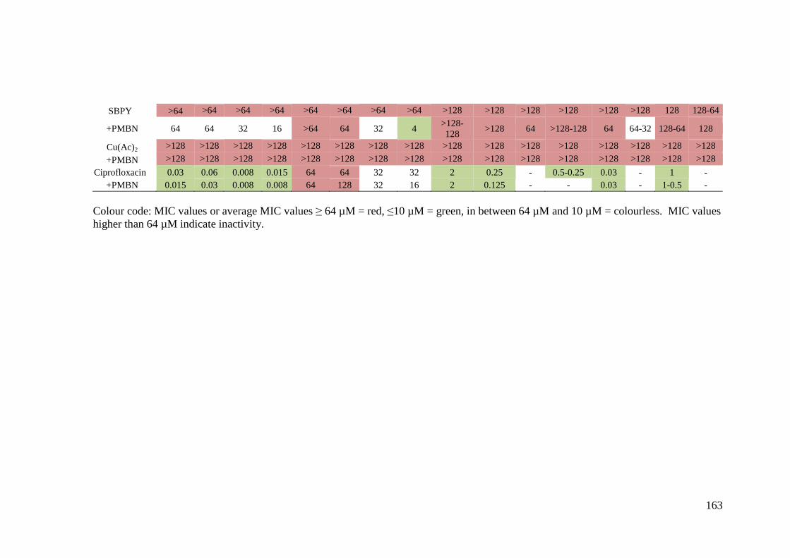
163
Colour code: MIC values or average MIC values 6 64 µM = red, "10 µM = green, in between 64 µM and 10 µM = colourless. MIC values higher than 64 µM indicate inactivity.
SBPY >64 >64 >64 >64 >64 >64 >64 >64 >128 >128 >128 >128 >128 >128 128 128-64
+PMBN 64 64 32 16 >64 64 32 4 >128-128 >128 64 >128-128 64 64-32 128-64 128
Cu(Ac)2 >128 >128 >128 >128 >128 >128 >128 >128 >128 >128 >128 >128 >128 >128 >128 >128 +PMBN >128 >128 >128 >128 >128 >128 >128 >128 >128 >128 >128 >128 >128 >128 >128 >128
Ciprofloxacin 0.03 0.06 0.008 0.015 64 64 32 32 2 0.25 - 0.5-0.25 0.03 - 1 - +PMBN 0.015 0.03 0.008 0.008 64 128 32 16 2 0.125 - - 0.03 - 1-0.5 -
164
Differences could be observed among the MIC values obtained with different
concentrations of DMSO (0.5% or 5%) for certain molecules, particularly for
CuSMHD against the mutated strains E. coli acrAB- and E. aerogenes TolC-. For
this molecule, the MICs obtained with concentrations of DMSO, 2.5%, 1.5% and
1% were all > 128 µM while with 5% DMSO, the MIC value was in the range 1-2
and 0.5-1 µM, respectively. Because of the effect of DMSO on bacterial growth, it
was not possible to confirm that the value truly reflects specific antimicrobial
activity of the compound itself but it more likely corresponds to a synergetic effect
of DMSO and the compound. The MIC values at 0.5% DMSO are more significant
and will be discussed. However, in 0.5% DMSO, partial precipitation of the
compounds could not be excluded which would lead to the determination of
overestimated MIC values i.e. underestimation of the biological effect at the
apparent concentration.
Influence of membrane permeabilizing agent on bioactivity
Since it has been reported that the low permeability of the outer membrane (Stratton,
2003; Cloete, 2003; Tenover, 2006; Strøm et al., 2003; Zasloff, 2002; Pagès and
Amaral, 2009; Nikaido and Pagès, 2012) is a prime factor limiting intracellular
activity of potential antimicrobial compounds, it is expected that the presence of a
membrane permeabilizing agent would act synergistically with the compounds under
study to promote their antimicrobial efficiency by facilitating increased uptake of the
compounds. Among the permeabilizing agents, Polymyxin B nonapeptide (PMBN)
has been used. PMBN is a cationic cyclic peptide derived from the antibacterial
peptide polymyxin B (PMB). PMBN is an extremely poor antimicrobial agent but it
is still capable of binding to lipid A of Gram-negative bacteria lipopolysaccharide
165
(LPS) like its parent compound, rendering the bacteria susceptible to various
hydrophobic antibiotics. This capacity of PMBN to bind to bacteria with relatively
high affinity and to permeabilize their outer membrane is often referred to as
“sensitizing activity” and points to a novel therapeutic direction (Tsubery et al.,
2000 and Tsubery et al., 2001). The compounds were therefore tested in the
presence and absence of sub-inhibitory concentrations of PMBN (used at 1/5 its
direct MIC value). Without PMBN all compounds were inactive against the strains
tested (MIC > 64 µM) except for the ligands SMHD and SBHD against S. aureus.
However, a significant increase of up to 3-fold improvement of MIC values on both
Gram-negative and Gram-positive bacteria was observed for the organic compounds
SMHD, SBHD and SBDP in presence of PMBN. These results strongly suggest that
the compounds, although toxic, do not penetrate the bacteria membrane efficiently.
This lack of penetration hinders their intrinsic toxicity and leads to an apparent high
MIC.
Efflux of the compounds
The role of efflux pumps was investigated using pump-deleted strains of Gram-
negative E. coli and E. aerogenes. SMHD also seems more active (16 µM) towards
the isogenic derived strain, in which the efflux pump AcrAB genes are deleted as
compared to wild-type E. coli (64 µM). No significant activity was observed for
SMHD in the absence likewise in presence of efflux pump for E. aerogenes. SBPY
show differences in the MDR clinical isolate EA289 overexpressing the AcrAB
efflux pump and on its efflux negative TolC- derivative EA298 with improvement in
MIC from >64 µM to 32 µM. These results confirmed that SBPY is recognized by
the efflux pumps and expelled from the bacteria thus limiting their bioactivity.
166
Effect of the dithiocarbazate substituent
Nevertheless, the ligand SMHD showed a broad range of moderate activity in the
presence of PMBN with the most promising MIC values at or around 16 µM against
E.Coli acrAB-, A. Baumannii, P. aeroginosa and S. aureus thus making it a potential
candidate for improvement. It is known that the biological activity of dithiocarbazate
compounds can be greatly modified in the presence of different substituents. For
instance, inhibition of E. coli and S. aureus by the Schiff base prepared from 2-
benzoylpyridine with SMDTC is highly effective whereas that of the SBDTC
compound shows no activity (Hossain et al., 1996).
Differences in activity against gram-positive and gram-negative bacteria
Apart from that, both SMHD and SBHD were moderately active against gram
positive S. aureus. Typically, antibacterial molecules are more active toward Gram-
positive than Gram-negative bacteria (Lessa et al., 2012; Bolla et al., 2011), as the
additional outer membrane of the latter organisms impairs or slow down the drug
uptake, which could be the case here.
Effect of complexation
Contrary to what has been usually reported in the literature that metal complexation
enhances the bioactivity of ligands (Nandi et al., 1984; Joseph et al., 2012), in this
case, the formation of the copper complexes induces a loss of antibacterial potency
of the compounds. Similar losses in activity were previously reported with Pd(II)
and Pt(II) complexes with acetone Schiff bases (Ali et al., 2002). The observation
for this tetradentate series of compounds can be explained by the lower solubility of
the metal complexes or by the lower stability of the hydrazone moiety in the case of
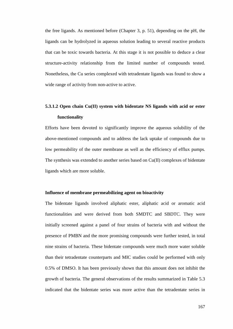
167
the free ligands. As mentioned before (Chapter 3, p. 51), depending on the pH, the
ligands can be hydrolyzed in aqueous solution leading to several reactive products
that can be toxic towards bacteria. At this stage it is not possible to deduce a clear
structure-activity relationship from the limited number of compounds tested.
Nonetheless, the Cu series complexed with tetradentate ligands was found to show a
wide range of activity from non-active to active.
5.3.1.2 Open chain Cu(II) system with bidentate NS ligands with acid or ester
functionality
Efforts have been devoted to significantly improve the aqueous solubility of the
above-mentioned compounds and to address the lack uptake of compounds due to
low permeability of the outer membrane as well as the efficiency of efflux pumps.
The synthesis was extended to another series based on Cu(II) complexes of bidentate
ligands which are more soluble.
Influence of membrane permeabilizing agent on bioactivity
The bidentate ligands involved aliphatic ester, aliphatic acid or aromatic acid
functionalities and were derived from both SMDTC and SBDTC. They were
initially screened against a panel of four strains of bacteria with and without the
presence of PMBN and the more promising compounds were further tested, in total
nine strains of bacteria. These bidentate compounds were much more water soluble
than their tetradentate counterparts and MIC studies could be performed with only
0.5% of DMSO. It has been previously shown that this amount does not inhibit the
growth of bacteria. The general observations of the results summarized in Table 5.3
indicated that the bidentate series was more active than the tetradentate series in
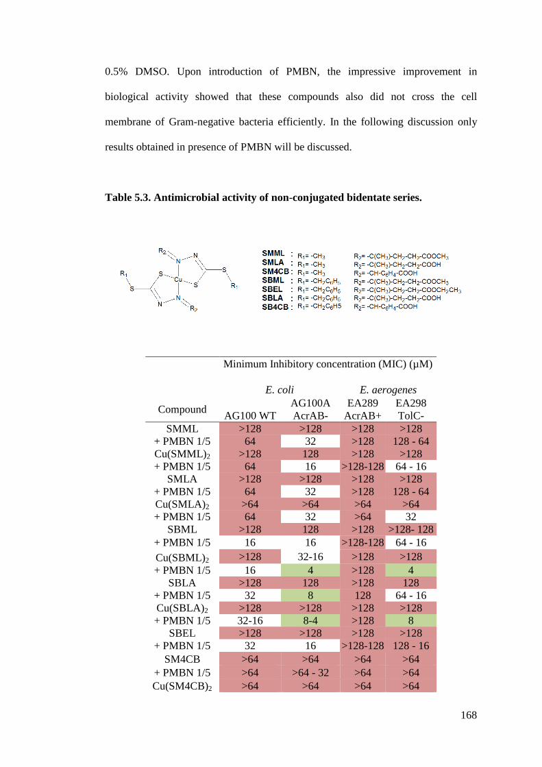
168
0.5% DMSO. Upon introduction of PMBN, the impressive improvement in
biological activity showed that these compounds also did not cross the cell
membrane of Gram-negative bacteria efficiently. In the following discussion only
results obtained in presence of PMBN will be discussed.
Table 5.3. Antimicrobial activity of non-conjugated bidentate series.
Minimum Inhibitory concentration (MIC) (µM)
E. coli E. aerogenes
Compound AG100 WT AG100A AcrAB-
EA289 AcrAB+
EA298 TolC-
SMML >128 >128 >128 >128 + PMBN 1/5 64 32 >128 128 - 64 Cu(SMML)2 >128 128 >128 >128 + PMBN 1/5 64 16 >128-128 64 - 16
SMLA >128 >128 >128 >128 + PMBN 1/5 64 32 >128 128 - 64 Cu(SMLA)2 >64 >64 >64 >64 + PMBN 1/5 64 32 >64 32
SBML >128 128 >128 >128- 128 + PMBN 1/5 16 16 >128-128 64 - 16 Cu(SBML)2 >128 32-16 >128 >128 + PMBN 1/5 16 4 >128 4
SBLA >128 128 >128 128 + PMBN 1/5 32 8 128 64 - 16 Cu(SBLA)2 >128 >128 >128 >128 + PMBN 1/5 32-16 8-4 >128 8
SBEL >128 >128 >128 >128 + PMBN 1/5 32 16 >128-128 128 - 16
SM4CB >64 >64 >64 >64 + PMBN 1/5 >64 >64 - 32 >64 >64 Cu(SM4CB)2 >64 >64 >64 >64
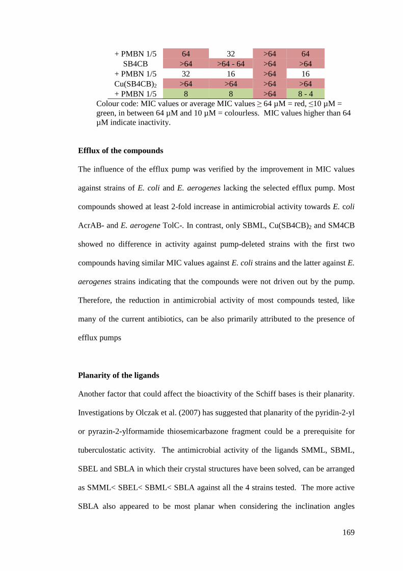
169
+ PMBN 1/5 64 32 >64 64 SB4CB >64 >64 - 64 >64 >64
+ PMBN 1/5 32 16 >64 16 Cu(SB4CB)2 >64 >64 >64 >64 + PMBN 1/5 8 8 >64 8 - 4
Colour code: MIC values or average MIC values 6 64 µM = red, "10 µM = green, in between 64 µM and 10 µM = colourless. MIC values higher than 64 µM indicate inactivity.
Efflux of the compounds
The influence of the efflux pump was verified by the improvement in MIC values
against strains of E. coli and E. aerogenes lacking the selected efflux pump. Most
compounds showed at least 2-fold increase in antimicrobial activity towards E. coli
AcrAB- and E. aerogene TolC-. In contrast, only SBML, Cu(SB4CB)2 and SM4CB
showed no difference in activity against pump-deleted strains with the first two
compounds having similar MIC values against E. coli strains and the latter against E.
aerogenes strains indicating that the compounds were not driven out by the pump.
Therefore, the reduction in antimicrobial activity of most compounds tested, like
many of the current antibiotics, can be also primarily attributed to the presence of
efflux pumps
Planarity of the ligands
Another factor that could affect the bioactivity of the Schiff bases is their planarity.
Investigations by Olczak et al. (2007) has suggested that planarity of the pyridin-2-yl
or pyrazin-2-ylformamide thiosemicarbazone fragment could be a prerequisite for
tuberculostatic activity. The antimicrobial activity of the ligands SMML, SBML,
SBEL and SBLA in which their crystal structures have been solved, can be arranged
as SMML< SBEL< SBML< SBLA against all the 4 strains tested. The more active
SBLA also appeared to be most planar when considering the inclination angles
170
between (i) the allylic (=C(CH3)CH2CH2) and the terminal carbocyclic acid/ester (-
COOR) planes (ii) the dithiocarbazate and the allylic planes, but appears to acquire
greater perpendicularity of the benzyl ring towards the dithiocarbazate plane.
Stability of the ligand
The results demonstrated that the free ligand SB4CB with the aromatic acid
substituent exhibited the lowest MIC values indicating a higher potency among the
compounds in this series. This efficacy might be due to the presence of the aromatic
ring that increases the stability of the compounds. It has been noted in the Chapter 3
(p. 52) that the aliphatic Schiff bases (SMML, SMLA, SBML, SBLA and SBEL)
were hydrolyzed when chromatographed by RP-HPLC with solvent system
containing 0.1% TFA. On the other hand, the aromatic Schiff bases (SM4CB and
SB4CB) were found to be stable when monitored by HPLC and UV-Vis
spectroscopy under similar conditions. No hydrolysis was observed even after 24
hours (18 hours is the duration required for MIC assay).
Effect of complexation
Many of the well known antibiotics like tetracyclines, quinolones and bleomycin are
chelating agents and their actions are improved by the presence of metal ions, in
particular copper ion (Ming, 2003; Efthimiadou et al., 2008). Thus, the synthesis and
study of metal complexes with drugs used in clinical practice as well as promising
ligands which may exhibit synergistic activity has attracted much attention as an
approach to new drug development (Turel et al., 2010). Upon complexation with
copper, most of the non-conjugated bidentate series compounds tested in this work
showed equal or lower MIC values. The free copper (II) acetate was also screened

171
against all strains and was found inactive (MIC >128 !M). The lack of growth
inhibition activity for copper (II) acetate confirmed that the observed growth
inhibition within experimental threshold was not due to the intrinsic biological
activity of copper (II) free metal (Ng et al., 2013). To compare the complexation
effect independently from the efficiency of the ligands, the ratio of MIC of the free
ligand to that of the Cu(II) complex (which had been multiplied by 2 due to the
coordination of two ligands in a complex) was calculated for the series of molecules
and for each strain. A ratio above one is taken to mean that the enhanced biological
activity is the result of a synergistic effect afforded by the complexation (the activity
is due not only to the ligand itself) whereas a ratio less than one implies that the
complexation is deleterious. As shown in Figure 5.7, the beneficial effect of the
complexation for the ligands highly depends on the bacteria strain.
Figure 5.7. Effect of complexation on the non-conjugated bidentate series of molecules against the different strains of E. coli (AG100 T and AG100A acrAB-) and E. aerogenes (EA289 acrAB- and EA298 tolC-). The ratio MIC (free ligand) / MIC(complexed ligand) has been calculated with the MIC (in presence of PMBN) reported according to the stoichiometry of the complex.
172
The complexation did not play a crucial role on the three first tested strains E. coli
WT, E. coli acrAB- and E. aerogenes acrAB+ since majority of the MIC ratio values
were 1. Only Cu(SB4CB)2, Cu(SBML)2 and Cu(SMLA)2 showed positive
complexation effect towards E. coli WT, E. coli AcrAB- and E. aerogenes AcrAB+,
respectively. Interestingly, the complexation improved the activity for most of
compounds with the exception of Cu(SM4CB)2 against the efflux pump TolC
deleted E. aerogenes strain whereas such an effect is not observed on the strain
possessing the TolC pump. This also demonstrated that complexation effectively
decreases the efflux of the active molecules in the TolC pump deleted strain. The
observed efficiency of the metal complexes could be linked to the significant
changes in the physico-chemical properties of the compounds upon chelation. For
instance, the molecular weight is doubled, the spatial geometry varied and new
redox properties appear since the compounds now involved a metal. Besides that,
the polarity of the metal complex is reduced to a greater extent in comparison to its
free ion because of the overlap of the ligand orbital and partial sharing of the
positive charge of the metal ion with the donor groups. Furthermore, it increases the
delocalization of ,-electrons over the whole chelate ring and enhances the
lipophilicity of complexes in aqueous solutions, which may modify the interactions
with cellular membranes (Raman, et al., 2003; Tiwari et al., 2012; Lobana et al.,
2009). The changes in the metal complexes may also be compatibility with the
hydrophobic pocket in the target site of the bacteria which further strengthen the
binding of the complexes to the microbe contributing to their enhanced activity
(Ming, 2003; Efthimiadou et al., 2008).
173
Effect of the dithiocarbazate substituent
There is also a general trend that the S-benzyl derived ligands and their metal
complexes showed better activity than the S-methyl derivatives in this series. There
have been mixed past reports on the bioactivity between SBDTC derivatives as
compared to the SMDTC (Hossain, et al., 1996; Pavan et al., 2010). The stronger
activity of S-benzyl derivatives could possibly be attributed to a higher cellular
uptake due to an increased lipophilicity. In order to further access the structure-
activity relationship, the biological activities of the compounds were correlated with
the electrochemical properties as well as their structures in both solid state (SCXRD)
and solution (EPR).
Redox potential of metal complexes
The redox potentials for the two series were recorded by cyclic voltammetry in
anhydrous DMF. The obvious differences between the two series were that Cu(II)
complexes of the bidentate series revealed a quasi-reversible reduction wave for
Cu(II)/Cu(I) couples with Epc ranging from -0.066 V to -0.114 V/(Ag/AgCl with
Fc/Fc+ = 0.563 V) (Chapter 3, page 88-94). On the other hand, both CuSMHD and
CuSBHD in the tetradentate ligands series underwent an electrochemically
irreversible one-electron reduction at Epc = -0.328 and -0.285 V which were more
negative than their bidentate counterparts. The more positive redox potential could
be an explanation for the greater activity of the bidentate series. A higher redox
potential means that Cu(II) reduction is easier, and consequently a higher content of
Cu(I) could be continuously generated due to the reversibilty. Cu(I) is prone to
participate in Fenton-type reactions that produce reactive oxygen species (ROS),
which can damage biomolecules within cells (Jansson et al., 2010). Radical
174
generation has also been proposed as a mechanism of cytotoxicity of Cu(II) and
Fe(III) thiosemicarbazone (Jansson et al., 2010). This certainly suggests that the role
of the reduction of the Cu(II) complexes in the antimicrobial action of the
compounds cannot be excluded. In both series, the redox potentials were higher in
the case of the benzyl-substituted compound than in the case of the methyl, which
could be explained by a weaker electron-donating effect of the benzyl group. Again,
the complexes having the higher redox potential were found to be more active. This
supports the suggestion that the easier reduction to Cu(I) may be responsible for the
increased activity.
Geometry of metal complexes
As previously mentioned in Chapter 3 (page 25, 88), there are correlation among
redox potential, geometry and biological activity. Because of the difference in
geometric preferences for Cu(II) (Jahn-Teller distortion favors square planar
geometry) and Cu(I) (as a d10 ion, there is no electronic preference for a particular
geometry) (Rorabacher, 2004), it would be anticipated that compounds with more
positive Cu(II)/Cu(I) reduction potential (which goes with Cu(I) being more stable)
are the least planar. The enhanced flexibility of the bidentate series as compared to
the tetradentate series may facilitate increased square planar distortion leading to
incorporation of Cu(I) ion that prefer non-square planar geometries. The information
with regard to the deviation from planarity in the solid state can be accessed from
the crystal structures. The crystal structures were obtained for Cu(SMML)2,
Cu(SMLA)2, CuSMHD and CuSBHD. The bond angles about the central copper
metal were consistent with a distorted square planar geometry with the sum of the
angles around Cu exceeds 360°. The bidentate series, Cu(SMML)2 and Cu(SMLA)2
175
(374.33° and 380.12°, respectively) displayed a more significant distortion than
CuSMHD and CuSBHD (367.65° and 360.9° respectively). The angles between two
N-Cu-S planes confirmed the deviation from planarity of the complexes in the order
Cu(SMLA)2 > Cu(SMML)2 > CuSMHD > CuSBHD. The bidentate series with more
positive Cu(II)/Cu(I) redox potentials also demonstrated higher antimicrobial
activity than their more planar tetradentate counterparts although the difference in
bioactivity between the compounds in the same series was less pronounced. The
EPR spectra recorded in DMF for both series indicated distorted square planar
geometry for most Cu(II) complexes. Referring to the empirical factor f (= g||/0||)
(Joseph et al., 2012; Krishna et al., 2008) of the compounds shown in Chapter 3 (p.
83), Cu(II) complexes with aliphatic acid and ester bidentate ligands (145-147 cm)
confirm slightly higher degree of tetrahedral distortion than those with tetradentate
ligands (141 and 143 cm) in solution. However, there are exceptions with
Cu(SM4CB)2 and Cu(SB4CB)2 formed by the more active aromatic acid bidentate
ligands. Both complexes revealed lower f values (128 and 121 cm, respectively)
which are in the square planar range.
At this stage, the growth inhibitory mechanism of action and molecular target for the
compounds carried out in this work is unclear. Nonetheless, others mechanisms of
action are possible. For example, square planar Cu(II) complexes have often been
associated with their ability to interact with DNA, the central target for most
therapeutic agents (Manikandamathavan, et al., 2013). One of the proposed
mechanism for these complexes involved the reduction of the DNA-intercalated
Cu(II) complexes by glutathione or ascorbate, and followed by reoxidation of Cu(I)
with dioxygen or in the presence of hydrogen peroxide. This leads to the generation
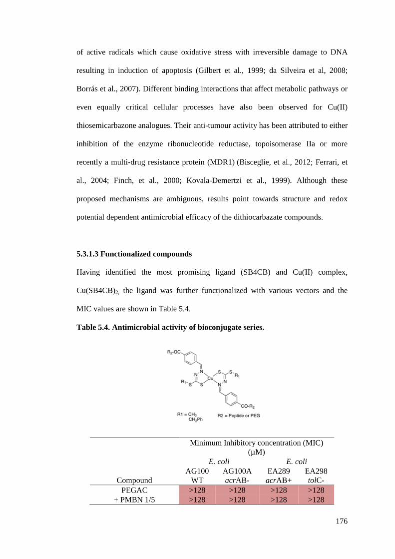
176
of active radicals which cause oxidative stress with irreversible damage to DNA
resulting in induction of apoptosis (Gilbert et al., 1999; da Silveira et al, 2008;
Borrás et al., 2007). Different binding interactions that affect metabolic pathways or
even equally critical cellular processes have also been observed for Cu(II)
thiosemicarbazone analogues. Their anti-tumour activity has been attributed to either
inhibition of the enzyme ribonucleotide reductase, topoisomerase IIa or more
recently a multi-drug resistance protein (MDR1) (Bisceglie, et al., 2012; Ferrari, et
al., 2004; Finch, et al., 2000; Kovala-Demertzi et al., 1999). Although these
proposed mechanisms are ambiguous, results point towards structure and redox
potential dependent antimicrobial efficacy of the dithiocarbazate compounds.
5.3.1.3 Functionalized compounds
Having identified the most promising ligand (SB4CB) and Cu(II) complex,
Cu(SB4CB)2, the ligand was further functionalized with various vectors and the
MIC values are shown in Table 5.4.
Table 5.4. Antimicrobial activity of bioconjugate series.
Minimum Inhibitory concentration (MIC) (µM)
E. coli E. coli
Compound AG100
WT AG100A acrAB-
EA289 acrAB+
EA298 tolC-
PEGAC >128 >128 >128 >128 + PMBN 1/5 >128 >128 >128 >128
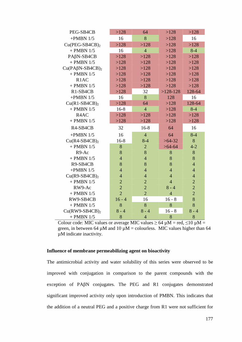
177
PEG-SB4CB >128 64 >128 >128 +PMBN 1/5 16 8 >128 16
Cu(PEG-SB4CB)2 >128 >128 >128 >128 + PMBN 1/5 16 4 >128 8-4
PA%N-SB4CB >128 >128 >128 >128 + PMBN 1/5 >128 >128 >128 >128
Cu(PA%N-SB4CB)2 >128 >128 >128 >128 + PMBN 1/5 >128 >128 >128 >128
R1AC >128 >128 >128 >128 + PMBN 1/5 >128 >128 >128 >128 R1-SB4CB >128 32 >128-128 128-64 +PMBN 1/5 16 8 128 16
Cu(R1-SB4CB)2 >128 64 >128 128-64 + PMBN 1/5 16-8 4 >128 8-4
R4AC >128 >128 >128 >128 + PMBN 1/5 >128 >128 >128 >128 R4-SB4CB 32 16-8 64 16 +PMBN 1/5 16 4 64 8-4
Cu(R4-SB4CB)2 16-8 8-4 >64-32 8 + PMBN 1/5 8 2 >64-64 4-2
R9-Ac 8 8 8 8 + PMBN 1/5 4 4 8 8 R9-SB4CB 8 8 8 4 +PMBN 1/5 4 4 4 4
Cu(R9-SB4CB)2 4 4 4 4 + PMBN 1/5 2 2 4 2
RW9-Ac 2 2 8 - 4 2 + PMBN 1/5 2 2 4 2 RW9-SB4CB 16 - 4 16 16 - 8 8 + PMBN 1/5 8 8 8 8
Cu(RW9-SB4CB)2 8 - 4 8 - 4 16 - 8 8 - 4 + PMBN 1/5 8 4 8 8
Colour code: MIC values or average MIC values 6 64 µM = red, "10 µM = green, in between 64 µM and 10 µM = colourless. MIC values higher than 64 µM indicate inactivity.
Influence of membrane permeabilizing agent on bioactivity
The antimicrobial activity and water solubility of this series were observed to be
improved with conjugation in comparison to the parent compounds with the
exception of PA%N conjugates. The PEG and R1 conjugates demonstrated
significant improved activity only upon introduction of PMBN. This indicates that
the addition of a neutral PEG and a positive charge from R1 were not sufficient for
178
uptake across the cell membrane of the bacteria. In contrast, it is positive to
highlight that R4, R9 and RW9 conjugates were already active without the presence
of PMBN. The increase in the cationic nature of the polyarginine derivatives allows
better interaction and subsequently permeability across the negatively charged
bacteria membrane as anticipated. Therefore, the MIC values obtained for these
compounds either showed no difference or improved only 2-fold with the presence
of PMBN. The slightly better activity in some cases of the polyarginine derivatives
with PMBN point out that conjugation has yet to bring about optimum uptakes and
the improved MIC values may be a result of synergistic effect between PMBN and
conjugated ligands. Nonetheless, polyarginine conjugates appeared to be most
efficient in crossing the cell membrane compared to the unconjugated compounds
and other vectors.
Efflux of the compounds
The PEG, R1 and R4 conjugates also showed at least 2-fold increase in MIC values
against pump deleted strains E. coli AcrAB- and E. aerogene TolC-. However, for
R9 and RW9 derivatives, the MIC values remained the same with or without the
presence of efflux pump. This is another encouraging observation that conjugation
to CPP R9 and RW9 is beneficial as they are not affected by the efflux pumps. The
PA%N conjugate was inactive against all strains tested. This conjugate was initially
designed to assess the effect of efflux pump as well as to reveal any synergistic
effect of the conjugate with possible new mechanism of action. However, the lack of
activity reported with regards to this conjugate certainly demised the potential for
positive synergistic effect.
179
Influence of arginine chain lengths on bioactivity
In addition to the noticeable trend with PMBN and efflux pumps, there is also
correlation of higher bacterial growth inhibition potency with arginine chain lengths.
The improvement of the MIC values averaged against all four strains upon
conjugation of SB4CB to polyarginine is in the following order: R9-SB4CB > RW9-
SB4CB> R4-SB4CB > R1-SB4CB > SB4CB. In order to identify the contribution of
each moiety in the conjugates, acetylated free peptides and PEG were tested as well.
The acetylated vectors PEGAC, R1AC and R4AC were totally inactive with MIC
>128 µM. The improved activity observed for the conjugates of these three vectors
could have benefited from the better water solubility, permeability and cell uptakes
of the conjugates. In contrast, RW9AC proved to be highly active against all the
strains while R9AC also showed activity against them. As previously mentioned
(Chapter 5, p. 149), CPPs and antimicrobial peptides are known to show similar
characteristics, in particular amphiphilicity, which is a crucial factor determining the
antibacterial activity of peptides. This seems to be the case for the CPP RW9.
Noting the antimicrobial activity of the acetylated R9AC and RW9AC, these two
vectors could have contributed their activity to the improved MIC values as
demonstrated by the conjugates.
Effect of complexation
According to the Figure 5.8, most of the bioconjugated Cu(II) complexes showed
equal or lower MIC values compared to the ligands indicating that complexation
with Cu(II) did not contribute significantly to the observed activity of the ligands.
The conjugates also showed resemblance to the parent compound in which
complexation improved the efficiency against E. aerogenes TolC- strain as
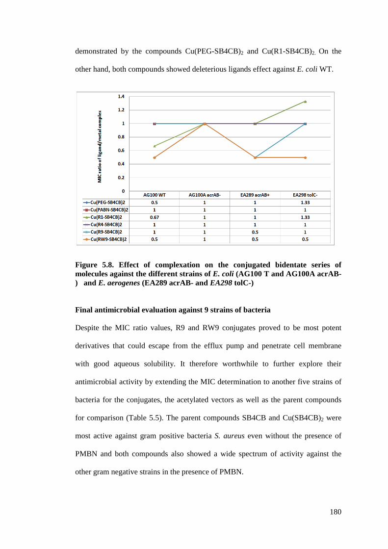
180
demonstrated by the compounds Cu(PEG-SB4CB)2 and Cu(R1-SB4CB)2. On the
other hand, both compounds showed deleterious ligands effect against E. coli WT.
Figure 5.8. Effect of complexation on the conjugated bidentate series of molecules against the different strains of E. coli (AG100 T and AG100A acrAB-) and E. aerogenes (EA289 acrAB- and EA298 tolC-)
Final antimicrobial evaluation against 9 strains of bacteria
Despite the MIC ratio values, R9 and RW9 conjugates proved to be most potent
derivatives that could escape from the efflux pump and penetrate cell membrane
with good aqueous solubility. It therefore worthwhile to further explore their
antimicrobial activity by extending the MIC determination to another five strains of
bacteria for the conjugates, the acetylated vectors as well as the parent compounds
for comparison (Table 5.5). The parent compounds SB4CB and Cu(SB4CB)2 were
most active against gram positive bacteria S. aureus even without the presence of
PMBN and both compounds also showed a wide spectrum of activity against the
other gram negative strains in the presence of PMBN.
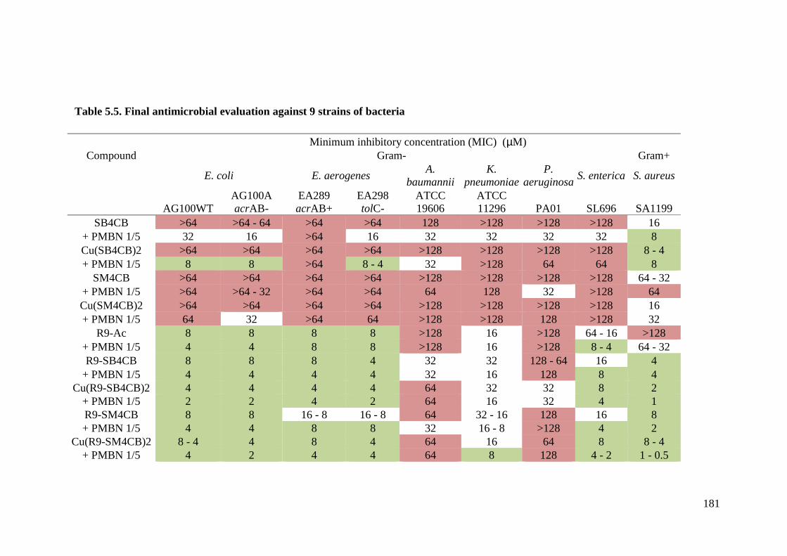
181
Minimum inhibitory concentration (MIC) (µM) Compound Gram- Gram+
E. coli E. aerogenes A. baumannii
K. pneumoniae
P. aeruginosa S. enterica S. aureus
AG100WT AG100A acrAB-
EA289 acrAB+
EA298 tolC-
ATCC 19606
ATCC 11296 PA01 SL696 SA1199
SB4CB >64 >64 - 64 >64 >64 128 >128 >128 >128 16 + PMBN 1/5 32 16 >64 16 32 32 32 32 8 Cu(SB4CB)2 >64 >64 >64 >64 >128 >128 >128 >128 8 - 4 + PMBN 1/5 8 8 >64 8 - 4 32 >128 64 64 8
SM4CB >64 >64 >64 >64 >128 >128 >128 >128 64 - 32 + PMBN 1/5 >64 >64 - 32 >64 >64 64 128 32 >128 64 Cu(SM4CB)2 >64 >64 >64 >64 >128 >128 >128 >128 16 + PMBN 1/5 64 32 >64 64 >128 >128 128 >128 32
R9-Ac 8 8 8 8 >128 16 >128 64 - 16 >128 + PMBN 1/5 4 4 8 8 >128 16 >128 8 - 4 64 - 32 R9-SB4CB 8 8 8 4 32 32 128 - 64 16 4
+ PMBN 1/5 4 4 4 4 32 16 128 8 4 Cu(R9-SB4CB)2 4 4 4 4 64 32 32 8 2
+ PMBN 1/5 2 2 4 2 64 16 32 4 1 R9-SM4CB 8 8 16 - 8 16 - 8 64 32 - 16 128 16 8 + PMBN 1/5 4 4 8 8 32 16 - 8 >128 4 2
Cu(R9-SM4CB)2 8 - 4 4 8 4 64 16 64 8 8 - 4 + PMBN 1/5 4 2 4 4 64 8 128 4 - 2 1 - 0.5
Table 5.5. Final antimicrobial evaluation against 9 strains of bacteria
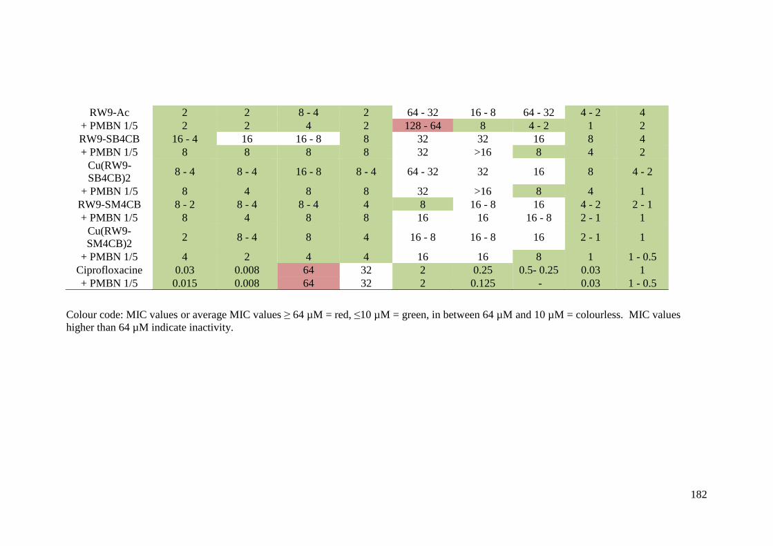
182
Colour code: MIC values or average MIC values 6 64 µM = red, "10 µM = green, in between 64 µM and 10 µM = colourless. MIC values higher than 64 µM indicate inactivity.
RW9-Ac 2 2 8 - 4 2 64 - 32 16 - 8 64 - 32 4 - 2 4 + PMBN 1/5 2 2 4 2 128 - 64 8 4 - 2 1 2 RW9-SB4CB 16 - 4 16 16 - 8 8 32 32 16 8 4 + PMBN 1/5 8 8 8 8 32 >16 8 4 2
Cu(RW9-SB4CB)2 8 - 4 8 - 4 16 - 8 8 - 4 64 - 32 32 16 8 4 - 2
+ PMBN 1/5 8 4 8 8 32 >16 8 4 1 RW9-SM4CB 8 - 2 8 - 4 8 - 4 4 8 16 - 8 16 4 - 2 2 - 1 + PMBN 1/5 8 4 8 8 16 16 16 - 8 2 - 1 1
Cu(RW9-SM4CB)2 2 8 - 4 8 4 16 - 8 16 - 8 16 2 - 1 1
+ PMBN 1/5 4 2 4 4 16 16 8 1 1 - 0.5 Ciprofloxacine 0.03 0.008 64 32 2 0.25 0.5- 0.25 0.03 1 + PMBN 1/5 0.015 0.008 64 32 2 0.125 - 0.03 1 - 0.5
183
It is likely that the complexes were more active against the Gram-positive bacteria
due to the ease of penetration through the cell wall as compared to the Gram-
negative bacteria. It should be noted that SM4CB, the less active S-methyl analogue
of SB4CB ligand was also functionalized with R9 and RW9 in order to better
understand the conjugation effect. This led to further positive results including the
synthesis of a new compound, R9-SM4CB that was active against most of the
bacteria. It is particularly interesting to note the construct of active R9-SM4CB
against S. aureus with MIC values of 8 µM (without the presence of PMBN) from
R9AC (MIC >128 µM) and SM4CB (MIC= 64 µM), both of which were inactive
under identical conditions. Comparing the efficacy of the compounds against the
different strains of bacteria, the MIC values of the ligands conjugates were
considerably better as compared to the parent compounds. Upon conjugation, the
functionalized compounds exhibited improved antimicrobial activity with MIC
values in the micromolar concentration range, as low as 1-0.5 µM. However, in
comparison with the R9 and RW9 acetylated free peptides, the improvement in MIC
values of the conjugates were less striking although there were some exceptions that
will be discussed herein. RW9AC proved to be active against E. coli, E. aerogenes,
K. pneumoniae, S. enterica and S. aureus but less active against A. baumannii and P.
aeruginosa. While R9AC also showed activity against some of the bacteria strains, it
was completely inactive against A. baumannii, P. aeruginosa and S. aureus.
Conjugation to R9 did not essentially change the antibacterial activity of R9 against
gram-negative bacteria E. coli, E. aerogenes ArcAB+, K. pneumoniae, P.
aeruginosa and S. enterica. The no improvement observation could mean that the
activity is only due to the peptide and not to the parent compound. The RW9
conjugates showed either similar or increased MIC values as compared to acetylated
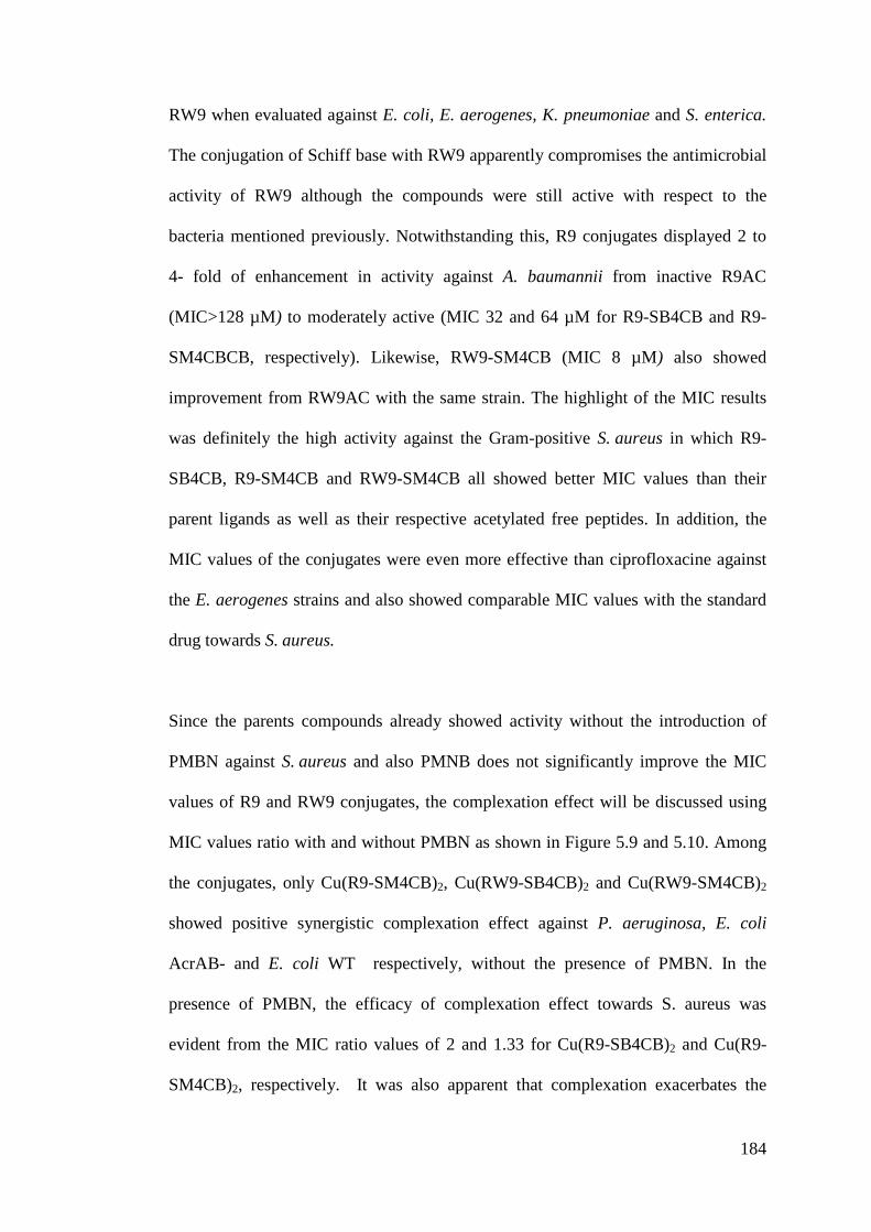
184
RW9 when evaluated against E. coli, E. aerogenes, K. pneumoniae and S. enterica.
The conjugation of Schiff base with RW9 apparently compromises the antimicrobial
activity of RW9 although the compounds were still active with respect to the
bacteria mentioned previously. Notwithstanding this, R9 conjugates displayed 2 to
4- fold of enhancement in activity against A. baumannii from inactive R9AC
(MIC>128 µM) to moderately active (MIC 32 and 64 µM for R9-SB4CB and R9-
SM4CBCB, respectively). Likewise, RW9-SM4CB (MIC 8 µM) also showed
improvement from RW9AC with the same strain. The highlight of the MIC results
was definitely the high activity against the Gram-positive S. aureus in which R9-
SB4CB, R9-SM4CB and RW9-SM4CB all showed better MIC values than their
parent ligands as well as their respective acetylated free peptides. In addition, the
MIC values of the conjugates were even more effective than ciprofloxacine against
the E. aerogenes strains and also showed comparable MIC values with the standard
drug towards S. aureus.
Since the parents compounds already showed activity without the introduction of
PMBN against S. aureus and also PMNB does not significantly improve the MIC
values of R9 and RW9 conjugates, the complexation effect will be discussed using
MIC values ratio with and without PMBN as shown in Figure 5.9 and 5.10. Among
the conjugates, only Cu(R9-SM4CB)2, Cu(RW9-SB4CB)2 and Cu(RW9-SM4CB)2
showed positive synergistic complexation effect against P. aeruginosa, E. coli
AcrAB- and E. coli WT respectively, without the presence of PMBN. In the
presence of PMBN, the efficacy of complexation effect towards S. aureus was
evident from the MIC ratio values of 2 and 1.33 for Cu(R9-SB4CB)2 and Cu(R9-
SM4CB)2, respectively. It was also apparent that complexation exacerbates the
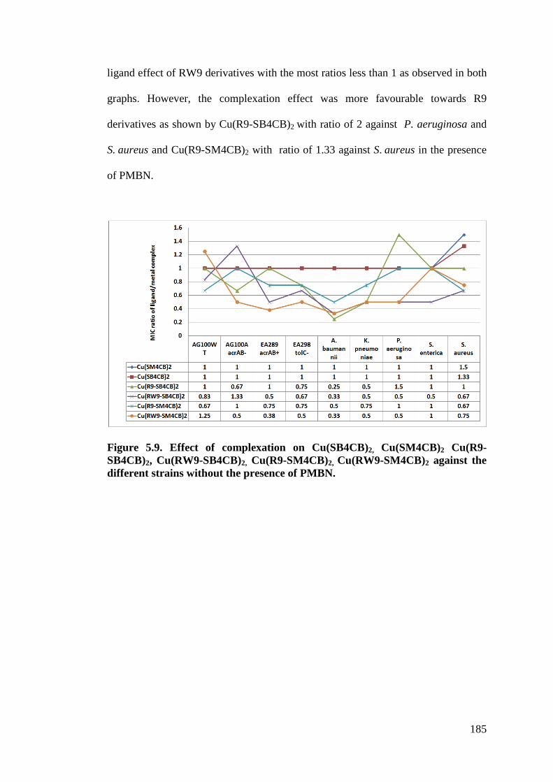
185
ligand effect of RW9 derivatives with the most ratios less than 1 as observed in both
graphs. However, the complexation effect was more favourable towards R9
derivatives as shown by Cu(R9-SB4CB)2 with ratio of 2 against P. aeruginosa and
S. aureus and Cu(R9-SM4CB)2 with ratio of 1.33 against S. aureus in the presence
of PMBN.
Figure 5.9. Effect of complexation on Cu(SB4CB)2, Cu(SM4CB)2 Cu(R9-SB4CB)2, Cu(RW9-SB4CB)2, Cu(R9-SM4CB)2, Cu(RW9-SM4CB)2 against the different strains without the presence of PMBN.
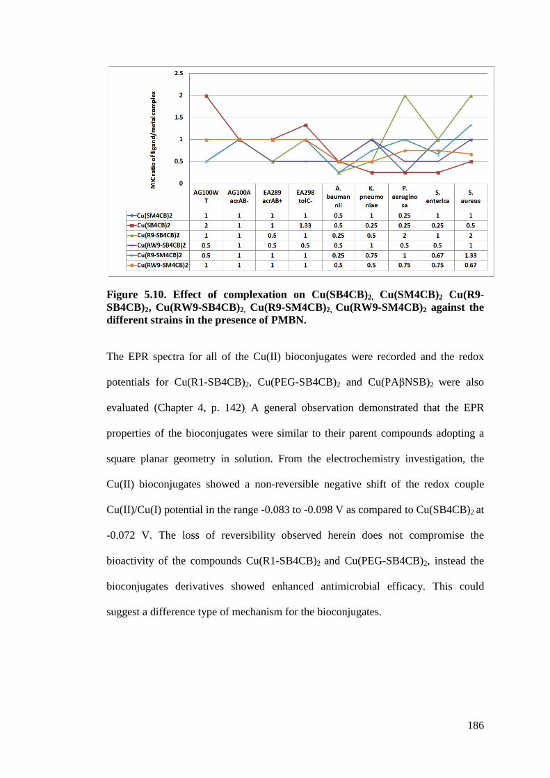
186
Figure 5.10. Effect of complexation on Cu(SB4CB)2, Cu(SM4CB)2 Cu(R9-SB4CB)2, Cu(RW9-SB4CB)2, Cu(R9-SM4CB)2, Cu(RW9-SM4CB)2 against the different strains in the presence of PMBN.
The EPR spectra for all of the Cu(II) bioconjugates were recorded and the redox
potentials for Cu(R1-SB4CB)2, Cu(PEG-SB4CB)2 and Cu(PA%NSB)2 were also
evaluated (Chapter 4, p. 142). A general observation demonstrated that the EPR
properties of the bioconjugates were similar to their parent compounds adopting a
square planar geometry in solution. From the electrochemistry investigation, the
Cu(II) bioconjugates showed a non-reversible negative shift of the redox couple
Cu(II)/Cu(I) potential in the range -0.083 to -0.098 V as compared to Cu(SB4CB)2 at
-0.072 V. The loss of reversibility observed herein does not compromise the
bioactivity of the compounds Cu(R1-SB4CB)2 and Cu(PEG-SB4CB)2, instead the
bioconjugates derivatives showed enhanced antimicrobial efficacy. This could
suggest a difference type of mechanism for the bioconjugates.
187
5.3.2 Cytotoxicity
In view of the well known cytotoxic activity of sulfur–nitrogen Schiff base chelating
agents and their metal complexes and also to judge their utility for further
development as anticancer agents, selected compounds were tested for their
cytotoxicity against two breast cancer cell lines MDA-MB-231 (human breast
carcinoma cells not expressing nuclear estrogen receptors, ER-) and MCF-7 (human
breast carcinoma cells expressing nuclear estrogen receptors, ER+). Measurement of
the cytotoxicity was carried out using MTT assay (Mosmann, 1983) based on the
metabolic reduction of tetrazolium salt to form water insoluble formazan crystals
with tamoxifen as standard. DMSO was used as negative control in the assay and the
final content of DMSO for each compound tested was 0.5% or less. The
concentration required to reduce growth of cancer cells by 50% (IC50) are shown in
Table 5.6 for both tetradentate series and bidentate NS series derived from 3-
acetylcoumarin.
Table 5.6. Cytotoxic assay results.
IC50 (!M) MCF7 MDA-MB231
Tetradentate series SMHD 138.90 9.61 SBHD 9.69 1.05 CuSMHD 2.60 2.34 CuSBHD 1.49 0.71 Bidentate series SBCM inactive inactive Re2(SBCM)2 19.41 8.61 Zn(SBCM)2 inactive inactive Cu(SBCM)2 5.97 8.31 Tamoxifen 11.20 13.40
188
For the tetradentate series, it interesting to note that the structure-activity
relationship observed in the antibacterial tests differed from their cytotoxicity. Both
ligands displayed at least 9-fold better toxicity towards the MDA-MB231 cell line
that does not express estrogen nuclear receptors, indicating that ligand toxicity is not
only mediated by these receptors. The more lipophilic SBHD exhibits a stronger
toxicity in comparison to SMHD (Pavan et al., 2010). Complexation of Schiff base
ligands with metal ions has been found to produce synergistic effects on the
antiproliferative activities of the parent ligands since the complexes showed a
marked cytotoxicity with IC50 values < 5.0 µM towards both cell lines. Again the
bioactivity of the complexes does not involve estrogen receptors because they are
more active on MDA-MB231 cells. On both cell lines, the benzyl substituted
complex CuSBHD showed slightly better IC50 values. The stronger activity of
CuSBHD could possibly be attributed to a higher cellular uptake due to an increased
lipophilicity. Likewise, the 3-acetylcoumarin derived bidentate series also
highlighted that complexation played a significant role in improving the bioactivity
of the compound. The ligand SBCM was found to be inactive towards both MCF-7
and MDA-MB231 cell lines. However, complexation of the Schiff base ligand with
Cu(II) and Re(I) with the exception of Zn(II) showed marked cytotoxicity with IC50
values < 10 !M. Re2(SBCM)2 displayed better selectivity towards MDA-MB231
whereas Cu(SBCM)2 was more potent against MCF7. All the Cu(II) complexes were
most potent in both series and have lower IC50 values in comparison to standard
tamoxifen making them potential anticancer agents for consideration. The difference
in selectivity observed between the Cu(II) complexes of bidentate (MCF7) and
tetradentate (MDA-MB231) series may result from their differences in ligand
substituents, physico-chemical and redox potential. The bioactive Re2(SBCM)2 with
189
the presence of tricarbonyl core may also offer novel exploratory derivatives for
future investigations in the treatment of cancer as novel pharmaceutical as well as
potential probe for sub-cell imaging. While the mechanism of action against breast
cancer cell lines for dithiocarbazate compounds has yet to be definitively determined
but an in vitro study by Cheah (2007) and Awidat (2005) on some dithiocarbazato
compounds suggested that they induce apotopsis by DNA fragmentation and
suppression of the expression of certain oncogenes. Previously mentioned
mechanisms in the antimicrobial section i.e. inhibition of the enzyme ribonucleotide
reductase, topoisomerase IIa or multi-drug resistance protein as well as production
of ROS have also been linked to their anti-cancer activity.
5.4 Conclusion
It was demonstrated that the bidentate Schiff base (SB4CB) and its Cu(II) complex
Cu(SB4CB)2 with aromatic acid functionality possess the most remarkable
antibacterial effect against a wide spectrum of bacteria. The pioneering conjugation
strategy investigated in this work demonstrated the utility of the combination of
functionalized dithiocarbazate derivatives with vectors (CPPs) to generate
bioconjugates with enhanced antimicrobial activity, membrane permeability and
water solubility. The results highlighted various trends and factors that govern
antimicrobial activities of the tested compounds. It was clearly evident that
antimicrobial activities of the compounds are strongly dependent on their
substitutents. Introduction of the carboxylic acid moiety and SBDTC increases the
antimicrobial activity within the bidentate series. The complexation with copper has
a synergetic effect on the antimicrobial activity of these compounds. The increased
activity of the complexes could be associated with their increased lipophilicity,
190
better fitness and size of the molecules towards binding sites as well as various
geometrical arrangements including square planar and distorted forms inducing a
tuning in redox potential. The observed cyclic voltammetric behaviour of bidentate
copper(II) complexes showed a Cu(II)/Cu(I) redox quasi-reversibility and a positive
shift as compared to the tetradentate series which may also contribute to their
toxicity. In addition, the biological activity depends on factors such as the bacteria
strain and the vectors used for bioconjugation. A deep understanding of the structure
and activity of metal complexes against microorganisms is invaluable toward future
development of antibiotic agents. The antimicrobial activity of the compounds can
be summarized as follows:
Tetradentate series (S-benzyl derivatives<S-methyl derivatives, Cu (II)
complexes<Ligands) <Bidentate series (Aliphatic<Aromatic, S-methyl
derivatives<S-benzyl derivatives, Ligands<Cu (II) complexes) <Peptide conjugate
series (PA%N<PEG<R1<R4<RW9/R9)< their Cu (II) complexes (MIC * 1-0.5 µ M)
All the selected Cu(II) complexes assayed against breast cancer cells lines (MCF-7
and MDA-MB-231) exhibited good cytotoxicity with lower IC50 values (0.71-8.31
µM) in comparison to their respective ligands and standard drug tamoxifen. This
highlights the relevance of metal complexation strategy to stabilize the ligands and
improve their bioactivity.

191
CHAPTER 6
CONCLUSION AND RECOMMENDATIONS
In conclusion, a total of 43 compounds were synthesized and characterized in this
work. These compounds comprised of 10 Schiff base ligands derived from either
SMDTC or SBDTC, 8 ligand-peptides/PEG conjugates, 5 acetylated peptides/PEG
and 19 metal complexes. Single crystal structures were solved for 11 compounds.
Only SMHD and SBHD were NNSS tetradentate ligands while the others behaved
as bidentate NS ligands coordinating to the central metal through the azomethine
nitrogen atom and the thiolate sulphur atom in their respective complexes. The
antimicrobial activity of the compounds showed improvement going from
macroacyclic tetradentate series to open chain bidentate series and finally the
bioconjugates series. This work also allows new insight into the relationship
between the structural/electrochemical properties and biological activity of the
compounds. The bidentate series demonstrated higher antimicrobial activity than
their more planar tetradentate counterparts as in the solid state, the crystal structures
of Cu(SMML)2 and Cu(SMLA)2 from the bidentate series displayed a more
significant distortion from square planar than CuSMHD and CuSBHD of the
macroacyclic tetradentate series. EPR data also support the slightly greater distortion
of the bidentate series in comparison to the tetradentate series in solution with the
exception of Cu(SB4CB)2 and Cu(SM4CB)2 that fall in the square planar range.
Both series also differed in their electrochemical properties in which bidentate series
showed Cu(II)/Cu(I) quasi reversibility at more positive potential whereas
tetradentate series was not reversible. The Cu(II) R9 derivatives of SB4CB and
SM4CB possess the most remarkable antibacterial effect against a wide spectrum of
192
bacteria. In addition, these R9-conjugates managed to address drawbacks that are
often associated with most metal complexes involving the issues of poor water
solubility and lack of uptake of the compounds due to low permeability of the outer
membrane as well as the efficiency of bacteria efflux pump. The successful
conjugation and facile complexation with copper for Schiff bases SB4CB and
SM4CB offer exciting potential for them to be further optimized with different
vectors in the future. The selected Cu(II) compound assayed against breast cancer
cells also exhibited good cytotoxicity. The fact that the Cu (II) complexes are more
efficient than the ligands is appealing. Taking into account, the serious side effects
and upcoming resistance of clinical reference drugs, these new compounds are
useful lead candidates for the development of novel therapeutic agents to treat
bacterial infections and cancer.
Hitherto, the mechanism of action of the compounds has yet to be verified.
Therefore, efforts to determine the concentration and location of the compounds
inside cells will be the next crucial step for intracellular understanding. This is
important in order to increases the compounds chances of succeeding in in vivo
assay, clinical trials and ultimately to be used as therapeutic drugs. Attaching the
lead compound to a probe or fluorescence moiety would allow cell imaging
experiments to be carried out. Parallel studies involving the recognition of
biomolecules as specific targets such DNA binding and cleavage, protein or enzyme
inhibitors as well as depolarization 3,3$-Dipropylthiadicarbocyanine iodide
(DiSC3(5)) or ortho-nitrophenyl-%-D-galactopyranoside (OPNG) membrane
permeabilization assay (for bacteria) are also essential in order shed light on the
mode of action of these compounds under biological conditions. It is also expected
193
that the application of computational analysis like docking will facilitate a deeper
understanding of the molecular interaction. In addition, Density Functional Theory
(DFT) and Quantitative Structure-Activity Relationship (QSAR) studies will enable
elucidation of key structural and chemical parameters required to develop potent
compounds. The data compilation will be beneficial in the long run as the
information may allow utilization of virtual high throughput screening (HTS)
approach to prescreen lead dithiocarbazate compounds in silico for diseases prior to
synthesis or bioactivity assay validation. This approach would be an excellent
strategy in both time- and cost-effective manner as compared to the carpet bombing
strategy.
194
REFERENCES
Abramkin, S., Valiahdi, S. M., Jakupec, M. A., Galanski, M., Metzler-Nolte, N., & Keppler, B. K. (2012). Solid-phase synthesis of oxaliplatin-TAT peptide bioconjugates. Dalton Transactions, 41(10), 3001-3005. Afrasiabi, Z., Sinn, E., Padhye, S., Dutta, S., Padhye, S., Newton, C., ... & Powell, A. K. (2003). Transition metal complexes of phenanthrenequinone thiosemicarbazone as potential anticancer agents: synthesis, structure, spectroscopy, electrochemistry and in vitro anticancer activity against human breast cancer cell-line, T47D. Journal of Inorganic Biochemistry, 95(4), 306-314. Ahmad, J. Ma, A. Jemal, (2013) in Breast Cancer Metastasis and Drug Resistance, Springer New York, pp. 1. Ali, M. A., & Livingstone, S. E. (1974). Metal complexes of sulphur-nitrogen chelating agents. Coordination Chemistry Reviews, 13(2), 101-132. Ali, M. A., & Tarafdar, M. T. H. (1977). Metal complexes of sulphur and nitrogen-containing ligands: Complexes of S-benzyldithiocarbazate and a Schiff base formed by its condensation with pyridine-2-carboxaldehyde. Journal of Inorganic and Nuclear Chemistry, 39(10), 1785-1791. Ali, M. A., Hossain, S. M., Majumder, S. M. M. H., Uddin, M. N., & Tarafder, M. T. H. (1987). Synthesis and characterization of some new nickel (II), zinc (II) and cadmium (II) complexes of quadridentate SNNS ligands. Polyhedron, 6(8), 1653-1656. Ali, M. A., Nazimuddin, M., Shaha, R., Butcher, R. J., & Bryan, J. (1999). Synthesis and characterization of bis-chelated nickel (II) complexes of the methylpyruvate Schiff bases of S-alkyldithiocarbazates and the X-ray crystal structure of the [Ni (ONSMe)2] complex. Polyhedron, 17(22), 3955-3961. Ali, M. A., Mirza, A. H., Butcher, R. J., Tarafder, M. T. H., & Ali, M. A. (2001a). Synthetic, spectroscopic, biological and X-ray crystallographic structural studies on a novel pyridine-nitrogen-bridged dimeric nickel (II) complex of a pentadentate N3S2 ligand. Inorganica Chimica Acta, 320(1), 1-6. Ali, M. A., Mirza, A. H., & Butcher, R. J. (2001b). Synthesis and characterization of copper (II) complexes of the methylpyruvate Schiff base of S-methyldithiocarbazate (Hmpsme) and the X-crystal structures of Hmpsme and [Cu (mpsme) Cl]. Polyhedron, 20(9), 1037-1043. Ali, M. A., Mirza, A. H., Butcher, R. J., Tarafder, M. T. H., Keat, T. B., & Ali, A. M. (2002). Biological activity of palladium (II) and platinum (II) complexes of the acetone Schiff bases of S-methyl-and S-benzyldithiocarbazate and the X-ray crystal structure of the [Pd(asme)2](asme= anionic form of the acetone Schiff base of S-methyldithiocarbazate) complex. Journal of Inorganic Biochemistry, 92(3), 141-148.
195
Akbar Ali, M., Mirza, A. H., Nazimuddin, M., Ahmed, R., Gahan, L. R., & Bernhardt, P. V. (2003). Synthesis and characterization of mono-and bis-ligand zinc (II) and cadmium (II) complexes of the di-2-pyridylketone Schiff base of S-benzyl dithiocarbazate (Hdpksbz) and the X-ray crystal structures of the [Zn (dpksbz)2] and [Cd (dpksbz) NCS]2 complexes. Polyhedron, 22(11), 1471-1479. Ali, M. A., Mirza, A. H., & Fong, G. A. (2004). Synthesis, characterization and x-ray crystal structures of the bis–ligand zinc (II) and cadmium (II) complexes of the methylpyruvate schiff base of S-methyldithiocarbazate. Transition Metal Chemistry, 29(6), 613-619. Ali, M. A., Mirza, A. H., Fereday, R. J., Butcher, R. J., Fuller, J. M., Drew, S. C., ... & Murray, K. S. (2005). Synthetic, EPR spectroscopic, magnetic and X-ray crystallographic structural studies on copper (II) complexes of the tridentate N2S donor ligand formed from 6-methyl-2-formylpyridine and S-methyldithiocarbazate (Hmpsme). Inorganica Chimica Acta, 358(13), 3937-3948. Ali, M. A., Mirza, A. H., Butcher, R. J., & Crouse, K. A. (2006). The preparation, characterization and biological activity of palladium (II) and platinum (II) complexes of tridentate NNS ligands derived from S-methyl-and S-benzyldithiocarbazates and the X-ray crystal structure of the [Pd (mpasme) Cl] complex. Transition Metal Chemistry, 31(1), 79-87. Ali, M. A., Hj Abu Bakar, H. J., Mirza, A. H., Smith, S. J., Gahan, L. R., and Bernhardt, P. V. (2008) Preparation, spectroscopic characterization and X-ray crystal and molecular structures of nickel(II), copper(II) and zinc(II) complexes of the Schiff base formed from isatin and S-methyldithiocarbazate (Hisa-sme). Polyhedron 27, 71-79 Ali, M. A., Mirza, A. H., Mei, C. C., Bernhardt, P. V., & Karim, M. R. (2013a). Template synthesis and X-ray structural characterization of nickel (II) and zinc (II) complexes of tetradentate SNNS ligands formed by condensation of phthalaldehyde with S-methyldithiocarbazate and 4N-methyl-3-thiosemicarbazide. Polyhedron, 49 (1) , 277-283. Ali, M. A., Bernhardt, P. V., Brax, M. A., England, J., Farlow, A. J., Hanson, G. R., ... & Wieghardt, K. (2013b). The trivalent copper complex of a conjugated bis-dithiocarbazate Schiff base: Stabilization of Cu in three different oxidation states. Inorganic Chemistry, 52(3), 1650-1657. Alimi, M., Allam, A., Selkti, M., Tomas, A., Roussel, P., Galardon, E., & Artaud, I. (2012). Characterization of cobalt (III) hydroxamic acid complexes based on a tris (2-pyridylmethyl) amine scaffold: Reactivity toward cysteine methyl ester. Inorganic Chemistry, 51(17), 9350-9356. Altomare, A. G. C. A. M. C. G. M., Cascarano, G., Giacovazzo, C., Guagliardi, A., Burla, M. C., Polidori, G. T., & Camalli, M. (1994). SIRPOW. 92-a program for automatic solution of crystal structures by direct methods optimized for powder data. Journal of Applied Crystallography, 27(3), 435-436.
196
Amoroso, A. J., Coogan, M. P., Dunne, J. E., Fernández-Moreira, V., Hess, J. B., Hayes, A. J., ... & Williams, C. (2007). Rhenium fac tricarbonyl bishydrazone complexes: biologically useful fluorochromes for cell imaging applications. Chemical Communications, (29), 3066-3068. Andrews, J. M. (2001). Determination of minimum inhibitory concentrations. Journal of Antimicrobial Chemotherapy, 48(suppl 1), 5-16. Ansel, H. C., Norred, W. P., & Roth, I. L. (1969). Antimicrobial activity of dimethyl sulfoxide against Escherichia coli, Pseudomonas aeruginosa, and Bacillus megaterium. Journal of Pharmaceutical Sciences, 58(7), 836-839. Artaud, I., Allam, A., Alimi, M., Maigre, L., Galardon, E., de Sousa, R. A., & Pages, J. (2014, March). Metallodrugs, as new strategy to improve cell uptake in bacteria of molecules known to be active in vitro. In Journal of Biological Inorganic Chemistry (Vol. 19, pp. S185-S185). 233 Spring St, New York, NY 10013 USA: SPRINGER. Awidat, K. (2005). Biological Activities and Molecular Analysis of Novel Dithiocarbazate Complex Compoundson Glioma Cell Lines (Doctoral dissertation, Universiti Putra Malaysia). Bacher, F., Enyedy, E. A., Nagy, N. V., Rockenbauer, A., Bognár, G. M., Trondl, R., ... & Arion, V. B. (2013). Copper (II) complexes with highly water-soluble l-and d-proline–thiosemicarbazone conjugates as potential inhibitors of topoisomerase II&. Inorganic Chemistry, 52(15), 8895-8908. Bagihalli, G. B., Avaji, P. G., Patil, S. A., & Badami, P. S. (2008). Synthesis, spectral characterization, in vitro antibacterial, antifungal and cytotoxic activities of Co (II), Ni (II) and Cu (II) complexes with 1, 2, 4-triazole Schiff bases. European Journal of Medicinal Chemistry, 43(12), 2639-2649. Balamurugan, R., Palaniandavar, M., & Halcrow, M. A. (2006). Copper (II) complexes of sterically hindered Schiff base ligands: synthesis, structure, spectra and electrochemistry. Polyhedron, 25(5), 1077-1088. Baldini, M., Belicchi-Ferrari, M., Bisceglie, F., Dall'Aglio, P. P., Pelosi, G., Pinelli, S., & Tarasconi, P. (2004). Copper (II) complexes with substituted thiosemicarbazones of &-ketoglutaric acid: synthesis, X-ray structures, DNA binding studies, and nuclease and biological activity. Inorganic Chemistry, 43(22), 7170-7179. Bandow, J. E., & Metzler#Nolte, N. (2009). New ways of killing the beast: Prospects for inorganic–organic hybrid nanomaterials as antibacterial agents. ChemBioChem, 10(18), 2847-2850. Barve, V., Ahmed, F., Adsule, S., Banerjee, S., Kulkarni, S., Katiyar, P., ... & Sarkar, F. H. (2006). Synthesis, molecular characterization, and biological activity of novel synthetic derivatives of chromen-4-one in human cancer cells. Journal of Medicinal Chemistry, 49(13), 3800-3808.

197
Basha, M. T., Chartres, J. D., Pantarat, N., Ali, M. A., Mirza, A. H., Kalinowski, D. S., ... & Bernhardt, P. V. (2012). Heterocyclic dithiocarbazate iron chelators: Fe coordination chemistry and biological activity. Dalton Transactions, 41(21), 6536-6548. Beraldo, H., & Gambinob, D. (2004). The wide pharmacological versatility of semicarbazones, thiosemicarbazones and their metal complexes. Mini Reviews in Medicinal Chemistry, 4(1), 31-39. Beshir, A. B., Guchhait, S. K., Gascon, J. A., & Fenteany, G. (2008). Synthesis and structure–activity relationships of metal–ligand complexes that potently inhibit cell migration. Bioorganic & Medicinal Chemistry Letters, 18(2), 498-504. Betteridge, P. W., Carruthers, J. R., Cooper, R. I., Prout, K., & Watkin, D. J. (2003). CRYSTALS version 12: software for guided crystal structure analysis. Journal of Applied Crystallography, 36(6), 1487-1487. Bharti, N., Naqvi, F., & Azam, A. (2002). Synthesis, characterization, and screening for antiamoebic activity of palladium (II), platinum (II), and ruthenium (II) complexes with ns#donor ligands. Helvetica Chimica Acta, 85(9), 2713-2720. Bisceglie, F., Pinelli, S., Alinovi, R., Tarasconi, P., Buschini, A., Mussi, F., ... & Pelosi, G. (2012). Copper (II) thiosemicarbazonate molecular modifications modulate apoptotic and oxidative effects on U937 cell line. Journal of Inorganic Biochemistry, 116, 195-203. Blower, P. J., Castle, T. C., Cowley, A. R., Dilworth, J. R., Donnelly, P. S., Labisbal, E., ... & Went, M. J. (2003). Structural trends in copper (II) bis (thiosemicarbazone) radiopharmaceuticals. Dalton Transactions, (23), 4416-4425. Blumberg, W. E., & Peisach, J. (2003). Bis (thiosemicarbazone) and other nitrogen and sulfur ligated complexes of copper (II). The Journal of Chemical Physics, 49(4), 1793-1802. Bolla, J. M., Alibert-Franco, S., Handzlik, J., Chevalier, J., Mahamoud, A., Boyer, G., ... & Pagès, J. M. (2011). Strategies for bypassing the membrane barrier in multi-drug resistant Gram-negative bacteria. FEBS letters, 585(11), 1682-1690. Borel, M., Rappi, M., Pasqualini, R., Madelmont, J. C., Godeneche, D., & Veyre, A. (1992). Synthesis of potential 99mTc nitrido tumor imaging disposition in mice. International Journal of Radiation Applications and Instrumentation. Part A. Applied Radiation and Isotopes, 43(3), 425-436. Borrás, J., Alzuet, G., González#Alvarez, M., García#Giménez, J. L., Macías, B., & Liu#González, M. (2007). Efficient DNA cleavage induced by copper (II) complexes of hydrolysis derivatives of 2, 4, 6#tri (2#pyridyl)#1, 3, 5#triazine in the presence of reducing agents. European journal of inorganic chemistry, 2007(6), 822-834.
198
Boschi, A., Massi, A., Uccelli, L., Pasquali, M., & Duatti, A. (2010). PEGylated N-methyl-S-methyl dithiocarbazate as a new reagent for the high-yield preparation of nitrido Tc-99m and Re-188 radiopharmaceuticals. Nuclear Medicine and Biology, 37(8), 927-934. Boucher, H. W., Talbot, G. H., Bradley, J. S., Edwards, J. E., Gilbert, D., Rice, L. B., ... & Bartlett, J. (2009). Bad bugs, no drugs: no ESKAPE! An update from the Infectious Diseases Society of America. Clinical Infectious Diseases, 48(1), 1-12. Brogden, K. A. (2005). Antimicrobial peptides: pore formers or metabolic inhibitors in bacteria? Nature Reviews Microbiology, 3(3), 238-250. Brunner, J., & Barton, J. K. (2006). Targeting DNA mismatches with rhodium intercalators functionalized with a cell-penetrating peptide. Biochemistry, 45(40), 12295-12302. Cattabriga, M., Marchi, A., Marvelli, L., Rossi, R., Vertuani, G., Pecoraro, R., ... & Ferretti, V. (1998). Synthesis and structural characterization of technetium and rhenium complexes containing derivatized amino acids. J. Chem. Soc., Dalton Transactions, (9), 1453-1460. Carballo, R., Casas, J. S., Garcı7a-Martı 7nez, E., Pereiras-Gabián, G., Sánchez, A., Sordo, J., ... & Abram, U. (2002). Reaction of bromopentacarbonylrhenium (I) with ferrocenylcarbaldehyde thiosemicarbazones: the first X-ray diffraction studies of metal carbonyl complexes containing bidentate thiosemicarbazone ligands. Journal of Organometallic Chemistry, 656(1), 1-10. Casas, J. S., Castellano, E. E., Ellena, J., García-Tasende, M. S., Pérez-Parallé, M. L., Sánchez, A., ... & Touceda, Á. (2008). New Pd (II) and Pt (II) complexes with N, S-chelated pyrazolonate ligands: Molecular and supramolecular structure and preliminary study of their in vitro antitumoral activity. Journal of Inorganic Biochemistry, 102(1), 33-45. Centore, R., Takjoo, R., Capobianco, A., & Peluso, A. (2013). Ring to open-chain transformation induced by selective metal coordination in a new dithiocarbazate ligand. Inorganica Chimica Acta, 404, 29-33. Chakraborty, A., Kumar, P., Ghosh, K., & Roy, P. (2010). Evaluation of a Schiff base copper complex compound as potent anticancer molecule with multiple targets of action. European Journal of Pharmacology, 647(1), 1-12. Chan, M. H. E., Crouse, K. A., Tahir, M. I. M., Rosli, R., Umar-Tsafe, N., & Cowley, A. R. (2008). Synthesis and characterization of cobalt (II), nickel (II), copper (II), zinc (II) and cadmium (II) complexes of benzyl N-[1-(thiophen-2-yl) ethylidene] hydrazine carbodithioate and benzyl N-[1-(thiophen-3-yl) ethylidene] hydrazine carbodithioate and the X-ray crystal structure of bis {benzyl N-[1-(thiophen-2-yl) ethylidene] hydrazine carbodithioate} nickel (II). Polyhedron, 27(4), 1141-1149.
199
Chandra, S., & Sangeetika, X. (2004). EPR, magnetic and spectral studies of copper (II) and nickel (II) complexes of schiff base macrocyclic ligand derived from thiosemicarbazide and glyoxal. Spectrochimica Acta Part A: Molecular and Biomolecular Spectroscopy, 60(1), 147-153. Chantson, J. T., Falzacappa, M. V. V., Crovella, S., & Metzler-Nolte, N. (2005). Antibacterial activities of ferrocenoyl-and cobaltocenium-peptide bioconjugates. Journal of Organometallic Chemistry, 690(21), 4564-4572. Chantson, J. T., Vittoria Verga Falzacappa, M., Crovella, S., & Metzler#Nolte, N. (2006). Solid#phase synthesis, characterization, and antibacterial activities of metallocene–peptide bioconjugates. ChemMedChem, 1(11), 1268-1274. Chaviara, A. T., Cox, P. J., Repana, K. H., Pantazaki, A. A., Papazisis, K. T., Kortsaris, A. H., ... & Bolos, C. A. (2005). The unexpected formation of biologically active Cu (II) Schiff mono-base complexes with 2-thiophene-carboxaldehyde and dipropylenetriamine: crystal and molecular structure of CudptaSCl2. Journal of Inorganic Biochemistry, 99(2), 467-476. Cheah, P. S., Ling, K. H., Crouse, K. A., & Rosli, R. (2007). Characterization of the S-benzyldithiocarbazate effects on cell proliferation and oncogene expression in human breast cancer cells. J Med Biol Sci, 1, 1-7.
Chen, C. L., Zhu, X. F., Li, M. X., Guo, H. M., & Niu, J. Y. (2011). Antitumor activity of manganese (II) and cobalt (III) complexes of 2-acetylpyridine schiff bases derived from S-methyldithiocarbazate: Synthesis, characterization, and crystal structure of the manganese (II) complex of 2-acetylpyridine S-methyldithiocarbazate. Russian Journal of Coordination Chemistry, 37(6), 435-438. Chen, X. B., Ye, Q., Wu, Q., Song, Y. M., Xiong, R. G., & You, X. Z. (2004). The first organometallic carbonyl tungsten complex of antibacterial drug norfloxacin. Inorganic Chemistry Communications, 7(12), 1302-1305. Chew, K. B., Tarafder, M. T. H., Crouse, K. A., Ali, A. M., Yamin, B. M., & Fun, H. K. (2004). Synthesis, characterization and bio-activity of metal complexes of bidentate N–S isomeric Schiff bases derived from S-methyldithiocarbazate (SMDTC) and the X-ray structure of the bis [S-methyl-%-N-(2-furyl-methylketone) dithiocarbazato] cadmium (II) complex. Polyhedron, 23(8), 1385-1392. Chikate, R. C., Belapure, A. R., Padhye, S. B., & West, D. X. (2005). Transition metal quinone–thiosemicarbazone complexes 1: Evaluation of EPR covalency parameters and redox properties of pseudo-square-planar copper (II)-naphthoquinone thiosemicarbazones. Polyhedron, 24(8), 889-899. Christlieb, M., & Dilworth, J. R. (2006). Ligands for molecular imaging: the synthesis of bis (thiosemicarbazone) ligands. Chemistry - A European Journal, 12(24), 6194-6206.
200
Cisnetti, F., Maréchal, J. D., Nicaise, M., Guillot, R., Desmadril, M., Lambert, F., & Policar, C. (2012). Metal Complexation of a D#Ribose#Based Ligand Decoded by Experimental and Theoretical Studies. European Journal of Inorganic Chemistry, 2012(20), 3308-3319. Clède, S., Lambert, F., Sandt, C., Gueroui, Z., Refregiers, M., Plamont, M.-A., Dumas, P., Vessieres, A., and Policar, C. (2012). A rhenium tris-carbonyl derivative as a single core multimodal probe for imaging (SCoMPI) combining infrared and luminescent properties. Chemical Communications 48, 7729-7731. Clède, S., Lambert, F., Sandt, C., Kascakova, S., Unger, M., Harté, E., ... & Policar, C. (2013). Detection of an estrogen derivative in two breast cancer cell lines using a single core multimodal probe for imaging (SCoMPI) imaged by a panel of luminescent and vibrational techniques. Analyst, 138(19), 5627-5638. Cloete, T. E. (2003). Resistance mechanisms of bacteria to antimicrobial compounds. International Biodeterioration & Biodegradation, 51(4), 277-282. Coates, A., Hu, Y., Bax, R., & Page, C. (2002). The future challenges facing the development of new antimicrobial drugs. Nature Reviews Drug Discovery, 1 (11), 895-910. Cowley, A. R., Dilworth, J. R., Donnelly, P. S., Gee, A. D., & Heslop, J. M. (2004). Acetylacetonate bis (thiosemicarbazone) complexes of copper and nickel: towards new copper radiopharmaceuticals. Dalton Transactions, (16), 2404-2412. Cowley, A. R., Dilworth, J. R., Donnelly, P. S., Heslop, J. M., & Ratcliffe, S. J. (2007). Bifunctional chelators for copper radiopharmaceuticals: the synthesis of [Cu (ATSM)–amino acid] and [Cu (ATSM)–octreotide] conjugates. Dalton Transactions, (2), 209-217. Crouse, K. A., Chew, K. B., Tarafder, M. T. H., Kasbollah, A., Ali, A. M., Yamin, B. M., & Fun, H. K. (2004). Synthesis, characterization and bio-activity of S-2-picolyldithiocarbazate (S2PDTC), some of its Schiff bases and their Ni (II) complexes and X-ray structure of S-2-picolyl-%-N-(2-acetylpyrrole) dithiocarbazate. Polyhedron, 23(1), 161-168. Creaven, B. S., Devereux, M., Karcz, D., Kellett, A., McCann, M., Noble, A., & Walsh, M. (2009). Copper (II) complexes of coumarin-derived Schiff bases and their anti-Candida activity. Journal of Inorganic Biochemistry, 103(9), 1196-1203. Czerwieniec, R., Kapturkiewicz, A., & Nowacki, J. (2005). Re (I)(tricarbonyl)+ complexes with anionic N8S" thioxalato ligand. Inorganic Chemistry Communications, 8(1), 34-37. Da Silva, A. S., De Silva, M. A. A., Carvalho, C. E. M., Antunes, O. A. C., Herrera, J. O. M., Brinn, I. M., & Mangrich, A. S. (1999). Coordination complexes of bifunctional compounds: I. Synthesis and properties of bis [5-(2-oxyphenyl)-3-phenyl-1, 2, 4-oxadiazolyl] copper (II). A fluorescent coordination compound of Cu (II). Inorganica Chimica Acta, 292(1), 1-6.
201
Da Silveira, V. C., Luz, J. S., Oliveira, C. C., Graziani, I., Ciriolo, M. R., & Ferreira, A. M. D. C. (2008). Double-strand DNA cleavage induced by oxindole-Schiff base copper (II) complexes with potential antitumor activity. Journal of inorganic biochemistry, 102(5), 1090-1103. Datta, P., Mukhopadhyay, A. P., Manna, P., Tiekink, E. R., Sil, P. C., & Sinha, C. (2011). Structure, photophysics, electrochemistry, DFT calculation, and in-vitro antioxidant activity of coumarin Schiff base complexes of Group 6 metal carbonyls. Journal of Inorganic Biochemistry, 105(4), 577-588. Dawara, L., Fahmi, N., & Singh, R. V. (2011). Synthesis, characterization, antimicrobial, pesticidal and DNA cleavage activity of germanium (IV) derivatives of 3-(2-methyl-2, 3-dihydro-benzthiazo-2-yl)-chromen-2-one and N$-[1-2-oxo-2H-chrome-3yl-ethylidene]-hydrazinecarbodithionic acid benzyl ester ligands. Main Group Metal Chemistry, 34(5-6), 139-146. Dawara, L., Joshi, S. C., & Singh, R. V. (2012). Synthesis, characterization, and antimicrobial and antispermatogenic activity of bismuth (iii) and arsenic (iii) derivatives of biologically potent nitrogen and sulfur donor ligands. International Journal of Inorganic Chemistry, 2012, 1-9. Deisingh, A. K., & Thompson, M. (2002). Detection of infectious and toxigenic bacteria. Analyst, 127(5), 567-581. Dhar, S., Kolishetti, N., Lippard, S. J., & Farokhzad, O. C. (2011). Targeted delivery of a cisplatin prodrug for safer and more effective prostate cancer therapy in vivo. Proceedings of the National Academy of Sciences, 108(5), 1850-1855. Diaz, A., Cao, R., & Garcia, A. (1994). Characterization and biological properties of a copper (II) complex with pyruvic acid thiosemicarbazone. Monatshefte für Chemie/Chemical Monthly, 125(8-9), 823-825. Diaz, A., Pogni, R., Cao, R., & Basosi, R. (1998). EPR characterization of a series of mono-and bis-thiosemicarbazone copper (II) complexes. Inorganica Chimica Acta, 275, 552-556. Diaz, A., Cao, R., Fragoso, A., & Sánchez, I. (1999). Interpretation of the sod-like activity of a series of copper (II) complexes with thiosemicarbazones. Inorganic Chemistry Communications, 2(8), 361-363. Dietz, G. P., & B)hr, M. (2004). Delivery of bioactive molecules into the cell: the Trojan horse approach. Molecular and Cellular Neuroscience, 27(2), 85-131. Dirscherl, G., Knape, R., Hanson, P., & König, B. (2007). Solid-phase synthesis of metal-complex containing peptides. Tetrahedron, 63(23), 4918-4928. Dirscherl, G., & Koenig, B. (2008). The use of solid#phase synthesis techniques for the preparation of peptide–metal complex conjugates. European Journal of Organic Chemistry, 2008(4), 597-634.
202
Dolan, C., Moriarty, R. D., Lestini, E., Devocelle, M., Forster, R. J., & Keyes, T. E. (2013). Cell uptake and cytotoxicity of a novel cyclometalated iridium (III) complex and its octaarginine peptide conjugate. Journal of Inorganic Biochemistry, 119, 65-74. Donnelly, P. S. (2011). The role of coordination chemistry in the development of copper and rhenium radiopharmaceuticals. Dalton Transactions, 40(5), 999-1010. dos Santos Claro, P. C., González#Baró, A. C., Parajón#Costa, B. S., & Baran, E. J. (2005). Spectroscopic and electrochemical behavior of the methyl and ethyl derivatives of bis (acetylacetonato) oxovanadium (IV). Zeitschrift für anorganische und allgemeine Chemie, 631(10), 1903-1908. Drew, M. G., Harding, C. J., McKee, V., Morgan, G. G., & Nelson, J. (1995). Geometric control of manganese redox state. J. Chem. Soc., Chem. Commun., (10), 1035-1038. Duncan, C., & White, A. R. (2012). Copper complexes as therapeutic agents. Metallomics, 4(2), 127-138. 'ura(ková, Z., Mendiola, M. A., Sevilla, M. T., & Valent, A. (1999). Thiohydrazone copper (II) complexes. The relationship between redox properties and superoxide dismutase mimetic activity. Bioelectrochemistry and Bioenergetics, 48(1), 109-116. Durot, S., Policar, C., Cisnetti, F., Lambert, F., Renault, J. P., Pelosi, G., ... & Mahy, J. P. (2005). Series of Mn complexes based on n#centered ligands and superoxide–reactivity in an anhydrous medium and SOD#$ike activity in an aqueous medium correlated to MnII/MnIII redox potentials. European Journal of Inorganic Chemistry, 2005(17), 3513-3523. Efthimiadou, E. K., Katsarou, M. E., Karaliota, A., & Psomas, G. (2008). Copper (II) complexes with sparfloxacin and nitrogen-donor heterocyclic ligands: structure–activity relationship. Journal of Inorganic Biochemistry, 102(4), 910-920. Enyedy, É. A., Nagy, N. V., Zsigó, É., Kowol, C. R., Arion, V. B., Keppler, B. K., & Kiss, T. (2010). Comparative solution equilibrium study of the interactions of copper (II), iron (II) and zinc (II) with triapine (3#aminopyridine#2#carbaldehyde thiosemicarbazone) and related ligands. European Journal of Inorganic Chemistry, 2010(11), 1717-1728. Evans, D. H., O'Connell, K. M., Petersen, R. A., & Kelly, M. J. (1983). Cyclic voltammetry. Journal of chemical education, 60(4), 290. Faller, P., Hureau, C., Dorlet, P., Hellwig, P., Coppel, Y., Collin, F., & Alies, B. (2012). Methods and techniques to study the bioinorganic chemistry of metal–peptide complexes linked to neurodegenerative diseases. Coordination Chemistry Reviews, 256(19), 2381-2396.
203
Farrugia, L. J. (1999). WinGX suite for small-molecule single-crystal crystallography. Journal of Applied Crystallography, 32(4), 837-838. Ferrari, M. B., Gasparri Fava, G., Pelosi, G., & Tarasconi, P. (2000). Versatile chelating behavior of aliphatic thiosemicarbazones in zinc and cobalt complexes. Polyhedron, 19(16), 1895-1901. Ferrari, M. B., Bisceglie, F., Pelosi, G., Sassi, M., Tarasconi, P., Cornia, M., ... & Pinelli, S. (2002a). Synthesis, characterization and X-ray structures of new antiproliferative and proapoptotic natural aldehyde thiosemicarbazones and their nickel (II) and copper (II) complexes. Journal of Inorganic Biochemistry, 90(3), 113-126. Ferrari, M. B., Bisceglie, F., Fava, G. G., Pelosi, G., Tarasconi, P., Albertini, R., & Pinelli, S. (2002b). Synthesis, characterization and biological activity of two new polymeric copper (II) complexes with &-ketoglutaric acid thiosemicarbazone. Journal of Inorganic Biochemistry, 89(1-2), 36-44. Ferrari, M. B., Bisceglie, F., Pelosi, G., Tarasconi, P., Albertini, R., Dall’Aglio, P. P., ... & Sava, G. (2004). Synthesis, characterization and biological activity of copper complexes with pyridoxal thiosemicarbazone derivatives. X-ray crystal structure of three dimeric complexes. Journal of Inorganic Biochemistry, 98(2), 301-312. Ferguson, L. N. (1975) Cancer. How can chemists help? Journal of Chemical Education, 52(11), 688. Finch, R. A., Liu, M. C., Grill, S. P., Rose, W. C., Loomis, R., Vasquez, K. M., ... & Sartorelli, A. C. (2000). Triapine (3-aminopyridine-2-carboxaldehyde-thiosemicarbazone): A Potent Inhibitor of Ribonucleotide Reductase Activity with Broad Spectrum Antitumor Activity. Biochemical Pharmacology, 59(8), 983-991. Francois, A., Auzanneau, C., Le Morvan, V., Galaup, C., Godfrey, H. S., Marty, L., Boulay, A., Artigau, M., Mestre-Voegtle, B., Leygue, N., Picard, C., Coulais, Y., Robert, J., and Benoist, E. (2014). A functionalized heterobimetallic 99mTc/Re complex as a potential dual-modality imaging probe: synthesis, photophysical properties, cytotoxicity and cellular imaging investigations. Dalton Transactions 43, 439-450. Fonseca, S. B., Pereira, M. P., & Kelley, S. O. (2009). Recent advances in the use of cell-penetrating peptides for medical and biological applications. Advanced Drug Delivery Reviews, 61(11), 953-964. Fricker, S. P. (2007). Metal based drugs: from serendipity to design. Dalton Transactions, (43), 4903-4917. Fuks, L., Gniazdowska, E., & Ko9mi:ski, P. (2010). Tricarbonylrhenium (I) complexes with anionic ligands containing S and O Donor atoms–potential radiopharmaceutical precursors. Polyhedron, 29(1), 634-638.
204
Fürstner, A. (2003). Chemistry and biology of roseophilin and the prodigiosin alkaloids: a survey of the last 2500 years. Angewandte Chemie International Edition, 42(31), 3582-3603. Gandin, V., Porchia, M., Tisato, F., Zanella, A., Severin, E., Dolmella, A., & Marzano, C. (2013). Novel mixed-ligand copper (I) complexes: role of dihydrazone ligands on cytotoxicity and genotoxicity. Journal of Medicinal Chemistry, 56(18), 7416-7430. Gennari, M., Pécaut, J., Collomb, M. N., & Duboc, C. (2012). A copper thiolate centre for electron transfer: mononuclear vs. dinuclear complexes. Dalton Transactions, 41(11), 3130-3133. Ghajar, B. M., & Harmon, S. A. (1968). The effect of dimethyl sulfoxide (DMSO) on permeability of Staphylococcusaureus. Biochemical and Biophysical Research Communications, 32(6), 940-944. Gilbert, B., Walton, P., & Whitwood, A. (1999). DNA damage via intercalation of copper complexes and activation by ascorbate and peroxides: direct EPR evidence for hydroxyl radical formation and reaction. Journal of the Chemical Society, Perkin Transactions 2, (9), 1891-1895.
Gingras, B. A., Suprunchuk, T., & Bayley, C. H. (1962). The preparation of some thiosemicarbazones and their copper complexes: Part III. Canadian Journal of Chemistry, 40(6), 1053-1059. Grossoehme, N. E., Spuches, A. M., & Wilcox, D. E. (2010). Application of isothermal titration calorimetry in bioinorganic chemistry. Journal of Biological Inorganic Chemistry, 15(8), 1183-1191. Haas, K. L., & Franz, K. J. (2009). Application of metal coordination chemistry to explore and manipulate cell biology. Chemical Reviews, 109(10), 4921-4960. Hambley, T. W. (2007). Developing new metal-based therapeutics: challenges and opportunities. Dalton Transactions, (43), 4929-4937. Hancock, R. E., & Lehrer, R. (1998). Cationic peptides: a new source of antibiotics. Trends In Biotechnology, 16(2), 82-88. Harris, J. M., & Chess, R. B. (2003). Effect of pegylation on pharmaceuticals. Nature Reviews Drug Discovery, 2(3), 214-221. Harvey, A. L. (2008). Natural products in drug discovery. Drug Discovery Today, 13(19), 894-901. Heinze, K., Beckmann, M., & Hempel, K. (2008). Solid#phase synthesis of transition#metal complexes. Chemistry - A European Journal, 14(31), 9468-9480.
205
Heldt, J. M., Fischer-Durand, N., Salmain, M., Vessieres, A., & Jaouen, G. (2004). Preparation and characterization of poly (amidoamine) dendrimers functionalized with a rhenium carbonyl complex and PEG as new IR probes for carbonyl metallo immunoassay. Journal of Organometallic Chemistry, 689(25), 4775-4782. Hoeschele, J. D. (2009) In remembrance of Barnett Rosenberg. Dalton Transactions, 48, 10648-10650. Holland, J. P., Aigbirhio, F. I., Betts, H. M., Bonnitcha, P. D., Burke, P., Christlieb, M., ... & Warren, J. E. (2007). Functionalized bis (thiosemicarbazonato) complexes of zinc and copper: Synthetic platforms toward site-specific radiopharmaceuticals. Inorganic Chemistry, 46(2), 465-485. Holland, J. P., Barnard, P. J., Bayly, S. R., Betts, H. M., Churchill, G. C., Dilworth, J. R., ... & Hueting, R. (2008). Synthesis, radiolabelling and confocal fluorescence microscopy of styrene#derivatised bis(thiosemicarbazonato) zinc and#copper Complexes. European Journal of Inorganic Chemistry, 2008(12), 1985-1993. Hossain, M. E., Alam, M. N., Begum, J., Akbar Ali, M., Nazimuddin, M., Smith, F. E., & Hynes, R. C. (1996). The preparation, characterization, crystal structure and biological activities of some copper (II) complexes of the 2-benzoylpyridine Schiff bases of S-methyl-and S-benzyldithiocarbazate. Inorganica Chimica Acta, 249(2), 207-213. How, F. N. F., Crouse, K. A., Tahir, M. I. M., Tarafder, M. T. H., & Cowley, A. R. (2008). Synthesis, characterization and biological studies of S-benzyl-%-N-(benzoyl) dithiocarbazate and its metal complexes. Polyhedron, 27(15), 3325-3329. How, F. N. F. (2008). Synthesis, Characterization and Elucidation of the Structure–Activity Relationship of Heteroatom Donor Ligands and Their Complexes Derived From Substituted Dithiocarbazate Derivatives (Doctoral dissertation, Universiti Putra Malaysia). Hoyer, J., Schatzschneider, U., Schulz-Siegmund, M., & Neundorf, I. (2012). Dimerization of a cell-penetrating peptide leads to enhanced cellular uptake and drug delivery. Beilstein Journal of Organic Chemistry, 8(1), 1788-1797. Hueting, R., Christlieb, M., Dilworth, J. R., Garayoa, E. G., Gouverneur, V., Jones, M. W., ... & Tourwé, D. A. (2010). Bis (thiosemicarbazones) as bifunctional chelators for the room temperature 64-copper labeling of peptides. Dalton Transactions, 39(15), 3620-3632. Huguet, F., Melet, A., Alves de Sousa, R., Lieutaud, A., Chevalier, J., Maigre, L., ... & Artaud, I. (2012). Hydroxamic acids as potent inhibitors of Fe(II) and Mn(II) E. coli methionine aminopeptidase: Biological activities and x#ray structures of oxazole hydroxamate–ecmetap#Mn complexes. ChemMedChem, 7(6), 1020-1030.
206
Hunoor, R. S., Patil, B. R., Badiger, D. S., Vadavi, R. S., Gudasi, K. B., Chandrashekhar, V. M., & Muchchandi, I. S. (2010). Spectroscopic, magnetic and thermal studies of Co (II), Ni (II), Cu (II) and Zn (II) complexes of 3-acetylcoumarin–isonicotinoylhydrazone and their antimicrobial and anti-tubercular activity evaluation. Spectrochimica Acta Part A: Molecular And Biomolecular Spectroscopy, 77(4), 838-844. Iskander, M. F., El-Sayed, L., El-Toukhy, A., & Tawflk, M. (1982). Coordination compounds of hydrazine derivatives with transition metals. Part 24. Coordination chemistry of hydrazine-S-methyl carbodithioate schiff bases derived from %-dicarbonyl compounds. Transition Metal Chemistry, 7(3), 135-140. Iskander, M. F., Shaban, M. A., & El-Badry, S. M. (2003). Sugar hydrazone–metal complexes: transition-and non-transition metal complexes of monosaccharide S-alkylhydrazonecarbodithioates and dehydro-l-ascorbic acid bis (S-alkylhydrazonecarbodithioates). Carbohydrate Research, 338(22), 2341-2347. Islam, M. A. A. A., Sheikh, M. C., Alam, M. S., Zangrando, E., Alam, M. A., Tarafder, M. T. H., & Miyatake, R. (2014) Synthesis, characterization and bio-activity of a bidentate NS Schiff base of S-allyldithiocarbazate and its divalent metal complexes: X-ray crystal structures of the free ligand and its nickel (II) complex. Transition Metal Chemistry, 1-9. Jakupec, M. A., Galanski, M., Arion, V. B., Hartinger, C. G., & Keppler, B. K. (2008). Antitumour metal compounds: more than theme and variations. Dalton Transactions, (2), 183-194. Jansson, P. J., Sharpe, P. C., Bernhardt, P. V., & Richardson, D. R. (2010). Novel thiosemicarbazones of the ApT and DpT series and their copper complexes: identification of pronounced redox activity and characterization of their antitumor activity. Journal of Medicinal Chemistry, 53(15), 5759-5769. Jasinski, J. P., Bianchani, J. R., Cueva, J., El#Saied, F. A., El#Asmy, A. A., & West, D. X. (2003). Spectral and structural studies of the copper (II) complexes of 3, 4#hexanedione bis (3#azacyclothiosemicarbazones). Zeitschrift Für Anorganische Und Allgemeine Chemie, 629(2), 202-206. Jones, C. J., & McCleverty, J. A. (1970). Complexes of transition metals with Schiff bases and the factors influencing their redox properties. Part I. Nickel and copper complexes of some diketone bisthiosemicarbazones. Journal of the Chemical Society A: Inorganic, Physical, Theoretical, 2829-2836. Joseph, J., Nagashri, K., & Janaki, G. B. (2012). Novel metal based anti-tuberculosis agent: Synthesis, characterization, catalytic and pharmacological activities of copper complexes. European Journal of Medicinal Chemistry, 49, 151-163. Kaatz, G. W., Barriere, S. L., Schaberg, D. R., & Fekety, R. (1987). The emergence of resistance to ciprofloxacin during treatment of experimental Staphylococcus aureus endocarditis. Journal of Antimicrobial Chemotherapy, 20(5), 753-758.
207
Kalia, S., Lumba, K., and Sharma, A. (2012) Screening of some newly synthesized transition metal complexes of salicylaldehyde schiff base of isonicotinoyldithiocarbazic acid against some pathogenic microbial strains. International Journal of advances in Pharmacy, Biology and Chemistry, 1(4), 461-464. Kanwar, S. S., Lumba, K., Gupta, S. K., Katoch, V. M., Singh, P., Mishra, A. K., & Kalia, S. B. (2008). Synthesis and mycobactericidal properties of metal complexes of isonicotinoyldithiocarbazic acid. Biotechnology Letters, 30(4), 677-680. Khan, K. M., Ambreen, N., Hussain, S., Perveen, S., & Iqbal Choudhary, M. (2009). Schiff bases of 3-formylchromone as thymidine phosphorylase inhibitors. Bioorganic & Medicinal Chemistry, 17(8), 2983-2988. Khoo, T. J. (2008). Structure Elucidation And Biological Activity Of Dithiocarbazate Derivatives, Their Schiff Base Ligands And Metal Complexes (Doctoral dissertation, Universiti Putra Malaysia). Khoo, T. J., Break, M. K. B., Crouse, K. A., Tahir, M. I. M., Ali, A. M., Cowley, A. R., ... & Tarafder, M. T. H. (2014). Synthesis, characterization and biological activity of two Schiff base ligands and their nickel (II), copper (II), zinc (II) and cadmium (II) complexes derived from S-4-picolyldithiocarbazate and X-ray crystal structure of cadmium (II) complex derived from pyridine-2-carboxaldehyde. Inorganica Chimica Acta. 413, 68-76. Kivelson, D., & Neiman, R. (2004). ESR studies on the bonding in copper complexes. The Journal of Chemical Physics, 35(1), 149-155. Kirin, S. I., Dübon, P., Weyhermüller, T., Bill, E., & Metzler-Nolte, N. (2005). Amino acid and peptide bioconjugates of copper (II) and zinc (II) complexes with a modified N, N-bis (2-picolyl) amine ligand. Inorganic Chemistry, 44(15), 5405-5415. Knoblauch, S., Benedix, R., Ecke, M., Gelbrich, T., Sieler, J., Somoza, F., & Hennig, H. (1999). Synthesis, crystal structure, spectroscopy, and theoretical investigations of tetrahedrally distorted copper (ii) chelates with [CuN2S2] coordination sphere. European Journal of Inorganic Chemistry, 1999(8), 1393-1403. Kovala-Demertzi, D., Miller, J. R., Kourkoumelis, N., Hadjikakou, S. K., & Demertzis, M. A. (1999). Palladium (II) and platinum (II) complexes of pyridine-2-carbaldehyde thiosemicarbazone with potential biological activity. Synthesis, structure and spectral properties. Extended network via hydrogen bond linkages of [Pd (PyTsc) Cl]. Polyhedron, 18(7), 1005-1013. Krasowska, M., Kochel, A., & Filarowski, A. (2010). The conformational analysis of 2-hydroxyaryl Schiff thiosemicarbazones. CrystEngComm, 12(6), 1955-1962.
208
Krishna, P. M., Reddy, K. H., Pandey, J. P., & Siddavattam, D. (2008). Synthesis, characterization, DNA binding and nuclease activity of binuclear copper (II) complexes of cuminaldehyde thiosemicarbazones. Transition Metal Chemistry, 33(5), 661-668. Kubota, S., Uda, M., Mori, Y., Kametani, F., and Terada, H. (1978) Syntheses and uncoupling activities of alkyl dithiocarbazates and alkyl pyridinecarbonyldithiocarbazates. Journal of Medicinal Chemistry 21, 591-594. Kuete, V., Alibert-Franco, S., Eyong, K. O., Ngameni, B., Folefoc, G. N., Nguemeving, J. R., ... & Pagès, J. M. (2011). Antibacterial activity of some natural products against bacteria expressing a multi-drug-resistant phenotype. International Journal of Antimicrobial Agents, 37(2), 156-161. Kulkarni, A., Patil, S. A., & Badami, P. S. (2009). Synthesis, characterization, DNA cleavage and in vitro antimicrobial studies of La (III), Th (IV) and VO (IV) complexes with Schiff bases of coumarin derivatives. European Journal of Medicinal Chemistry, 44(7), 2904-2912. Kumar, S., Dhar, D. N., & Saxena, P. N. (2009). Applications of metal complexes of Schiff bases - a review. J Sci Ind Res, 68(3), 181-187. Lanfredi, A. M. M., Tiripicchio, A., Camellini, M. T., Monaci, A., & Tarli, F. (1977). X-Ray and infrared structural studies on the methyl ester of dithiocarbazic acid and its N-substituted derivatives. Journal of The Chemistry Society., Dalton Transactions, (5), 417-422. Latheef, L., & Prathapachandra Kurup, M. R. (2008) Spectral and structural studies of nickel (II) complexes of salicylaldehyde 3-azacyclothiosemicarbazones. Polyhedron 27, 35-43. Leigh, M., Raines, D. J., Castillo, C. E., & Duhme#Klair, A. K. (2011). Inhibition of xanthine oxidase by thiosemicarbazones, hydrazones and dithiocarbazates derived from hydroxy#substituted benzaldehydes. ChemMedChem, 6(6), 1107-1118. Lessa, J. A., Reis, D. C., Da Silva, J. G., Paradizzi, L. T., da Silva, N. F., de Fátima A Carvalho, M., ... & Beraldo, H. (2012). Coordination of thiosemicarbazones and bis (thiosemicarbazones) to bismuth (III) as a strategy for the design of metal#based antibacterial agents. Chemistry & Biodiversity, 9(9), 1955-1966. Li, J. W. H., & Vederas, J. C. (2009). Drug discovery and natural products: end of an era or an endless frontier? Science, 325(5937), 161-165. Li, H.-Q., Luo, Y., Li, D.-D., and Zhu, H.-L. (2009) (E)-4-Chlorobenzyl 3-(3-nitrobenzylidene) dithiocarbazate. Acta Crystallographica, Section E 65, o3101. Li, M. X., Zhang, L. Z., Chen, C. L., Niu, J. Y., & Ji, B. S. (2012). Synthesis, crystal structures, and biological evaluation of Cu (II) and Zn (II) complexes of 2-benzoylpyridine Schiff bases derived from S-methyl-and S-phenyldithiocarbazates. Journal of Inorganic Biochemistry, 106(1), 117-125.
209
Li, Q. X., Tang, H. A., Li, Y. Z., Wang, M., Wang, L. F., & Xia, C. G. (2000). Synthesis, characterization, and antibacterial activity of novel Mn (II), Co (II), Ni (II), Cu (II), and Zn (II) complexes with vitamin K3-thiosemicarbazone. Journal of Inorganic Biochemistry, 78(2), 167-174. Li, S. P. Y., Liu, H. W., Zhang, K. Y., & Lo, K. K. W. (2010). Modification of luminescent iridium (III) polypyridine complexes with discrete poly (ethylene glycol)(PEG) pendants: synthesis, emissive behavior, intracellular uptake, and PEGylation properties. Chemistry - A European Journal, 16(28), 8329-8339. Lim, S., Price, K. A., Chong, S. F., Paterson, B. M., Caragounis, A., Barnham, K. J., ... & Donnelly, P. S. (2010). Copper and zinc bis (thiosemicarbazonato) complexes with a fluorescent tag: synthesis, radiolabelling with copper-64, cell uptake and fluorescence studies. JBIC Journal of Biological Inorganic Chemistry, 15(2), 225-235. Liolios, C. C., Zikos, C., Fragogeorgi, E., Benaki, D., Pelecanou, M., Pirmettis, I., ... & Varvarigou, A. D. (2012). A bombesin copper complex based on a bifunctional cyclam derivative. European Journal of Inorganic Chemistry, 2012(17), 2877-2888. Liu, K., Lu, H., Hou, L., Qi, Z., Teixeira, C., Barbault, F., ... & Xie, L. (2008). Design, synthesis, and biological evaluation of N-carboxyphenylpyrrole derivatives as potent HIV fusion inhibitors targeting gp41. Journal of Medicinal Chemistry, 51(24), 7843-7854. Liu, Y. T., Lian, G. D., Yin, D. W., & Su, B. J. (2012). Synthesis and antimicrobial activity of some novel ferrocene-based Schiff bases containing a ferrocene unit. Research on Chemical Intermediates, 38(3-5), 1043-1053. Liu, Y. T., Lian, G. D., Yin, D. W., & Su, B. J. (2013). Synthesis, characterization and biological activity of ferrocene-based Schiff base ligands and their metal (II) complexes. Spectrochimica Acta Part A: Molecular and Biomolecular Spectroscopy, 100, 131-137. Lobana, T. S., Sharma, R., Bawa, G., & Khanna, S. (2009). Bonding and structure trends of thiosemicarbazone derivatives of metals - an overview. Coordination Chemistry Reviews, 253(7), 977-1055. Lolis, E., & Bucala, R. (2003). Therapeutic approaches to innate immunity: severe sepsis and septic shock. Nature Reviews Drug Discovery, 2(8), 635-645. Low, M. L., Ravoof, T. B. S., Tahir, M. I. M., Crouse, K. A., & Tiekink, E. R. (2013). (Pyridin-4-yl) methyl N'-(3-phenylallylidene) hydrazinecarbodithioate. Acta Crystallographica Section E: Structure Reports Online, 69(2), o167-o168. Lundberg, P., & Langel, Ü. (2003). A brief introduction to cell#penetrating peptides. Journal of Molecular Recognition, 16(5), 227-233.
210
Ma, M. T., Cooper, M. S., Paul, R. L., Shaw, K. P., Karas, J. A., Scanlon, D., ... & Donnelly, P. S. (2011). Macrobicyclic cage amine ligands for copper radiopharmaceuticals: A single bivalent cage amine containing two Lys3-bombesin targeting peptides. Inorganic Chemistry, 50(14), 6701-6710. Maia, P. I. D. S., Fernandes, A. G. D. A., Silva, J. J. N., Andricopulo, A. D., Lemos, S. S., Lang, E. S., ... & Deflon, V. M. (2010). Dithiocarbazate complexes with the [M(PPh3)]2+ (M; Pd or Pt) moiety: Synthesis, characterization and anti-Tripanosoma cruzi activity. Journal of Inorganic Biochemistry, 104(12), 1276-1282. Mallea, M., Chevalier, J., Bornet, C., Eyraud, A., Davin-Regli, A., Bollet, C., & Pages, J. M. (1998). Porin alteration and active efflux: two in vivo drug resistance strategies used by Enterobacter aerogenes. Microbiology, 144(11), 3003-3009. Mamelli, L., Petit, S., Chevalier, J., Giglione, C., Lieutaud, A., Meinnel, T., ... & Pagès, J. M. (2009). New antibiotic molecules: bypassing the membrane barrier of gram negative bacteria increases the activity of peptide deformylase inhibitors. PloS One, 4(7), e6443. Manan, M. A. F. A., Crouse, K. A., Tahir, M. I. M., Rosli, R., How, F. N. F., Watkin, D. J., & Slawin, A. M. (2011a). Synthesis, characterization and cytotoxic activity of s-benzyldithiocarbazate schiff bases derived from 5-fluoroisatin, 5-chloroisatin, 5-bromoisatin and their crystal structures. Journal of Chemical Crystallography, 41(11), 1630-1641. Manan, M. A. F. A., Tahir, M. I. M., Crouse, K. A., Rosli, R., How, F. N. F., & Watkin, D. J. (2011b). The crystal structure and cytotoxicity of centrosymmetric copper (II) complex derived from S-methyldithiocarbazate with isatin. Journal of Chemical Crystallography, 41(12), 1866-1871. Manikandamathavan, V. M., & Unni Nair, B. (2013). DNA binding and cytotoxicity of copper (II) imidazole terpyridine complexes: Role of oxyanion, hydrogen bonding and ,–, interaction. European Journal of Medicinal Chemistry, 68, 244-252. Masters, P. A., O'Bryan, T. A., Zurlo, J., Miller, D. Q., & Joshi, N. (2003). Trimethoprim-sulfamethoxazole revisited. Archives of Internal Medicine, 163(4), 402-410. Maurya, M. R., Khurana, S., Azam, A., Zhang, W., & Rehder, D. (2003). Synthesis, characterisation and antiamoebic studies of dioxovanadium (V) complexes containing ONS donor lgands derived from S#%enzyldithiocarbazate. European Journal of Inorganic Chemistry, 2003(10), 1966-1973. Maurya, M. R., Haldar, C., Khan, A. A., Azam, A., Salahuddin, A., Kumar, A., & Costa Pessoa, J. (2012). Synthesis, characterization, catalytic and antiamoebic activity of vanadium complexes of binucleating bis (dibasic tridentate ONS donor) ligand systems. European Journal of Inorganic Chemistry, 2012(15), 2560-2577.
211
Meggers, E. (2009). Targeting proteins with metal complexes. Chemical Communications, (9), 1001-1010. Metzler-Nolte, N. (2010). Biomedical applications of organometal–peptide conjugates. In Medicinal Organometallic Chemistry (pp. 195-217). Springer Berlin Heidelberg. Mevellec, F., Roucoux, A., Noiret, N., & Patin, H. (2002). Novel six-coordinate oxorhenium (V)‘3+ 2’mixed-ligand complexes carrying the SNO/SN donor atom set. Inorganica Chimica Acta, 332(1), 30-36. Miklán, Z., Szabó, R., Zsoldos-Mády, V., Reményi, J., Bánóczi, Z. and Hudecz, F. (2007). New ferrocene containing peptide conjugates: Synthesis and effect on human leukemia (HL#60) cells. Peptide Science, 88(2), 108-114. Milunovic, M. N., Enyedy, E. A., Nagy, N. V., Kiss, T., Trondl, R., Jakupec, M. A., ... & Arion, V. B. (2012). L-and D-proline thiosemicarbazone conjugates: coordination behavior in solution and the effect of copper (II) coordination on their antiproliferative activity. Inorganic Chemistry, 51(17), 9309-9321. Ming, L. J. (2003). Structure and function of “metalloantibiotics”. Medicinal Research Reviews, 23(6), 697-762. Mitchell, D. J., Steinman, L., Kim, D. T., Fathman, C. G., & Rothbard, J. B. (2000). Polyarginine enters cells more efficiently than other polycationic homopolymers. The Journal of Peptide Research, 56(5), 318-325. Mohamed, G. G., Omar, M. M., & Ibrahim, A. A. (2009). Biological activity studies on metal complexes of novel tridentate Schiff base ligand. Spectroscopic and thermal characterization. European Journal of Medicinal Chemistry, 44(12), 4801-4812. Mosmann, T. (1983). Rapid colorimetric assay for cellular growth and survival: application to proliferation and cytotoxicity assays. Journal of Immunological Methods, 65(1), 55-63. Nair, M. S., & Joseyphus, R. S. (2008). Synthesis and characterization of Co (II), Ni (II), Cu (II) and Zn (II) complexes of tridentate Schiff base derived from vanillin and DL-&-aminobutyric acid. Spectrochimica Acta Part A: Molecular and Biomolecular Spectroscopy, 70(4), 749-753. Nandi, A. K., Chaudhuri, S., Mazumdar, S. K., & Ghosh, S. (1984). Crystal and molecular structure of hexan-2,5-dione bis (4-phenylthiosemicarbazonato) nickel (II),(C20H22N6S2Ni): a model study of the enhancement of the antibacterial activity of a tetradentate N, S donor ligand on metal complexation. Inorganica Chimica Acta, 92(3), 235-240. Navneet, A., & Pradeep, M. (2005). Synthesis and evaluation of 4-substituted semicarbazones of levulinic acid for anticonvulsant activity. Journal of Zhejiang University Science B, 6(7), 617-621.
212
Neelam, B., Mannar R, M., Fehmida, N., Alok, B., Sudha, B., & Amir, A. (2000). Palladium (II) complexes of NS donor ligandsderived from S-methyl-dithiocarbazate, S-benzyldithiocarbazateand thiosemicarbazide as antiamoebic agents. European Journal of Medicinal Chemistry, 35(5), 481-486. Neu HC & Gootz TD. Antimicrobial Chemotherapy. In: Baron S, editor. Medical Microbiology. 4th edition. Galveston (TX): University of Texas Medical Branch at Galveston; 1996. Chapter 11. Available from: http://www.ncbi.nlm.nih.gov/books/NBK7986/ Ng, N. S., Leverett, P., Hibbs, D. E., Yang, Q., Bulanadi, J. C., Wu, M. J., & Aldrich-Wright, J. R. (2013). The antimicrobial properties of some copper (II) and platinum (II) 1, 10-phenanthroline complexes. Dalton Transactions, 42(9), 3196-3209. Ngarivhume, T., Díaz, A., Cao, R., Ortiz, M., & Sánchez, I. (2005). Association capacity of ribose bis (thiosemicarbazonato) copper (II) with nitric oxide. Synthesis And Reactivity In Inorganic, Metal-Organic, And Nano-Metal Chemistry, 35(10), 795-800. Nikaido, H. & Pagès, J. M. (2012). Broad#specificity efflux pumps and their role in multi-drug resistance of Gram#negative bacteria. FEMS Microbiology Reviews, 36(2), 340-363. Notman, R., Noro, M., O'Malley, B., & Anwar, J. (2006). Molecular basis for dimethylsulfoxide (DMSO) action on lipid membranes. Journal of the American Chemical Society, 128(43), 13982-13983. O'Connell, K. M., Hodgkinson, J. T., Sore, H. F., Welch, M., Salmond, G. P., & Spring, D. R. (2013). Combating multi-drug#resistant bacteria: Current strategies for the discovery of novel antibacterials. Angewandte Chemie International Edition, 52(41), 10706-10733. Ohya, Y., Nagatomi, K., & Ouchi, T. (2001). Synthesis and cytotoxic activity of macromolecular prodrug of cisplatin using poly (ethylene glycol) with galactose residues or antennary galactose units. Macromolecular Bioscience, 1(8), 355-363. Olczak, A., G#ówka, M. L., Go#ka, J., Szczesio, M., Bojarska, J., Koz#owska, K., ... & Orlewska, C. (2007). Is planarity of pyridin-2-yl-and pyrazin-2-yl-formamide thiosemicarbazones related to their tuberculostatic activity? X-ray structures of two pyrazine-2-carboxamide-N’-carbonothioyl-hydrazones. Journal of Molecular Structure, 830(1), 171-175. Olczak, A., Szczesio, M., Golka, J., Orlewska, C., Gobis, K., Foks, H., & Glowka, M. L. (2010). Planarity of heteroaryldithiocarbazic acid derivatives showing tuberculostatic activity. II. Crystal structures of 3-[amino (pyrazin-2-yl) methylidene]-2-methylcarbazic acid esters. Acta Crystallographica Section C: Crystal Structure Communications, 67(1), o37-o42.
213
Okandeji, B. O., Greenwald, D. M., Wroten, J., & Sello, J. K. (2011). Synthesis and evaluation of inhibitors of bacterial drug efflux pumps of the major facilitator superfamily. Bioorganic & Medicinal Chemistry, 19(24), 7679-7689. Ostermeier, M., Berlin, M. A., Meudtner, R. M., Demeshko, S., Meyer, F., Limberg, C., & Hecht, S. (2010). Complexes of click#derived bistriazolylpyridines: remarkable electronic influence of remote substituents on thermodynamic stability as well as electronic and magnetic properties. Chemistry - A European Journal, 16(33), 10202-10213. Padhye, S., Yang, H., Jamadar, A., Cui, Q. C., Chavan, D., Dominiak, K., ... & Sarkar, F. H. (2009). New difluoro Knoevenagel condensates of curcumin, their Schiff bases and copper complexes as proteasome inhibitors and apoptosis inducers in cancer cells. Pharmaceutical Research, 26(8), 1874-1880. Pagès, J. M., & Amaral, L. (2009). Mechanisms of drug efflux and strategies to combat them: challenging the efflux pump of Gram-negative bacteria. BBA-Proteins and Proteomics, 1794(5), 826-833. Pagès, J. M., Kascàkovà, S., Maigre, L., Allam, A., Alimi, M., Chevalier, J., ... & Artaud, I. (2013). New peptide-based antimicrobials for tackling drug resistance in bacteria: Single-cell fluorescence imaging. ACS Medicinal Chemistry Letters, 4(6), 556-559. Parajón-Costa, B. S., Wagner, C. C., & Baran, E. J. (2004, July). Vibrational spectra and electrochemical behavior of bispicolinate copper (II). In Anales de la Asociación Química Argentina (Vol. 92, No. 1-3, pp. 109-117). Asociación Química Argentina. Patel, R. N., Shukla, K. K., Singh, A., Choudhary, M., Patel, D. K., Niclós-Gutiérrez, J., & Choquesillo-Lazarte, D. (2009). Spectroscopic, structural and magnetic studies of nickel (II) complexes with tetra-and pentadentate ligands. Transition Metal Chemistry, 34(2), 239-245. Paterson, B. M., & Donnelly, P. S. (2011). Copper complexes of bis (thiosemicarbazones): from chemotherapeutics to diagnostic and therapeutic radiopharmaceuticals. Chemical Society Reviews, 40(5), 3005-3018. Paterson, B. M., Karas, J. A., Scanlon, D. B., White, J. M., & Donnelly, P. S. (2010). Versatile new bis (thiosemicarbazone) bifunctional chelators: synthesis, conjugation to bombesin (7" 14)-NH2, and copper-64 radiolabeling. Inorganic Chemistry, 49(4), 1884-1893. Patra, M., Gasser, G., Pinto, A., Merz, K., Ott, I., Bandow, J. E., & Metzler#Nolte, N. (2009). Synthesis and biological evaluation of chromium bioorganometallics based on the antibiotic platensimycin lead structure. ChemMedChem, 4(11), 1930-1938. Patra, M., Gasser, G., Wenzel, M., Merz, K., Bandow, J. E., & Metzler-Nolte, N. (2012a). Sandwich and half-sandwich derivatives of platensimycin: Synthesis and biological evaluation. Organometallics, 31(16), 5760-5771.
214
Patra, M., Gasser, G., & Metzler-Nolte, N. (2012b). Small organometallic compounds as antibacterial agents. Dalton Transactions, 41(21), 6350-6358. Pavan, F. R., Maia, P. I. D. S., Leite, S. R., Deflon, V. M., Batista, A. A., Sato, D. N., ... & Leite, C. Q. (2010). Thiosemicarbazones, semicarbazones, dithiocarbazates and hydrazide/hydrazones: Anti–Mycobacterium tuberculosis activity and cytotoxicity. European Journal of Medicinal Chemistry, 45(5), 1898-1905. Pedras, M. S. C., & Jha, M. (2006). Toward the control of Leptosphaeria maculans : Design, syntheses, biological activity, and metabolism of potential detoxification inhibitors of the crucifer phytoalexin brassinin. Bioorganic & Medicinal Chemistry, 14(14), 4958-4979. Pelosi, G. (2010). Thiosemicarbazone metal complexes: From structure to activity. Open Crystallography Journal. 3(1), 16-28. Petit, S., Duroc, Y., Larue, V., Giglione, C., Léon, C., Soulama, C., ... & Artaud, I. (2009). Structure–activity relationship analysis of the peptide deformylase inhibitor 5#bromo#1h#indole#3#acetohydroxamic acid. ChemMedChem, 4(2), 261-275. Phaniband, M. A., Dhumwad, S. D., & Pattan, S. R. (2011). Synthesis, characterization, antimicrobial, and DNA cleavage studies of metal complexes of coumarin Schiff bases. Medicinal Chemistry Research, 20(4), 493-502. Piddock, L. J. (2006). Clinically relevant chromosomally encoded multi-drug resistance efflux pumps in bacteria. Clinical Microbiology Reviews, 19(2), 382-402. Plesiat, P., & Nikaido, H. (1992). Outer membranes of Gram#negative bacteria are permeable to steroid probes. Molecular Microbiology, 6(10), 1323-1333. Policar, C., Waern, J. B., Plamont, M.-A., Clède, S., Mayet, C., Prazeres, R., Ortega, J.-M., Vessières, A., and Dazzi. (2011). A Subcellular IR imaging of a metal–carbonyl moiety using photothermally induced resonance. Angewandte Chemie International Edition 50, 860-864. Pogni, R., Baratto, M. C., Diaz, A., & Basosi, R. (2000). EPR characterization of mono (thiosemicarbazones) copper (II) complexes. Note II. Journal of Inorganic Biochemistry, 79(1), 333-337. Pradel, E., & Pagès, J. M. (2002). The AcrAB-TolC efflux pump contributes to multi-drug resistance in the nosocomial pathogen Enterobacter aerogenes. Antimicrobial Agents and Chemotherapy, 46(8), 2640-2643. Pro, C. (2011). Agilent Technologies. Yarnton, Oxfordshire, England. Puckett, C. A., & Barton, J. K. (2009). Fluorescein redirects a ruthenium" octaarginine conjugate to the nucleus. Journal of the American Chemical Society, 131(25), 8738-8739.
215
Raja, D.S, Bhuvanesh, N. S., & Natarajan, K. (2011). Biological evaluation of a novel water soluble sulphur bridged binuclear copper (II) thiosemicarbazone complex. European Journal of Medicinal Chemistry, 46(9), 4584-4594. Raman, N., Muthuraj, V., Ravichandran, S., & Kulandaisamy, A. (2003). Synthesis, characterisation and electrochemical behaviour of Cu (II), Co (II), Ni (II) and Zn (II) complexes derived from acetylacetone andp-anisidine and their antimicrobial activity. Journal of Chemical Sciences, 115(3), 161-167. Randhawa, M. A. (2006). The effect of dimethyl sulfoxide (DMSO) on the growth of dermatophytes. Japanese Journal of Medical Mycology, 47(4). Rapheal, P. F., Manoj, E., & Prathapachandra Kurup, M. R. (2007). Copper (II) complexes of N(4)-substituted thiosemicarbazones derived from pyridine-2-carbaldehyde: Crystal structure of a binuclear complex. Polyhedron, 26(4), 818-828. Ravoof, T. B., Crouse, K. A., Tahir, M. I. M., Cowley, A. R., & Ali, M. A. (2004). Synthesis, characterization and bioactivity of mixed-ligand Cu (II) complexes containing S-methyldithiocarbazate derivatives and saccharinate ligands and the X-ray crystal structure of the copper–saccharinate complex containing S-methyl-%-N-(6-methylpyrid-2-yl) methylenedithiocarbazate. Polyhedron, 23(16), 2491-2498. Ravoof, T. B., Crouse, K. A., Tahir, M. I. M., Cowley, A. R., & Ali, M. A. (2007). Synthesis, characterization and bioactivity of mixed-ligand Cu (II) complexes containing Schiff bases derived from S-benzyldithiocarbazate and saccharinate ligand and the X-ray crystal structure of the copper-saccharinate complex containing S-benzyl-%-N-(acetylpyrid-2-yl)methylenedithiocarbazate. Polyhedron, 26(6), 1159-1165. Ravoof, T. B. (2008). Synthesis, characterisation and biological activities of nitrogen-sulphur ligands and their transition metal complexes (Doctoral dissertation, Universiti Putra Malaysia). Ravoof, T. B., Crouse, K. A., Tahir, M. I. M., How, F. N., Rosli, R., & Watkins, D. J. (2010). Synthesis, characterization and biological activities of 3-methylbenzyl 2-(6-methyl pyridin-2-ylmethylene) hydrazine carbodithioate and its transition metal complexes. Transition Metal Chemistry, 35(7), 871-876. Ravoof, T. B., Crouse, K. A., Tahir, M. I. M., Rosli, R., Watkin, D. J., & How, F. N. (2011). Synthesis, Characterisation and Biological Activities of 2-Methylbenzyl 2-(dipyridin-2-yl methylene) hydrazinecarbodithioate. Journal of Chemical Crystallography, 41(4), 491-495. Rebolledo, A. P., Vieites, M., Gambino, D., Piro, O. E., Castellano, E. E., Zani, C. L., ... & Beraldo, H. (2005). Palladium (II) complexes of 2-benzoylpyridine-derived thiosemicarbazones: spectral characterization, structural studies and cytotoxic activity. Journal of Inorganic Biochemistry, 99(3), 698-706.
216
Regberg, J., Srimanee, A., & Langel, Ü. (2012). Applications of cell-penetrating peptides for tumor targeting and future cancer therapies. Pharmaceuticals, 5(9), 991-1007. Rijt, S. H. V., Kostrhunova, H., Brabec, V., & Sadler, P. J. (2011). Functionalization of osmium arene anticancer complexes with (poly) arginine: effect on cellular uptake, internalization, and cytotoxicity. Bioconjugate Chemistry, 22(2), 218-226. Rijt, S. H., & Sadler, P. J. (2009). Current applications and future potential for bioinorganic chemistry in the development of anticancer drugs. Drug Discovery Today, 14(23), 1089-1097. Riley, T., & Riggs-Sauthier, J. (2008). The benefits and challenges of PEGylating small molecules. 32(7), 88-94. Ronconi, L., Marzano, C., Zanello, P., Corsini, M., Miolo, G., Maccà, C., ... & Fregona, D. (2006). Gold (III) dithiocarbamate derivatives for the treatment of cancer: solution chemistry, DNA binding, and hemolytic properties. Journal of Medicinal Chemistry, 49(5), 1648-1657. Ronconi, L., & Sadler, P. J. (2007). Using coordination chemistry to design new medicines. Coordination Chemistry Reviews, 251(13), 1633-1648. Ronconi, L., & Fregona, D. (2009). The Midas touch in cancer chemotherapy: from platinum-to gold-dithiocarbamato complexes. Dalton Transactions, (48), 10670-10680. Rorabacher, D. B. (2004). Electron transfer by copper centers. Chemical Reviews, 104(2), 651-698. Roy, S., Mandal, T. N., Barik, A. K., Pal, S., Gupta, S., Hazra, A., ... & Kar, S. K. (2007). Metal complexes of pyrimidine derived ligands–Syntheses, characterization and X-ray crystal structures of Ni (II), Co (III) and Fe (III) complexes of Schiff base ligands derived from S-methyl/S-benzyl dithiocarbazate and 2-S-methylmercapto-6-methylpyrimidine-4-carbaldehyde. Polyhedron, 26(12), 2603-2611. Sadler, P. J. (2009) Dalton Transactions themed issue on Metal Anticancer Compounds. Dalton Transactions , 48, 10647 Sadler: N. J. Farrer & P. J. Sadler, Medicinal Inorganic Chemistry: State of Art, New Trends and Vision of the Future in Bioinorganic Medicinal Chemistry, ed. E. Alessio, Wiley, Weinheim, 2011, Chapter 1 Salton MRJ & Kim KS. Structure. In: Baron S, editor. Medical Microbiology. 4th edition. Galveston (TX): University of Texas Medical Branch at Galveston; 1996. Chapter 2. Available from: http://www.ncbi.nlm.nih.gov/books/NBK8477/
217
Santos, I. G., Abram, U., Alberto, R., Lopez, E. V., & Sanchez, A. (2004). Tricarbonylrhenium (I) complexes with thiosemicarbazone derivatives of 2-acetylpyridine and 2-pyridine formamide showing two unusual coordination modes of tridentate thiosemicarbazone ligands. Inorganic Chemistry, 43(6), 1834-1836. Santos, S., Torcato, I., & Castanho, M. A. (2012). Biomedical applications of dipeptides and tripeptides. Peptide Science, 98(4), 288-293. Sasmal, P. K., Patra, A. K., & Chakravarty, A. R. (2008). Synthesis, structure, DNA binding and DNA cleavage activity of oxovanadium (IV) N-salicylidene-S-methyldithiocarbazate complexes of phenanthroline bases. Journal of Inorganic Biochemistry, 102(7), 1463-1472. Sarkar, S., Patra, A., Drew, M. G. B., Zangrando, E., & Chattopadhyay, P. (2009). Copper (II) complexes of tetradentate N2S2 donor sets: synthesis, crystal structure characterization and reactivity. Polyhedron, 28(1), 1-6. Sheldrick, G. M. SHELXS-97, Program for Crystal Structure Solution, University of Göttingen, Göttingen, Germany, 1997a. Sheldrick, G. M. SHELXL-97, Program for the refinement of crystal structures from diffraction data, University of Göttingen, Göttingen, Germany, 1997b. Singh, R. V., Chaudhary, P., Chauhan, S., & Swami, M. (2009). Microwave-assisted synthesis, characterization and biological activities of organotin (IV) complexes with some thio Schiff bases. Spectrochimica Acta Part A: Molecular and Biomolecular Spectroscopy, 72(2), 260-268. Soliman, A. A., & Linert, W. (2007). Structural features of ONS-donor salicylidene Schiff base complexes. Monatshefte Für Chemie-Chemical Monthly, 138(3), 175-189. Splith, K., Neundorf, I., Hu, W., N'Dongo, H. W. P., Vasylyeva, V., Merz, K., & Schatzschneider, U. (2010). Influence of the metal complex-to-peptide linker on the synthesis and properties of bioactive CpMn (CO) 3 peptide conjugates. Dalton Transactions, 39(10), 2536-2545. Stavri, M., Piddock, L. J., & Gibbons, S. (2007). Bacterial efflux pump inhibitors from natural sources. Journal of Antimicrobial Chemotherapy, 59(6), 1247-1260. Stefani, C., Jansson, P. J., Gutierrez, E., Bernhardt, P. V., Richardson, D. R., & Kalinowski, D. S. (2012). Alkyl substituted 2 -benzoylpyridine thiosemicarbazone chelators with potent and selective anti-neoplastic activity: Novel ligands that limit methemoglobin formation. Journal of Medicinal Chemistry, 56(1), 357-370. Stephan, H., Geipel, G., Appelhans, D., Bernhard, G., Tabuani, D., Komber, H., & Voit, B. (2005). Pegylation of 1, 4, 8, 11-tetraazacyclotetradecane (cyclam) and its Cu (II) complexation. Tetrahedron Letters, 46(18), 3209-3212.
218
Stewart, K. M., Horton, K. L., & Kelley, S. O. (2008). Cell-penetrating peptides as delivery vehicles for biology and medicine. Org. Biomol. Chem., 6(13), 2242-2255. Storr, T., Thompson, K. H., & Orvig, C. (2006). Design of targeting ligands in medicinal inorganic chemistry. Chemical Society Reviews, 35(6), 534-544. Stratton, C. W. (2003). Dead bugs don’t mutate: susceptibility issues in the emergence of bacterial resistance. Emerging Infectious Diseases, 9(1), 10. Strohl, W. R. (2000). The role of natural products in a modern drug discovery program. Drug Discovery Today, 5(2), 39-41. Strøm, M. B., Haug, B. E., Skar, M. L., Stensen, W., Stiberg, T., & Svendsen, J. S. (2003). The pharmacophore of short cationic antibacterial peptides. Journal of Medicinal Chemistry, 46(9), 1567-1570. Swarts, J. C., Cook, M. J., & Baker, E. N. (2008). Metal-containing proteins, macrocycles, and coordination complexes in therapeutic applications and disease. Metal-Based Drugs, 2008. Sun, J., Liu, D. M., & Yan, C. G. (2009). Transition metal complexes of bidentate p-tert-butylcalix [4] arene S-alkyldithiocarbazate Schiff bases. Journal of Coordination Chemistry, 62(14), 2337-2346. Takjoo, R., Centore, R., Hakimi, M., Ali Beyramabadi, S., & Morsali, A. (2011). S-allyl-3-(2-pyridyl-methylene) dithiocarbazate ligand and its manganese (II), cobalt (III) and nickel (II) complexes. Inorganica Chimica Acta, 371(1), 36-41. Tarafder, M. T. H., Ali, M. A., Wee, D. J., Azahari, K., Silong, S., & Crouse, K. A. (2000a). Complexes of a tridentate ONS Schiff base. Synthesis and biological properties. Transition Metal Chemistry, 25(4), 456-460. Tarafder, M. T. H., Ali, A. M., Elias, M. S., Crouse, K. A., & Silong, S. (2000b). Coordination chemistry and biological activity of bidentate and quadridentate nitrogen–sulfur donor ligands and their complexes. Transition Metal Chemistry, 25(6), 706-710. Tarafder, M. T. H., Ali, M. A., Saravanan, N., Weng, W. Y., Kumar, S., Umar-Tsafe, N., & Crouse, K. A. (2000c). Coordination chemistry and biological activity of two tridentate ONS and NNS Schiff bases derived from S-benzyldithiocarbazate. Transition Metal Chemistry, 25(3), 295-298. Tarafder, M. T. H., Saravanan, N., & Crouse, K. A. (2001a). Coordination chemistry and biological activity of nickel (II) and copper (II) ion complexes with nitrogen–sulphur donor ligands derived from S-benzyldithiocarbazate (SBDTC). Transition Metal Chemistry, 26(6), 613-618.
219
Tarafder, M. T. H., Kasbollah, A., Crouse, K. A., Ali, A. M., Yamin, B. M., & Fun, H. K. (2001b). Synthesis and characterization of Zn (II) and Cd (II) complexes of S-benzyl-%-N-(2-pyridyl) methylenedithiocarbazate (HNNS): bioactivity of the HNNS Schiff base and its Zn (II), Cu (II) and Cd (II) complexes and the X-ray structure of the [Zn (NNS)2] complex. Polyhedron, 20(18), 2363-2370. Tarafder, M. T. H., Jin, K. T., Crouse, K. A., Ali, A. M., Yamin, B. M., & Fund, H. K. (2002a). Coordination chemistry and bioactivity of Ni2+, Cu2+, Cd2+ and Zn2+ complexes containing bidentate Schiff bases derived from S-benzyldithiocarbazate and the X-ray crystal structure of bis [S-benzyl-%-N-(5-methyl-2-furylmethylene) dithiocarbazato] cadmium (II). Polyhedron, 21(25-26), 2547-2554. Tarafder, M. T. H., Khoo, T. J., Crouse, K. A., Ali, A. M., Yamin, B. M., & Fun, H. K. (2002b). Coordination chemistry and bioactivity of some metal complexes containing two isomeric bidentate NS Schiff bases derived from S-benzyldithiocarbazate and the X-ray crystal structures of S-benzyl-%-N-(5-methyl-2-furylmethylene) dithiocarbazate and bis [S-benzyl-%-N-(2-furylmethylketone) dithiocarbazato] cadmium (II). Polyhedron, 21(27), 2691-2698. Tarafder, M. T. H., Chew, K. B., Crouse, K. A., Ali, A. M., Yamin, B. M., & Fun, H. K. (2002c). Synthesis and characterization of Cu (II), Ni (II) and Zn (II) metal complexes of bidentate NS isomeric Schiff bases derived from S-methyldithiocarbazate (SMDTC): bioactivity of the bidentate NS isomeric Schiff bases, some of their Cu (II), Ni (II) and Zn (II) complexes and the X-ray structure of the bis [S-methyl-%-N-(2-furyl-methyl) methylenedithiocarbazato] zinc (II) complex. Polyhedron, 21(27), 2683-2690. Tarafder, M. T. H., Islam, M. A. A. A. A., Crouse, K. A., Chantrapromma, S., & Fun, H. K. (2008). (E)-4-(Benzyloxy) benzaldehyde thiosemicarbazone. Acta Crystallographica Section E: Structure Reports Online, 64(6), o988-o989. Taubes, G. (2008). The bacteria fight back. Science (New York, NY), 321(5887), 356-361. Tenover, F. C. (2006). Mechanisms of antimicrobial resistance in bacteria. The American Journal of Medicine, 119(6), S3-S10. Terada, H., Uda, M., Kametani, F., & Kubota, S. (1978). Structural requirements of alkyl acyldithiocarbazates for the uncoupling of oxidative phosphorylation in mitochondria. Biochimica et Biophysica Acta (BBA)-Bioenergetics, 504(2), 237-247. Thompson, K. H., & Orvig, C. (2003). Boon and bane of metal ions in medicine. Science, 300(5621), 936-939. Thompson, K. H., & Orvig, C. (2006). Metal complexes in medicinal chemistry: new vistas and challenges in drug design. Dalton Transactions, (6), 761-764.
220
Timerbaev, A. R., Hartinger, C. G., Aleksenko, S. S., & Keppler, B. K. (2006). Interactions of antitumor metallodrugs with serum proteins: advances in characterization using modern analytical methodology. Chemical Reviews, 106(6), 2224-2248. Tiwari, K. N., Monserrat, J. P., Hequet, A., Ganem-Elbaz, C., Cresteil, T., Jaouen, G., ... & Jolivalt, C. (2012). In vitro inhibitory properties of ferrocene-substituted chalcones and aurones on bacterial and human cell cultures. Dalton Transactions, 41(21), 6451-6457. Tsubery, H., Ofek, I., Cohen, S., & Fridkin, M. (2000). Structure-function studies of polymyxin b nonapeptide: implications to sensitization of gram-negative bacteria. Journal of Medicinal Chemistry, 43(16), 3085-3092. Tsubery, H., Ofek, I., Cohen, S., & Fridkin, M. (2001). N-terminal modifications of polymyxin B nonapeptide and their effect on antibacterial activity. Peptides,22(10), 1675-1681. Turel, I., Kljun, J., Perdih, F., Morozova, E., Bakulev, V., Kasyanenko, N., ... & Osheroff, N. (2010). First ruthenium organometallic complex of antibacterial agent ofloxacin. Crystal structure and interactions with DNA. Inorganic Chemistry, 49(23), 10750-10752. Uccelli, L., Pasquali, M., Boschi, A., Giganti, M., & Duatti, A. (2011). Automated preparation of Re-188 lipiodol for the treatment of hepatocellular carcinoma. Nuclear Medicine and Biology, 38(2), 207-213. Umamaheswari, V., Cias, P., Pöppl, A., Kaupp, M., & Gescheidt, G. (2014). Ligand spheres in asymmetric hetero Diels–Alder reactions catalyzed by Cu (II) box complexes: experiment and modeling. Dalton Transactions, 43(2), 698-705 Vecchione, J. J., Alexander, B., & Sello, J. K. (2009). Two distinct major facilitator superfamily drug efflux pumps mediate chloramphenicol resistance in Streptomyces coelicolor. Antimicrobial Agents and Chemotherapy, 53(11), 4673-4677. Veronese, F. M., & Pasut, G. (2005). PEGylation, successful approach to drug delivery. Drug Discovery Today, 10(21), 1451-1458. Vigato, P. A., & Tamburini, S. (2004). The challenge of cyclic and acyclic schiff bases and related derivatives. Coordination Chemistry Reviews, 248(17), 1717-2128. Viveiros, M., Jesus, A., Brito, M., Leandro, C., Martins, M., Ordway, D., ... & Amaral, L. (2005). Inducement and reversal of tetracycline resistance in Escherichia coli K-12 and expression of proton gradient-dependent multi-drug efflux pump genes. Antimicrobial Agents and Chemotherapy, 49(8), 3578-3582. Vives, E. (2005). Present and future of cell-penetrating peptide mediated delivery systems:“is the Trojan horse too wild to go only to Troy?”. Journal of Controlled Release, 109(1), 77-85.
221
Walrant, A., Correia, I., Jiao, C. Y., Lequin, O., Bent, E. H., Goasdoué, N., ... & Alves, I. D. (2011). Different membrane behaviour and cellular uptake of three basic arginine-rich peptides. Biochimica et Biophysica Acta (BBA) -Biomembranes, 1808(1), 382-393. Walsh, C. (2000). Molecular mechanisms that confer antibacterial drug resistance. Nature, 406(6797), 775-781. Wender, P. A., Mitchell, D. J., Pattabiraman, K., Pelkey, E. T., Steinman, L., & Rothbard, J. B. (2000). The design, synthesis, and evaluation of molecules that enable or enhance cellular uptake: peptoid molecular transporters. Proceedings of the National Academy of Sciences, 97(24), 13003-13008. West, D. X., Liberta, A. E., Padhye, S. B., Chikate, R. C., Sonawane, P. B., Kumbhar, A. S., & Yerande, R. G. (1993). Thiosemicarbazone complexes of copper (II): structural and biological studies. Coordination Chemistry Reviews, 123(1), 49-71. Wiecek, J., Dokorou, V., Ciunik, Z., & Kovala-Demertzi, D. (2009). Organotin complexes of pyruvic acid thiosemicarbazone: synthesis, crystal structures and antiproliferative activity of neutral and cationic diorganotin complexes. Polyhedron, 28(15), 3298-3304. Xu, L., Zhou, J. H., Chen, X. T., & You, X. Z. (2002). Dibenzyl 2, 2'-(ethane-1, 2-diylidene) dihydrazinecarbodithioate bis (dimethylformamide) solvate. Acta Crystallographica Section C: Crystal Structure Communications, 58(8), o513-o514. Yang, G., Nowsheen, S., Aziz, K., & Georgakilas, A. G. (2013). Toxicity and adverse effects of Tamoxifen and other anti-estrogen drugs. Pharmacology & Therapeutics, 139(3), 392-404. Yu, Z., & Quinn, P. (1994). Dimethyl sulphoxide: a review of its applications in cell biology. Bioscience Reports, 14, 259-281. Yu, Z. W., & Quinn, P. J. (1998). The modulation of membrane structure and stability by dimethyl sulphoxide (Review). Molecular Membrane Biology, 15(2), 59-68. Zasloff, M. (2002). Antimicrobial peptides of multicellular organisms. Nature, 415(6870), 389-395. Zhang, L. Z., Ding, T., Chen, C. L., Li, M. X., Zhang, D., & Niu, J. Y. (2011a). Biological activities of pyridine-2-carbaldehyde Schiff bases derived from S-methyl-and S-benzyldithiocarbazate and their zinc (II) and manganese (II) complexes. Crystal Structure of the Manganese (II) complex of pyridine-2-carbaldehyde S-benzyldithiocarbazate. Russian Journal of Coordination Chemistry, 37(5), 356-361.
222
Zhang, H. J., Qian, Y., Zhu, D. D., Yang, X. G., & Zhu, H. L. (2011b). Synthesis, molecular modeling and biological evaluation of chalcone thiosemicarbazide derivatives as novel anticancer agents. European Journal of Medicinal Chemistry, 46(9), 4702-4708.
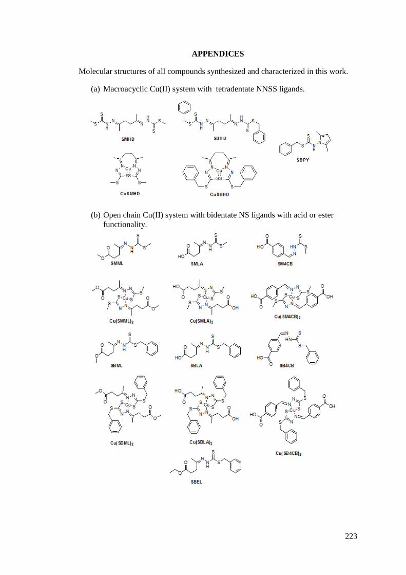
223
APPENDICES
Molecular structures of all compounds synthesized and characterized in this work.
(a) Macroacyclic Cu(II) system with tetradentate NNSS ligands.
(b) Open chain Cu(II) system with bidentate NS ligands with acid or ester functionality.
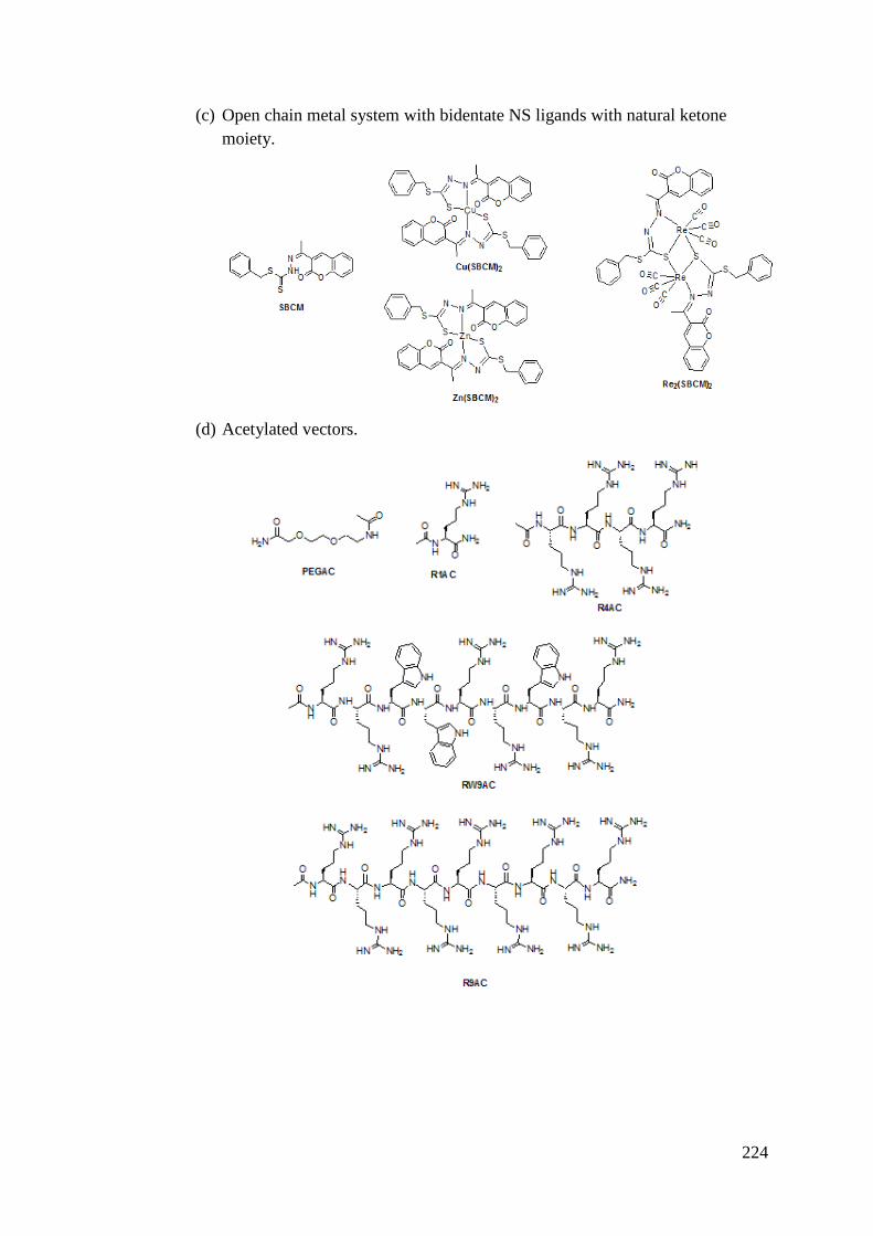
224
(c) Open chain metal system with bidentate NS ligands with natural ketone moiety.
(d) Acetylated vectors.
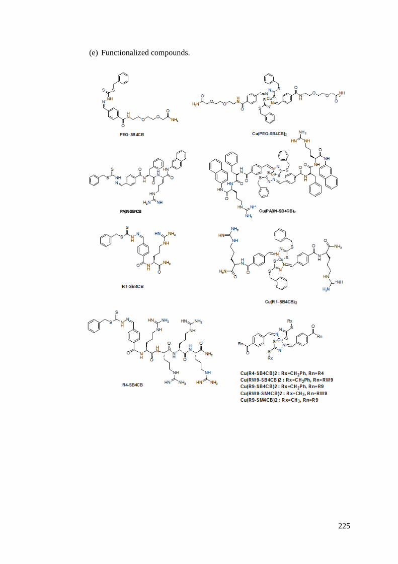
225
(e) Functionalized compounds.
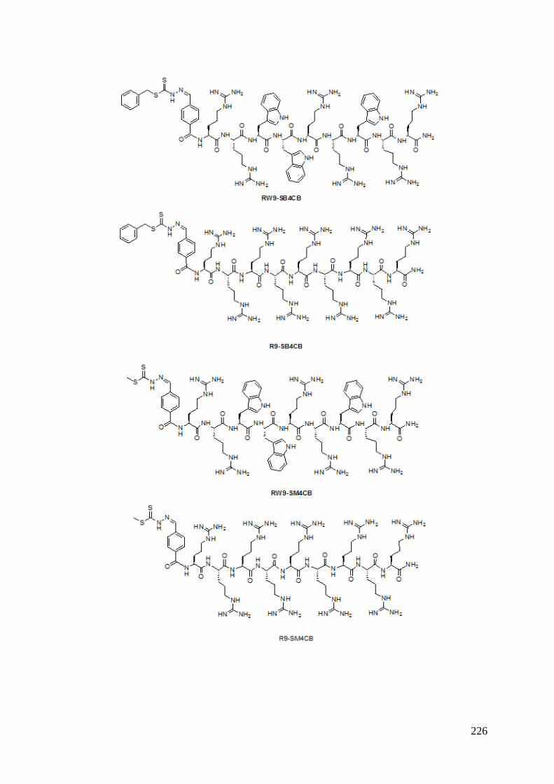
226
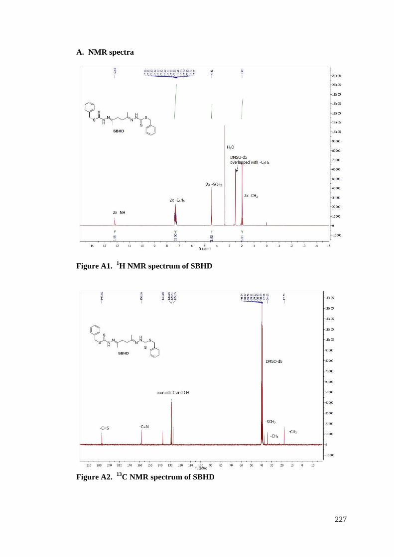
227
A. NMR spectra
Figure A1. 1H NMR spectrum of SBHD
Figure A2. 13C NMR spectrum of SBHD
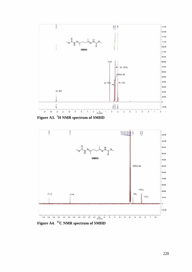
228
Figure A3. 1H NMR spectrum of SMHD
Figure A4. 13C NMR spectrum of SMHD
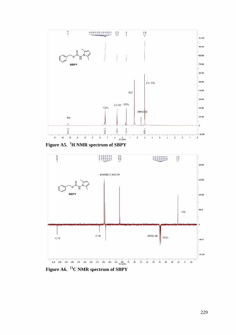
229
Figure A5. 1H NMR spectrum of SBPY
Figure A6. 13C NMR spectrum of SBPY
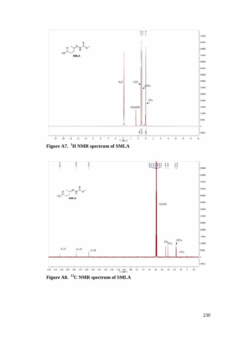
230
Figure A7. 1H NMR spectrum of SMLA
Figure A8. 13C NMR spectrum of SMLA
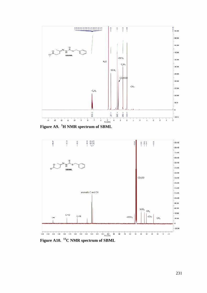
231
Figure A9. 1H NMR spectrum of SBML
Figure A10. 13C NMR spectrum of SBML
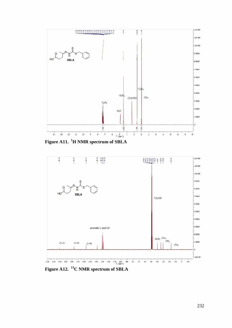
232
Figure A11. 1H NMR spectrum of SBLA
Figure A12. 13C NMR spectrum of SBLA
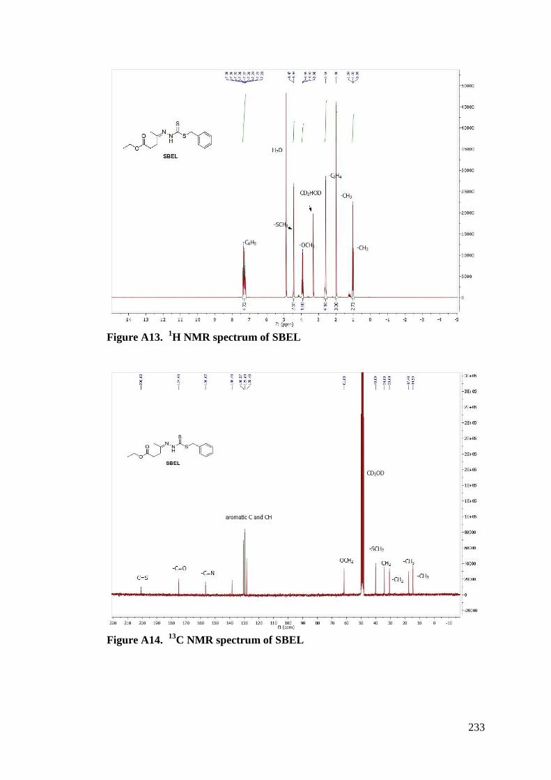
233
Figure A13. 1H NMR spectrum of SBEL
Figure A14. 13C NMR spectrum of SBEL
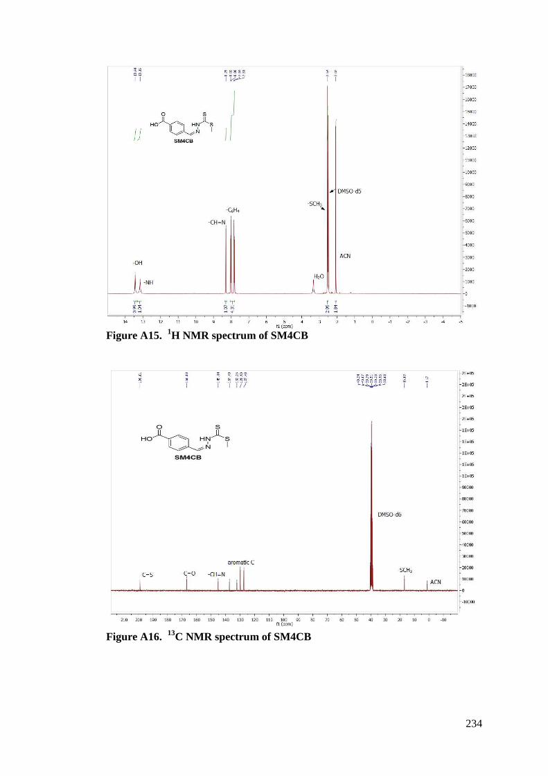
234
Figure A15. 1H NMR spectrum of SM4CB
Figure A16. 13C NMR spectrum of SM4CB
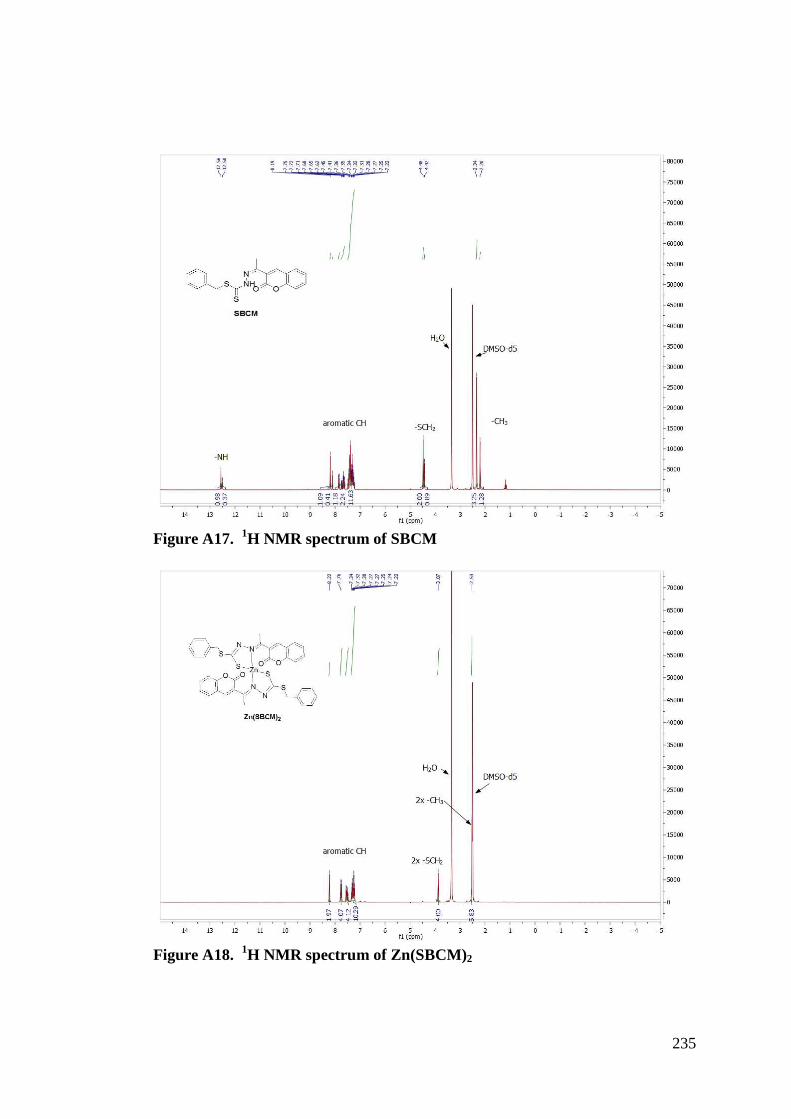
235
Figure A17. 1H NMR spectrum of SBCM
Figure A18. 1H NMR spectrum of Zn(SBCM)2
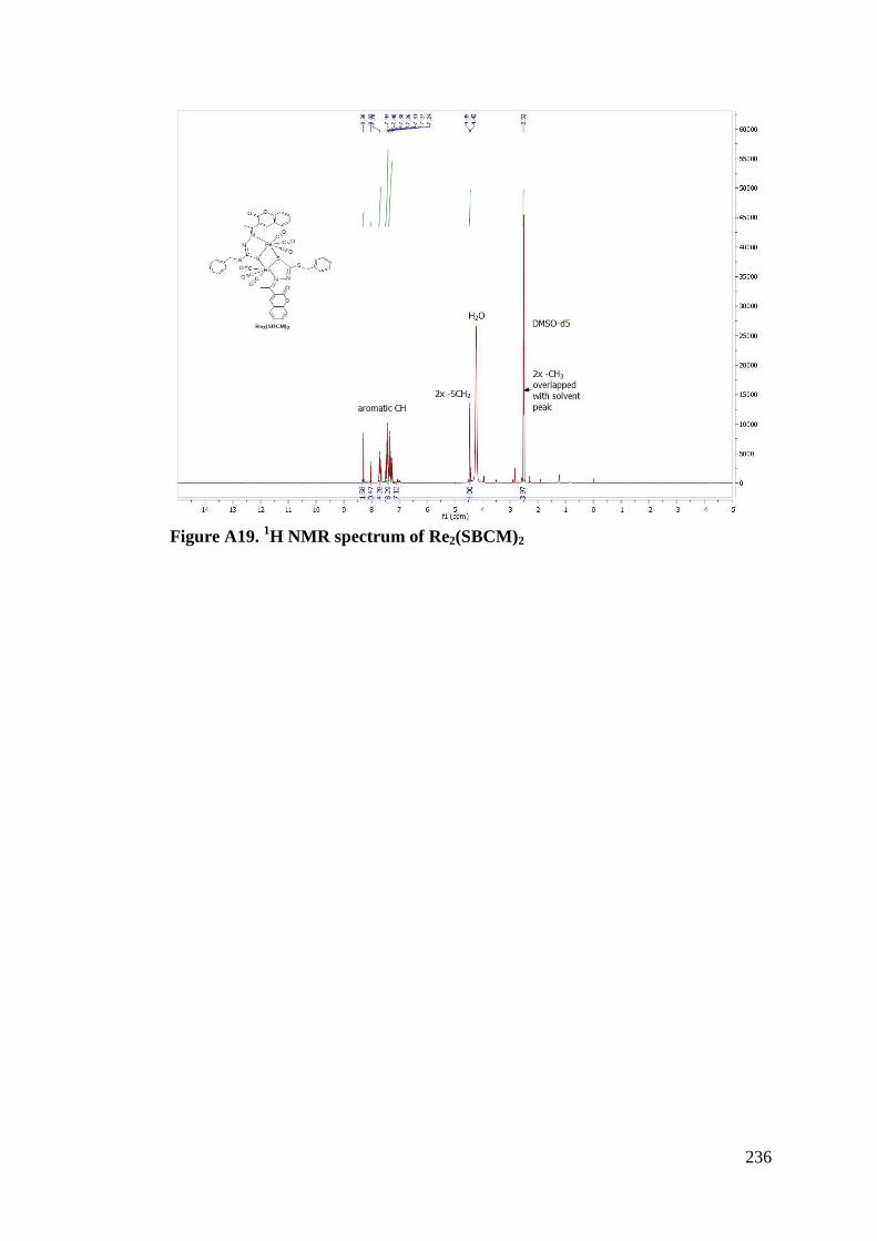
236
Figure A19. 1H NMR spectrum of Re2(SBCM)2
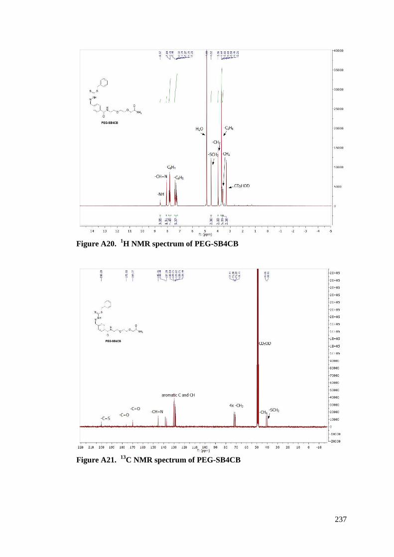
237
Figure A20. 1H NMR spectrum of PEG-SB4CB
Figure A21. 13C NMR spectrum of PEG-SB4CB
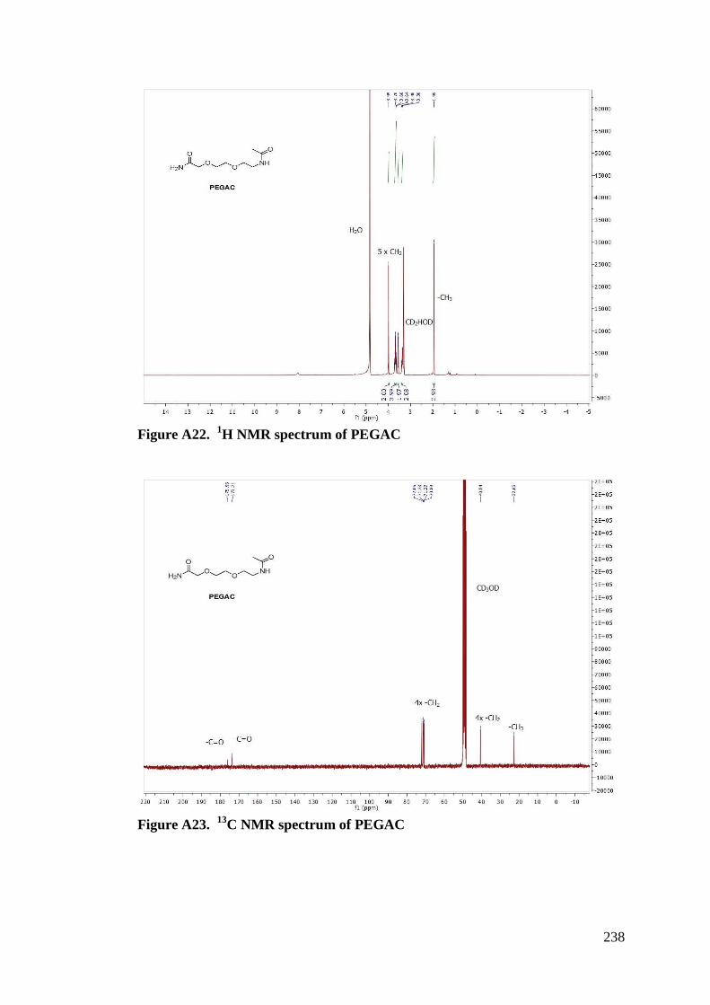
238
Figure A22. 1H NMR spectrum of PEGAC
Figure A23. 13C NMR spectrum of PEGAC
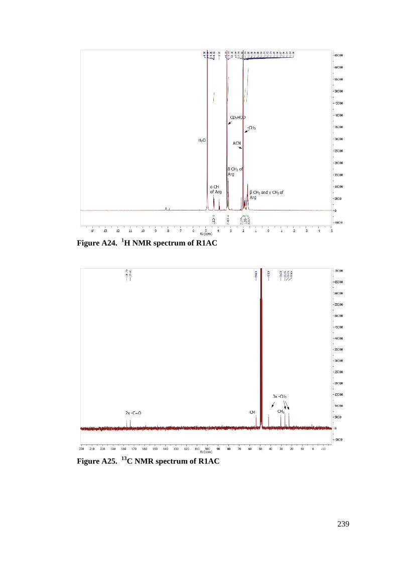
239
Figure A24. 1H NMR spectrum of R1AC
Figure A25. 13C NMR spectrum of R1AC
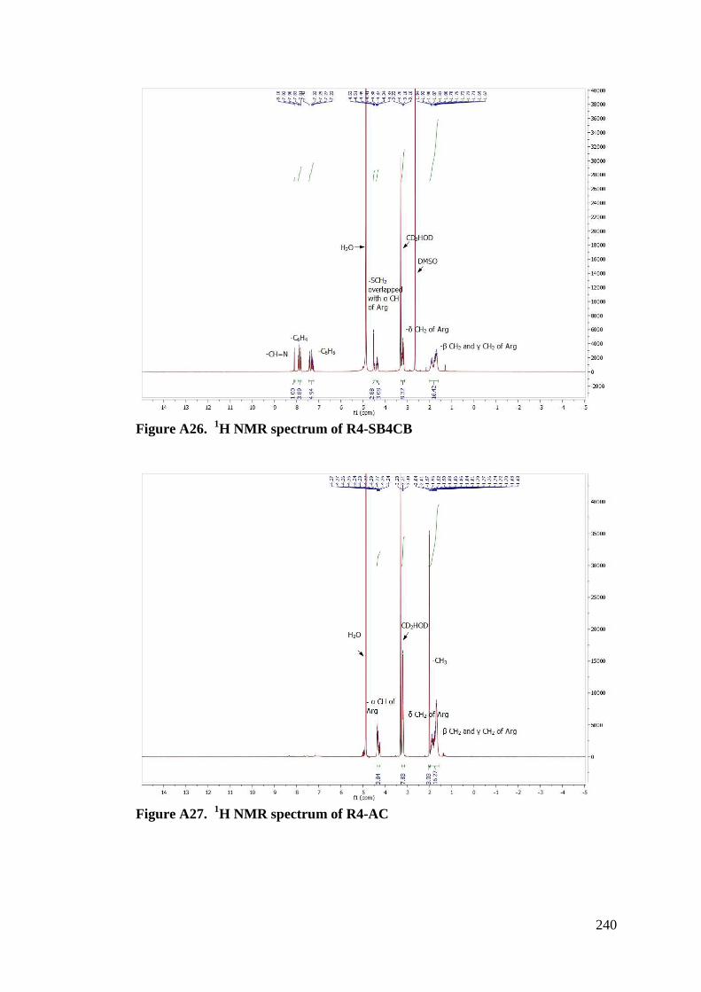
240
Figure A26. 1H NMR spectrum of R4-SB4CB
Figure A27. 1H NMR spectrum of R4-AC
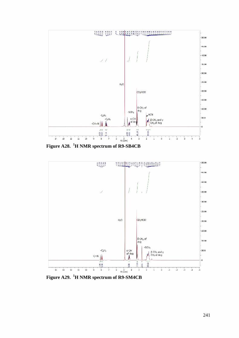
241
Figure A28. 1H NMR spectrum of R9-SB4CB
Figure A29. 1H NMR spectrum of R9-SM4CB
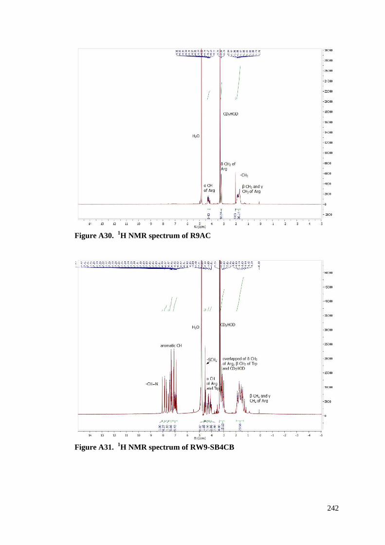
242
Figure A30. 1H NMR spectrum of R9AC
Figure A31. 1H NMR spectrum of RW9-SB4CB
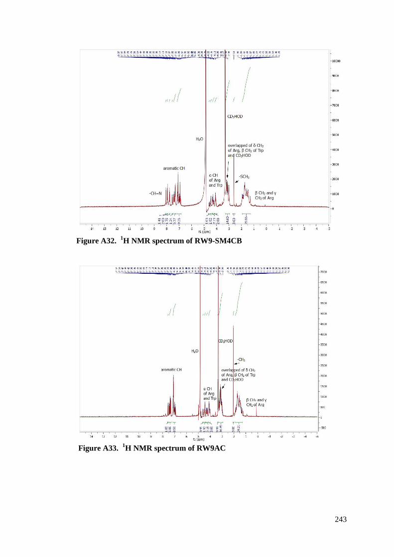
243
Figure A32. 1H NMR spectrum of RW9-SM4CB
Figure A33. 1H NMR spectrum of RW9AC
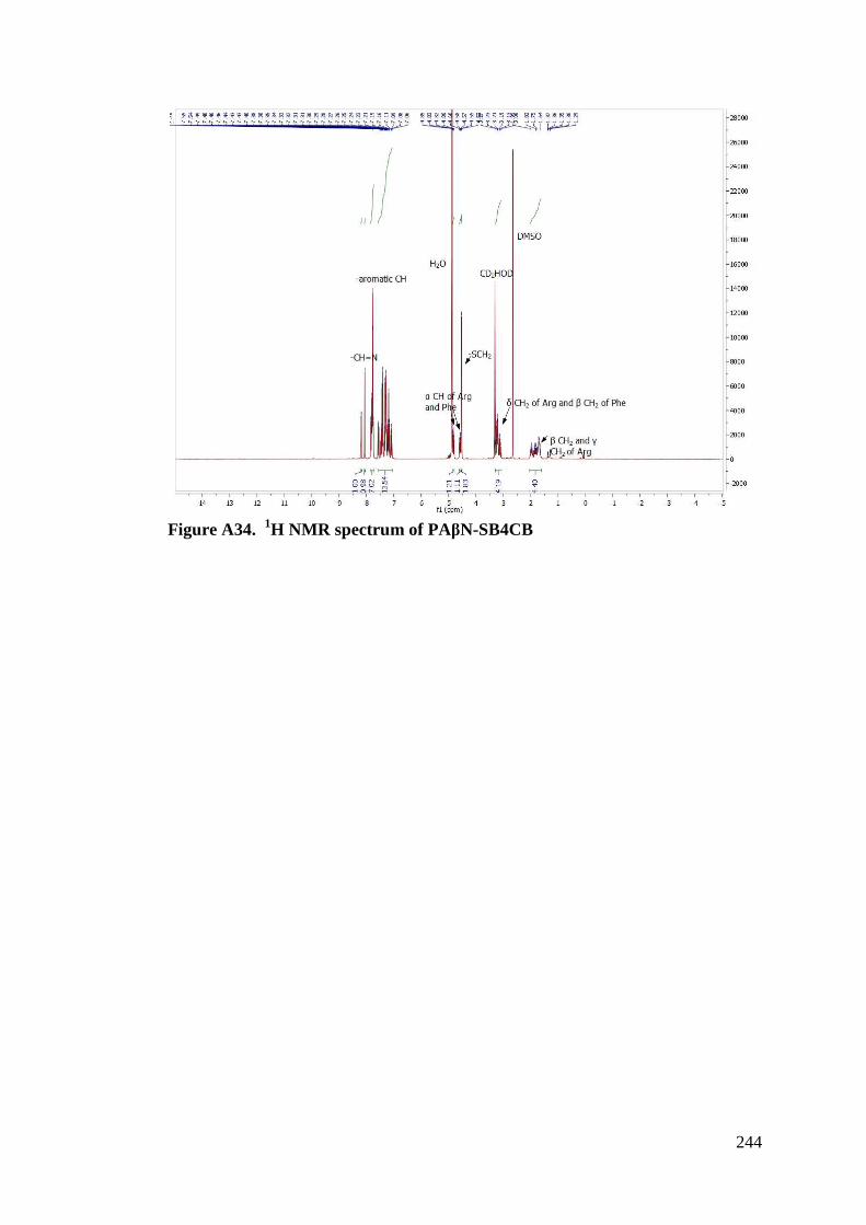
244
Figure A34. 1H NMR spectrum of PA"N-SB4CB
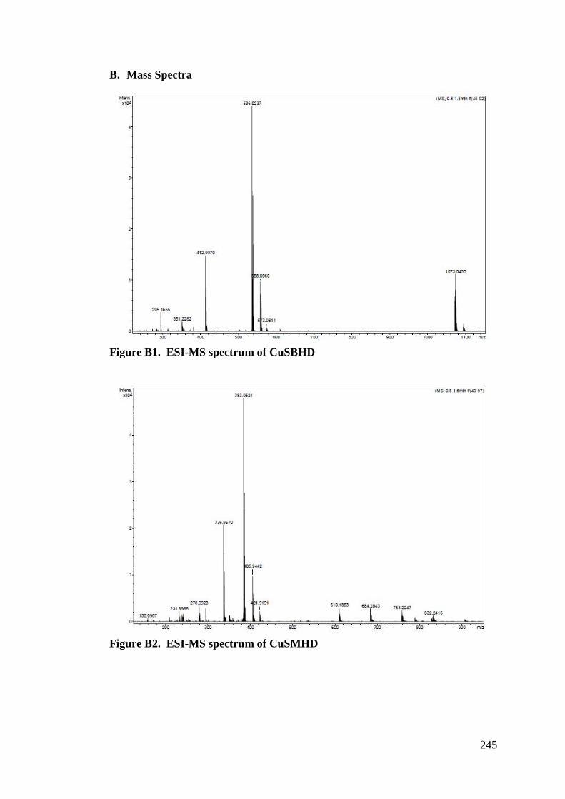
245
B. Mass Spectra
Figure B1. ESI-MS spectrum of CuSBHD
Figure B2. ESI-MS spectrum of CuSMHD
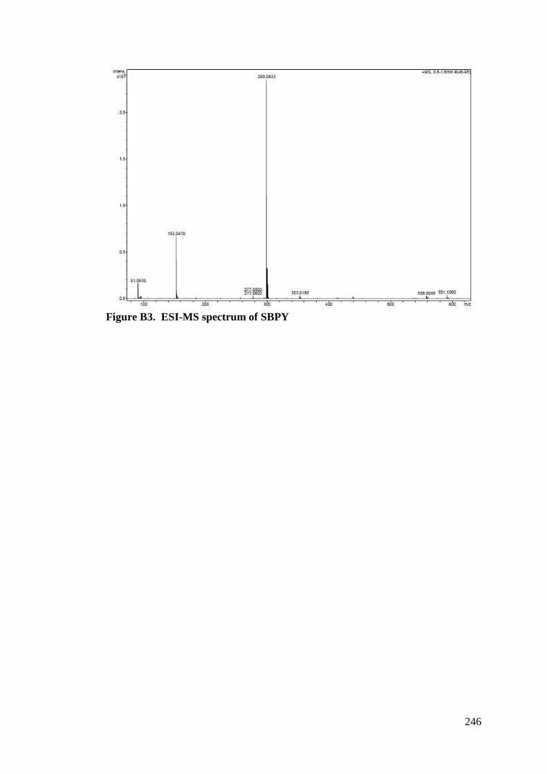
246
Figure B3. ESI-MS spectrum of SBPY
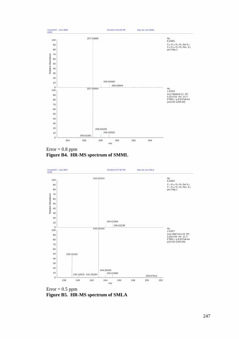
247
Error = 0.8 ppm Figure B4. HR-MS spectrum of SMML
Error = 0.5 ppm Figure B5. HR-MS spectrum of SMLA
\\tsclient\F\...\A12-0846 6/1/2012 8:35:09 PM May lee Low SMML24/05
254 256 258 260 262 264m/z
0
10
20
30
40
50
60
70
80
90
1000
10
20
30
40
50
60
70
80
90
100
Rel
ativ
e Ab
unda
nce
257.03889
259.03469
260.03804257.03934
259.03505258.04228
256.62391
NL:8.15E5C 8 H 14 O 2 N 2 Na S 2: C 8 H 14 O 2 N 2 Na 1 S 2pa Chrg 1
NL:1.02E8A12-0846#9-21 RT: 0.20-0.53 AV: 13 T: FTMS + p ESI Full ms [110.00-1200.00]
\\tsclient\F\...\A12-0847 6/1/2012 8:37:50 PM May lee Low SMLA24/05
238 240 242 244 246 248 250 252m/z
0
10
20
30
40
50
60
70
80
90
1000
10
20
30
40
50
60
70
80
90
100
Rel
ativ
e Ab
unda
nce
243.02324
245.01904
246.02239243.02334
239.16192
245.01883244.02630
240.16515 242.26284 250.67614
NL:8.24E5C 7 H 12 O 2 N 2 Na S 2: C 7 H 12 O 2 N 2 Na 1 S 2pa Chrg 1
NL:1.51E7A12-0847#12-23 RT: 0.28-0.59 AV: 12 T: FTMS + p ESI Full ms [110.00-1200.00]
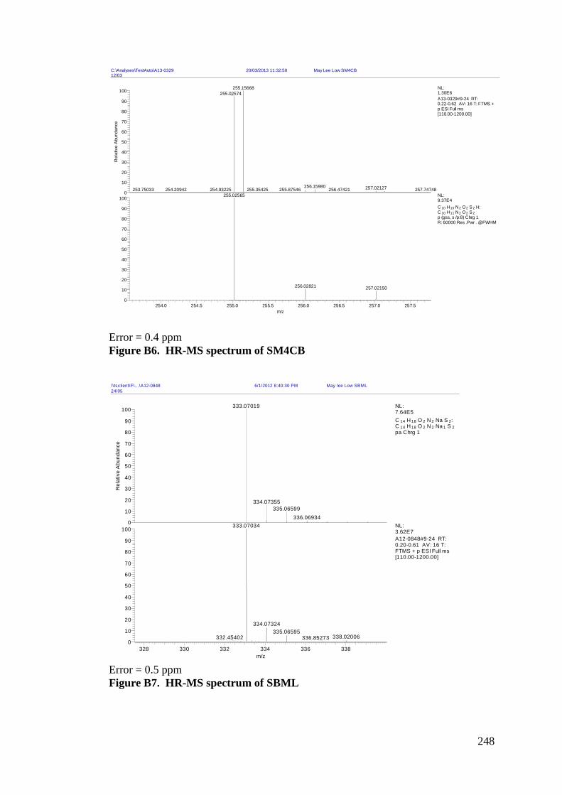
248
Error = 0.4 ppm Figure B6. HR-MS spectrum of SM4CB
Error = 0.5 ppm Figure B7. HR-MS spectrum of SBML
C:\Analyses\TestAuto\A13-0329 20/03/2013 11:32:50 May Lee Low SM4CB12/03
254.0 254.5 255.0 255.5 256.0 256.5 257.0 257.5m/z
0
10
20
30
40
50
60
70
80
90
1000
10
20
30
40
50
60
70
80
90
100
Rel
ativ
e Ab
unda
nce
255.15668255.02574
256.15980 257.02127255.35425 256.47421254.93225 257.74748254.20942253.75033 255.87546255.02565
256.02821 257.02150
NL:1.30E6A13-0329#9-24 RT: 0.22-0.62 AV: 16 T: FTMS + p ESI Full ms [110.00-1200.00]
NL:9.37E4C 10 H10 N2 O2 S 2 H: C 10 H11 N2 O2 S 2p (gss, s /p:8) Chrg 1R: 60000 Res .Pwr . @FWHM
\\tsclient\F\...\A12-0848 6/1/2012 8:40:30 PM May lee Low SBML24/05
328 330 332 334 336 338m/z
0
10
20
30
40
50
60
70
80
90
1000
10
20
30
40
50
60
70
80
90
100
Rel
ativ
e A
bund
ance
333.07019
334.07355335.06599
336.06934333.07034
334.07324335.06595
338.02006332.45402 336.85273
NL:7.64E5C 14 H 18 O 2 N 2 Na S 2: C 14 H 18 O 2 N 2 Na 1 S 2pa Chrg 1
NL:3.62E7A12-0848#9-24 RT: 0.20-0.61 AV: 16 T: FTMS + p ESI Full ms [110.00-1200.00]
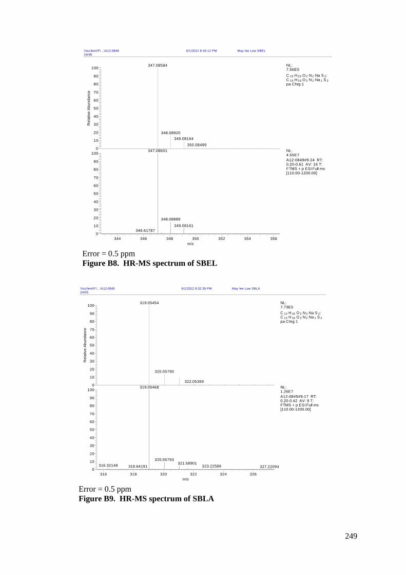
249
Error = 0.5 ppm Figure B8. HR-MS spectrum of SBEL
Error = 0.5 ppm Figure B9. HR-MS spectrum of SBLA
\\tsclient\F\...\A12-0849 6/1/2012 8:43:12 PM May lee Low SBEL24/05
344 346 348 350 352 354 356m/z
0
10
20
30
40
50
60
70
80
90
1000
10
20
30
40
50
60
70
80
90
100
Rel
ativ
e Ab
unda
nce
347.08584
348.08920349.08164
350.08499347.08601
348.08889349.08161
346.61787
NL:7.56E5C 15 H 20 O 2 N 2 Na S 2: C 15 H 20 O 2 N 2 Na 1 S 2pa Chrg 1
NL:4.55E7A12-0849#9-24 RT: 0.20-0.61 AV: 16 T: FTMS + p ESI Full ms [110.00-1200.00]
\\tsclient\F\...\A12-0845 6/1/2012 8:32:30 PM May lee Low SBLA24/05
316 318 320 322 324 326m/z
0
10
20
30
40
50
60
70
80
90
1000
10
20
30
40
50
60
70
80
90
100
Rel
ativ
e A
bund
ance
319.05454
320.05790
322.05369319.05468
320.05793316.32148 323.22589318.64191
321.58901327.22094
NL:7.73E5C 13 H 16 O 2 N 2 Na S 2: C 13 H 16 O 2 N 2 Na 1 S 2pa Chrg 1
NL:1.26E7A12-0845#9-17 RT: 0.20-0.42 AV: 9 T: FTMS + p ESI Full ms [110.00-1200.00]
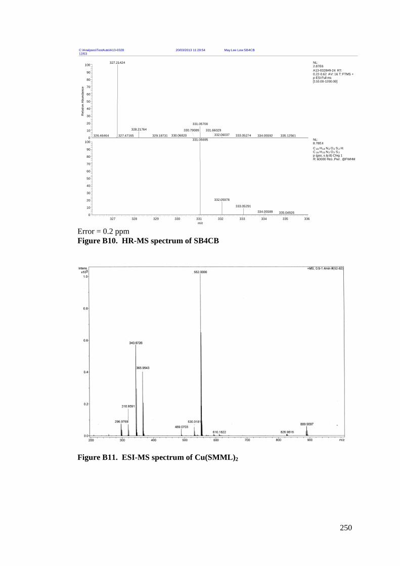
250
Error = 0.2 ppm Figure B10. HR-MS spectrum of SB4CB
Figure B11. ESI-MS spectrum of Cu(SMML)2
C:\Analyses\TestAuto\A13-0328 20/03/2013 11:29:54 May Lee Low SB4CB12/03
327 328 329 330 331 332 333 334 335 336m/z
0
10
20
30
40
50
60
70
80
90
1000
10
20
30
40
50
60
70
80
90
100
Rel
ativ
e Ab
unda
nce
327.21424
331.05700328.21764
332.06037 333.05274330.06820329.18731327.47165 335.12561334.05592326.46464330.79089 331.66029
331.05695
332.05978
333.05291334.05589 335.04926
NL:2.87E6A13-0328#9-24 RT: 0.22-0.62 AV: 16 T: FTMS + p ESI Full ms [110.00-1200.00]
NL:8.78E4C 16 H14 N2 O2 S 2 H: C 16 H15 N2 O2 S 2p (gss, s /p:8) Chrg 1R: 60000 Res .Pwr . @FWHM
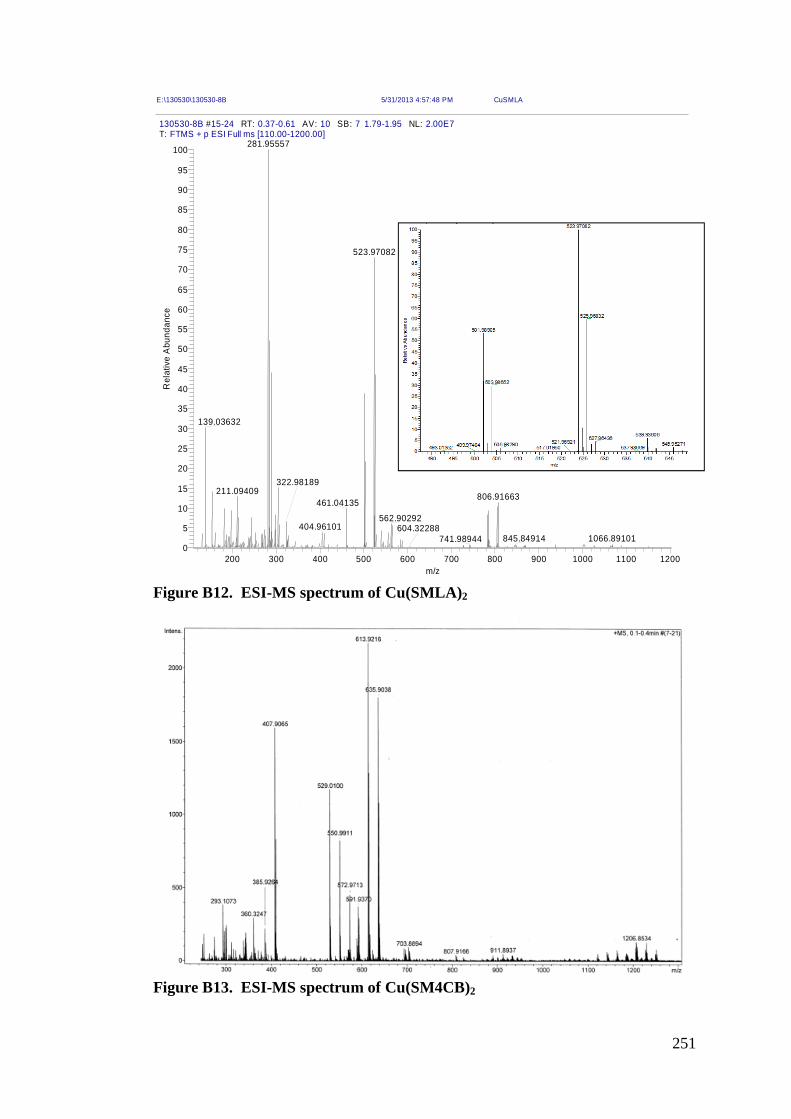
251
Figure B12. ESI-MS spectrum of Cu(SMLA)2
Figure B13. ESI-MS spectrum of Cu(SM4CB)2
E:\130530\130530-8B 5/31/2013 4:57:48 PM CuSMLA
130530-8B #15-24 RT: 0.37-0.61 AV: 10 SB: 7 1.79-1.95 NL: 2.00E7T: FTMS + p ESI Full ms [110.00-1200.00]
200 300 400 500 600 700 800 900 1000 1100 1200m/z
0
5
10
15
20
25
30
35
40
45
50
55
60
65
70
75
80
85
90
95
100
Rel
ativ
e A
bund
ance
281.95557
523.97082
139.03632
211.09409 806.91663461.04135
322.98189
562.90292404.96101
1066.89101845.84914741.98944604.32288
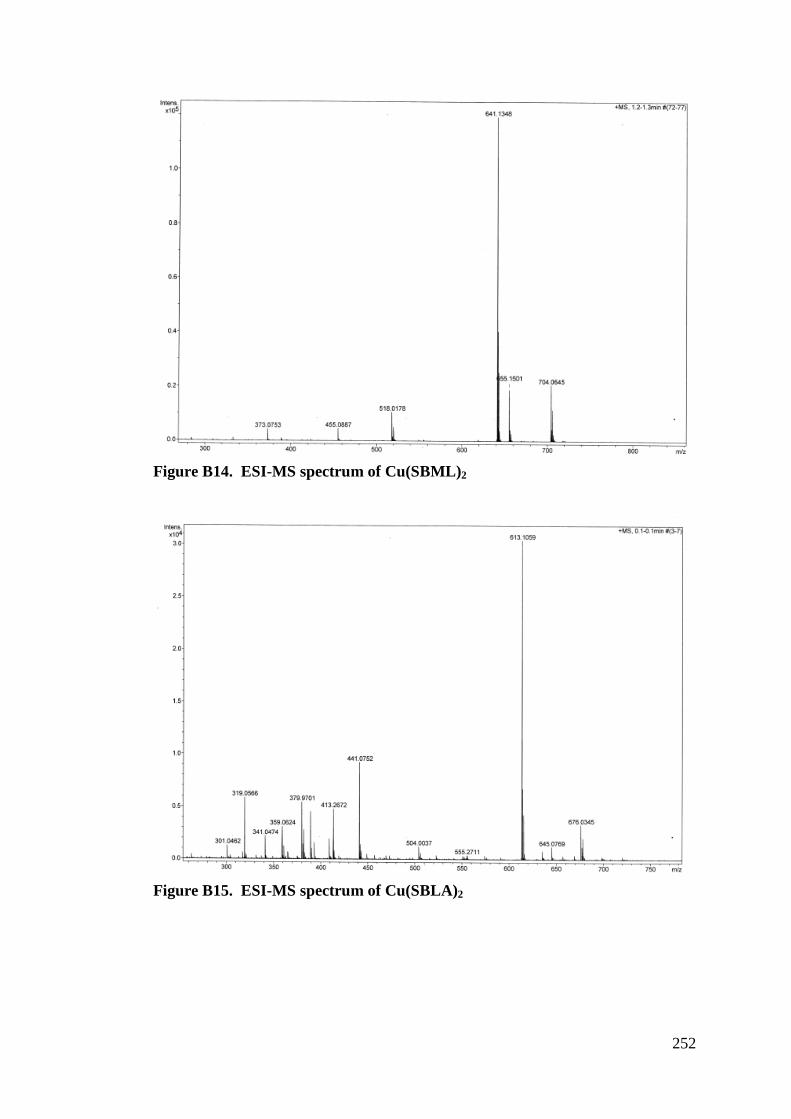
252
Figure B14. ESI-MS spectrum of Cu(SBML)2
Figure B15. ESI-MS spectrum of Cu(SBLA)2
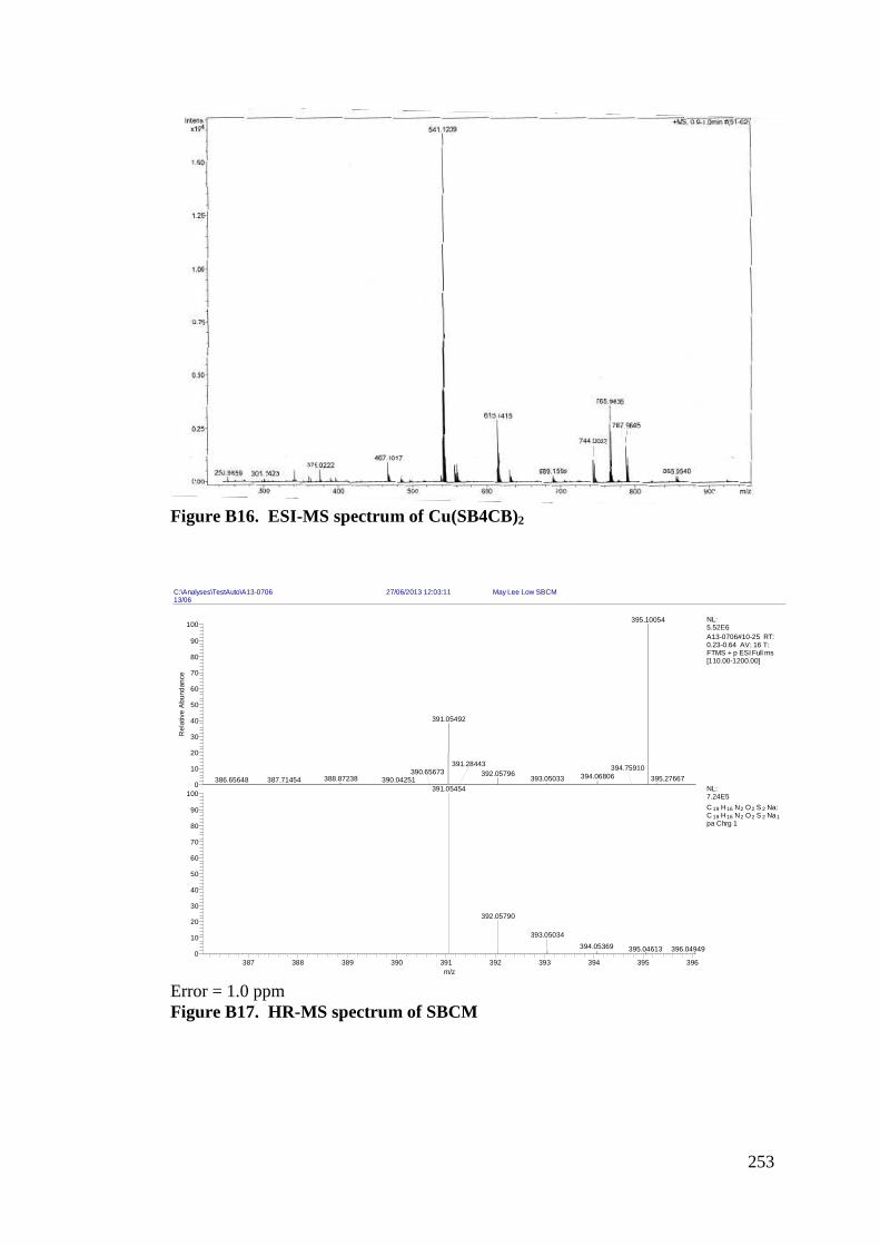
253
Figure B16. ESI-MS spectrum of Cu(SB4CB)2
Error = 1.0 ppm Figure B17. HR-MS spectrum of SBCM
C:\Analyses\TestAuto\A13-0706 27/06/2013 12:03:11 May Lee Low SBCM13/06
387 388 389 390 391 392 393 394 395 396m/z
0
10
20
30
40
50
60
70
80
90
1000
10
20
30
40
50
60
70
80
90
100
Rel
ativ
e Ab
unda
nce
395.10054
391.05492
392.05796 394.06806 395.27667393.05033388.87238
391.28443 394.75910390.04251386.65648
390.65673387.71454
391.05454
392.05790
393.05034394.05369 395.04613 396.04949
NL:5.52E6A13-0706#10-25 RT: 0.23-0.64 AV: 16 T: FTMS + p ESI Full ms [110.00-1200.00]
NL:7.24E5C 19 H16 N2 O2 S 2 Na: C 19 H16 N2 O2 S 2 Na1pa Chrg 1
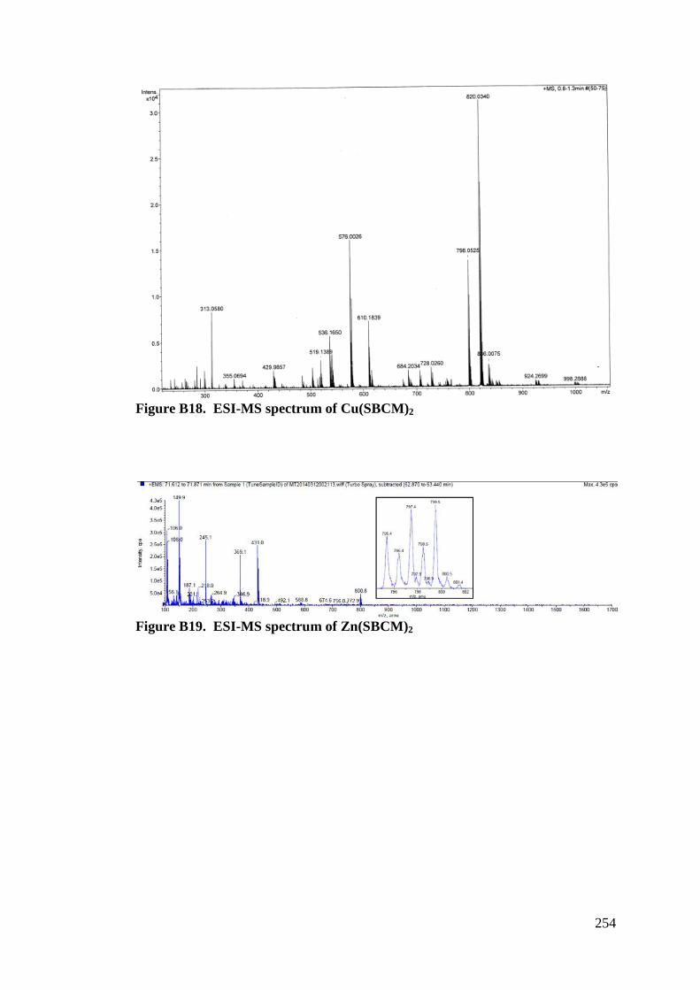
254
Figure B18. ESI-MS spectrum of Cu(SBCM)2
Figure B19. ESI-MS spectrum of Zn(SBCM)2
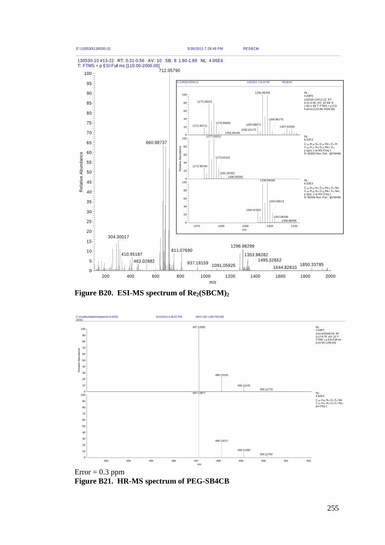
255
Figure B20. ESI-MS spectrum of Re2(SBCM)2
Error = 0.3 ppm Figure B21. HR-MS spectrum of PEG-SB4CB
E:\130530\130530-10 5/30/2013 7:28:49 PM RESBCM
130530-10 #13-22 RT: 0.31-0.56 AV: 10 SB: 8 1.80-1.99 NL: 4.06E6T: FTMS + p ESI Full ms [110.00-2000.00]
200 400 600 800 1000 1200 1400 1600 1800 2000m/z
0
5
10
15
20
25
30
35
40
45
50
55
60
65
70
75
80
85
90
95
100R
elat
ive
Abun
danc
e
712.05790
660.98737
304.30017
1296.98299811.07690
410.95187463.02882 937.18159 1850.337851091.05925
1353.98282
1644.828101495.32652
C:\Xcalibur\data\Analyses\A14-0255 3/12/2014 4:48:52 PM MAY LEE LOW PEGSB20/02
493 494 495 496 497 498 499 500 501 502m/z
0
10
20
30
40
50
60
70
80
90
1000
10
20
30
40
50
60
70
80
90
100
Rel
ativ
e A
bund
ance
497.12892
498.13159
499.12470500.12778
497.12877
498.13212
499.12456500.12792
NL:2.54E7A14-0255#8-29 RT: 0.17-0.75 AV: 22 T: FTMS + p ESI Full ms [110.00-1200.00]
NL:6.92E5C 22 H26 N4 O4 S 2 Na: C 22 H26 N4 O4 S 2 Na 1pa Chrg 1
E:\130530\130530-10 5/30/2013 7:28:49 PM RESBCM
1270 1280 1290 1300 1310m/z
0
20
40
60
80
1000
20
40
60
80
100
Rel
ativ
e A
bund
ance
0
20
40
60
80
1001296.98299
1275.00055
1294.980711272.99721 1307.02630
1300.981751278.99858
1283.481881292.41175
1277.00251
1279.00321
1272.99766
1281.003021285.00262
1298.98446
1300.98515
1294.97961
1302.984961306.98456
NL:4.44E5130530-10#13-22 RT: 0.31-0.56 AV: 10 SB: 8 1.80-1.99 T: FTMS + p ESI Full ms [110.00-2000.00]
NL:6.02E3C 44 H 30 N 4 O 10 Re 2 S 4 H: C 44 H 31 N 4 O 10 Re 2 S 4p (gss, s /p:40) Chrg 1R: 60000 Res .Pwr . @FWHM
NL:6.03E3C 44 H 30 N 4 O 10 Re 2 S 4 Na: C 44 H 30 N 4 O 10 Re 2 S 4 Na 1p (gss, s /p:40) Chrg 1R: 60000 Res .Pwr . @FWHM
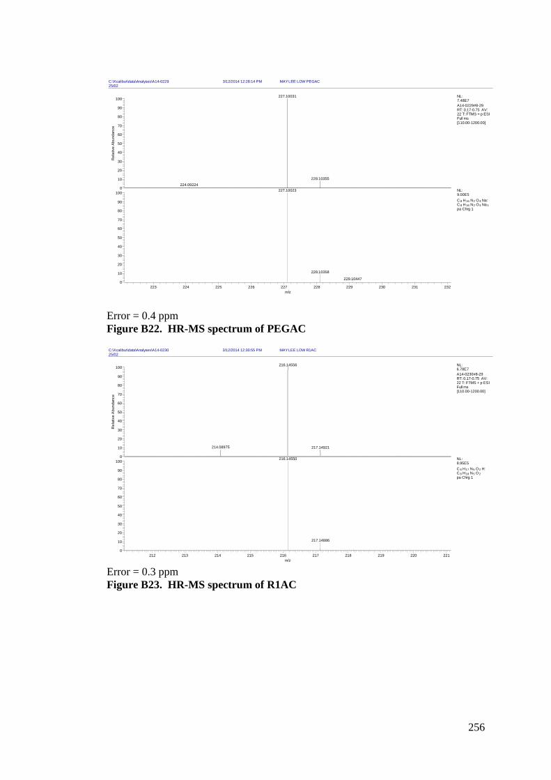
256
Error = 0.4 ppm Figure B22. HR-MS spectrum of PEGAC
Error = 0.3 ppm Figure B23. HR-MS spectrum of R1AC
C:\Xcalibur\data\Analyses\A14-0229 3/12/2014 12:28:14 PM MAY LEE LOW PEGAC25/02
223 224 225 226 227 228 229 230 231 232m/z
0
10
20
30
40
50
60
70
80
90
1000
10
20
30
40
50
60
70
80
90
100
Rel
ativ
e Ab
unda
nce
227.10031
228.10355224.09224
227.10023
228.10358
229.10447
NL:7.48E7A14-0229#8-29 RT: 0.17-0.75 AV: 22 T: FTMS + p ESI Full ms [110.00-1200.00]
NL:9.00E5C 8 H16 N2 O4 Na: C 8 H16 N2 O4 Na 1pa Chrg 1
C:\Xcalibur\data\Analyses\A14-0230 3/12/2014 12:30:55 PM MAY LEE LOW R1AC25/02
212 213 214 215 216 217 218 219 220 221m/z
0
10
20
30
40
50
60
70
80
90
1000
10
20
30
40
50
60
70
80
90
100
Rel
ativ
e A
bund
ance
216.14556
214.08975 217.14921
216.14550
217.14886
NL:6.78E7A14-0230#8-29 RT: 0.17-0.75 AV: 22 T: FTMS + p ESI Full ms [110.00-1200.00]
NL:8.95E5C 8 H17 N5 O2 H: C 8 H18 N5 O2pa Chrg 1
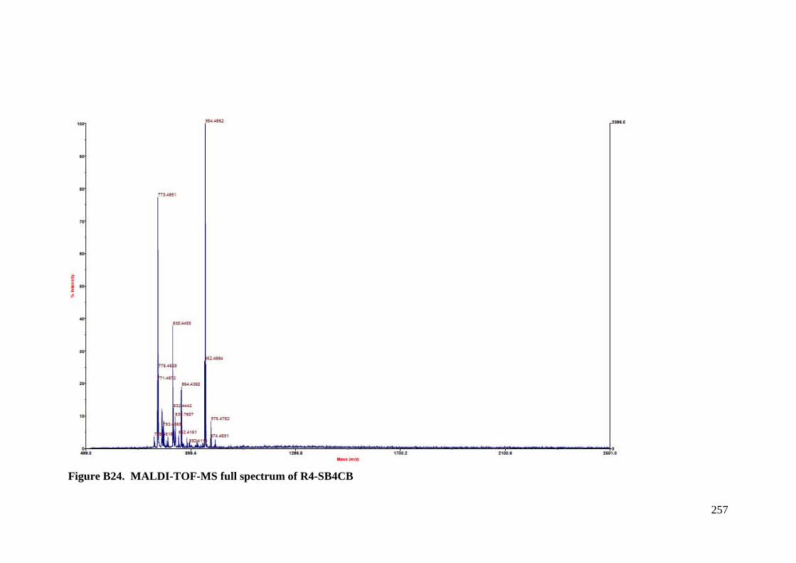
257
Figure B24. MALDI-TOF-MS full spectrum of R4-SB4CB
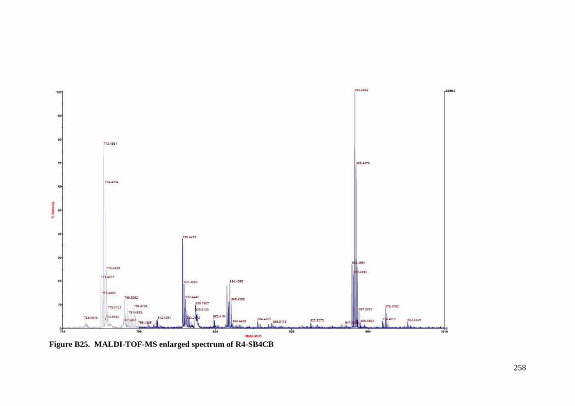
258
Figure B25. MALDI-TOF-MS enlarged spectrum of R4-SB4CB
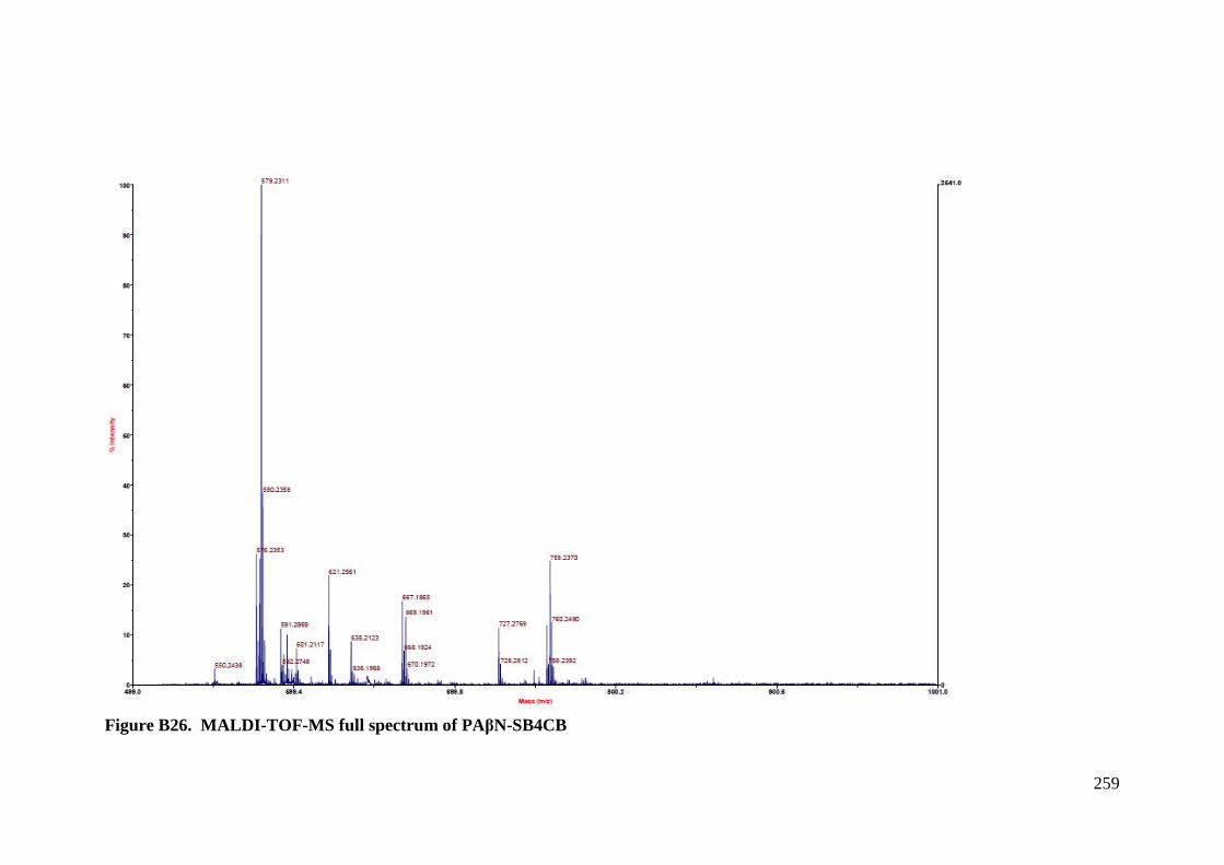
259
Figure B26. MALDI-TOF-MS full spectrum of PA"N-SB4CB
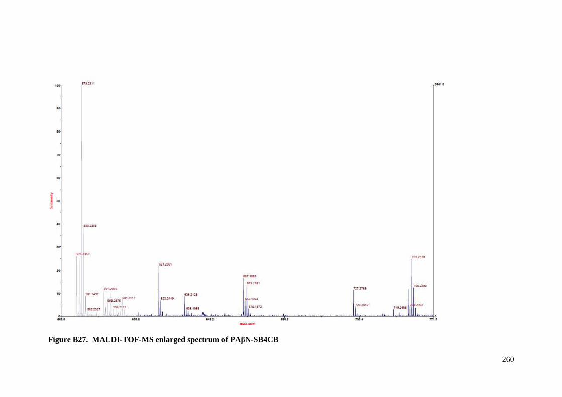
260
Figure B27. MALDI-TOF-MS enlarged spectrum of PA"N-SB4CB
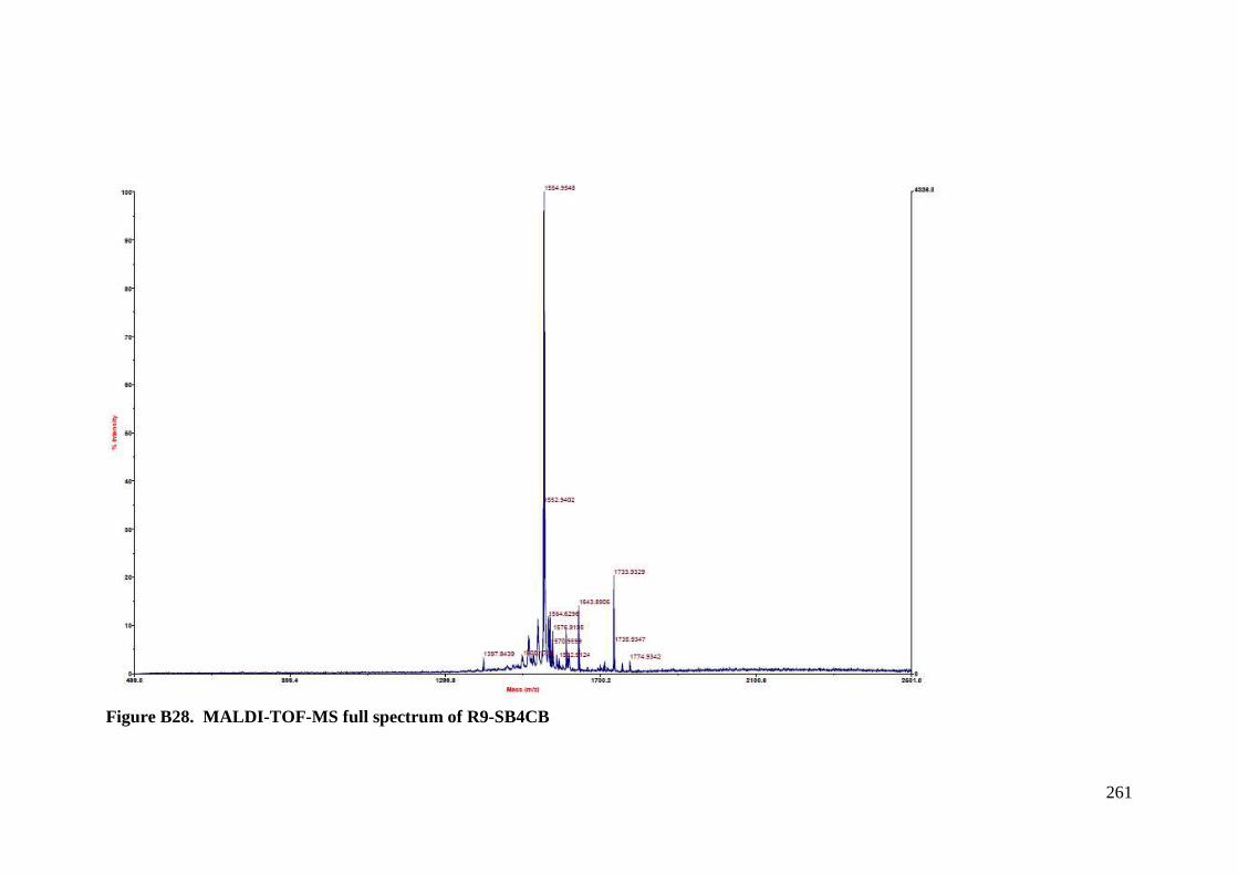
261
Figure B28. MALDI-TOF-MS full spectrum of R9-SB4CB
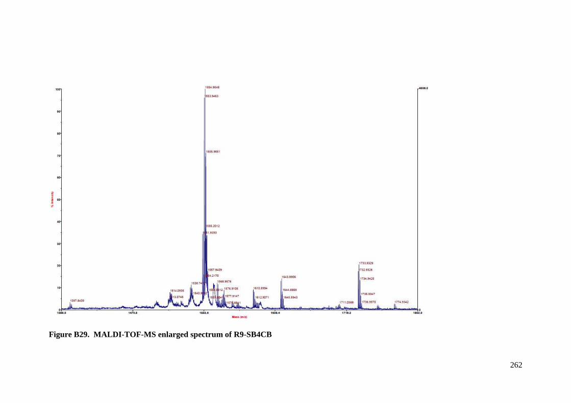
262
Figure B29. MALDI-TOF-MS enlarged spectrum of R9-SB4CB
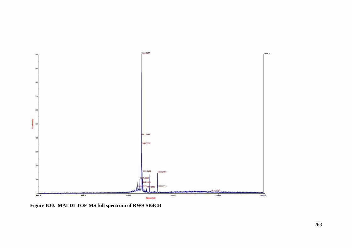
263
Figure B30. MALDI-TOF-MS full spectrum of RW9-SB4CB
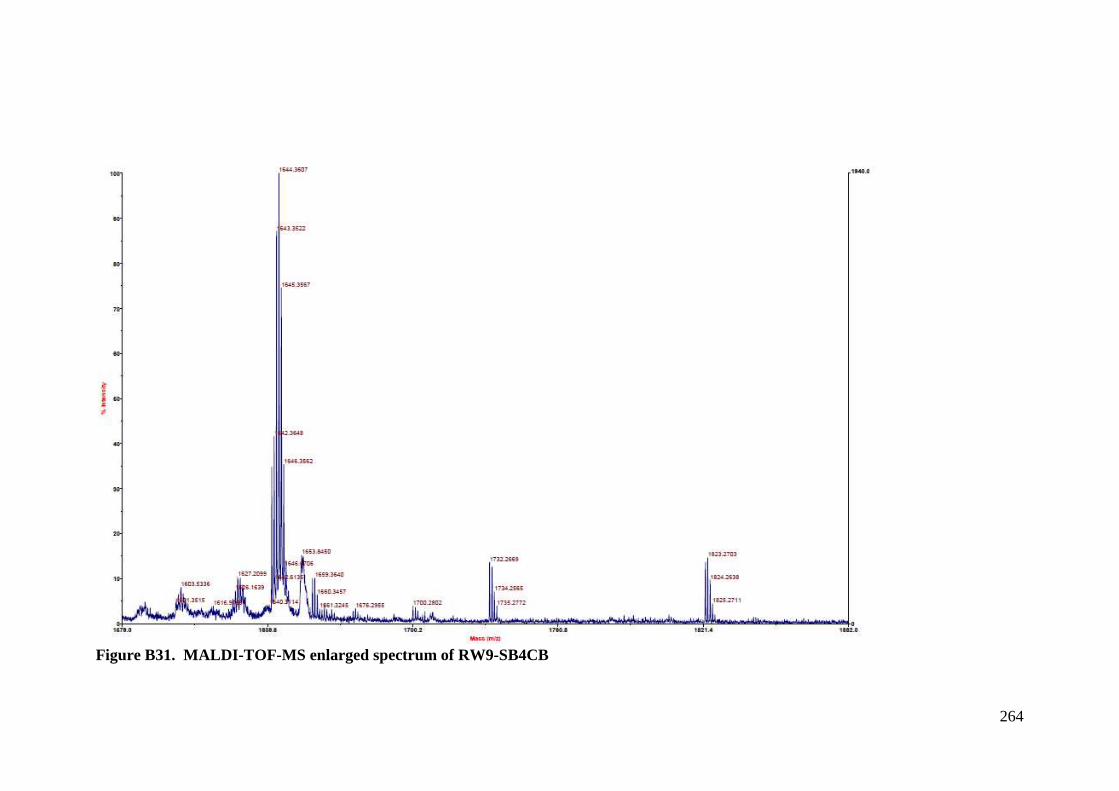
264
Figure B31. MALDI-TOF-MS enlarged spectrum of RW9-SB4CB
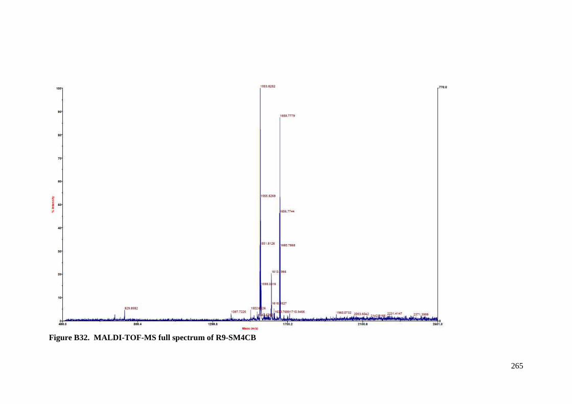
265
Figure B32. MALDI-TOF-MS full spectrum of R9-SM4CB
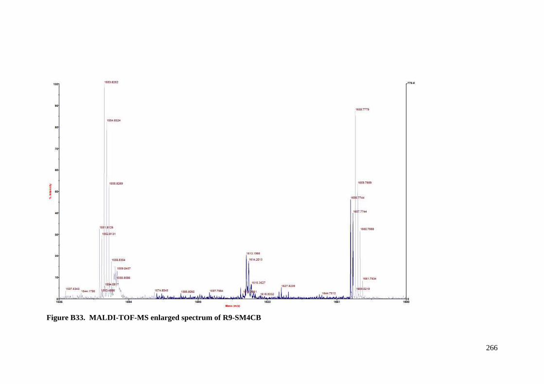
266
Figure B33. MALDI-TOF-MS enlarged spectrum of R9-SM4CB
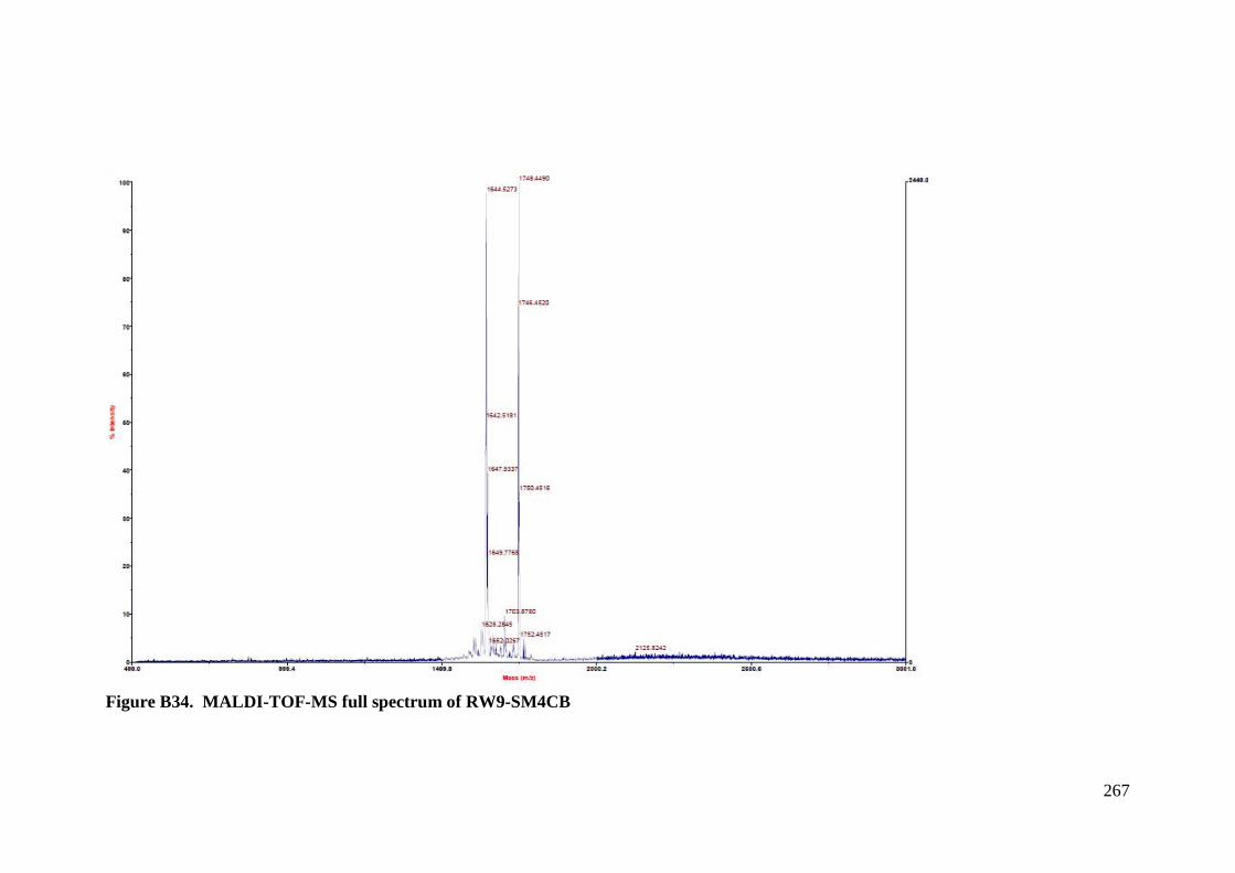
267
Figure B34. MALDI-TOF-MS full spectrum of RW9-SM4CB
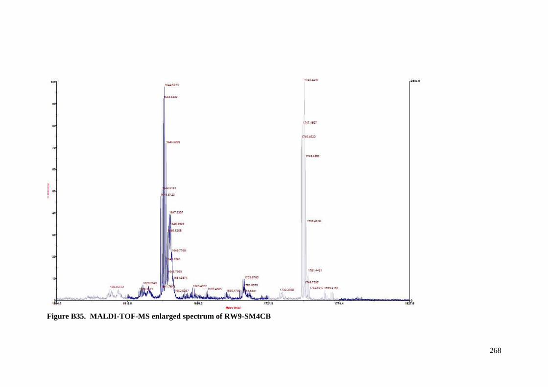
268
Figure B35. MALDI-TOF-MS enlarged spectrum of RW9-SM4CB
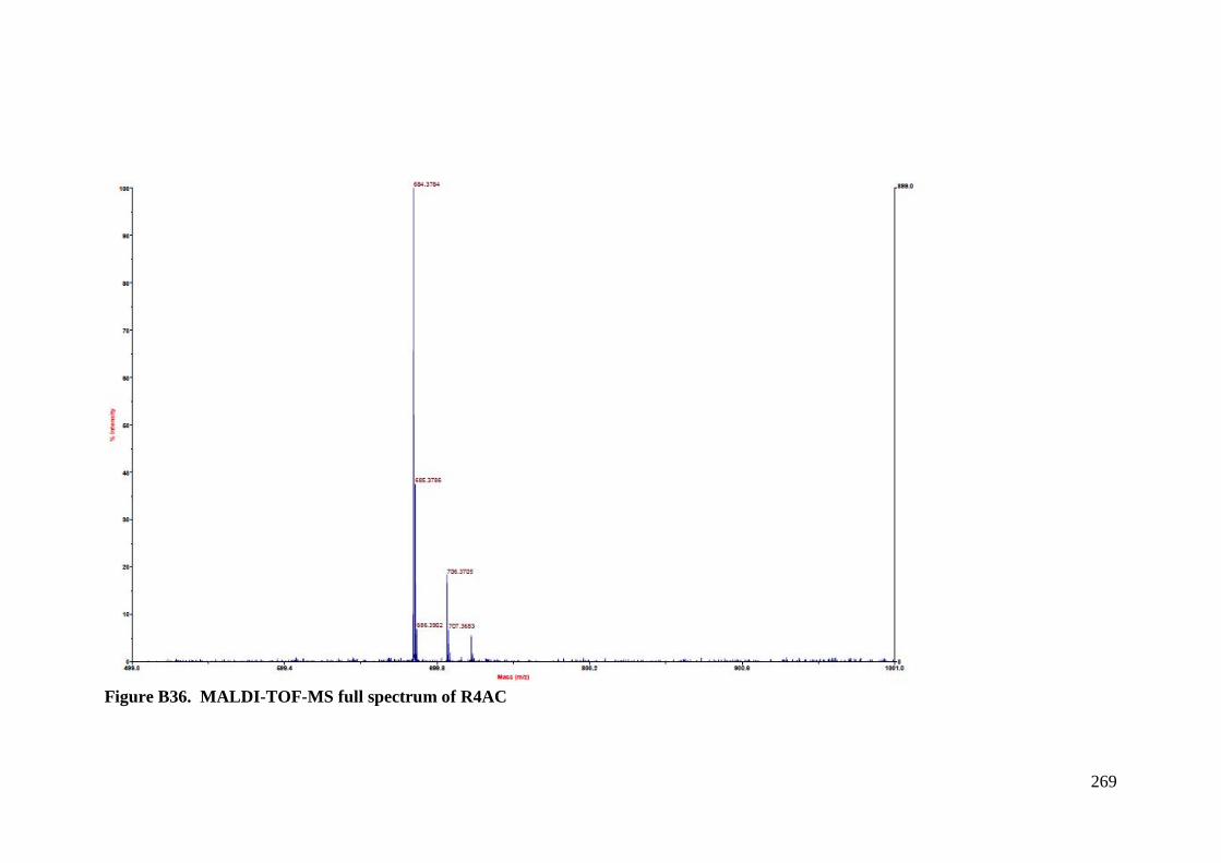
269
Figure B36. MALDI-TOF-MS full spectrum of R4AC
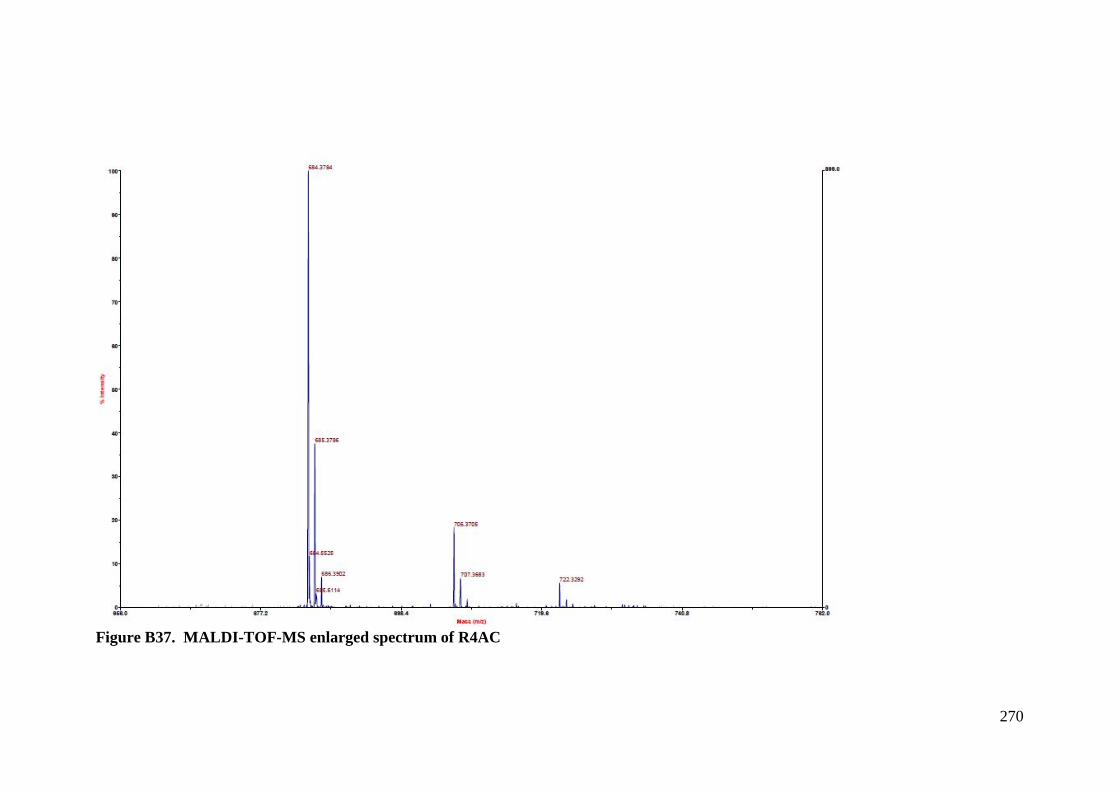
270
Figure B37. MALDI-TOF-MS enlarged spectrum of R4AC
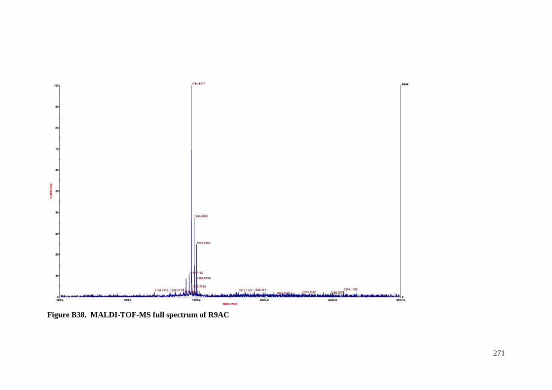
271
Figure B38. MALDI-TOF-MS full spectrum of R9AC
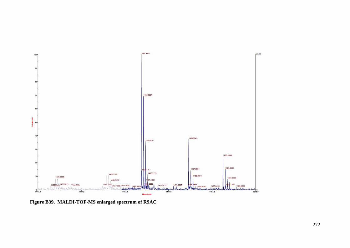
272
Figure B39. MALDI-TOF-MS enlarged spectrum of R9AC
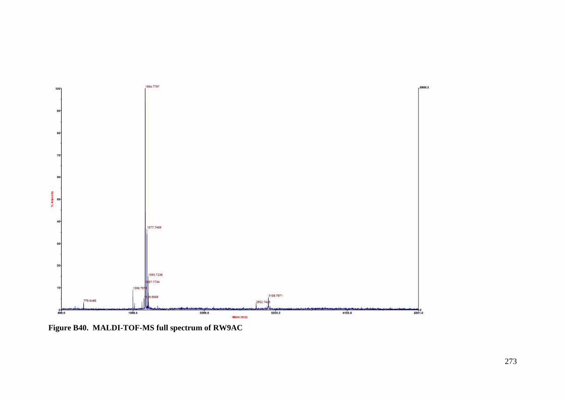
273
Figure B40. MALDI-TOF-MS full spectrum of RW9AC
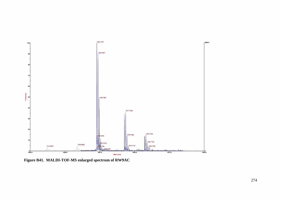
274
Figure B41. MALDI-TOF-MS enlarged spectrum of RW9AC
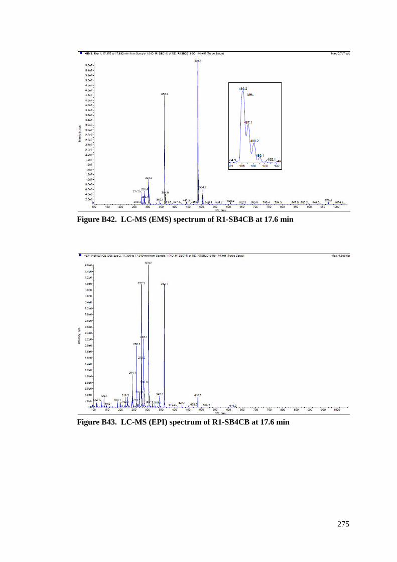
275
Figure B42. LC-MS (EMS) spectrum of R1-SB4CB at 17.6 min
Figure B43. LC-MS (EPI) spectrum of R1-SB4CB at 17.6 min
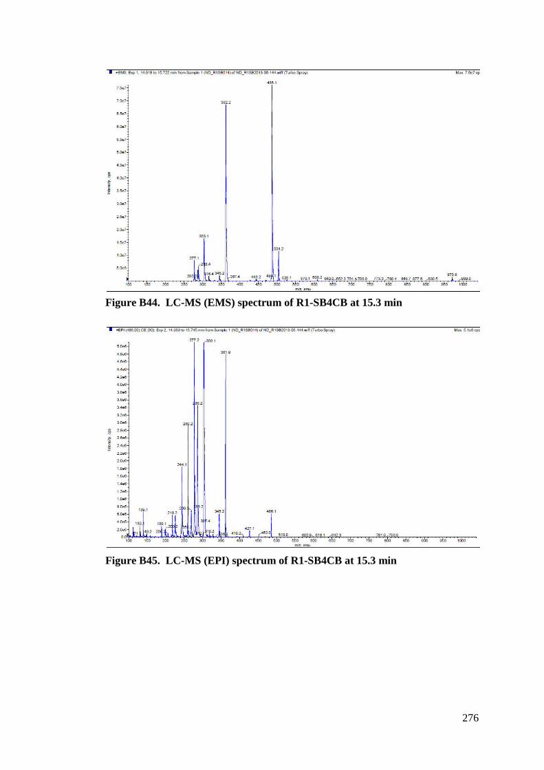
276
Figure B44. LC-MS (EMS) spectrum of R1-SB4CB at 15.3 min
Figure B45. LC-MS (EPI) spectrum of R1-SB4CB at 15.3 min
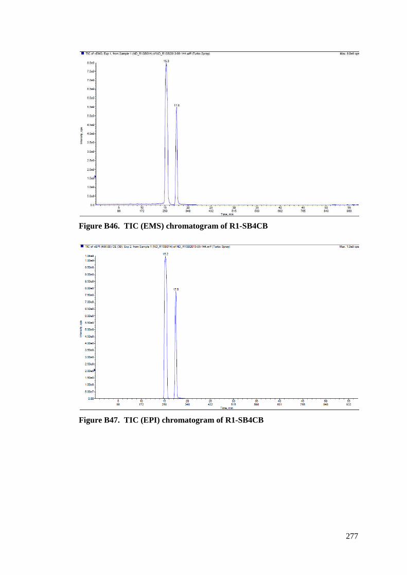
277
Figure B46. TIC (EMS) chromatogram of R1-SB4CB
Figure B47. TIC (EPI) chromatogram of R1-SB4CB
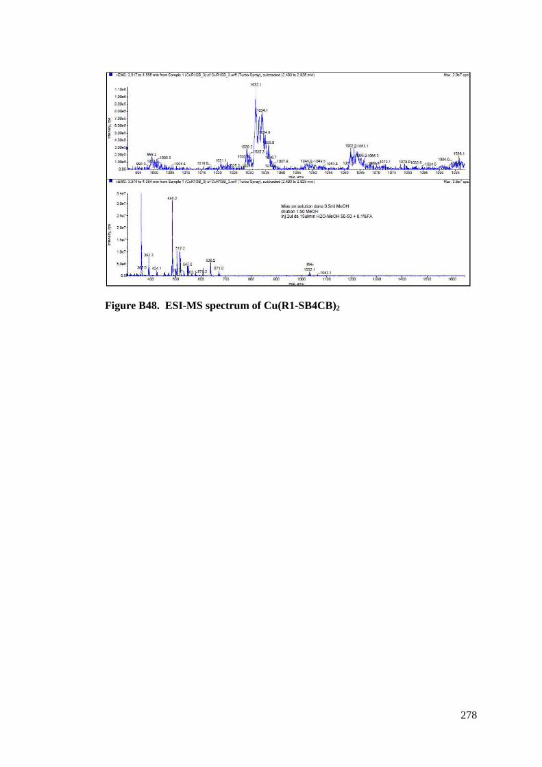
278
Figure B48. ESI-MS spectrum of Cu(R1-SB4CB)2
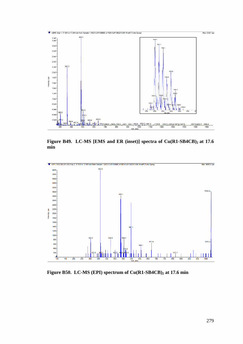
279
Figure B49. LC-MS [EMS and ER (inset)] spectra of Cu(R1-SB4CB)2 at 17.6 min
Figure B50. LC-MS (EPI) spectrum of Cu(R1-SB4CB)2 at 17.6 min
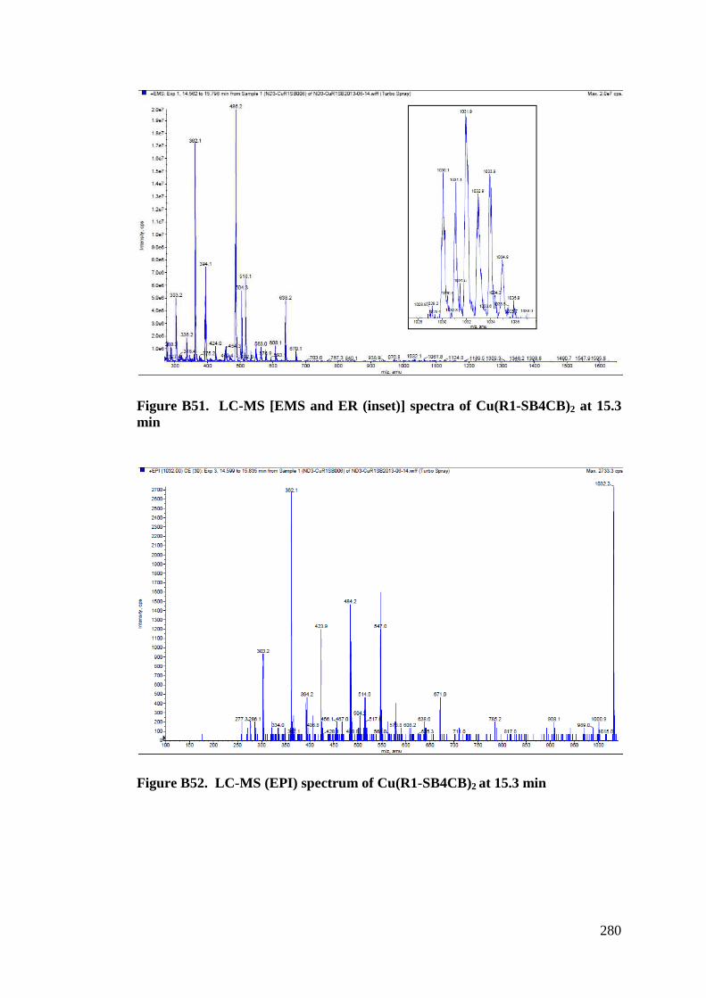
280
Figure B51. LC-MS [EMS and ER (inset)] spectra of Cu(R1-SB4CB)2 at 15.3 min
Figure B52. LC-MS (EPI) spectrum of Cu(R1-SB4CB)2 at 15.3 min
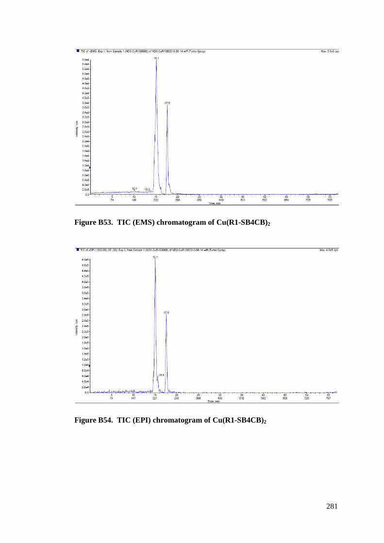
281
Figure B53. TIC (EMS) chromatogram of Cu(R1-SB4CB)2
Figure B54. TIC (EPI) chromatogram of Cu(R1-SB4CB)2
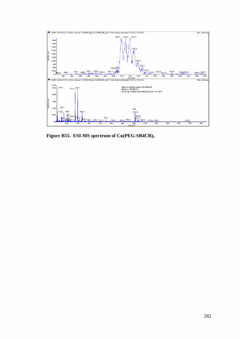
282
Figure B55. ESI-MS spectrum of Cu(PEG-SB4CB)2
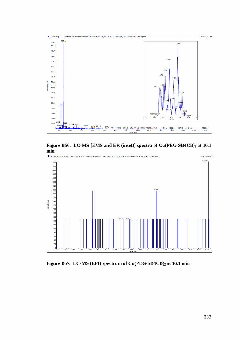
283
Figure B56. LC-MS [EMS and ER (inset)] spectra of Cu(PEG-SB4CB)2 at 16.1 min
Figure B57. LC-MS (EPI) spectrum of Cu(PEG-SB4CB)2 at 16.1 min
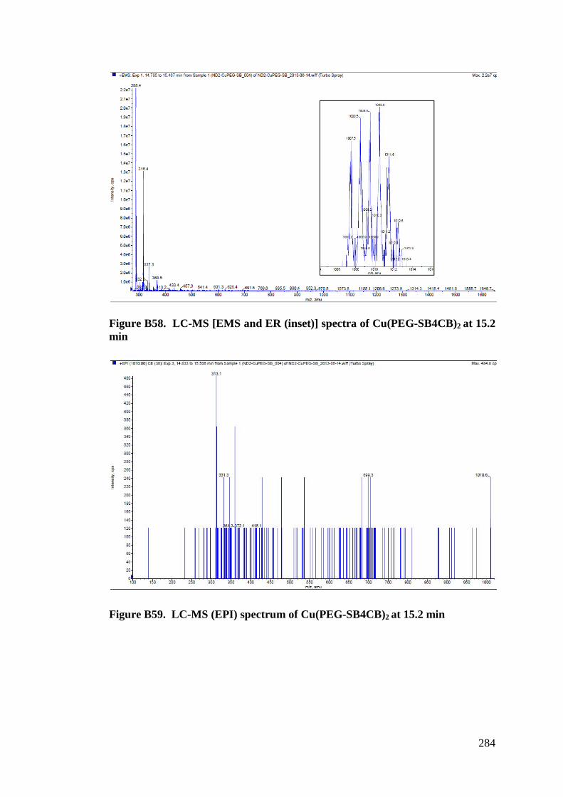
284
Figure B58. LC-MS [EMS and ER (inset)] spectra of Cu(PEG-SB4CB)2 at 15.2 min
Figure B59. LC-MS (EPI) spectrum of Cu(PEG-SB4CB)2 at 15.2 min
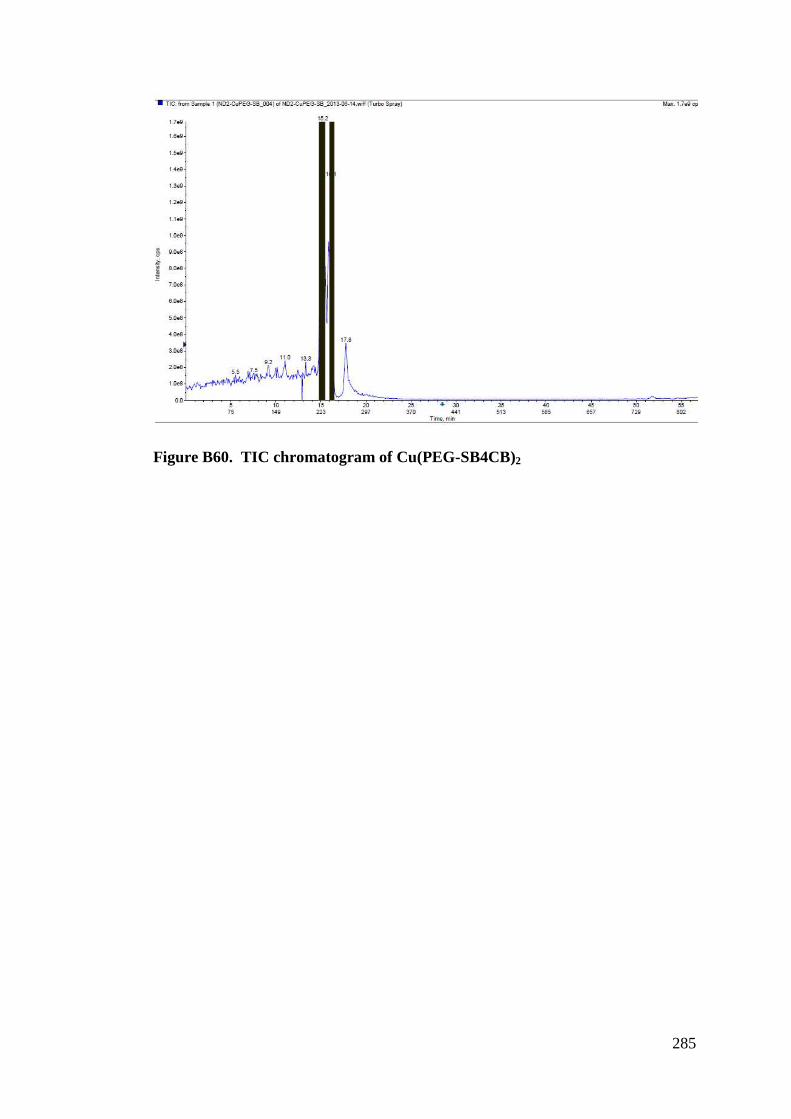
285
Figure B60. TIC chromatogram of Cu(PEG-SB4CB)2
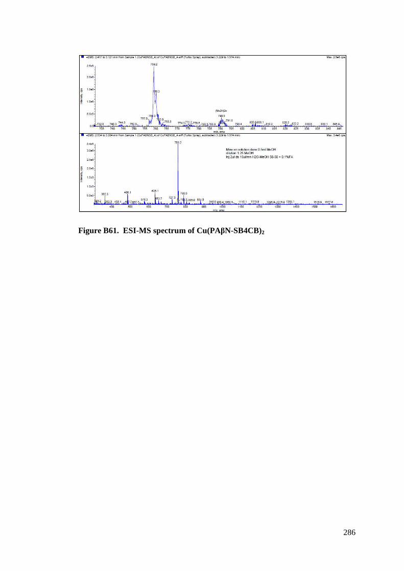
286
Figure B61. ESI-MS spectrum of Cu(PA"N-SB4CB)2
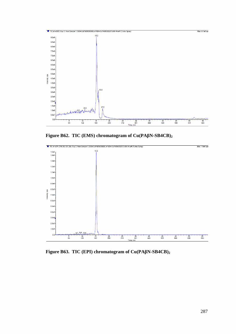
287
Figure B62. TIC (EMS) chromatogram of Cu(PA"N-SB4CB)2
Figure B63. TIC (EPI) chromatogram of Cu(PA"N-SB4CB)2
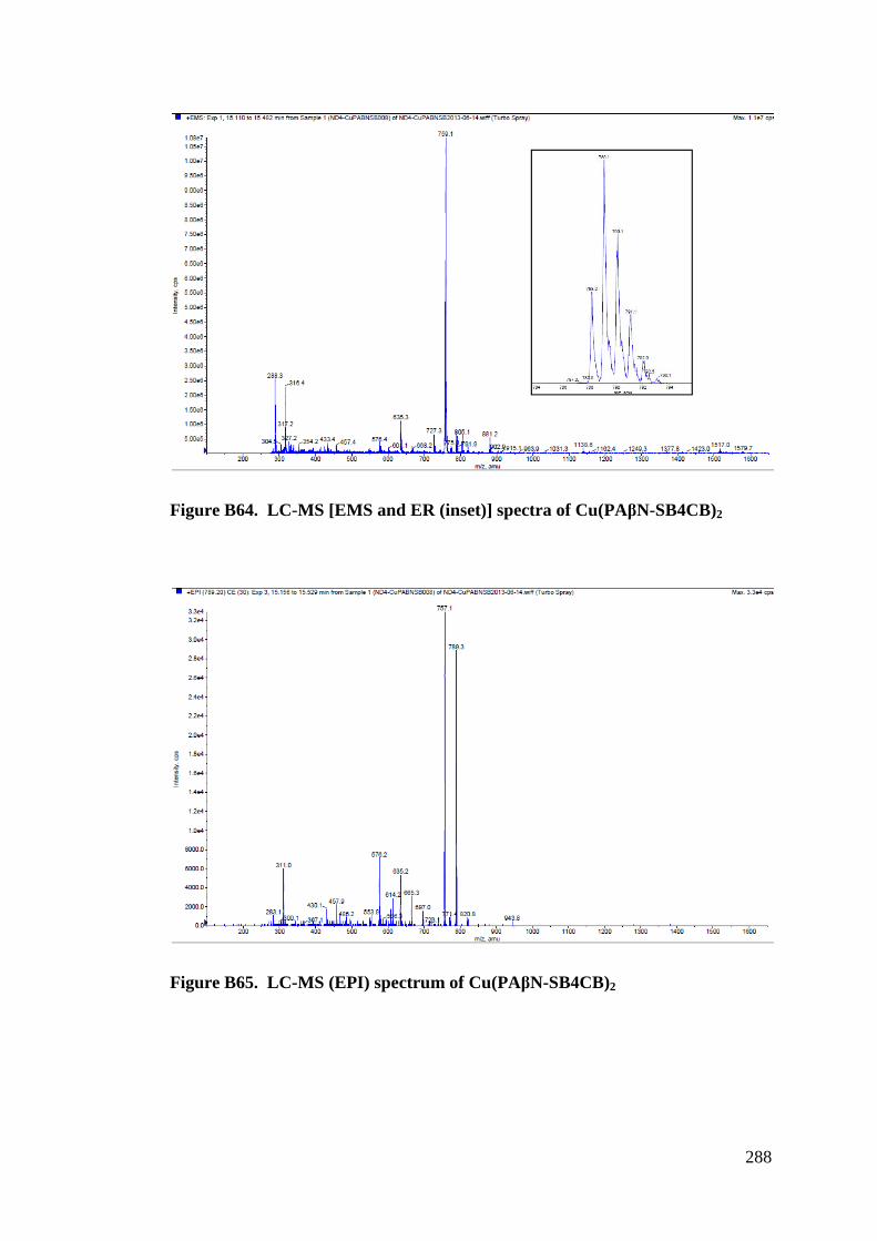
288
Figure B64. LC-MS [EMS and ER (inset)] spectra of Cu(PA"N-SB4CB)2
Figure B65. LC-MS (EPI) spectrum of Cu(PA"N-SB4CB)2
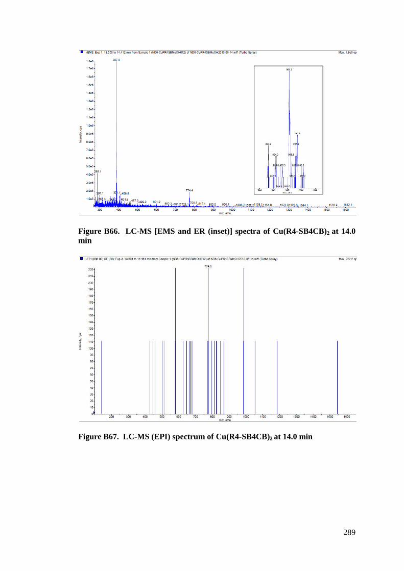
289
Figure B66. LC-MS [EMS and ER (inset)] spectra of Cu(R4-SB4CB)2 at 14.0 min
Figure B67. LC-MS (EPI) spectrum of Cu(R4-SB4CB)2 at 14.0 min
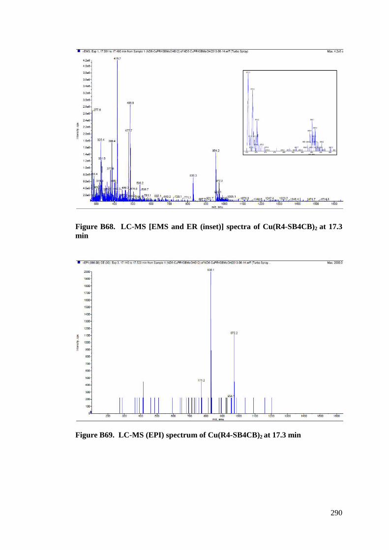
290
Figure B68. LC-MS [EMS and ER (inset)] spectra of Cu(R4-SB4CB)2 at 17.3 min
Figure B69. LC-MS (EPI) spectrum of Cu(R4-SB4CB)2 at 17.3 min
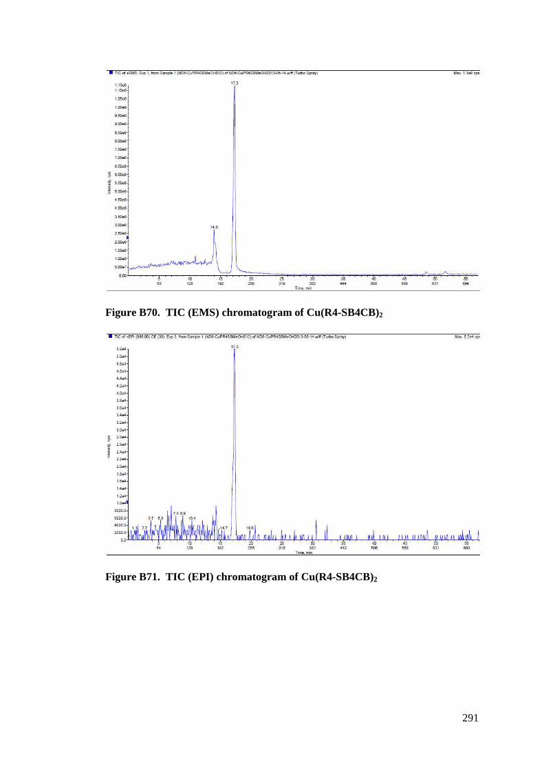
291
Figure B70. TIC (EMS) chromatogram of Cu(R4-SB4CB)2
Figure B71. TIC (EPI) chromatogram of Cu(R4-SB4CB)2
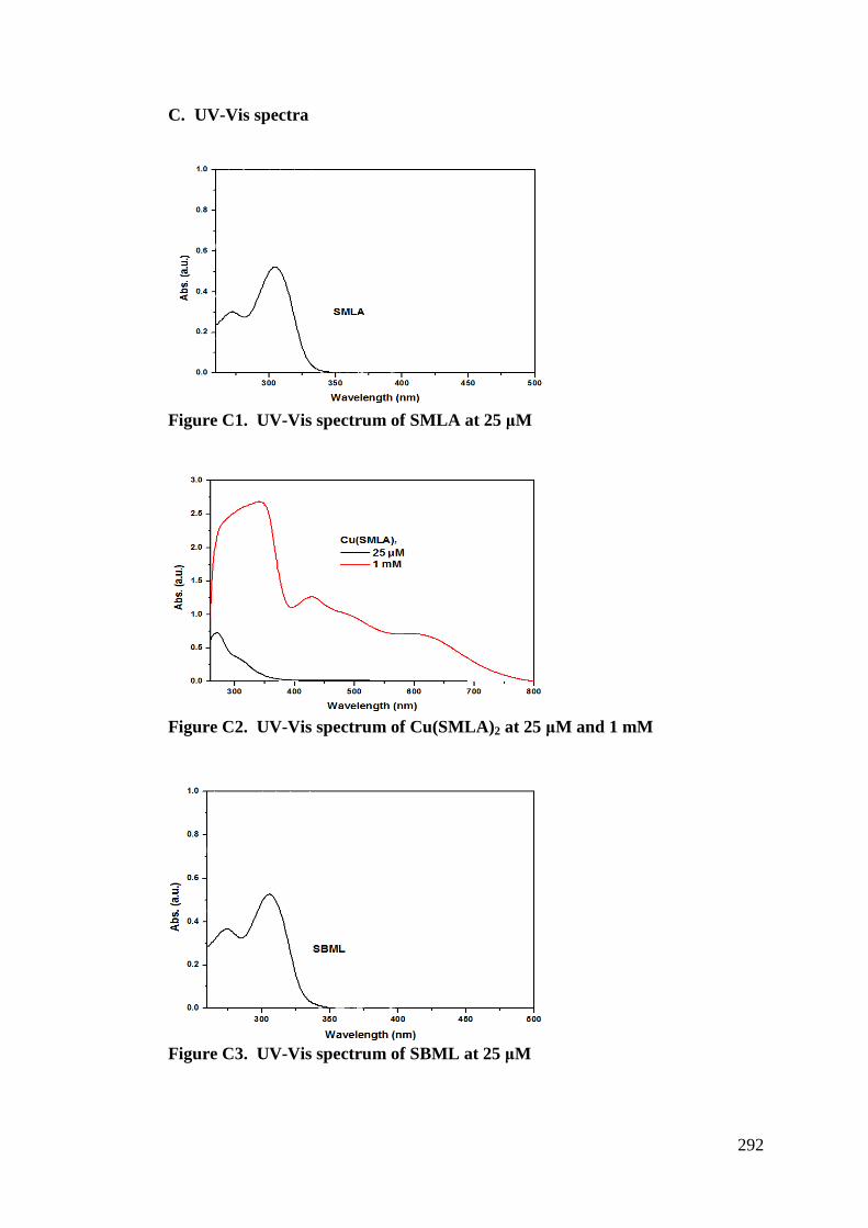
292
C. UV-Vis spectra
Figure C1. UV-Vis spectrum of SMLA at 25 !M
Figure C2. UV-Vis spectrum of Cu(SMLA)2 at 25 !M and 1 mM
Figure C3. UV-Vis spectrum of SBML at 25 !M
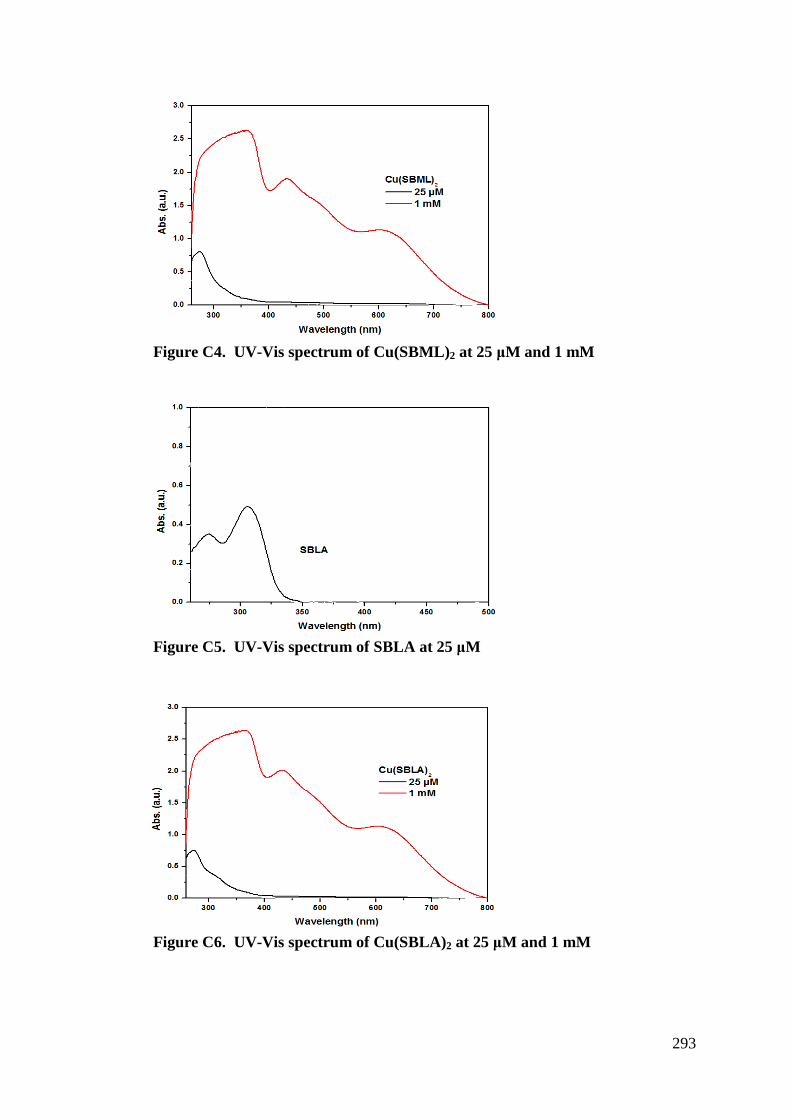
293
Figure C4. UV-Vis spectrum of Cu(SBML)2 at 25 !M and 1 mM
Figure C5. UV-Vis spectrum of SBLA at 25 !M
Figure C6. UV-Vis spectrum of Cu(SBLA)2 at 25 !M and 1 mM
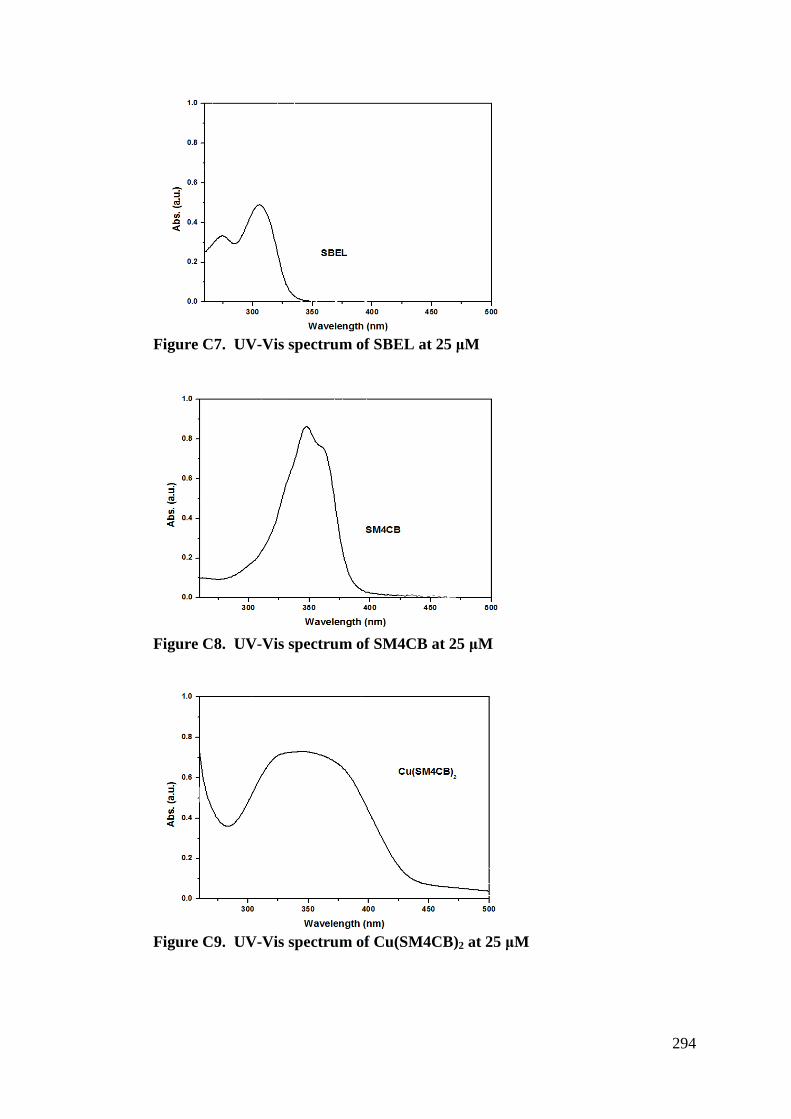
294
Figure C7. UV-Vis spectrum of SBEL at 25 !M
Figure C8. UV-Vis spectrum of SM4CB at 25 !M
Figure C9. UV-Vis spectrum of Cu(SM4CB)2 at 25 !M
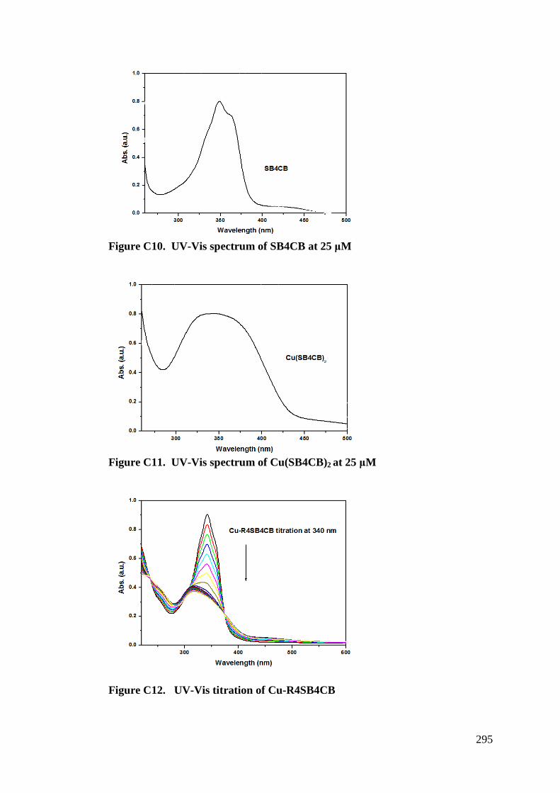
295
Figure C10. UV-Vis spectrum of SB4CB at 25 !M
Figure C11. UV-Vis spectrum of Cu(SB4CB)2 at 25 !M
Figure C12. UV-Vis titration of Cu-R4SB4CB
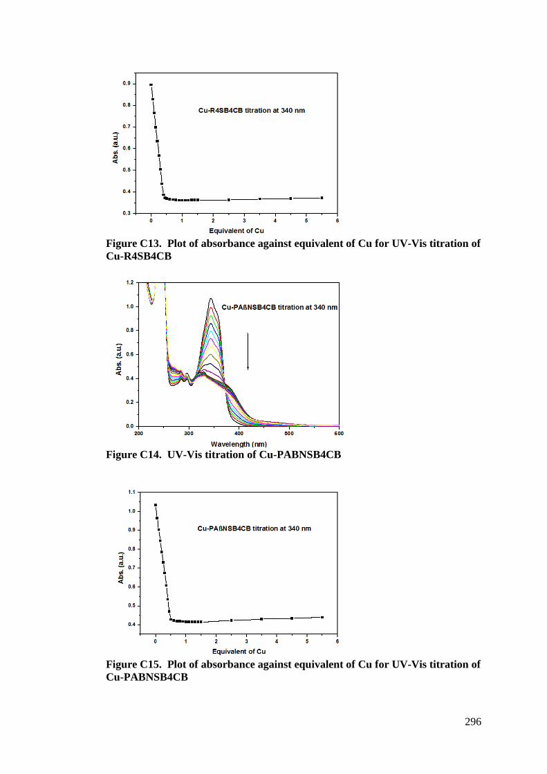
296
Figure C13. Plot of absorbance against equivalent of Cu for UV-Vis titration of Cu-R4SB4CB
Figure C14. UV-Vis titration of Cu-PABNSB4CB
Figure C15. Plot of absorbance against equivalent of Cu for UV-Vis titration of Cu-PABNSB4CB
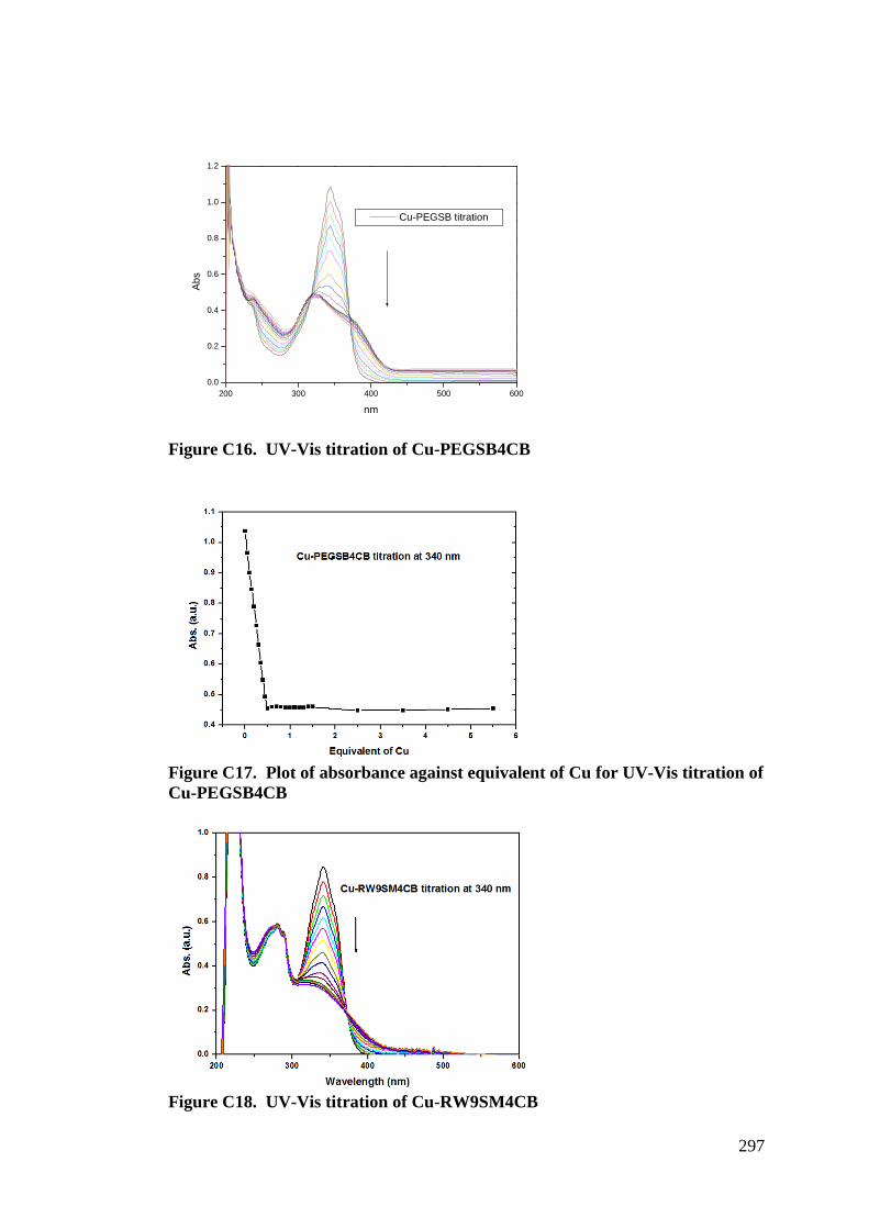
297
Figure C16. UV-Vis titration of Cu-PEGSB4CB
Figure C17. Plot of absorbance against equivalent of Cu for UV-Vis titration of Cu-PEGSB4CB
Figure C18. UV-Vis titration of Cu-RW9SM4CB
200 300 400 500 6000.0
0.2
0.4
0.6
0.8
1.0
1.2
Abs
nm
Cu-PEGSB titration
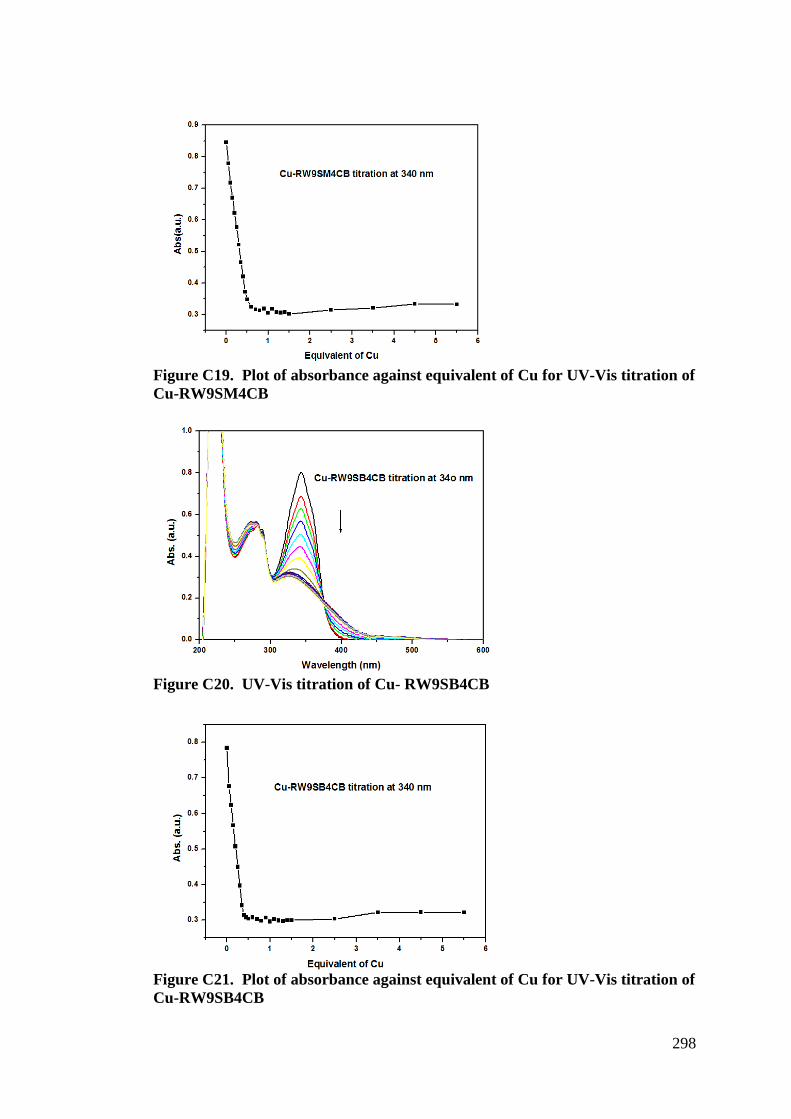
298
Figure C19. Plot of absorbance against equivalent of Cu for UV-Vis titration of Cu-RW9SM4CB
Figure C20. UV-Vis titration of Cu- RW9SB4CB
Figure C21. Plot of absorbance against equivalent of Cu for UV-Vis titration of Cu-RW9SB4CB
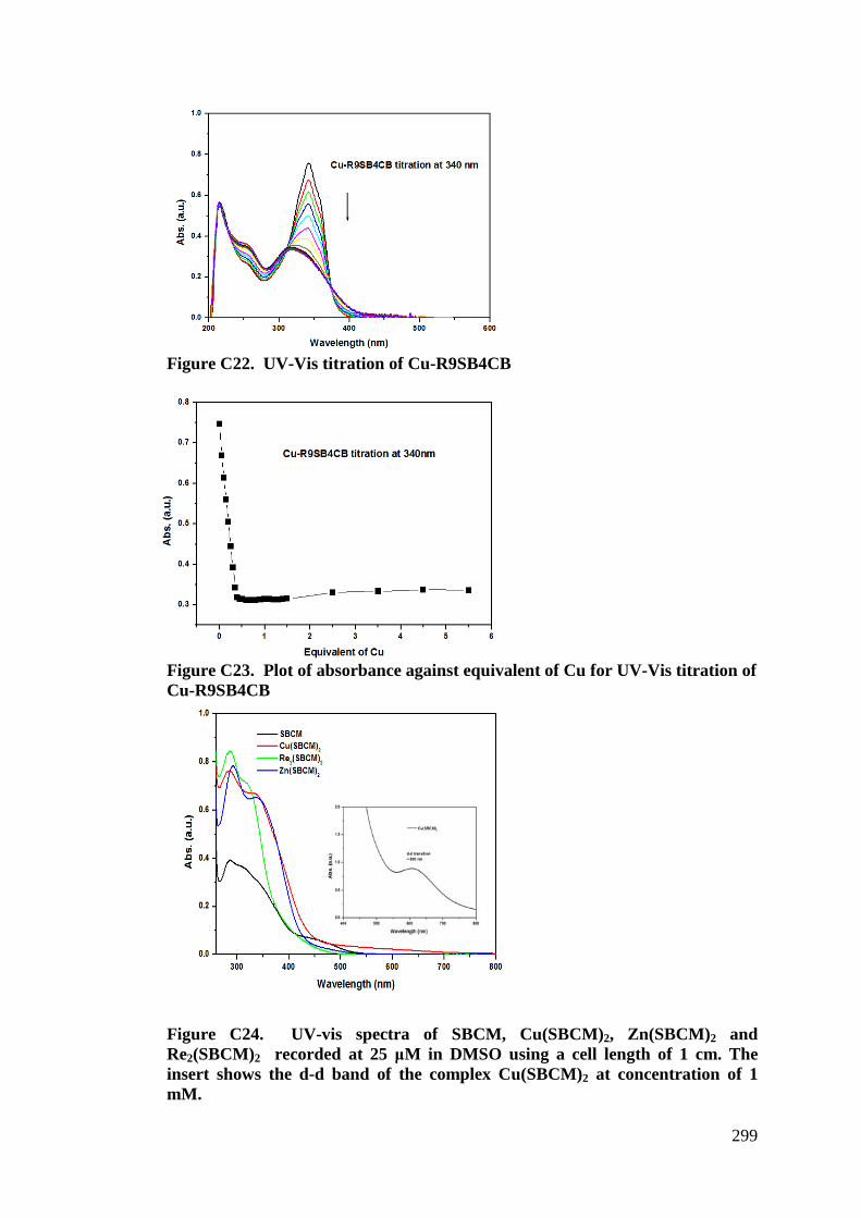
299
Figure C22. UV-Vis titration of Cu-R9SB4CB
Figure C23. Plot of absorbance against equivalent of Cu for UV-Vis titration of Cu-R9SB4CB
Figure C24. UV-vis spectra of SBCM, Cu(SBCM)2, Zn(SBCM)2 and Re2(SBCM)2 recorded at 25 !M in DMSO using a cell length of 1 cm. The insert shows the d-d band of the complex Cu(SBCM)2 at concentration of 1 mM.
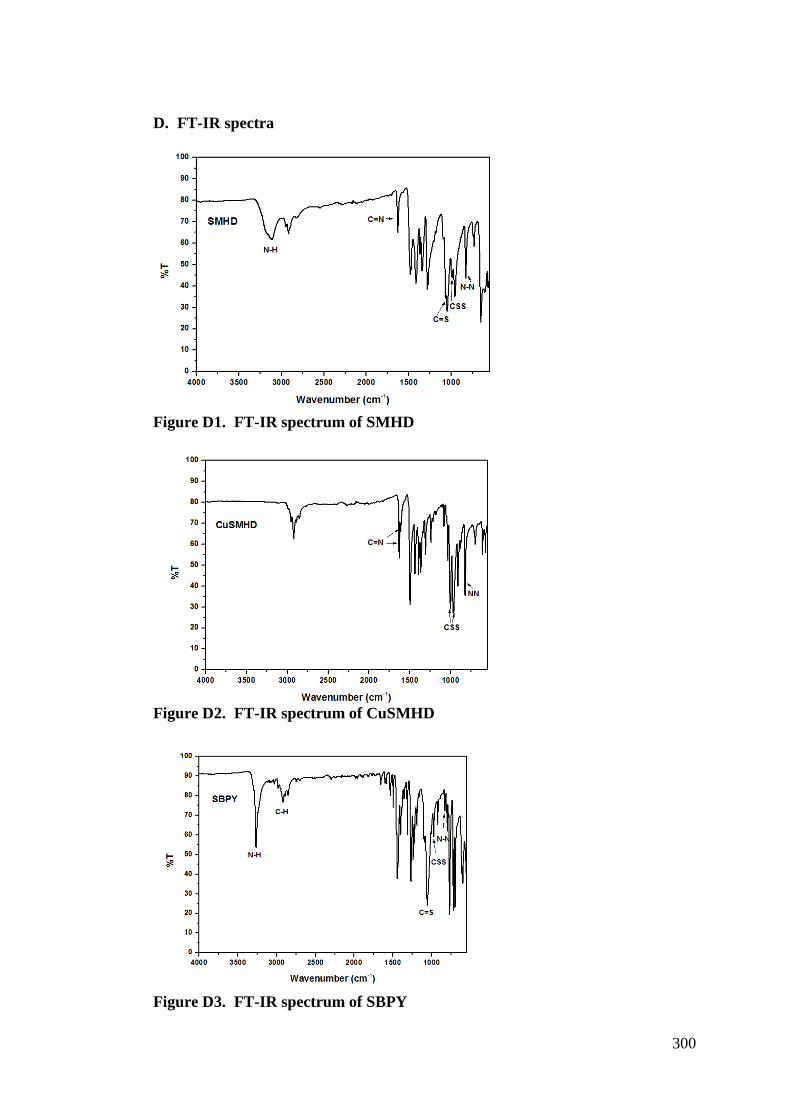
300
D. FT-IR spectra
Figure D1. FT-IR spectrum of SMHD
Figure D2. FT-IR spectrum of CuSMHD
Figure D3. FT-IR spectrum of SBPY
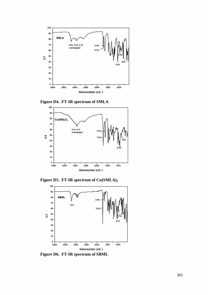
301
Figure D4. FT-IR spectrum of SMLA
Figure D5. FT-IR spectrum of Cu(SMLA)2
Figure D6. FT-IR spectrum of SBML
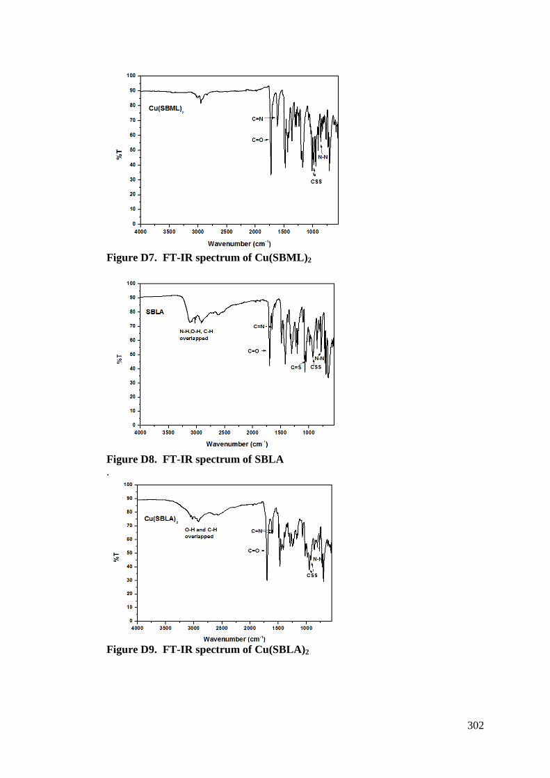
302
Figure D7. FT-IR spectrum of Cu(SBML)2
Figure D8. FT-IR spectrum of SBLA .
Figure D9. FT-IR spectrum of Cu(SBLA)2
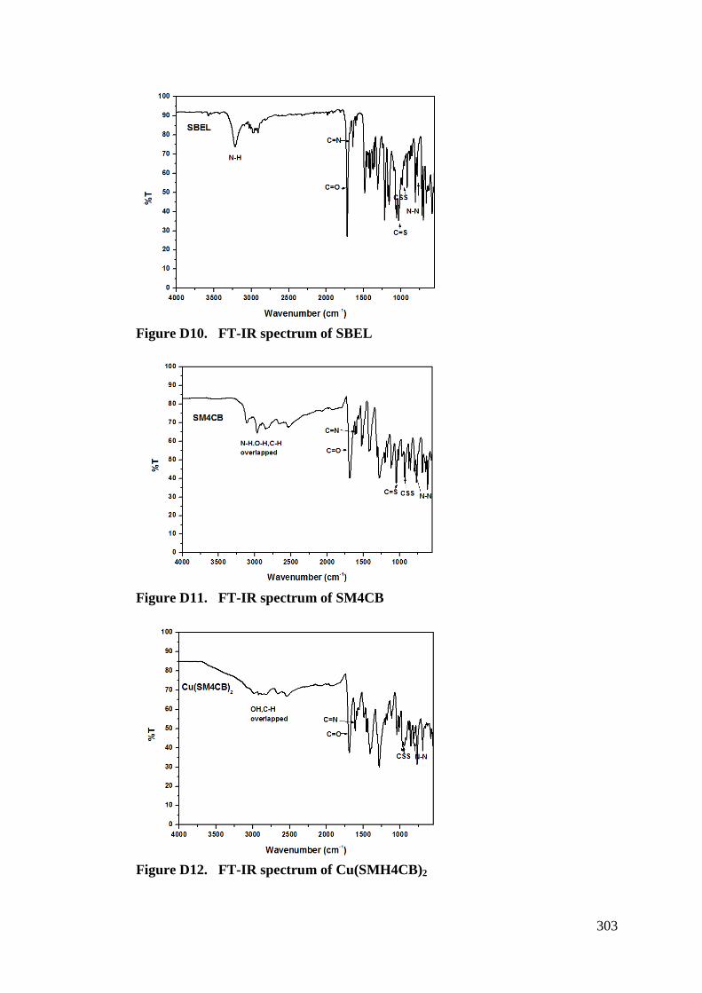
303
Figure D10. FT-IR spectrum of SBEL
Figure D11. FT-IR spectrum of SM4CB
Figure D12. FT-IR spectrum of Cu(SMH4CB)2
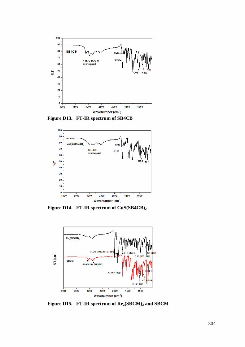
304
Figure D13. FT-IR spectrum of SB4CB
Figure D14. FT-IR spectrum of CuS(SB4CB)2
Figure D15. FT-IR spectrum of Re2(SBCM)2 and SBCM
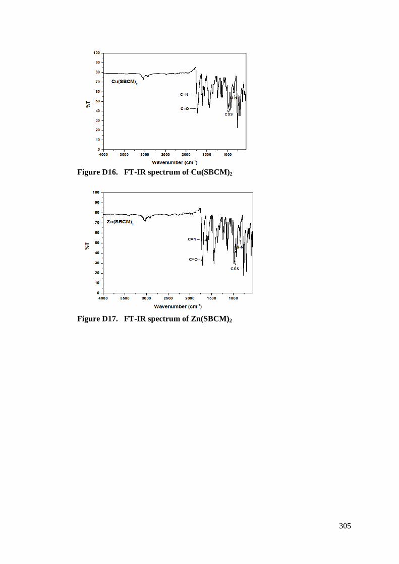
305
Figure D16. FT-IR spectrum of Cu(SBCM)2
Figure D17. FT-IR spectrum of Zn(SBCM)2
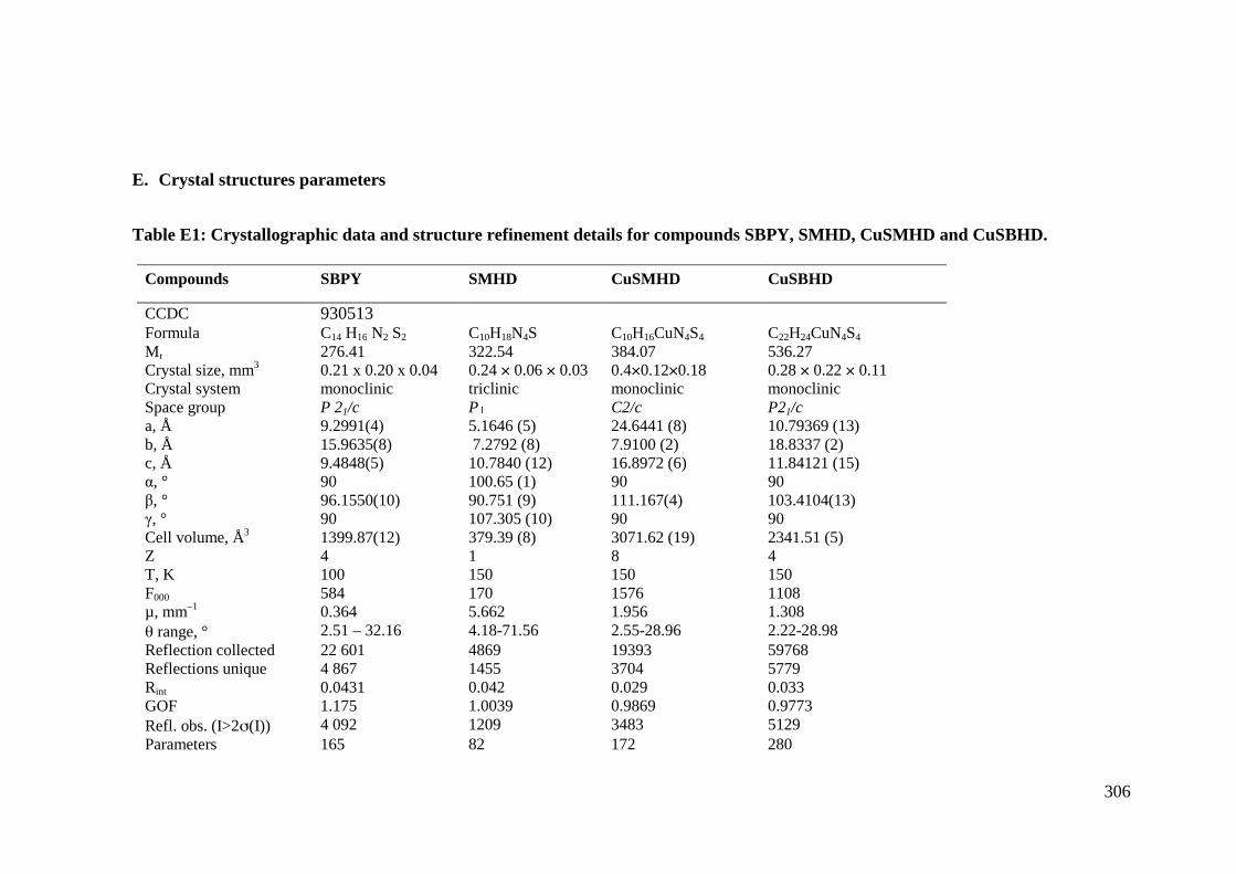
E. Crystal structures parameters
Table E1: Crystallographic data and structure refinement details for compounds SBPY, SMHD, CuSMHD and CuSBHD.
Compounds SBPY
CCDC 930513 Formula C14 H16 N2 S2 Mr 276.41 Crystal size, mm3 0.21 x 0.20 x 0.04 Crystal system monoclinic Space group P 21/c a, Å 9.2991(4) b, Å 15.9635(8) c, Å 9.4848(5) &, ° 90 %, ° 96.1550(10) -, ° 90 Cell volume, Å3 1399.87(12) Z 4 T, K 100 F000 584 µ, mm–1 0.364 " range, ° 2.51 – 32.16 Reflection collected 22 601 Reflections unique 4 867 Rint 0.0431 GOF 1.175 Refl. obs. (I>2#(I)) 4 092 Parameters 165
Crystallographic data and structure refinement details for compounds SBPY, SMHD, CuSMHD and CuSBHD.
SMHD CuSMHD CuSBHD
C10H18N4S C10H16CuN4S4 C22H24CuN4S4 322.54 384.07 536.27 0.24 ! 0.06 ! 0.03 0.4!0.12!0.18 0.28 ! 0.22 ! 0.11 triclinic monoclinic monoclinic P C2/c P21/c 5.1646 (5) 24.6441 (8) 10.79369 (13) 7.2792 (8) 7.9100 (2) 18.8337 (2) 10.7840 (12) 16.8972 (6) 11.84121 (15) 100.65 (1) 90 90 90.751 (9) 111.167(4) 103.4104(13) 107.305 (10) 90 90 379.39 (8) 3071.62 (19) 2341.51 (5) 1 8 4 150 150 150 170 1576 1108 5.662 1.956 1.308 4.18-71.56 2.55-28.96 2.22-28.98 4869 19393 59768 1455 3704 5779 0.042 0.029 0.033 1.0039 0.9869 0.9773 1209 3483 5129 82 172 280
306
Crystallographic data and structure refinement details for compounds SBPY, SMHD, CuSMHD and CuSBHD.
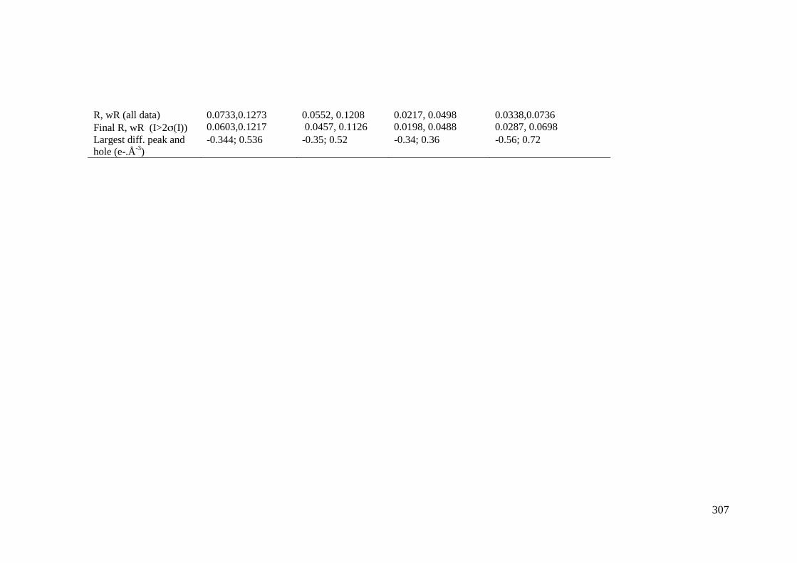
307
R, wR (all data) 0.0733,0.1273 0.0552, 0.1208 0.0217, 0.0498 0.0338,0.0736 Final R, wR (I>2#(I)) 0.0603,0.1217 0.0457, 0.1126 0.0198, 0.0488 0.0287, 0.0698 Largest diff. peak and hole (e-.Å-3)
-0.344; 0.536 -0.35; 0.52 -0.34; 0.36 -0.56; 0.72
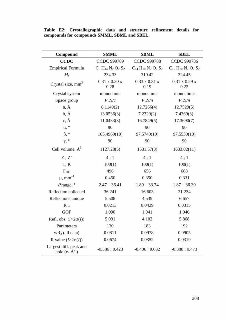
308
Table E2: Crystallographic data and structure refinement details for compounds for compounds SMML, SBML and SBEL.
Compound SMML SBML SBEL CCDC CCDC 999789 CCDC 999788 CCDC 999786
Empirical Formula C8 H14 N2 O2 S2 C14 H18 N2 O2 S2 C15 H20 N2 O2 S2 Mr 234.33 310.42 324.45
Crystal size, mm3 0.31 x 0.30 x 0.28
0.33 x 0.31 x 0.19
0.31 x 0.29 x 0.22
Crystal system monoclinic monoclinic monoclinic Space group P 21/c P 21/n P 21/n
a, Å 8.1149(2) 12.7266(4) 12.7529(5) b, Å 13.0536(3) 7.2329(2) 7.4369(3) c, Å 11.0433(3) 16.7849(5) 17.3690(7) &, ° 90 90 90 %, ° 105.4960(10) 97.5740(10) 97.5530(10) -, ° 90 90 90
Cell volume, Å3 1127.28(5) 1531.57(8) 1633.02(11)
Z ; Z’ 4 ; 1 4 ; 1 4 ; 1 T, K 100(1) 100(1) 100(1) F000 496 656 688
µ, mm–1 0.450 0.350 0.331 " range, ° 2.47 – 36.41 1.89 – 33.74 1.87 – 36.30
Reflection collected 36 241 16 603 21 234 Reflections unique 5 508 4 539 6 657
Rint 0.0213 0.0429 0.0315 GOF 1.090 1.041 1.046
Refl. obs. (I>2#(I)) 5 091 4 102 5 868 Parameters 130 183 192
wR2 (all data) 0.0811 0.0978 0.0905 R value (I>2#(I)) 0.0674 0.0352 0.0319
Largest diff. peak and hole (e-.Å-3) -0.386 ; 0.423 -0.406 ; 0.632 -0.380 ; 0.473
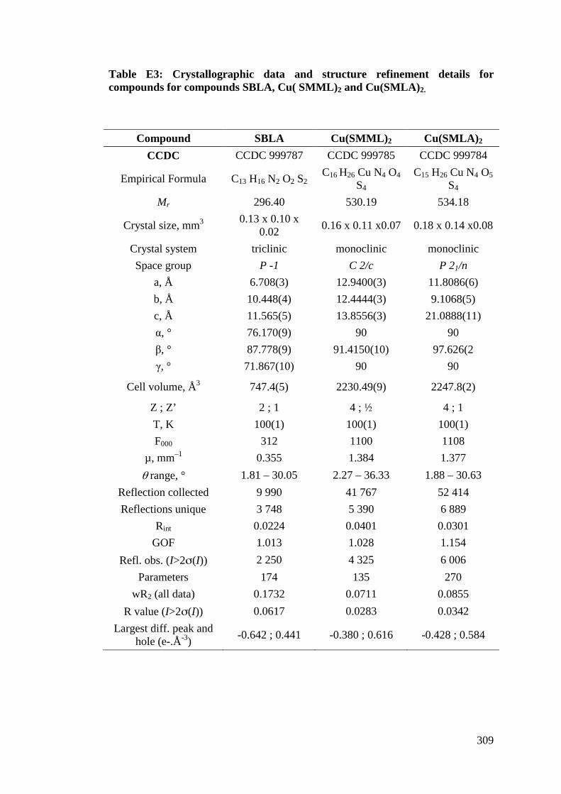
309
Table E3: Crystallographic data and structure refinement details for compounds for compounds SBLA, Cu( SMML)2 and Cu(SMLA)2.
Compound SBLA Cu(SMML)2 Cu(SMLA)2 CCDC CCDC 999787 CCDC 999785 CCDC 999784
Empirical Formula C13 H16 N2 O2 S2 C16 H26 Cu N4 O4
S4 C15 H26 Cu N4 O5
S4 Mr 296.40 530.19 534.18
Crystal size, mm3 0.13 x 0.10 x 0.02 0.16 x 0.11 x0.07 0.18 x 0.14 x0.08
Crystal system triclinic monoclinic monoclinic Space group P -1 C 2/c P 21/n
a, Å 6.708(3) 12.9400(3) 11.8086(6) b, Å 10.448(4) 12.4444(3) 9.1068(5) c, Å 11.565(5) 13.8556(3) 21.0888(11) &, ° 76.170(9) 90 90 %, ° 87.778(9) 91.4150(10) 97.626(2 -, ° 71.867(10) 90 90
Cell volume, Å3 747.4(5) 2230.49(9) 2247.8(2)
Z ; Z’ 2 ; 1 4 ; $ 4 ; 1 T, K 100(1) 100(1) 100(1) F000 312 1100 1108
µ, mm–1 0.355 1.384 1.377 " range, ° 1.81 – 30.05 2.27 – 36.33 1.88 – 30.63
Reflection collected 9 990 41 767 52 414 Reflections unique 3 748 5 390 6 889
Rint 0.0224 0.0401 0.0301 GOF 1.013 1.028 1.154
Refl. obs. (I>2#(I)) 2 250 4 325 6 006 Parameters 174 135 270
wR2 (all data) 0.1732 0.0711 0.0855 R value (I>2#(I)) 0.0617 0.0283 0.0342
Largest diff. peak and hole (e-.Å-3) -0.642 ; 0.441 -0.380 ; 0.616 -0.428 ; 0.584
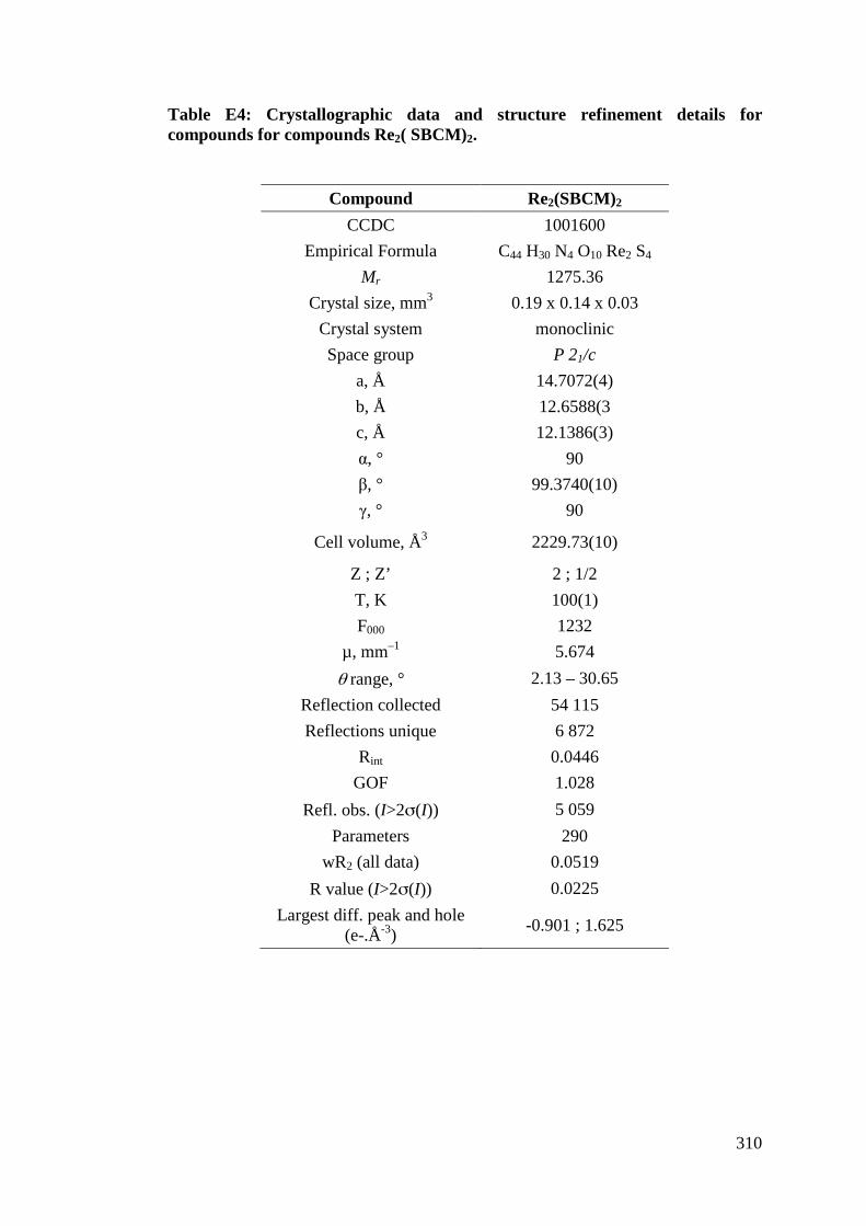
310
Table E4: Crystallographic data and structure refinement details for compounds for compounds Re2( SBCM)2.
Compound Re2(SBCM)2 CCDC 1001600
Empirical Formula C44 H30 N4 O10 Re2 S4 Mr 1275.36
Crystal size, mm3 0.19 x 0.14 x 0.03 Crystal system monoclinic Space group P 21/c
a, Å 14.7072(4) b, Å 12.6588(3 c, Å 12.1386(3) &, ° 90 %, ° 99.3740(10) -, ° 90
Cell volume, Å3 2229.73(10)
Z ; Z’ 2 ; 1/2 T, K 100(1) F000 1232
µ, mm–1 5.674 " range, ° 2.13 – 30.65
Reflection collected 54 115 Reflections unique 6 872
Rint 0.0446 GOF 1.028
Refl. obs. (I>2#(I)) 5 059 Parameters 290
wR2 (all data) 0.0519 R value (I>2#(I)) 0.0225
Largest diff. peak and hole (e-.Å-3) -0.901 ; 1.625
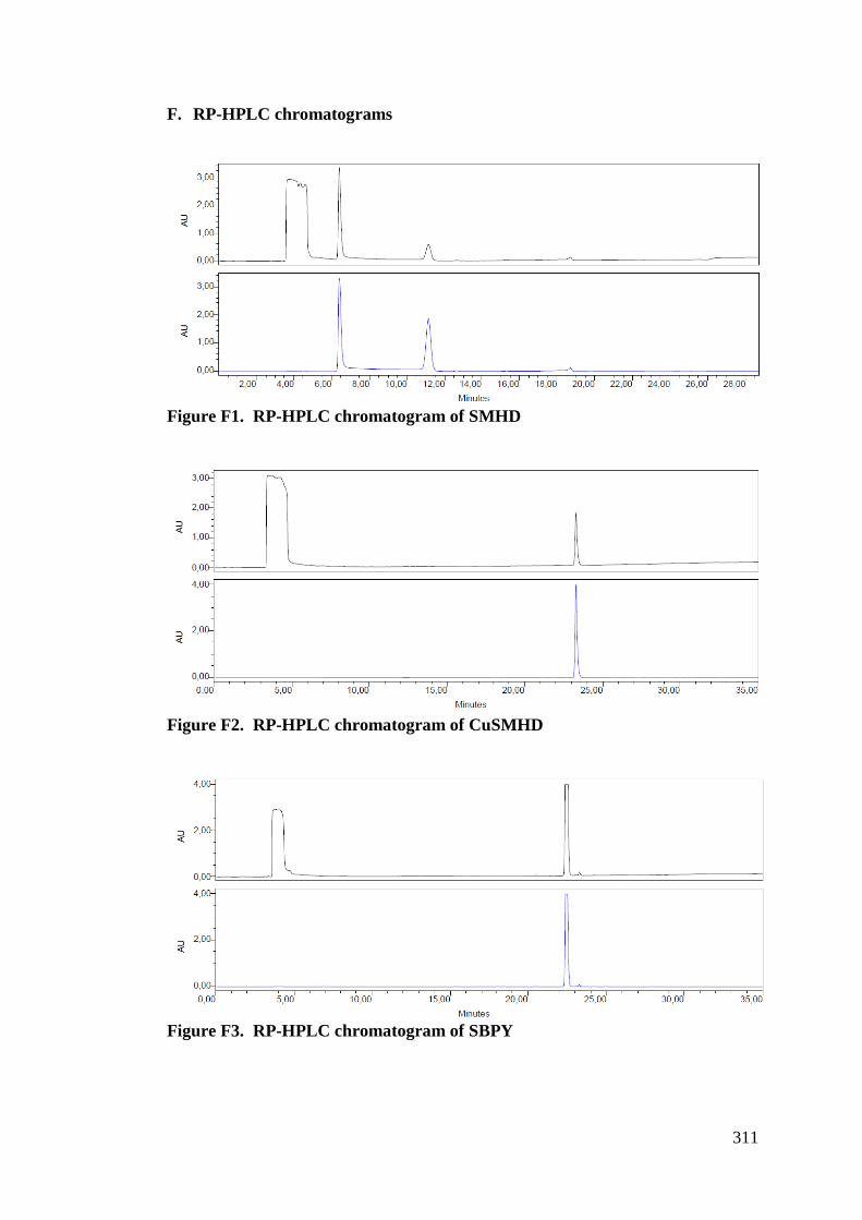
311
F. RP-HPLC chromatograms
Figure F1. RP-HPLC chromatogram of SMHD
Figure F2. RP-HPLC chromatogram of CuSMHD
Figure F3. RP-HPLC chromatogram of SBPY
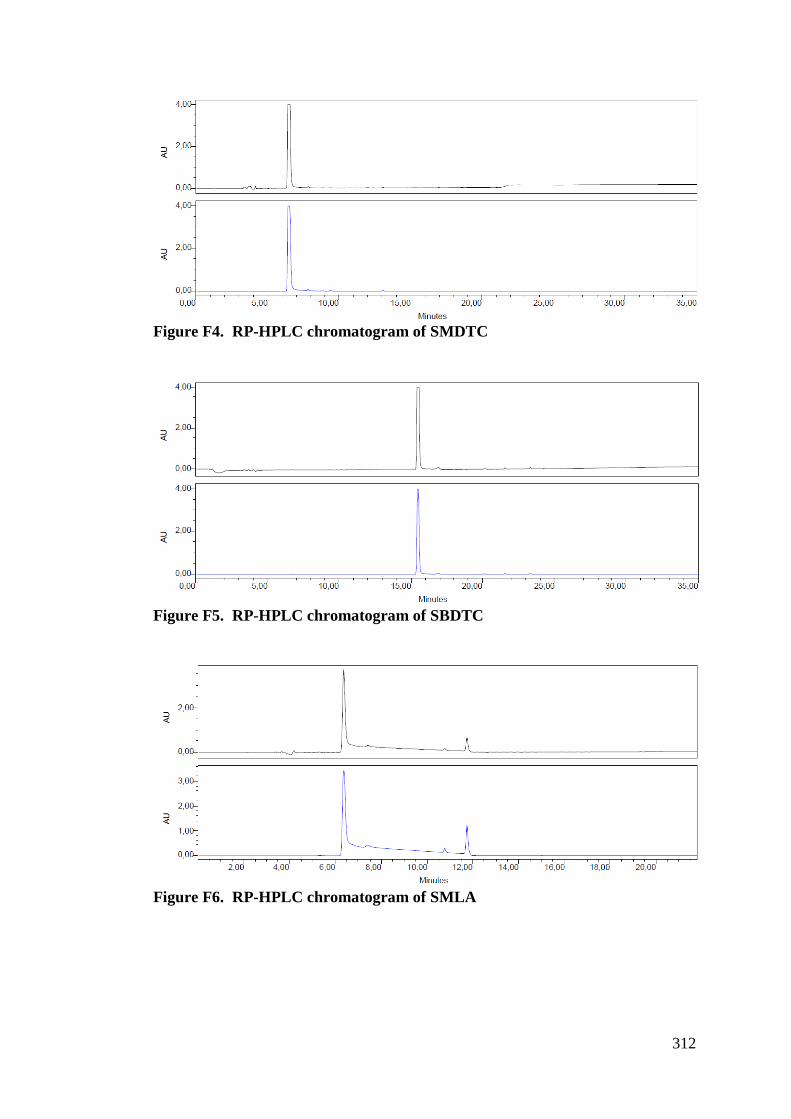
312
Figure F4. RP-HPLC chromatogram of SMDTC
Figure F5. RP-HPLC chromatogram of SBDTC
Figure F6. RP-HPLC chromatogram of SMLA
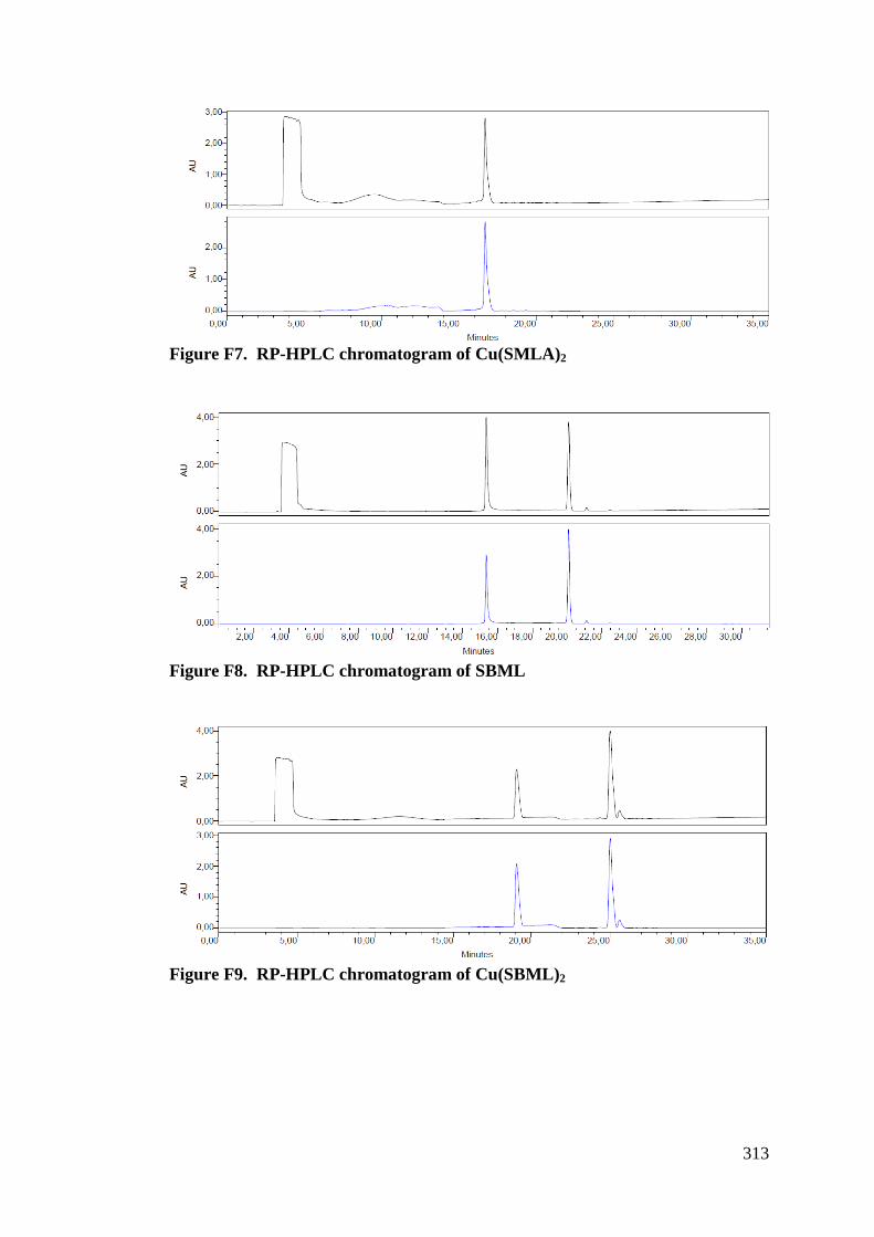
313
Figure F7. RP-HPLC chromatogram of Cu(SMLA)2
Figure F8. RP-HPLC chromatogram of SBML
Figure F9. RP-HPLC chromatogram of Cu(SBML)2
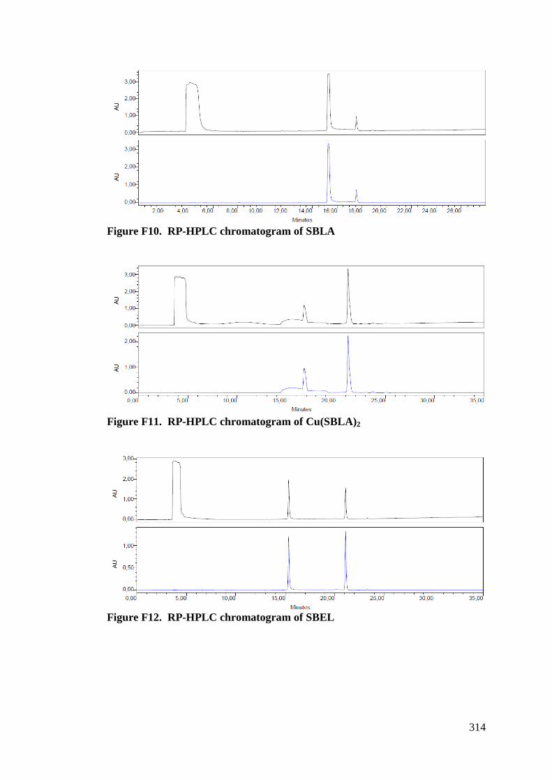
314
Figure F10. RP-HPLC chromatogram of SBLA
Figure F11. RP-HPLC chromatogram of Cu(SBLA)2
Figure F12. RP-HPLC chromatogram of SBEL
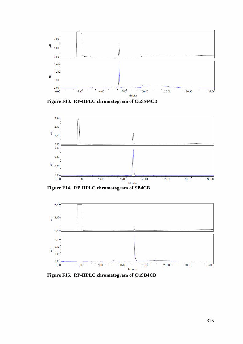
315
Figure F13. RP-HPLC chromatogram of CuSM4CB
Figure F14. RP-HPLC chromatogram of SB4CB
Figure F15. RP-HPLC chromatogram of CuSB4CB
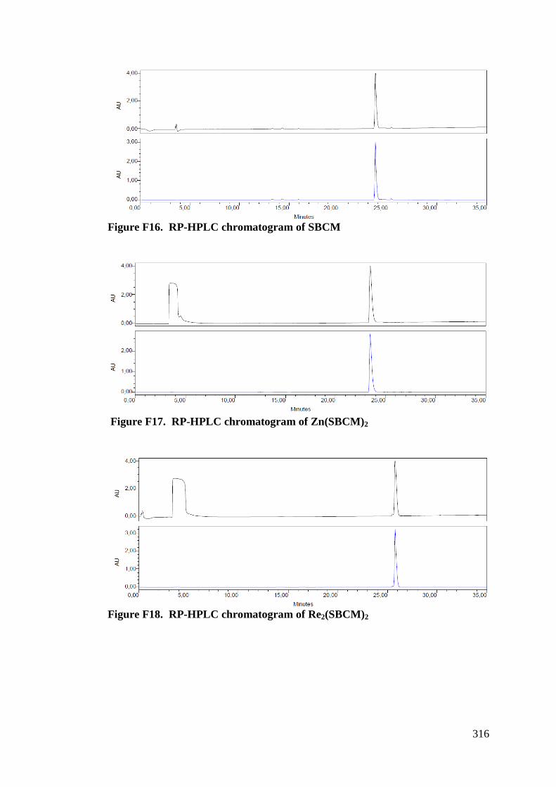
316
Figure F16. RP-HPLC chromatogram of SBCM
Figure F17. RP-HPLC chromatogram of Zn(SBCM)2
Figure F18. RP-HPLC chromatogram of Re2(SBCM)2
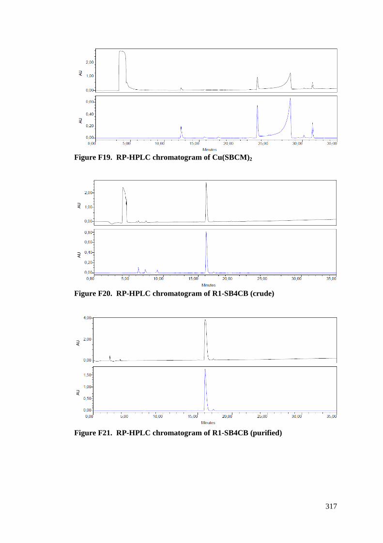
317
Figure F19. RP-HPLC chromatogram of Cu(SBCM)2
Figure F20. RP-HPLC chromatogram of R1-SB4CB (crude)
Figure F21. RP-HPLC chromatogram of R1-SB4CB (purified)
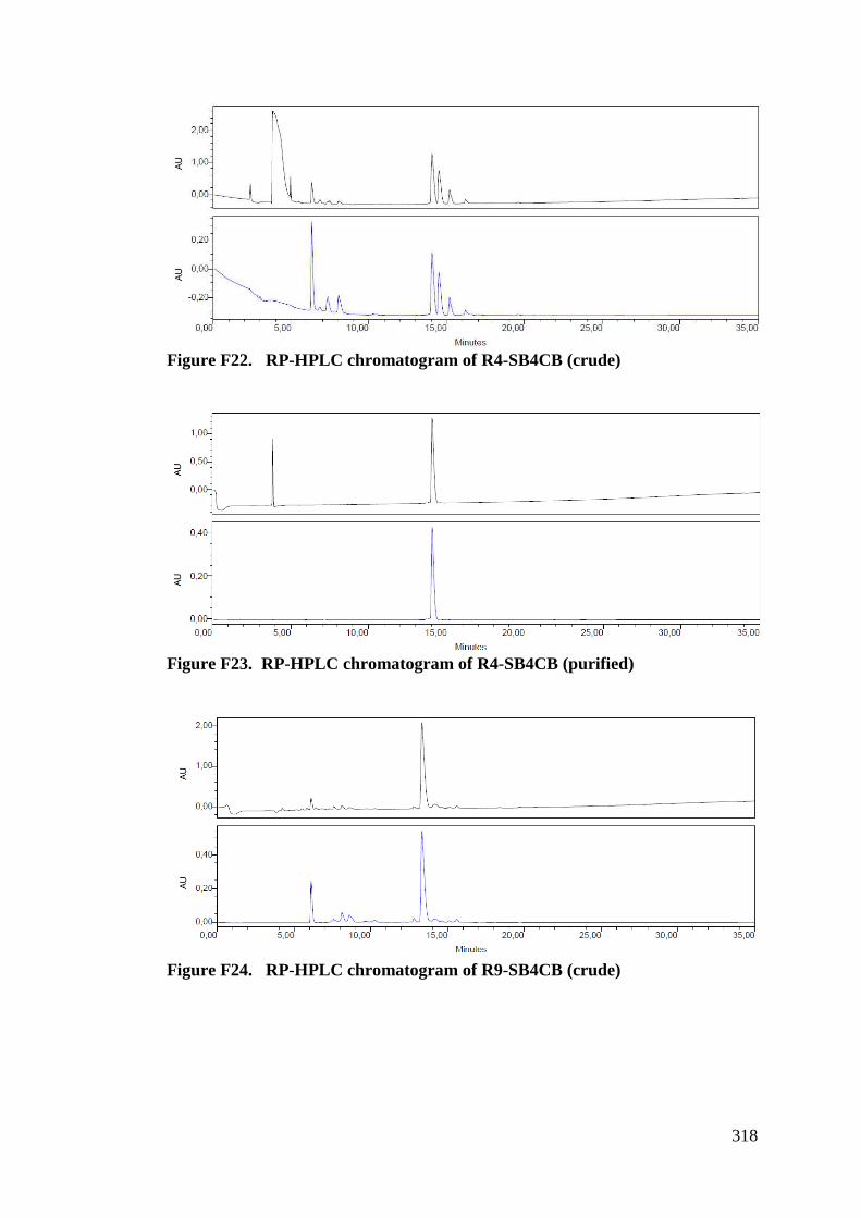
318
Figure F22. RP-HPLC chromatogram of R4-SB4CB (crude)
Figure F23. RP-HPLC chromatogram of R4-SB4CB (purified)
Figure F24. RP-HPLC chromatogram of R9-SB4CB (crude)
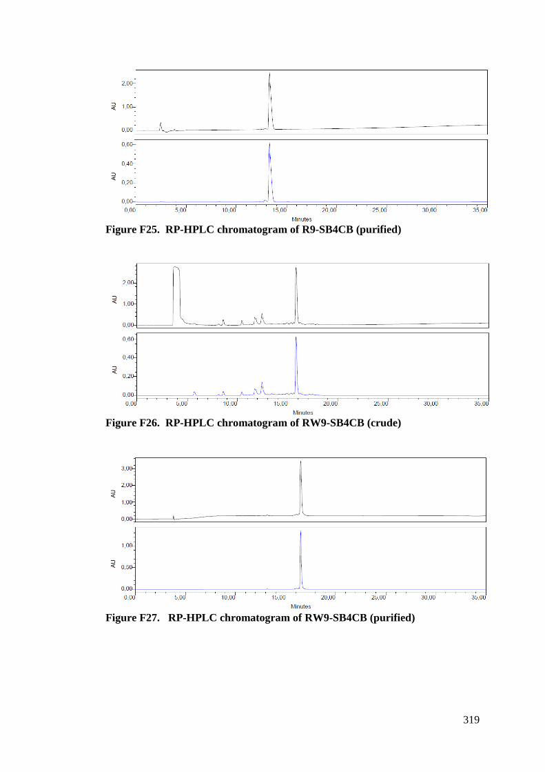
319
Figure F25. RP-HPLC chromatogram of R9-SB4CB (purified)
Figure F26. RP-HPLC chromatogram of RW9-SB4CB (crude)
Figure F27. RP-HPLC chromatogram of RW9-SB4CB (purified)
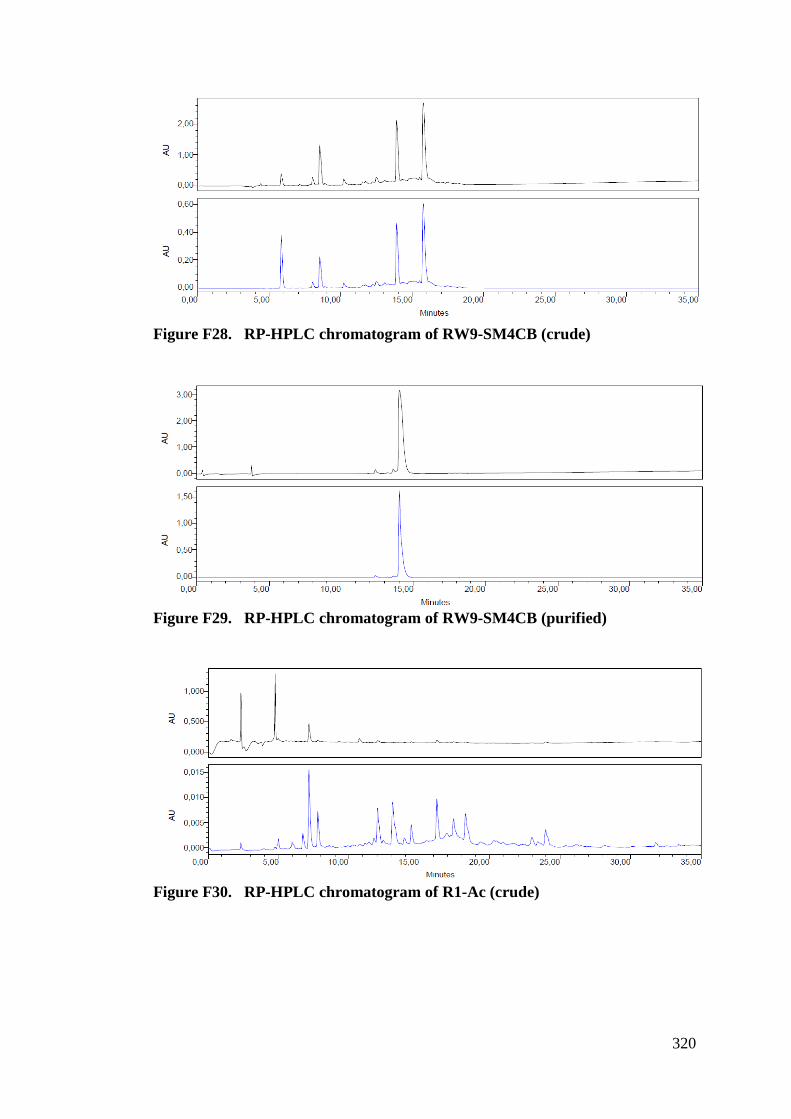
320
Figure F28. RP-HPLC chromatogram of RW9-SM4CB (crude)
Figure F29. RP-HPLC chromatogram of RW9-SM4CB (purified)
Figure F30. RP-HPLC chromatogram of R1-Ac (crude)
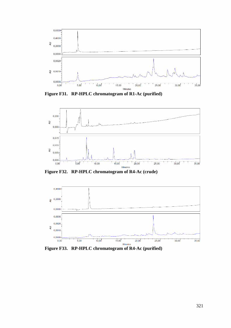
321
Figure F31. RP-HPLC chromatogram of R1-Ac (purified)
Figure F32. RP-HPLC chromatogram of R4-Ac (crude)
Figure F33. RP-HPLC chromatogram of R4-Ac (purified)
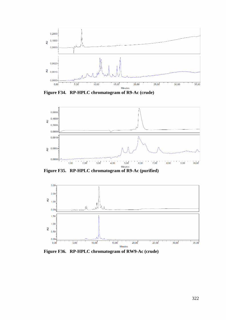
322
Figure F34. RP-HPLC chromatogram of R9-Ac (crude)
Figure F35. RP-HPLC chromatogram of R9-Ac (purified)
Figure F36. RP-HPLC chromatogram of RW9-Ac (crude)
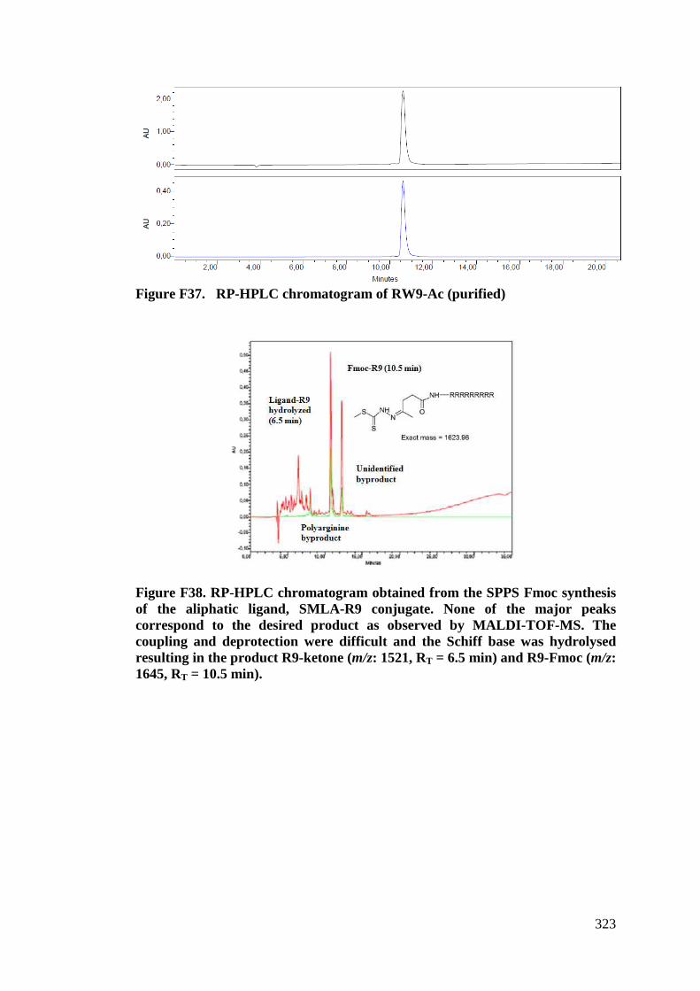
323
Figure F37. RP-HPLC chromatogram of RW9-Ac (purified)
Figure F38. RP-HPLC chromatogram obtained from the SPPS Fmoc synthesis of the aliphatic ligand, SMLA-R9 conjugate. None of the major peaks correspond to the desired product as observed by MALDI-TOF-MS. The coupling and deprotection were difficult and the Schiff base was hydrolysed resulting in the product R9-ketone (m/z: 1521, RT = 6.5 min) and R9-Fmoc (m/z: 1645, RT = 10.5 min).
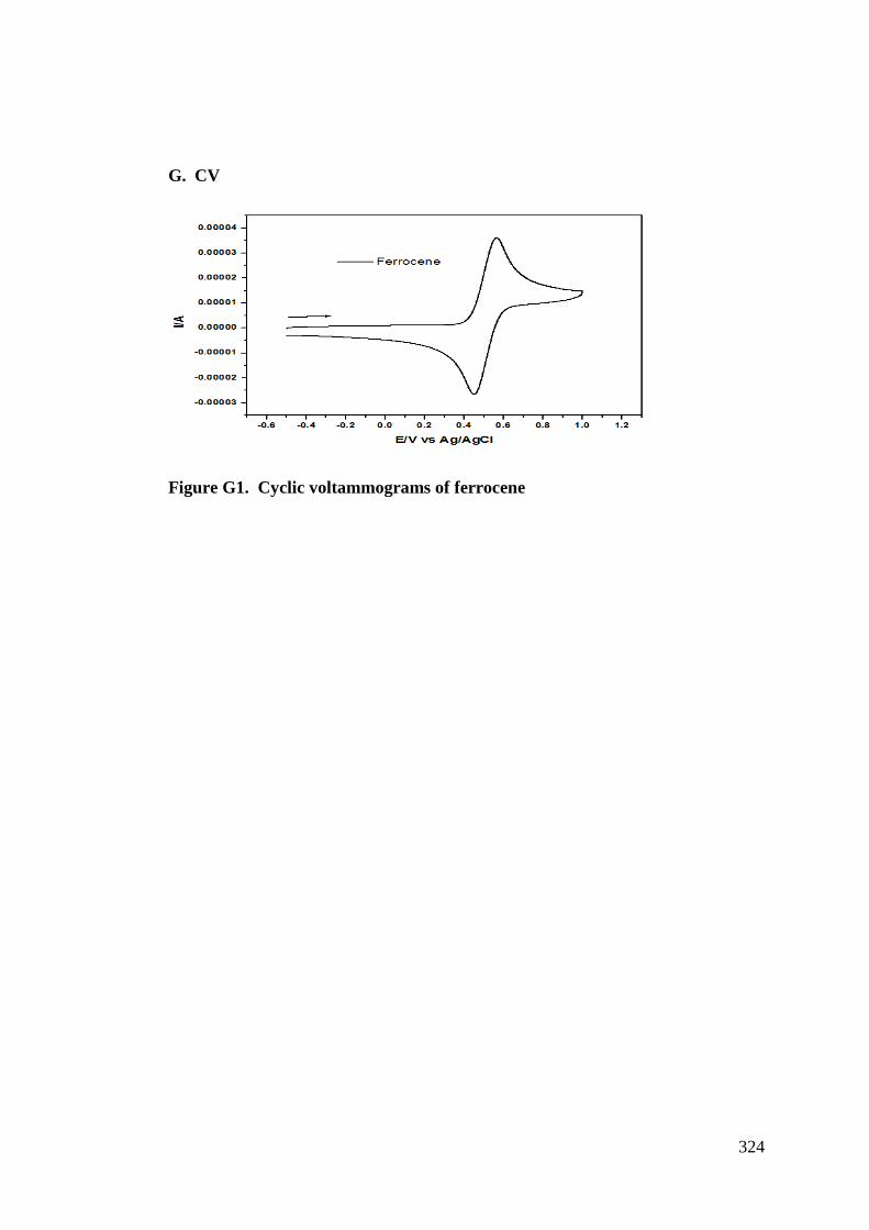
324
G. CV
Figure G1. Cyclic voltammograms of ferrocene
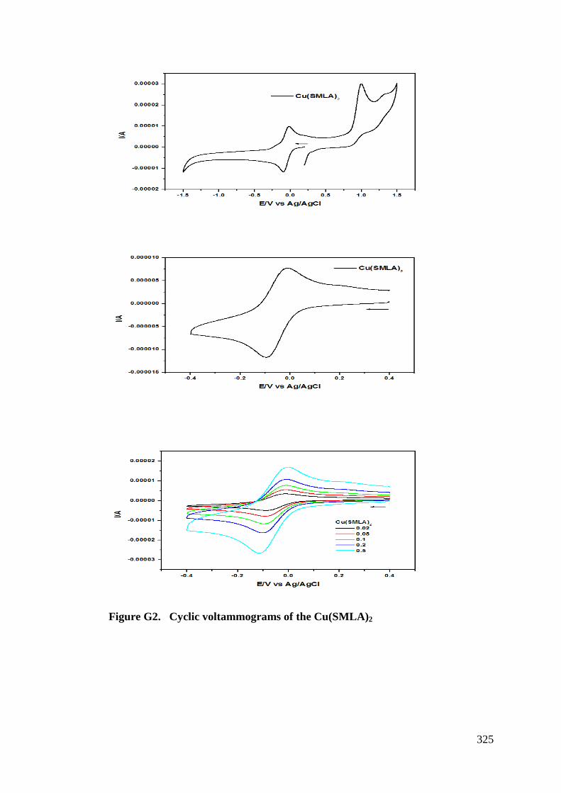
325
Figure G2. Cyclic voltammograms of the Cu(SMLA)2
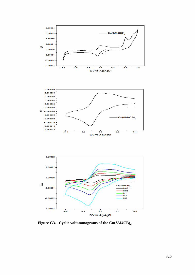
326
Figure G3. Cyclic voltammograms of the Cu(SM4CB)2
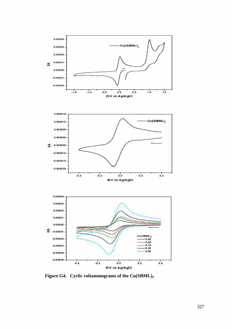
327
Figure G4. Cyclic voltammograms of the Cu(SBML)2
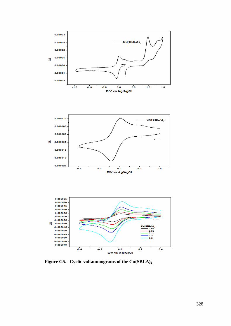
328
Figure G5. Cyclic voltammograms of the Cu(SBLA)2
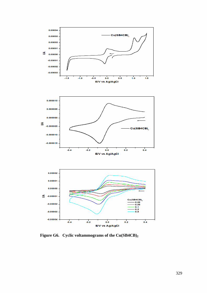
329
Figure G6. Cyclic voltammograms of the Cu(SB4CB)2
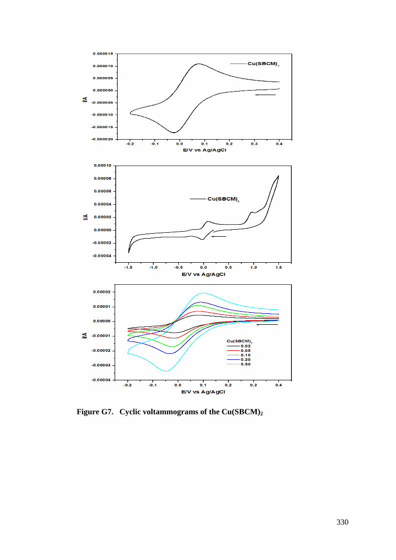
330
Figure G7. Cyclic voltammograms of the Cu(SBCM)2
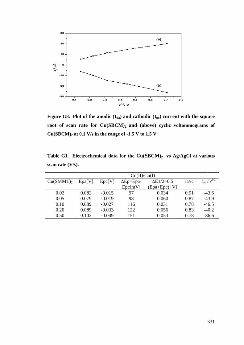
331
Figure G8. Plot of the anodic (Ipa) and cathodic (Ipc) current with the square root of scan rate for Cu(SBCM)2 and (above) cyclic voltammograms of
Cu(SBCM)2 at 0.1 V/s in the range of -1.5 V to 1.5 V.
Table G1. Electrochemical data for the Cu(SBCM)2 vs Ag/AgCl at various
scan rate (V/s).
Cu(II)/Cu(I) Cu(SMML)2 Epa[V] Epc[V] 2Ep=Epa-
Epc[mV] 2E1/2=0.5
(Epa+Epc) [V] ia/ic ipc / +1/2
0.02 0.082 -0.015 97 0.034 0.91 -43.6 0.05 0.079 -0.019 98 0.060 0.87 -43.9 0.10 0.089 -0.027 116 0.031 0.78 -46.5 0.20 0.089 -0.033 122 0.056 0.83 -40.2 0.50 0.102 -0.049 151 0.053 0.78 -36.6
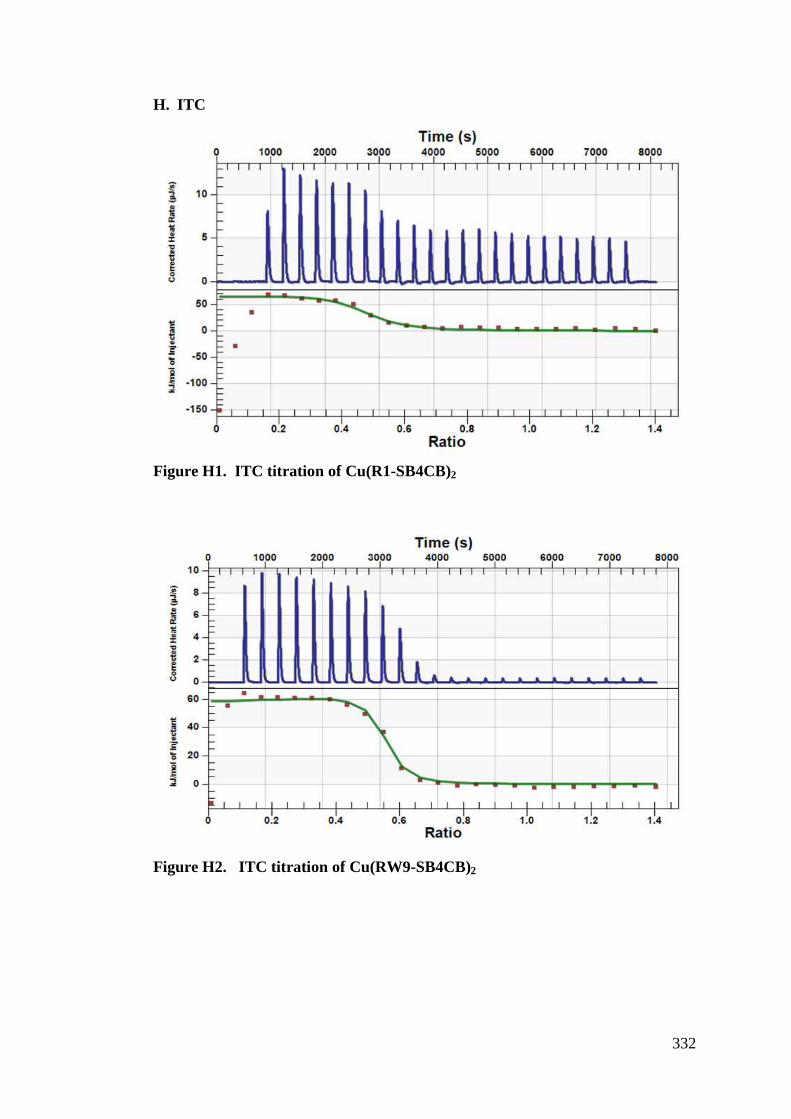
332
H. ITC
Figure H1. ITC titration of Cu(R1-SB4CB)2
Figure H2. ITC titration of Cu(RW9-SB4CB)2
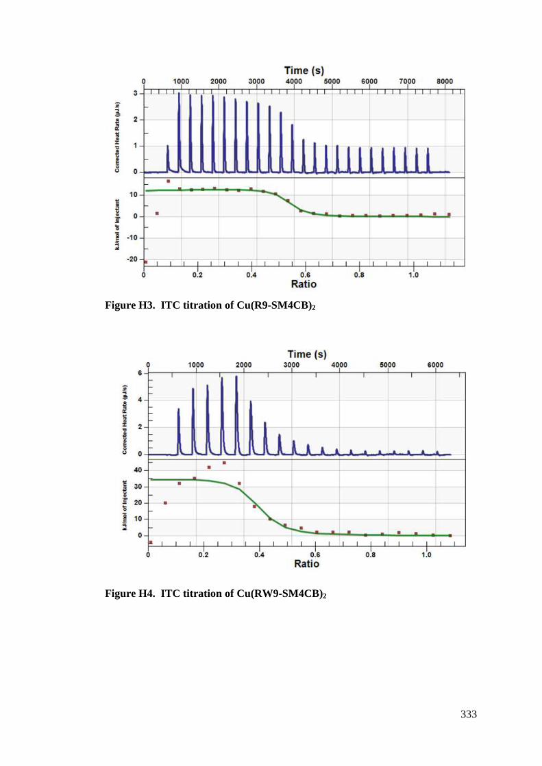
333
Figure H3. ITC titration of Cu(R9-SM4CB)2
Figure H4. ITC titration of Cu(RW9-SM4CB)2
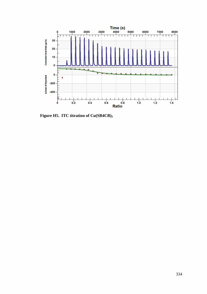
334
Figure H5. ITC titration of Cu(SB4CB)2
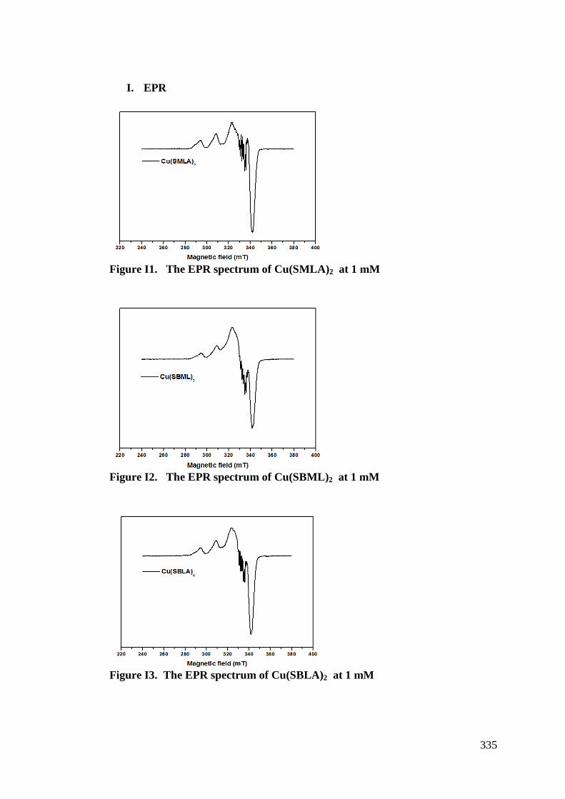
335
I. EPR
Figure I1. The EPR spectrum of Cu(SMLA)2 at 1 mM
Figure I2. The EPR spectrum of Cu(SBML)2 at 1 mM
Figure I3. The EPR spectrum of Cu(SBLA)2 at 1 mM
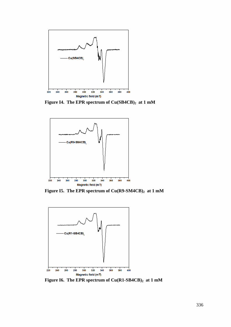
336
Figure I4. The EPR spectrum of Cu(SB4CB)2 at 1 mM
Figure I5. The EPR spectrum of Cu(R9-SM4CB)2 at 1 mM
Figure I6. The EPR spectrum of Cu(R1-SB4CB)2 at 1 mM
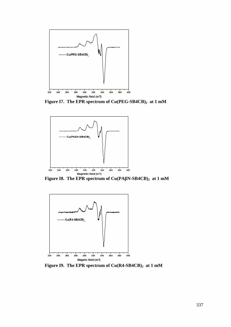
337
Figure I7. The EPR spectrum of Cu(PEG-SB4CB)2 at 1 mM
Figure I8. The EPR spectrum of Cu(PA"N-SB4CB)2 at 1 mM
Figure I9. The EPR spectrum of Cu(R4-SB4CB)2 at 1 mM
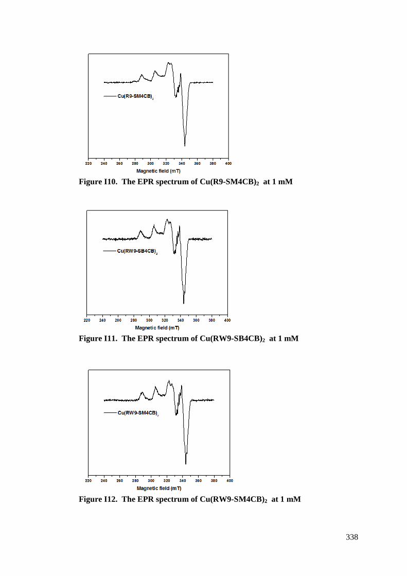
338
Figure I10. The EPR spectrum of Cu(R9-SM4CB)2 at 1 mM
Figure I11. The EPR spectrum of Cu(RW9-SB4CB)2 at 1 mM
Figure I12. The EPR spectrum of Cu(RW9-SM4CB)2 at 1 mM

339
Figure I13. The EPR spectrum of Cu(SBCM)2 at 1 mM
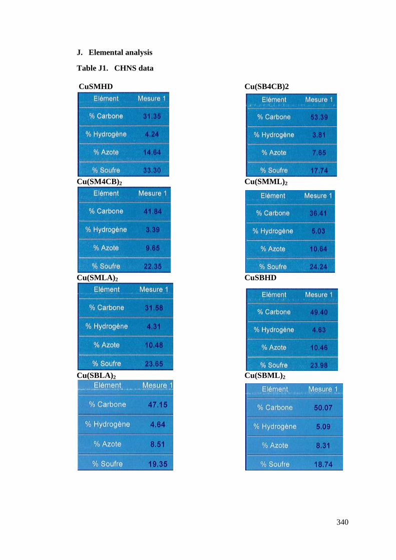
340
J. Elemental analysis
Table J1. CHNS data CuSMHD Cu(SB4CB)2
Cu(SM4CB)2 Cu(SMML)2
Cu(SMLA)2 CuSBHD
Cu(SBLA)2 Cu(SBML)2
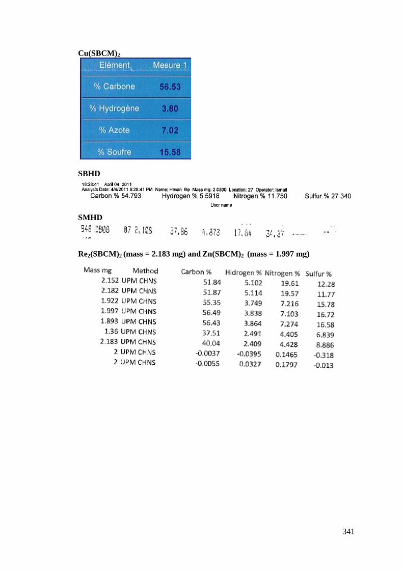
341
Cu(SBCM)2
SBHD
SMHD
Re2(SBCM)2 (mass = 2.183 mg) and Zn(SBCM)2 (mass = 1.997 mg)
342
LIST OF PUBLICATIONS AND CONFERENCES ATTENDED
List of Publications 1. Low, M. L., Ravoof, T. B. S., Tahir, M. I. M., Crouse, K. A., & Tiekink, E.
R. (2013). (Pyridin-4-yl) methyl N'-(3-phenylallylidene) hydrazinecarbodithioate. Acta Crystallographica Section E: Structure Reports Online, 69(2), o167-o168.
2. May Lee Low, Laure Maigre, Pierre Dorlet, Régis Guillot, Jean-Marie Pagès,
Nicolas Delsuc, Clotilde Policar and Karen A. Crouse. A New Series of Copper(II) Dithiocarbazate Schiff Bases: Synthesis, Conjugation to Polyarginine and Effect On Antimicrobial Activity. Manuscript in preparation.
3. May Lee Low, Georgiana Paulus, Pierre Dorlet, Régis Guillot, Rozita Rosli,
Nicolas Delsuc, Clotilde Policar and Karen A. Crouse. Synthesis, Characterization and Biological Activity of Cu(II), Zn(II) And Re(I) Complexes Derived from S-Benzyl Dithiocarbazate and 3-Acetylcoumarin. Manuscript in preparation.
4. May Lee Low, Laure Maigre, Mohamed Ibrahim M. Tahir, Pierre Dorlet,
Régis Guillot, Thahira Begum Ravoof, Rozita Rosli, Jean-Marie Pagès, Nicolas Delsuc, Clotilde Policar and Karen A. Crouse. X-Ray and Solution Structure of Copper (II) Macroacyclic Bis(dithiocarbazate): Influence on Their Redox Properties and Bioactivities. Manuscript in preparation.
List of Conferences Attended 1. May Lee Low, Laure Maigre, Pierre Dorlet, Régis Guillot, Jean-Marie Pagés,
Nicolas Delsuc, Clotilde Policar, Karen A. Crouse. A New Series of Copper(II) Dithiocarbazate Schiff Bases: Synthesis, Conjugation to Polyarginine and Effect on Antimicrobial Activity. 16th International Conference of Bioinorganic Chemistry (ICBIC), Grenoble, France (22nd - 26th July 2013). Poster presentation.
2. May Lee Low, Laure Maigre, Pierre Dorlet, Régis Guillot, Jean-Marie Pagés,
Nicolas Delsuc, Clotilde Policar, Karen A. Crouse. A New Series of Copper(II) Dithiocarbazate Schiff Bases: Synthesis, Conjugation to Polyarginine and Effect on Antimicrobial Activity. Group Français de Chimie Bio-Inorganique, Reunion Annuelle, Aussois, France (24-27th March 2013). Oral communication.
3. May Lee Low, Laure Maigre, Mohamed Ibrahim M. Tahir, Pierre
Dorlet, Régis Guillot, Thahira Begum Ravoof, Rozita Rosli, Jean-Marie Pagés, Nicolas Delsuc, Clotilde Policar, Karen A. Crouse. Macroacyclic Bis(dithiocarbazate): Synthesis, Characterization and Bioactivities. 40th International Conference on Coordination Chemistry (ICCC), Valencia, Spain (9-13th September 2012). Poster presentation.
343
4. May Lee Low, Laure Maigre, Mohamed Ibrahim M. Tahir, Pierre Dorlet, Régis Guillot, Thahira Begum Ravoof, Rozita Rosli, Jean-Marie Pagés, Nicolas Delsuc, Clotilde Policar, Karen A. Crouse. Synthesis, Characterisation and Biological Activities of Cu(II) Complex Containing Macroacyclic Tetradentate (N2S2) Schiff Base Ligand Derived from Dithiocarbazate. Club Metalloproteines et Modeles, Reunion Annuelle, Mittelwihr, France (25-28th March 2012). Poster presentation.
5. The 3rd Fundamental Science Congress 2011, Universiti Putra Malaysia (5-
6th July 2011), As Participant. 6. 13th BCA/CCG Intensive Teaching School in X-Ray Structure Analysis,
Durham, UK (26th March-3rd April 2011), As Participant.
344
BIODATA OF THE AUTHOR
Low May Lee was born on the 27th of March 1988 in Melaka, Malaysia. She
received her primary and secondary education in Air Panas Setapak Girls School,
Kuala Lumpur. In 2010, she obtained her B.Sc. (Honours) in Chemistry with first
class from Universiti Putra Malaysia and was awarded for the Highest Academic
Achievement in the B.Sc. (Honours) in Chemistry program for the 2010 graduating
class. During her undergraduate studies, she did her industrial training with the
Material Characterization Unit (MCU) at Rubber Research Institute of Malaysia
(RRIM). She also carried out her final year project in the field of solid state
chemistry working on the synthesis and characterization of bismuth ferrite materials
under the supervision of Dr. Tan Kar Ban. In the same year 2010, she started her
Doctor of Philosophy in Synthesis program at Universiti Putra Malaysia under the
supervision of Professor Karen A. Crouse and in November 2011, she was granted
the Erasmus Mundus: MAHEVA Scholarship to continue her doctoral research in
the group of Professor Clotilde Policar at the Laboratoire des BioMolécules (UMR
7203) in Paris, France for 22 months.