synthesis of ha and beta-tcp using sol-gel process … · study by many researchers worldwide...
TRANSCRIPT
Proceedings of COBEM 2011 21st Brazilian Congress of Mechanical Engineering
Copyright © 2011 by ABCM October 24-28, 2011, Natal, RN, Brazil
SYNTHESIS OF HA AND BETA-TCP USING SOL-GEL PROCESS AND ANALYSIS WITH FTIR
Leonardo Ribeiro Rodrigues, [email protected]
Carmen Gilda Barroso Tavares Dias, [email protected] DEMA-FEM-UNICAMP / INCT Biofabris and DEM-UFPA
Fernando Jorge Mendes Monteiro, [email protected] IBMC-INEB / DEMM-FEUP
Cecília Amélia de Carvalho Zavaglia, [email protected] DEMA-FEM-UNICAMP / INCT Biofabris
Abstract. Nanotechnology has been attracting researchers´ attention due to its innovative concepts and associated
technology development. Nowadays, one approach to control the surface´s chemistry and in vivo solubility of calcium
phosphate compounds are associated to the employment of nanoparticles. The sol-gel process is used for synthesis of
many materials in nanoscale, because your process starts on a colloidal suspension. The hydroxyapatite Ca10(PO4)6
(OH)2 is the mineral phase of bone tissue. The hydroxyapatite bioceramic is a bioactive material and because of this
reason its application with bone substitute and also as a coating or film in materials with a biological response less
then hydroxyapatite. Β-TCP, [β-Ca3(PO4)2], is widely used on the development of bone defects fulfilling since its
chemical composition leads to implants that exhibit excellent biocompatibility, bioactivity and resorbable. This
material could be employed as bone cement, as porous or dense blocks, as granulated material and in all cases the
particle size and morphology are key factors to achieve appropriate chemical, biological and mechanical properties.
In this context, the goal of this work is to synthesize nanostructured HA and β-TCP using sol-gel method which has
proved to be very efficient in obtaining nanomaterials with high homogeneity. The DSC showed the optimal
temperature range of gel work and the best temperature range from 50 to 90ºC. For characterization was used
scanning electron microscopy (MEV), X-ray fluorescence (XFR), and Scherrer equation associated with X-ray
diffraction (XRD). Surfaces good bioactivity have investigated using bands derived from PO3-4
by Fourier transform
infrared spectroscopy (FTIR).
Keywords: Hydroxyapatite, beta-tricalcium phosphate, Fourier transform infrared spectroscopy, sol-gel process with
sucrose, nanotechnology.
1. INTRODUCTION
The calcium phosphates are the most important inorganic components of bone tissue “Abdel Fattah et al. (2008)”.
The common calcium phosphate laboratory syntheses are: hydroxyapatite, beta-tricalcium phosphate and alpha-
tricalcium phosphate.
The hydroxyapatite (HA) is a bioactive calcium phosphate bioceramic, that in biological environment is not
stoichiometric as the synthetic material (Ca/P: 1,67). The mineral phase of synthetic material is similar to bone tissue
and it is used for bone replacement. The bioactivity of HA induces bone growth and facilitates the regeneration process
“Guha et al. (2009)”.
Beta-tricalcium phosphate β-TCP is a bioceramic with Ca/P of 1,5. This material is used as bone cement for filling
small facial defects, and also be possible used for the fabrication of dense or porous ceramic composites. Beta-TCP is a
biodegradable material that can be replaced by bone tissue “Ohura et al. (1996)”.
Calcium phosphates are being applied on scaffolds construction for tissue engineering “Kawachi et al. (2000)”.
The nanotechology is an innovative concept with numerous technological advances, therefore became the object of
study by many researchers worldwide “Souza et al. (2007)”. The nanotechnology in biomaterials enable functional
regulation of cells, which plays a central role in nanomedicine targeting at regenerative medicine and intractable
diseases. It is possible design the nanostructure of materials that induces cell differentiation at specific positions of the
living body. The nanostructures are based on design bioinspired performing necessary function at the right time and the
right place by manipulating assembly of molecules and atoms forming the natural tissue, for example bone tissue.
During the development of new biomaterials, nanoparticles play an important role because their greater superficial
are often leads to distinct values of reactivity, mechanical, chemical and biological properties in vitro and in vivo when
compared to micro and macroscopic particles “Quina et al. (2004)”.
The sol-gel product is characterized by primary nanoparticles, and this is a crucial parameter to improve the surface
reaction and the stability at implant/natural bone interface. Another approach to the sol-gel process is the sucrose
addition, which allows the synthesis of particles smaller than the conventional process “Rodrigues et al. (2009)”.
In this work, two types of nanostructured calcium phosphate were synthetized by sol-gel process with sucrose, were:
hydroxyapatite and beta-tricalcium phosphate.

Proceedings of COBEM 2011 21st Brazilian Congress of Mechanical Engineering
Copyright © 2011 by ABCM October 24-28, 2011, Natal, RN, Brazil
For characterization of this bioceramics were used: X-ray diffraction (XRD), X-ray fluorescence (XFR), scanning
electron microscope (SEM) and Fourier transform infrared spectroscopy (FTIR).
In XRD were analyzed for crystalline materials. The standards used for the comparison and confirmation of results it
was obtained in the JCPDS library.
The XRF is used to characterize the materials by semi-quantitative analysis of each chemical element present in the
sample.
The SEM is used to visualize the nanoparticles size and identify the morphology of material.
The FTIR analysis indicated characteristic spectra bands for all the studied materials and identifies a composition of
sample “Laranjeira et al. (2010)”.
2. MATERIALS AND METHODS
2.1. Materials
The reagents used were: phosphoric acid - LAFAN 85% (H3PO4), calcium nitrate tetrahydrate – Synth 99%
[Ca(NO3)2 . 4 H2O] and Sucrose – Synth (C12H22O11).
The hydroxyapatite Ca/P: 1.67 and β-tricalcium phosphate Ca/P: 1.5.
2.2. Methods
Hydroxyapatite was prepared from two aqueous solutions of sucrose, one with phosphoric acid and, the other with
calcium nitrate tetrahydrate. The Ca/P ratio was maintained equal to 1.67. Sucrose was used as a gel forming and
dispersant agent. Then, the gel was fired at 700ºC.
Beta-tricalcium phosphate was prepared of same way that hydroxyapatite, but maintaining the Ca/P ratio in 1.5. The
gel was fired at 1050ºC.
Water + calcium
nitrate + sucrose
Water + calcium nitrate
+ phosphoric acid +
sucrose
Sol
Aging
Sol-gel
Calcination
Powder
Water +
phosphoric acid +
sucrose
Figure 1. Fluxogram of sol-gel process with sucrose. Both bioceramics were calcined, but hydroxyapatite at 700ºC and
β-TCP at 1050ºC.
2.3. Characterizations
The morphology and microstructure were analyzed by scanning electron microscopy (SEM). The equipment used
was a JOEL model JXA-840A of DEMA-FEM-UNICAMP, SEM-FEG JEOL JSM 6330F belonging to LNLS-LME.
The X-ray diffraction was performed to verify the crystallinity and phases present in the material. The equipment
belong to the DEMA-FEM-UNICAMP, model DMAX 2200-Rigaku, Cu-Kα radiation with Ni filter. Analyses were
performed at 40kV and 30mA in a 2θ range from 20º to 50º, with step size of 0.02º and 2 second of integration time.
The crystallite size were calculated using the Scherrer Equation “Nag et al. (2007)” (Eq. 1) after fitting a gaussian
curve on the principal peak to determine their width at the half height intensity “Briks et al. (1946)”.
t= 0,9λ / βcos θ (1)
Where, “t” is the crystallite mean size, “λ” is the X-ray wavelength, “β” given by Eq. (2) is the peak width at half
height intensity, and “θ” is the Bragg angle.
β = β2
exp – β2
int (2)
Where, “βexp” is the peak width at half height intensity of the sample and “βinst” is the instrumental correction
obtained by the width at half height intensity of the monocrystal pattern.

Proceedings of COBEM 2011 21st Brazilian Congress of Mechanical Engineering
Copyright © 2011 by ABCM October 24-28, 2011, Natal, RN, Brazil
The X-ray fluorescence spectroscopy was used to determine the impurities quantitative and the equipment used was
Rigaku RIX 3100 spectrometer. The sample were conformed into 25mm disks and analyzed by a semi-quantitative
routine.
Infrared spectra analysis was performed with a Fourier transform infrared spectroscopy (FTIR) and the equipment
used was Thermo Scientific Nicolet IR100 FT-IR with spectral resolution of 128 to 4 cm-1
. The spectral used was 4000
to 400cm-1
and the samples were ground with dried potassium bromide (KBr) powder, and
tablet pressed into a disc for this analyses.
3. RESULTS AND DISCUSSION
3.1 X-ray diffraction
The standard graphics of XRD were obtained in database Joint Committee on Powder Diffraction Standards
(JCPDS) and the HA has the number 09-0169 and β-TCP has the number 09-0432.
(a) (b) Figure 2. X-ray diffraction of sample. (a) Graphic of HA with a peak contamination of B-TCP. (b) Graphic of β-TCP.
After heat treatment the HA shown the peaks in Fig. 2 (a) indicating a contamination with beta-TCP. The sample B-
TCP shown in Fig. 2 (b) the peaks of beta-TCP and Hydroxyapatite, this problem occurred because the sol-gel process
with sucrose to synthetize beta-tcp is a new application. This process for synthesis of calcium phosphates as
hydroxyapatite and beta-tcp is new and it was developed based on the article “Souza et al. (2007)”. Sucrose is
introduced into the process to decrease the size of the particles.
Table 1. The crystallite size of HA and β-TCP obtained by Scherrer equation:
Sample Crystallite (nm)
HA ~58
Β-TCP ~95
The results of X-ray diffraction were used to perform analysis of crystallite size by Scherrer equation. The results
were consistent due to temperature difference of sample calcination. The Hydroxyapatite crystallite had an average size
of about 58nm and beta-TCP calcined at 1050ºC had an average size of 95nm.
Table 2. Impurities´ XRF semi-quantitative analysis.
Materials Sr Fe Na S Cl Mg Si Al Ni K Mn Zr
HA (wt.%) 0.028 0.004 0.018 0.007 0.028 b.l.d. b.l.d. b.l.d. b.l.d. b.l.d. b.l.d. b.l.d.
Β-TCP (wt.%) 0,018 b.l.d 0,011 0,006 b.l.d. b.l.d. b.l.d. 0,005 0,003 b.l.d. b.l.d. 0,006
HA-VETEC (wt.%) 0.012 0.015 0.074 0.021 0.032 0.325 0.020 0.009 0.002 0.012 0.009 b.l.d.
b.l.d.: bellow the limit of detection
Proceedings of COBEM 2011 21st Brazilian Congress of Mechanical Engineering
Copyright © 2011 by ABCM October 24-28, 2011, Natal, RN, Brazil
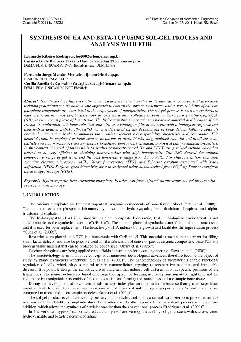
The X-ray fluorescence showed a very important result for the use of these materials in the biomedical are because it
has not significant amounts of contaminants and quantities of each element is small compared to commercial material as
HA-VETEC shown in the Tab. 2.
(a) (b)
Figure 3. Image of SEM. (a) Shown the HA nanoparticle aggregates. (b) Shown the β-TCP nanoparticle aggregates.
The scanning electron microscopy proved that the samples have the approximated results obtained with Scherrer
equation. The Fig. 3 (a) shown the HA nanoparticle clusters with nanometer size. Fig. 3 (b) shown Beta-TCP with
larger particle size when compared with HA, but the results are similar the results of Scherrer equation when compared
the particle size with scale.
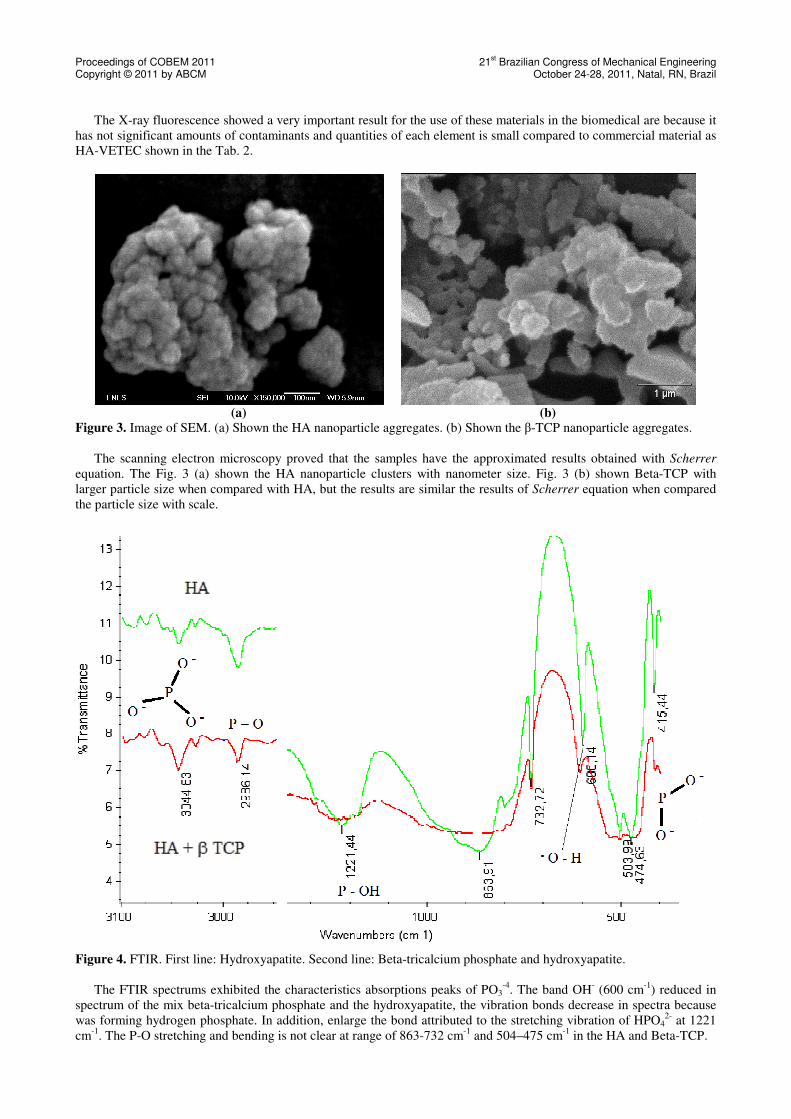
Figure 4. FTIR. First line: Hydroxyapatite. Second line: Beta-tricalcium phosphate and hydroxyapatite.
The FTIR spectrums exhibited the characteristics absorptions peaks of PO3-4
. The band OH- (600 cm
-1) reduced in
spectrum of the mix beta-tricalcium phosphate and the hydroxyapatite, the vibration bonds decrease in spectra because
was forming hydrogen phosphate. In addition, enlarge the bond attributed to the stretching vibration of HPO42-
at 1221
cm-1
. The P-O stretching and bending is not clear at range of 863-732 cm-1
and 504–475 cm-1
in the HA and Beta-TCP.

Proceedings of COBEM 2011 21st Brazilian Congress of Mechanical Engineering
Copyright © 2011 by ABCM October 24-28, 2011, Natal, RN, Brazil
4. CONCLUSION
In the results of X-ray diffraction, the hydroxyapatite only have a Beta-TCP peak contamination, this is due to Ca/P
has not been perfect during the synthesis. The Fig. 2 (b) shown peaks of beta-TCP and HA, this problem occurred due
to sol-gel process with sucrose to synthesis of beta-TCP it is a new method and it needs modifications in your process
parameters.
The results of Scherrer equation shown the crystallite size below 100nm, therefore these are nanomaterials. The HA
with ~58nm and beta-TCP with ~95nm.
The X-ray fluorescence to semi-quantitative analysis showed a low amount chemical elements presents in the
samples and it showed also low wt.% of each element when to compare HA and B-TCP with commercial HA.
The scanning electron microscopy showed the morphology and an idea about the particle size. The hydroxyapatite
showed the agglomerate structure with nanoparticles bellow 100nm. The beta-TCP with HA contamination showed
nanoparticles and large dense blocks due to calcination temperature.
By FTIR, the evidence the key factors influence at morphologies nonconventional in the case were non-
stoichiometric phosphocalcic in HA and Beta-TCP.
3. ACKNOWLEDGEMENTS
The authors would like to thank CAPES and INCT Biofabris for the financial support. The SEM-FEG micrographs
were supported by LME-LNLS - Brazilian Synchrotron Light Laboratory/MCT.
4. REFERENCES
Abdel Fattah, W.I., Reicha, F.M. Elkhooly, T.A., 2008. “Nano-beta-tricalcium phosphate synthesis and biodegradation:
1. Effect of microwave and SO4 2-ions on beta-TCP synthesis and its characterization”. Biomedical Materials, 3, 1-
13.
Briks, L. S. and Friedman, H., 1946. “Particle Size Determination from X-ray Line Broadening”. Journal of Applied
Physics, 17, 8, 687-692.
Guha, A.K., Singh, S., Kumaresan, R., Nayar, S., Sinha, A., 2009. “Mesenchymal cell response to nanosized biphasic
calcium phosphate composites”. Colloids and Surfaces B: Biointerfaces, 73, 146-151.
Kawachi, E.Y., Bertran, C.A., Reis, R.R., Alves, O.L., 2000. “Biocerâmicas: Tendências e perspactivas de uma área
interdiciplinar, química nova, 23, 4, 443-450.
Laranjeira, M.S., Fernandes, M.H., Monteiro, F.J., 2010. “Innovative macroporous granules of nanostructured-
hydroxyapatite agglomerates: Bioactivity and osteoblast-like cell behavior”. Journal of Biomedical Materials
Research Part A, 95A, 3, 891-900.
Nag, M., Basak, P., Manorama, S. V., 2007. “Low-Temperature hydrothermal synthesis of phase -pure rutile titania
nanocrystals: Time temperature tuning of morphology and photocatalytic activity”. Materials Research Bulletin, 42,
1691 – 1704.
Ohura, K., Bohner, M., Hardouin, P., Hardouin, P., Lemaítre, J., Pasquier, G., Flautre, B., 1996. “Resorption of, and
bone formation from, new β-tricalcium phosphate-monocalcium phosphate cements: an in vivo study”. Journal
Biomedical Materials Research, 30, 193-200.
Quina, F.H., 2004. “Nanotecnologia e o meio ambiente: perspectiva e riscos”. Química nova, 27, 6, 1028-1029.
Rodrigues, L.R., Motisuke, M., Zavalgia, C.A.C., 2009. “Synthesis of nanostructured hydroxyapatite: A comparative
study between sol-gel and aqueous solution precipitation”. Key Engineering Materials, 396-398, 623-626.
Souza, E.A., Duque, J.G.S., Kubota, L., Meneses, C.T., 2009. “Synthesis and characterization of NiO and NiFe2O4
nanoparticles obtained by a sucrose-based route”. Journal of Physics and Chemistry of Solids, 68, 594-599.
5. RESPONSIBILITY NOTICE
I Leonardo Ribeiro Rodrigues, Carmen Gilda Barroso Tavares Dias, Fernando Jorge Mendes Monteiro and Cecília
Amélia de Carvalho Zavaglia are the only responsible for the printed material included in this paper.