synthesis of netilmicin and apramycin derivatives for the
TRANSCRIPT
Wayne State University
Wayne State University Dissertations
1-1-2017
Synthesis Of Netilmicin And ApramycinDerivatives For The Treatment Of Multidrug-Resistant Infectious DiseasesAmr Sayed Motawi SonousiWayne State University,
Follow this and additional works at: https://digitalcommons.wayne.edu/oa_dissertations
Part of the Organic Chemistry Commons
This Open Access Dissertation is brought to you for free and open access by DigitalCommons@WayneState. It has been accepted for inclusion inWayne State University Dissertations by an authorized administrator of DigitalCommons@WayneState.
Recommended CitationSonousi, Amr Sayed Motawi, "Synthesis Of Netilmicin And Apramycin Derivatives For The Treatment Of Multidrug-ResistantInfectious Diseases" (2017). Wayne State University Dissertations. 1880.https://digitalcommons.wayne.edu/oa_dissertations/1880

SYNTHESIS OF NETILMICIN AND APRAMYCIN DERIVATIVES FOR THE
TREATMENT OF MULTIDRUG-RESISTANT INFECTIOUS DISEASES
by
AMR SONOUSI
DISSERTATION
Submitted to the Graduate School
of Wayne State University,
Detroit, Michigan
in partial fulfillment of the requirements
for the degree of
DOCTOR OF PHILOSOPHY
2017
MAJOR: CHEMISTRY (Organic)
Approved By:
Advisor Date

ii
DEDICATION
I dedicate my PhD work to my parents Sayed Sonousi and Hoda Fayed for nursing me with
affection and love and for their dedicated partnership for success in my life. I also dedicate my
work to my wife Tasnim Kandeel for her endless love and support for me throughout the process.

iii
ACKNOWLEDGEMENTS
I would first like to express my utmost appreciation to my advisor, Professor David
Crich, for his endless patience, brilliant guidance, continuous encouragement, and constant
support during the past five years of my Ph.D. studies. His passion and dedication for science
will always be my example to follow. Without the help and support from him, I would not have
been able to finish this thesis.
I would like to extend my appreciation to all of my committee members, Professors
James Rigby, John SantaLucia and Steven Sucheck, for their precious time and for their
suggestions made for my thesis dissertation. I also would like to thank our collaborators
Professors Andrea Vasella, Erik C Böttger, Patrice Courvalin and Jochen Schacht for their
contribution to my project
My sincere thanks to my mentors who taught me during my first-year courses, especially
Dr. Cha for the advanced organic reactions course as well as Drs Guo, Kodanko, Stockdill, Ahn
and Hendrickson for the other organic and bio-organic courses.
My profound gratitude goes to my present and past lab mates. Dr. Takayuki Kato, Dr.
Takayuki Furukawa, Dr. Suresh Dharuman, and Dr. Takahiko Matsushita gave me much advice
on my research. They were always willing to share their ideas with me in long discussions. I also
thank Dr. Mandhapati for his help teaching me laboratory techniques and helping me familiarize
myself with the lab in my first year; Philip, with whom I enjoyed working with on the same
bench, and Dr. Buda, Dr. Popik, Peng, Harsha, Jessica, Girish, Bibek, Sandeep, Xiaoxiao,
Guanyu, Mike, Mohamed, Philemon, and Jonathan for their timely help in the lab. Finally, I
would like to thank the helpful staff in the chemistry department, as well as my supportive
friends and family.
iv
TABLE OF CONTENTS
Dedication ....................................................................................................................................... ii
Acknowledgements ........................................................................................................................ iii
Table of Contents ........................................................................................................................... iv
List of Tables ............................................................................................................................... viii
List of Figures ................................................................................................................................ ix
List of Schemes ............................................................................................................................. xii
List of Abbreviations ................................................................................................................... xiv
Chapter 1. Introduction ................................................................................................................... 1
1.1. Background and significance ............................................................................................... 1
1.2. Structural features and classifications .................................................................................. 3
1.3. Mechanism of action of aminoglycosides ............................................................................ 5
1.3.1. AGA uptake ............................................................................................................. 5
1.3.2. Protein synthesis and AGA binding to the ribosomes ............................................. 6
1.4. Aminoglycoside resistance and toxicity ............................................................................ 11
1.4.1. Aminoglycoside resistance .................................................................................... 11
1.4.1.1. Reduction of aminoglycoside internal concentration ................................. 12
1.4.1.2. Ribosomal binding site modifications ........................................................ 13
1.4.1.3. Aminoglycoside modifying enzymes (AMEs) ........................................... 13
v
1.4.2. Adverse effects of aminoglycosides ...................................................................... 16
1.4.2.1. Nephrotoxicity ............................................................................................ 17
1.4.2.2. Ototoxicity .................................................................................................. 18
1.5. Recent advances ................................................................................................................. 21
1.6. Overall goals ...................................................................................................................... 23
Chapter 2. Investigations in the 4,6-Class of AGAs ..................................................................... 24
2.1. Mode of binding of netilmicin to the bacterial ribosomal A-site....................................... 24
2.2. Synthesis of netilmicin ....................................................................................................... 25
2.3. Modifications of netilmicin................................................................................................ 26
2.4. Rational .............................................................................................................................. 29
2.5. Chemistry ........................................................................................................................... 29
2.5.1. Triazenes as a selective protecting group for secondary amines ........................... 29
2.5.2. Aryl triazenes ........................................................................................................ 30
2.5.3. Examples of selective protection ........................................................................... 33
2.5.4. Application to aminoglycosides ............................................................................ 35
2.5.5. Synthesis of netilmicin derivatives ........................................................................ 37
2.5.6. Synthesis of plazomicin ......................................................................................... 41
2.6. Biological Evaluation......................................................................................................... 44
2.7. Conclusion ......................................................................................................................... 48
Chapter 3. Development of Apramycin Derivatives with Modification at the 5-Position and
Examination of their Antiribosomal and Antibacterial Activity................................................... 50

vi
3.1. Mode of binding of apramycin to the bacterial ribosomal A-site ...................................... 50
3.2. Modifications of apramycin ............................................................................................... 51
3.2.1. Modifications at the 5- and 6-positions of apramycin ........................................... 52
3.2.2. Modifications at the N1,N2',N7',N4''-positions of apramycin ............................... 55
3.2.3. Modifications at the 6''-position of apramycin ...................................................... 58
3.2.4. Modifications at the N7’-Me and 6’-positions of apramycin ................................ 59
3.3. Rationale ............................................................................................................................ 60
3.4. Synthesis of apramycin derivatives ................................................................................... 63
3.4.1. Synthesis of a key apramycin intermediate ........................................................... 63
3.4.2. Synthesis of 5-O--ribofuranosyl apramycin, 5-O--paromobiosyl apramycin and
5-O--[3-O-(2-aminoethyl) ribofuranosyl] apramycin ......................................................... 64
3.4.3 Synthesis of 5-O--[3-O-(2-hydroxyethyl) ribofuranosyl] apramycin .................. 67
3.4.4. Modification of the 5’’-hydroxyl group of 5-O--ribofuranosyl apramycin and 5-
O--[3-O-(2-aminoethyl) ribofuranosyl] apramycin ............................................................ 68
3.4.5. Synthesis of 6-O-propyl apramycin, 6-O-(2,3-dihydroxypropyl) apramycin and 6-
O-(2-hydroxyethyl) apramycin ............................................................................................. 74
3.4.6. Synthesis of 5-epi-apramycin, 5-deoxy-5-fluoro apramycin, 5-deoxy-5-epi-fluoro-
apramycin and 5-deoxy apramycin ....................................................................................... 75
3.4.7. Synthesis of 3-N-formyl apramycin and 3-N-acetyl apramycin ............................ 77
3.5. Biological Evaluation......................................................................................................... 79
3.6. Conclusion ......................................................................................................................... 86
Chapter 4. Conclusions ................................................................................................................. 88

vii
Chapter 5. Experimental Section .................................................................................................. 90
References ................................................................................................................................... 176
Abstract ....................................................................................................................................... 189
Autobiographical Statement........................................................................................................ 191

viii
LIST OF TABLES
Table 1. Examples of selective protection of secondary amines as the N-phenyl triazenes ......... 34
Table 2. Catalysts used in the B-alkyl Suzuki reaction and their yields ....................................... 40
Table 3. Inhibition of wild-type bacterial and hybrid ribosomes (IC50, M). ............................. 44
Table 4. In vivo minimal inhibitory concentrations (MIC, μg/ml) of clinical isolates. ............... 46
Table 5. %Inhibition of in vitro R17 phage RNA-directed polypeptide synthesis by various
aminoglycoside antibiotics at four concentrations. ....................................................................... 62
Table 6. In vivo minimal inhibitory concentrations (MIC, μg/ml) of clinical isolates. ................ 62
Table 7. Inhibition of wild-type bacterial and hybrid ribosomes (IC50, M). ............................. 79
Table 8. Minimal inhibitory concentrations (MIC, μg/mL) of clinical isolates. ......................... 82
Table 9. Minimal inhibitory concentrations (MIC, μg/mL) of engineered E coli strains carrying
known AMEs ................................................................................................................................ 84
ix
LIST OF FIGURES
Figure 1. A graphical representation showing the number of antibacterial approvals from 1980
till 2012 ........................................................................................................................................... 1
Figure 2. A graphical representation showing the classification of antibiotics according to their
mode of action................................................................................................................................. 2
Figure 3. Streptomycin (1) .............................................................................................................. 3
Figure 4. Classification of aminoglycosides ................................................................................... 4
Figure 5. Unusual aminoglycoside structures ................................................................................. 5
Figure 6. Schematic diagram showing the stages of AGAs uptake ................................................ 6
Figure 7. The four fundamental nucleotides found in RNA and their Watson-Crick base pairs. ... 7
Figure 8. Schematic diagram showing ribosomes in protein synthesis .......................................... 8
Figure 9. a) Crystal structure of paromomycin binding to the bacterial A site (PDB code: 1FJG).
b) Schematic diagram showing the binding of paromomycin (3) with rRNA nucleobases. .......... 9
Figure 10. Schematic diagram showing a) the equilibrium of A-site between "flipped in" and
"flipped out" conformations in protein translation and b) aminoglycoside bound to ribosome A
site stabilizing the "flipped out" conformation ............................................................................. 10
Figure 11. Schematic diagram showing aminoglycoside bound to ribosome and causing codon
misreading. .................................................................................................................................... 10
Figure 12. Schematic diagram showing different resistance mechanisms ................................... 12
Figure 13. Target sites of aminoglycosides modifying enzymes on a) kanamycin B b) neomycin
B c) netilmicin and d) apramycin.................................................................................................. 14
Figure 14. Structures of tobramycin (12) and amikacin (13)........................................................ 16
Figure 15. A schematic representation of the mechanisms of aminoglycoside ototoxicity. ......... 19
Figure 16. Secondary-structure comparison of decoding-site rRNA sequences in the small
ribosomal subunit. (A) Decoding region of 16S rRNA helix 44 in wild-type ribosomes of M.
smegmatis. (B) Homologous 18S rRNA sequence in human ribosomes. (C) Homologous 12S

x
rRNA sequence in human mitochondrial ribosomes. (D) Mitochondrial 12S rRNA sequence with
mutation A1555G conferring hypersusceptibility to AGA ototoxicity. ....................................... 20
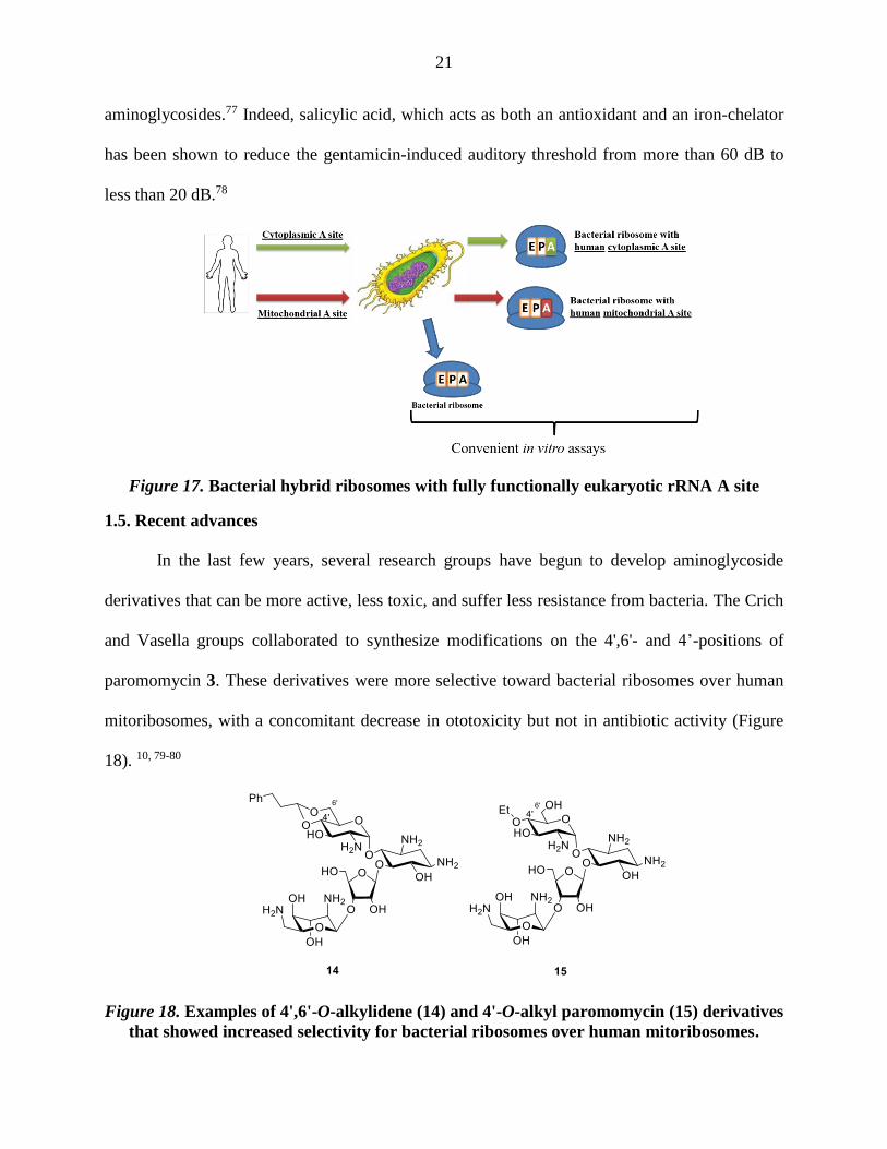
Figure 17. Bacterial hybrid ribosomes with fully functionally eukaryotic rRNA A site .............. 21
Figure 18. Examples of 4',6'-O-alkylidene (14) and 4'-O-alkyl paromomycin (15) derivatives that
showed increased selectivity for bacterial ribosomes over human mitoribosomes. ..................... 21
Figure 19. The structure of plazomicin [6'-N-(2-hydroxyethyl)-1-N-(4-amino-2(S)-
hydroxybutyryl) sisomicin] (16) ................................................................................................... 22
Figure 20. a) Crystal structure of sisomicin bound to the deep/major groove of the bacterial A
site showing π-stacking with G1491 base and pseudo base-pair interaction with A1408 (PDB
code: 4F8U), b) a schematic diagram showing the pseudo base-pair interaction of ring I with
A1408 and c) detailed interaction of sisomicin with the bacterial A site. .................................... 25
Figure 21. Resonance structures of aryl triazenes and the coalescence temperatures of triazene
N-methyl protons .......................................................................................................................... 32
Figure 22. Room-temperature 600 MHz 1H NMR spectra of (a) 58 and (b) 51 in CD3OD. ........ 35
Figure 23. The outer hair cells (OHC) in different parts of the cochlea (apex, mid and base) at 3
M (OHC are still intact) and 30 M (considerable loss in OHC) of plazomicin. ...................... 48
Figure 24. The effect of concentration of plazomicin (16) in comparison to gentamicin (7) and
apramycin (9) in the percentage of outer hair cell (OHC) loss. .................................................... 48
Figure 25. a) X-Ray crystal structure of apramycin bound to the bacterial A site showing CH/π-
stacking of ring I with G1491 base, pseudo base-pair interaction of ring I with A1408 and
interaction of ring III with A1492 (PDB code: 4AQY) and b) detailed interaction of apramycin
with the bacterial A site. ............................................................................................................... 51
Figure 26. Structures of apramycin (9) and aprosamine (91) ....................................................... 51
Figure 27. A schematic diagram showing known modifications of apramycin and the year
modified; modifications in green showed increased bacterial activity, modifications in red
showed a decreased bacterial activity, and modifications in black showed an unreported or
comparable bacterial activity. ....................................................................................................... 52
Figure 28. Paramomycin structure showing rings A-D, its hybrid with apramycin and a
simplified form of the hybrid. ....................................................................................................... 61

xi
Figure 29. A Schematic diagram showing the 5-ribosylation of the 4,6 AGAs ........................... 63
Figure 30. Reaction co-ordinate diagram showing the formation of 6-O-allyl apramycin
derivative 172 under Curtin-Hammett control .............................................................................. 74
xii
LIST OF SCHEMES
Scheme 1. Synthesis of netilmicin ................................................................................................ 26
Scheme 2. Synthesis of 5-deoxy-5,5-difluoro-netilmicin (23) ..................................................... 27
Scheme 3. Synthesis of 5-deoxy-5-fluoronetilmicin 29 and 5-epi-netilmicin 30 ......................... 28
Scheme 4. Synthesis of 6’-N-glycyl-netilmicin 32 ....................................................................... 28
Scheme 5. Rotamers of secondary amine carbamates .................................................................. 29
Scheme 6. General protocol for selective protection of secondary amines as the N-phenyl
triazenes ........................................................................................................................................ 30
Scheme 7. Literature reactions showing the compatibility of phenyltriazenes with oxidizing,
reducing and alkylating agents...................................................................................................... 30
Scheme 8. Literature reactions showing the compatibility of phenyltriazenes with acylating
reagents and basic conditions........................................................................................................ 31
Scheme 9. Uses of triazenes in solid-phase organic chemistry (SPOS) ...................................... 31
Scheme 10. Application to sisomicin............................................................................................ 36
Scheme 11. Application to Apramycin ......................................................................................... 37
Scheme 12. Synthesis of a protected netilmicin intermediate ...................................................... 38
Scheme 13. Synthesis of 4'-iodo netilmicin, 4'-bromo netilmicin and 4'-chloro netilmicin ......... 39
Scheme 14. A proposed mechanism for nitrate catalysis of bromination of glycals .................... 39
Scheme 15. Synthesis of 4'-phenyl netilmicin and 4'-butyl netilmicin ......................................... 40
Scheme 16. Synthesis of 4'-(ethylsulfanyl) netilmicin ................................................................. 41
Scheme 17. Synthesis of plazomicin. .......................................................................................... 43
Scheme 18. Preparation of the key intermediate 95 for modification at the 5,6-positions ........... 53

xiii
Scheme 19. Synthesis of 5-O-glycosides 98 and 6-O-glycosides 99............................................ 54
Scheme 20. Synthesis of 5-deoxyapramycin 102 and 5,6-dideoxyapramycin 105 ...................... 55
Scheme 21. Transition metal directed derivatization .................................................................... 56
Scheme 22. Synthetic scheme for the modification at the 4’-position ......................................... 57
Scheme 23. Synthetic scheme for the modification at the 7’-position ......................................... 58
Scheme 24. Synthetic scheme for the modification at the 4’’-position ........................................ 59
Scheme 26. Synthesis of key apramycin intermediate 124. ......................................................... 63
Scheme 27. Synthesis of 5-O--ribofuranosyl apramycin 127. .................................................... 64
Scheme 28. Synthesis of 5-O--paromobiosyl apramycin 133. .................................................. 65
Scheme 29. Synthesis of compounds 138 and 138. ................................................................ 66
Scheme 31. Synthesis of compound 145. .................................................................................... 69
Scheme 32. Synthesis of compound 152. .................................................................................... 70
Scheme 34. Synthesis of the compounds 165 and 166 ................................................................ 72
Scheme 35. The formation of the pyranose side product 170 ....................................................... 73
Scheme 36. Synthesis of compounds 173, 175 and 177. ............................................................. 75
Scheme 37. Synthesis of compounds 179, 182, 184 and 186. ..................................................... 77
Scheme 38. Synthesis of compounds 190 and 191. ..................................................................... 78
Scheme 39. Diagnostic nOe interactions in the cis and trans rotamers of 3-N-formyl apramycin
....................................................................................................................................................... 79
xiv
LIST OF ABBREVIATIONS
A Adenine
AAC Aminoglycoside acetyltransferases
Ac Acetyl
ACN Acetonitrile
ADP Adenosine diphosphate
AGA Aminoglycoside antibiotics
AIBN Azobisisobutyronitrile
AME Aminoglycoside modifying enzyme
ANT Aminoglycoside nucleotidyltransferases
APH Aminoglycoside acetyltransferases
Ar Aryl
ATP Adenosine triphosphate
BAIB Bis(acetoxy)iodobenzene
Boc tert-Butyloxycarbonyl
Bn Benzyl
Bu Butyl
Bz Benzoyl
c Concentration
C Cytosine
oC Celsius
Calcd. Calculated
Cbz Benzyloxycarbonyl
xv
COSY Homonuclear correlation spectroscopy
m-CPBA m-Chloroperbenzoic acid
DAST Diethylaminosulfur trifluoride
DBU 1,8-Diazabicyclo[5.4.0]undec-7-ene
DCC N,N'-Dicyclohexylcarbodiimide
DCM Dichloromethane
DIPEA Diisopropylethylamine
DMAP 4-Dimethylaminopyridine
DMF Dimethylformamide
DMP Dess-Martin Periodinane
DMSO Dimethyl sulfoxide
DNA Deoxyribonucleic acid
DOS Deoxystreptamine
dppf 1,1'-Bis(diphenylphosphino)ferrocene
ESI Electrospray ionization
EDP Energy-dependent phase
ESIHRMS Electrospray ionization high resolution mass spectrometry
Et Ethyl
Fmoc 9-Fluorenylmethoxycarbonyl
FT/IR Fourier transform infrared
G Guanine
Gal Galactose
h Hour

xvi
HMBC Heteronuclear multiple bond correlation
HMPA Hexamethylphosphoramide
HSQC Heteronuclear single quantum coherence
Hz Hertz
KHMDS Potassium bis(trimethylsilyl)amide
L-HABA L--amino--hydroxybutyryl
LPS lipopolysaccharides
MDR Multi-drug-resistant
Me Methyl
mmol Millimole
mp Melting point
mRNA Messenger ribonucleic acid
MRSA Methicillin-resistant Staphylococcus aureus
MS Molecular sieves
Ms Methanesulfonyl
NADPH Nicotinamide adenine dinucleotide phosphate
NBS N-Bromosuccinamide
NIS N-Iodosuccinamide
NMMO N-Methylmorpholine-N-oxide
nOe Nuclear Overhauser effect
NOS N-O-succinimde
OHC Outer hair cells
PCC Pyridinium chlorochromate

xvii
Ph Phenyl
Phth Phthaloyl
PMB p-Methoxybenzyl
ppm Parts per million
pTSA 4-Toluene sulfonic acid
Py Pyridine
ROS Reactive oxygen species
RNA Ribonucleic acid
rRNA Ribosomal ribonucleic acid
Stick’s reagent Imidazole-1-sulfonyl azide hydrochloride
TBAF Tetrabutylammonium fluoride
TBAI Tetrabutylammonium iodide
TEA Triethylamine
Tf Trifluoromethanesulfonyl
TFA Trifluoroacetic acid
TfOH Trifluoromethanesulfonic acid
THF Tetrahydrofuran
TMSOTf Trimethylsilyl trifluoromethanesulfonate
tRNA Transfer ribonucleic acid
Troc 2,2,2-Trichloroethoxycarbonyl
TTMS Tris(trimethylsilyl)silane
U Uracil

1
CHAPTER 1. INTRODUCTION
1.1. Background and Significance
Infectious bacteria are becoming progressively more resilient to existing antibiotic drugs.
It has been estimated that multi-drug resistant bacterial diseases are directly responsible for
23000 deaths annually in the United States and more than 25000 in the European Union.1-2 These
multi-drug resistant diseases are also estimated to cause economic loses of $55 billion dollars
annually in the United States.1 Despite the need for new antibiotics to overcome the huge loses in
lives and money, the development pipeline is constrained (Figure 1).1, 3 Pharmaceutical
companies are unwilling to develop novel antibiotics because of risky market failures. Therefore
many incentive strategies have been proposed to encourage research facilities and
pharmaceutical companies to develop new antibiotics.4
Figure 1. A graphical representation showing the number of antibacterial
approvals from 1980 till 20121
Antibiotics "opposing life" are compounds that are produced by microorganisms that
selectively inhibit the growth of or kill other microorganisms. Antibiotics can be classified
according to their chemical structure, mechanism of action, spectrum of activity or their source.
0
2
4
6
8
10
12
14
16
18
1980-19841985-19891990-19941995-19992000-20042005-20092010-2012
Number of Antibacterial Approvals per Year
2
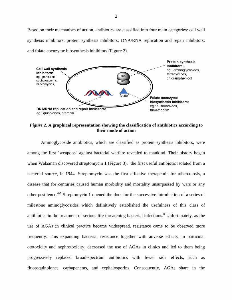
Based on their mechanism of action, antibiotics are classified into four main categories: cell wall
synthesis inhibitors; protein synthesis inhibitors; DNA/RNA replication and repair inhibitors;
and folate coenzyme biosynthesis inhibitors (Figure 2).
Figure 2. A graphical representation showing the classification of antibiotics according to
their mode of action
Aminoglycoside antibiotics, which are classified as protein synthesis inhibitors, were
among the first "weapons" against bacterial warfare revealed to mankind. Their history began
when Waksman discovered streptomycin 1 (Figure 3),5 the first useful antibiotic isolated from a
bacterial source, in 1944. Streptomycin was the first effective therapeutic for tuberculosis, a
disease that for centuries caused human morbidity and mortality unsurpassed by wars or any
other pestilence.6-7 Streptomycin 1 opened the door for the successive introduction of a series of
milestone aminoglycosides which definitively established the usefulness of this class of
antibiotics in the treatment of serious life-threatening bacterial infections.8 Unfortunately, as the
use of AGAs in clinical practice became widespread, resistance came to be observed more
frequently. This expanding bacterial resistance together with adverse effects, in particular
ototoxicity and nephrotoxicity, decreased the use of AGAs in clinics and led to them being
progressively replaced broad-spectrum antibiotics with fewer side effects, such as
fluoroquinolones, carbapenems, and cephalosporins. Consequently, AGAs share in the

3
antibiotics market declined to only 2.7% in 2010.9 Recently, with the ever-growing bacterial
resistance to the newer classes of antibiotics, many researchers decided to revisit AGAs with
renewed emphasis on chemical modification, which includes structural modifications,10-11
dimerization,12 and conjugation to other antibiotics13 or biomolecules.14
Figure 3. Streptomycin (1)
1.2. Structural features and classifications
Structurally aminoglycosides are low molecular weight (300-800 Dalton)
pseudosaccharide molecules consisting of a central aminocyclitol ring, mostly a 2-
deoxystreptamine (2-DOS) ring, linked to one or more amino sugars by glycosidic bonds. The
most prominent features of AGAs are the presence of multiple amines attached to their rings.
These amines together with the various hydroxyl groups gave them high water solubility and
basic characteristics. As a result of their high polarity, AGAs are poorly absorbed orally with less
than 1% reaching the blood stream through the gastrointestinal tract, which makes parenteral
injections the common mode of administration for systemic diseases. Moreover, their high
polarity prevents them from crossing the blood-brain barrier and reaching the central nervous
system.15
Aminoglycosides are classified according to the linkage type with the 2-
deoxystreptamine ring into two major classes: 4,5-aminoglycosides and 4,6-aminoglycosides in

4
which the 2-deoxystreptamine ring is disubstituted at the positions 4 and 5, or 4 and 6 (Figure 4).
Although most of the aminoglycosides fit to this classification, some others have unusual
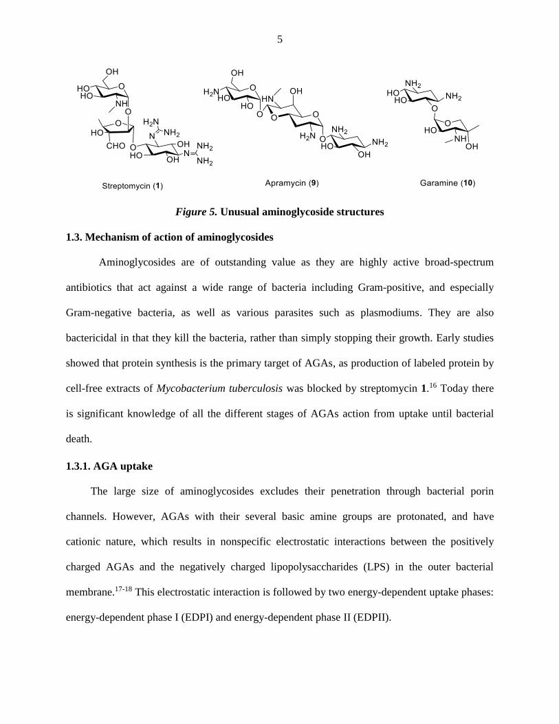
structures (Figure 5) such as bicyclic rings (e.g. apramycin 9), mono-substitution of the 2-DOS
ring (e.g. apramycin 9 and garamine 10) or incorporation of a streptamine ring instead of 2-DOS
(e.g. streptomycin 1). In addition, aminoglycosides that are derived from bacteria of the
Streptomyces genus are named with the suffix mycin, whereas those that are derived from
Micromonospora are named with the suffix micin.
Figure 4. Classification of aminoglycosides
5
Figure 5. Unusual aminoglycoside structures
1.3. Mechanism of action of aminoglycosides
Aminoglycosides are of outstanding value as they are highly active broad-spectrum
antibiotics that act against a wide range of bacteria including Gram-positive, and especially
Gram-negative bacteria, as well as various parasites such as plasmodiums. They are also
bactericidal in that they kill the bacteria, rather than simply stopping their growth. Early studies
showed that protein synthesis is the primary target of AGAs, as production of labeled protein by
cell-free extracts of Mycobacterium tuberculosis was blocked by streptomycin 1.16 Today there
is significant knowledge of all the different stages of AGAs action from uptake until bacterial
death.
1.3.1. AGA uptake
The large size of aminoglycosides excludes their penetration through bacterial porin
channels. However, AGAs with their several basic amine groups are protonated, and have
cationic nature, which results in nonspecific electrostatic interactions between the positively
charged AGAs and the negatively charged lipopolysaccharides (LPS) in the outer bacterial
membrane.17-18 This electrostatic interaction is followed by two energy-dependent uptake phases:
energy-dependent phase I (EDPI) and energy-dependent phase II (EDPII).

6
Energy-dependent phase I (EDPI) is a slow rate uptake to the cytosol and depends on
AGA concentration. Inhibitors of oxidative phosphorylation or electron transport can halt this
phase (Figure 6). In the energy-dependent phase II (EDPII), aminoglycosides bind to the 30S
ribosomal subunit through a rapid process, and this binding perturbs the translational accuracy
(misreading) and leads to defective proteins. The so-formed defective cell membrane proteins
eventually alter cell membrane permeability, stimulating further aminoglycoside influx and
leading to an autocatalytic cycle of AGA uptake and protein synthesis disruption, followed by
cell death.19
Figure 6. Schematic diagram showing the stages of AGAs uptake
1.3.2. Protein synthesis and AGA binding to the ribosomes
Normal protein synthesis starts by producing a mRNA copy of the genetic information in
the DNA in a process called transcription. This process is followed by the translation process in
which the ribosomes translate mRNA-encoded genetic information to proteins. RNA is
assembled as a single-stranded chain of nucleotides that fold upon themselves, rather than as
paired double-strands as in the DNA. Ribonucleotides consist of three parts: the phosphate
backbone, ribofuranose sugars, and nucleobases. There are four nucleobases found in RNA:
adenine, guanine, cytosine and uracil. Adenine and guanine are purine bases while cytosine and

7
uracil are pyrimidine bases. The bases usually pair together according to the Watson-Crick rule
in which adenine associates with uracil by two hydrogen bonds, and guanine pairs with cytosine
by three hydrogen bonds (Figure 7).
Figure 7. The four fundamental nucleotides found in RNA and their Watson-Crick base
pairs.
The high accuracy of protein translation, with errors estimated at only 4 × 10⁻⁴ per
codon,20 suggested that fidelity does not simply come from mRNA-codon/tRNA-anticodon
recognition, but that ribosomes also play a crucial role in translation accuracy and do not just act
as an inert platform. The fidelity of protein synthesis requires the binding of a correct tRNA to
the A-site, which is a small loop in the small ribosomal subunit that serves as aminoacyl-tRNA
acceptor site. tRNA interacts with the ribosomal A-site containing three unpaired adenines
(A1408, A1492 and A1493) and makes it adopt a “flipped-out” conformation. This leads to a

8
faster step in which other conformational changes occur, and results in tight binding of the
cognate tRNA to the A-site.21 The amino acid that the tRNA was carrying is transferred and
bonds to the growing polypeptide chain (Figure 8). The ribosome then moves one codon step
along the mRNA to accept the new tRNA, which codes to the next codon.
Figure 8. Schematic diagram showing ribosomes in protein synthesis
Aminoglycoside bactericidal activity is attributed to their binding of aminoacyl-tRNA
acceptor site A-site in the bacterial 16S rRNA.22-27 Recognition and binding of aminoglycosides
to their target is due to two primary types of interactions. The most prominent interaction comes
from the electrostatic interaction between the cationic aminoglycosides and the negatively
charged backbone of rRNA. Other interactions mainly arise from hydrogen bonding of
aminoglycosides with rRNA bases. The location of these hydrogen bonds differs between
aminoglycosides, but some are common. For example, the 2-DOS ring (ring II) of paromomycin
interacts with bases A1406, U1495 and G1494 by hydrogen bonds.23 Moreover, hydrogen bonds
are formed between the paromomycin ring 1 and A1408, A1492, A1493 and G1491 in the
bacterial rRNA (Figure 9). On the other hand, neither rings III nor IV have any direct interaction
with rRNA.23

9
Figure 9. a) Crystal structure of paromomycin binding to the bacterial A site (PDB code:
1FJG). b) Schematic diagram showing the binding of paromomycin (3) with rRNA
nucleobases.
When bound to the A-site, AGAs stabilize the conformation of the internal loop with
A1492 and A1493 "flipped out" (Figure 10). This reduces the energetic cost for both cognate and
noncognate tRNA to bind, thereby reducing the ability of the ribosome to recognize the correct
tRNA, and leads to misreading of the mRNA and synthesis of defective proteins (Figure 11).28-30
In addition, this "flipped out" conformation increases the affinity of the tRNA for the A site, thus
stabilizing the pre-translocation state and increasing the energy barrier for translocation.31-32

10
Figure 10. Schematic diagram showing a) the equilibrium of A-site between "flipped in"
and "flipped out" conformations in protein translation and b) aminoglycoside bound to
ribosome A site stabilizing the "flipped out" conformation
The connection between the resulting faulty proteins and bacterial cell death is a subject
of debate. One hypothesis is that the damaged protein can insert in the bacterial inner membrane
and cause its destabilization and so cell death.33-34 Another hypothesis suggests that defective
metabolic and respiratory enzymes lead to oxidative stress and production of toxic free
radicals.35
Figure 11. Schematic diagram showing aminoglycoside bound to ribosome and causing
codon misreading.
11
1.4. Aminoglycoside resistance and toxicity
The excellent characteristics of AGAs as broad-spectrum antibacterial agents, with
desirable bactericidal activity against difficult-to-treat Gram-negative bacteria and mycobacteria,
are counterbalanced by two major problems, namely resistance and toxicity (ototoxicity, i.e.,
damage to the inner ear, and nephrotoxicity, i.e., damage to the kidney).36 Due to these problems
the importance of AGAs has waned, but a deep understanding of these problems may help to
overcome them.
1.4.1. Aminoglycoside resistance
AGAs are isolated from soil-dwelling bacteria in particular Streptomyces and
Micromonospora species. However, their bacterial origins also are the source of most of the
resistance problems encountered today, as most of the AGA producing species have established
strategies to prevent the deleterious effects of the antimicrobial metabolites they produce
themselves.9 Resistance problems also arise because of the frequent use of AGAs against human
and animal pathogens. Improper and incomplete treatment with AGAs will allow mutant
resistant bacteria to flourish. Thus, establishment of regulations to address proper use of
antibiotics, promotion of public awareness of rational administration of antibiotics, and
encouragement of the development of new antibiotics are three strategies that Food and Drug
Administration (FDA) is pursuing to solve bacterial resistance problems.37
Resistance mechanisms can be categorized into three types (Figure 12). First, bacteria can
reduce the internal concentration of AGAs by decreasing the drug uptake (influx) or increasing
the drug expulsion (efflux). Second, some bacteria are able to modify their ribosomal A-site so
that AGAs can no longer bind to it. Finally, the most common mechanism for bacterial resistance
arises from the structural modification of the aminoglycosides themselves by specific enzymes

12
expressed by resistant strains. There are three classes of these aminoglycoside modifying
enzymes (AME): aminoglycoside phosphotransferases (APHs), aminoglycoside
acetyltransferases (AACs) and aminoglycoside nucleotidyltransferases (ANTs).38-39
Figure 12. Schematic diagram showing different resistance mechanisms
1.4.1.1. Reduction of aminoglycoside internal concentration
Bacteria can reduce aminoglycoside concentration by decreasing the drug uptake (influx)
or by increasing the drug expulsion (efflux). As discussed in the previous section AGA uptake
goes through three stages. While the first step is electrostatic attraction between AGAs and the
bacterial cell surface, the other two steps are energy and oxygen dependent which give anaerobic
bacteria an inherent resistance to AGAs.40 Also mutations to the ATP synthases of E. coli, S.
aureus, and P. aeruginosa have been shown to decrease their susceptibility to AGAs.41
The other strategy of decreasing AGA concentration in the bacterial cells is by increasing
the drug expulsion (efflux). This is done by efflux energy-dependent active pumps. There are
many types of transporters, such as the resistance nodulation cell division (RND)-type
transporter superfamily,42-43 which plays an important role in Gram-negative bacteria like P.

13
aeruginosa. Another example is the major facilitator superfamily (MFS) of transporters which
contributes to aminoglycoside resistance of E. coli.44
1.4.1.2. Ribosomal binding site modifications
Alteration of the aminoglycosides target, the 16S RNA by bacteria, is another mode of
resistance. There are two types of target modifications: nucleotide mutation and nucleotide
methylation. The most common example of nucleotide mutations is the A1408G mutation in the
ribosomal A-site. This mutation gives bacteria resistance to the 6’-NH2 aminoglycosides by
interrupting key interactions with the AGAs.45
Many aminoglycoside-producing bacteria (Streptomyces and Micromonospora) protect
themselves from their own AGAs by producing rRNA methylases, which can methylate the 16S
rRNA.46-47 Examples of these methylase enzymes that are now well known include RmtA, in P.
aeruginosa,48 the RmtB that was found to be responsible for aminoglycoside resistance in
Serratia marcescens,46 and ArmA, that was first found in a Klebsiella pneumoniae clinical
isolate.49 These mutations are of low clinical importance at present, but they pose a potential
threat because of the almost complete resistance they bring against AGAs, especially 4,6-
disubstituted AGAs. The mono-substituted 2-DOS AGA apramycin (9), on the other hand, is not
susceptible to the ArmA methylation mechanism due to its unusual bicyclic structure.50
1.4.1.3. Aminoglycoside modifying enzymes (AMEs)
Aminoglycoside modifying enzymes catalyze covalent modification at hydroxyl or amino
groups of both the 2-deoxystreptamine nucleus and the sugar moieties. The modified drugs fail
to properly bind to the ribosomes. As AMEs are encoded on plasmids they are highly mobile and
are easily spread between bacterial species. There are three classes of aminoglycoside modifying
enzymes (AME): aminoglycoside acetyltransferases (AACs), aminoglycoside
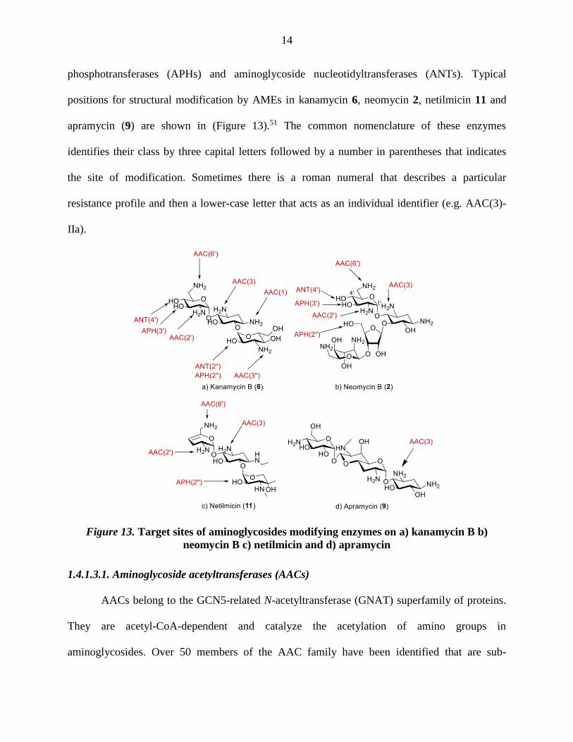
14
phosphotransferases (APHs) and aminoglycoside nucleotidyltransferases (ANTs). Typical
positions for structural modification by AMEs in kanamycin 6, neomycin 2, netilmicin 11 and
apramycin (9) are shown in (Figure 13).51 The common nomenclature of these enzymes
identifies their class by three capital letters followed by a number in parentheses that indicates
the site of modification. Sometimes there is a roman numeral that describes a particular
resistance profile and then a lower-case letter that acts as an individual identifier (e.g. AAC(3)-
IIa).
Figure 13. Target sites of aminoglycosides modifying enzymes on a) kanamycin B b)
neomycin B c) netilmicin and d) apramycin
1.4.1.3.1. Aminoglycoside acetyltransferases (AACs)
AACs belong to the GCN5-related N-acetyltransferase (GNAT) superfamily of proteins.
They are acetyl-CoA-dependent and catalyze the acetylation of amino groups in
aminoglycosides. Over 50 members of the AAC family have been identified that are sub-

15
classified to AAC(1), which has no further subclasses, AAC(3)-I to X, AAC(2′)-I, and AAC(6′)-I
and -II. AAC(1) enzymes do not cause a substantial drop in antibiotic activity and are of little
importance as they are rarely found in clinical isolates.52 AAC(3) enzymes are found only in
Gram-negative bacteria where AAC(3)-IIa are found in a large variety of genera, AAC(2′)-I
confers resistance to neomycin, kanamycin, and gentamicin and is found in Gram-negative
bacteria and mycobacteria.53 AAC(6′) enzymes are present in Gram-negative as well as Gram-
positive bacteria and are by far the most common of all AMEs as acetylation of the 6’-amino
group blocks a crucial interaction with A1408 and renders the AGA inactive.54
1.4.1.3.2. Aminoglycoside phosphotransferases (APHs)
APHs are ATP-dependent enzymes that catalyze the regiospecific transfer of the -
phosphoryl group of the ATP to hydroxyl groups of AGAs. This phosphorylation introduces a
negative charge into the molecule, which decrease the ability to bind to the A-site in the
ribosome. APHs are often found on multidrug-resistant R plasmids leading to problems of gene
transfer between Gram-positive and Gram-negative bacteria. Seven classes of such enzymes,
APH(3’), APH(2’’), APH(3’’), APH(4), APH(7’’), APH(6), and APH(9) have been identified in
clinical isolates of which the APH(3′) class is the most common.55
1.4.1.3.3. Aminoglycoside nucleotidyltransferases (ANTs)
ANTs are another class of ATP-dependent enzyme that catalyze the transfer of an AMP
group to a hydroxyl group in the AGA. Different classes of ANTs, the ANT(6), ANT(9),
ANT(4′), ANT(2″), and ANT(3″), are now known. Although they are the smallest AME family
by number, ANTs are of significant clinical importance because of the ability of ANT(2″) to
neutralize tobramycin 12 and amikacin 13 as well as gentamicin 7 (Figure 14).56

16
Figure 14. Structures of tobramycin (12) and amikacin (13)
1.4.1.3.4. Avoiding AME resistance
There are two viable ways to avoid the resistance from AMEs. One way is to develop
inhibitors of the modifying enzymes that can be co-administered with the AGA. Alternatively, a
better way is to synthesize analogs of natural aminoglycosides resistant to the modifying
enzymes. In comparison to kanamycin B (6), netilmicin (11) suffers from fewer resistance
enzymes as the installation of the N1-ethyl group sterically protects it from aminoglycoside
modifying enzymes at this position (Figure 13). Netilmicin (11) is also protected from several
AMEs, such as APH(3′) and ANT(4′), by the absence of the 3′- and 4′-OH groups in ring I.
Aminoglycoside modifying enzymes for apramycin also are rare, with only AAC(3) known thus
far. This absence of resistance makes apramycin (9) a candidate for human use and a good
candidate for modification and development.57
1.4.2. Adverse effects of aminoglycosides
The adverse effects of AGAs are one of the main problems that prevent their wide use in
clinics. The main side effects a patient can encounter when given AGAs are nephrotoxicity, or
kidney damage, and ototoxicity, or hearing loss. Along with these main side effects, there are
minor acute side effects like neuromuscular blocking action that can be referred to as curare-like
activity. The mechanism of this latter effect was confirmed to be action as a calcium channel
17
blocker; subsequently AGAs have been used as chemical tools to explore the functions of
calcium channels.58
1.4.2.1. Nephrotoxicity
Nephrotoxicity is kidney damage cause by AGAs and is clinically presented as non-
oligouric kidney injury. Kidney damage can lead to the inability of the body to clear urine and
other wastes. Its manifestations include aminoaciduria, glycosuria, enzymuria, hypomagnesemia,
hypocalcemia, and hypokalemia. Although nephrotoxicity is reversible, if it is untreated it causes
increased electrolyte levels in the body and may lead to permanent kidney damage and
eventually kidney failure.27 This toxicity can be explained by the accumulation of AGAs in the
renal cortical tissue especially the proximal tubules. AGAs are absorbed by endocytosis and once
transferred to the lysosome, the positively-charged AGAs strongly bind to the negatively-
charged phospholipids resulting in a decrease of lysosomal phospholipase activity.59 An
abnormal increase in size and number of lysosomes was found with decreased lysosome stability
can lead eventually to cell death.60
Several strategies have been used to prevent nephrotoxicity including: 1) hydration
therapy, which can often decrease the symptoms of aminoglycoside-induced nephrotoxicity. 2)
The use of a once daily large dose as opposed to the same daily dose taken as separate three
doses or by continuous infusion.61 This latter is explained by the finding that uptake by the renal
cells will be saturable at relatively low concentrations such that the excess drug passes the
lumen, is not reabsorbed, and is excreted without causing toxicity. 3) Aminoglycoside
modifications in which the N-1 atom has been made non-ionizable (i.e., by acylation) decrease
AGA basicity and thus reduce binding to acidic phospholipids, and decrease inhibition of the
lysosomal phospholipases.39 4) Co-administration of polyaspartic acid prevents aminoglycoside

18
binding to negatively charged phospholipids bilayers and thereby prevents the drug from
inhibiting the activities of lysosomal phospholipases.62 Overall, nephrotoxicity is reversible, can
be easily monitored and can largely be prevented.
1.4.2.2. Ototoxicity
Unlike nephrotoxicity, ototoxicity is irreversible and difficult to monitor. It is reported to
affect as much as 20% of the patient population, which makes it the main concern.63 Ototoxicity
includes damage to the vestibular system, resulting in imbalance disorders, and damage to the
cochlea, resulting in tinnitus and hearing loss. There is no apparent correlation between
nephrotoxicity potential or with the concentrations reached in the inner ear by different
aminoglycosides with the magnitude of their ototoxic potential.63 However, longer AGA
treatments, kidney malfunction and the nutritional state of the patient may also contribute to the
magnitude of ototoxicity.64 While some AGAs (e.g. gentamicin) are more vestibulotoxic than
cochleotoxic, which can be used for vestibular chemical ablation, others (e.g. amikacin and
neomycin) can be more cochleotoxic than vestibulotoxic.
AGAs quickly penetrate to the inner ear within minutes of parenteral administration, and
although the half-life of the AGAs in the plasma ranges from 3-5 h, their half-lives in the inner
ear can reach 30 days. This long resident time was earlier misinterpreted as accumulation of
AGAs in the inner ear, but this was ruled out by the fact the concentration of the drug in the
inner ear was the same as in other organs and never reached the plasma concentration.65
AGAs cause damage to hair cells in the cochlea located in the inner ear. The cochlear
hair cells function is to convert sound waves to electric impulses, which are transferred to the
brain to give the hearing sensation. AGAs affect first the cochlear hair cells in the basal part that
are responsible for the higher frequency (high pitched) sound, and so causing higher frequency

19
deafness, before they affect the cochlear hair cells in the apical part that are responsible of the
lower frequency (low pitched) sound.66 The fact that the damaged hair cells do not regenerate
makes ototoxicity irreversible.
Figure 15. A schematic representation of the mechanisms of aminoglycoside ototoxicity.
Ototoxicity caused by AGAs involves the formation of reactive oxygen species (ROS) in
the inner ear tissues that lead to ultimate cell death (Figure 15). The mode of formation of ROS
has not been clear,67-69 but some hypothesis will be outlined: 1) a non-catalytical pathway, in
which AGAs binds to iron and mediate the formation a reactive oxygen species from arachidonic
acid. Electrospray Ionization Mass Spectrometry (ESIMS) confirmed the existence of ternary
complexes between Fe2+/3+, gentamicin 7, and arachidonic acid.70 2) A catalytical pathway, in
which AGAs activate Rho-GTPase (Rac-1), which in turn activates the NADPH oxidase
complex to form superoxide radicals.71 However, recent evidence suggests that inhibition of
mitochondrial protein synthesis is the main reason.24, 72-74 The potency of a series of AGAs in
inhibiting mitochondrial hybrid ribosome function correlated with the relative cochleotoxicity of
the respective compounds in humans.75 The molecular rationale for the increased affinity to
mitochondrial rRNA is that its A site retains the critical A1408 residue, similar to the bacterial A

20
site (Figure 16). This rationale is bolstered by the hyper-susceptibility of individuals with
mutations in mitochondrial rRNA to ototoxicity, in particular the transition mutation A1555G in
the A-site of the mitoribosomal small subunit which renders such A sites even more similar to
the bacterial A site.73, 75 The problem of efficient genetic tools to study the interaction between
eukaryotic rRNA with aminoglycosides has been overcome by the pioneering work of the
Böttger group. They replaced the bacterial drug binding site in 16S rRNA with its eukaryotic
counterpart, resulting in bacterial hybrid ribosomes with a fully functional eukaryotic rRNA
decoding site (Figure 17).76 This technique allows the fast screening of aminoglycosides
derivatives in vitro to evaluate their potential ototoxicity in addition to evaluation of their
bactericidal activity against wild type and clinically resistant strains. It is one of the key
techniques in this collaborative project with the Böttger group.
Figure 16. Secondary-structure comparison of decoding-site rRNA sequences in the small
ribosomal subunit. (A) Decoding region of 16S rRNA helix 44 in wild-type ribosomes of M.
smegmatis. (B) Homologous 18S rRNA sequence in human ribosomes. (C) Homologous 12S
rRNA sequence in human mitochondrial ribosomes. (D) Mitochondrial 12S rRNA sequence
with mutation A1555G conferring hypersusceptibility to AGA ototoxicity.
Ototoxicity side effects can be reduced by concurrent administration of antioxidants
which act as radical scavengers to neutralize any ROS formed and prevent their delirious actions.
Antioxidants do not affect the serum concentration or the antibacterial efficacy of
21
aminoglycosides.77 Indeed, salicylic acid, which acts as both an antioxidant and an iron-chelator
has been shown to reduce the gentamicin-induced auditory threshold from more than 60 dB to
less than 20 dB.78
Figure 17. Bacterial hybrid ribosomes with fully functionally eukaryotic rRNA A site
1.5. Recent advances
In the last few years, several research groups have begun to develop aminoglycoside
derivatives that can be more active, less toxic, and suffer less resistance from bacteria. The Crich
and Vasella groups collaborated to synthesize modifications on the 4',6'- and 4’-positions of
paromomycin 3. These derivatives were more selective toward bacterial ribosomes over human
mitoribosomes, with a concomitant decrease in ototoxicity but not in antibiotic activity (Figure
18). 10, 79-80
Figure 18. Examples of 4',6'-O-alkylidene (14) and 4'-O-alkyl paromomycin (15) derivatives
that showed increased selectivity for bacterial ribosomes over human mitoribosomes.

22
Apramycin (9) is considered to be one of the least ototoxic AGAs, which this can
possibly be attributed to its unique bicyclic aminosugar moiety.81 Considerable work has been
done by the Crich group in an effort to increase apramycin activity as well as it's selectivity by
modification of 6' and 7'-positions.82 These studies, however, showed these positions to be
essential for activity as most manipulations of them decreased the activity.
Figure 19. The structure of plazomicin [6'-N-(2-hydroxyethyl)-1-N-(4-amino-2(S)-
hydroxybutyryl) sisomicin] (16)
Plazomicin, 16 (Figure 19) developed by the Achaogen company, is a semisynthetic
aminoglycoside antibiotic currently in phase 3 clinical trials for complicated urinary tract
infections.83-84 Like netilmicin, plazomicin is prepared from the readily available sisomicin.83-84
Plazomicin 16 was designed to thwart the most common aminoglycoside modifying enzymes
(AMEs) by the judicious positioning of substituents and so to respond to the ever-growing
problem of drug-resistant infectious diseases.85 The placement of the 4-amino-2(S)-
hydroxybutyryl (L-HABA) unit on the 1-position not only protect the drug against enzymes
acting on position 1 [AAC(1) enzymes], but also on both position 3 [AAC(3) enzymes] and
position 2'' [ANT(2’’) and APH(2’’)]. L-HABA substitution also serves to decrease the drug
nephrotoxicity as discussed before. The inclusion of a 2-hydroxyethyl group on the 6'-position
protects the drug against enzymes acting on position 6’.

23
1.6. Overall goals
Toward the goal of preparing next generation AGAs two sub-projects were defined: 1)
examining the effects on 4’-modification in the 4,6-series on the netilmicin skeleton 11.
Netilmicin 11 was chosen as substrate for this investigation because its ototoxicity is low, which
makes it ideal for further investigation.86-88 Likewise, the knowledge that modification of the 4’-
position of paromomycin10, 79-80 reduces its ototoxicity but not it’s antibiotic activity, suggested
derivatization of netilmicin at that position.
2) Improvement of apramycin 9 by hybridization: A number of reports have described the
synthesis and evaluation of hybrid AGAs combining fragments from both the 4,5- and 4,6-series
of 2-deoxystrptamine AGAs.89-92 Our attention was captured by an early report on the
moderately increased activity of the 5-O-(β-D-ribofuranosyl) derivative of apramycin against
selected Gram-negative organisms,93 suggesting derivatization at the 5-position of apramycin 9
and apramycin-paromomycin hybrids; the goal being to combine paromomycin’s high activity
with apramycin’s low ototoxicity as a possible avenue for further improving the profile of this
promising antibiotic.

24
CHAPTER 2. INVESTIGATIONS IN THE 4,6-CLASS OF AGAs
2.1. Mode of binding of netilmicin to the bacterial ribosomal A-site
Netilmicin 11 is a N1-ethyl semisynthetic analog of sisomicin, an antibiotic extracted
from Micromonospora inyoensis.94 Netilmicin was developed and marketed in the United States
as Netromycin® by the Schering Corporation.95 Netilmicin belongs to the 4,6-disubstituted class
of aminoglycosides and is active against most Gram-negative and some Gram-positive bacteria,
including many gentamicin-resistant strains.96 The N1-ethyl modification protects netilmicin
from both adenylating and phosphorylating enzymes and consequently maintains its activity
against most strains of bacteria that harbor these enzymes.97 It is also reported to be less ototoxic
than amikacin and gentamicin in in vivo rabbit studies.87
Although a crystal structure of the rRNA A-site bound to netilmicin has yet to be
determined, it is assumed that netilmicin has the same binding mode as its congener sisomicin,
which specifically binds to the deep/major groove of the bacterial A site and makes 11 hydrogen
bonds to base and phosphate oxygen atoms (Figure 20).98 Sisomicin 8 has a double bond
between the C4′ and the C5′ atoms, which force ring I to adopt a flattened conformation with the
possibility of classical π-stacking with the G1491 base. This is in contrast to AGAs with a
saturated carbohydrate ring I, which adopt a chair conformation that interacts with the aromatic
of G1491 through CH/π interactions.98 Ring I also forms a pseudo base-pair interaction with
A1408 in which the ring oxygen of ring 1 accepts a hydrogen bond from N6-adenine and the
protonated 6'-amino group donates a hydrogen bond to adenine N1. The C3-amine in ring II
makes hydrogen bonds with the phosphate oxygen atoms of A1493 and A1494 so that A1492
and A1493 can take flipped-out conformations. Ring III binds to the upper side of the A-site
helix through four hydrogen bonds.98

25
Figure 20. a) Crystal structure of sisomicin bound to the deep/major groove of the bacterial
A site showing π-stacking with G1491 base and pseudo base-pair interaction with A1408
(PDB code: 4F8U), b) a schematic diagram showing the pseudo base-pair interaction of
ring I with A1408 and c) detailed interaction of sisomicin with the bacterial A site.
2.2. Synthesis of netilmicin
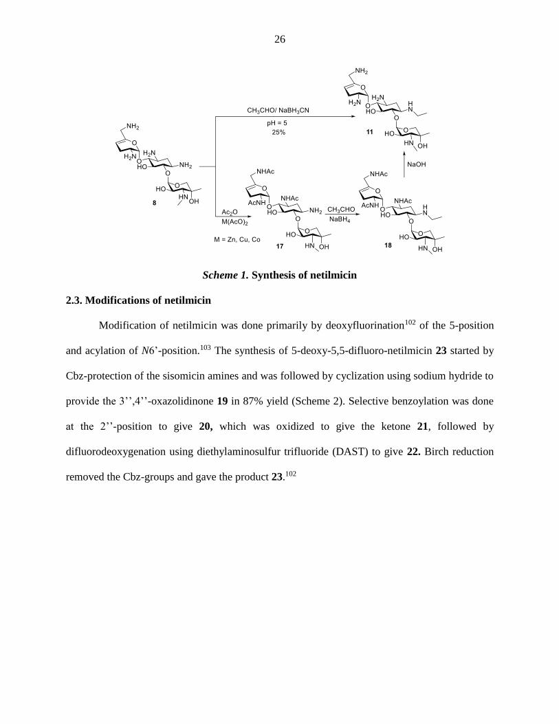
Netilmicin 11 was first synthesized in one step by reductive alkylation of sisomicin95
without the conventional protection of the other amine groups (Scheme 1). The synthesis took
advantage of the fact that the 1-amino group is the least basic and under acidic conditions (pH =
5), all other amines are protonated and so unreactive. The only problem with this direct synthesis
was its low yield (25%). Subsequently, several papers and patents were published that describe
improved overall yields albeit employing three main steps.99-101 In these sequences the first step
was selective protection of the 3,2’,6’-amino groups mediated by transition metal acetates such
as zinc, copper or cobalt acetates to give 17. This was followed by reductive alkylation of the
N1-amine to afford compound 18 and finally deprotection; the best overall yield (73%) was
obtained using zinc acetate in the first step (Scheme 1).101
G1491
A1408
a) b)
c)
26
Scheme 1. Synthesis of netilmicin
2.3. Modifications of netilmicin
Modification of netilmicin was done primarily by deoxyfluorination102 of the 5-position
and acylation of N6’-position.103 The synthesis of 5-deoxy-5,5-difluoro-netilmicin 23 started by
Cbz-protection of the sisomicin amines and was followed by cyclization using sodium hydride to
provide the 3’’,4’’-oxazolidinone 19 in 87% yield (Scheme 2). Selective benzoylation was done
at the 2’’-position to give 20, which was oxidized to give the ketone 21, followed by
difluorodeoxygenation using diethylaminosulfur trifluoride (DAST) to give 22. Birch reduction
removed the Cbz-groups and gave the product 23.102

27
Scheme 2. Synthesis of 5-deoxy-5,5-difluoro-netilmicin (23)
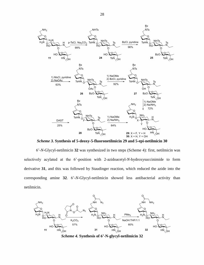
The synthesis of 5-deoxy-5-fluoronetilmicin 29 started by tosyl protection of the
sisomicin amines to give the pentatosyl derivative 24, which was benzoylated at the N1’ and 2’’-
positions to afford 25 (Scheme 3). Inversion of configuration of the 5-hydroxyl group and
adjustment of the protecting groups was done by a sequence of the following steps: mesylation
of the 5-hydroxyl group, substitution of the mesylate with sodium acetate, cleavage of acetate
and benzoate esters, and finally benzoylation at the N1’ and 2’’-positions to give the 5-epi-
netilmicin derivative 27. Deoxyfluorination then was done using DAST to afford the 5-fluoro-
netilmicin derivative 28 in 25%, which upon deprotection with sodium in liquid ammonia
yielded the desired product 29. In the same fashion, 27 was deprotected to afford 5-epi-netimicin
30 in 72% yield. 5-Deoxy-5-fluoronetilmicin 29 and 5-epi-netimicin 30 showed higher
antibacterial activity than the parent, while 5-deoxy-5,5-difluoro-netilmicin 23 was slightly less
active. Moreover, in acute toxicity studies, compound 30 showed less toxicity that netilmicin.102
28
Scheme 3. Synthesis of 5-deoxy-5-fluoronetilmicin 29 and 5-epi-netilmicin 30
6’-N-Glycyl-netilmicin 32 was synthesized in two steps (Scheme 4): first, netilmicin was
selectively acylated at the 6’-position with 2-azidoacetyl-N-hydroxysuccinimide to form
derivative 31, and this was followed by Staudinger reaction, which reduced the azide into the
corresponding amine 32. 6’-N-Glycyl-netilmicin showed less antibacterial activity than
netilmicin.
Scheme 4. Synthesis of 6’-N-glycyl-netilmicin 32
29
2.4. Rational
As discussed in the previous chapter, because modification of the 4’-position of
paromomycin10, 79-80 reduces its ototoxicity but not its antibiotic activity, it was of interest to
examine if these effects on 4’-modification in the 4,5-series (i.e. paromomycin) extends to 4,6-
series (i.e. netilmicin). Netilmicin was chosen as substrate for modification in this manner
because it is considered to be one of the least ototoxic aminoglycosides, which makes it ideal for
further investigation.86-88
2.5. Chemistry
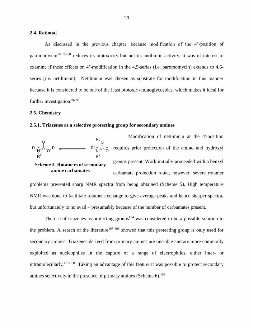
2.5.1. Triazenes as a selective protecting group for secondary amines
Modification of netilmicin at the 4'-position
requires prior protection of the amino and hydroxyl
groups present. Work initially proceeded with a benzyl
carbamate protection route, however, severe rotamer
problems prevented sharp NMR spectra from being obtained (Scheme 5). High temperature
NMR was done to facilitate rotamer exchange to give average peaks and hence sharper spectra,
but unfortunately to no avail – presumably because of the number of carbamates present.
The use of triazenes as protecting groups104 was considered to be a possible solution to
the problem. A search of the literature105-106 showed that this protecting group is only used for
secondary amines. Triazenes derived from primary amines are unstable and are more commonly
exploited as nucleophiles in the capture of a range of electrophiles, either inter- or
intramolecularly.107-108 Taking an advantage of this feature it was possible to protect secondary
amines selectively in the presence of primary amines (Scheme 6).109
Scheme 5. Rotamers of secondary
amine carbamates

30
Scheme 6. General protocol for selective protection of secondary amines as the N-phenyl
triazenes
2.5.2. Aryl triazenes
Trisubstituted triazenes have been widely employed in organic synthesis for the
protection and/or derivatization of aryl amines.104-105, 109-110 As a protecting group, aryl triazenes
are compatible with oxidizing agents (e.g. pyridinium dichromate (PDC)) and reducing agents
(e.g. LiAlH4 and NaBH4) as well as with basic conditions, acylating and alkylating reagents
(Schemes 7 and 8). The free amine can be easily regenerated by treatment with trifluoroacetic
acid.105, 111
Scheme 7. Literature reactions showing the compatibility of phenyltriazenes with
oxidizing, reducing and alkylating agents

31
Scheme 8. Literature reactions showing the compatibility of phenyltriazenes with acylating
reagents and basic conditions
Triazenes are also used as linkers to solid supports in solid-phase organic synthesis
(SPOS) (Scheme 9). After functionalization on the bead, the triazene linker can be easily cleaved
from the solid support using acidic conditions. Triazenes can also act as concealed diazonium
salts upon cleavage from the resin and as such are used for conversion of aryl amines to many
functionalized arenes as well as in cyclizations to generate various heterocyclic structures,
namely, benzoannelated nitrogen heterocycles.104, 110
Scheme 9. Uses of triazenes in solid-phase organic chemistry (SPOS)
32
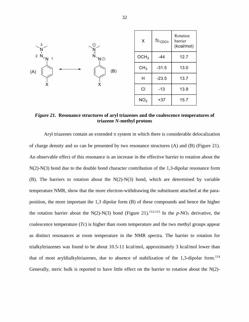
Figure 21. Resonance structures of aryl triazenes and the coalescence temperatures of
triazene N-methyl protons
Aryl triazenes contain an extended system in which there is considerable delocalization
of charge density and so can be presented by two resonance structures (A) and (B) (Figure 21).
An observable effect of this resonance is an increase in the effective barrier to rotation about the
N(2)-N(3) bond due to the double bond character contribution of the 1,3-dipolar resonance form
(B). The barriers to rotation about the N(2)-N(3) bond, which are determined by variable
temperature NMR, show that the more electron-withdrawing the substituent attached at the para-
position, the more important the 1,3 dipolar form (B) of these compounds and hence the higher
the rotation barrier about the N(2)-N(3) bond (Figure 21).112-113 In the p-NO2 derivative, the
coalescence temperature (Tc) is higher than room temperature and the two methyl groups appear
as distinct resonances at room temperature in the NMR spectra. The barrier to rotation for
trialkyltriazenes was found to be about 10.5-11 kcal/mol, approximately 3 kcal/mol lower than
that of most aryldialkyltriazenes, due to absence of stabilization of the 1,3-dipolar form.114
Generally, steric bulk is reported to have little effect on the barrier to rotation about the N(2)-

33
N(3) bond, but in the extreme case of 2,2-dimethyl- and 2,2,6,6-tetramethylpiperidine-based
triazenes the barrier is reduced to ∼11 kcal mol−1 in CS2.115
2.5.3. Examples of Selective Protection
A general reaction protocol was established for this synthetic method. A series of primary
and secondary diamines were treated with 1.1 equivalents of benzenediazonium tetrafluoroborate
in methanol/water in the presence of powdered potassium or sodium carbonate followed by
protection of the primary amines as the azides, benzyloxy carbamates or fluorenyl methyl
carbamates. Workup and silica gel chromatography then gave the product in moderate to good
yield. Yields were not improved by the use of excess benzenediazonium tetrafluoroborate as this
leads to complications in isolation arising from the decomposition of the reagent. All these
results are summarized in Table 1.

34
Table 1. Examples of selective protection of secondary amines as the N-phenyl
triazenes

35
Consistent with expectations, the 1H NMR spectra of the azido triazenes are mostly sharp
in CDCl3 and CD3OD at room temperature with the exception of those compounds containing
multiple Cbz groups. The contrast between the 1H NMR spectra of phenyl triazene protected
dissymmetric secondary amines and those of the corresponding carbamates is illustrated in
(Figure 22). The room-temperature 1H NMR spectrum of 58, obtained by sequential treatment of
spermidine with imidazole sulfonyl azide and benzyl chloroformate, displays significant
broadening of all resonances in this pseudosymmetric secondary carbamate (Figure 22a). In
contrast, the 1H NMR spectrum of the corresponding diazido triazene 51 is sharp (Figure 22b).
Figure 22. Room-temperature 600 MHz 1H NMR spectra of (a) 58 and (b) 51 in CD3OD.
2.5.4. Application to Aminoglycosides
Having established the viability of the method, its application to the aminoglycosides was
explored. First, sisomicin 8 with its single secondary and four primary amino groups was

36
investigated. Reaction with one equivalent of benzenediazonium tetrafluoroborate under the
standard conditions was followed by treatment with either an excess of imidazole sulfonyl azide
or benzyloxy carbonyl chloride resulting in the isolation of 59 and 60 respectively, in excellent
yields (Scheme 10).
Scheme 10. Application to sisomicin
The method was also applied to the monosubstituted deoxystreptamine aminoglycoside
apramycin 9, when the azido and carbamate-protected triazenes 61 and 62 were both obtained in
moderate yield (Scheme 11). Triazenes can be easily deprotected using trifluoroacetic acid in
essentially quantitative yield.
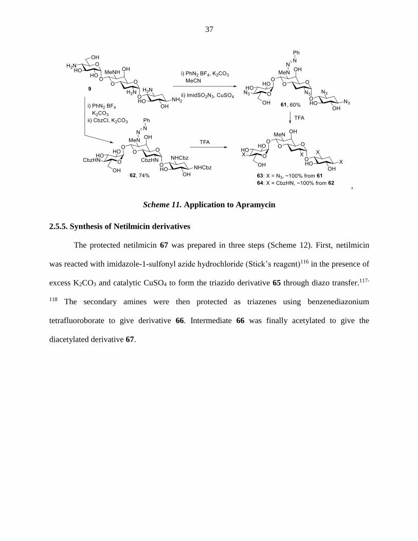
37
,
Scheme 11. Application to Apramycin
2.5.5. Synthesis of Netilmicin derivatives
The protected netilmicin 67 was prepared in three steps (Scheme 12). First, netilmicin
was reacted with imidazole-1-sulfonyl azide hydrochloride (Stick’s reagent)116 in the presence of
excess K2CO3 and catalytic CuSO4 to form the triazido derivative 65 through diazo transfer.117-
118 The secondary amines were then protected as triazenes using benzenediazonium
tetrafluoroborate to give derivative 66. Intermediate 66 was finally acetylated to give the
diacetylated derivative 67.

38
Scheme 12. Synthesis of a protected netilmicin intermediate
After the common intermediate 67 was obtained, iodination using N-iodosuccinimide119
in the presence of silver nitrate installed iodine in the 4'-position. Attempted bromination of the
4'-position using bromine120 was unsuccessful. However, the reaction proceeded using N-
bromosuccinimide and K2CO3 as a base in acetonitrile as a solvent in 29% yield. The yield
increased to 50% when a catalytic amount of tetrabutylammonium nitrate was added (Scheme
13). This can be explained either by the lipophilic tetrabutylammonium ion acting as phase
transfer catalyst facilitating the deprotonation or by the effect of nitrate ion which traps the
oxocarbenium ion and facilitates subsequent deprotonation (Scheme 14) as has been proposed in
related processes.121 Chlorination was best achieved with iodobenzene dichloride122 (Scheme
13). Intermediates 68, 69 and 70 were subjected to a stepwise sequential deprotection:
deacetylation, reduction of the azide groups under Staudinger conditions,123 and removal of the
triazene group with trifluoroacetic acid to give the final products 72, 73 and 74.
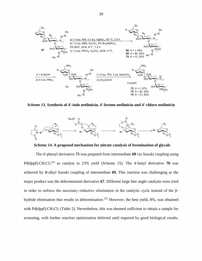
39
Scheme 13. Synthesis of 4'-iodo netilmicin, 4'-bromo netilmicin and 4'-chloro netilmicin
Scheme 14. A proposed mechanism for nitrate catalysis of bromination of glycals
The 4'-phenyl derivative 75 was prepared from intermediate 69 via Suzuki coupling using
Pd(dppf).CH2Cl2124 as catalyst in 23% yield (Scheme 15). The 4'-butyl derivative 70 was
achieved by B-alkyl Suzuki coupling of intermediate 69. This reaction was challenging as the
major product was the debrominated derivative 67. Different large bite angle catalysts were tried
in order to enforce the necessary reductive elimination in the catalytic cycle instead of the β-
hydride elimination that results in debromination.125 However, the best yield, 8%, was obtained
with Pd(dppf).CH2Cl2 (Table 2). Nevertheless, this was deemed sufficient to obtain a sample for
screening, with further reaction optimization deferred until required by good biological results.

40
Deprotection of intermediates 75 and 76 was done as described in (Scheme 13) to give the
desired products 78 and 79.
Scheme 15. Synthesis of 4'-phenyl netilmicin and 4'-butyl netilmicin
Table 2. Catalysts used in the B-alkyl Suzuki reaction and their yields
Entry Catalyst Solvent % Yield
of 76
% Yield
of 69
1 Pd(dppf).CH2Cl2 dioxane:water (3:1) 0 -
2 Pd(dppf).CH2Cl2 toluene:water (3:1) 8 32
3 Pd(PPh3)4 toluene:water (4:1) traces -
4 Pd(PCy3) Cl2 toluene:water (4:1) 5 14

41
Triazene protecting groups were found to be incompatible with the ethylsulfenyl chloride
needed to prepare 4'-(ethylsulfanyl) netilmicin 83; therefore, all amines were protected as the
2,2,2-trichloroethyl carbamate (Troc) group,126 followed by acetylation of the hydroxyl groups
(Scheme 16). The resulting compound 81 was reacted with ethylsulfenyl chloride,127 formed in
situ from diethyl disulfide and sulfuryl chloride, to give the desired intermediate 82. One step
deprotection using 6 N NaOH afforded the desired product 83.
Scheme 16. Synthesis of 4'-(ethylsulfanyl) netilmicin
2.5.6. Synthesis of plazomicin
As discussed in the introduction, plazomicin 16 is one of the recently developed AGAs
and is currently in phase 3 clinical trials for complicated urinary tract infections.83-84 An
authentic sample was needed to serve as a standard for the comparison of biological activity of
the synthesized compounds. However, although the synthesis of plazomicin from sisomicin has
been outlined in the patent84 and in the open literature,83 few details and characterization data
were available. Accordingly, the semisynthesis of plazomicin from sisomicin was undertaken by
a novel synthetic route featuring the use of the phenyltriazenyl protecting group for secondary

42
amines in only 7 steps and 8.5% overall yield, which compares favorably to the published 0.16%
yield.83
The synthesis of plazomicin started by selective protection of the 6'-NH2 group of
sisomicin 8 using 4-nitrobenzyl N-hydroxysuccinimidyl carbonate128 to give 6'-N-(4-
nitrobenzyloxycarbonyl) sisomicin 84 in 75% yield. The amino substituents at positions 2' and 3
were then selectively protected as the Boc derivatives by the temporary protection of the other
amines in the form of zinc chelates, resulting in the formation of 85 in 52% yield.129 The
reaction of 85 with a 10 mol% excess of phenyl diazonium tetrafluoroborate in acetonitrile in the
presence of potassium carbonate gave an 88% yield of the desired triazene 86 (Scheme 17),
whose sharp, well-resolved NMR spectra facilitated spectral elucidation in this and the following
steps. Thus, the triazene moiety fulfills the expectation of a disymmetric secondary amine
protecting group, free of the rotamer problems associated with the more common carbamates.
Furthermore, as suggested by the model studies, it was selectively introduced onto a secondary
amine in the presence of a primary amine without the need for an excess of the diazonium salt.
The remaining amine at position N1 was then coupled to N-Boc-4-amino-2(S)-hydroxy-
butyric acid130 under standard carbodiimide conditions in the presence of hydroxy-5-norbornene-
2,3-dicarboximide131 and afforded the amide 87 in high yield. The 4-nitrobenzyl carbamate
group was then cleaved with aqueous sodium hydroxide in dioxane to yield 70% of the amine
88, which was subjected to reductive amination with tert-butyldimethylsilyloxy acetaldehyde
and sodium triacetoxyborohydride132 to give 89 in 52% yield. The reductive amination was
conducted in the presence of Hünig’s base133 so as to avoid premature cleavage of the triazene
group. Finally, removal of the Boc and triazene groups was achieved with 50% trifluoroacetic
acid in dichloromethane to give plazomicin 16 in 77% yield after purification by

43
chromatography over Sephadex C-25. A sample of the analog N1-(4-amino-2(S)-hydroxybutyryl
sisomycin 88 was obtained in 54% by simple acid-mediated deprotection of 90.
Scheme 17. Synthesis of plazomicin.

44
2.6. Biological Evaluation
The above synthesized samples were screened for ribosomal selectivity and antibacterial
activity by the Böttger lab in Zurich. To assess the effect of the antibiotic on translation, IC50
values of cell-free translation assays with purified 70S ribosomes of both wild-type and mutant
M. smegmatis strains that carry the rRNA A sites of either eukaryotic cytoplasmic or
mitochondrial ribosomes were determined. The IC50 value is the concentration required to inhibit
protein synthesis by 50% (Table 3).134 In addition, the MIC values for Mycobacterium smegmatis
(Sms), Methicillin-resistant Staphylococcus aureus (MRSA), E. coli and P. aeruginosa were
recorded.72 The minimal inhibitory concentration (MIC) is the lowest concentration of an
antimicrobial agent (AGA) that inhibits 50% of the visible growth of a microorganism (Table 4).
Table 3. Inhibition of wild-type bacterial and hybrid ribosomes (IC50, M).
IN VITRO (IC50, M) Sms (M) Mit (M)
(selectivity)
A1555G (M)
(selectivity)
Cyt (M)
(selectivity)
Sisomicin (8) 0.02 25.4 (1137) 1.4 (64) 93.8 (4196)
Netilmicin (11) 0.02 31.3 (1490) 1.4 (66) 98.8 (4698)
Apramycin (8) 0.17 124.8 (747) 51.5 (308) 108.7 (651)
Gentamicin (7) 0.03 19 (594) 1.1 (34) 58 (1812)
4'-Iodo netilmicin (72) 0.15 81.7 (545) 2.6 (17) 366.8 (2451)
4'-Bromo netilmicin (73) 0.09 71.3 (791) 1.8 (20) 261.5 (2900)
4'-Chloro netilmicin (74) 0.18 305.9 (1733) 84.8 (481) 980.3 (5555)
4'-Phenyl netilmicin (78) 0.27 28.9 (106) 3.5 (13) 81.4 (299)
4'-Butyl netilmicin (79) 0.58 407.6 (699) 21.9 (37) 940.4 (1612)
4-(Ethylsulfanyl) netilmicin (83) 0.67 256.3 (381) 46.3 (69) 783.3 (1165)
Plazomicin (16) 0.08 68.2 (808) 4.7 (56) 388.5 (4605)
1-[4-Amino-2-hydroxy-butyryl]
sisomicin (90) 0.02 8.5 (467) 0.4 (24) 169.1 (9278)
Sms: M. Smegmatis ribosome, Cyt: M. Smegmatis ribosome with human cytosolic A-site, Mit: M. Smegmatis
ribosome with human mitochondrial A-site, A1490G: M. Smegmatis ribosome with human mitochondrial A-site
with A1555G mutation. Selectivities are obtained by dividing the hybrid ribosomal activity by M. Smegmatis
ribosomal activity

45
The addition of an iodo group at the 4’-position of apramycin results in a 7-fold reduction
in ribosomal activity and reduction of overall selectivities. When compared to the parent
netilmicin, 4'-bromo netilmicin 73 exhibits a 4 to 5-fold loss in activity and a smaller (2 to 3-
fold) reduction in activities against the mitochondrial wild-type, the A1555G mitochondrial
mutant, and the cytosolic hybrid. On the other hand, 4'-chloro netilmicin 74 showed a 9-fold loss
in activity against the bacterial ribosome, but an increase in selectivity overall, especially over
the A1555G mitochondrial mutant hybrid that is considered to be the allele responsible for
hyper-susceptibility to AGAs ototoxicity. A clear trend is visible in which the selectivities
increase as the electronegativity of the halogen, introduced at the 4’-position, increases. The
inclusion of phenyl, butyl and ethylsulfide groups at 4’-position of netilmicin is detrimental for
activities against the wild-type bacterial ribosome, and also causes an overall reduction in
ribosomal selectivities.
1-L-HABA sisomicin 90 shows a similar bacterial ribomosal activity when compared to
sisomicin and netilmicin. The L-HABA group decreases the selectivities in the mitochondrial
and A1555G mutant hybrid ribosomes but increases the selectivity over the cytosolic hybrid.
Plazomicin 16, on the other hand, showed a 4-fold reduction in the bacterial ribomosal activity
and decreased selectivities over the mitochondrial and A1555G hybrid ribosomes but to a lesser
extent than 1-L-HABA sisomicin 90. Therefore, the 6’-(2-hydroxyethyl) group results in
decreased antibacterioribosomal activity probably due to moderation of the hydrogen bond
between the 6’-amine and A1408, resulting in a weakened pseudobase pair interaction. Inclusion
of the 6’-N-(2-hydroxyethyl) does however increase selectivity over the mitoribosomes when
compared to L-HABA sisomicin 90. A similar effect of 6’-N-(2-hydroxyethyl) substituent on the
selectivities of neomycin was recently reported by the Crich group.135

46
Table 4. In vivo minimal inhibitory concentrations (MIC, μg/ml) of clinical isolates.
IN VIVO
(MIC, μg/ml) Sms
MRSA E coli P aeruginosa
AG03
8 AG039
AG04
2
AG04
4
AG00
1
AG05
5
AG00
3
AG03
1
AG03
2
AG03
3
AG08
6
Sisomicin 1 0.25-
0.5
0.25-
0.5
16-
32 4 1-2 1-2 32 16 0.5 >128 4
Netilmicin 2 0.5 0.5 2 1 2 2 8 2 2 >128 >128
4'-Iodo
netilmicin
(72)
2 4-8 16 64 16 2 1-2 16 4-8 4-8 >128 >128
4'-Bromo
netilmicin
(73)
2 4 8-16 64 8-16 2 1-2 16 8 8 64 2
4'-Chloro
netilmicin
(74)
2 2-4 8 32 8 4 2-4 16-
32 4 64 >128 n.d.
4'-Phenyl
netilmicin
(78)
8 8 16 64 16 4 2 64 16-
32
16-
32 >64 2
4'-Butyl
Netilmicin
(79)
32 16 16 64 32 8-16 5 8-16 64 64 >128 8-16
4-
(Ethylsulfanyl
) netilmicin
(83)
64 32 32 128 32-
64 16 8-16 8-16 >128 128 >128 16
Plazomicin
(16)
≤0.2
5 2 2 2 2 2 2 2 4 4 16 32
1-[4-amino-
2-hydroxy-
1-oxobutyl]
sisomicin
(90)
≤0.2
5 1 1-2 1 1 2 2 1 2 2 >128 16
MIC (μg/ml) in vivo: minimal inhibitory concentrations of netilmicin derivatives to inhibit Sms (M smegmatis),
MRSA (Methicillin-resistant S aureus), E coli and P aeruginosa.
Consideration of the MIC values reveals that the 4’-iodo, bromo and chloro derivatives of
netilmicin have almost the same activity against Sms and E. coli as netilmicin, but that they are
less active against MRSA. On the other hand, the presence of phenyl, butyl and ethylsulfanyl
groups in the 4'-position of netilmicin results in considerable loss of activity in most strains. All
47
4’-derivatives synthesized (except the 4’-iodo derivative) have good to moderate activity against
the AG086 clinical strain of P. aeruginosa (Table 4). This is relevant as this strain is resistant to
both netilmicin and sisomicin, and indicates that modification at the 4’-position gives some
protection against the aminoglycoside modifying enzyme responsible for resistance to netilmicin
and sisomicin in this strain. Overall it is apparent that 4’-modifications reduce activity in the 4,6-
series to a greater extent than in the 4,5-series. The same effect was encountered with 4’-
modifications of the 4,6-aminoglycoside kanamycin B.11
As expected, plazomicin 16 has good activity with all strains tested including the strains
that are resistant to netilmicin, suggesting that L-HABA and hydroxyethyl groups were
successfully able to shield the drug from most AMEs. Compound 90, lacking the 6’-N-
hydroxyethyl group displayed slightly better activity in all strains than plazomicin, except for the
P aeruginosa strain AG033, which was completely resistant to it.
A mouse cochlear explant study, conducted by Professor Jochen Schacht at the Kresge
hearing institute of the University of Michigan, was done to compare the ototoxicities of
plazomicin 16, apramycin 9 and gentamicin 7. This study depends on extracting the cochleae of
mice and soaking them in various concentrations of the drug. The extent of the loss of outer hair
cells (OHC) is examined and quantified (Figure 23). The results showed that plazomicin is more
cochleotoxic than apramycin and comparable to gentamicin (Figure 24). The cochlear damage
caused by plazomicin in comparison to apramycin and gentamicin is consistent with predictions
from the A1555G hypersusceptibility mutant of the mitochondrial ribosome, albeit the
differences are less apparent on the mitochondrial ribosomes themselves.
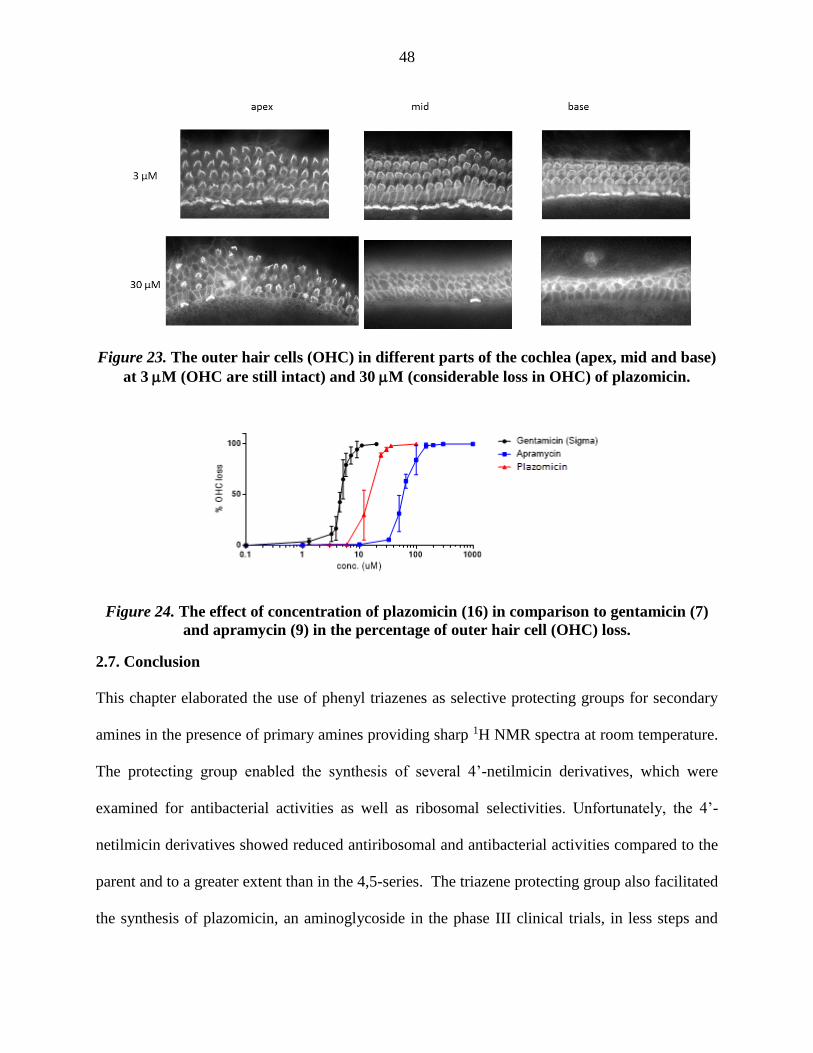
48
Figure 23. The outer hair cells (OHC) in different parts of the cochlea (apex, mid and base)
at 3 M (OHC are still intact) and 30 M (considerable loss in OHC) of plazomicin.
Figure 24. The effect of concentration of plazomicin (16) in comparison to gentamicin (7)
and apramycin (9) in the percentage of outer hair cell (OHC) loss.
2.7. Conclusion
This chapter elaborated the use of phenyl triazenes as selective protecting groups for secondary
amines in the presence of primary amines providing sharp 1H NMR spectra at room temperature.
The protecting group enabled the synthesis of several 4’-netilmicin derivatives, which were
examined for antibacterial activities as well as ribosomal selectivities. Unfortunately, the 4’-
netilmicin derivatives showed reduced antiribosomal and antibacterial activities compared to the
parent and to a greater extent than in the 4,5-series. The triazene protecting group also facilitated
the synthesis of plazomicin, an aminoglycoside in the phase III clinical trials, in less steps and

49
higher yield than the published synthesis, and enabled the study of the ribosomal selectivity and
ototoxicity of this compound.
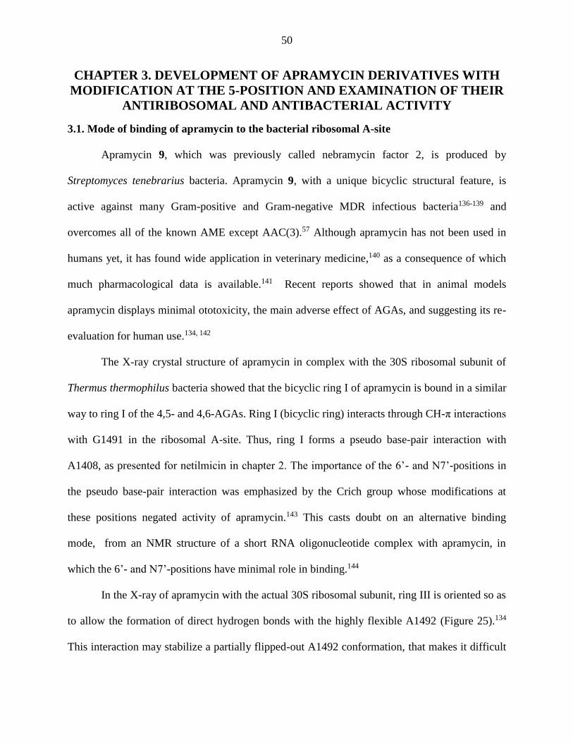
50
CHAPTER 3. DEVELOPMENT OF APRAMYCIN DERIVATIVES WITH
MODIFICATION AT THE 5-POSITION AND EXAMINATION OF THEIR
ANTIRIBOSOMAL AND ANTIBACTERIAL ACTIVITY
3.1. Mode of binding of apramycin to the bacterial ribosomal A-site
Apramycin 9, which was previously called nebramycin factor 2, is produced by
Streptomyces tenebrarius bacteria. Apramycin 9, with a unique bicyclic structural feature, is
active against many Gram-positive and Gram-negative MDR infectious bacteria136-139 and
overcomes all of the known AME except AAC(3).57 Although apramycin has not been used in
humans yet, it has found wide application in veterinary medicine,140 as a consequence of which
much pharmacological data is available.141 Recent reports showed that in animal models
apramycin displays minimal ototoxicity, the main adverse effect of AGAs, and suggesting its re-
evaluation for human use.134, 142
The X-ray crystal structure of apramycin in complex with the 30S ribosomal subunit of
Thermus thermophilus bacteria showed that the bicyclic ring I of apramycin is bound in a similar
way to ring I of the 4,5- and 4,6-AGAs. Ring I (bicyclic ring) interacts through CH-π interactions
with G1491 in the ribosomal A-site. Thus, ring I forms a pseudo base-pair interaction with
A1408, as presented for netilmicin in chapter 2. The importance of the 6’- and N7’-positions in
the pseudo base-pair interaction was emphasized by the Crich group whose modifications at
these positions negated activity of apramycin.143 This casts doubt on an alternative binding
mode, from an NMR structure of a short RNA oligonucleotide complex with apramycin, in
which the 6’- and N7’-positions have minimal role in binding.144
In the X-ray of apramycin with the actual 30S ribosomal subunit, ring III is oriented so as
to allow the formation of direct hydrogen bonds with the highly flexible A1492 (Figure 25).134
This interaction may stabilize a partially flipped-out A1492 conformation, that makes it difficult

51
for A1492 to contact with the minor groove of the codon-anticodon helix and cause misreading.
This explains the failure of apramycin to induce misreading leaving the inhibition of
translocation as the main cause of bacterial protein synthesis inhibition.79 This rationale for the
absence of misreading by apramycin is supported by work on the cleavage of ring III to give
aprosamine 91 (Figure 26), which, albeit with lower levels of activity, induces misreading.23
Figure 25. a) X-Ray crystal structure of apramycin bound to the bacterial A site showing
CH/π-stacking of ring I with G1491 base, pseudo base-pair interaction of ring I with A1408
and interaction of ring III with A1492 (PDB code: 4AQY) and b) detailed interaction of
apramycin with the bacterial A site.
Figure 26. Structures of apramycin (9) and aprosamine (91)
3.2. Modifications of apramycin
Many modifications of apramycin have been done in order to increase its potency. Most
are reported in patents and include modifications at the 5- and 6-positions,93, 145-146 modifications
G1491
A1408
A1492
A1493

52
at the N1, N2', N7', and N4''-positions,142-143, 147-150 modifications at the 6''-position,151 and
modifications at the N7’-Me and 6’-position.143 These modifications are summarized in (Figure
27) and show how apramycin has started to gain attention recently after having been neglected
since the 1980s. This ensemble of studies reveals that enhanced bacterial activity might best be
achieved through modifications to ring II (2-DOS), and most modifications to ring I (bicyclic
ring) decreased the bacterial activity. The following sub-sections will discuss the art of selective
modification at these positions.
Figure 27. A schematic diagram showing known modifications of apramycin and the year
modified; modifications in green showed increased bacterial activity, modifications in red
showed a decreased bacterial activity, and modifications in black showed an unreported or
comparable bacterial activity.
3.2.1. Modifications at the 5- and 6-positions of apramycin
A method of derivatizing apramycin at the 5- and/or 6-positions was reported that passes
through a common intermediate 95, which requires only four steps to synthesize. In this
sequence, all amines were first protected as carbamates, which was followed by protection of the
5- and 6-hydroxyl groups as a cyclohexylidene ketal. Subsequent protection of the remaining

53
hydroxyl groups as esters was followed by acid hydrolysis of the ketal to give the 5,6-diol
compound 95, that is the key intermediate for modifications at these positions (Scheme 18).93, 145-
146
Scheme 18. Preparation of the key intermediate 95 for modification at the 5,6-positions
Reaction of 95 with dihydropyran was unselective and gave both the 5- and 6-protected
alcohols 96 and 97. The alcohols 96 and 97 were separated, glycosylated and deprotected to
achieve the 5-O-glycoside 98 and the 6-O-glycosides 99 (Scheme 19).93

54
Scheme 19. Synthesis of 5-O-glycosides 98 and 6-O-glycosides 99
Additionally, the 5,6-diol intermediate 95 was selectively acetylated at the 6-position
followed by chlorination at 5-position to afford the compound 100. This was followed by radical
de-chlorination to give the compound 101. Global deprotection by catalytic hydrogenolysis and
basic hydrolysis afforded the 5-deoxyapramycin 102.145 Further, the key intermediate 95 was
subjected to mesylation to give 103, which was reacted with sodium iodide and then zinc dust to
give the 5,6-alkene 104. Finally, hydrolysis and catalytic hydrogenation gave 5,6-
dideoxyapramycin derivative 105 (Scheme 20).146

55
Scheme 20. Synthesis of 5-deoxyapramycin 102 and 5,6-dideoxyapramycin 105
3.2.2. Modifications at the N1,N2',N7',N4''-positions of apramycin
A novel method for regioselective derivatization at the N1, N3 or N2'-positions of
apramycin was introduced by Kirst and co-workers.152 The method reported facilitates the
synthesis of N1, N3 or N2'-derivatives of apramycin by transition metal-directed acylations of
apramycin. The transition metals co-ordinate with all except the amine to be acylated and so act
as transient protecting groups that save the conventional steps of protection and deprotection.
Selectively among the different amines is achieved by changing the transition metal cations. Zinc
salts are used to achieve regioselective acylation and alkylation of N1 of apramycin in a single
reaction. Copper salts are used for the synthesis of N3-acyl derivatives of apramycin, while
nickel salts are used to accomplished the regioselective acylation of N2' of apramycin (Scheme
21). The so-formed acyl derivatives can be reduced subsequently by diborane or lithium
aluminium hydride to give the corresponding N-alkyl derivatives.149-150, 152

56
Scheme 21. Transition metal directed derivatization
The regioselective derivatization at the 4'' position of apramycin is reported in a US
patent.147 The approach for the selective modification at 4'' position involves protection of all
amines as their Cbz derivatives, then the 5,6- and 2'',3''- vicinal diols are both protected with an
isopropylidine group. The 6''-hydroxyl group is next acylated prior to deprotection of the
isopropylidine groups. The key step in this method involves releasing the free amines by
hydrogenolysis so that migration of 6''-O-acyl group to the proximal 4’’-amine takes place and
gives the 4''-derivatives of apramycin. The 4''-N-alkyl analogues are prepared by reduction of
corresponding acyl derivatives with diborane or lithium aluminum hydride (Scheme 22).

57
Scheme 22. Synthetic scheme for the modification at the 4’-position
The synthesis of 7'-N-alkylapramycin derivatives proceeded by the initial preparation of
an apramycin-carbon dioxide complex that then was reacted with acetic anhydride to form
1,3,2',4''-tetra-N-acetyl apramycin 113 (Scheme 23). This was followed by alkylation of the
acetylated intermediate to provide the 7'-N-alkyl-1,3,2',4''-tetra-N-acetyl apramycin derivatives
114. General deprotection strategies finally yielded the 7'-N-alkyl apramycins 115.148

58
Scheme 23. Synthetic scheme for the modification at the 7’-position
3.2.3. Modifications at the 6''-position of apramycin
Modification at the 6''-position of apramycin is achieved by a comparable approach to
that used to modify the N4’’-position. In this chemistry, instead of 6’’-acylation of the derivative
110, the 6’’-hydroxyl is converted to the corresponding mesylates, tosylates, chlorides, and
amines or is deoxygenated. Unlike acyl groups, these groups were not to migrate after
unmasking of the apramycin amines (Scheme 24).151

59
Scheme 24. Synthetic scheme for the modification at the 4’’-position
3.2.4. Modifications at the N7’-Me and 6’-positions of apramycin
Crich and co-workers reported modifications at N7’ and 6’-postions (Scheme 25).143 All
amines and hydroxyl groups except at the N7’ and 6’-O-postions of apramycin were selectively
protected in five steps. First, all the primary amines were protected as azides using triflyl azide.
Subsequently, the secondary amine was protected as the benzyl carbamate, which upon
cyclization using sodium hydride provided the 6’,7’-oxazolidinone ring. Benzylation of all the
remaining hydroxyl groups and finally the cleavage of oxazolidinone ring afforded the key
intermediate 122. This key intermediate allowed the subsequent modifications at the 6’-position,
including the inversion of the hydroxyl group configuration or replacement of hydroxyl group
with both inversion and retention of configuration by an amine group. The introduction of CH3
and CF3 groups geminal to the 6’-alcohol was also achieved in both configurations.
Modifications can also be made at the 7’-position from intermediate 122, including the

60
preparation of analogues in which the methyl group is removed or replaced by longer alkyl
chains.
Scheme 25. Synthetic scheme for the modification at the N7’-Me and 6’-positions
3.3. Rationale
Comparison of the antiribosomal and antibacterial activities of 4,5- or 4,6-disubstituted 2-
deoxystreptamine derivatives with the corresponding 4-monosubstituted 2-deoxystreptamine
derivatives generally shows the higher activity of the disubstituted compounds. For example,
AGAs like kanamycin B, neomycin and ribostamycin are more active than neamine (Figure 28
and Table 5).153 Correspondingly, in apramycin, there was an enhancement of activity when it
was glycosylated at the 6-position to give 6-O-(3-amino-deoxy-α-D-glucopyranosyl) apramycin
99, and when it was glycosylated at the 5-position to give 5-O-(β-D-ribofuranosyl) apramycin 98

61
(Table 6).93 Thus, modification of apramycin at the 5- and/or 6-positions was considered to offer
the most promise for the development of improved derivatives. The knowledge that bacterial
resistance arising from the presence of ribosomal methyltransferases (e.g., ArmA) renders most
of the 4,6-disubstituted AGAs obsolete focused the project on substitution of apramycin at the 5-
position. Consequently, it was decided to re-evaluate the 5-O-β-D-ribofuranosyl apramycin (98,
Scheme 19) previously prepared by a Japanese group,93 preferably by a shortened and improved
route. Extrapolating further, the attachment of aminosugars such as the paromomycin CD ring to
the 5-position was projected to give an apramycin-paromomycin hybrid; the goal being to
combine paromomycin’s high activity with apramycin’s low ototoxicity (Figure 28). A
simplified form of this hybrid, in which a 2-aminoethyl group replaces the D ring paromomycin,
was also designed.
Figure 28. Paramomycin structure showing rings A-D, its hybrid with apramycin and a
simplified form of the hybrid.

62
Table 5. %Inhibition of in vitro R17 phage RNA-directed polypeptide synthesis by
various aminoglycoside antibiotics at four concentrations.
Antibiotic
Drug concentration (g/ml)
0.1 1 10 50
Neamine 2 6 37 75
Ribostamycin 5 45 65 92
Kanamycin B 22 48 58 82
Neomycin 28 55 80 96
Table 6. In vivo minimal inhibitory concentrations (MIC, μg/ml) of clinical isolates.
While this work was underway, Fridman and coworkers described the ribosylation of the
4,6-AGAs at the 5-position to give 4,5,6-trisubstitued AGAs (Figure 29).92 The resulting
compounds were slightly less active than the parents, but apparently gave improved ribosomal
selectivity in the 5-O-ribosylated kanamycin B (X = OH, Y = NH2, Z = H) and tobramycin (X =
H, Y = NH2, Z = H) series, both of which contain 2′-amines. The 2′-amine in these AG structures
forms a hydrogen bond with the ribofuranose sugar ring oxygen,154 which may assist in orienting
the ribofuranose ring in the A-site. However, these compounds are still expected to have the
ArmA resistance problem of 4,6 AGAs.
IN VIVO (MIC, μg/ml) E coli
NIHJ
P. aeruginosa
A9930
K. pneumoniae
D11
Apramycin 3.1 3.1 0.8
5-O-(β-D-ribofuranosyl)
apramycin (98) 1.6 0.8 0.8
6-O-(3-amino-deoxy-α-D-
glucopyranosyl) apramycin (99) 1.6 1.6 0.4
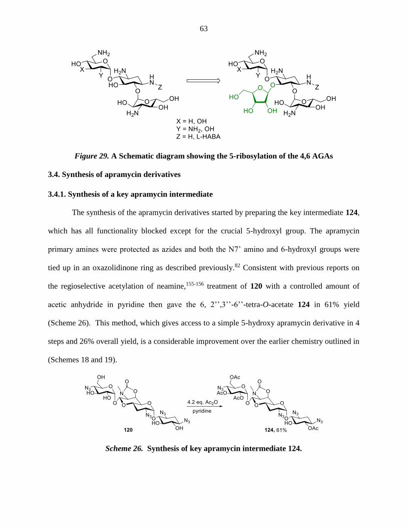
63
Figure 29. A Schematic diagram showing the 5-ribosylation of the 4,6 AGAs
3.4. Synthesis of apramycin derivatives
3.4.1. Synthesis of a key apramycin intermediate
The synthesis of the apramycin derivatives started by preparing the key intermediate 124,
which has all functionality blocked except for the crucial 5-hydroxyl group. The apramycin
primary amines were protected as azides and both the N7’ amino and 6-hydroxyl groups were
tied up in an oxazolidinone ring as described previously.82 Consistent with previous reports on
the regioselective acetylation of neamine,155-156 treatment of 120 with a controlled amount of
acetic anhydride in pyridine then gave the 6, 2’’,3’’-6’’-tetra-O-acetate 124 in 61% yield
(Scheme 26). This method, which gives access to a simple 5-hydroxy apramycin derivative in 4
steps and 26% overall yield, is a considerable improvement over the earlier chemistry outlined in
(Schemes 18 and 19).
Scheme 26. Synthesis of key apramycin intermediate 124.

64
3.4.2. Synthesis of 5-O--ribofuranosyl apramycin, 5-O--paromobiosyl apramycin and 5-
O--[3-O-(2-aminoethyl) ribofuranosyl] apramycin
Glycosylation of 124 with 2,3,5-tri-O-acetyl-α,β-D-ribofuranosyl trichloroacetimidate157
125 with promotion by BF3.OEt2 afforded the glycoside 126 in 95% yield as a separable
anomeric mixture ( = 1:9). Deprotection of the β-anomer by saponification then
hydrogenolysis allowed the formation of 127 (Scheme 27).
Scheme 27. Synthesis of 5-O--ribofuranosyl apramycin 127.
Treatment of pentaazido per-O-acetylparomomycin158 with p-thiocresol and BF3.OEt2, as
described for neomycin B,159-160 gave a 43% yield of the parombiosyl thioglycoside 128.
Complications of anomer separation following glycosylations of 124 with 128 prompted its
conversion to the 4-methoxybenzoate 130, after which oxidation with ozone gave the disaccharyl
sulfoxide 131 (Scheme 28). Triflic anhydride activation161-162 of 131 and exposure to 124 then
afforded the anticipated saccharide 132 in 48% yield as a single β-anomer. Finally,
saponification of the carbamate and ester groups followed by Staudinger reduction of the azides

65
gave the apramycin-paromomycin chimera 133 in 35% yield, which was isolated in the form of
the peracetate salt (Scheme 28).
Scheme 28. Synthesis of 5-O--paromobiosyl apramycin 133.
Preparation of the 3-O-(2-azidoethyl) ribofuranosyl donor 136 from 5-O-benzyl-1,2-O-
isopropylidene-α-D-ribofuranose 134 was achieved by alkylation with 2-azidoethyl tosylate163 to
give 135. Acidic hydrolysis of the acetonide, p-nitrobenzoylation gave the donor 136 in 45%
yield. This is a considerable improvement over an earlier synthesis of the related 3-O-(2-
azidoethyl) ribofuranosyl groups reported by the Wong group.164 Activation of 136 with
BF3.OEt2 in the presence of 124 gave a separable anomeric mixture ( = 1.15:1) of the
glycosides 137 in 43% yield. Deprotection of individual isomers resulted in the formation of

66
138 and 138, both of which were isolated as the peracetate salts in excellent yields (Scheme
29).
Scheme 29. Synthesis of compounds 138 and 138.
The synthesized compounds 127, 133, 138 and 138 were screened for the
antiribosomal and antibacterial activities. Compared to apramycin, the 5-O-β-D-ribofuranosyl
apramycin 127 showed moderately increased antiribosomal activity and especially selectivity
over the mitoribosome (Table 7), but reduced antibacterial activity against all strains tested
(Table 8). On the other hand, the 5-O-paromobiosyl apramycin 133 shows better antiribosomal
activity and better antibacterial activity towards MRSA and E coli. Unfortunately, 133 has less
mitoribosomal selectivity and less activity towards P aeruginosa. 5-O-[3-(2-Aminoethyl)-β-D-
ribofuranosyl] apramycin (138 shows an enhanced antiribosomal activity and mitoribosomal
selectivity with comparable antibacterial activity towards MRSA and E coli but is less active
towards P aeruginosa. In addition, both compounds 133 and 138 were active towards AAC(3)

67
containing bacteria, that inactivate apramycin by acetylation at the 3-position. Biological results
will be discussed in detail in Section 3.5. In view of the increased activity of paromobiosyl and
aminoethylribosyl apramycin compared to the parent compound, and their ability to overcome
AAC(3) AME resistance, more derivatives were explored and evaluated.
3.4.3 Synthesis of 5-O--[3-O-(2-hydroxyethyl) ribofuranosyl] apramycin
Two hypotheses were prompted by the observation that 5-[3-(2-aminoethyl)-β-D-
ribofuranosyl] apramycin 138 shows activity towards AAC(3) containing bacteria: i) the
aminoethyl group was bulky enough to block the AAC(3) enzyme’s active site or ii) the
increased affinity for the ribosome resulting from the addition of the extra amine outweighs the
reduction in activity due to N3 acetylation by the AME. To differentiate between these
hypotheses, a similar compound was synthesized in which a hydroxyethyl side chain replaced the
aminoethyl group of 138. The preparation of compound 142 started by alkylation of the
reported intermediate 134165 with 2-(benzyloxy)ethyl tosylate166 to afford the compound 139 in
83% yield (Scheme 30). Acetonide cleavage then p-nitrobenzoylation of intermediate 139
enabled the preparation of the donor 140, which was glycosylated with 124 to allow the
formation of the ribofuranoside product 141 in 25% yield as an anomeric mixture ( = 1:1).
Finally, deprotection of the -isomer allowed the formation of the target compound 142. When
this compound was screened for activity against AAC(3) containing bacteria (Table 9) it showed
a loss in activity, which supports the hypothesis that the amino group of 138is essential and
plays role in increasing the overall activity of the aminoglycoside. Biological results will be
discussed in detail in Section 3.5.

68
Scheme 30. Synthesis of compound 142.
3.4.4. Modification of the 5’’-hydroxyl group of 5-O--ribofuranosyl apramycin and 5-O--
[3-O-(2-aminoethyl) ribofuranosyl] apramycin
The presence of the ribosyl 5-hydroxyl group in compounds 127 and 138 renders them
prone to antibacterial resistance due to the action of the APH(3’,5’’) AMEs. Previous
modifications to the ribosyl 5-hydroxyl group in the 4,5-aminoglycoside series carried out with a
view to circumventing this AME include deoxygenation accompanied with cyclization with the
2’-amine167 or substitution by a halogen atom.168 However, these modifications were reported to
reduce antibacterial activity substantially and so were not considered useful. Therefore, two
alternative approaches to the solution of this problem were developed. Thus, it was decided i) to
explore the effect of removing the offending CH2OH group from the ribosyl ring altogether, and
ii) to substitute a formamido group for the 5-hydroxyl group in the ribose ring. Consequently, a
compound analogous to the 5-O--ribofuranosyl apramycin 127 lacking the 5’’-hydroxymethyl
group (-O--D-erythrofuranosyl apramycin) 152 was synthesized and screened. The -O-(5''-
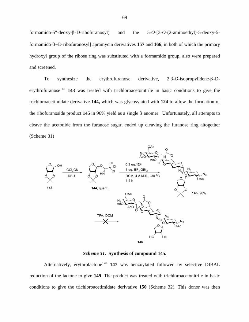
69
formamido-5''-deoxy--D-ribofuranosyl) and the 5-O-[3-O-(2-aminoethyl)-5-deoxy-5-
formamido-D-ribofuranosyl] apramycin derivatives 157 and 166, in both of which the primary
hydroxyl group of the ribose ring was substituted with a formamido group, also were prepared
and screened.
To synthesize the erythrofuranose derivative, 2,3-O-isopropylidene--D-
erythrofuranose169 143 was treated with trichloroacetonitrile in basic conditions to give the
trichloroacetimidate derivative 144, which was glycosylated with 124 to allow the formation of
the ribofuranoside product 145 in 96% yield as a single anomer. Unfortunately, all attempts to
cleave the acetonide from the furanose sugar, ended up cleaving the furanose ring altogether
(Scheme 31)
Scheme 31. Synthesis of compound 145.
Alternatively, erythrolactone170 147 was benzoylated followed by selective DIBAL
reduction of the lactone to give 149. The product was treated with trichloroacetonitrile in basic
conditions to give the trichloroacetimidate derivative 150 (Scheme 32). This donor was then
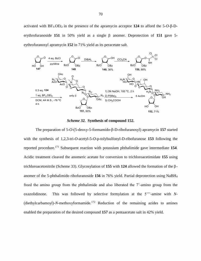
70
activated with BF3.OEt2 in the presence of the apramycin acceptor 124 to afford the 5-O-β-D-
erythrofuranoside 151 in 50% yield as a single anomer. Deprotection of 151 gave 5-
eythrofuranosyl apramycin 152 in 71% yield as its peracetate salt.
Scheme 32. Synthesis of compound 152.
The preparation of 5-O-(5-deoxy-5-formamido-β-D-ribofuranosyl) apramycin 157 started
with the synthesis of 1,2,3-tri-O-acetyl-5-O-p-tolylsulfonyl-D-ribofuranose 153 following the
reported procedure.171 Subsequent reaction with potassium phthalimide gave intermediate 154.
Acidic treatment cleaved the anomeric acetate for conversion to trichloroacetimidate 155 using
trichloroacetonitrile (Scheme 33). Glycosylation of 155 with 124 allowed the formation of the -
anomer of the 5-phthalimido ribofuranoside 156 in 76% yield. Partial deprotection using NaBH4
freed the amino group from the phthalimide and also liberated the 7’-amino group from the
oxazolidinone. This was followed by selective formylation at the 5’’’-amine with N-
(diethylcarbamoyl)-N-methoxyformamide.172 Reduction of the remaining azides to amines
enabled the preparation of the desired compound 157 as a pentaacetate salt in 42% yield.
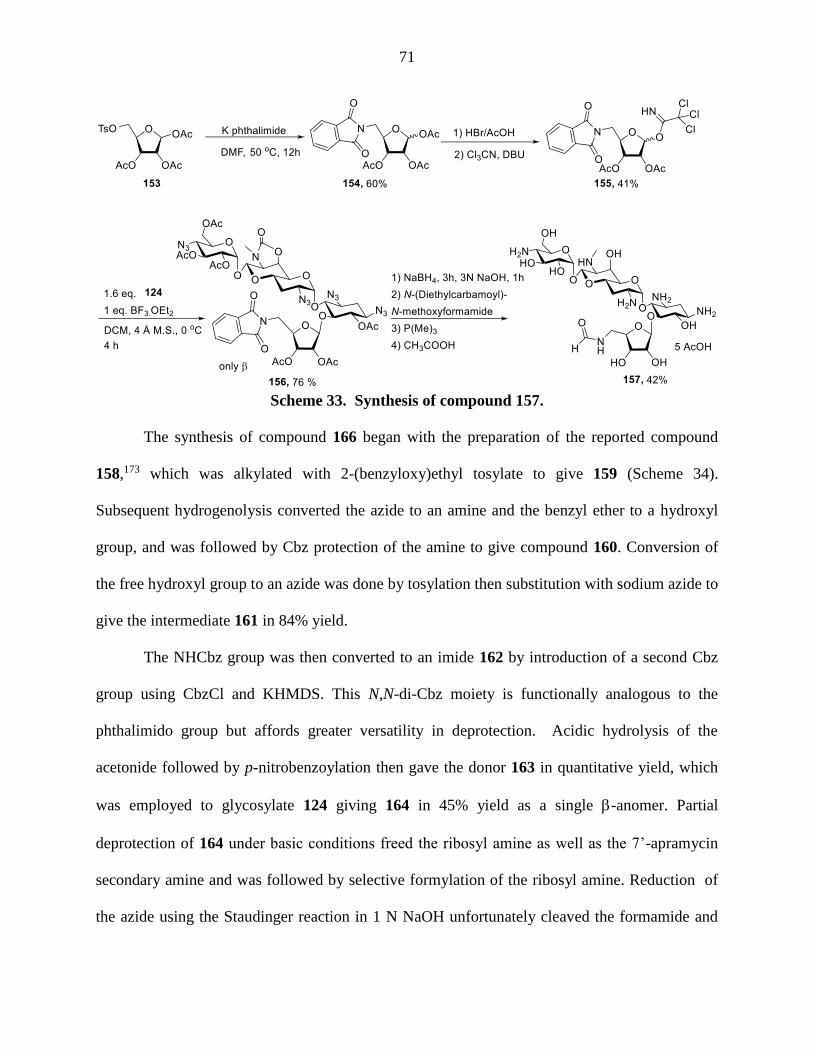
71
Scheme 33. Synthesis of compound 157.
The synthesis of compound 166 began with the preparation of the reported compound
158,173 which was alkylated with 2-(benzyloxy)ethyl tosylate to give 159 (Scheme 34).
Subsequent hydrogenolysis converted the azide to an amine and the benzyl ether to a hydroxyl
group, and was followed by Cbz protection of the amine to give compound 160. Conversion of
the free hydroxyl group to an azide was done by tosylation then substitution with sodium azide to
give the intermediate 161 in 84% yield.
The NHCbz group was then converted to an imide 162 by introduction of a second Cbz
group using CbzCl and KHMDS. This N,N-di-Cbz moiety is functionally analogous to the
phthalimido group but affords greater versatility in deprotection. Acidic hydrolysis of the
acetonide followed by p-nitrobenzoylation then gave the donor 163 in quantitative yield, which
was employed to glycosylate 124 giving 164 in 45% yield as a single -anomer. Partial
deprotection of 164 under basic conditions freed the ribosyl amine as well as the 7’-apramycin
secondary amine and was followed by selective formylation of the ribosyl amine. Reduction of
the azide using the Staudinger reaction in 1 N NaOH unfortunately cleaved the formamide and

72
provided 5-O-[5-amino-3-O-(2-aminoethyl)-5-deoxy--D-ribofuranosyl] apramycin 165. The
cleavage of the formamide was avoided by conducting the Staudinger reaction under neutral
conditions when the desired product 166 was obtained in 57% yield as the peracetate salt
(Scheme 34). The 1H-NMR spectrum of compound 166 showed two rotamers about the
formamide N-CHO bond in a ratio of 9:1. NMR analysis of the major rotamer showed a 3JH-
5’’,C=O = 3.1 and 2JCHO,C=O = 197.1, which are in agreement with it being the expected trans-
rotamer.174-175
Scheme 34. Synthesis of the compounds 165 and 166

73
In an attempt to cleave the acetonide of 161 followed by p-nitrobenzoylation of the
resulting diol, complications arose from the formation of a pyranose side product 170 due to ring
closure of the 6-amino group with the aldose moiety of 168 (Scheme 35).176 This problem was
solved by introduction of a second Cbz onto the amine to give the imide 162 as presented above
in (Scheme 34).
Scheme 35. The formation of the pyranose side product 170
Compared to 5-O--ribofuranosyl apramycin 127, both the dehydroxmethyl analog, 5-O-
(β-D-erythrofuranosyl) apramycin 152, and its 5’’’-formamido analog, -O-(5-deoxy-5-
formamido--D-ribofuranosyl) apramycin 157, were more active towards MRSA and P
aeruginosa, suggesting that the presence of an APH (3’,5’’) resistance determinant may at least
in part be responsible for the poor activity of the simple ribofuranosyl derivative 127 (Tables 8
and 9). Similarly, when compared to 5-O-[3-O-(2-aminoethyl)--D-ribofuranosyl] apramycin
138, the 5-amino-5-deoxy-ribosyl analog 165 and 5-deoxy-5-formamido ribosyl analog 166
showed a higher activity in the presence of the APH(3’,5’’) AME (Table 9). In addition, both of
these compounds, but especially 165 showed i) potent antibacterial activity exceeding that of
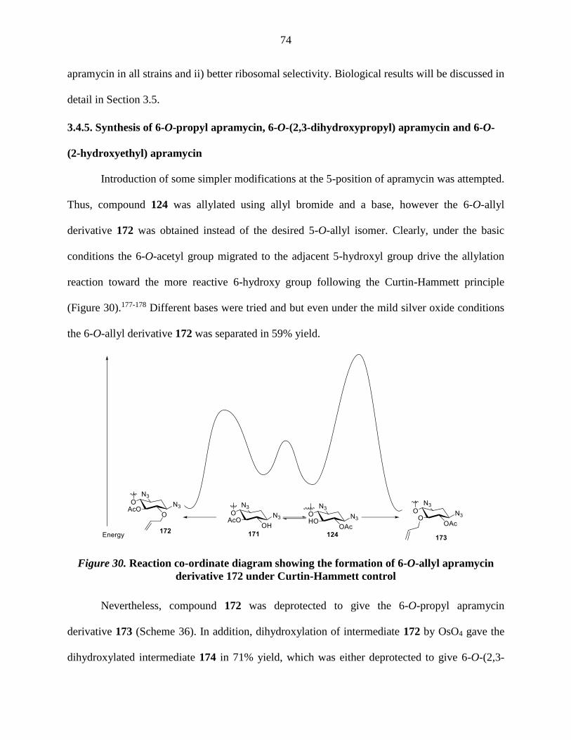
74
apramycin in all strains and ii) better ribosomal selectivity. Biological results will be discussed in
detail in Section 3.5.
3.4.5. Synthesis of 6-O-propyl apramycin, 6-O-(2,3-dihydroxypropyl) apramycin and 6-O-
(2-hydroxyethyl) apramycin
Introduction of some simpler modifications at the 5-position of apramycin was attempted.
Thus, compound 124 was allylated using allyl bromide and a base, however the 6-O-allyl
derivative 172 was obtained instead of the desired 5-O-allyl isomer. Clearly, under the basic
conditions the 6-O-acetyl group migrated to the adjacent 5-hydroxyl group drive the allylation
reaction toward the more reactive 6-hydroxy group following the Curtin-Hammett principle
(Figure 30).177-178 Different bases were tried and but even under the mild silver oxide conditions
the 6-O-allyl derivative 172 was separated in 59% yield.
Figure 30. Reaction co-ordinate diagram showing the formation of 6-O-allyl apramycin
derivative 172 under Curtin-Hammett control
Nevertheless, compound 172 was deprotected to give the 6-O-propyl apramycin
derivative 173 (Scheme 36). In addition, dihydroxylation of intermediate 172 by OsO4 gave the
dihydroxylated intermediate 174 in 71% yield, which was either deprotected to give 6-O-(2,3-

75
dihydroxypropyl) apramycin 175 or exposed to oxidative cleavage then reduction before
deprotection to allow the formation of 6-O-(2-hydroxyethyl) apramycin 177. Biological
screening of 173, 175 and 177 didn’t show any advantage over the parent apramycin (Tables 7
and 8).
Scheme 36. Synthesis of compounds 173, 175 and 177.
3.4.6. Synthesis of 5-epi-apramycin, 5-deoxy-5-fluoro apramycin, 5-deoxy-5-epi-fluoro-
apramycin and 5-deoxy apramycin
A previous report showed that the 5-hydroxyl group is not essential for activity, and that
removing it even increases antibacterial activity,145 suggesting further exploration of this
position. Thus, inversion of configuration at the 5-position and replacement of the 5-hydroxy
group with fluoride in both configurations were explored. Moreover, 5-deoxyapramycin was also

76
synthesized as a comparator. To synthesize 5-epi-apramycin 179, compound 124 was triflated at
the 5-position and the triflate displaced by potassium acetate to afford the inverted acetate
intermediate 178 (Scheme 37). Basic hydrolysis followed by Staudinger reaction of the
intermediate 178 enabled the preparation of compound 179 in 65% yield as the pentaacetate salt.
Preparation of 5-deoxy-5-fluoro apramycin 182 started by triflation and was followed by
substitution with sodium nitrite, which upon workup gave the inverted hydroxyl group
intermediate 180.179 Subsequent treatment with diethylaminosulfur trifluoride (DAST)
substituted the hydroxyl group with fluoride with inversion of configuration giving 181 in 81%
yield. Deprotection of 181 by basic hydrolysis of the esters and subsequent Staudinger reaction
enabled the preparation of 182 in 58% yield. The 5-deoxy-5-epi-fluoro apramycin derivative 183
was accessed from 124 in 74% yield by displacement of the 5-hydroxyl group with fluoride
using DAST. Global deprotection of 183 gave the 5-deoxy-5-epi-fluoro apramycin pentaacetate
salt 184 in 59% yield. Finally, 5-deoxyapramycin was synthesized by the initial displacement of
the 5-hydroxyl group of 124 with iodide via the triflate intermediate giving 185. This was
followed by deacetylation and parallel hydrogenolysis of the azides and of the C-I bond. Basic
hydrolysis of the oxazolidinone ring gave 186, which was isolated as the peracetate salt in 47%
yield. Biological screening (Table 7 and Table 8) of 5-deoxyapramycin was consistent with the
previous report.145 All compounds tested in this series (5-epi-OH, 5-fluoro, 5-epi-fluoro and 5-
deoxy) showed better antiribosomal activity than apramycin. Derivatives 5-epi apramycin 179,
5-fluoro-5-deoxy apramycin 182, 5-epifluoro apramycin 184 and 5-deoxy apramycin 186 had
better antibacterial effect against MRSA and E. coli strains and comparable antibacterial effects
toward P aeruginosa compared to the parent compound (Table 7 and Table 8). Biological results
will be discussed in detail in Section 3.5.

77
Scheme 37. Synthesis of compounds 179, 182, 184 and 186.
3.4.7. Synthesis of 3-N-formyl apramycin and 3-N-acetyl apramycin
The only known AME that affects apramycin is AAC(3)-IV that acetylates the 3-amino
group of apramycin.57 As formamido groups are tolerated at other positions on aminoglycoside
skeletons when acetamido groups are not,180 it was possible that installation of a formamide
group at the 3-amino group of apramycin would block the action of AAC(3)-IV but not
compromise the antibacterial activity. Consequently, 3-N-formyl apramycin was synthesized
together with 3-N-acetyl apramycin that was needed as a comparison standard. The synthesis of
these derivatives began with the preparation of the compound 187 using copper-based selective
acylation as reported.152 Subsequently, the remaining primary amines were protected as azides
and the 3-amino group was freed in basic conditions to afford the compound 188. Selective
formylation at the 3-position was achieved with using N-(diethylcarbamoyl)-N-

78
methoxyformamide.172 Reduction of the remaining azides to amines enabled the preparation of
the desired compound 190. In a similar way, 3-amino group of 188 was acetylated using N-
(diethylcarbamoyl)-N-methoxyacetamide 189 and the azides were reduced to afford compound
191 (Scheme 38). Interestingly, the 1H-NMR spectra of 3-N-formyl apramycin shows two
rotamers in 4:3 ratio favoring the trans-rotamer, with the assignment-based on an nOe spectrum
of 190, which showed clear enhancement of the resonances for H-3 and CHO in the cis-form but
not of H-3 on irradiation of the formyl proton in the trans-rotomer (Scheme 39). The biological
data for compounds 190 and 191 showed complete loss of antibacterial activity (Table 7 and
Table 8) thereby negating the hypothesis that a formamido group might be tolerated at the 3-
position of apramycin.
Scheme 38. Synthesis of compounds 190 and 191.

79
Scheme 39. Diagnostic nOe interactions in the cis and trans rotamers of 3-N-formyl
apramycin
3.5. Biological Evaluation
All the synthesized compounds were submitted to the Böttger lab in Zurich, where they were
screened for antiribosomal activity (Table 7) and antibacterial activity against strains of E. coli,
MRSA and P aeruginosa (Table 8). The methods were identical to the ones applied in the
netilmicin series (Chapter 2). In addition, the MIC of the compounds were obtained against
engineered E. coli strains with known AME resistance mechanisms.
Table 7. Inhibition of wild-type bacterial and hybrid ribosomes (IC50, M).
IN VITRO (IC50, M) Sms (M) Mit (M) A1555G (M) Cyt (M)
Apramycin (9) 0.17 124.8 (747) 51.5 (308) 108.7 (651)
Neomycin (2) 0.03 3.8 (127) 0.4 (14) 32.8 (1094)
5-O-(β-D-Ribofuranosyl)
apramycin (127) 0.09 368.9 (4130) 272.8 (3054) 436.2 (4883)
5-O-(β-Paramobiosyl)
apramycin (133) 0.13 2.1 (16) 0.5 (4) 29.7 (225)
5-O-[3-(2-Aminoethyl)--
ribofuranosyl] apramycin
(138
0.07 77.4 (1106) 16.9 (241) 202.6 (2896)
5-O-[3-(2-Aminoethyl)-α-
ribofuranosyl] apramycin
(138
0.35 192.4 (550) 284.8 (814) 257.2 (735)

80
5-O-[3-(2-Hydroxyethyl)-β-D-
ribofuranosyl] apramycin (142) 0.31 904.0 (2945) 456.0 (1483) 1227.0 (3991)
5-O-(β-D-Erythrofuranosyl)
apramycin (152) 0.12 312.2 (2504) 235.0 (1885) 448.6 (3598)
5-O-β-(5'''-Formamido-5'''-
deoxy-D-ribofuranosyl)
apramycin (157)
0.33 571.5 (1744) 263.2 (803) >
5-O-[5-Deoxy-5-amino-3-(2-
aminoethyl)-β-D-
ribofuranosyl] apramycin (165)
0.04 48.0 (1143) 22 (524) 37.0 (881)
5-O-[5-Deoxy-5-formamido-3-
(2-
aminoethyl)-β-D-ribofuranosyl]
apramycin (166)
0.09 93.0 (1094) 11.0 (129) 142.0 (1671)
6-O-Propyl apramycin (173) 2.49 448.7 (180) 828.5 (332) 646.0 (259)
6-O-(2,3-Dihydroxypropyl)
apramycin (175) 0.57 403.1 (707) 565.3 (991) 462.4 (811)
6-O-(2-Hydroxyethyl)
apramycin (177) 0.39 638.1 (1619) 388.6 (986) 425.34 (1079)
5-Epi apramycin (179) 0.11 39.9 (359) 17.8 (160) 25.5 (229)
5-Deoxy-5-fluoro-apramycin
(182) 0.12 113.7 (978) 125.2 (1076) 136.6 (1175)
5-Deoxy-5-epifluoro-
apramycin (184) 0.10 55.8 (559) 50.4 (506) 51.7 (519)
5-Deoxy apramycin (186) 0.11 86.5 (824) 67.4 (642) 80.2 (764)
3-N-Formyl apramycin (190) 69 > 2000 > 2000 > 2000
3-N-Acetyl apramycin (191) n.d. n.d. n.d. n.d.
Sms: M. Smegmatis ribosome, Cyt: M. Smegmatis ribosome with human cytosolic A-site, Mit: M. Smegmatis
ribosome with human mitochondrial A-site, A1490G: M. Smegmatis ribosome with human mitochondrial A-site
with A1555G mutation. Selectivities are obtained by dividing the hybrid ribosomal activity by M. Smegmatis
ribosomal activity.
The inhibitory activity of apramycin against the bacterial ribosome is less than that of
neomycin but much greater selectivities is displayed toward the wild-type mitochondrial and the
A1555G mutant mitochondrial ribosomes, which is reflected in lower cochleotoxicity of
apramycin compared to neomycin75, 134 Compared to apramycin, the installation of a ribosyl

81
group at the 5-position, as in compound 127, resulted in moderately increased antiribosomal
activity (2-fold) and greater selectivity over the wild-type mitoribosome hybrid (5- to 6-fold) and
the A1555G mutant mitoribosome hybrid (10-fold). On the other hand, the 5-O-paromobiosyl
group shifted the activities of apramycin to resemble more those of neomycin: like neomycin, 5-
O-paromobiosyl apramycin 133 exhibits strong antibacterioribosomal activity but very low
mitoribosomal selectivity. 5-O-[3-(2-Aminoethyl)-β-D-ribofuranosyl] apramycin 138 shows an
enhanced antiribosomal activity (2 to 3-fold) and mitoribosomal selectivity in wild-type but not
in the A1555G mutant hybrid. Both changing the anomeric configuration of the ribose ring as in
compound 138 or subtituting the 2-aminoethyl group with hydroxyethyl group 142 had a
detrimental effect on the ribosomal activity. On the other hand, the 5’’’-amine analog 165 of
compound 138 showed enhanced antibacterioribosomal activity (2-fold) and a comparable
mitoribosomal selectivity when compared to the parent. Replacing the 5’’’-hydroxyl group of
138 with a 5’’’-fomamido group as in compound 166 did not change much the ribosomal
activity and selectivities Compared to 5-O--ribofuranosyl apramycin 127, both the
dehydroxmethyl analog 152, and its 5’’’-formamido analog 157, exhibited reduced
antibacterioribosomal activities and mitoribosomal selectivities.
Derivatives 5-epi apramycin 179, 5-fluoro-5-deoxy apramycin 182, 5-epifluoro
apramycin 184 and 5-deoxy apramycin 186 showed better antiribosomal activity than apramycin.
Enhancement of selectivity over the wild-type and mutant mitoribosomal were also obtained in
case of 5-fluoro-5-deoxy apramycin 182 and 5-deoxy apramycin 186.
All the compounds synthesized with substitutions at the 6-position and the 3-position
abolish the ribosomal activity.

82
Table 8. Minimal inhibitory concentrations (MIC, μg/mL) of clinical isolates.
(MIC, μg/ml)
MRSA E coli P aeruginosa
AG03
8
AG03
9
AG04
2
AG04
4
AG00
1
AG05
5
AG00
3
AG03
1
AG03
2
AG03
3 AG086
Apramycin (9) 8 8 8 16 16 8 8-16 8 8 8 4
Neomycin (2) 0.5-1 128 128 0.5-1 ---- 4 4 32 32-
64 ≥128 ≥128
5-O-(β-D-
Ribofuranosyl)
apramycin (127)
64 64 64 32-
64 16
16-
32 16 64
32-
64 128 >128
5-O-(β-
Paramobiosyl)
apramycin (133)
2-4 2-4 2 2 4 8 4 64 64 64 ≥128
5-O-[3-(2-
Aminoethyl)--
ribofuranosyl]
apramycin (138
8-16 8 8 8-16 8 8 4-8 32 32 128 >128
5-O-[3-(2-
Aminoethyl)-α-
ribofuranosyl]
apramycin (138
16 16 16 16 32 16-
32
16-
32
32-
64 64 128 >128
5-O-[3-(2-
Hydroxyethyl)-β-D-
ribofuranosyl]
apramycin (142)
32 32 32 n.d. 32 16 16 >32 >32 >32 n.d.
5-O-(β-D-
Erythrofuranosyl)
apramycin (152)
16-
32
16-
32 16 16 16 16 16 32 32
64-
128 >128
5-O-β-(5'''-
Formamido-5'''-
deoxy-D-
ribofuranosyl)
apramycin (157)
32 16-
32 32 n.d. 8 16 8 >32 >32 >32 n.d.
5-O-[5-Deoxy-5-
amino-3-(2-
aminoethyl)-β-D-
ribofuranosyl]
apramycin (165)
1 1 1 n.d. 2 2 1-2 2 2 2 n.d.
5-O-[5-Deoxy-5-
formamido-3-(2-
aminoethyl)-β-D-
ribofuranosyl]
apramycin (166)
4 4-8 4-8 n.d. 2 4 2 16 32 64 n.d.
6-O-Propyl
apramycin (173) ≥128 ≥128
64-
128 128
64-
128
64-
128
64-
128 >128 >128 >128 >128
6-O-(2,3-
Dihydroxypropyl)
apramycin (175)
64 64 64 64-
128 32 32 32 64 64 ≥128 ≥128

83
6-O-(2-
Hydroxyethyl)
apramycin (177)
16-
32
16-
32 64 64 16 16 16 32 32
64-
128 4-8
5-Epi apramycin
(179) 4 4 2-4 4 4 4 4 4 4 8 32
5-Deoxy-5-fluoro-
apramycin (182) 4-8 4-8 8 n.d. 8 4 8 8-16 16 32 n.d.
5-Deoxy-5-
epifluoro-apramycin
(184)
2-4 2-4 4 n.d. 8 4 4 8-16 8-16 32 n.d.
5-Deoxy apramycin
(186) 2-4 2 4 n.d. 4 4 4 8 8 16 n.d.
3-N-Formyl
apramycin (190) >64 n.d. n.d. n.d. >64 n.d. n.d. >64 n.d. n.d. n.d.
3-N-Acetyl
apramycin (191) >64 >32 >32 n.d. >32 >32 >32 >32 >32 >32 n.d.
MIC (μg/mL) of apramycin derivatives to inhibit Sms (Mycobacterium smegmatis), MRSA (Methicillin-resistant
Staphylococcus aureus), E coli and P aeruginosa.
Compared to apramycin, the 5-O-β-D-ribofuranosyl apramycin 127 showed a reduced
antibacterial activity against all strains tested (Table 8). On the other hand, the 5-O-paromobiosyl
apramycin 133 shows an increase in antibacterial activity towards MRSA and E coli but a
reduced activity towards P aeruginosa. 5-O-[3-(2-Aminoethyl)-β-D-ribofuranosyl] apramycin
138 exhibits comparable antibacterial activity towards MRSA and E coli but is less active
towards P aeruginosa. Both the dehydroxmethyl analog, 5-O-(β-D-erythrofuranosyl) apramycin
152, and the 5’’-formamido analog, 5-O-(5'’'-deoxy-5’’’-formamido--D-ribofuranosyl)
apramycin 157, were more active than their ribosyl congener towards MRSA and P aeruginosa,
suggesting that the presence of an APH (3’,5’’) resistance determinant may at least in part be
responsible for the poor activity of the simple ribofuranosyl derivative 127. Similarly, when
compared to 5-O-[3-O-(2-aminoethyl)--D-ribofuranosyl] apramycin 138, the 5-amino-5-
deoxy-ribosyl analog 165 and 5-deoxy-5-formamido ribosyl analog 166 showed a higher activity
towards MRSA and E coli.
Compared to apramycin, 5-epi apramycin 179, 5-fluoro-5-deoxy apramycin 182, 5-

84
epifluoro apramycin 184 and 5-deoxy apramycin 186 had better antibacterial effects against
MRSA and E. coli strains and comparable antibacterial effects toward P aeruginosa. This
highlights the fact that a 5-hydroxy group is not necessary for activity and that its removal or
substitution may be beneficial.
All the compounds synthesized with the substitution at the 6-position (6-O-propyl 173, 6-
O-(2,3-dihydroxypropyl) 175 and 6-O-(2-hydroxyethyl) 177) were less active in all tested
bacterial strains. Similarly, 3-N-formyl apramycin 190 and 3-N-acetyl apramycin 191 showed
greatly reduced activity.
Table 9. Minimal inhibitory concentrations (MIC, μg/mL) of engineered E coli strains
carrying known AMEs
(MIC, μg/ml)
E coli
AG173 AG163 AG166 AG182 AG037
AAC(3)-IV APH(3’)-Ia APH(3’)-IIa AAC(3)-IV
APH(3’)-Ia APH(3’)-IIIa
Apramycin (9) 256 4 4-8 >128 1
Neomycin (2) n.d. >64 >64 n.d. >64
5-O-(β-D-Ribofuranosyl)
apramycin (127) 16-32 8-16 8 64 4
5-O-(β-Paramobiosyl) apramycin
(133) 1-2 8 2 64 1
5-O-[3-(2-Aminoethyl)--
ribofuranosyl] apramycin (138 4 4-8 2 16 0.5
5-O-[3-(2-Aminoethyl)-α-
ribofuranosyl] apramycin (138 n.d. n.d. n.d. n.d. n.d.
5-O-[3-(2-Hydroxyethyl)-β-D-
ribofuranosyl] apramycin (142) 32-64 8 8 n.d. n.d.
5-O-(β-D-Erythrofuranosyl)
apramycin (152) 128 8 8 >128 1-2
5-O-β-(5'''-Formamido-5'''-deoxy-
D-ribofuranosyl) apramycin (157) >64 8 8-16 n.d. n.d.
5-O-[5-Deoxy-5-amino-3-(2-
aminoethyl)-β-D-ribofuranosyl]
apramycin (165)
4 2 2 8 0.25
5-O-[5-Deoxy-5-formamido-3-(2-
aminoethyl)-β-D-ribofuranosyl]
apramycin (166)
32 2 4 32-64 0.25
6-O-Propyl apramycin (173) n.d. n.d. n.d. n.d. n.d.
6-O-(2,3-Dihydroxypropyl)
apramycin (175) n.d. n.d. n.d. n.d. n.d.
6-O-(2-Hydroxyethyl) apramycin
(177) >128 8-16 8-16 >128 2

85
5-Epi apramycin (179) 64 n.d. n.d. >128 n.d.
5-Deoxy-5-fluoro-apramycin (182) 64 n.d. n.d. >128 n.d.
5-Deoxy-5-epifluoro-apramycin
(184) 128 n.d. n.d. >128 n.d.
5-Deoxy apramycin (186) 32 n.d. n.d. 128 n.d.
3-N-Formyl apramycin (190) n.d. n.d. n.d. n.d. n.d.
3-N-Acetyl apramycin (191) n.d. n.d. n.d. n.d. n.d.
AAC(3)‐IV AME is the only resistance determinant currently effective in the abrogation
of apramycin activity,57 consequently it was important to examine the synthesized compounds
against bacteria known to carry this and other enzymes. Interestingly, adding the β-D-
ribofuranosyl group 127 at the 5-position protects the compound from the action of AAC(3)-IV.
This protection is increased at least 4-fold by further incorporation of a 2-aminoethyl group at
the 3’’’-position of compound 138 and 8-fold if a diaminoidose sugar is incorporated instead as
in 5-O-(β-paramobiosyl) apramycin 133. Compared to 5-O-(β-D-ribofuranosyl) apramycin 127,
removal of the 5’’’-CH2OH 152 or substituting the 5’’’-OH with a formamido group 157 made
the compounds more prone to AAC(3)-IV. Similarly, when compared to 5-O-(2-aminoethyl)--
ribofuranosyl] apramycin 138, its 5’’’ formamide analog 166 or substituting the 3’’’-
aminoethyl group with a hydroxyethyl group 142 reduced the activity at least 8-fold against
AAC(3)-IV containing bacteria. On the other hand, activity was not reduced in the 5’’’ amine
analog 165 of 5-O-[3-(2-aminoethyl)--ribofuranosyl] apramycin 138.
Compared to apramycin, the 5-epi apramycin 179, 5-fluoro-5-deoxy apramycin 182, and
5-epifluoro apramycin 184 showed moderately increased activity (2- to 4-fold) against AAC(3)-
IV containing E coli , whereas 5-deoxy apramycin 186 showed a more substantial increase in
activity (8-fold).

86
Apramycin is inheritably not susceptible to APH(3’) due to the absence of a hydroxy
group at the 3’-position, thus all synthesized compounds tested showed results with APH(3’)-Ia
and APH(3’)-IIa containing bacteria comparable to the wild type. However, APH(3’)-IIIa
enzymes are also known to phosphorylate the 5-hydroxyl of the ribose ring in 4,5-AGAs.38 Thus,
the presence of APH (3’)-IIIa results in a 4-fold reduction activity in antibacterial activity when a
ribofuranosyl group is attached at the 5-position as in compound 127. Removal of the ribosyl 5-
OH as in the erythrofuranose restores activity in the presence of the APH(3’)-IIIa. Similarly, the
replacement of the ribosyl 5-OH by a formamido or amino group overcomes the presence of
APH(3’)-IIIa as is clear from the comparison of 5-O-[3-(2-aminoethyl)--ribofuranosyl]
apramycin 138, with its formamido 166 and amino 165 analogs.
3.6. Conclusion
An easy and efficient method has been developed for derivatization of apramycin at the
5-position. Compounds 5-O-[3-(2-aminoethyl)--ribofuranosyl] apramycin 138, 5-O-(β-
paramobiosyl) apramycin 133, 5-O-[5-deoxy-5-formamido-3-(2-aminoethyl)-β-D-ribofuranosyl]
apramycin 166 and 5-Deoxy apramycin 186 showed improved antibacterial activity against
MRSA and E. coli strains and reduced antibacterial activity toward P aeruginosa in comparison to
the parent apramycin. 5-Epi apramycin 179 exhibits higher activity against all bacterial strains,
albeit with less mitoribosomal selectivity. The apramycin-paromomycin hybrid 133and its
simplified form 138were active in the presence of the AAC(3)-IV class of AME that is the only
resistance determinant currently effective in the abrogation of apramycin activity The 5-O-[5-
deoxy-5-amino-3-(2-aminoethyl)-β-D-ribofuranosyl] apramycin 165 is considered a
breakthrough in so far as it satisfies all the project needs with improved antibacterial activity
compared to apramycin against all bacterial strains screened including P aeruginosa, and strains

87
carrying the AAC(3) and APH(3’,5’’) AMEs. In addition, compound 165 showed improved ribosomal
selectivity. Moreover, this study showed that a hydroxy group at the 5-position of apramycin is
not essential, in fact its removal, inversion of configuration, or replacement by a fluoro group as
in compounds 179, 182, 184 and 186 increases activity. Overall, new apramycin derivatives were
developed which are more active than the parent drug. Moreover, aminoglycosides with better
mitoribosomal selectivity and the ability to overcome the AAC(3)resistance determinant were
developed for potential treatment of multidrug resistant bacterial infections.

88
CHAPTER 4. CONCLUSIONS
In order to investigate the effect of 4’-modifications of the 4,6-AGA netilmicin on the
reduction of ototoxicity but not antibiotic activity several netilmicin derivatives have been
prepared. These derivatives were screened for antiribosomal activity in cell-free translation
assays as well as antibacterial activity against clinical isolates of methicillin-resistant
Staphylococcus aureus, Escherichia coli (E coli) and Pseudomonas aeruginosa. Unfortunately,
all modifications of netilmicin at the 4’-position showed similar or reduced antibacterial
activities to the parent. All derivatives except 4'-chloro netilmicin showed less selectivity than
netilmicin for inhibition of the bacterial ribosome as compared to the eurkaryotic ribosomes.
Overall, the 4’-modifications reduce activity in 4,6-series to a greater extent than in the 4,5-
series. This conclusion is consistent with the results observed in a series of 4’-modifications of
the 4,6-aminoglycoside kanamycin B.11
This work made use of phenyl triazenes as selective protecting groups for secondary
amines in the presence of primary amines. A number of polyamine substrates were used in the
study including aliphatic and heterocyclic polyamines, amino acids and aminoglycosides. The
secondary amines in these substrates were selectively protected as phenyl triazenes, and the
primary amines were subsequently protected as the azides, benzyloxy carbamates or
fluorenylmethyl carbamates. Phenyl triazenes also gave the advantage over carbamates of
showing sharp 1H NMR spectra at room temperature. This protecting group enabled the synthesis
of plazomicin, an aminoglycoside in the 3rd phase of clinical trials, in fewer steps and higher
yield than previously reported.
A series of novel apramycin derivatives were designed by focusing on derivatization and
modifications at the 5-position. An apramycin-paromomycin hybrid, in which the paromobiosyl

89
group (rings III and IV) of paromomycin was attached to the 5-postion of apramycin, was
synthesized with the aim of combining paromomycin’s high activity with apramycin’s low
ototoxicity. The hybrid 133 showed better antibacterial activity against MRSA and E coli strains
compared to the parent apramycin, but unfortunately with reduced antibacterial activity toward P
aeruginosa and reduced ribosomal selectivity. Further truncation and modifications of this hybrid
led to the development of 5-O-[5-amino-3-O-(2-aminoethyl)-5-deoxy-β-D-ribofuranosyl]
apramycin 165, 5-O-[3-O-(2-aminoethyl)-5-deoxy-5-formamido-β-D-ribofuranosyl] apramycin
166, 5-epi apramycin 179 and 5-deoxy apramycin 186 that showed an overall better antibacterial
effect with better ribosomal selectivity than the parent. Compound 165 is considered the best of
all derivatives synthesized as it was more active against all bacterial strains than apramycin
including P aeruginosa and AAC(3) and APH(3’)-IIIa- containing bacteria, and also showed a
better ribosomal selectivity. Overall, apramycin-class aminoglycoside derivatives with better
antibacterial activities and ribosomal selectivities, and the ability to overcome the AAC(3) and
APH(3’)-IIIa resistance determinants were developed with for the treatment of multidrug
resistant bacterial infections.

90
CHAPTER 5. EXPERIMENTAL SECTION
General Information
All reagents and solvents were purchased from commercial suppliers and were used
without further purification unless otherwise specified. Chromatographic purifications were
carried out over silica gel. Analytical thin-layer chromatography was performed with pre-coated
glass backed plates (w/UV 254) and visualized by UV irradiation (254 nm) or by staining with
25% H2SO4 in EtOH or ceric ammonium molybdate (ceric sulfate: (4.0 g); ammonium
molybdate (10 g); H2SO4: 40 mL, H2O: 360 mL) solution. Specific rotations were obtained using
a digital polarimeter in the solvent specified at 589 nm and 23 ºC on an Autopol III polarimeter
(Rudolph Research Analytical, Hackettstown, NJ) with a path length of 10 cm. Infrared spectra
were recorded on a FT/IR instrument. High resolution mass spectra were recorded with an
electrospray source coupled to a time-of-flight mass analyzer (Waters). 1H, 13C and 2D NMR
spectra were recorded on 400 MHz, 500 MHz or 600 MHz instruments as specified.
Assignments in 1H and 13C NMR were done by the assistance of H−H COSY, HSQC and/or
HMBC experiments
91
General procedure A. Selective protection of secondary amines as phenyl triazenes
with subsequent protection of primary amines as azides: An ice cooled solution of the amine
(1 equiv) in a water/methanol mixture (3:7, 0.1 M) was treated with Na2CO3 (8 equiv), and then
dropwise over 0.5 h with a solution of phenyldiazonium tetrafluoroborate (1.1 equiv) in water
(0.1 M). After completion of the addition, Stick's reagent (imidazole-1-sulfonyl azide
hydrochloride, 0.3 mmol per primary amine) and a catalytic amount of CuSO4 were added. The
reaction mixture was allowed to warm to rt and stirred overnight before it was diluted with ethyl
acetate and washed with water and brine. The organic layer was dried and concentrated. The
residue was purified by chromatography over silica gel.
General procedure B: Selective protection of secondary amines as phenyl triazenes
with subsequent protection of primary amines as azides: The substrate (1 equiv) was
dissolved in acetonitrile:H2O (1:1, 0.1 M). K2CO3 (8 equiv) was added and the reaction mixture
was cooled to 0 oC using an ice bath. A solution of phenyldiazonium tetrafluoroborate (1.1
equiv) in acetonitrile (0.1 M) was added using a syringe pump over 0.5 h. After completion of
the addition, 0.22 mmol per amine of imidazole-1-sulfonyl azide hydrochloride and a catalytic
amount of CuSO4 were added. The reaction mixture was allowed to warm to rt and stirred
overnight. The reaction mixture was diluted with ethyl acetate and washed with water and brine.
The organic layer was dried and concentrated. The residue was purified by chromatography over
silica gel.
General procedure C: Selective protection of secondary amines as phenyl triazenes
with subsequent protection of primary amines as benzyl carbamates: An ice cooled solution
of the amine (1 equiv) in a water/methanol mixture (3:7, 0.1 M) was treated with Na2CO3 (8
equiv), and then dropwise over 0.5 h with a solution of phenyldiazonium tetrafluoroborate (1.1
92
equiv) in water (0.1 M). After completion of the addition, 0.24 mmol per amine of benzyl
chloroformate was added. The reaction mixture was allowed to warm to rt and stirred overnight.
The reaction mixture was diluted with ethyl acetate and washed with water and brine. The
organic layer was dried and concentrated. The residue was purified by chromatography over
silica gel.
General procedure D: Deprotection by deacetylation, Staudinger and acid cleavage
of phenyl triazenes: A stirred solution of substrate (0.04 mmol, 1 equiv) in dioxane (1.5 mL)
was treated with 1N NaOH (0.5 mL) and heated with stirring at 60 °C for 1.5 h. The reaction
mixture was cooled to rt before 1M P(CH3)3 in THF (4.5 equiv) was added and the reaction
mixture stirred at rt for 6h. The reaction mixture was then concentrated and purified by column
chromatography (eluent: 5% to 12% ammonical MeOH in DCM). The product-containing
fractions were concentrated, dissolved in ethanol (0.9 mL) and treated with sodium
hypophosphite (2 equiv) and trifluoroacetic acid (12 equiv), and stirred at rt for 8 h. The reaction
mixture was neutralized using Amberlite® IRA400 hydroxide form, filtered and dried. The crude
product was dissolved in D.I. water (1 mL), acidified by glacial acetic acid till pH = 3-4 and
loaded to a Sephadex column (CM Sephadex C-25) from which it was flushed with D.I. water
(20 mL), then by gradient elution of 0.1% - 1.0% NH4OH in D.I. water. The fractions containing
the product were combined, acidified with glacial acetic acid and lyophilized to give a white
solid.
Methyl 2-azido-6-(1-methyl-3-phenyltriaz-2-en-1-yl) hexanoate (50). This compound
was prepared according to the general procedure A using compound 42 (38 mg, 0.22 mmol) to
afford the desired product 7 (38 mg, 58 %) as a yellow oil; Rf = 0.8 (40% EtOAc in hexanes);
[α]D25= -26.1 (c 0.025, MeOH); IR (film) (cm-1): 2105, 1745, 1594 (w); 1H NMR (400 MHz,

93
CD3OD) δ 7.36 (dd, J = 8.5, 1.2 Hz, 2H), 7.33 – 7.24 (m, 2H), 7.09 (t, J = 7.3 Hz, 1H), 4.00 (dd,
J = 8.2, 5.3 Hz, 1H), 3.77 (t, J = 7.0 Hz, 2H), 3.71 (s, 3H), 3.19 (s, 3H), 1.91 – 1.81 (m, 1H),
1.79-1.66 (m, 3H), 1.47 – 1.36 (m, 2H); 13C NMR (101 MHz, CD3OD) δ 171.2, 151.0, 128.3,
124.9, 120.0, 61.6, 51.6, 30.5, 27.2, 22.4; ESI-HRMS: m/z calcd. for C14H20N6NaO2 [M+Na]+
327.1545; found, 327.1561.
3-(4-Azidobutyl)-3-(3-azidopropyl)-1-phenyltriaz-1-ene (51). This compound was
prepared according to the general procedure A using compound 43 (50 mg, 0.20 mmol) to afford
the desired product 51 (36 mg, 61 %) as a yellow oil; Rf = 0.5 (20% EtOAc in hexanes); IR
(film) (cm-1): 2093, 1593 (w); 1H NMR (600 MHz, CD3OD) δ 7.35 (d, J = 7.4 Hz, 2H), 7.28 (t, J
= 7.9 Hz, 2H), 7.09 (t, J = 7.3 Hz, 1H), 3.77 (t, J = 7.0 Hz, 2H), 3.73 (t, J = 7.2 Hz, 2H), 3.35 (t,
J = 6.6 Hz, 2H), 3.32 (t, J = 6.8 Hz, 2H), 1.93 (p, J = 6.7 Hz, 2H), 1.74 (p, J = 7.3 Hz, 2H), 1.60
(p, J = 7.1 Hz, 2H); 13C NMR (151 MHz, CD3OD) δ 150.8, 128.3, 125.0, 120.1, 50.7, 48.7, 27.7,
25.9, 23.8; ESI-HRMS: m/z calcd. for C13H20N9 [M+H]+, 302.1842; found, 302.1845.
3-(3-Azidobenzyl)-3-methyl-1-phenyltriaz-1-ene (52). This compound was prepared
according to the general procedure A using compound 44 (100 mg, 0.74 mmol) to afford the
desired product 52 (135 mg, 69 %) as a yellow oil; Rf= 0.7 (20% EtOAc in hexanes); IR (film)
(cm-1): 2109, 1590 (w); 1H NMR (400 MHz, CD2Cl2) δ 7.53 (d, J = 8.2 Hz, 2H), 7.42-7.31 (m,
3H), 7.23 (t, J = 7.3 Hz, 1H), 7.11 (d, J = 7.6 Hz, 1H), 7.05 – 6.92 (m, 2H), 4.97 (s, 2H), 3.21 (br
s, 3H); 13C NMR (101 MHz, CD2Cl2) δ 150.6, 140.5, 139.0, 130.1, 128.9, 125.8, 124.4, 120.8,
118.5, 118.3, 59.0, 34.9; ESI-HRMS: m/z calcd. for C14H15N6 [M+H]+, 267.1358; found,
267.1363.
4-Azido-1-(phenyldiazenyl)piperidine (53). This compound was prepared according to
the general procedure A using compound 45 (100 mg, 1 mmol) to afford the desired product 53

94
(160 mg, 70 %) as a yellow oil; Rf= 0.65 (20% EtOAc in hexanes); IR (film) (cm-1): 2092, 1593
(w); 1H NMR (400 MHz, CDCl3) δ 7.46 (dd, J = 8.4, 1.4 Hz, 2H), 7.40 – 7.33 (m, 2H), 7.21 (t, J
= 7.3 Hz, 1H), 4.16 (ddd, J = 13.6, 6.4, 4.3 Hz, 2H), 3.74 (tt, J = 8.3, 3.7 Hz, 1H), 3.56 (ddd, J =
13.6, 8.8, 3.9 Hz, 2H), 2.06 – 1.96 (m, 2H), 1.77 (dtd, J = 13.1, 8.7, 4.2 Hz, 2H); 13C NMR (101
MHz, CDCl3) δ 150.3, 128.9, 126.3, 120.7, 57.3, 44.2, 29.9; ESI-HRMS: m/z calcd. for C11H15N6
[M+H]+, 231.1358; found, 231.1367.
3-(3-Azidopropyl)-3-cyclohexyl-1-phenyltriaz-1-ene (54). This compound was
prepared according to the general procedure A using compound 46 (100 mg, 0.64 mmol) to
afford the desired product 54 (110 mg, 60 %) as a yellow oil; Rf= 0.75 (20% EtOAc in hexanes);
IR (film) (cm-1): 2094, 1594 (w); 1H NMR (600 MHz, CD2Cl2) δ 7.43 (dd, J = 8.5, 1.2 Hz, 2H),
7.37 – 7.32 (m, 2H), 7.14 (t, J = 7.3 Hz, 1H), 3.77 – 3.72 (m, 2H), 3.66 (br s, 1H), 3.37 (t, J = 6.7
Hz, 2H), 2.05-1.94 (m, 4H), 1.90 (d, J = 13.6 Hz, 2H), 1.76 – 1.61 (m, 3H), 1.42 (qt, J = 13.1,
3.3 Hz, 2H), 1.24 (qt, J = 13.0, 3.7 Hz, 1H); 13C NMR (151 MHz, CD2Cl2) δ 151.2, 128.7, 125.0,
120.4, 64.4, 49.6, 43.6, 32.4, 26.6, 25.8, 25.5; ESI-HRMS: m/z calcd. for C15H23N6 [M+H]+,
287.1984; found, 287.1979.
3-(2-Azidoethyl)-1,3-diphenyltriaz-1-ene (55). This compound was prepared according
to the general procedure A using compound 47 (100 mg, 0.74 mmol) to afford the desired
product 55 (81 mg, 41 %) as a red oil; Rf= 0.7 (20% EtOAc in hexanes); IR (film) (cm-1): 2098,
1601; 1H NMR (400 MHz, CDCl3) δ 7.62 (d, J = 8.2 Hz, 2H), 7.52 (d, J = 8.5 Hz, 2H), 7.47-
7.37 (m, 4H), 7.28 (t, J = 7.3 Hz, 1H), 7.17 (t, J = 7.3 Hz, 1H), 4.46 (t, J = 6.4 Hz, 2H), 3.69 (t, J
= 6.4 Hz, 2H); 13C NMR (101 MHz, CDCl3) δ 149.8, 144.6, 129.3, 129.0, 127.2, 123.9, 121.5,
117.4, 47.4, 44.7; ESI-HRMS: m/z calcd. for C14H15N6 [M+H]+, 267.1358; found, 267.1350.

95
4-Fluorenylmethyloxycarbonylamino-1-(phenyldiazenyl)piperidine (56). An ice
cooled solution of 45 (100 mg, 1 mmol) in a water/methanol mixture (3:7, 4 mL) was treated
with Na2CO3 (5 mmol), and then dropwise over 0.5 h with a solution of phenyldiazonium
tetrafluoroborate (1.1 mmol) in water (4 mL). After completion of the addition, Fmoc-Cl (1.1
mmol) was added. The reaction mixture was allowed to warm to rt and stirred 7 h before it was
diluted with brine and extracted with DCM thrice. The organic layer was dried and concentrated.
The residue was purified by column chromatography (15% EtOAc in hexanes) over silica gel to
give the desired product 56 (329 mg, 77 %) as a yellow solid. Rf= 0.25 (20% EtOAc in hexanes);
IR (film) (cm-1): 1687, 1538; 1H NMR (400 MHz, CDCl3) δ 7.77 (d, J = 7.5 Hz, 2H), 7.60 (d, J =
7.4 Hz, 2H), 7.47 – 7.30 (m, 8H), 7.19 (t, J = 7.3 Hz, 1H), 4.71 (br d, J = 6.1 Hz, 1H), 4.45 (d, J
= 6.0 Hz, 4H), 4.22 (t, J = 6.3 Hz, 1H), 3.90 – 3.75 (br s, 1H), 3.32 – 3.14 (m, 2H), 2.16 – 2.04
(m, 2H), 1.59 – 1.34 (m, 2H); 13C NMR (101 MHz, CDCl3) δ 155.5, 150.3, 143.9, 141.4, 128.9,
127.7, 127.1, 126.2, 124.9, 120.7, 120.0, 66.5, 48.3, 47.3, 45.7, 31.5; ESI-HRMS: m/z calcd. for
C26H26N4NaO2 [M+H]+, 449.1953; found, 449.1939.
Benzyl N-(4-(1-(3-(benzyloxycarbonylamino)propyl)-3-phenyltriaz-2-en-1-yl)butyl)
carbamate (57). This compound was prepared according to the general procedure C using
compound 43 (50 mg, 0.2 mmol) to afford the desired product 57 (55 mg, 54 %) as a yellow oil;
Rf= 0.5 (60% EtOAc in hexanes); IR (film) (cm-1): 1700, 1591 (w); 1H NMR (600 MHz,
CD2Cl2) δ 7.42 (d, J = 7.6 Hz, 2H), 7.33 (m, 12H), 7.14 (t, J = 7.3 Hz, 1H), 5.09 (m, 4H), 3.76 (t,
J = 6.5 Hz, 2H), 3.74 – 3.68 (m, 2H), 3.25 – 3.20 (m, 2H), 3.19 (br s, 2H), 1.84 (br s, 2H), 1.72
(br s, 2H), 1.59 – 1.47 (m, 2H); 13C NMR (101 MHz, CD2Cl2) δ 156.3, 150.7, 137.1, 137.0,
128.8, 127.92, 127.88, 127.85, 127.8, 125.3, 120.3, 66.3, 40.5, 38.4, 27.9, 27.2, 24.7; ESI-
HRMS: m/z calcd. for C29H36N5O4 [M+H]+, 518.2767; found, 518.2770.

96
Benzyl (4-azidobutyl)(3-azidopropyl)carbamate (58). A solution of 43 (100 mg, 0.39
mmol) in a water/methanol mixture (1:1, 4 mL) was treated with Na2CO3 (3.9 mmol), imidazole-
1-sulfonyl azide hydrochloride (3 equiv) and a catalytic amount of CuSO4. The mixture was
stirred for 3 h, after which 5 equiv of benzyl chloroformate was added. The reaction mixture was
stirred for another hour before it was diluted with brine and extracted with DCM thrice. The
organic layer was dried using Na2SO4 and concentrated. The residue was purified by column
chromatography to give the desired product 58 (77 mg, 59 %) as a colorless oil; Rf= 0.4 (25%
EtOAc in hexanes); 1H NMR (600 MHz, CD3OD) δ 7.43 – 7.20 (m, 5H), 5.11 (s, 2H), 3.40 –
3.17 (m, 11H), 1.87 – 1.71 (m, 2H), 1.66 – 1.57 (m, 2H), 1.57 – 1.45 (m, 2H); 13C NMR (151
MHz, CD3OD) δ 156.5, 136.3, 128.1, 127.7, 127.6, 66.9, 50.7, 48.63, 48.59, 46.7, 46.4, 44.7,
44.2, 27.6, 27.0, 25.7, 25.4, 24.8. (151 MHz, CD3OD); ESI-HRMS: m/z calcd. for
C15H21N7NaO2 [M+Na]+, 354.1654; found, 154.1666.
1,3,2’,6’-Tetra-deamino-1,3,2',6'-tetraazido-3''-N-(phenylazo)sisomicin (59). This
compound was prepared according to the general procedure A using compound 8 (100 mg, 0.14
mmol) to afford the desired product 59 (83 mg, 88 %) as a yellow solid; Rf= 0.4 (5% MeOH in
CH2Cl2); [α]D25= +143.4 (c 0.037, MeOH); IR (film) (cm-1):3442, 2102, 1594 (w); 1H NMR
(400 MHz, CD3OD) δ 7.40 (d, J = 7.4 Hz, 2H), 7.29 (t, J = 7.9 Hz, 2H), 7.11 (t, J = 7.3 Hz, 1H),
5.89 (d, J = 2.3 Hz, 1H), 5.48 (d, J = 4.2 Hz, 1H), 4.98 (dd, J = 5.2, 2.2 Hz, 1H), 4.40 (dd, J =
11.4, 3.8 Hz, 1H), 4.30 (d, J = 12.1, 1H), 4.29 (d, J = 11.2, 1H) 3.83 – 3.60 (m, 6H), 3.43 (m,
3H), 3.34 (s, 3H), 2.45 (dd, J = 15.5, 11.5 Hz, 1H), 2.28 (m, 2H), 1.42 (q, J = 12.6 Hz, 1H), 1.11
(s, 3H); 13C NMR (101 MHz, CD3OD) δ 151.1, 145.7, 128.4, 125.0, 120.3, 98.9, 98.0, 97.0,
80.1, 79.7, 74.6, 73.2, 68.8, 68.5, 65.3, 60.7, 60.2, 55.1, 54.6, 51.9, 32.6, 21.1, 20.8; ESI-HRMS:
m/z calcd. for C25H34N15O7 [M+H]+, 656.2766; found, 656.2747.

97
1,3,2',6'-Tetra-N-(benzyloxycarbonyl)-3''-N-(phenylazo)sisomicin (60). This
compound was prepared according to the general procedure C using compound 8 (100 mg, 0.14
mmol) to afford the desired product 60 (125 mg, 79 %) as a yellow solid; Rf= 0.33 (5% MeOH
in CH2Cl2); [α]D25= +9.8 (c 5.7, CH2Cl2); IR (film) (cm-1): 3416, 1702, 1592 (w); 1H NMR (600
MHz, CD2Cl2 + CD3OD) δ 7.42 – 7.22 (m, 24H), 7.16 (t, J = 7.0 Hz, 1H), 5.41 (br s, 1H), 5.18 –
4.94 (m, 9H), 4.56 (br s, 1H), 4.37 – 4.30 (m, 1H), 3.99 – 3.88 (m, 2H), 3.86 – 3.79 (m, 1H),
3.72 – 3.59 (m, 3H), 3.59 – 3.45 (m, 3H), 3.43 – 3.28 (m, 5H), 2.31 (br s, 1H), 2.11 (s, 1H), 2.00
(br s, 1H), 1.30 (br s, 1H), 1.08 (br s, 3H); 13C NMR (151 MHz, CD2Cl2 + CD3OD) δ 156.8,
156.2, 150.7, 146.2, 136.7, 136.5, 128.9, 128.5, 128.4, 128.3, 128.03, 127.95, 127.9, 127.7,
125.7, 120.5, 100.8, 97.3, 96.3, 85.6, 79.7, 75.8, 73.1, 69.1, 68.9, 67.3, 66.7, 66.5, 50.3, 49.6,
47.3, 42.5, 36.4, 33.8, 22.7, 22.2; ESI-HRMS: m/z calcd. for C57H65N7NaO15 [M+Na]+
1110.4436; found, 1110.4463.
1,3,2’,4’’-Tetra-deamino-1,3,2',4''-tetraazido-7'-N-(phenylazo) apramycin (61). This
compound was prepared according to the general procedure B using compound 9 (free base, 100
mg, 0.19 mmol) to afford the desired product 61 (83 mg, 60 %) as a buff solid; Rf= 0.5 (40%
EtOAc in hexanes); [α]D25= +52.3 (c 0.7, MeOH); IR (film) (cm-1): 3410, 2105, 1595 (w); 1H
NMR (600 MHz, CD3OD) δ 7.35 (d, J = 7.5 Hz, 2H), 7.30 (t, J = 7.7 Hz, 2H), 7.13 (d, J = 7.2
Hz, 1H), 5.60 (d, J = 3.2 Hz, 1H), 5.53 (d, J = 8.4 Hz, 1H), 5.26 (d, J = 3.7 Hz, 1H), 4.35 (s, 1H),
4.03 – 3.98 (m, 1H), 3.95 (dd, J = 10.8, 4.3 Hz, 1H), 3.92 (t, J = 6.3 Hz, 1H), 3.56 (t, J = 9.6 Hz,
1H), 3.53 – 3.46 (m, 3H), 3.45 – 3.36 (m, 3H), 3.33 (s, 1H), 3.32 – 3.30 (m, 4H), 3.29 – 3.19 (m,
4H), 3.19 – 3.10 (m, 2H), 2.28 – 2.19 (m, 2H), 2.08 (q, J = 11.3 Hz, 1H), 1.47 – 1.36 (m, 1H);
13C NMR (151 MHz, CD3OD) δ 150.8, 128.4, 125.3, 120.6, 97.7, 95.3, 93.9, 79.4, 76.6, 76.5,

98
72.0, 71.4, 71.0, 70.1, 68.8, 67.8, 66.5, 61.2, 60.3, 60.1, 59.7, 56.4, 35.2, 31.8, 28.0; ESI-HRMS:
m/z calcd. for C27H37N15NaO11 [M+Na]+, 770.2695; found, 770.2660.
1,3,2',4''-Tetra-N-(benzyloxycarbonyl)-7'-N-(phenylazo)apramycin (62).This
compound was prepared according to the general procedure C using compound 9 (free base, 100
mg, 0.19 mmol) to afford the desired product 62 (162 mg, 74 %) as a buff solid; Rf= 0.6 (10%
MeOH in CH2Cl2); [α]D25= +19.8 (c 2.6, MeOH); IR (film) (cm-1): 3396, 1691, 1532 (w); 1H
NMR (600 MHz, CD3OD) δ 7.35 – 7.16 (m, 24H), 7.04 (t, J = 7.3 Hz, 1H), 5.53 (d, J = 8.4 Hz,
1H), 5.33 – 5.25 (m, 3H), 5.09 – 5.02 (m, 5H), 4.97 (d, J = 12.5 Hz, 1H), 4.87 (d, J = 12.9 Hz,
1H), 4.39 (s, 1H), 3.97 (d, J = 7.7 Hz, 1H), 3.95 – 3.88 (m, 1H), 3.80 – 3.70 (m, 2H), 3.66 – 3.37
(m, 9H), 3.35 – 3.30 (m, 1H), 3.27 – 3.13 (m, 4H), 2.09 – 1.96 (m, 2H), 1.82 - 1.71 (m, 1H), 1.50
– 1.39 (m, 1H); 13C NMR (151 MHz, CD3OD) δ 157.7, 157.2, 156.9, 156.7, 150.6, 136.8, 128.5,
128.1, 128.0, 127.6, 127.5, 127.4, 125.3, 120.5, 98.2, 95.5, 93.9, 81.4, 77.1, 75.1, 72.2, 72.0,
70.4, 70.2, 69.3, 67.5, 66.5, 66.3, 66.2, 66.1, 60.8, 52.8, 51.4, 50.3, 49.9, 35.0, 34.0, 30.0; ESI-
HRMS: m/z calcd. for C59H69N7NaO19 [M+Na]+, 1202.4546; found, 1202.4500.
1,3,2’,4’’-Tetra-deamino-1,3,2',4''-tetraazidoapramycin (63). Compound 61 (10 mg,
0.015 mmol)) was dissolved in a CH2Cl2/ethanol mixture (1:1, 0.3 mL) and cooled in an ice bath
before trifluoroacteic acid (0.05 mL) was added. The reaction mixture was allowed to warm to rt
and was stirred for 0.5 h. After completion the solution was concentrated, toluene (1 mL) was
added and the solution concentrated again. The crude product was purified using column
chromatography (10% methanol/CH2Cl2) to afford 63 (8.5 mg, 98 %) as a buff solid with
spectral data identical to the literature.181
1,3,2',4''-Tetra-N-(benzyloxycarbonyl)apramycin (64). Compound 62 (40 mg, 0.034
mmol)) was dissolved in a CH2Cl2/ethanol mixture (1:1, 0.5 mL) and cooled in an ice bath before

99
trifluoroacteic acid (0.1 mL) was added. The reaction mixture was allowed to warm to rt and was
stirred for 0.5 h. After completion the solution was concentrated, toluene (1 mL) was added and
the solution concentrated again. The crude product was purified using column chromatography
(10% methanol/CH2Cl2) to afford 64 (35 mg, 96 %) as a buff solid; Rf= 0.2 (10% MeOH in
CH2Cl2); [α]D25= +12.5 (c 0.002, MeOH); 13C NMR (101 MHz, CD3OD) δ 157.3, 156.9, 156.7,
136.8, 128.3, 128.1, 128.1, 127.7, 127.6, 127.6, 127.5, 127.4, 127.3, 97.6, 94.3, 92.1, 80.4, 77.2,
75.1, 71.7, 69.6, 69.1, 66.8, 66.4, 66.3, 66.1, 63.0, 61.5, 60.7, 53.7, 51.5, 50.2, 49.7, 33.9, 29.7,
29.5, 29.3; ESI-HRMS: m/z calcd. for C53H66N5O19 [M+H]+, 1076.4352; found, 1076.4324. 1H-
NMR spectral data are not listed for this compound owing to complications owing to the
presence of multiple rotamers arising from the presence of the four Cbz groups.
3,2',6'-Triazido netilmicin (65). K2CO3 (8.06 g, 58.3 mmol) was added slowly to a
stirred solution of netilmicin sulfate (5.00 g, 6.94 mmol) in 1:1 dioxane:water (150 mL). A
catalytic amount of copper sulfate (110.7 mg, 0.69 mmol) was added before the solution was ice
cooled. Stick’s reagent (5.27 g, 25.0 mmol) was added and the reaction mixture was stirred for 4
h at rt. After completion, the reaction mixture was concentrated in vacuo and purified by gradient
chromatography over silica gel (eluent: 5% to 12% ammonical MeOH in DCM) to give 65 (2.2
g, 57%) as a white solid; [α]D25= +94.1 (c 2.8, DCM); 1H NMR (600 MHz, CDCl3): δ 5.89 (s,
1H, H-1’), 4.93 (d, J = 5.1 Hz, 1H, H-4’), 4.85 (d, J = 3.3 Hz, 1H, H-1’’), 3.78 – 3.68 (m, 3H, H-
4, H-6’, H-5’’), 3.63 (m, 2H, H-5, H-6’), 3.58 – 3.52 (m, 2H, H-2’’, H-5’’), 3.37 (ddd, J = 11.0,
6.1, 2.0 Hz, 1H, H-2’), 3.33 – 3.26 (m, 1H, H-3), 3.05 (t, J = 9.5 Hz, 1H, H-6), 2.86 – 2.78 (m,
1H, NCH2), 2.62 – 2.49 (m, 5H, H-1, NCH2, NCH3), 2.48 – 2.42 (m, 1H, H-3’), 2.41 (d, J = 10.2
Hz, 1H, H-3’’), 2.33 (m, 1H, H-2), 2.27 (dt, J = 16.2, 5.8 Hz, 1H, H-3’), 1.13 (s, 3H, 4’’CH3),
1.12 – 1.06 (m, 3H, NCH2CH3, H-2); 13C NMR (151 MHz, CDCl3): δ 145.4 (C-5’), 102.7 (C-

100
1’’), 98.3 (C-4’), 96.4 (C-1’), 88.3 (C-6), 79.4 (C-4), 76.6 (C-5), 71.1 (C-4’’), 69.7 (C-2’’), 67.4
(C-5’’), 65.7 (C-3’’), 60.6 (C-3), 56.0 (C-1), 54.6 (C-2’), 52.3 (C-6’), 41.5 (NCH2), 39.0 (NCH3),
33.3 (2), 24.3 (4’’CH3), 21.1 (C-3’), 15.0 (NCH2CH3); ESI-HRMS: m/z calcd. for C21H36N11O7
[M+H]+ 554.2799; found, 554.2789.
Triazido-1,3''-bis(phenyltriaz-2-en-1-yl) netilmicin (66). A stirred solution of 65 (2.26
g, 4.07 mmol) in 2:1 acetonitrile:water (50 mL) was treated with K2CO3 (5.61 g, 40.7 mmol) and
cooled to 0 °C. Benzenediazonium tetrafluoroborate (2.34 g, 12.2 mmol) was added slowly and
the reaction mixture was stirred for 2 h. The reaction mixture was diluted with EtOAc and
washed with aqueous NaHCO3 and brine, then dried with sodium sulfate, filtered and
concentrated. The crude product was purified by gradient chromatography over silica gel (eluent:
20% to 40% EtOAc/hexanes) to yield 66 (2.5 g, 82%) as a buff solid; [α]D25= +33.0 (c 0.3,
DCM); 1H NMR (600 MHz, CD2Cl2): δ 7.42 – 7.37 (m, 2H, ArH), 7.35 – 7.28 (m, 6H, ArH),
7.19 – 7.12 (m, 2H, ArH), 5.93 (d, J = 2.4 Hz, 1H, H-1’), 5.32 (t, J = 1.1 Hz, 1H, OH), 5.08 (d, J
= 3.9 Hz, 1H, H-1’’), 4.99 (dd, J = 5.5, 2.3 Hz, 1H, H-4’), 4.66 (s, 1H, OH), 4.26 (td, J = 10.9,
3.8 Hz, 1H, H-2’’), 4.00 – 3.93 (m, 2H, H-6, H-5’’), 3.93 – 3.86 (m, 2H, H-5, NCH2), 3.82 (t, J =
9.4 Hz, 1H, H-4), 3.78 (d, J = 14.0 Hz, 1H, H-6), 3.71 (d, J = 14.0 Hz, 1H, H-6), 3.66 – 3.55 (m,
3H, H-3’’, H-5’’, NCH2), 3.52 (ddd, J = 12.5, 9.7, 4.5 Hz, 1H, H-3), 3.46 (ddd, J = 11.1, 6.2, 2.4
Hz, 1H, H-2’), 3.09 (s, 3H, NCH3), 2.58 – 2.45 (m, 1H, H-3’), 2.37 – 2.26 (m, 2H, H-2, H-3’),
1.82 (q, J = 13.0 Hz, 1H, H-2), 1.68 (d, J = 11.0 Hz, 1H, OH), 1.26 (t, J = 7.2 Hz, 3H,
NCH2CH3), 1.14 (s, 3H, 4’’CH3); 13C NMR (151 MHz, CD2Cl2): δ 150.6 (ArC), 150.3 (ArC),
145.5 (ArC), 128.8 (ArC), 126.1(ArC), 125.8 (ArC), 120.6 (ArC), 120.5 (ArC), 101.0 (C-1’’),
98.3 (C-4’), 96.9 (C-1’), 84.5 (C-6), 79.9 (C-4), 76.5 (C-5), 73.3 (C-4’’), 69.2 (C-5’’, C-3’’),
67.7 (C-2’’), 60.5 (C-3), 54.9 (C-2’), 52.4 (C-6’), 42.4 (NCH2), 37.0 (NCH3), 32.8 (C-2), 22.5

101
(4’’CH3), 21.3 (C-3’), 12.5 (NCH2CH3); ESI-HRMS: m/z calcd. for C33H43N15NaO7 [M+Na]+
784.3368; found:784.3348.
5,2'''-Di-O-acetyl-3,2',6'-triazido-1,3''-bis(phenyltriaz-2-en-1-yl) netilmicin (67). 4-
Dimethylaminopyridine (7.95 g, 65.1 mmol) and 66 (2.48 g, 3.2 mmol) were dissolved in dry
DCM and the resulting solution was stirred and ice cooled before acetic anhydride (1.2 mL, 13.0
mmol) was added dropwise. The reaction mixture was stirred overnight at rt under argon, then it
was diluted with DCM and the organic layer was washed with aqueous NaHCO3 followed by
brine, dried with Na2SO4, and concentrated in vacuo. The crude product was purified via silica
gel chromatography eluting with 15% to 40 % EtOAc in hexanes to give 67 (2.10 g, 75%) as a
white foam; [α]D25= -19.7 (c 1.0, DCM); 1H NMR (600 MHz, CD2Cl2): δ 7.39 – 7.28 (m, 8H,
ArH), 7.19 – 7.13 (m, 2H, ArH), 5.43 (d, J = 10.4 Hz, 1H, H-2’’), 5.34 (t, J = 9.7 Hz, 1H, H-5),
5.32 (s, 1H, H-1’), 5.08 (d, J = 2.8 Hz, 1H, H-1’’), 5.03 (dd, J = 5.5, 1.8 Hz, 1H, H-4’), 4.10 (s,
3H, H-6, H-3’’, NCH2), 3.88 – 3.80 (m, 2H, H-1, H-5’’), 3.79 (d, J = 14.0 Hz, 2H, H-6’, H-1),
3.73 (d, J = 14.0 Hz, 1H, H-6’), 3.68 – 3.60 (m, 1H, NCH2), 3.60 – 3.53 (m, 1H, H-3), 3.42 (ddd,
J = 11.3, 6.1, 2.4 Hz, 1H, H-2’), 3.35 (d, J = 12.3 Hz, 1H, H-5’’), 3.15 (s, 3H, NCH3), 2.54 –
2.46 (m, 1H, H-3’), 2.33 (dt, J = 15.4, 5.5 Hz, 1H, H-3’), 2.31 – 2.25 (m, 1H, H-2), 2.20 (s, 3H,
COCH3), 2.08 – 1.95 (m, 1H, H-2), 1.69 (s, 3H, COCH3), 1.27 (s, 3H, NCH2CH3), 1.14 (s, 3H,
4’’CH3); 13C NMR (151 MHz, CD2Cl2): δ 170.1 (C=O), 169.8 (C=O), 150.5 (ArC), 150.4 (ArC),
145.3 (C-5’), 128.8 (ArC), 128.8 (ArC), 125.9 (ArC), 125.8 (ArC), 120.6 (ArC), 120.5 (ArC),
98.6 (C-1’’), 98.3 (C-4’), 97.1 (C-1’), 81.4 (C-6), 77.8 (C-4), 74.9 (C-5), 73.6 (C-4’’), 68.9 (C-
2’’, C-5’’), 65.9 (C-3’’), 61.6 (C-1), 60.7 (C-3), 54.4 (C-2’), 52.2 (C-6’), 43.9 (NCH2), 38.2
(NCH3), 34.2 (C-2), 22.4 (4’’CH3), 21.5 (COCH3), 20.6 (COCH3), 20.1 (C-3’); ESI-HRMS: m/z
calcd. for C37H48N15O9 [M+H]+ 846.3759; found, 846.3747.

102
5,2'''-Di-O-acetyl-3,2',6'-triazido-4'-iodo-1,3''-bis(phenyltriaz-2-en-1-yl) netilmicin
(68). To a stirred solution of compound 67 (100 mg, 0.11 mmol) in dry acetonitrile (1 mL), N-
iodosuccinimide (37 mg, 0.16 mmol) and silver nitrate (500 mg, 0.59 mmol) were added. The
reaction mixture was stirred at 80 °C for 2.5 h then cooled to rt and filtered through Celite®. The
filtrate was diluted with EtOAc and washed with saturated aqueous Na2S2O3 and brine, dried and
concentrated. The residue was purified by silica gel column chromatography eluting with 12%
acetone/hexanes to give 68 (52 mg, 45%) as white solid; [α]D25= +33.5 (c 0.4, DCM); 1H NMR
(600 MHz, CD2Cl2): δ 7.38 – 7.28 (m, 8H, ArH), 7.20 – 7.12 (m, 2H, ArH), 5.48 – 5.39 (m, 2H,
H-1’, H-2’’), 5.35 (t, J = 9.6 Hz, 1H, H-5), 5.12 – 5.05 (s, 1H, H-1’’), 4.24 (d, J = 13.8 Hz, 1H,
H-6’), 4.17 – 4.06 (m, 2H, H-6, NCH2), 4.04 (d, J = 13.8 Hz, 1H, H-6’), 3.90 – 3.74 (m, 3H, H-4,
H-3’’, H-5’’), 3.69 – 3.58 (m, 2H, H-3, NCH2), 3.55 (ddd, J = 11.1, 6.2, 2.5 Hz, 1H, H-2’), 3.35
(d, J = 12.3 Hz, 1H, H-5’’), 3.15 (s, 3H, NCH3), 2.95 (dd, J = 16.1, 11.5 Hz, 1H, H-3’), 2.80 (dd,
J = 16.3, 6.2 Hz, 1H, H-3’), 2.33 – 2.25 (m, 1H, H-2), 2.19 (s, 3H, COCH3), 2.09 – 2.00 (m, 1H,
H-2), 1.69 (s, 3H, COCH3), 1.27 (s, 3H, NCH2CH3), 1.14 (s, 3H, 4’’CH3); 13C NMR (151 MHz,
CD2Cl2): δ 170.0 (C=O), 169.8 (C=O), 150.5 (ArC), 150.4 (ArC), 145.3 (C-5’), 128.8 (ArC),
128.8 (ArC), 125.9 (ArC), 125.8 (ArC), 120.6 (ArC), 120.5 (ArC), 98.6 (C-1’’), 97.1 (C-1’),
81.4 (C-6), 78.3 (C-4), 74.8 (C-5), 73.6 (C-4’’), 68.9 (C-2’’, 5’’), 66.1 (C-4’), 65.9 (C-3’’), 61.5
(C-1), 60.7 (C-3), 55.0 (C-2’), 54.5 (C-6’), 43.9 (NCH2), 34.0 (C-3’), 22.4 (4’’CH3), 21.4
(COCH3), 20.6 (COCH3), 11.9 (NCH2CH3); ESI-HRMS: m/z calcd. for C37H47IN15O9 [M+H]+
972.2726; found, 972.2701.
5,2'''-Di-O-acetyl-3,2',6'-triazido-4'-bromo-1,3''-bis(phenyltriaz-2-en-1-yl)
netilmicin (69). In a round bottom flask, protected netilmicin 67 (500 mg, 0.59 mmol), K2CO3
(408 mg, 2.95 mmol), tetrabutylammonium nitrate (5 mol%) and BHT (5 mol%) were dissolved

103
with stirring in dry acetonitrile (5 mL) and the flask was wrapped with aluminum foil before N-
bromosuccinimide (126 mg, 0.71 mmol) was added. The reaction mixture was stirred for 1.5 h
for completion, then was diluted with EtOAc and washed with saturated aqueous Na2S2O3 and
brine, dried and concentrated. The resulting crude product was purified using silica gel
chromatography (eluent: 25% to 50% EtOAc/hexanes) to yield 69 (270 mg, 50%) as an orange
solid; [α]D25= +14.03 (c 0.5, DCM); 1H NMR (499 MHz, CD2Cl2): δ 7.36 (p, J = 10.7, 9.4 Hz,
8H, ArH), 7.21 – 7.15 (m, 2H, ArH), 5.46 (dd, J = 11.0, 3.0 Hz, 1H, H-2’’), 5.41 (d, J = 2.5 Hz,
1H, H-1’), 5.38 (t, J = 11.9 Hz, 1H, H-5), 5.10 (d, J = 3.1 Hz, 1H, H-1’’), 4.24 (d, J = 13.8 Hz,
1H, H-6’), 4.19 – 4.03 (m, 2H, H-6, NCH2), 4.01 – 3.93 (m, 2H, H-6’, H-3’’), 3.85 (t, J = 9.9 Hz,
1H, H-4), 3.89 – 3.83 (m, 1H, H-1), 3.81 (d, J = 13.6 Hz, 1H, H-5’’), 3.71 – 3.62 (m, 2H, H-3,
NCH2), 3.59 (ddd, J = 11.2, 6.3, 2.6 Hz, 1H, H-2’), 3.37 (d, J = 12.4 Hz, 1H, H-5’’), 3.17 (s, 3H,
NCH3), 2.95 (dd, J = 16.6, 11.1 Hz, 1H, H-3’), 2.75 (dd, J = 16.2, 6.4 Hz, 1H, H-3’), 2.36 – 2.26
(m, 1H, H-2), 2.22 (s, 3H, COCH3), 2.11 – 2.01 (m, 1H, H-2), 1.72 (s, 3H, COCH3), 1.29 (t, J =
5.7 Hz, 3H, NCH2CH3), 1.16 (s, 3H, 4’’CH3); 13C NMR (126 MHz, CD2Cl2): δ 170.1 (C=O),
169.8 (C=O), 150.5 (ArC), 143.3 (C-5’), 128.9 (ArC), 128.8 (ArC), 126.0 (ArC), 125.8 (ArC),
120.6 (ArC), 120.5 (ArC), 98.7 (C-1’’), 97.0 (C-4’), 96.2 (C-1’), 81.5 (C-6), 78.5 (C-4), 74.8 (C-
5), 73.6 (C-4’’), 68.9 (C-2’’), 66.0 (C-3’’), 61.6 (C-1), 60.6 (C-3), 54.7 (C-2’), 50.9 (C-6’), 41.9
(NCH2), 33.9 (C-2), 30.4 (C-3’), 22.4 (4’’CH3), 21.5 (COCH3), 20.6 (COCH3); ESI-HRMS: m/z
calcd. for C37H47BrN15O9[M+H]+ 924.2865; found, 924.2836.
5,2'''-Di-O-acetyl-3,2',6'-triazido-4'-chloro-1,3''-bis(phenyltriaz-2-en-1-yl) netilmicin
(70). A mixture of K2CO3 (123 mg, 0.89 mmol) and compound 67 (150 mg, 0.18 mmol) in dry
acetonitrile (2 mL) was dried over 3Å molecular sieves for 1 h, then cooled to 0 °C and the flask
wrapped with aluminium foil before iodobenzene dichloride122 (58.5 mg, 0.21 mmol) was added.

104
The reaction mixture was stirred for 45 min for complete consumption of the starting material,
then was diluted with EtOAc and washed with saturated aqueous Na2S2O3 and brine, dried and
concentrated. The crude product was purified by gradient chromatography over silica gel (eluent:
15% to 30% EtOAc/hexanes) to yield 70 (45 mg, 30%) as a foam; [α]D25= +17.25 (c 0.1, DCM);
1H NMR (600 MHz, CD2Cl2): δ 7.41 – 7.27 (m, 8H, ArH), 7.19 – 7.13 (m, 2H, ArH), 5.43 (d, J
= 11.7 Hz, 1H, H-2’’) 5.39 – 5.31 (m, 2H, H-1’, H-5), 5.08 (d, J = 2.9 Hz, 1H, H-1’’), 4.19 (d, J
= 13.9 Hz, 1H, H-6’), 4.16 – 3.97 (m, 2H, H-6, NCH2), 3.92 (d, J = 13.9 Hz, 1H, H-6’), 3.89 –
3.75 (m, 3H, H-4, H-3’’, H-5’’), 3.64 – 3.59 (m, 2H, H-3, NCH2), 3.56 (ddd, J = 11.3, 6.3, 2.5
Hz, 1H, H-2’), 3.34 (d, J = 12.4 Hz, 1H, H-5’’), 3.15 (s, 3H, NCH3), 2.84 (dd, J = 16.0, 11.4 Hz,
1H, H-3’), 2.61 (dd, J = 16.1, 6.3 Hz, 1H, H-3’), 2.33 – 2.24 (m, 1H, H-2), 2.19 (s, 3H, COCH3),
2.08 – 1.99 (m, 1H, H-2), 1.69 (s, 3H, COCH3), 1.31 – 1.23 (m, 3H, NCH2CH3), 1.14 (s, 3H,
4’’CH3); 13C NMR (151 MHz, CD2Cl2): δ 170.1 (C=O), 169.8 (C=O), 150.5 (ArC), 150.4 (ArC),
142.3 (C-5’), 128.8 (ArC), 128.8 (ArC), 126.0 (ArC), 125.8 (ArC), 120.5 (ArC), 120.5 (ArC),
107.9 (C-4’), 98.7 (C-1’’), 96.9 (C-1’), 81.6 (C-6), 78.5 (C-4), 74.8 (C-5), 73.6 (C-4’’), 69.3 (C-
2’’), 68.9 (C-5’’), 65.9 (C-3’’), 61.5 (C-1), 60.6 (C-3), 54.3 (C-2’), 48.8 (C-6’), 44.0 (NCH2),
34.0 (C-2), 28.5 (C-3’), 22.4 (4’’CH3), 21.5 (COCH3), 20.6 (COCH3), 11.8 (NCH2CH3); ESI-
HRMS: m/z calcd. for C37H46ClN15NaO9 [M+Na]+ 902.3189; found, 902.3177.
4'-Iodonetilmicin pentaacetate salt (72). Substrate 68 (30 mg, 0.03 mmol) was
deprotected using the general procedure D to yield 72 (13 mg, 57%) as a white solid; [α]D25=
+87.3 (c 0.3, H2O); 1H NMR (600 MHz, D2O): δ 5.63 (s, 1H, H-1’), 4.91 (s, 1H, H-1’’), 4.09 (d,
J = 10.8 Hz, 1H, H-2’’), 3.95 (d, J = 14.6 Hz, 1H, H-6’), 3.81 (d, J = 13.4 Hz, 1H, H-5’’), 3.79 –
3.68 (m, 3H, H-4, H-2’, H-6’), 3.64 – 3.55 (m, 2H, H-5, H-6), 3.37 – 3.28 (m, 3H, H-1, H-3’’, H-
5’’), 3.23 (t, J = 10.0 Hz, 1H, H-3), 3.12 (dq, J = 13.1, 7.2 Hz, 1H, NCH2), 2.97 (dd, J = 18.3, 5.3

105
Hz, 1H, H-3’), 2.92 (dt, J = 13.4, 7.3 Hz, 1H, NCH2), 2.76 (s, 3H, NCH3), 2.67 (dd, J = 18.2, 4.1
Hz, 1H, H-3’), 2.44 – 2.35 (m, 1H, H-2), 1.66 (q, J = 13.0 Hz, 1H, H-2), 1.18 (s, 3H, 4’’CH3),
1.14 (t, J = 6.6 Hz, 3H, NCH2CH3); 13C NMR (151 MHz, D2O): δ 180.9 (C=O), 142.6 (C-5’),
101.6 (C-1’’), 96.7 (C-1’), 83.2 (C-6), 79.6 (C-4), 73.6 (C-5), 69.8 (C-4’’), 68.3 (C-4’), 67.6 (C-
5’’), 66.4 (C-2’’), 63.6 (C-3’’), 56.1 (C-1), 48.0 (C-3), 47.9 (C-2’), 43.1 (NCH2), 40.8 (C-6’),
36.2 (C-3’), 34.6 (NCH3), 26.2 (C-2), 23.0 (COCH3), 20.7 (4’’CH3), 10.9 (NCH2CH3); ESI-
HRMS: m/z calcd. for C21H41IN5O7 [M+H]+ 602.2051; found, 602.2043.
4'-Bromonetilmicin pentaacetate salt (73). Substrate 69 (40 mg, 0.04 mmol) was
deprotected using the general procedure D to yield 73 (16.2 mg, 58%) as a white solid; [α]D25=
+50.0 (c 1.1, H2O); 1H NMR (600 MHz, D2O): δ 5.56 (s, 1H, H-1’), 4.89 (s, 1H, H-1’’), 4.07 (dt,
J = 10.8, 3.0 Hz, 1H, H-2’’), 3.91 (d, J = 14.5 Hz, 1H, H-6’), 3.82 (d, J = 12.9 Hz, 1H, H-5’’),
3.70 – 3.64 (m, 2H, H-6’, H-2’), 3.64 – 3.59 (m, 1H, H-4), 3.59 – 3.51 (m, 2H, H-5, H-6), 3.36 –
3.23 (m, 3H, H-1, H-3’’, H-5’’), 3.12 – 3.01 (m, 2H, H-3, NCH2), 2.92 – 2.81 (m, 2H, H-3’,
NCH2), 2.75 (s, 3H, NCH3), 2.63 – 2.54 (m, 1H, H-3’), 2.33 – 2.25 (m, 1H, H-2), 1.49 (q, J =
12.7, 12.3 Hz, 1H, H-2), 1.17 (s, 3H, 4’’CH3), 1.11 (t, J = 7.2 Hz, 3H, NCH2CH3); 13C NMR
(151 MHz, D2O): δ 181.2 (C=O), 140.3 (C-5’), 101.5 (C-1’’), 98.1 (C-4’), 96.8 (C-1’), 83.8 (C-
6), 80.8 (C-4), 73.9 (C-5), 69.8 (C-4’’), 67.6 (C-5’’), 66.5 (C-2’’), 63.6 (C-3’’), 56.4 (C-1), 48.1
(C-3), 47.3 (C-2’), 40.7 (NCH2), 39.5 (C-6’), 34.6 (NCH3), 32.6 (C-3’), 27.4 (C-2), 23.1
(COCH3), 20.8 (4’’CH3), 11.1 (NCH2CH3); ESI-HRMS: m/z calcd. for C21H41BrN5O7 [M+H]+
554.2189; found, 554.2200.
4'-Chloronetilmicin pentaacetate salt (74). Substrate 70 (37 mg, 0.04 mmol) was
deprotected using the general procedure D to yield 74 (19.6 mg, 59%) as a white solid; [α]D25=
+41.8 (c 1.2, H2O); 1H NMR (600 MHz, D2O): δ 5.56 (s, 1H, H-1’), 4.91 (d, J = 3.7 Hz, 1H, H-
106
1’’), 4.09 (dd, J = 10.8, 3.7 Hz, 1H, H-2’’), 3.93 (d, J = 14.5 Hz, 1H, H-6’), 3.82 (d, J = 12.9 Hz,
1H, H-5’’), 3.79 (t, J = 5.9 Hz, 1H, H-2’), 3.77 – 3.73 (m, 1H, H-4), 3.65 (d, J = 14.5 Hz, 1H, H-
6’), 3.63 – 3.55 (m, 2H, H-5, H-6), 3.36 –3.28 (m, 3H, H-1, H-3’’, H-5’’), 3.20 (td, J = 11.4,
10.7, 3.9 Hz, 1H, H-3), 3.11 (dq, J = 14.2, 7.1 Hz, 1H, NCH2), 2.91 (dq, J = 14.4, 7.2 Hz, 1H,
NCH2), 2.84 (dd, J = 18.1, 6.1 Hz, 1H, H-3’), 2.76 (s, 3H, NCH3), 2.52 (dd, J = 18.0, 4.8 Hz, 1H,
H-3’), 2.38 (dt, J = 12.4, 4.1 Hz, 1H, H-2), 1.63 (q, J = 12.5 Hz, 1H, H-2), 1.18 (s, 3H, 4’’CH3),
1.13 (t, J = 7.2 Hz, 3H, NCH2CH3); 13C NMR (151 MHz, D2O): δ 181.1 (C=O), 139.7 (C-5’),
110.0 (C-4’), 101.5 (C-1’’), 96.8 (C-1’), 83.3 (C-6), 80.0 (C-4), 73.6 (C-5), 69.8 (C-4’’), 67.6 (C-
5’’), 66.4 (C-2’’), 63.6 (C-3’’), 56.2 (C-1), 48.0 (C-3), 46.9 (C-2), 40.8 (NCH2), 37.4 (C-6’),
34.6 (NCH3), 30.7 (C-3’), 26.4 (C-2), 23.0 (COCH3), 20.8 (4’’CH3), 10.9 (NCH2CH3); ESI-
HRMS: m/z calcd. for C21H41ClN5O7 [M+H]+ 510.2695; found, 510.2687.
5,2'''-Di-O-acetyl-3,2',6'-triazido-4'-phenyl-1,3''-bis(phenyltriaz-2-en-1-yl)
netilmicin (75). A round bottom flask was charged with compound 69 (146 mg, 0.16 mmol),
K2CO3 (66 mg, 0.47 mmol), phenylboronic acid (29 mg, 0.24 mmol) and Pd(dppf)Cl2 (10 mol%)
then evacuated and and flushed with argon three times. Dry dioxane (3 mL), dried over 5 Å MS,
was added and the reaction mixture was heated at 80 °C with stirring for 36 h. The reaction
mixture was concentrated and purified by gradient chromatography over silica gel (eluent: 3% to
5% IPA in hexanes) to give 75 in 23% yield; [α]D25= +54.6 (c 0.3, DCM); 1H NMR (500 MHz,
CD2Cl2): δ 7.45 – 7.23 (m, 13H, ArH), 7.22 – 7.13 (m, 2H, ArH), 5.47 (dd, J = 11.7, 2.3 Hz, 1H,
H-2’’), 5.44 (d, J = 2.5 Hz, 1H, H-1’), 5.40 (t, J = 9.3 Hz, 1H, H-5), 5.12 (d, J = 3.0 Hz, 1H, H-
1’’), 4.21 – 3.99 (m, 3H, H-6, H-3’’, NCH2), 3.96 (t, J = 9.9 Hz, 1H, H-4), 3.88 (d, J = 13.5 Hz,
1H, H-6’), 3.84 (d, J = 12.1 Hz, 1H, H-5’’), 3.78 (d, J = 13.4 Hz, 1H, H-6’), 3.72 – 3.64 (m, 2H,
H-3, NCH2), 3.63 (ddd, J = 11.4, 6.0, 2.6 Hz, 1H, H-2’), 3.38 (d, J = 12.4 Hz, 1H, H-5’’), 3.18

107
(s, 3H, NCH3), 2.90 (dd, J = 15.9, 11.7 Hz, 1H, H-3’), 2.60 (dd, J = 16.1, 6.0 Hz, 1H, H-3’), 2.40
– 2.29 (m, 1H, H-2), 2.25 (s, 3H, COCH3), 2.18 – 2.08 (m, 1H, H-2), 1.73 (s, 3H, COCH3), 1.37
– 1.25 (m, 3H, NCH2CH3), 1.17 (s, 3H, 4’’CH3); 13C NMR (126 MHz, CD2Cl2): δ 170.1 (C=O),
169.9 (C=O), 150.5 (ArC), 142.4 (C-5’), 138.4 (ArC), 128.9 (ArC), 128.8 (ArC), 128.6 (ArC),
128.4 (ArC), 127.4 (ArC), 126.0 (ArC), 125.8 (ArC), 120.6 (ArC), 113.0 (C-4’), 98.6 (C-1’’),
96.9 (C-1’), 81.6 (C-6), 78.2 (C-4), 75.0 (C-5), 73.6 (C-4’’), 68.9 (C-5’’), 66.0 (C-3’’), 61.7 (C-
1), 60.6 (C-3), 54.6 (C-2’), 49.9 (C-6’), 33.9 (NCH2), 26.3 (C-3’), 22.4 (4’’CH3), 21.5 (COCH3),
20.7 (COCH3), 11.9 (NCH2CH3); ESI-HRMS: m/z calcd. for C43H52N15O9 [M+H]+ 922.4072;
found, 922.4070.
5,2'''-Di-O-acetyl-3,2',6'-triazido-4'-butyl-1,3''-bis(phenyltriaz-2-en-1-yl) netilmicin
(76). A round bottom flask was charged with compound 69 (300 mg, 0.32 mmol), cesium
carbonate (527 mg, 1.62 mmol), potassium butyltrifluoroborate (106 mg, 0.64 mmol) and
Pd(dppf)Cl2 (10 mol%) then evacuated and flushed with argon three times. Degassed toluene (2
mL) and degassed water (1 mL) were added and the reaction mixture was heated at 90 °C for 12
h under argon. On completion, the reaction mixture was cooled and directly loaded in a silica gel
column and eluted with 15% acetone in hexanes to give 76 in 8% yield as white solid; [α]D25=
+23.14 (c 0.5, DCM); 1H NMR (600 MHz, CD2Cl2): δ 7.41 – 7.24 (m, 8H, ArH), 7.21 – 7.11 (m,
2H, ArH), 5.43 (d, J = 11.6 Hz, 1H, H-2’’), 5.32 – 5.29 (m, 1H, H-5), 5.27 (d, J = 2.4 Hz, 1H, H-
1’), 5.07 (d, J = 2.8 Hz, 1H, H-1’’), 4.23 – 4.00 (m, 3H, H-6, H-3’’, NCH2), 3.96 (d, J = 13.7 Hz,
1H, H-6’), 3.87 – 3.79 (m, 2H, H-4, H-5’’), 3.77 (d, J = 13.6 Hz, 1H, H-6’), 3.68 – 3.60 (m, 1H,
NCH2), 3.60 – 3.54 (m, 1H, H-3), 3.40 (dq, J = 8.7, 2.8, 2.3 Hz, 1H, H-2’), 3.34 (d, J = 12.3 Hz,
1H, H-5’’), 3.14 (s, 3H, NCH3), 2.45 (dd, J = 16.0, 11.8 Hz, 1H, H-3’), 2.27 (m, 2H, H-2, H-3’),
2.19 (s, 3H, COCH3), 2.11 (m, 1H, CH2CH2CH2CH3), 2.07 – 1.99 (m, 1H, H-2), 1.95 (M, 1H,

108
CH2CH2CH2CH3), 1.69 (s, 3H, COCH3), 1.46 – 1.23 (m, 7H, CH2CH2CH2CH3,
CH2CH2CH2CH3, NCH2CH3), 1.13 (s, 3H, 4’’CH3), 0.91 (t, J = 5.7 Hz, 3H, CH2CH2CH2CH3);
13C NMR (151 MHz, CD2Cl2): δ 169.9 (C=O), 150.4 (ArC), 139.5 (C-5), 128.8 (ArC), 128.8
(ArC), 125.9 (ArC), 125.8 (ArC), 120.5 (ArC), 111.3 (C-4’), 98.5 (C-1’’), 96.6 (C-1’), 81.53 (C-
6), 77.9 (C-4), 74.9 (C-5), 73.6 (C-4’’), 68.9 (C-5’’), 65.9 (C-3’’), 61.6 (C-1), 60.6 (C-3), 54.7
(C-2’), 48.6 (C-6’), 43.9 (NCH2), 34.1 (C-2), 31.3 (CH2CH2CH2CH3), 30.9 (CH2CH2CH2CH3),
23.9 (C-3’), 22.5 (CH2CH2CH2CH3), 22.4 (4’’CH3), 21.5 (COCH3), 20.6 (COCH3), 13.7
(CH2CH2CH2CH3), 11.8 (NCH2CH3); ESI-HRMS: m/z calcd. for C41H56N15O9 [M+H]+
902.4385; found, 902.4344.
4'-Phenylnetilmicin pentaacetate salt (78). Substrate 75 (29 mg, 0.03 mmol) was
deprotected using the general procedure D to yield 78 (14 mg, 54%) as a white solid; [α]D25=
+64.09 (c 0.5, H2O); 1H NMR (600 MHz, D2O): δ 7.36 – 7.26 (m, J = 26.9, 7.3 Hz, 3H, ArH),
7.14 (d, J = 7.5 Hz, 2H, ArH), 5.66 (s, 1H, H-1’), 4.95 (d, J = 3.7 Hz, 1H, H-1’’), 4.12 (dd, J =
10.7, 3.7 Hz, 1H, H-2’’), 3.92 – 3.83 (m, 2H, H-2’, H-5’’), 3.83 – 3.77 (m, 1H, H-4), 3.69 – 3.63
(m, 2H, H-5, H-6), 3.61 (d, J = 14.4 Hz, 1H, H-6’), 3.53 (d, J = 14.3 Hz, 1H, H-6’), 3.41 – 3.32
(m, 3H, H-1, H-3’’, H-5’’), 3.22 (td, J = 11.9, 4.2 Hz, 1H, H-3), 3.15 (dq, J = 14.5, 7.2 Hz, 1H,
NCH2), 2.95 (dq, J = 14.7, 7.3 Hz, 1H, NCH2), 2.86 (dd, J = 18.2, 5.8 Hz, 1H, H-3’), 2.79 (s, 3H,
NCH3), 2.55 (dd, J = 18.3, 5.2 Hz, 1H, H-3’), 2.41 (dt, J = 12.3, 4.0 Hz, 1H, H-2), 1.70 – 1.61
(m, 1H, H-2), 1.21 (s, 3H, 4’’CH3), 1.17 (t, J = 7.2 Hz, 3H, NCH2CH3); 13C NMR (151 MHz,
D2O): δ 180.9 (C=O), 139.1 (C-5’), 136.5 (ArC), 129.0 (ArC), 128.5 (ArC), 128.3 (ArC), 114.7
(C-4’), 101.6 (C-1’’), 96.3 (C-1’), 83.5 (C-6), 80.1 (C-4), 73.9 (C-5), 69.9 (C-4’’), 67.7 (C-5’’),
66.5 (C-2’’), 63.6 (C-3’’), 56.4 (C-1), 48.2 (C-3), 46.7 (C-2’), 40.8 (NCH2), 37.9 (C-6’), 34.6
109
(NCH3), 29.2 (C-3’), 26.8 (C-2), 23.0 (COCH3), 20.8 (4’’CH3), 11.0 (NCH2CH3); ESI-HRMS:
m/z calcd. for C27H46N5O7 [M+H]+ 552.3397; found, 552.3391.
4'-Butylnetilmicin pentaacetate salt (79). Substrate 76 (27 mg, 0.03 mmol) was
deprotected using the general procedure D to yield 79 (7 mg, 27%) as a white solid;; [α]D25=
+38.02 (c 0.3, H2O); 1H NMR (600 MHz, D2O): δ 5.32 (s, 1H, H-1’), 4.86 (d, J = 3.7 Hz, 1H, H-
1’’), 3.93 (dd, J = 10.7, 3.6 Hz, 1H, H-2’’), 3.86 (d, J = 12.8 Hz, 1H, H-5’’), 3.69 (d, J = 14.2
Hz, 1H, H-6’’), 3.49 (t, J = 9.1 Hz, 1H, H-6), 3.44 (d, J = 14.4 Hz, 1H, H-6’’), 3.41 – 3.37 (m,
1H, H-4), 3.35 (t, J = 9.6 Hz, 1H, H-5), 3.30 – 3.22 (m, 2H, H-5’’, H-2’), 3.01 (d, J = 10.9 Hz,
1H, H-3’’), 2.98 – 2.90 (m, 1H, H-1), 2.89 – 2.80 (m, 1H, NCH2), 2.80 – 2.71 (m, 1H, H-3), 2.69
– 2.57 (m, 4H, NCH3, NCH2), 2.19 (dd, J = 17.1, 5.9 Hz, 1H, H-3’), 2.11 (dt, J = 12.9, 3.9 Hz,
1H, H-2), 2.06 (dd, J = 17.6, 9.3 Hz, 1H, H-3’), 1.96 (dt, J = 14.4, 7.4 Hz, 1H,
CH2CH2CH2CH3), 1.91 – 1.85 (m, 1H, CH2CH2CH2CH3), 1.28 – 1.15 (m, 3H, CH2CH2CH2CH3,
H-2), 1.14 (s, 3H, 4’’CH3), 1.10 (dq, J = 14.8, 7.6 Hz, 2H, CH2CH2CH2CH3), 1.03 (t, J = 7.1 Hz,
3H, NCH2CH3), 0.71 (t, J = 7.3 Hz, 3H, CH2CH2CH2CH3); 13C NMR (151 MHz, D2O): δ 181.4
(C=O), 135.9 (C-5’), 113.4 (C-4’), 101.3 (C-1’’), 96.8 (C-1’), 84.9 (C-5), 81.4 (C-4), 74.4 (C-6),
70.5 (C-4’’), 67.5 (C-5’’), 67.4 (C-2’’), 63.7 (C-3’’), 56.9 (C-1), 48.7 (C-3), 46.5 (C-2’), 40.5
(NCH2), 37.0 (C-6’), 35.2 (NCH3), 30.0 (CH2CH2CH2CH3), 29.8 (CH2CH2CH2CH3), 29.7 (C-2),
27.5 (C-3’), 23.1 (COCH3), 21.6 (CH2CH2CH2CH3), 20.9 (4’’CH3), 13.0 (CH2CH2CH2CH3),
12.1 (NCH2CH3); ESI-HRMS: m/z calcd. for C25H50N5O7 [M+H]+ 532.3710; found, 532.3694.
1,3,2',6',3''-Penta(trichloroethyloxycarbonyl) netilmicin (80). A stirred solution of
netilmicin sulfate (400 mg, 0.55 mmol) in 1:1 dioxane:water (10 mL) was ice-cooled and treated
with Na2CO3 (883 mg, 8.33 mmol) and 2,2,2-trichloroethyl chloroformate (0.46 mL, 3.33
mmol). The reaction mixture was stirred at rt for 4 h. After completion, the reaction mixture was

110
concentrated in vacuo then diluted with EtOAc and washed with brine, dried and concentrated.
The crude product was purified by silica gel column chromatography eluting with 4%
methanol/DCM to give 80 (722 mg, 96%) as a white solid; ESI-HRMS: m/z calcd. for
C36H46Cl15N5NaO17 [M+Na]+ 1367.8114; found, 1367.8109.
5,2'''-Di-O-acetyl-1,3,2',6',3''-penta(trichloroethyloxycarbonyl) netilmicin (81). A
stirred solution of compound 80 (1.06 g, 0.78 mmol) in pyridine (10 mL) was cooled to 0 °C and
treated with acetic anhydride (1 mL). The resulting solution was stirred at rt for 12 h after which
it was diluted with EtOAc and the organic layer was washed with aqueous NaHCO3 followed by
brine, dried with Na2SO4, and concentrated. The crude product was purified via silica gel
chromatography eluting with 20% to 35 % EtOAc in hexanes to give 81 (880 mg, 78%) as a
white solid; ESI-HRMS: m/z calcd. for C40H50Cl15N5NaO19 [M+Na]+ 1367.8114; found,
1367.8109.
5,2'''-Di-O-acetyl-1,3,2',6',3''-penta(trichloroethyloxycarbonyl)-4'-(ethylsulfanyl)-
netilmicin (82). Ethanesulfanyl chloride was freshly prepared for the reaction as described.
Diethyl disulfide (1.22 mL, 10 mmol) was dissolved in dry DCM (20 mL) and cooled to -30 °C
before sulfuryl chloride (0.80 mL, 10 mmol) was added dropwise. The resulting ethanesulfanyl
chloride solution (0.52 mL, 0.52 mmol) was added to a stirred solution of 81 (500 mg, 0.35
mmol) previously cooled to -50 °C. The reaction mixture was stirred for 1 h before DBU (78 L,
0.52 mmol) was added, then it was stirred for an additional 4 h. After completion, the reaction
mixture was diluted with DCM and washed with aqueous NaHCO3 followed by brine, dried, and
concentrated. The crude product was purified via silica gel chromatography eluting with 20% to
30 % EtOAc in hexanes to give 82 (225 mg, 43%) as a white foam; ESI-HRMS: m/z calcd. for
C42H54Cl15N5NaO19S [M+Na]+ 1511.8359; found, 1511.8390.

111
4'-(Ethylsulfanyl) netilmicin pentaacetate salt (83). Compound 82 (100 mg, 0.066
mmol) was suspended in 6N NaOH (4 mL) in a closed vial and heated to 120 °C for 4 h during
which the reaction mixture become clear. Upon completion, the reaction mixture was neutralized
by 12 N H2SO4 till pH = 9 before it was lyophilized. The resulting solid residue was extracted
with isopropyl alcohol (10 mL) and concentrated. The crude product was desalted and purified
using Sephadex column (elution: D.I. water (20 mL), then gradient elution of 0.1% - 1.0%
NH4OH in D.I. water). The fractions containing the product were combined, acidified with
glacial acetic acid and lyophilized to afford 83 (8 mg, 15%) as the pentaacetate salt in the form
of white solid; [α]D25= +87.4 (c 0.3, H2O); 1H NMR (600 MHz, D2O): δ 5.54 (s, 1H, H-1’), 4.89
(d, J = 3.5 Hz, 1H, H-1’’), 4.15 (d, J = 14.3 Hz, 1H, H-6’), 4.07 (dd, J = 10.8, 3.6 Hz, 1H, H-
2’’), 3.81 (d, J = 12.9 Hz, 1H, H-5’’), 3.75 (t, J = 5.7 Hz, 1H, H-2’), 3.73 – 3.65 (m, 2H, H-6’,
H-4), 3.60 – 3.54 (m, 2H, H-5, H-6), 3.37 – 3.25 (m, 3H, H-1, H-3’’, H-5’’), 3.16 – 3.05 (m, 2H,
H-3, NCH2), 2.94 – 2.84 (m, 1H, NCH2), 2.74 (s, 4H, H-3’, NCH3), 2.51 (hept, J = 6.5, 5.9 Hz,
2H, SCH2CH3), 2.42 (dd, J = 18.0, 5.5 Hz, 1H, H-3’), 2.34 (dt, J = 14.1, 4.3 Hz, 1H, H-2), 1.62 –
1.51 (m, 1H, H-2), 1.16 (s, 3H, 4’’CH3), 1.11 (t, J = 7.2 Hz, 3H, NCH2CH3), 1.00 (t, J = 7.4 Hz,
3H, SCH2CH3); 13C NMR (151 MHz, D2O): δ 181.1(C=O), 145.2 (C-5’’), 106.1 (C-4’’), 101.5
(C-1’’), 96.5 (C-1’), 83.4 (C-6), 80.2 (C-4), 73.7 (C-5), 69.8 (C-4’’), 67.6 (C-5’’), 66.4 (C-2’’),
63.5 (C-3’’), 56.3 (C-1), 48.0 (C-3), 46.8 (C-2’), 40.7 (NCH2), 38.2 (C-6’), 34.5 (NCH3), 28.4
(C-3’), 26.7 (C-2), 26.0 (SCH2CH3), 23.0 (COCH3), 20.7 (4’’CH3), 14.0 (SCH2CH3), 10.9
(NCH2CH3); ESI-HRMS: m/z calcd. for C23H46N5O7S [M+H]+ 536.3118; found, 536.3099.
6'-N-(p-Nitrobenzyloxycarbonyl) sisomicin (84). Sisomicin sulfate 8 (2.00 g, 2.88
mmol) was added to a stirred suspension of Amberlite IRA-400 (OH form) (20 g) in methanol
(15 mL) and stirred overnight. The suspension was filtered from the resin and the filtrate was

112
concentrated to give sisomicin free base (1.54 g). A solution of sisomicin free base (1.29 g) in
methanol (30 mL) was treated with Zn(AcO)2.2H2O (1.90 g, 8.67 mmol) and stirred for 1 h. A
solution of N-(4-nitrobenzyloxycarbonyloxy) succinimide128, 182 (0.85 g, 2.88 mmol) in DCM
(10 mL) was added using syringe pump over 3h and the reaction mixture was left to stir
overnight. The reaction mixture was concentrated to dryness then dissolved in 10% aqueous
NH4OH solution (8 mL). The aqueous layer was washed with DCM (3 mL) four times and
extracted with 30% IPA in DCM which was washed with a 10% NH4OH:brine (7:3) mixture.
The organic layer was dried over Na2SO4, filtered and dried to give 84 (1.35 g, 75%) as a foam;
Rf = 0.55 (DCM:MeOH:NH4OH = 3:2:1); [α]D25 = +101.1 (c = 0.90, MeOH); 1H NMR (600
MHz, CDCl3): δ 8.19 (d, J = 8.4 Hz, 2H, PNZ-mHs), 7.49 (d, J = 8.4 Hz, 2H, PNZ-oHs), 5.44 (s,
1H, H-1'), 5.21-5.15 (m, 2H, CH2-4-NO2C6H4), 4.90 (d, J = 2.6 Hz, 1H, H-1''), 4.80 (d, J = 2.9
Hz, 1H, H-4'), 3.89 (dd, J = 13.9 Hz, 5.9 Hz, 1H, H-6'), 3.79 (d, J = 12.1 Hz, 1H, H-5''), 3.57-
3.44 (m, 5H, H-2'', H-4, H-5, H-5'', H-6'), 3.07-3.04 (m, 1H, H-2'), 3.01 (t, J = 9.4 Hz, 1H, H-6),
2.82-2.77 (m, 2H, H-1, H-3), 2.57 (s, 3H, NCH3), 2.45 (d, J = 9.9 Hz, 1H, H-3''), 2.16-2.12 (m,
1H, H-3'), 2.00-1.91 (m, 2H, H-2, H-3'), 1.23-1.15 (m, 1H, H-2), 1.13 (s, 3H, 4''-CH3); 13C NMR
(151 MHz, CDCl3): δ 156.2 (s, C=O), 147.4 (s, C-5'), 145.8, 144.5, 128.0, 123.7 (s, arom.), 101.2
(s, C-1''), 99.1 (s, C-1'), 98.3 (s, C-4'), 89.5 (s, C-6), 82.6 (s, C-4), 75.6 (s, C-5), 71.1 (s, C-4''),
69.4 (s, C-2''), 67.3 (s, C-5''), 65.2 (s, C-3''), 64.8 (s, CH2-4-NO2C6H4), 50.6 (s, C-1), 49.7 (s, C-
3), 47.0 (s, C-2'), 43.5 (s, C-6'), 39.3 (s, C-2), 38.7 (s, C- NCH3), 25.9 (s, C-3'), 24.2 (s, C-4''-
CH3); ESI-HRMS: m/z calcd. for C27H43N6O11 [M+H]+ 627.2990, found: 627.2971.
6'-N-(p-Nitrobenzyloxycarbonyl)-2',3-di-N-(tert-butyloxycarbonyl) sisomicin (85). A
solution of 84 (1.00 g, 1.60 mmol) and Zn(AcO)2.2H2O (1.05 g, 8.67 mmol) in methanol (20
mL) was stirred for 1 h before a solution of N-(tert-butoxycarbonyloxy)succinimide (0.65 g, 3.02
113
mmol) in THF (10 mL) was added using a syringe pump over 4 h. After stirring overnight (TEA
0.17 mL) was added, followed by N-(tert-butoxycarbonyloxy)succinimide (0.18 g, 0.81 mmol) in
THF (2 mL) and the resulting solution was stirred for 24 h. The reaction mixture was quenched
by addition of glycine (0.48 g, 6.4 mmol) and evaporated to dryness. The crude product was
dissolved in DCM (50 mL) and washed with 30% aqueous NH4OH and dried over Na2SO4 and
concentrated. The residue was purified by chromatography over silica gel (eluent: gradient of 6%
to 8% of ammonical methanol in dichloromethane) to give 85 (0.69 g, 52%) as a white solid Rf=
0.5 (25% ammonical MeOH in DCM); [α]D2 5= +131.7 (c = 0.25, MeOH); 1H NMR (600 MHz,
CD3OD): δ 8.21 (d, J = 8.4 Hz, 2H, PNZ-mHs), 7.58 (d, J = 8.4 Hz, 2H, PNZ-oHs), 5.40 (s, 1H,
H-1'), 5.26-5.17 (m, 2H, CH2-4-NO2C6H4), 4.95 (d, J = 3.7 Hz, 1H, H-1''), 4.73 (br s, 1H, H-4'),
3.97 (d, J = 12.1 Hz, 1H, H-5''), 3.87 (d, J =15.4, 1H, H-6'), 3.75 (dd, J = 10.5 Hz, 3.5 Hz, 1H,
H-2''), 3.69 (t, J = 7.9, 1H, H-5), 3.65 (d, J = 15.4, 1H, H-6'), 3.50 (t, J = 8.1, 1H, H-5), 3.45 (m,
2H, H-3, H-4), 3.32 (d, J = 12.1 Hz, 1H, H-5''), 3.08 (t, J = 9.4, 1H, H-6), 2.82 (t, J = 8.4, 1H, H-
1), 2.57 (s, 3H, NCH3), 2.53 (d, J = 10.3, 1H, H-3''), 2.08-2.03 (m, 2H, H-3'), 1.99 (br d, J =11.4,
1H, H-2), 1.42 (s, 9H, C(CH3)3), 1.40 (s, 9H, C(CH3)3), 1.26(m, 1H, H-2), (, J =, H, H-'), 1.16 (s,
3H, 4''-CH3); 13C NMR (151 MHz, CD3OD): δ 157.0, 156.5, 156.3(s, C=O), 147.4 (s, arom.),
146.4 (s, C-5'), 144.7 (s, arom.) 127.6, 123.2 (s, arom.), 101.0 (s, C-1''), 97.0 (s, C-1'), 95.9 (s, C-
4'), 88.6 (s, C-6), 80.2 (s, C-4), 78.8 (s, C(CH3)3), 75.6 (s, C-5), 71.4 (s, C-4''), 69.2 (s, C-2''),
68.0 (s, C-5''), 64.7 (s, CH2-4-NO2C6H4), 64.0 (s, C-3''), 50.3 (s, C-1), 49.3 (s, C-3), 46.9 (s, C-
2'), 42.6 (s, C-6'), 36.9 (s, NCH3), 35.4 (s, C-2), 27.4 (s, C(CH3)3), 27.3 (s, C(CH3)3), 22.1 (s, C-
3'), 22.0 (s, C-4''-CH3); ESI-HRMS: m/z calcd. for C37H59N6O15 [M+H]+ 827.4038, found:
827.4023.
114
6'-N-(p-Nitrobenzyloxycarbonyl)-3''-N-phenylazo-2',3-di-N-(tert-butyloxycarbonyl)
sisomicin (86). A stirred solution of 85 (675 mg, 0.82 mmol) in acetonitrile:H2O (1:1, 14 mL)
was treated with K2CO3 (1.13 g, 8.17 mmol) and cooled to 0 oC in an ice bath. A solution of
phenyldiazonium tetrafluoroborate (173 mg, 0.90 mmol) in acetonitrile (7 mL) was added using
a syringe pump over 1 h at 0 oC, after which the reaction was complete. The reaction mixture
was diluted with ethyl acetate and washed with water, and brine. The organic layer was dried
over Na2SO4 and concentrated. The residue was purified by chromatography over silica gel
(eluent: gradient of 6% to 10% of methanol in dichloromethane) to give 86 (0.67 g, 88%) as a
buff gum; Rf = 0.3 (10% MeOH in DCM); [α]D25 = +127.6 (c = 2.7, MeOH); 1H NMR (600
MHz, CD3OD): δ 8.22 (d, J = 8.4 Hz, 2H, PNZ-mHs), 7.58 (d, J = 8.1 Hz, 2H, PNZ-oHs), 7.38
(d, J = 7.7 Hz, 2H, Ph-oHs), 7.28 (t, J = 7.7 Hz, 2H, Ph-mHs), 7.10 (t, J = 7.2 Hz, 1H, Ph-pH),
5.41 (s, 1H, H-1'), 5.26-5.17 (m, 2H, CH2-4-NO2C6H4), 5.16 (d, J = 3.7 Hz, 1H, H-1''), 4.73 (br
s, 1H, H-4'), 4.44 (dd, J = 11 Hz, 3.7 Hz, 1H, H-2''), 4.21 (d, J = 11 Hz, 1H, H-3''), 4.13 (d, J
=11.7, 1H, H-5''), 3.88 (d, J = 15.4, 1H, H-6'), 3.71 (t, J = 8.1, 1H, H-2'), 3.65 (d, J = 15.4, 1H,
H-6'), 3.56 (m, 1H, H-5), 3.46 (m, 2H, H-3, H-4), 3.38 (d, J =11.7, 1H, H-5''), 3.31(s, 3H,
NCH3), 3.17 (t, J = 9.4, 1H, H-6), 2.88 (t, J = 8.4, 1H, H-1), 2.10-2.06 (m, 2H, H-3'), 1.99 (br d,
J =13.2, 1H, H-2), 1.42 (s, 9H, C(CH3)3), 1.41 (s, 9H, C(CH3)3), 1.31-1.25(m, 1H, H-2), 1.09 (s,
3H, 4''-CH3); 13C NMR (151 MHz, CD3OD): δ 157.1, 156.5, 156.4(s, C=O), 151.0, 147.4 (s,
arom.), 146.5 (s, C-5'), 144.7, 128.5, 128.3, 127.7, 127.6, 124.9, 123.2, 120.2 (s, arom.), 101.5 (s,
C-1''), 97.1 (s, C-1'), 95.9 (s, C-4'), 88.4 (s, C-6), 80.2 (s, C-4), 78.9 (s, C(CH3)3), 75.6 (s, C-5),
72.9 (s, C-4''), 69.1 (s, C-5''), 68.0 (s, C-3''), 65.6 (s, C-2''), 64.7 (s, CH2-4-NO2C6H4), 50.4 (s, C-
1), 49.3 (s, C-3), 46.9 (s, C-2'), 42.6 (s, C-6'), 35.4 (s, C-2), 32.6 (s, NCH3), 27.4 (s, C(CH3)3),
115
27.2 (s, C(CH3)3), 22.1 (s, C-3'), 21.2 (s, C-4''-CH3); ESI-HRMS: m/z calcd. for C43H63N8O15
[M+H]+ 931.4413, found: 931.4388.
1-N-(N-tert-butyloxycarbonyl-4-amino-2(S)-hydroxybutyryl)-6'-N-(p-
nitrobenzyloxycarbonyl)-3''-N-phenylazo-2',3-di-N-(tert-butyloxycarbonyl) sisomicin (87).
A solution of N-Boc-4-amino-2(S)-hydroxy-butyric acid (15.3 mg, 0.069 mmol), HONB183 (12.5
mg, 0.069 mmol), DIPEA (18.0 mg, 0.14 mmol) and EDCI (13.4 mg, 0.069 mmol) in DMF (0.2
mL) was stirred for 0.5 h before 86 (50 mg, 0.053 mmol) in DMF (2 mL) was added. Stirring
was continued for 24 h at room temperature before the reaction mixture was cooled to 0°C,
quenched with saturated aqueous NaHCO3 (0.5 mL), and extracted with ethyl acetate. The
organic layer was washed with saturated aqueous NaHCO3, brine, dried over Na2SO4, filtered
and concentrated to dryness. The residue was purified via silica gel chromatography eluting with
25% to 40% acetone in hexanes to give 87 (54 mg, 89%) as a white foam; Rf = 0.3 (50%
Acetone in hexanes); [α]D25 = +83.0 (c = 1.4, MeOH); 1H NMR (600 MHz, CD3OD): δ 8.23 (d, J
= 8.4 Hz, 2H, PNZ-mHs), 7.59 (d, J = 8.4 Hz, 2H, PNZ-oHs), 7.38 (d, J = 7.7 Hz, 2H, Ph-oHs),
7.28 (t, J = 7.9 Hz, 2H, Ph-mHs), 7.09 (t, J = 7.2 Hz, 1H, Ph-pH), 5.40 (s, 1H, H-1'), 5.27-5.18
(m, 3H, CH2-4-NO2C6H4, H-1''), 4.74 (br s, 1H, H-4'), 4.34 (dd, J = 11.2 Hz, 3.5 Hz, 1H, H-2''),
4.19 (m, 2H, H-3'', H-5''), 3.96 (dd, J = 8.4 Hz, 3.7 Hz, 1H, CH(OH)CH2CH2), 3.88 (m, 2H, H-1,
H-6'), 3.71 (t, J = 8.1, 1H, H-2'), 3.68-3.60 (m, 3H, H-5, H-6, H-6'), 3.51-3.47 (m, 2H, H-3, H-
4), 3.33 (d, J = 12.1, 1H, H-5''), 3.29(s, 3H, NCH3), 3.13-3.00 (m, 2H, CH(OH)CH2CH2), 2.11-
2.00 (m, 3H, H-3', H-2), 1.85-1.82 (m, 1H, CH(OH)CH2CH2), 1.73-1.67 (m, 1H,
CH(OH)CH2CH2), 1.54-1.51 (m, 1H, H-2), 1.42- 1.40 (m, 27H, C(CH3)3), 1.08 (s, 3H, 4''-CH3);
13C NMR (151 MHz, CD3OD): δ 175.7, 157.2, 157.1, 156.4 (s, C=O), 151.0, 147.5 (s, arom.),
146.5 (s, C-5'), 144.7, 128.3, 127.6, 124.9, 123.2, 120.2 (s, arom.), 95.6 (s, C-1''), 97.2 (s, C-1'),
116
96.0 (s, C-4'), 81.5 (s, C-6), 79.8 (s, C-4), 79.0, 78.6 (s, C(CH3)3), 75.9 (s, C-5), 73.0 (s, C-4''),
69.4 (s, CH(OH)CH2CH2), 69.1 (s, C-5''), 68.6 (s, C-3''), 65.4 (s, C-2''), 64.7 (s, CH2-4-
NO2C6H4), 49.2 (s, C-3), 49.0 (s, C-1), 47.0 (s, C-2'), 42.6 (s, C-6'), 36.3 (s, CH(OH)CH2CH2),
33.9 (s, CH(OH)CH2CH2), 33.7 (s, C-2), 32.8 (s, NCH3), 27.4 (s, C(CH3)3), 27.2 (s, C(CH3)3),
22.1 (s, C-3'), 21.2 (s, C-4''-CH3); ESI-HRMS: m/z calcd. for C52H77N9NaO19 [M+Na]+
1154.5233, found: 1154.5212.
1-N-(N-tert-Butyloxycarbonyl-4-amino-2(S)-hydroxybutyryl)-3''-N-phenylazo-2',3-
di-N-(tert-butyloxycarbonyl) sisomicin (88). A stirred solution of 87 (50 mg, 0.08 mmol) in
1,4-dioxane (1 mL) was treated with 1 N aqueous NaOH (1 mL) and heated to 40 oC for 14 h.
The reaction mixture was diluted with H2O (2 mL) and extracted with ethyl acetate twice. The
combined organic layers were washed with H2O and brine, dried over Na2SO4, filtered and
concentrated. The residue was purified by column chromatography over silica gel (eluent:
gradient of 6% to 15% of ammonical methanol in dichloromethane) to give 88 (38 mg, 70%) as a
white solid; Rf = 0.5 (20% ammonical MeOH in DCM); [α]D25 = +116.5 (c = 0.086, MeOH); 1H
NMR (600 MHz, CD3OD): δ 7.38 (d, J = 7.7 Hz, 2H, Ph-oHs), 7.28 (t, J = 7.9 Hz, 2H, Ph-mHs),
7.10 (t, J = 7.2 Hz, 1H, Ph-pH), 5.44 (s, 1H, H-1'), 5.20 (d, J = 3.3, H-1''), 4.72 (br s, 1H, H-4'),
4.35 (dd, J = 11.4 Hz, 3.7 Hz, 1H, H-2''), 4.23-4.19 (m, 2H, H-3'', H-5''), 3.96 (dd, J = 8.4 Hz, 3.7
Hz, 1H, CH(OH)CH2CH2), 3.90 (m, 1H, H-1), 3.75-3.69 (m, 2H, H-6, H-2'), 3.65-3.60 (m, 1H,
H-5), 3.55 (t, 1H, J = 9.4, H-4), 3.51-3.45 (m, 1H, H-3), 3.33 (d, J =12.1, 1H, H-5''), 3.29(s, 3H,
NCH3), 3.23-3.15 (m, 2H, H-6'), 3.13-3.02 (m, 2H, CH(OH)CH2CH2), 2.14-2.00 (m, 3H, H-3',
H-2), 1.88-1.82 (m, 1H, CH(OH)CH2CH2), 1.73-1.67 (m, 1H, CH(OH)CH2CH2), 1.57-1.51 (m,
1H, H-2), 1.42- 1.40 (m, 27H, C(CH3)3), 1.08 (s, 3H, 4''-CH3); 13C NMR (151 MHz, CD3OD): δ
175.7, 157.1, 156.4, 156.3 (s, C=O), 151.0 (s, arom.), 149.4 (s, C-5'), 128.3, 124.9, 120.2 (s,

117
arom.), 95.6 (s, C-1''), 97.3 (s, C-1'), 95.2 (s, C-4'), 81.5 (s, C-6), 80.1 (s, C-4), 78.9, 78.6 (s,
C(CH3)3), 75.9 (s, C-5), 73.0 (s, C-4''), 69.4 (s, CH(OH)CH2CH2), 69.1 (s, C-5''), 68.6 (s, C-3''),
65.4 (s, C-2''), 49.2 (s, C-3), 49.0 (s, C-1), 47.0 (s, C-2'), 43.4 (s, C-6'), 36.4 (s,
CH(OH)CH2CH2), 33.9 (s, CH(OH)CH2CH2), 33.7 (s, C-2), 32.7 (s, NCH3), 27.43 (s, C(CH3)3),
27.42 (s, C(CH3)3), 22.1 (s, C-3'), 21.2 (s, C-4''-CH3); ESI-HRMS: m/z calcd. for C44H72N8NaO15
[M+Na]+ 975.5015, found: 975.4996.
6'-N-(2-tert-Butyldimethylsiloxyethyl)-1-N-(N-tert-butyloxycarbonyl-4-amino-2(S)-
hydroxybutyryl)-3''-N-phenylazo-2',3-di-N-(tert-butyloxycarbonyl) sisomicin (89). tert-
Butyldimethylsilyloxy acetaldehyde (Sigma-Aldrich, 20 μL, 0.11 mmol) and DIPEA (27.1 mg,
0.21 mmol) were added to a stirred solution of 88 (100 mg, 0.11 mmol) in dry THF (2.5 mL).
The reaction mixture was stirred for 10 min before sodium triacetoxyborohydride (26.7 mg, 0.13
mmol) was added, after which stirring was continued for 24 h at room temperature. The solvent
was evaporated under vacuum and the residue was purified by column chromatography on silica
gel eluting with gradient of 4% to 15% ammonical methanol in dichloromethane to give 89
(59mg, 51%) as a white foam; Rf = 0.5 (10% ammonical MeOH in DCM); [α]D25 = +95.3 (c =
0.01, MeOH); 1H NMR (600 MHz, CD3OD): δ 7.38 (d, J = 7.7 Hz, 2H, Ph-oHs), 7.28 (t, J = 7.7
Hz, 2H, Ph-mHs), 7.10 (t, J = 7.3 Hz, 1H, Ph-pH), 5.43 (s, 1H, H-1'), 5.20 (d, J = 3.3, H-1''),
4.76 (br s, 1H, H-4'), 4.34 (dd, J = 11.4 Hz, 3.3 Hz, 1H, H-2''), 4.22-4.19 (m, 2H, H-3'', H-5''),
3.96 (dd, J = 8.4 Hz, 3.7 Hz, 1H, CH(OH)CH2CH2), 3.88 (m, 1H, H-1), 3.77-3.67 (m, 4H,
NHCH2CH2O, H-6, H-2'), 3.65-3.60 (m, 1H, H-5), 3.57 (t, 1H, J = 8.4, H-4), 3.51-3.46 (m, 1H,
H-3), 3.33 (d, J =12.1, 1H, H-5''), 3.30(m, 1H, H-6'), 3.29(s, 3H, NCH3), 3.16 (d, J =13.6, 1H,
H-6'), 3.12-3.02 (m, 2H, CH(OH)CH2CH2), 2.80-2.64 (m, 2H, NHCH2CH2O), 2.14-2.00 (m,
3H, H-3', H-2), 1.88-1.82 (m, 1H, CH(OH)CH2CH2), 1.73-1.67 (m, 1H, CH(OH)CH2CH2), 1.59-

118
1.51 (m, 1H, H-2), 1.42-1.40 (m, 27H, C(CH3)3), 1.08 (s, 3H, 4''-CH3), 0.91 (s, 9H, SiC(CH3)3),
0.09 (s, 6H, Si(CH3)2); 13C NMR (151 MHz, CD3OD): δ 175.7, 157.1, 156.5, 156.4 (s, C=O),
151.0 (s, arom.), 146.5 (s, C-5'), 128.3, 124.9, 120.2 (s, arom.), 99.6 (s, C-1''), 97.6 (s, C-4'),
97.2 (s, C-1'), 81.5 (s, C-6), 79.8 (s, C-4), 78.9, 78.8, 78.6 (s, C(CH3)3), 75.9 (s, C-5), 73.0 (s, C-
4''), 69.4 (s, CH(OH)CH2CH2), 69.2 (s, C-5''), 68.6 (s, C-3''), 65.4 (s, C-2''), 61.6 (s,
NHCH2CH2O), 50.7 (s, C-6'), 49.9 (s, NHCH2CH2O), 49.3 (s, C-3), 49.0 (s, C-1), 47.1 (s, C-2'),
36.4 (s, CH(OH)CH2CH2), 33.93 (s, CH(OH)CH2CH2), 33.87 (s, C-2), 32.7 (s, NCH3), 27.5 (s,
C(CH3)3), 27.4 (s, C(CH3)3), 25.0(s, SiC(CH3)3), 22.2 (s, C-3'), 21.2 (s, C-4''-CH3), 17.8 (s,
SiC(CH3)3), -6.6(s, Si(CH3)2); ESI-HRMS: m/z calcd. for C52H91N8O16Si [M+H]+ 1111.6322,
found: 1111.6307.
6'-N-(2-Hydroxyethyl)-1-N-(4-amino-2(S)-hydroxybutyryl)sisomicin pentaacetate
salt [Plazomicin.5AcOH] (16). Compound 89 (59 mg, 0.054 mmol) was dissolved in DCM (1
mL) and cooled to 0 °C before trifluoroacetic acid (1 mL) was added. The reaction mixture was
stirred for 6 h. Upon completion of the reaction, the reaction mixture was diluted with dry DCM
(10 mL) and toluene (10 mL) and concentrated. The crude product was diluted with dry DCM
(10 mL) and concentrated twice and the residue was purified by Sephadex C-25 chromatography
eluting with a gradient of 0.1% to 3% ammonia in deionized H2O. Fractions containing the
product were combined, acidified with glacial acetic acid and lyophilized to give 16 (19 mg,
40%) as a white solid; [α]D25 = +46.5 (c = 0.67, H2O); 1H NMR (600 MHz, D2O): δ 5.51 ( s, 1H,
H-1'), 5.16 (t, J = 3.5 Hz, H, H-4'), 4.99 (d, J = 4.0 Hz, 1H, H-1''), 4.11(dd, J =9.4 Hz, 3.9 Hz,
1H, CH(OH)CH2CH2), 4.00 (d, J = 12.8 Hz, 1H, H-5''), 3.95 (m, 1H, H-1), 3.84 (dd, J = 11.0
Hz, 4.0 Hz, 1H, H-2''), 3.81 (t, J = 9.9 Hz, 1H, H-4), 3.77 (t, J = 5.3 Hz, 1H, H-2'), 3.71(t, J = 5.1
Hz, 2H, NHCH2CH2O), 3.69-3.65 (m, 2H, H-6, H-6'), 3.64-3.94 (m, 2H, H-5, H-6'), 3.31 (m, 1H,
119
H-3), 3.24 (d, J = 12.8 Hz, 1H, H-5''), 3.15 (d, J = 11.0 Hz, 1H, H-3''), 3.07 (q, J = 4.9, 2H,
NHCH2CH2O) 3.01 (t, J =7.2 Hz, 2H, CH(OH)CH2CH2), 2.74 (s, 3H, NCH3), 2.58-2.52 (m, 1H,
H-3'), 2.29-2.24 (m, 1H, H-3'), 2.07(dt, J = 13.2 Hz, 4.4 Hz, 1H, H-2), 2.04-1.98 (m, 1H,
CH(OH)CH2CH2), 1.84-1.79 (m, 1H, CH(OH)CH2CH2), 1.63-1.57 (q, 1H, J = 12.5 Hz, H-2),
1.17 (s, 3H, 4''-CH3); 13C NMR (151 MHz, D2O): δ 181.2 (s, CH3COOH), 175.4 (s, NHCO),
141.7 (s, C-5'), 102.5 (s, C-4'), 98.0 (s, C-1''), 96.9 (s, C-1'), 79.8 (s, C-4), 78.8 (s, C-6), 73.8 (s,
C-5), 69.8 (s, C-4''), 69.4 (s, CH(OH)CH2CH2), 66.8 (s, C-5''), 65.9 (s, C-2''), 64.2 (s, C-3''), 56.4
(s, NHCH2CH2O), 48.8 (s, C-1), 48.31 (s, NHCH2CH2O), 48.26 (s, C-3), 47.9 (s, C-6'), 45.9 (s,
C-2'), 36.8 (s, CH(OH)CH2CH2), 34.9 (s, NCH3), 30.7 (s, CH(OH)CH2CH2), 30.4 (s, C-2), 23.1
(s, CH3COOH), 23.0 (s, C-3'), 20.8 (s, 4''-CH3); ESI-HRMS: m/z calcd. for C25H49N6O10 [M+H]+
593.3510, found: 593.3481.
1-N-(4-Amino-2(S)-hydroxy-butyryl) sisomicin pentaacetate salt (90). Compound 88
(50 mg, 0.052 mmol) was dissolved in DCM (1 mL) and cooled to 0 °C before trifluoroacetic
acid (1 mL) was added. The reaction mixture was stirred for 45 min. Upon completion of the
reaction, the reaction was diluted with dry DCM (10 mL) and toluene (10 ml) and concentrated.
The crude product was diluted with dry DCM (10 mL) and concentrated twice and the residue
was purified by a Sephadex column eluting with gradient of 0.1% to 3% ammonia in deionized
H2O. Fractions containing the product were combined, acidified with glacial acetic acid and
lyophilized to give 90 (24 mg, 54%) as a white solid; [α]D25= +34.5 (c=0.07, H2O); 1H NMR
(600 MHz, D2O): δ 5.43 ( s, 1H, H-1'), 5.02 (t, J = 3.5 Hz, H, H-4'), 4.96(d, J = 4.0 Hz, 1H, H-
1''), 4.08(dd, J =9.2 Hz, 3.7 Hz, 1H, CH(OH)CH2CH2), 3.98 (d, J = 12.8 Hz, 1H, H-5''), 3.92 (m,
1H, H-1), 3.84(dd, J = 11.0 Hz, 3.9 Hz, 1H, H-2''), 3.74 (t, J = 9.7 Hz, 1H, H-4), 3.70 (t, J = 5.1
Hz, 1H, H-2'), 3.64 (m, 1H, H-6), 3.57 (m, 1H, H-5), 3.49 (s, 2H, H-6'), 3.25 (m, 1H, H-3), 3.21

120
(d, J = 12.8 Hz, 1H, H-5''), 3.11(d, J = 11.0 Hz, 1H, H-3''), 2.98 (t, J =7.3 Hz, 2H,
CH(OH)CH2CH2), 2.71 (s, 3H, NCH3), 2.52-2.46 (m, 1H, H-3'), 2.23-2.17 (m, 1H, H-3'), 2.03-
1.95 (m, 2H, H-2, CH(OH)CH2CH2), 1.82-1.75 (m, 1H, CH(OH)CH2CH2), 1.63-1.57 (m, 1H, H-
2), 1.14 (s, 3H, 4''-CH3); 13C NMR (151 MHz, D2O): δ 181.2 (s, CH3COOH), 175.4 (s, NHCO),
143.3 (s, C-5'), 100.3 (s, C-4'), 98.1 (s, C-1''), 97.0 (s, C-1'), 80.0 (s, C-4), 78.8 (s, C-6), 73.8 (s,
C-5), 69.8 (s, C-4''), 69.5 (s, CH(OH)CH2CH2), 66.8 (s, C-5''), 66.0 (s, C-2''), 64.2 (s, C-3''), 48.8
(s, C-1), 48.4 (s, C-3), 46.0 (s, C-2'), 40.5 (s, C-6'), 36.8 (s, CH(OH)CH2CH2), 34.9 (s, NCH3),
30.7 (s, CH(OH)CH2CH2), 30.4 (s, C-2), 23.2 (s, CH3COOH), 23.0 (s, C-3'), 20.8 (s, 4''-CH3);
ESI-HRMS: m/z calcd. for C23H45N6O9 [M+H]+ 549.3248, found: 549.3234.
6,2’’,3’’,6’’-Tetra-O-acetyl-1,3,2′,4′′-tetraazido-6′,7′-oxazolidino-apramycin (124). A
stirred solution of 1,3,2′,4′′-tetraazido-6′,7′-oxazolidino-apramycin82 120 (100 mg, 0.15 mmol) in
dry pyridine (0.3 mL) was cooled to 0 °C and treated with acetic anhydride (60 L, 0.62 mmol).
The reaction mixture was allowed to warm up to rt and stirred overnight. The reaction progress
was monitored by TLC and additional acetic anhydride (0.5-1 equiv) was added as needed. After
completion, the reaction mixture was diluted with EtOAc and the organic layer was washed with
aqueous NaHCO3 followed by brine, dried with Na2SO4, and concentrated. The crude product
was purified via silica gel chromatography eluting with 0.6% to 0.8 % methanol in DCM to give
124 (76 mg, 61%) as a white solid; [α]D25= +101.6 (c 3.1, DCM); 1H NMR (600 MHz, CDCl3): δ
5.35 (t, J = 9.9 Hz, 1H, H-3''), 5.30 (d, J = 3.7 Hz, 1H, H-1''), 5.15 (d, J = 3.5 Hz, 1H, H-1'), 4.93
(d, J = 2.9 Hz, 1H, H-8'), 4.93 – 4.87 (m, 2H, H-6, H-2''), 4.81 (dd, J = 8.6, 3.2 Hz, 1H, H-6'),
4.61 (dd, J = 10.5, 3.2 Hz, 1H, H-5'), 4.31 (dd, J = 12.3, 2.2 Hz, 1H, H-6''), 4.20 (dd, J = 12.3,
5.1 Hz, 1H, H-6''), 3.84 (dd, J = 8.7, 3.0 Hz, 1H, H-7'), 3.80 (dt, J = 10.9, 5.5 Hz, 1H, H-4'), 3.72
(ddd, J = 10.7, 5.1, 2.3 Hz, 1H, H-5''), 3.66 (ddd, J = 12.3, 9.9, 4.7 Hz, 1H, H-3), 3.62 – 3.53 (m,
121
3H, H-2', H-4'', H-5), 3.49 (ddd, J = 12.5, 10.0, 4.5 Hz, 1H, H-1), 3.45 (t, J = 9.5 Hz, 1H, H-4),
2.89 (s, 3H, NCH3), 2.41 (dt, J = 13.3, 4.6 Hz, 1H, H-2), 2.29 (dt, J = 10.9, 4.5 Hz, 1H, H-3'),
2.13 (s, 3H, COCH3), 2.10 (s, 3H, COCH3), 2.10 (s, 3H, COCH3), 2.07 (s, 3H, COCH3), 1.89 (q,
J = 11.7 Hz, 1H, H-3'), 1.60 (q, J = 12.7 Hz, 1H, H-2); 13C NMR (151 MHz, CDCl3): δ 170.37
(C=O), 170.34 (C=O), 169.91 (C=O), 169.88 (C=O), 157.0 (NC=O), 98.8 (C-1'), 94.9 (C-8'),
94.5 (C-1''), 83.8 (C-4), 74.9 (C-6), 74.3 (C-5), 70.7 (C-3''), 70.1 (C-2''), 69.8 (C-6'), 68.8 (C-5''),
65.5 (C-5'), 65.1 (C-4'), 62.8 (C-6''), 60.1 (C-2'), 59.9 (C-7'), 58.4 (C-3), 58.0 (C-1), 57.6 (C-4''),
32.0 (C-2), 30.6 (C-3'), 29.8 (C- NCH3), 20.9 (COCH3), 20.7 (COCH3); ESI-HRMS: m/z calcd.
for C30H39N13NaO16 [M+Na]+ 860.2535; found, 860.2522.
-O--2’’’,3’’’,5’’’-Tri-O-acetyl-D-ribofuranosyl]-6,2’’,3’’,6’’-tetra-O-acetyl-
1,3,2’,4’’-tetraazido-6’,7’-oxazolidino-apramycin (126). A suspension of 2’’’,3’’’,5’’’-tri-O-
acetyl-D-ribofuranosyl trichloroacetimidate157 125 (150 mg, 0.36 mmol), acceptor 124 (100 mg,
0.12 mmol) and activated 4 Å MS in dry DCM was stirred at rt for 1 h before cooling to 0 °C and
addition of BF3.OEt2 (132 L, 0.54 mmol). After 4 h of stirring at 0 °C, the reaction was
quenched with triethylamine (0.5 mL) and filtered through Celite®. The reaction mixture was
diluted with EtOAc and washed with aqueous NaHCO3 and brine then concentrated. The crude
product was purified using silica gel column chromatography (eluent: 20% - 40%
EtOAc/hexanes) to give 126 = 1:9 (126 mg, 95%), further purification was done to give 126
(47 mg) as the -anomer in the form of a white solid while the rest remained as a mixture.
[α]D25= +47.2 (c 2.7, DCM); 1H NMR (600 MHz, CDCl3): δ 5.67 (d, J = 3.5 Hz, 1H, H-1'), 5.43
(d, J = 10.1 Hz, 1H, H-3''), 5.40 (d, J = 2.3 Hz, 1H, H-1'''), 5.37 (d, J = 3.8 Hz, 1H, H-1''), 5.11 (t,
J = 5.0 Hz, 1H, H-3'''), 5.07 (dd, J = 4.9, 3.3 Hz, 1H, H-2'''), 4.92 (t, J = 9.9 Hz, 1H, H-6), 4.85
(dd, J = 10.4, 3.9 Hz, 1H, H-2''), 4.82 (d, J = 4.6 Hz, 1H, H-8'), 4.76 (dd, J = 7.4, 3.3 Hz, 1H, H-
122
6'), 4.41 (dd, J = 10.3, 3.3 Hz, 1H, H-5'), 4.32 (m, 2H, H-6'', H-5'''), 4.24 (q, J = 4.0 Hz, 1H, H-
4'''), 4.21 (dd, J = 12.2, 5.4 Hz, 1H, H-6''), 4.09 (dd, J = 12.1, 4.1 Hz, 1H, H-5'''), 3.79 (d, J = 9.2
Hz, 1H, H-5), 3.77 – 3.64 (m, 4H, H-4, H-7', H-4', H-5''), 3.60 – 3.53 (m, 2H, H-4'', H-3), 3.46 –
3.38 (m, 1H, H-1), 3.22 (dt, J = 12.9, 4.1 Hz, 1H, H-2'), 2.93 (s, 3H, NCH3), 2.41 (dt, J = 13.0,
4.5 Hz, 1H, H-2), 2.26 – 2.21 (m, 1H, H-3'), 2.20 (s, 3H, COCH3), 2.14 – 2.02 (m, 18H,
6*COCH3), 1.93 (q, J = 11.7 Hz, 1H, H-3'), 1.58 (q, J = 12.6 Hz, 1H, H-2); 13C NMR (151 MHz,
CDCl3): δ 170.8 (C=O), 170.4 (C=O), 170.2 (C=O), 169.8 (C=O), 169.6 (C=O), 169.5 (C=O),
169.5 (C=O), 157.0 (C=O), 106.1 (C-1'''), 97.4 (C-8'), 96.4 (C-1'), 94.0 (C-1''), 80.4 (C-5), 79.5
(C-4'''), 77.5 (C-4), 74.5 (C-6), 74.1 (C-2'''), 71.0 (C-6'), 70.9 (C-3'''), 70.3 (C-3''), 69.9 (C-2''),
69.1 (C-5''), 66.0 (C-5'), 65.7 (C-4'), 63.4 (C-5'''), 62.9 (C-6''), 60.2 (C-7'), 60.1 (C-4''), 59.1 (C-
3), 58.1 (C-1), 56.6 (C-2'), 31.5 (C-2), 30.1 (NCH3), 29.9 (C-3'), 20.8 (COCH3), 20.8 (COCH3),
20.7 (COCH3), 20.7 (COCH3), 20.7 (COCH3), 20.4 (COCH3), 20.4 (COCH3); ESI-HRMS: m/z
calcd. for C41H54N13O23 [M+H]+ 1118.3275; found, 1118.3234.
-O-(-D-Ribofuranosyl) apramycin pentaacetate salt (127). A stirred solution of
substrate 129 (47 mg, 0.04 mmol) in dioxane (0.5 mL) was treated with 3 N NaOH (0.25 mL)
and heated at 100 °C for 12 h. The reaction mixture was cooled to rt and neutralized with glacial
acetic acid before it was concentrated in vacuo. The crude product was purified through a silica
gel column (eluent: 10-20% methanol/DCM). The product-containing fractions were
concentrated, dissolved in dioxane:water:glacial acetic acid = 1:2:0.2 (0.3 mL) and Pd/C (0.5
equiv) was added. The reaction mixture was stirred at room temperature under 1 atm of hydrogen
(balloon) for 1 h. After completion, the reaction mixture was filtered over Celite® and the filtrate
concentrated to dryness and dissolved in aqueous acetic acid solution (pH 4, 1 mL) before it was
charged to a Sephadex column (CM Sephadex C-25). The column was flushed with D.I. water
123
(20 mL), then eluted with a gradient of 0.1% - 1.0% NH4OH in D.I. water. The fractions
containing the product were combined, acidified with glacial acetic acid and lyophilized to afford
127 (29 mg, 69%) as pentaacetate salt in the form of a white solid [α]D25= +66.25 (c 0.8, H2O);
1H NMR (600 MHz, D2O): δ 5.66 (d, J = 4.0 Hz, 1H, H-1'), 5.29 (d, J = 3.8 Hz, 1H, H-1''), 5.14
(s, 1H, H-1'''), 5.01 (d, J = 8.5 Hz, 1H, H-8'), 4.38 (s, 1H, H-6'), 4.00 (d, J = 4.7 Hz, 1H, H-2'''),
3.98 – 3.93 (m, 1H, H-3'''), 3.86 – 3.74 (m, 3H, H-4''', H-4, H-3''), 3.74 – 3.63 (m, 5H, H-4', H-5,
H-5'', H-5''', H-6''), 3.58 (dd, J = 12.4, 4.4 Hz, 1H, H-6''), 3.53 – 3.41 (m, 5H, H-5', H-2', H-2'',
H-6, H-5'''), 3.29 – 3.21 (m, 1H, H-3), 3.19 (dd, J = 8.5, 2.4 Hz, 1H, H-7'), 3.12 (td, J = 11.6, 4.3
Hz, 1H, H-1), 3.06 (t, J = 10.4 Hz, 1H, H-4''), 2.58 (s, 3H, NCH3), 2.24 (dt, J = 11.3, 3.1 Hz, 1H,
H-2), 2.21 – 2.10 (m, 1H, H-3'), 1.92 – 1.81 (m, 1H, H-3'), 1.66 – 1.57 (m, 1H, H-2); 13C NMR
(151 MHz, D2O): δ 110.3 (C-1'''), 94.5 (C-1'), 94.3 (C-1''), 92.8 (C-8'), 84.9 (C-5), 82.3 (C-4'''),
75.9 (C-4), 75.1 (C-2''') 72.5 (C-6), 70.2 (C-5''), 69.6 (C-2''), 69.6 (C-4'), 68.9 (C-3''), 68.4 (C-
3'''), 66.0 (C-5'), 62.6 (C-6'), 60.8 (C-5'''), 60.2 (C-6''), 59.3 (C-7'), 52.0 (C-4''), 49.8 (C-3), 48.3
(C-1), 47.8 (C-2'), 30.0 (NCH3), 28.6 (C-2), 26.7 (C-3'); ESI-HRMS: m/z calcd. for
C26H49N5NaO15 [M+Na]+ 694.3123; found, 694.3122.
p-Cresyl-2’,6’-diazido-2,5,3’4’-tetra(4-methoxybenzoyl)-1-thio--paromobioside
(130). p-Cresyl-2’,6’-diazido-2,5,3’4’-tetra-O-acetyl--thioparomobioside159-160 128 (375 mg,
0.59 mmol) was dissolved in dry methanol, then NaOMe (4.7 mg, 0.12 mmol) was added and the
reaction mixture was stirred for 1.5 h. The reaction was quenched with glacial acetic acid and
concentrated till dryness. The crude product was dissolved in pyridine (5 mL) and p-
methoxybenzoyl chloride (802 mg, 4.72 mmol) was added followed by stirring for 48 h then
diluting with EtOAc. The organic layer was washed with aqueous NaHCO3 followed by brine,
dried with Na2SO4, and concentrated. The crude product was purified via silica gel

124
chromatography eluting with 10% to 40 % EtOAc in hexanes to give 130 (398 mg, 76%) as a
white solid; [α]D25= -14.2 (c 13.2, DCM); 1H NMR (600 MHz, CDCl3): δ 8.08 (d, J = 8.8 Hz,
2H, ArH), 8.04 (d, J = 8.8 Hz, 2H, ArH), 7.95 (d, J = 8.8 Hz, 2H, ArH), 7.85 (d, J = 8.9 Hz, 2H,
ArH), 7.40 (d, J = 8.0 Hz, 2H, ArH), 7.02 (d, J = 7.8 Hz, 2H, ArH), 6.97 – 6.88 (m, 6H, ArH),
6.78 (d, J = 8.9 Hz, 2H, ArH), 5.51 (d, J = 4.5 Hz, 1H, H-1), 5.43 (t, J = 4.9 Hz, 1H, H-2), 5.23
(t, J = 2.8 Hz, 1H, H-3'), 5.16 (d, J = 1.8 Hz, 1H, H-1'), 5.04 (d, J = 1.8 Hz, 1H, H-4'), 4.78 (t, J
= 5.6 Hz, 1H, H-3), 4.68 (dd, J = 12.0, 2.7 Hz, 1H, H-5), 4.56 (td, J = 5.8, 5.2, 2.7 Hz, 1H, H-4),
4.50 (dd, J = 12.1, 4.6 Hz, 1H, H-5), 4.24 (ddd, J = 8.5, 4.1, 1.9 Hz, 1H, H-5'), 3.87 (s, 3H,
OCH3), 3.85 – 3.73 (m, 9H, 3 OCH3), 3.57 (dd, J = 13.1, 8.4 Hz, 1H, H-6'), 3.41 (t, J = 2.2 Hz,
1H, H-2'), 3.27 (dd, J = 13.1, 4.0 Hz, 1H, H-6'), 2.22 (s, 3H, CH3); 13C NMR (151 MHz,
CDCl3): δ 166.0 (C=O), 165.4 (C=O), 165.0 (C=O), 164.03 (C=O), 163.97 (Ar-C), 163.9 (Ar-C),
163.7 (Ar-C), 163.4 (Ar-C), 138.6 (Ar-C), 133.9 (Ar-C), 132.3 (Ar-C), 132.0 (Ar-C), 131.9 (Ar-
C), 131.8 (Ar-C), 129.8 (Ar-C), 127.7 (Ar-C), 122.3 (Ar-C), 121.1 (Ar-C), 120.9 (Ar-C), 120.8
(Ar-C), 99.3 (C-1'), 88.2 (C-1), 81.2 (C-4), 76.7 (C-3), 74.5 (C-2), 74.2 (C-5'), 69.1 (C-3'), 65.7
(C-4'), 63.8 (C-5), 56.9 (C-2'), 55.52 (OCH3), 55.46 (OCH3), 55.4 (2 OCH3), 50.8 (C-6'), 21.1
(CH3); ESI-HRMS: m/z calcd. for C50H48N6NaO15S [M+Na]+ 1027.2796; found, 1027.2749.
p-Cresyl-2’,6’-diazido-2,5,3’4’-tetra(4-methoxybenzoyl)--thio-paromobiosyl S-
oxide (131). A solution of compound 130 (198 mg, 0.2 mmol) in dry DCM (15 mL) was cooled
to -78 °C before ozone gas was bubbled in for 5 min till the solution turned blue. The solution
was then warmed to rt, concentrated and the crude product purified by gradient chromatography
over silica gel (eluent: 40% EtOAc/hexanes) to give 131 (162 mg, 80%) as a white solid; [α]D25=
+60.0 (c 0.5, DCM); 1H NMR (400 MHz, CDCl3): δ 8.16 (d, J = 9.0 Hz, 2H, ArH), 8.12 – 8.03
(m, 4H, ArH), 7.68 (d, J = 9.0 Hz, 2H, ArH), 7.51 (d, J = 8.2 Hz, 2H, ArH), 7.24 (d, J = 7.9 Hz,

125
2H, ArH), 7.00 (d, J = 9.0 Hz, 2H, ArH), 6.96 (d, J = 4.3 Hz, 2H, ArH), 6.93 (d, J = 4.4 Hz, 2H,
ArH), 6.78 (d, J = 9.0 Hz, 2H, ArH), 6.14 (dd, J = 5.0, 1.9 Hz, 1H, H-2), 5.28 (d, J = 1.6 Hz, 1H,
H-1'), 5.23 (t, J = 2.7 Hz, 1H, H-3'), 5.09 – 5.03 (m, 1H, H-4'), 5.00 (dd, J = 7.1, 5.1 Hz, 1H, H-
3), 4.90 (d, J = 1.9 Hz, 1H, H-1), 4.76 (dd, J = 11.3, 1.5 Hz, 1H, H-5), 4.64 – 4.52 (m, 2H, H-5,
H-4), 4.28 (ddd, J = 7.7, 4.4, 1.7 Hz, 1H, H-5'), 3.92 (s, 3H, OCH3), 3.86 (s, 3H, OCH3), 3.85 (s,
3H, OCH3), 3.82 (s, 3H, OCH3), 3.54 (dd, J = 13.0, 8.1 Hz, 1H, H-6'), 3.42 – 3.36 (m, 1H, H-2'),
3.32 (dd, J = 13.0, 4.5 Hz, 1H, H-6'), 2.30 (s, 3H, CH3); 13C NMR (101 MHz, CDCl3): δ 166.0
(C=O), 165.0 (C=O), 164.8 (C=O), 164.1 (C=O), 163.94 (Ar-C), 163.86 (Ar-C), 163.6 (Ar-C),
142.2 (Ar-C), 136.6 (Ar-C), 132.3 (Ar-C), 132.1 (Ar-C), 131.9 (Ar-C), 131.7 (Ar-C), 130.0 (Ar-
C), 124.6 (Ar-C), 122.1 (Ar-C), 121.1 (Ar-C), 121.0 (Ar-C), 120.6 (Ar-C), 113.94 (Ar-C),
113.86 (Ar-C), 113.8 (Ar-C), 113.7 (Ar-C), 100.1 (C-1), 99.1 (C-1'), 81.6 (C-4), 75.9 (C-3), 74.1
(C-5'), 70.1 (C-2), 69.1 (C-3'), 65.7 (C-4'), 62.7 (C-5), 56.7 (C-2'), 55.54 (OCH3), 55.46 (OCH3),
55.4 (OCH3), 50.7 (C-6'); ESI-HRMS: m/z calcd. for C50H48N6NaO16S [M+Na]+ 1043.2745;
found, 1043.2717.
5-O--[2’’’’,6’’’’-Diazido-2’’’,5,3’’’’4’’’’-tetra(4-methoxybenzoyl) paromobiosyl]-
6,2’’,3’’,6’’-tetra-O-acetyl-1,3,2′,4′′-tetraazido-6′,7′-oxazolidino-apramycin (132). A
suspension of donor 131 (140 mg, 0.14 mmol), acceptor 124 (150 mg, 0.18 mmol) and activated
4 Å MS in dry DCM (2 mL) was stirred at rt for 1 h before addition of freshly distilled triflic
anhydride (33 L, 0.2 mmol). After 5 h of stirring at rt, the reaction was quenched with
triethylamine (0.1 mL) and filtered through Celite® then concentrated in vacuo. The crude
product was purified by flash column chromatography on silica gel (20% EtOAc/toluene) to
afford 132 (113 mg, 48%) as a white foam; [α]D25= +84.9 (c 0.3, DCM); 1H NMR (600 MHz,
CDCl3): δ 8.10 – 8.04 (m, 4H, ArH), 8.02 (d, J = 8.9 Hz, 2H, ArH), 7.83 (d, J = 8.9 Hz, 2H,

126
ArH), 7.01 – 6.94 (m, 4H, ArH), 6.92 (d, J = 8.9 Hz, 2H, ArH), 6.82 (d, J = 9.0 Hz, 2H, ArH),
5.58 (d, J = 3.5 Hz, 1H, H-1'), 5.44 (d, J = 1.7 Hz, 1H, H-1'''), 5.39 (t, J = 10.0 Hz, 1H, H-3''),
5.33 (d, J = 3.8 Hz, 1H, H-1''), 5.24 – 5.18 (m, 2H, H-2''', H-3''''), 5.14 (d, J = 1.5 Hz, 1H, H-1''''),
5.07 – 5.04 (m, 1H, H-4''''), 4.89 – 4.80 (m, 4H, H-2'', H-5''', H-6, H-8'), 4.77 – 4.72 (m, 2H, H-
3''', H-6'), 4.50 – 4.44 (m, 2H, H-4''', H-5'''), 4.43 (dd, J = 10.4, 3.3 Hz, 1H, H-5'), 4.32 – 4.26 (m,
2H, H-5'''', H-6''), 4.20 (dd, J = 12.2, 5.4 Hz, 1H, H-6''), 3.87 (s, 3H, OCH3), 3.85 (s, 3H, OCH3),
3.83 (s, 3H, OCH3), 3.81 (s, 3H, OCH3), 3.75 – 3.68 (m, 3H, H-5, H-5'', H-7'), 3.64 – 3.49 (m,
4H, H-3, H-4', H-4'', H-6''''), 3.41 – 3.30 (m, 4H, H-1, H-2'''', H-4, H-6''''), 3.12 (dt, J = 12.9, 4.1
Hz, 1H, H-2'), 2.92 (s, 3H, NCH3), 2.38 (dt, J = 12.9, 4.4 Hz, 1H, H-2), 2.17 – 2.12 (m, 1H, H-
3'), 2.11 – 2.07 (m, 9H, 3COCH3), 2.03 (s, 3H, COCH3), 1.88 – 1.81 (m, 1H, H-3'), 1.44 (q, J =
12.6 Hz, 1H, H-2); 13C NMR (151 MHz, CDCl3): δ 170.4 (C=O), 170.1 (C=O), 169.9 (C=O),
169.6 (C=O), 165.9 (C=O), 165.5 (C=O), 165.0(C=O), 164.1(C=O), 164.0 (Ar-C), 163.9 (Ar-C),
163.8 (Ar-C), 163.6 (Ar-C), 157.1(C=O), 132.3 (Ar-C), 132.1 (Ar-C), 131.8 (Ar-C), 122.2 (Ar-
C), 121.1 (Ar-C), 121.0 (Ar-C), 120.7 (Ar-C), 113.9 (Ar-C), 113.9 (Ar-C), 113.9 (Ar-C), 113.8
(Ar-C), 106.8 (C-1'''), 99.3 (C-1''''), 96.9 (C-1'), 96.6 (C-8'), 94.1 (C-1''), 81.4 (C-5), 79.9 (C-4'''),
78.0 (C-4), 75.7 (C-3'''), 74.9 (C-6), 74.5 (C-5''''), 74.2 (C-3''), 70.8 (C-3''''), 70.5 (C-6'), 69.9 (C-
2''), 69.2 (C-2'''), 69.0 (C-5''), 65.7 (C-4''''), 65.6 (C-5'), 63.0 (C-5'''), 62.9 (C-6''), 60.1 (C-7', C-3),
58.8 (C-4''), 58.1 (C-1), 56.8 (C-2''''), 56.6 (C-2'), 55.5 (OCH3), 55.5 (OCH3), 50.7 (C-6''''), 31.2
(C-2'), 30.4 (C-3'), 30.0 (NCH3), 21.0 (COCH3), 20.8 (COCH3), 20.8 (COCH3), 20.7 (COCH3);
ESI-HRMS: m/z calcd. for C73H79N19NaO31 [M+Na]+ 1740.5087; found, 1740.5109.
5-O--(Paromobiosyl) apramycin heptaacetate salt (133). A stirred solution of
compound 132 (40 mg, 0.02 mmol) in dioxane (0.5 mL) was treated with 3N NaOH (0.5 mL)
and heated at 120 °C for 2 h. The reaction mixture was cooled to rt and neutralized with glacial

127
acetic acid before it was concentrated in vacuo. The crude product was purified with silica gel
column chromatography (eluent: 5%-15% methanol/DCM) to give a residue that was directly
subjected to Staudinger reaction by dissolving in THF (0.6 mL) followed by the addition of 0.3N
NaOH (0.3 mL) and 1M P(CH3)3 in THF (0.3 mL). The reaction mixture was stirred at 55 °C for
2h, then concentrated and purified by column chromatography (eluent: 5% to 50%
ammonia/MeOH). The product-containing fractions were concentrated and dissolved in D.I.
water (1 mL), acidified by glacial acetic acid till pH = 3-4 and loaded to Sephadex column (CM
Sephadex C-25) from which the product was flushed with D.I. water (20 mL), then gradient
elution of 0.1% - 1.0% NH4OH in D.I. water. The fractions containing the product were
combined, acidified with glacial acetic acid and lyophilized to afford 133 (8 mg, 35%) as a
peracetate salt in the form of a white foam; [α]D25 = +54.4 (c 0.5, H2O); 1H NMR (600 MHz,
D2O): δ 5.68 (d, J = 3.9 Hz, 1H, H-1'), 5.32 (d, J = 3.9 Hz, 1H, H-1''), 5.22 (d, J = 2.4 Hz, 1H, H-
1'''), 5.10 (d, J = 1.4 Hz, 1H, H-1''''), 5.03 (d, J = 8.5 Hz, 1H, H-8'), 4.41 (s, 1H, H-6'), 4.31 (t, J =
5.7 Hz, 1H, H-3'''), 4.21 (dd, J = 3.8, 2.8 Hz, 1H, H-2'''), 4.15 – 4.09 (m, 1H, H-5''''), 4.08 – 4.00
(m, 2H, H-3'''', H-4'''), 3.83 (t, J = 9.7 Hz, 1H, H-4), 3.79 (dt, J = 10.2, 3.9 Hz, 1H, H-5''), 3.77 –
3.65 (m, 5H, H-5''', H-3'', H-4', H-5, H-6''), 3.65 – 3.58 (m, 2H, H-4'''', H-6''), 3.58 – 3.44 (m, 5H,
H-6, H-2', H-5', H-2'', H-5'''), 3.39 (s, 1H, H-2''''), 3.32 – 3.25 (m, 1H, H-3), 3.25 – 3.19 (m, 2H,
H-7', H-6''''), 3.19 – 3.12 (m, 2H, H-1, H-6''''), 3.09 (t, J = 10.3 Hz, 1H, H-4''), 2.60 (s, 3H,
NCH3), 2.27 (dt, J = 11.8, 3.1 Hz, 1H, H-2), 2.22 – 2.13 (m, 1H, H-3'), 1.87 (dd, J = 24.1, 11.8
Hz, 1H, H-3'), 1.64 (q, J = 13.0 Hz, 1H, H-2); 13C NMR (151 MHz, D2O): δ 110.1 (C-1'''), 95.3
(C-1''''), 94.5 (C-1'), 94.4 (C-1''), 92.8 (C-8'), 84.9 (C-5), 81.3 (C-4'''), 76.0 (C-4), 75.2 (C-3'''),
73.4 (C-2'''), 72.5 (C-2''), 70.2 (C-5''''), 70.1 (C-5''), 69.7 (C-6), 69.5 (C-5'), 68.4 (C-3''), 67.5 (C-
3''''), 67.2 (C-4''''), 65.9 (C-4'), 62.6 (C-6'), 60.2 (C-5'''), 60.0 (C-6''), 59.3 (C-7'), 52.0 (C-4''), 50.7

128
(C-2''''), 49.8 (C-1), 48.4 (C-3), 47.8 (C-2'), 40.3 (C-6''''), 30.0 (NCH3), 28.6 (C-2), 26.7 (C-3');
ESI-HRMS: m/z calcd. for C32H62N7O18 [M+H]+ 832.4151; found, 832.4131.
3-O-(2-Azidoethyl)-5-O-benzyl-1,2-O-isopropylidene-D-ribofuranose (135). 5-O-
Benzyl-1,2-O-isopropylidene--D-ribofuranose164 134 (1000 mg, 3.57 mmol) was dissolved in
dry THF (3 mL) and NaH (214 mg, 5.36 mmol) was added under argon. After stirring for 15
min, a solution of 2-azidoethyl tosylate163 (1.72 g, 7.14 mmol) in dry THF (3 mL) was added
dropwise followed by stirring for 12 h. More NaH (150 mg, 3.75 mmol) and 2-azidoethyl
tosylate (860 mg, 3.57 mmol) were added and stirring continued for 24 h. After completion, the
reaction was quenched with methanol and concentrated in vacuo and the crude product was
purified by column chromatography (eluent: 5% to 30% EtOAc/hexanes) to give 135 (974 mg,
78%) as a gum; [α]D25 = = +71.2 (c = 1.0); 1H NMR (400 MHz, CDCl3): δ 7.37 – 7.26 (m, 5H,
ArH), 5.81 (d, J = 3.8 Hz, 1H, H-1), 4.67 – 4.61 (m, 2H, H-2, PhCH2), 4.55 (d, J = 12.2 Hz, 1H,
PhCH2), 4.12 (ddd, J = 9.0, 3.7, 2.1 Hz, 1H, H-4), 3.89 – 3.84 (m, 2H, H-3, H-5), 3.81 (dd, J =
11.5, 2.2 Hz, 1H, OCH2), 3.67 – 3.58 (m, 2H, OCH2, H-5 ), 3.45 (ddd, J = 13.3, 7.4, 3.5 Hz, 1H,
CH2N3), 3.28 (ddd, J = 13.3, 5.6, 3.5 Hz, 1H, CH2N3), 1.57 (s, 3H, CH3), 1.35 (s, 3H, CH3); 13C
NMR (101 MHz, CDCl3): δ 137.99 (Ar-C), 128.37 (Ar-C), 127.78 (Ar-C), 127.68 (Ar-C),
112.98 (CMe2), 104.13 (C-1), 78.61 (C-2), 77.89 (C-3), 77.17 (C-4), 73.52 (PhCH2), 69.38 (C-
5), 67.65 (OCH2), 50.59 (CH2N3), 26.69 (CH3), 26.58 (CH3); ESI-HRMS: m/z calcd for
C17H23N3O5Na [M+Na]+ 372.1535, found 372.1533.
3-O-(2-Azidoethyl)-5-O-benzyl-1,2-di-O-(4-nitrobenzoyl)--D-ribofuranose (136). A
solution of 135 (822 mg, 2.36 mmol) in a 1:3 mixture of 1 N aqueous hydrochloric acid and p-
dioxane (16 mL) was stirred for 1.5 h at ambient temperature then was concentrated and dried
under reduce pressure. The residue was diluted with pyridine (40 mL) and treated with 4-

129
dimethylaminopyridine (30 mg, 0.236 mmol) and p-nitrobenzoyl chloride (1.53 g, 8.26 mmol) at
ambient temperature and stirred for 10 h. The reaction mixture was concentrated and the residue
was purified by silica gel column chromatography (hexane: ethyl acetate 8:1 to 4:1). to give 136
as a white foam (643 mg, 1.06 mmol, 45%); [α]D25 = +66.9 (c = 1.0); 1H NMR (600 MHz,
CDCl3) = 8.26 (s, 4H, ArH); 8.20 (d, J = 8.8 Hz, ArH); 8.12 (d, J = 8..8 Hz, ArH); 7.39-7.26
(m, 5H, ArH); 6.80 (d, J = 4.4 Hz, 1H, H-1); 5.45 (dd, J = 4.4 Hz, 6.6 Hz, 1H, H-2); 4.65 (d, J =
11.7 Hz, 1H, PhCH); 4.58 (d, J = 11.7 Hz, 1H, PhCH2); 4.58 (m, 1H, H-4); 3.86 (dd, J = 6.6 Hz,
2.9 Hz, 1H, H-3); 3.74-3.65 (m, 4H, H-5, CH2CH2N3); 3.33 (m, 1H, CH2CH2N3); 3.28 (m, 1H,
CH2CH2N3); 13C NMR (150 MHz, CDCl3) = 163.72, 163.56, 150.84, 150.79, 137.47, 131.00,
130.80, 128.55, 127.96, 127.70, 123.65 (18C, ArC.); 95.82 (C-1); 84.70 (C-4); 76.91 (C-3);
73.73 (PhCH2); 73.14 (C-2); 70.33 (-CH2CH2N3); 69.38 (C-5); 51.12 (CH2CH2N3); ESI-HRMS:
m/z calcd for C28H25N5O11Na [M+Na]+ 630.1448, found 630.1453.
-O--3’’’-O-(2-Azidoethyl)-5’’’-O-benzyl-2’’’-O-(4-nitrobenzoyl)-D-
ribofuranosyl]-6,2’’,3’’,6’’-tetra-O-acetyl-1,3,2’,4’’-tetraazido-6’,7’-oxazolidino-apramycin
(137) and -O--3’’’-O-(2-Azidoethyl)-5’’’-O-benzyl-2’’’-O-(4-nitrobenzoyl)-D-
ribofuranosyl]-6,2’’,3’’,6’’-tetra-O-acetyl-1,3,2’,4’’-tetraazido-6’,7’-oxazolidino-apramycin
(137) 3-O-(2-Azidoethyl)-5-O-benzyl-1,2-di-O-(4-nitrobenzoyl)--D-ribofuranose 136 (181
mg, 0.30 mmol) and apramycin derivative 124 (100 mg, 0.12 mmol) was were charged to a
round bottom flask, co-evaporated with toluene three times and dried in vacuo overnight. The
flask was purged with argon and the mixture dissolved in dry DCM (1.5 mL) before cooling to 0
°C, treatment with BF3.OEt2 (0.23 mL, 0.63 mmol) and stirring for 12 h. The reaction was
quenched with triethylamine (0.2 mL), diluted with EtOAc and washed with aqueous NaHCO3
and brine then concentrated. The crude product was purified using silica gel column

130
chromatography (eluent: 20% - 40% EtOAc/hexanes) to give 137α (35 mg, 23%) and 137β (31
mg, 20%); anomer: [α]D25= +79.1 (c 1.1, DCM); 1H NMR (600 MHz, CDCl3): δ 8.32 (d, J =
8.6 Hz, 2H, ArH), 8.23 (d, J = 8.9 Hz, 2H, ArH), 7.36 – 7.29 (m, 5H, ArH), 5.68 – 5.65 (m, 1H,
H-2'''), 5.56 (d, J = 3.8 Hz, 1H, H-1'''), 5.38 (t, J = 10.0 Hz, 1H, H-3''), 5.32 (d, J = 3.8 Hz, 1H,
H-1''), 5.15 (d, J = 3.5 Hz, 1H, H-1'), 4.94 – 4.87 (m, 3H, H-2'', H-6, H-8'), 4.75 (dd, J = 8.4, 3.1
Hz, 1H, H-6'), 4.66 (dd, J = 10.5, 3.1 Hz, 1H, H-5'), 4.60 (d, J = 11.9 Hz, 1H, CH2Ph), 4.54 (d, J
= 12.0 Hz, 1H, CH2Ph), 4.31 (dd, J = 12.1, 1.8 Hz, 1H, H-6''), 4.25 (dt, J = 7.0, 3.3 Hz, 1H, H-
4'''), 4.21 (dd, J = 12.2, 5.2 Hz, 1H, H-6''), 4.04 (dd, J = 7.6, 5.1 Hz, 1H, H-3'''), 3.79 (dd, J = 8.4,
3.0 Hz, 1H, H-7'), 3.75 – 3.69 (m, 4H, H-4', H-5, H-5', H-5''), 3.68 – 3.62 (m, 1H, H-3), 3.62 –
3.57 (m, 3H, H-5''', H-4'', OCH2CH2), 3.56 – 3.48 (m, 3H, H-1, H-4, OCH2CH2), 3.29 (dt, J =
12.8, 4.1 Hz, 1H, H-2'), 3.18 (t, J = 4.9 Hz, 2H, OCH2CH2), 2.91 (s, 3H, NCH3), 2.42 (dt, J =
12.9, 4.4 Hz, 1H, H-2), 2.25 (dt, J = 10.6, 4.3 Hz, 1H, H-3'), 2.13 (s, 3H, COCH3), 2.11 (s, 3H,
COCH3), 2.09 (m, 6H, COCH3), 1.86 (q, J = 11.5 Hz, 1H, H-3'), 1.55 (q, J = 12.6 Hz, 1H, H-2);
13C NMR (151 MHz, CDCl3): δ 170.3 (C=O), 169.9 (C=O), 169.8 (C=O), 164.3 (C=O), 156.9
(C=O), 150.9 (Ar-C), 137.5 (Ar-C), 134.6 (Ar-C), 131.1 (Ar-C), 128.5 (Ar-C), 127.9 (Ar-C),
127.8 (Ar-C), 123.8 (Ar-C), 102.8 (C-1'''), 97.8 (C-1'), 95.0 (C-8'), 94.2 (C-1''), 82.5 (C-5), 80.4
(C-4'''), 80.1 (C-4), 77.2 (C-3'''), 73.6 (C-6), 73.6 (CH2Ph), 71.3 (C-2'''), 70.7 (C-3''), 70.0 (C-2''),
69.9 (C-6'), 69.9 (OCH2CH2), 68.9 (C-5'''), 68.7 (C-5''), 65.5 (C-5'), 65.2 (C-4'), 62.8 (C-6''), 60.2
(C-7'), 60.1 (C-4''), 58.3 (C-3), 58.1 (C-1), 56.7 (C-2'), 50.6 (OCH2CH2), 31.4 (C-2), 30.3 (C-3'),
29.8 (NCH3), 21.2 (COCH3), 20.8 (COCH3), 20.8 (COCH3), 20.7 (COCH3); ESI-HRMS: m/z
calcd. for C51H59N17O23 [M+Na]+ 1300.3867; found, 1300.3887; anomer: [α]D25= +70.8 (c 1.8,
DCM); 1H NMR (600 MHz, CDCl3): δ 8.28 (d, J = 8.6 Hz, 2H, ArH), 8.20 (d, J = 8.7 Hz, 2H,
ArH), 7.46 – 7.26 (m, 5H, ArH), 5.79 (d, J = 3.4 Hz, 1H, H-1'), 5.42 (t, J = 10.0 Hz, 1H, H-3''),

131
5.38 (s, 1H, H-1'''), 5.37 (d, J = 3.8 Hz, 1H, H-1''), 5.23 (d, J = 4.2 Hz, 1H, H-2'''), 4.92 (t, J = 9.7
Hz, 1H, H-6), 4.86 (dd, J = 10.3, 3.8 Hz, 1H, H-2''), 4.81 (d, J = 4.4 Hz, 1H, H-8'), 4.76 (dd, J =
7.5, 3.2 Hz, 1H, H-6'), 4.60 (d, J = 11.9 Hz, 1H, CH2Ph), 4.51 (d, J = 11.9 Hz, 1H, CH2Ph), 4.39
(dd, J = 10.3, 3.2 Hz, 1H, H-5'), 4.33 (d, J = 12.0 Hz, 1H, H-6''), 4.26 – 4.16 (m, 2H, H-6'', H-
4'''), 4.14 (dd, J = 7.1, 4.5 Hz, 1H, H-3'''), 3.84 – 3.71 (m, 3H, H-5, H-7', H-5''), 3.71 – 3.52 (m,
8H, H-4, H-4', H-4'', H-3, H-5''', OCH2CH2), 3.47 – 3.37 (m, 1H, H-1), 3.17 (m, 2H, OCH2CH2),
3.09 (dt, J = 12.8, 3.9 Hz, 1H, H-2'), 2.94 (s, 3H, NHCH3), 2.41 (dt, J = 12.9, 4.3 Hz, 1H, H-2),
2.19 – 1.99 (m, 13H, 4COCH3, H-3'), 1.88 (q, J = 11.8 Hz, 1H, H-3'), 1.57 (q, J = 12.6 Hz, 1H,
H-2); 13C NMR (151 MHz, CDCl3): δ 170.4(C=O), 170.1(C=O), 169.8 (C=O), 169.7 (C=O),
163.8 (C=O), 157.1 (C=O), 150.8 (Ar-C), 137.7 (Ar-C), 134.5 (Ar-C), 131.1 (Ar-C), 130.9 (Ar-
C), 128.5 (Ar-C), 127.8 (Ar-C), 127.7 (Ar-C), 123.7 (Ar-C), 123.6 (Ar-C), 106.9 (C-1'''), 97.3
(C-1'), 96.4 (C-8'), 94.2 (C-1''), 82.0 (C-5), 80.5 (C-4'''), 77.8 (C-4), 76.8 (C-3'''), 75.7 (C-6), 75.0
(CH2Ph), 73.4 (C-2'''), 71.0 (C-3''), 70.4 (C-2''), 70.2 (C-6'), 70.0 (OCH2CH2), 69.7 (C-5'''), 69.1
(C-5''), 65.9 (C-5'), 65.7 (C-4'), 62.9 (C-6''), 60.2 (C-7'), 60.1 (C-4''), 59.1 (C-3), 58.1 (C-1), 56.5
(C-2'), 50.6 (OCH2CH2), 31.3 (C-2), 30.1 (C-3'), 29.7(NCH3), 20.9 (COCH3), 20.8 (COCH3),
20.7 (COCH3), 20.7 (COCH3); ESI-HRMS: m/z calcd. for C51H59N17O23 [M+Na]+ 1300.3867;
found, 1300.3835.
-O--3-O-(2-Aminoethyl)-D-ribofuranosyl] apramycin hexaacetate salt (138). A
stirred solution of compound 137 (30 mg, 0.02 mmol) in dioxane (0.5 mL) was treated with 3N
NaOH (0.25 mL) and heated at 100 °C for 18 h. The reaction mixture was cooled to rt and
neutralized with glacial acetic acid and concentrated in vacuo. The crude product was passed
through a silica gel column (eluent: 50% methanol/DCM). The product-containing fractions was
concentrated and dissolved in THF (0.6 mL) followed by the addition of 0.3N NaOH (0.3 mL)

132
and 1M P(CH3)3 in THF (0.3 mL). The reaction mixture was stirred at 55 °C for 2 h then
concentrated and purified by column chromatography (eluent: 5% to 50% ammonia/MeOH). The
product-containing fractions were concentrated, dissolved in dioxane:water:glacial acetic acid =
1:2:0.2 (0.3 mL). Pd(OH)2/C (0.5 equiv) was added and the reaction mixture was stirred at room
temperature under 1 atm of hydrogen (balloon) for 4 h. After completion, the reaction mixture
was filtered over Celite®, concentrated to dryness and dissolved in aqueous acetic acid solution
(pH 4, 1 mL) before it was charged to a Sephadex column (CM Sephadex C-25). The column
was flushed with D.I. water (20 mL), then eluted with a gradient of 0.1% - 1.0% NH4OH in D.I.
water. The fractions containing the product were combined, acidified with glacial acetic acid and
lyophilized to afford 138 (21 mg, 85%) as peracetate salt in the form of a white solid; [α]D25=
+52.3 (c 1.1, H2O); 1H NMR (600 MHz, D2O): δ 5.65 (d, J = 3.4 Hz, 1H, H-1'), 5.33 (s, 1H, H-
1''), 5.24 (d, J = 4.4 Hz, 1H, H-1'''), 5.03 (d, J = 8.4 Hz, 1H, H-8'), 4.40 (s, 1H, H-6'), 4.23 – 4.18
(m, 1H, H-2'''), 4.16 (q, J = 3.6 Hz, 1H, H-4'''), 3.87 – 3.80 (m, 1H, H-4), 3.80 – 3.64 (m, 8H, H-
3'', H-4', H-5, H-5'', H-3''', H-6'', OCH2CH2), 3.64 – 3.41 (m, 7H, H-6'', H-5', H-6, H-5''', H-2', H-
5', H-2''), 3.29 – 3.11 (m, 3H, H-1, H-3, H-7'), 3.05 (m, 3H, OCH2CH2, H-4''), 2.60 (s, 3H,
NCH3), 2.29 – 2.16 (m, 2H, H-2, H-3'), 1.91 – 1.80 (m, 1H, H-3'), 1.66 – 1.56 (m, 1H, H-2); 13C
NMR (151 MHz, D2O): δ 102.8 (C-1'''), 95.1 (C-1'), 94.4 (C-1''), 92.9 (C-8''), 84.3 (C-5), 83.2
(C-4'''), 78.4 (C-4), 78.0 (C-3'''), 71.6 (C-6), 70.9 (C-2'''), 70.3 (C-5''), 69.8 (C-2''), 68.7 (C-4'),
66.8 (OCH2CH2), 66.0 (C-5'), 62.8 (C-6'), 61.5 (C-5'''), 60.3 (C-6''), 59.5 (C-7'), 52.0 (C-4''), 50.0
(C-3), 48.6 (C-1), 47.8 (C-2'), 39.1 (OCH2CH2), 30.1 (NCH3), 29.2 (C-2), 27.0 (C-3'); ESI-
HRMS: m/z calcd. for C28H55N6O15 [M+H]+ 715.3725; found, 715.3742.
-O--3’’’-O-(2-Aminoethyl)-D-ribofuranosyl) apramycin hexaacetate salt (138).
Substrate 137 (35 mg, 0.03 mmol) was deprotected in the same manner as compound 137 to
133
yield 138 (30 mg, 95%) as a pentaacetate salt in the form of a white solid; [α]D25= 68.92 (c 0.5,
H2O); 1H NMR (600 MHz, D2O): δ 5.64 (d, J = 3.6 Hz, 1H, H-1'), 5.28 (d, J = 3.7 Hz, 1H, H-1''),
5.16 (s, 1H, H-1'''), 5.00 (d, J = 8.5 Hz, 1H, H-8'), 4.37 (s, 1H, H-6'), 4.19 (d, J = 2.9 Hz, 1H, H-
2'''), 3.97 – 3.88 (m, 1H, H-4'''), 3.86 – 3.54 (m, 12H, H-4, H-3''', H-3'', H-5''', H-6'', H-5'', H-4',
H-5, H-2', OCH2CH2), 3.53 – 3.39 (m, 5H, H-2', H-5', H-5''', H-2'', H-6), 3.31 – 3.21 (m, 1H, H-
3), 3.18 (d, J = 8.4 Hz, 1H, H-7'), 3.16 – 3.08 (m, 1H, H-1), 3.05 (t, J = 10.3 Hz, 1H, H-4''), 3.01
(t, J = 4.8 Hz, 2H, OCH2CH2), 2.57 (s, 3H, NHCH3), 2.24 (dd, J = 8.6, 4.0 Hz, 1H, H-2), 2.20 –
2.09 (m, 1H, H-3'), 1.84 (q, J = 11.9 Hz, 1H, H-3'), 1.66 – 1.59 (m, 1H, H-2); 13C NMR (151
MHz, D2O): δ 110.5 (C-1'''), 94.5 (C-1'), 94.3 (C-1''), 92.8 (C-8'), 84.9 (C-5), 81.0 (C-4'''), 77.0
(C-3'''), 75.8 (C-4), 73.2 (C-2'''), 72.5 (C-6), 70.2 (C-5''), 69.6 (C-2''), 69.5 (C-3''), 68.4 (C-4'),
66.0 (C-5'), 65.9 (OCH2CH2), 62.6 (C-6'), 61.0 (C-5'''), 60.2 (C-6''), 59.3 (C-7'), 51.9 (C-4''), 49.8
(C-3), 48.3 (C-1), 47.8 (C-2'), 39.2 (OCH2CH2), 30.0 (NCH3), 28.5 (C-2), 26.7 (C-3); ESI-
HRMS: m/z calcd. for C28H55N6O15 [M+H]+ 715.3725; found, 715.3690.
5-O-Benzyl-3-O-(2-benzyloxyethyl)-1,2-O-isopropylidene--D-ribofuranose (139). 5-
O-benzyl-1,2-O-isopropylidene--D-ribofuranose164 134 (1000 mg, 3.57 mmol) was dissolved in
dry THF (20 mL) and NaH (185 mg, 4.64 mmol) was added under argon. After stirring for 15
min, 2-benzyloxyethyl tosylate166 (1.31 g, 4.29 mmol) was added and stirring continued for 12 h.
More NaH (185 mg, 4.64 mmol) and 2-benzyloxyethyl tosylate (1.31 g, 4.29 mmol) were added
and the mixture stirred for 24 h. After completion, the reaction was quenched with methanol,
diluted with EtOAc and washed with aqueous NaHCO3 and brine. The organic layer was dried
over Na2SO4 and concentrated. The crude product was purified using silica gel column
chromatography (eluent: 5% to 12% EtOAc/hexanes) to give 139 (1.24 g, 83%) in the form of a
colorless oil; [α]D25= +42.22 (c 2.2, DCM); 1H NMR (400 MHz, CDCl3): δ 7.39 – 7.26 (m, 10H,

134
ArH), 5.77 (d, J = 3.7 Hz, 1H, H-1), 4.63 (d, J = 4.1 Hz, 1H, H-2), 4.61 (d, J = 4.1 Hz, 1H,
CH2Ph), 4.58 – 4.52 (m, 3H, CH2Ph), 4.14 (ddd, J = 9.1, 4.1, 2.0 Hz, 1H, H-4), 3.92 – 3.76 (m,
3H, H-3, H-5, CH2CH2), 3.76 – 3.56 (m, 4H, H-5, 3H-CH2CH2), 1.57 (s, 3H, CH3), 1.35 (s, 3H,
CH3); 13C NMR (101 MHz, CDCl3): δ 138.2 (ArC), 138.1 (ArC), 128.4 (ArC), 128.3 (ArC),
127.73 (ArC), 127.69 (ArC), 127.67 (ArC), 127.63 (ArC), 127.59 (ArC), 112.8 (C(CH3)2), 104.0
(C-1), 79.0 (C-3), 77.9 (C-4), 77.5 (C-2), 73.5 (CH2Ph), 73.2 (CH2Ph), 70.7 (CH2CH2), 70.1
(CH2CH2), 68.2 (C-5), 26.8 (CH3), 26.5 (CH3); ESI-HRMS: m/z calcd. for C24H30NaO6 [M+Na]+
437.1940; found, 437.1939.
5-O-Benzyl-3-O-(2-benzyloxyethyl)-1,2-di-O-(4-nitrobenzoyl)--D-ribofuranose
(140) and 5-O-Benzyl-3-O-(2-benzyloxyethyl)-1,2-di-O-(4-nitrobenzoyl)--D-ribofuranose
(140). To a stirred solution of compound 139 (600 mg, 1.45 mmol) in dioxane (10 mL), 1 N
HCl (5 mL) was added and the reaction mixture was heated at 80 °C for 45 min. The reaction
mixture was cooled, neutralized with solid NaHCO3 then the solvent was evaporated. The
residue was dissolved in EtOAc and washed with water and brine. The organic layer was dried
over Na2SO4 and concentrated. To a solution of the residue in dry pyridine (10 mL), p-
nitrobenzoyl chloride (672 mg, 3.6 mmol) and a catalytic amount of DMAP were added followed
by stirring overnight. The reaction mixture was concentrated then diluted with EtOAc and
washed with aqueous NaHCO3, brine, dried with Na2SO4 and concentrated. The crude product
was purified using silica gel column chromatography (eluent: 5% - 25% EtOAc/hexanes) to give
140 = 1:1.5 (820 g, 84%, yellow oil). Further purification was done to separate analytical
sample of anomers: 140 (86 mg, 9%, yellow oil), 140 (63 mg, 6%, yellow oil); anomer:
[α]D25= +74.69 (c 5.7, DCM); 1H NMR (400 MHz, CDCl3): δ 8.26 – 8.21 (m, 2H, ArH), 8.19 –
8.14 (m, 2H, ArH), 8.11 – 8.07 (m, 4H, ArH), 7.42 – 7.27 (m, 5H, ArH), 7.27 – 7.22 (m, 3H,

135
ArH), 7.20 – 7.06 (m, 2H, ArH), 6.81 (d, J = 4.4 Hz, 1H, H-1), 5.47 (dd, J = 6.3, 4.4 Hz, 1H, H-
2), 4.66 – 4.53 (m, 3H, H-4, CH2Ph), 4.49 – 4.41 (m, 2H, CH2Ph), 4.38 (dd, J = 6.3, 2.8 Hz, 1H,
H-3), 3.81 – 3.73 (m, 2H, CH2CH2), 3.71 – 3.65 (m, 2H, H-5), 3.66 – 3.56 (m, 2H, CH2CH2); 13C
NMR (101 MHz, CDCl3): δ 163.7 (C=O), 163.6 (C=O), 150.7 (ArC), 150.7 (ArC), 137.8 (ArC),
137.6 (ArC), 135.1 (ArC), 134.5 (ArC), 131.0 (ArC), 130.7 (ArC), 128.5 (ArC), 128.4 (ArC),
127.9 (ArC), 127.7 (ArC), 127.3 (ArC), 123.6 (ArC), 96.0 (C-1), 85.0 (C-4), 76.9 (C-3), 73.7
(CH2Ph), 73.4 (CH2Ph), 73.2 (C-2), 71.2 (CH2CH2), 69.9 (CH2CH2), 69.5 (C-5); ESI-HRMS:
m/z calcd. for C35H32N2NaO12 [M+Na]+ 695.1853; found, 695.1859; anomer: [α]D25= -11.33
(c 0.042, DCM); 1H NMR (400 MHz, CDCl3): δ 8.22 (m, 4H, ArH), 8.12 – 8.05 (m, 2H, ArH),
8.05 – 7.99 (m, 2H, ArH), 7.29 – 7.16 (m, 10H, ArH), 6.55 (s, 1H, H-1), 5.74 (d, J = 4.4 Hz, 1H,
H-2), 4.66 (dd, J = 7.7, 4.4 Hz, 1H, H-3), 4.51 (s, 2H, CH2Ph), 4.46 – 4.39 (m, 3H, H-4, CH2Ph),
3.85 (dd, J = 11.0, 2.7 Hz, 1H, H-5), 3.83 – 3.75 (m, 2H, CH2CH2), 3.72 (dd, J = 11.1, 3.5 Hz,
1H, H-5), 3.64 – 3.53 (m, 2H, CH2CH2); 13C NMR (101 MHz, CDCl3): δ 163.6 (C=O), 163.0
(C=O), 150.7 (ArC), 150.6 (ArC), 137.9 (ArC), 137.8 (ArC), 134.6 (ArC), 131.0 (ArC), 130.9
(ArC), 128.4 (ArC), 128.3 (ArC), 127.7 (ArC), 127.6 (ArC), 127.5 (ArC), 123.6 (ArC), 123.5
(ArC), 99.5 (C-1), 82.2 (C-4), 77.3 (C-3), 75.2 (C-2), 73.5 (CH2Ph), 73.2 (CH2Ph), 71.1
(CH2CH2), 69.7 (CH2CH2), 68.8 (C-5); ESI-HRMS: m/z calcd. for C35H32N2NaO12 [M+Na]+
695.1853; found, 695.1846.
-O--5’’’-O-Benzyl-3’’’-O-(2-benzyloxyethyl)-1’’’,2’’’-di-O-(4-nitrobenzoyl)-D-
ribofuranosyl]-6,2’’,3’’,6’’-tetra-O-acetyl-1,3,2’,4’’-tetraazido-6’,7’-oxazolidino-apramycin
(141). Donor 140 (161 mg, 0.24 mmol), acceptor 124 (67 mg, 0.08 mmol) and activated 4 Å MS
were stirred in dry DCM at rt for 1 h before cooling to 0 °C. BF3.OEt2 (100 L, 0.27 mmol) was
added and reaction mixture was stirred for 48 h at 0 °C. The reaction was quenched with

136
triethylamine (0.5 mL) and filtered through Celite® before dilution with EtOAc and washing with
aqueous NaHCO3 and brine and concentration. The crude product was purified using silica gel
column chromatography (eluent: 0.6% - 1.5% methanol/DCM) to give the anomer 141 (14 mg,
13%) as a white solid; [α]D25= +56.85 (c 0.7, DCM); 1H NMR (600 MHz, CDCl3): δ 8.20 – 8.11
(m, 4H, ArH), 7.40 –7.30 (m, 5H, ArH), 7.24 – 7.10 (m, 5H, ArH), 5.80 (d, J = 3.6 Hz, 1H, H-
1’), 5.43 (t, J = 10.0 Hz, 1H, H-3’’), 5.39 – 5.34 (m, 2H, H-1’’, H-1’’’), 5.26 (d, J = 4.3 Hz, 1H,
H-2’’’), 4.91 (t, J = 9.7 Hz, 1H, H-6), 4.87 (dd, J = 10.3, 3.9 Hz, 1H, H-2’’), 4.81 (d, J = 4.5 Hz,
1H, H-8’), 4.76 (dd, J = 7.4, 3.3 Hz, 1H, H-6’), 4.57 (d, J = 12.0 Hz, 1H, CH2Ph), 4.51 (d, J =
12.0 Hz, 1H, CH2Ph), 4.39 (dd, J = 10.3, 3.4 Hz, 1H, H-5’), 4.37 – 4.31 (m, 3H, CH2Ph, H-6’’),
4.27 – 4.19 (m, 2H, H-6’’, H-4’’’), 4.14 (dd, J = 7.5, 4.4 Hz, 1H, H-3’’’), 3.87 – 3.71 (m, 4H, H-
5, H-7’, H-5’’, H-5’’’), 3.71 – 3.63 (m, 3H, H-4, CH2CH2), 3.63 – 3.52 (m, 4H, H-3, H-5’’ H-4’,
H-4’’), 3.51 – 3.37 (m, 3H, H-1, CH2CH2), 3.08 (dt, J = 12.9, 4.2 Hz, 1H, H-2’), 2.95 (s, 3H,
NHCH3), 2.41 (dt, J = 13.1, 4.5 Hz, 1H, H-2), 2.23 – 1.97 (m, 13H, 4COCH3, H-3’), 1.87 (q, J =
11.7 Hz, 1H, H-3’), 1.57 (q, J = 12.6 Hz, 1H, H-2); 13C NMR (151 MHz, CDCl3): δ 170.3
(C=O), 170.1 (C=O), 169.8 (C=O), 169.7 (C=O), 163.8 (C=O), 157.1 (ArC), 150.7 (ArC), 137.9
(ArC), 137.9 (ArC), 134.7 (ArC), 130.8 (ArC), 128.5 (ArC), 128.2 (ArC), 127.7 (ArC), 127.6
(ArC), 127.5 (ArC), 127.4 (ArC), 123.6 (ArC), 107.2 (C-1’’’), 97.3 (C-8’), 96.4 (C-1’), 94.2 (C-
1’’), 82.0 (C-5), 80.7 (C-4’’’), 77.8 (C-4), 77.7 (C-3’’’), 75.7 (C-2’’), 75.0 (C-6), 73.4 (CH2Ph),
73.0 (CH2Ph), 71.0 (C-6’), 70.8 (C-3’’), 70.4 (CH2CH2), 70.3 (C-5’’’), 69.9 (C-2’’), 69.2 (C-5’’),
69.1 (CH2CH2), 65.9 (C-5’), 65.7 (C-4’), 62.9 (C-6’’), 60.20 (C-4’’), 60.17 (C-7’), 59.1 (C-3),
58.1 (C-1), 56.5 (C-2’), 31.3 (C-2), 30.1 (C-3’), 29.7 (NCH3), 20.9 (COCH3), 20.8 (COCH3),
20.7 (COCH3); ESI-HRMS: m/z calcd. for C58H66N14NaO24 [M+Na]+ 1365.4272; found,
1365.4260.
137
-O--[3’’’-O-(2-Hydroxyethyl)-D-ribofuranosyl] apramycin pentaacetate salt (142).
A stirred solution of compound 141 (10 mg, 0.01 mmol) in dioxane (0.5 mL) was treated with
3N NaOH (0.25 mL) and heated at 100 °C for 3 h. The reaction mixture was cooled to rt and
neutralized with Amberlyst® before concentration in vacuo. The crude product was dissolved in
dioxane:water:glacial acetic acid = 1:2:0.2 (0.3 mL) and Pd(OH)2/C (0.5 equiv) was added. The
reaction mixture was stirred at room temperature under 50 psi of hydrogen for 12 h. After
completion, the reaction mixture was filtered through Celite® and concentrated to dryness. The
residue was then dissolved in aqueous acetic acid solution (pH 4, 1 mL) before it was charged to
a Sephadex column (CM Sephadex C-25). The column was flushed with D.I. water (20 mL),
then gradient eluted with 0.1% - 1.0% NH4OH in D.I. water. The fractions containing the
product were combined, acidified with glacial acetic acid and lyophilized to afford 142 (3.5 mg,
48%) as peracetate salt in the form of a white solid; [α]D25= +72.0 (c 0.1, H2O); 1H NMR (600
MHz, D2O): δ 5.72 (d, J = 3.6 Hz, 1H, H-1’), 5.34 (d, J = 3.8 Hz, 1H, H-1’’), 5.20 (s, 1H, H-
1’’’), 5.06 (d, J = 8.5 Hz, 1H, H-8’), 4.43 (s, 1H, H-6’), 4.21 (d, J = 4.3 Hz, 1H, H-2’’’), 3.97 –
3.92 (m, 1H, H-4’’’), 3.91 – 3.86 (m, 1H, H-4), 3.86 – 3.79 (m, 2H, H-5’’, H-3’’’), 3.78 – 3.66
(m, 7H, H-5, H-4’, H-5’’’, H-6’’, H-3’’, CH2CH2), 3.63 (dd, J = 12.4, 4.6 Hz, 1H, H-6’’), 3.60 –
3.55 (m, 3H, H-2’’,CH2CH2), 3.55 – 3.45 (m, 4H, H-6, H-2’, H-6’, H-5’’’), 3.42 – 3.33 (m, 1H,
H-3), 3.24 (d, J = 8.9 Hz, 1H, H-7’), 3.21 – 3.09 (m, 2H, H-, H-1, H-4’’), 2.63 (s, 3H, NCH3),
2.32 (dt, J = 12.3, 3.8 Hz, 1H, H-2), 2.23 – 2.15 (m, 1H, H-3’), 1.94 – 1.90 (m, 1H, H-3’), 1.70
(q, J = 12.8 Hz, 1H, H-2); 13C NMR (151 MHz, D2O): δ 110.4 (C-1’’’), 94.6 (C-1’), 94.4 (C-
1’’), 92.8 (C-8’), 84.8 (C-5), 81.1 (C-4’’’), 77.0 (C-3’’’), 75.2 (C-4), 73.4 (C-2’’’), 72.4 (C-6),
71.4 (CH2CH2), 70.2 (C-5’’), 69.7 (C-2’’), 69.3 (C-3’’), 68.2 (C-4’), 66.0 (C-5’), 62.6 (C-6’),
60.9 (C-5’’’), 60.5 (CH2CH2), 60.3 (C-6’’), 59.3 (C-7’), 52.0 (C-4’’), 49.7 (C-3), 48.3 (C-1),

138
47.8 (C-2’), 30.0 (NCH3), 28.0 (C-2), 26.6 (C-3’); ESI-HRMS: m/z calcd. for C28H54N5O16
[M+H]+ 716.3566; found, 716.3541.
2,3-O-Isopropylidene--D-erythrofuranosyl trichloroacetimidate (144). 2,3-O-
Isopropylidene--D-erythrofuranose169 143 (300 mg, 1.88 mmol) and trichloroacetonitrile (2
mL) were dissolved with stirring in dry DCM (2 mL) and ice-cooled before addition of DBU (2
drops). The reaction mixture was stirred at rt for 5 min and concentrated. The crude mixture was
passed through a silica gel column that had been basified with 0.5% triethylamine/hexanes,
eluting with 0.5% triethylamine in EtOAc/hexanes to give 144 (564 mg, quant.) in the form of a
yellow oil; [α]D25= -84.04 (c 0.5, DCM);1H NMR (400 MHz, CDCl3): δ 8.58 (s, 1H, NH), 6.29
(s, 1H, H-1), 4.93 (dd, J = 5.9, 3.5 Hz, 1H, H-3), 4.83 (d, J = 5.9 Hz, 1H, H-2), 4.20 (d, J = 10.5
Hz, 1H, H-4), 4.09 (dd, J = 10.6, 3.6 Hz, 1H, H-4), 1.50 (s, 3H, CH3), 1.35 (s, 3H, CH3); 13C
NMR (101 MHz, CDCl3): δ 160.7(C=N), 113.1 (C(CH3)2), 105.4 (C-1), 84.4 (C-2), 79.4 (C-3),
74.0 (C-4), 26.2 (CH3), 24.9 (CH3); ESI-HRMS: m/z calcd. for C9H12Cl3NNaO4 [M+Na]+
325.9730; found, 325.9722.
-O--2''',3'''-O-Isopropylidene-D-erythrofuranosyl]-6,2’’,3’’,6’’-tetra-O-acetyl-
1,3,2’,4’’-tetraazido-6’,7’-oxazolidino-apramycin (145). Donor 144 (109 mg, 0.36 mmol),
acceptor 124 (100 mg, 0.12 mmol) and activated 4 Å MS were added to a flame-dried round-
bottom flask and stirred in dry DCM (2.5 mL) at rt for 1 h before cooling to -78 °C, addition of
BF3.OEt2 (200 L, 0.54 mmol) and stirring for 1.5 h at -30 °C. The reaction was allowed to
warm up to 0 °C before quenching with triethylamine (0.5 mL) and filtered through Celite®. The
reaction mixture was diluted with EtOAc, and washed with aqueous NaHCO3 and brine then
concentrated. The crude product was purified using silica gel column chromatography (eluent:
0.4% - 0.8% Methanol/DCM) to give 145 (112 mg, 96%) as white solid; [α]D25= +77.97 (c 0.7,
139
DCM); 1H NMR (400 MHz, CDCl3): δ 5.45 – 5.35 (m, 3H, H-1’, H-1’’, H-3’’), 5.23 (s, 1H, H-
1’’’), 4.93 (t, J = 9.8 Hz, 1H, H-6), 4.87 – 4.80 (m, 2H, H-8’, H-2’’), 4.76 (dd, J = 7.5, 3.5 Hz,
1H, H-6’), 4.72 (dd, J = 5.9, 3.5 Hz, 1H, H-3’’’), 4.45 – 4.36 (m, 2H, H-5’, H-2’’’), 4.31 (dd, J =
12.2, 2.3 Hz, 1H, H-6’’), 4.20 (dd, J = 12.2, 5.2 Hz, 1H, H-6’’), 4.04 (d, J = 10.7 Hz, 1H, H-
4’’’), 3.90 (dd, J = 10.8, 3.6 Hz, 1H, H-4’’’), 3.79 – 3.68 (m, 3H, H-5, H-7’, H-5’’), 3.68 – 3.62
(m, 2H, H-4, H-4’), 3.61 – 3.51 (m, 2H, H-3, H-4’’), 3.45 (ddd, J = 12.3, 10.1, 4.3 Hz, 1H, H-1),
3.11 (dt, J = 12.8, 4.0 Hz, 1H, H-2’), 2.92 (s, 3H, NCH3), 2.42 (dt, J = 13.1, 4.6 Hz, 1H, H-2),
2.24 (dt, J = 11.1, 4.4 Hz, 1H, H-3’), 2.19 (s, 3H, COCH3), 2.14 – 2.04 (m, 6H, 2*COCH3), 2.05
(s, 3H, COCH3), 2.03 – 1.93 (m, 1H, H-3’), 1.59 (q, J = 12.6 Hz, 1H, H-2), 1.40 (s, 3H, CH3),
1.25 (s, 3H, CH3); 13C NMR (101 MHz, CDCL3): δ 170.4 (C=O), 170.1 (C=O), 169.8 (C=O),
156.9 (C=O), 112.5 (C(CH3)2), 109.9 (C-1’’’), 97.7 (C-8’), 97.4 (C-1’), 94.3 (C-1’’), 85.6 (C-
2’’’), 80.9 (C-5), 79.7 (C-3’’’), 77.2 (C-4), 75.6 (C-6), 73.4 (C-4’’’), 71.0 (C-6’), 70.3 (C-3’’),
69.9 (C-2’’), 69.1 (C-5’’), 66.2 (C-5’), 65.8 (C-4’), 62.8 (C-6’’), 60.2 (C-7’), 60.1 (C-4’’), 59.1
(C-3), 57.9 (C-1), 55.9 (C-2’), 31.5 (C-2), 30.2 (NCH3), 29.4 (C-3’), 26.2 (CH3), 24.8 (CH3),
20.8 (COCH3), 20.7 (COCH3), 20.7 (COCH3); ESI-HRMS: m/z calcd. for C37H49N13NaO19
[M+Na]+ 1002.3165; found, 1002.3137.
Erythrolactone-2,3-dibenzoate (148). An aqueous solution of isoascorbic acid (7.04 g,
40 mmol) in water (100 mL) was ice-cooled and Na2CO3 (8.48 g, 80 mmol) was added slowly
followed by aqueous solution of H2O2 (30%, 9.2 mL). The reaction mixture was stirred at 42 °C
for 30 min after which charcoal (2 g) was added and the mixture stirred at 75 °C for 30 min to
destroy excess H2O2. The reaction mixture was filtered while hot and neutralized with 6 N HCl
then concentrated till dryness. The resulting residue was dissolved in dry pyridine (50 mL) and
cooled to 0 °C before addition of benzoyl chloride dropwise (11.6 mL, 100 mmol). The reaction

140
mixture was stirred at rt for 12 h before it was diluted with EtOAc and washed with aqueous
NaHCO3 and brine. The organic layer was dried over Na2SO4 and concentrated. The residue was
purified by gradient chromatography over silica gel (eluent: 10% to 35% EtOAc in hexanes) to
give 148 (9.0 g, 69%) as a white solid; [α]D25= -145.78 (c 1.2, DCM); 1H NMR (400 MHz,
CDCl3): δ 7.97 (m, 4H), 7.65 – 7.50 (m, 2H, ArH), 7.41 (m, 2H, ArH), 7.35 (m, 2H, ArH), 6.07 –
5.95 (m, 2H, H-2, H-3), 4.73 (dd, J = 11.4, 3.1 Hz, 1H, H-4), 4.65 (d, J = 11.4 Hz, 1H, H-4); 13C
NMR (101 MHz, CDCL3): δ 170.3 (C=O), 165.3 (ArC), 164.9 (ArC), 133.9 (ArC), 130.1 (ArC),
129.8 (ArC), 128.6 (ArC), 128.5 (ArC), 128.5 (ArC), 128.0 (ArC), 69.9 (C-4), 69.7 (C-2),
67.7(C-3); ESI-HRMS: m/z calcd. for C18H14NaO6 [M+Na]+ 349.0688; found, 349.0691.
2,3-Di-O-benzoyl--D-erythrofuranose (149). A stirred solution of erythrolactone-
2,3-dibenzoate 148 (1000 mg, 3.06 mmol) in dry THF was cooled to -78 °C and DIBAL (1 M in
hexanes, 6 mL) was added. The mixture was stirred for 4 h before it was quenched with
methanol (20 mL). The so-formed residue was filtered through Celite® and the filtrate was
concentrated and dissolved in EtOAc. The organic layer was washed with aqueous NaHCO3 and
brine. The organic layer was dried over Na2SO4 and concentrated. The residue was purified by
gradient chromatography over silica gel (eluent: 10% to 25% EtOAc in hexanes) to give 149
(360 mg, 36%) as an mixture (0.5:1) in the form of a gum; [α]D25= -29.1 (c 0.3, DCM); 1H
NMR (400 MHz, CDCl3): δ 8.18 – 8.01 (m, 2H, ArH), 8.00 – 7.93 (m, 2H, ArH), 7.92 – 7.84 (m,
2H, ArH), 7.57 – 7.42 (m, 2H, ArH), 7.42 – 7.24 (m, 7H, ArH), 5.90 – 5.78 (m, 1.5H, H-3b, H-
3), 5.74 (dd, J = 7.2, 4.7 Hz, 0.5H, H-1), 5.72 – 5.67 (m, 1H, H-1), 5.59 (dd, J = 5.3, 1.6 Hz,
1H, H-2), 5.33 (dd, J = 5.9, 4.7 Hz, 0.5H, H-2), 4.72 – 4.63 (m, 1H, OH), 4.58 (dd, J = 10.0,
6.0 Hz, 1H, H-4), 4.37 – 4.26 (m, 1H, H-4), 4.13 (dd, J = 10.0, 4.2 Hz, 1H, H-4); 13C NMR
(101 MHz, CDCl3): δ 165.8 (C=O), 165.7 (C=O), 133.5 (ArC), 133.5 (ArC), 133.4 (ArC), 133.4
141
(ArC), 133.3 (ArC), 130.00 (ArC), 139.95 (ArC), 129.9 (ArC), 129.83 (ArC), 129.79 (ArC),
129.7 (ArC), 129.18 (ArC), 129.15 (ArC), 128.6 (ArC), 128.51 (ArC), 128.45 (ArC), 128.41
(ArC), 128.36 (ArC), 100.4 (C-1), 94.9 (C-1), 77.0 (C-2), 76.9 (C-2), 72.3 (C-3), 72.2 (C-
3), 69.9 (C-4), 69.8 (C-4); ESI-HRMS: m/z calcd. for C18H16NaO6 [M+Na]+ 349.0688;
found, 349.0691.
2,3-Di-O-benzoyl--D-erythrofuranosyl trichloroacetimidate (150). 2,3-O-Dibenzoyl-
-D-erythrofuranose (149) (360 mg, 1.10 mmol) and trichloroacetonitrile (2 mL) were
dissolved in dry DCM (2 mL) and ice-cooled before addition of DBU (2 drops). The reaction
mixture was stirred at rt for 5 min and concentrated. The crude mixture was passed through a
silica gel column that had been basified with 0.5% triethylamine/hexanes, eluting with 0.5%
triethylamine in EtOAc/hexanes to give compound 150 (458 mg, 88%) as a gum; [α]D25= -78.9 (c
1.6, DCM); 1H NMR (400 MHz, CDCl3): δ 8.69 (s, 1H, NH), 8.05 – 7.97 (m, 2H, ArH), 7.93 –
7.86 (m, 2H, ArH), 7.54 (m, 2H, ArH), 7.40 (m, 2H, ArH), 7.32 (m, 2H, ArH), 6.58 (s, 1H, H-1),
5.94 – 5.84 (m, 2H, H-2, H-3), 4.66 – 4.58 (m, 1H, H-4), 4.30 (dd, J = 9.8, 3.4 Hz, 1H, H-4); 13C
NMR (101 MHz, CDCl3): δ 165.6 (ArC), 165.1 (ArC), 160.8 (C=N), 133.6 (ArC), 133.4 (ArC),
129.9 (ArC), 129.7 (ArC), 128.9 (ArC), 128.8 (ArC), 128.5 (ArC), 128.4 (ArC), 103.2 (C-1),
77.2 (CCl3), 75.5 (C-2), 71.8 (C-4), 71.5 (C-3); ESI-HRMS: m/z calcd. for C20H16Cl3NaO6
[M+Na]+ 493.9941; found, 493.9945.
-O--2''',3'''-Di-O-benzoyl-D-erythrofuranosyl]-6,2’’,3’’,6’’-tetra-O-acetyl-
1,3,2’,4’’-tetraazido-6’,7’-oxazolidino-apramycin (151). Donor 150 (109 mg, 0.36 mmol),
acceptor 124 (100 mg, 0.12 mmol) and activated 4 Å MS were stirred in dry DCM (2.5 mL) at rt
for 1 h before cooling to -78 °C. BF3.OEt2 (200 L, 0.54 mmol) was added and reaction mixture
was stirred for 3 h at -78 °C. The reaction was quenched at -78 °C with triethylamine (0.5 mL)

142
and filtered through Celite® before it was diluted with EtOAc. The organic layer was washed
with NaHCO3 and brine then concentrated. The crude product was purified using silica gel
column chromatography (eluent: 0.4% - 0.8% Methanol/DCM) to give the anomer 151 (68 mg,
50%) in the form of white solid; [α]D25= +64.1 (c 4.5, DCM); 1H NMR (400 MHz, CDCl3): δ
7.90 (m, 4H, ArH), 7.53 (m, 2H, ArH), 7.35 (m, 4H, ArH), 5.63 (m, 2H, H-3''', H-1'''), 5.54 (d, J
= 3.5 Hz, 1H, H-1'), 5.51 (dd, J = 5.0, 1.4 Hz, 1H, H-2'''), 5.44 (t, J = 10.0 Hz, 1H, H-3''), 5.39
(d, J = 3.8 Hz, 1H, H-1''), 5.02 (t, J = 9.9 Hz, 1H, H-6), 4.93 – 4.86 (m, 2H, H-2'', H-8'), 4.82
(dd, J = 7.7, 3.3 Hz, 1H, H-6'), 4.62 – 4.50 (m, 2H, H-5', H-4'''), 4.33 (dd, J = 12.1, 1.9 Hz, 1H,
H-6''), 4.23 (dd, J = 12.2, 5.3 Hz, 1H, H-6''), 4.16 (dd, J = 9.9, 4.9 Hz, 1H, H-4'''), 3.93 (t, J = 9.2
Hz, 1H, H-5), 3.84 – 3.70 (m, 4H, H-4, H-4', H-5'', H-7'), 3.70 – 3.54 (m, 2H, H-3, H-4''), 3.49
(ddd, J = 12.5, 10.5, 4.3 Hz, 1H, H-1), 3.34 (dt, J = 12.9, 4.1 Hz, 1H, H-2'), 2.94 (s, 3H, NCH3),
2.47 (dt, J = 12.9, 4.5 Hz, 1H, H-2), 2.33 – 2.23 (m, 1H, H-3'), 2.11 (m, 12H, 4COCH3), 1.97 (q,
J = 11.8 Hz, 1H, H-3'), 1.65 (q, J = 12.5 Hz, 1H, H-2); 13C NMR (101 MHz, CDCl3): δ 170.4
(C=O), 170.2 (C=O), 169.8 (C=O), 169.6 (C=O), 165.5 (Ar-C), 165.3 (Ar-C), 157.0 (Ar-C),
133.6 (Ar-C), 133.4 (Ar-C), 129.6 (Ar-C), 129.0 (Ar-C), 128.9 (Ar-C), 128.5 (Ar-C), 128.4 (Ar-
C), 106.4 (C-1'''), 97.4 (C-1'), 96.8 (C-8'), 94.1 (C-1''), 79.2 (C-5), 78.1 (C-4), 75.8 (C-2'''), 74.8
(C-6), 71.6 (C-3'''), 70.8 (C-6'), 70.6 (C-2''), 70.4 (C-4'''), 69.9 (C-3''), 69.1 (C-5''), 66.0 (C-5'),
65.6 (C-4'), 62.9 (C-6''), 60.2 (C-7'), 60.1 (C-3), 59.0 (C-4''), 58.2 (C-1), 56.5 (C-2'), 31.5 (C-2),
30.1 (NCH3), 29.7(C-3') 20.9 (2COCH3), 20.8 (COCH3), 20.7 (COCH3); ESI-HRMS: m/z calcd.
for C48H53N13NaO21 [M+Na]+ 1170.3377; found, 1170.3353.
-O--D-Erythrofuranosyl) apramycin pentaacetate salt (152). A stirred solution of
compound 151 (60 mg, 0.02 mmol) in dioxane (1mL) was treated with 3N NaOH (1 mL) and
heated at 100 °C for 2 h. The reaction mixture was cooled to 55 °C and 1M P(CH3)3 in THF (0.3
143
mL) was added and stirring continued for 2 h. The reaction mixture was neutralized with glacial
acetic acid, concentrated in vacuo, the residue dissolved in aqueous acetic acid solution (pH 4, 1
mL) and was charged to a Sephadex column (CM Sephadex C-25). The column was flushed with
D.I. water (20 mL), then gradient eluted with 0.1% - 1.0% NH4OH in D.I. water. The fractions
containing the product were combined, acidified with glacial acetic acid, and lyophilized to
afford compound 152 (35 mg, 71%) as peracetate salt in the form of white solid; [α]D25= +58.71
(c 2.3, H2O); 1H NMR (600 MHz, D2O): δ 5.67 (d, J = 4.1 Hz, 1H, H-1'), 5.28 (d, J = 3.9 Hz,
1H, H-1’’), 5.18 (d, J = 3.8 Hz, 1H, H-1'''), 5.00 (d, J = 8.5 Hz, 1H, H-8'), 4.38 (s, 1H, H-6'), 4.16
(q, J = 4.4 Hz, 1H, H-3'''), 4.04 (dd, J = 9.6, 4.9 Hz, 1H, H-4'''), 3.98 (t, J = 4.2 Hz, 1H, H-2'''),
3.85 (t, J = 9.7 Hz, 1H, H-4), 3.77 (dt, J = 10.2, 3.9 Hz, 1H, H-5''), 3.74 – 3.65 (m, 3H, H-4', H-
5, H-3''), 3.74 – 3.65 (m, 2H, H-4''', H-6''), 3.57 (dd, J = 12.5, 4.5 Hz, 1H, H-6''), 3.54 – 3.43 (m,
4H, H-2', H-2'', H-5', H-6), 3.34 – 3.27 (m, 1H, H-3), 3.18 (dd, J = 8.5, 2.7 Hz, 1H, H-7'), 3.13
(td, J = 12.0, 4.2 Hz, 1H, H-1), 3.08 (t, J = 10.4 Hz, 1H, H-4''), 2.57 (s, 3H, NCH3), 2.26 (dt, J =
12.5, 4.1 Hz, 1H, H-2), 2.16 (dt, J = 9.7, 4.4 Hz, 1H, H-3'), 1.86 – 1.80 (m, 1H, H-3'), 1.65 (q, J
= 12.6 Hz, 1H, H-2); 13C NMR (151 MHz, D2O): δ 110.0 (C-1'''), 94.3 (C-1'), 93.9 (C-1''), 92.7
(C-8'), 84.4 (C-5), 75.6 (C-2'''), 75.1 (C-4), 72.4 (C-6), 71.4 (C-4'''), 70.1 (C-5''), 69.6 (C-2''),
69.6 (C-4'), 69.3 (C-3''), 68.1 (C-3'''), 65.7 (C-5'), 62.5 (C-6'), 60.2 (C-6''), 59.2 (C-7'), 51.9 (C-
4''), 49.4 (C-3), 48.4 (C-1), 47.4 (C-2'), 29.9 (NCH3), 28.0 (C-2), 26.8 (C-3'); ESI-HRMS: m/z
calcd. for C25H48N5O14 [M+H]+ 642.3198; found, 642.3182.
1,2,3-Tri-O-acetyl-5-deoxy-5-phthalimido--D-ribofuranose (154). 1,2,3-Tri-O-
acetyl-5-O-p-tolylsulfonyl-D-ribofuranose171 153 (1000 mg, 2.3 mmol) was dissolved in dry
DMF (20 mL) and treated with potassium phthalimide (1000 mg, 5.4 mmol). The reaction
mixture was stirred at 50 °C for 12 h before it was diluted with water and extracted with DCM
144
three times. The organic layer wash then washed with 5% aqueous NaOH and brine, dried over
Na2SO4, and concentrated. The residue was purified using silica gel column chromatography
(eluent: 15% - 35% EtOAc/hexanes) to give 154 (566 mg, 60%) as a white solid; [α]D25= +49.66
(c 1.3, DCM); 1H NMR (400 MHz, CDCl3): δ 7.80 – 7.73 (m, 2H, ArH), 7.70 – 7.62 (dd, J = 5.4,
3.1 Hz, 2H, ArH), 6.34 (d, J = 4.5 Hz, 1H, H-1), 5.23 (dd, J = 6.8, 4.5 Hz, 1H, H-2), 5.15 (dd, J
= 6.7, 3.4 Hz, 1H, H-3), 4.48 (td, J = 6.8, 3.4 Hz, 1H, H-4), 3.86 (dd, J = 6.8, 5.2 Hz, 2H, H-5),
2.03 – 1.95 (m, 9H, COCH3); 13C NMR (101 MHz, CDCl3): δ 169.9 (C=O), 169.4 (C=O), 169.2
(C=O), 168.0 (C=O), 134.1 (ArC), 131.8 (ArC), 123.4 (ArC), 93.7 (C-1), 80.4 (C-4), 70.7 (C-3),
69.6 (C-2), 39.3 (C-5), 20.9 (COCH3), 20.5 (COCH3), 20.2 (COCH3); ESI-HRMS: m/z calcd. for
C19H19NNaO9 [M+Na]+ 428.0958; found, 428.0964.
2,3-Di-O-acetyl-5-deoxy-5-phthalimido-D-ribofuranose trichloroacetimidate (155).
To an ice-cooled solution of 154 (550 mg, 1.36 mmol) in DCM (5 mL), 33% HBr/acetic acid
(0.7 mL, 4.07 mmol) was added followed by stirring for 45 min. After completion, solid
NaHCO3 was added to neutralize the reaction, then water was added and the aqueous layer was
extracted with DCM three times. The organic layer was washed with brine, dried over Na2SO4
and concentrated. The residue was purified using silica gel column chromatography (eluent: 20%
- 60% EtOAc/hexanes) to give 2,3-di-O-acetyl-5-deoxy-5-phthalimido--D-ribofuranose as a
mixtureof anomers (200 mg, 41%) that was used directly in the next step. 2,3-Di-O-
acetyl-5-deoxy-5-phthalimido--D-ribofuranose (190 mg, 0.53 mmol) and trichloroacetonitrile
(2 mL) were dissolved in dry DCM (2 mL) and ice-cooled before addition of DBU (2 drops).
The reaction mixture was stirred at rt for 5 min and concentrated. The crude product was passed
through a silica gel column, basified with 0.5% triethylamine/hexanes, eluting with 0.5%

145
triethylamine in EtOAc/hexanes to give compound 155 (270 mg, quant) which was used in the
next step without further purification.
-O--(2''',3'''-Di-O-acetyl-5'''-deoxy-5'''-phthalimido-D-ribofuranosyl)-6,2’’,3’’,6’’-
tetra-O-acetyl-1,3,2',4''-tetraazido-6’,7’-oxazolidino-apramycin (156). Donor 155 (190 mg,
0.52 mmol), acceptor 124 (701 mg, 0.84 mmol) and activated 4 Å MS were stirred in dry DCM
(3 mL) at rt for 1 h before cooling to 0 °C. BF3.OEt2 (400 L, 1.08 mmol) was added and
reaction mixture was stirred for 2 h at 0 °C. The reaction was quenched with triethylamine (0.5
mL) and filtered through Celite® before it was diluted with EtOAc and washed with aqueous
NaHCO3 and brine. The organic layer was dried over Na2SO4 and concentrated. The crude
product was purified using silica gel column chromatography (eluent: 0.6% - 1.5%
methanol/DCM) to give the glycoside 156 (470 mg, 76%) as the anomer in the form of white
solid; [α]D25= +131.96 (c 5.3, DCM); 1H NMR (400 MHz, CDCl3): δ 7.95 – 7.88 (m, 2H, ArH),
7.77 – 7.70 (m, 2H, ArH), 5.39 (t, J = 9.9 Hz, 1H, H-3’’), 5.34 (d, J = 3.8 Hz, 2H, H-1’, H-1’’),
5.30 (s, 1H, H-1’’’), 5.12 (d, J = 4.9 Hz, 1H, H-2’’’), 5.06 (dd, J = 7.2, 4.8 Hz, 1H, H-3’’’), 4.95
– 4.88 (m, 2H, H-8’, H-2’’), 4.83 (dd, J = 8.2, 2.9 Hz, 1H, H-6’), 4.66 (dd, J = 10.5, 2.9 Hz, 1H,
H-5’), 4.46 (t, J = 9.9 Hz, 1H, H-6), 4.42 – 4.29 (m, 2H, H-6’’, H-4’’’), 4.22 (dd, J = 12.2, 5.2
Hz, 1H, H-6’’), 3.96 (d, J = 5.0 Hz, 2H, H-5’’’), 3.84 – 3.68 (m, 3H, H-4’, H-6’, H-5’’), 3.65 –
3.53 (m, 3H, H-3, H-4, H-4’’), 3.46 – 3.28 (m, 3H, H-1, H-5, H-2’), 2.94 (s, 3H, NCH3), 2.41 (dt,
J = 12.6, 4.3 Hz, 1H, H-2), 2.23 (s, 4H, H-3’, COCH3), 2.13 – 1.98 (m, 15H, 5*COCH3), 1.78 (q,
J = 11.7 Hz, 1H, H-3’), 1.43 (q, J = 12.6 Hz, 1H, H-2); 13C NMR (101 MHz, CDCl3): δ 170.3
(C=O), 170.2 (C=O), 169.9 (C=O), 169.8 (C=O), 169.5 (C=O), 168.2 (C=O), 157.2 (C=O),
134.1 (ArC), 132.0 (ArC), 123.7 (ArC), 106.9 (C-1’’’), 97.0 (C-1’), 94.8 (C-8’), 93.8 (C-1’’),
79.8 (C-5), 79.1 (C-4’’’), 78.9 (C-4), 74.0 (2’’’), 73.4 (C-6), 72.6 (C-3’’’), 70.7 (C-6’), 70.3 (C-

146
3’’), 69.9 (C-2’’), 68.9 (C-5’’), 65.4 (C-5’), 65.3 (C-4’), 62.9 (C-6’’), 60.21 (C-7’), 60.16 (C-
4’’), 58.4 (C-3), 58.2 (C-1), 57.7 (C-2), 39.5 (C-5’’’), 31.4 (C-2), 31.3 (C-3’), 29.9 (NCH3), 20.9
(COCH3), 20.8 (COCH3), 20.8 (COCH3), 20.7 (COCH3), 20.6 (COCH3), 20.4 (COCH3); ESI-
HRMS: m/z calcd. for C47H54N14NaO23 [M+Na]+ 1205.3384; found, 1205.3359.
-O--(5'''-Formamido-5'''-deoxy-D-ribofuranosyl) apramycin pentaacetate salt
(157). To a stirred solution of compound 156 (50 mg, 0.04 mmol) in an IPA:water mixture (7:3,
1.5 mL), NaBH4 (90 mg, 2.4 mmol) was added followed by stirring for 2 h. The reaction mixture
was diluted with methanol and glacial acetic acid was added dropwise until effervescence
stopped. The reaction mixture was concentrated in vacuo followed by the addition of 3 N NaOH
(0.5 mL) and water (0.5 mL). The reaction mixture was heated at 100 °C for 1 h before it was
cooled, neutralized with glacial acetic acid and concentrated. The crude mixture was desalted
using a Sephadex column and the product-containing fractions were concentrated. A part of the
solid residue (8.2 mg, 0.009 mmol) was dissolved in water (0.2 mL) and treated with N-
(diethylcarbamoyl)-N-methoxyformamide172 (2.4 L, 0.014 mmol) and triethylamine (1L).
The reaction mixture was stirred for 2 h and quenched with ammonium hydroxide (0.25 mL)
followed by addition of 1M P(CH3)3 in THF (0.3 mL) and stirring at 60 °C for 3h. The reaction
mixture was then concentrated to dryness and dissolved in aqueous acetic acid solution (pH 4, 1
mL) before it was charged to a Sephadex column (CM Sephadex C-25). The column was flushed
with D.I. water (20 mL), then gradient eluted with 0.1% - 1.0% NH4OH in D.I. water. The
fractions containing the product were combined, acidified with acetic acid and lyophilized to
afford 157 in (4.5 mg, 42%) as peracetate salt in the form of a white solid; [α]D25= +82.2 (c 0.2,
H2O); 1H NMR (600 MHz, D2O): δ 7.98 (s, 1H, CHO), 5.67 (d, J = 3.9 Hz, 1H, H-1’), 5.34 (d, J
= 3.9 Hz, 1H, H-1’’), 5.14 (d, J = 2.9 Hz, 1H, H-1’’’), 5.06 (d, J = 8.5 Hz, 1H, H-8’), 4.46 – 4.39

147
(m, 1H, H-6’), 4.02 (dd, J = 4.4, 3.2 Hz, 1H, H-2’’’), 3.94 (t, J = 5.3 Hz, 1H, H-3’’’), 3.90 (q, J =
5.7 Hz, 1H, H-3’’’), 3.85 – 3.67 (m, 6H, H-4, H-5, H-4’, H-3’’, H-5’’, H-6), 3.63 (dd, J = 12.5,
4.6 Hz, 1H, H-6’’), 3.56 (dd, J = 9.8, 3.8 Hz, 1H, H-2’’), 3.54 – 3.50 (m, 3H, H-6, H-2’, H-5’),
3.42 (dd, J = 14.6, 4.2 Hz, 1H, H-5’’’), 3.32 (dd, J = 14.6, 6.2 Hz, 1H, H-5’’’), 3.29 – 3.23 (m,
1H, H-3), 3.22 (dd, J = 8.5, 2.7 Hz, 1H, H-7’), 3.16 (td, J = 11.6, 10.9, 4.3 Hz, 1H, H-1), 3.10 (t,
J = 10.3 Hz, 1H, H-4’’), 2.63 (s, 3H, NCH3), 2.30 – 2.16 (m, 2H, H-2, H-3’), 1.94 – 1.83 (m, 1H,
H-3’), 1.68 – 1.55 (m, 1H, H-2); 13C NMR (151 MHz, D2O): δ 164.7(CHO), 110.0 (C-1’’’), 94.4
(C-1’’), 94.0 (C-1’), 92.9 (C-8’), 85.1 (C-5), 80.9 (C-4’’’), 76.4 (C-4), 74.7 (C-2’’’), 72.5 (C-6),
70.8 (C-3’’’), 70.2 (C-2’’), 69.7 (C-5’’), 69.7 (C-4’), 68.6 (C-3’’), 65.9 (C-5’), 62.7 (C-6’), 60.3
(C-6’’), 59.4 (C-7’), 52.0 (C-4’’), 49.8 (C-3), 48.5 (C-1), 47.7 (C-2’), 40.0 (C-5’’’), 30.0
(NCH3), 28.9 (C-2), 26.8 (C-3’); ESI-HRMS: m/z calcd. for C27H51N6O15 [M+H]+ 699.3412;
found, 699.3410.
5-Azido-3-O-(2-benzyloxyethyl)-5-deoxy-1,2-O-isopropylidene--D-ribofuranose
(159). 5-Azido-5-deoxy-1,2-O-isopropylidene--D-ribofuranose173 158 (4.0 g, 18.6 mmol) was
dissolved in dry THF (100 mL) and NaH (100 mg, 24.5 mmol) was added. After stirring for 15
min, 2-benzyloxyethyl tosylate (6.83 g, 22.3 mmol) was added and stirring continued for 36 h.
After completion, the reaction was quenched with methanol, diluted with EtOAc and washed
with aqueous NaHCO3 and brine then concentrated. The crude product was purified using silica
gel column chromatography (eluent: 10% to 20% EtOAc/hexanes) to give 159 (3.08 g, 47%) in
the form of a colorless oil; [α]D25= +119.83 (c 1.2, DCM); 1H NMR (400 MHz, CDCl3): δ 7.45 –
7.21 (m, 5H, ArH), 5.76 (d, J = 3.5 Hz, 1H, H-1), 4.64 (t, J = 3.9 Hz, 1H, H-2), 4.56 (s, 2H,
CH2Ph), 4.14 (dt, J = 8.5, 3.2 Hz, 1H, H-4), 3.91– 3.71 (m, 2H, H-3, CH2CH2), 3.76 – 3.64 (m,
4H, H-, CH2CH2, CH2CH2, H-5), 3.32 (dd, J = 13.5, 4.0 Hz, 1H, H-5), 1.57 (s, 3H, CH3), 1.35 (s,
148
3H, CH3); 13C NMR (101 MHz, CDCl3): δ 138.0 (ArC), 128.4 (ArC), 127.7 (ArC),
113.2(C(CH3)2), 103.9 (C-1), 79.5 (C-3), 77.4 (C-4), 77.3 (C-2), 73.3 (CH2Ph), 70.1 (CH2CH2),
69.7 (CH2CH2), 50.6 (C-5), 26.8(CH3), 26.5(CH3). ; ESI-HRMS: m/z calcd. for C17H23N3NaO5
[M+Na]+ 372.1535; found, 372.1538.
5-Benzyloxycarbonylamino-5-deoxy-3-O-(2-hydroxyethyl)-1,2-O-isopropylidene--
D-ribofuranose (160). To a solution of compound (159) (3.0 g, 8.6 mmol) in dioxane:water =
5:1 (30 mL), 20% Pd(OH)2/C (3.0 g, 0.5 equiv) was added and the reaction mixture stirred at
room temperature under 50 psi of hydrogen for 18 h. After completion, the reaction mixture was
filtered over Celite®, concentrated to dryness and dissolved in dioxane:water = 3:1 (50 mL).
K2CO3 (6.0 g, 43.5 mmol) and benzyloxychloroformate (2.5 mL, 17.2 mmol) were added and the
reaction mixture was stirred for 4 h. After completion, the reaction mixture was concentrated and
purified using silica gel column chromatography (eluent: 0.8% to 1% methanol/DCM) to give
160 (1.67 g, 53%) as a colorless oil; [α]D25= +35.47 (c 1.5, DCM); 1H NMR (400 MHz, CDCl3):
δ 7.41 – 7.28 (m, 5H, ArH), 5.73 (d, J = 3.8 Hz, 1H, H-1), 5.10 (d, J = 1.4 Hz, 2H (CH2Ph)),
4.60 (t, J = 4.1 Hz, 1H, H-2), 4.03 (dt, J = 9.0, 3.6 Hz, 1H, H-4), 3.77 – 3.61 (m, 5H, CH2CH2,
CH2CH2, H-5), 3.55 (dd, J = 9.0, 4.4 Hz, 1H, H-3), 3.45 (dt, J = 14.6, 4.2 Hz, 1H, H-5), 3.04 (t, J
= 5.8 Hz, 1H, OH), 1.56 (s, 3H, CH3), 1.35 (s, 3H, CH3); 13C NMR (101 MHz, CDCl3): δ 156.9
(C=O), 136.3 (ArC), 128.5 (ArC), 128.2 (ArC), 113.1(C(CH3)2), 104.1 (C-1), 79.3 (C-3), 77.1
(C-2), 77.0 (C-4), 72.0 (CH2CH2), 67.0 (CH2Ph), 61.6 (CH2CH2), 40.6 (C-5), 26.6 (CH3), 26.5
(CH3); ESI-HRMS: m/z calcd. for C18H25NNaO7 [M+Na]+ 390.1529; found, 390.1537.
3-O-(2-Azidoethyl)-5-benzyloxycarbonylamino-5-deoxy-1,2-O-isopropylidene--D-
ribofuranose (161). To a stirred solution of the alcohol 160 (1.0 g, 2.7 mmol) in dry THF (5
mL), triethylamine (2.8 mL, 20.4 mmol). The reaction mixture was ice-cooled before addition of
149
p-tolylsulfonyl chloride (975 mg, 5.13 mmol) in dry THF (5 mL). The reaction mixture was
stirred at 30 °C for 48 h before it was concentrated in vacuo. The crude product was dissolved in
EtOAc and washed with aqueous NaHCO3 and brine, dried over Na2SO4, and concentrated. The
resulted solid was dissolved in dry DMF (10 mL) and treated with NaN3 (1.05 g, 16.3 mmol) and
stirred at 40 °C for 48 h. After completion, the reaction mixture was diluted with acetone and
excess NaN3 was filtered off. The solvent was partially removed under vacuum and the residue
was diluted with EtOAc, washed with brine, dried over Na2SO4, and filtered. The solvent was
removed under vacuum and the resulting product was purified using silica gel column
chromatography (eluent: 10% to 25% EtOAc/hexanes) to give 161 (900 mg, 84% over two steps)
as a viscous oil; [α]D25= +35.38 (c 1.9, DCM); 1H NMR (400 MHz, CDCl3): δ 7.39 – 7.27 (m,
5H, ArH), 5.73 (d, J = 3.7 Hz, 1H, H-1), 5.10 (s, 2H, CH2Ph), 4.60 (t, J = 4.1 Hz, 1H, H-2), 4.05
(dt, J = 8.6, 4.1 Hz, 1H, H-4), 3.82 (ddd, J = 10.1, 6.0, 3.8 Hz, 1H, CH2O), 3.67 (ddd, J = 10.3,
6.5, 3.9 Hz, 1H, CH2O), 3.63 – 3.47 (m, 3H, H-3, H-5), 3.47 – 3.32 (m, 2H, CH2N3), 1.56 (s, 3H,
CH3), 1.34 (s, 3H, CH3); 13C NMR (101 MHz, CDCl3): δ 156.5 (C=O), 136.4 (ArC), 128.5
(ArC), 128.1 (ArC), 113.3(C(CH3)2), 104.1 (C-1), 80.0 (C-3), 77.1 (C-2), 77.0 (C-4), 69.4
(CH2O), 66.9 (CH2Ph), 50.7 (CH2N3), 41.1 (C-5), 26.7 (CH3), 26.6 (CH3); ESI-HRMS: m/z
calcd. for C18H24N4NaO6 [M+Na]+ 415.1594; found, 415.1589.
3-O-(2-Azidoethyl)-5-di(benzyloxycarbonyl)amino-5-deoxy-1,2-O-isopropylidene--
D-ribofuranose (162). A stirred solution of the compound 161 (200 mg, 0.51 mmol) in dry THF
(8 mL) and HMPA (2 mL), was cooled to -78 °C under argon before KHMDS (0.5 M in toluene,
1.5 mL, 0.66 mmol) and benzyloxychloroformate (0.3 mL, 2.1 mmol) were added. The reaction
mixture was stirred at -78 °C for 2 h before additional KHMDS (0.5 M in toluene, 3 mL, 1.5
mmol) was added. The reaction was stirred for 30 min and quenched with NH4Cl, diluted with
150
EtOAc, and washed with aqueous NaHCO3 and brine. The organic layer was dried over Na2SO4
and concentrated. The crude product was purified using silica gel column chromatography
(eluent: 10% to 25% EtOAc/hexanes) to give 162 (272 mg, quant) [α]D25= +14.73 (c 1.5, DCM);
1H NMR (400 MHz, CDCl3): δ 7.48 – 7.28 (m, 10H, ArH), 5.70 (d, J = 3.8 Hz, 1H, H-1), 5.42 –
5.12 (m, 4H, CH2Ph), 4.56 (t, J = 4.1 Hz, 1H, H-2), 4.20 (dt, J = 8.9, 5.4 Hz, 1H, H-4), 4.07 (dd,
J = 5.4, 1.2 Hz, 2H, H-5), 3.72 (ddd, J = 9.9, 6.0, 4.0 Hz, 1H, CH2O), 3.57 (dd, J = 8.8, 4.4 Hz,
1H, H-3), 3.45 (ddd, J = 10.1, 6.3, 4.2 Hz, 1H, CH2O), 3.34 – 3.16 (m, 2H, CH2N3), 1.51 (s, 3H,
CH3), 1.33 (s, 3H, CH3); 13C NMR (101 MHz, CDCl3): δ 153.6 (C=O), 135.2 (ArC), 128.5
(ArC), 128.3 (ArC), 128.2 (ArC), 128.1 (ArC), 113.1(C(CH3)2), 104.2 (C-1), 81.3 (C-3), 77.4
(C-2), 77.1 (C-4), 68.9 (CH2O), 68.8 (CH2Ph), 66.9 (CH2Ph), 50.5 (CH2N3), 47.1 (C-5), 26.7
(2CH3); ESI-HRMS: m/z calcd. for C26H30N4NaO8 [M+Na]+ 549.1961; found, 549.1962.
3-O-(2-Azidoethyl)-5-di(benzyloxycarbonyl)amino-5-deoxy-1,2-di-O-(p-
nitrobenzoyl)--D-ribofuranose (163). To a stirred solution of compound 162 (268 mg, 0.51
mmol) in dioxane (10 mL), 1 N HCl (4 mL) was added and the reaction mixture was heated at 80
°C for 2 h. The reaction mixture was cooled, neutralized with solid NaHCO3 and the solvent was
evaporated. The residue was dissolved in EtOAc and washed with water and brine, dried with
Na2SO4 and evaporated. To a solution of the crude mixture in dry pyridine (10 mL), p-
nitrobenzoyl chloride (672 mg, 3.6 mmol) and a catalytic amount of DMAP were added followed
by stirring overnight. The reaction mixture was diluted with EtOAc and washed with NaHCO3,
brine, dried with Na2SO4 then concentrated. The crude product was purified using silica gel
column chromatography (eluent: 15% - 40% EtOAc/hexanes) to give the isomer (235 mg,
59%) as a white solid and the isomer (165 mg, 41%) as a white solid; isomer: [α]D25=
+13.85 (c 0.4, DCM); 1H NMR (400 MHz, CDCl3): δ 8.31 – 8.17 (m, 6H, ArH), 8.12 (d, J = 8.8
151
Hz, 2H, ArH), 7.48 – 7.17 (m, 10H, ArH), 6.64 (d, J = 4.4 Hz, 1H, H-1), 5.36 (dd, J = 6.6, 4.4
Hz, 1H, H-2), 5.34 – 5.21 (m, 4H, 2*CH2Ph), 4.61 (td, J = 6.2, 3.8 Hz, 1H, H-4), 4.18 (dd, J =
6.6, 3.8 Hz, 1H, H-3), 4.15 – 3.99 (m, 2H, H-5), 3.47 (t, J = 4.8 Hz, 2H, CH2CH2O), 3.20 – 3.12
(m, 2H, CH2N3). 13C NMR (101 MHz, CDCl3): δ 163.6 (C=O), 163.3 (C=O), 153.5 (C=O), 150.9
(ArC), 150.8 (ArC), 148.4 (ArC), 137.3 (ArC), 134.9 (ArC), 134.8 (ArC), 134.1 (ArC), 131.0
(ArC), 130.9 (ArC), 130.8 (ArC), 128.6 (ArC), 128.4 (ArC), 123.7 (ArC), 123.4 (ArC), 95.4 (C-
1), 82.6 (C-4), 77.3 (C-3), 72.4 (C-2), 70.1 (CH2CH2O), 69.3 (CH2Ph), 50.9 (CH2N3), 47.7 (C-5);
ESI-HRMS: m/z calcd. for C37H32N6NaO14 [M+Na]+ 807.1874; found, 807.1852; isomer:
[α]D25= -32.63 (c 0.5, DCM); 1H NMR (400 MHz, CDCl3): δ 8.53 – 7.91 (m, 8H, ArH), 7.29 (s,
10H, ArH), 6.52 (s, 1H, H-1), 5.71 (d, J = 4.1 Hz, 1H, H-2), 5.19 (q, J = 12.3 Hz, 4H, 2*CH2Ph),
4.46 (dt, J = 8.1, 4.9 Hz, 1H, H-4), 4.39 (dd, J = 8.0, 4.2 Hz, 1H, H-3), 4.30 – 4.14 (m, 2H, H-5),
3.73 (ddd, J = 9.9, 6.8, 3.3 Hz, 1H, CH2CH2O), 3.57 (ddd, J = 9.5, 6.0, 3.3 Hz, 1H, CH2O), 3.30
– 3.04 (m, 2H, CH2N3); 13C NMR (101 MHz, CDCl3): δ 163.5 (C=O), 162.9 (C=O), 153.9
(C=O), 150.9 (ArC), 150.7 (ArC), 134.9 (ArC), 134.4 (ArC), 134.3 (ArC), 131.1 (ArC), 131.0
(ArC), 128.6 (ArC), 128.5 (ArC), 128.2 (ArC), 128.1 (ArC), 123.7 (ArC), 123.6 (ArC), 99.6 (C-
1), 80.2 (C-4), 79.2 (C-3), 74.5 (C-2), 70.4 (CH2CH2O), 69.2 (CH2Ph), 50.6 (CH2N3), 47.2 (C-5);
ESI-HRMS: m/z calcd. for C37H32N6NaO14 [M+Na]+ 807.1874; found, 807.1877.
5-O--[3-O-(2-Azidoethyl)-5-di(benzyloxycarbonyl)amino-5-deoxy-2-O-p-
nitrobenzoyl-D-ribofuranose]-6,2’’,3’’,6’’-tetra-O-acetyl-1,3,2’,4’’-tetraazido-6’,7’-
oxazolidino-apramycin (164). Donor 163 ( isomer, 165 mg, 0.21 mmol), acceptor 124 (440
mg, 0.52 mmol) and activated 4 Å MS were stirred in dry DCM (3 mL) at rt for 1 h before
cooling to 0 °C. BF3.OEt2 (300 L, 0.78 mmol) was added and reaction mixture was stirred for
48 h at 0 °C. The reaction was quenched with triethylamine (0.5 mL) and filtered through Celite®
152
before it was diluted with EtOAc. The organic layer was washed with aqueous NaHCO3 and
brine then concentrated. The crude product was purified using silica gel column chromatography
(eluent: 0.6% - 1.5% Methanol/DCM) to give the glycoside 164 (136 mg, 45%) as the anomer
in the form of a white solid; [α]D25= +46.26 (c 0.9, DCM); 1H NMR (600 MHz, CDCl3): δ 8.29
– 8.21 (m, 2H, ArH), 8.19 – 8.09 (m, 2H, ArH), 7.39 – 7.32 (m, 4H, ArH), 7.32 – 7.25 (m, 6H,
ArH), 5.43 – 5.35 (m, 3H, H-1’, H-3’’, H-1’’), 5.32 (d, J = 3.9 Hz, 1H, H-1’’’), 5.31 – 5.26 (m,
4H, 2*CH2Ph), 5.25 (d, J = 4.1 Hz, 1H, H-2’’’), 4.89 (dd, J = 10.3, 3.9 Hz, 1H, H-2’’), 4.87 (d, J
= 3.4 Hz, 1H, H-8’), 4.84 (t, J = 9.8 Hz, 1H, H-6), 4.78 (dd, J = 8.2, 3.2 Hz, 1H, H-6’), 4.60 (dd,
J = 10.5, 3.2 Hz, 1H, H-5’), 4.31 (dd, J = 12.3, 2.3 Hz, 1H, H-6’’), 4.28 – 4.18 (m, 2H, H-6’’, H-
4’’’), 4.18 – 4.10 (m, 2H, H-5’’’), 4.08 (dd, J = 7.8, 4.5 Hz, 1H, H-3’’’), 3.79 (dd, J = 8.2, 3.5
Hz, 1H, H-7’), 3.71 (ddd, J = 10.7, 5.3, 2.3 Hz, 1H, H-5’’), 3.66 (td, J = 10.9, 4.3 Hz, 1H, H-4’),
3.63 – 3.52 (m, 4H, H-3, H-5, H-4’’, CH2CH2O), 3.49 – 3.43 (m, 2H, H-4, CH2CH2O), 3.39
(ddd, J = 12.5, 10.2, 4.2 Hz, 1H, H-1), 3.27 (dt, J = 12.8, 4.3 Hz, 1H, H-2’), 3.12 (ddd, J = 13.3,
7.3, 3.2 Hz, 1H, CH2N3), 3.02 (ddd, J = 13.3, 5.7, 3.2 Hz, 1H, CH2N3), 2.92 (s, 3H, NCH3), 2.38
(dt, J = 12.9, 4.5 Hz, 1H, H-2), 2.20 (s, 3H, COCH3), 2.17 – 2.11 (m, 1H, H-3’), 2.08 (d, J = 4.4
Hz, 6H, 2*COCH3), 2.04 (s, 3H, COCH3), 1.77 (q, J = 11.8 Hz, 1H, H-3’), 1.41 (q, J = 12.6 Hz,
1H, H-2); 13C NMR (151 MHz, CDCl3): δ 170.3 (C=O), 170.0 (C=O), 169.9 (C=O), 169.4
(C=O), 163.9 (C=O), 157.0 (ArC), 153.7 (ArC), 150.8 (ArC), 135.3 (ArC), 134.4 (ArC), 130.9
(ArC), 128.5 (ArC), 128.2 (ArC), 127.9 (ArC), 123.7 (ArC), 106.7 (C-1’’’), 96.8 (C-1’), 95.4 (C-
8’), 94.0 (C-1’’), 80.4 (C-5), 79.5 (C-3’’’), 79.3 (C-4), 79.1 (C-4’’’), 74.8 (C-2’’’), 74.3 (C-6),
70.6 (C-3’’), 70.3 (C-6’), 70.0 (CH2CH2O), 69.9 (C-2’’), 68.9 (2*CH2Ph), 65.6 (C-5’), 65.3 (C-
4’), 62.9 (C-6’’), 60.14 (C-7’), 60.11 (C-4’’), 58.3 (C-3), 58.1 (C-1), 57.4 (C-2’), 50.6 (CH2N3),
48.2 (C-5’’’), 31.3 (C-3’), 31.0 (C-2), 29.9 (NCH3), 21.0 (COCH3), 20.9 (COCH3), 20.8

153
(COCH3), 20.7 (COCH3); ESI-HRMS: m/z calcd. for C60H66N18NaO26 [M+Na]+ 1477.4293;
found, 1477.4232.
5-O--[5-Amino-3-O-(2-aminoethyl)-5-deoxy-D-ribofuranosyl] apramycin
heptaacetate salt (165). A stirred solution of substrate 164 (67 mg, 0.046 mmol) in dioxane (1.5
mL) was treated with 3 N NaOH (1.5 mL) and heated at 100 °C for 18 h. The reaction mixture
was cooled to 0 °C and neutralized with glacial acetic acid before it was concentrated in vacuo.
The crude mixture was passed through a silica gel column (eluent: 25% methanol/DCM). The
resulting solid (20 mg, 0.023 mmol) was dissolved in a water methanol:water mixture (1:1, 0.5
mL) and treated with N-(diethylcarbamoyl)-N-methoxyformamide172 (30 L, 0.17 mmol) and
triethylamine (2L). The reaction mixture was stirred for 2 h and quenched with aqueous
ammonium hydroxide (0.25 mL) and concentrated. The crude product was purified using silica
gel column chromatography (eluent: 5% to 15% ammonical MeOH in DCM). A part of the solid
residue (12 mg, 0.014 mmol) dissolved in dioxane (3 mL) followed by the addition of 1 N NaOH
(0.5 mL) and 1 M P(CH3)3 in THF (0.2 mL), and stirred at 50 °C for 45 min. The reaction
mixture was then concentrated to dryness and dissolved in aqueous acetic acid (pH 4, 1 mL)
before it was charged to a Sephadex column (CM Sephadex C-25). The column was flushed with
D.I. water (20 mL), then gradient eluted of 0.1% - 1.0% NH4OH in D.I. water. The fractions
containing the products were combined, acidified with glacial acetic acid and lyophilized to
afford 165 in (14.5 mg, 56%) as a white solid; [α]D25= +72.53 (c 0.7, H2O); 1H NMR (600 MHz,
D2O): δ 5.73 (d, J = 4.0 Hz, 1H, H-1’), 5.31 (d, J = 3.7 Hz, 1H, H-1’’), 5.26 (s, 1H, H-1’’’), 5.03
(d, J = 8.6, 1H, H-8’), 4.40 (s, 1H, H-6’), 4.26 (d, J = 4.4 Hz, 1H, H-2’’’), 4.03 (td, J = 7.5, 3.8
Hz, 1H, H-4’’’), 3.93 – 3.86 (m, 2H, H-4, H-3’’’), 3.83 – 3.71 (m, 4H, H-5, H-4’, H-3’’, H-5’’),
3.69 – 3.57 (m, 4H, CH2CH2O, H-6’’), 3.56 – 3.44 (m, 4H, H-6, H-2’, H-5’, H-2’’), 3.35 – 3.26
154
(m, 1H, H-3), 3.23 – 3.11 (m, 3H, H-1, H-7’, H-5’’’), 3.11 – 2.99 (m, 4H, H-4’’, H-5’’’,
CH2CH2O), 2.59 (s, 3H, NCH3), 2.24 (dt, J = 13.1, 4.4 Hz, 1H, H-2), 2.20 (dt, J = 10.1, 4.7 Hz,
1H, H-3’), 1.93 – 1.83 (m, 1H, H-3’), 1.68 – 1.59 (m, 1H, H-2); 13C NMR (151 MHz, D2O): δ
108.9 (C-1’’’), 94.4 (C-1’), 92.7 (C-1’’), 92.5 (C-8’), 83.2 (C-5), 79.2 (C-3’’’), 77.0 (C-4’’’),
73.6 (C-4), 72.7 (C-2’’’), 72.0 (C-6), 70.2 (C-2’’), 69.5 (C-5’’, C-5’), 68.3 (C-3’’), 65.9 (C-4’),
65.8 (CH2CH2O), 62.6 (C-6’), 60.2 (C-6’’), 59.5 (C-7’), 52.0 (C-4’’), 50.0 (C-1), 48.7 (C-3),
47.6 (C-2’), 42.1 (C-5’’’), 39.2 (CH2CH2O), 30.0 (NCH3), 28.2 (C-2), 27.0 (C-3’); ESI-HRMS:
m/z calcd. for C28H56N7O14 [M+H]+ 714.3885; found, 714.3868.
5-O--[3-O-(2-Aminoethyl)-5-deoxy-5-formamido-D-ribofuranosyl] apramycin
hexaacetate salt (166). A stirred solution of substrate 164 (67 mg, 0.046 mmol) in dioxane (1.5
mL) was treated with 3 N NaOH (1.5 mL) and heated at 100 °C for 18 h. The reaction mixture
was cooled to 0 °C and neutralized with glacial acetic acid before it was concentrated in vacuo.
The crude mixture was passed through a silica gel column (eluent: 25% methanol/DCM). The
resulting solid (20 mg, 0.023 mmol) was dissolved in a water methanol:water mixture (1:1, 0.5
mL) and treated with N-(diethylcarbamoyl)-N-methoxyformamide172 (30 L, 0.17 mmol) and
triethylamine (2L). The reaction mixture was stirred for 2 h and quenched with aqueous
ammonium hydroxide (0.25 mL) and concentrated. The crude product was purified using silica
gel column chromatography (eluent: 5% to 15% ammonical MeOH in DCM). A part of the solid
residue (20 mg, 0.022 mmol) was dissolved in dioxane:water (1:1, 0.6 mL) followed by the
addition 1 M P(CH3)3 in THF (0.3 mL), and stirred at 50 °C for 45 min. The reaction mixture
was then concentrated to dryness and dissolved in aqueous acetic acid solution (pH 4, 1 mL)
before it was charged to a Sephadex column (CM Sephadex C-25). The column was flushed with
D.I. water (20 mL), then gradient elution of 0.1% - 1.0% NH4OH in D.I. water. The fractions

155
containing the product were combined, acidified with acetic acid, and lyophilized to afford 166
in (13.9 mg, 57%) as a white solid; [α]D25= +55.35 (c 0.2, H2O); 1H NMR (600 MHz, D2O): δ
7.94 (s, 1H, CHO), 5.69 (d, J = 3.9 Hz, 1H, H-1’), 5.30 (d, J = 4.0 Hz, 1H, H-1’’), 5.15 (d, J =
3.0 Hz, 1H, H-1’’’), 5.03 (d, J = 8.6 Hz, 1H, H-8’), 4.40 (t, J = 2.7 Hz, 1H, H-6’), 4.16 (dd, J =
4.9, 3.0 Hz, 1H, H-2’’’), 3.97 (q, J = 5.6 Hz, 1H, H-4’’’), 3.88 (t, J = 9.6 Hz, 1H, H-4), 3.83 –
3.68 (m, 5H, H-5, H-4’, H-3’’, H-5’’, H-3’’’), 3.68 – 3.55 (m, 4H, H-6’’, CH2CH2O), 3.54 – 3.44
(m, 4H, H-6, H-2’, H-5’, H-2’’), 3.40 (dd, J = 14.5, 4.6 Hz, 1H, H-5’’’), 3.33 (ddd, J = 14.3,
10.4, 4.3 Hz, 1H, H-3), 3.29 (dd, J = 14.5, 6.1 Hz, 1H, H-5’’’), 3.20 (dd, J = 8.6, 2.8 Hz, 1H, H-
7’), 3.18 – 3.12 (m, 1H, H-1), 3.10 (t, J = 10.3 Hz, 1H, H-4’’), 3.05 – 2.98 (m, 2H, CH2CH2O),
2.59 (s, 3H, NCH3), 2.28 (dt, J = 12.6, 4.3 Hz, 1H, H-2), 2.18 (dt, J = 11.2, 4.6 Hz, 1H, H-3’),
1.88 (d, J = 11.8 Hz, 1H, H-3’), 1.72 – 1.63 (m, 1H, H-2); 13C NMR (151 MHz, D2O): δ 164.8
(CHO), 110.3 (C-1’’’), 94.4 (C-1’), 93.6 (C-1’’), 92.8 (C-8’), 84.8 (C-5), 79.3 (C-4’’’), 78.8 (C-
3’’’), 74.8 (C-4), 73.1 (C-2’’’), 72.3 (C-6), 70.2 (C-2’’), 69.7 (C-5’’), 69.3 (C-5’), 68.2 (C-3’’),
65.9 (C-4’), 65.8 (CH2CH2O), 62.6 (C-6’), 60.2 (C-6’’), 59.3 (C-7’), 52.0 (C-4’’), 49.6 (C-1),
48.5 (C-3), 47.6 (C-2’), 40.1 (C-5’’’), 39.2 (CH2CH2O), 30.0 (NCH3), 27.9 (C-2), 26.7 (C-3’);
ESI-HRMS: m/z calcd. for C29H56N7O15 [M+H]+ 742.3834; found, 742.3861.
3-O-(2-Azidoethyl)-5-(benzyloxy)-5-deoxy-1,2,4-tri-O-(p-nitrobenzoyl)-1,5-imino-D
ribopyranose (170). To a stirred solution of compound 161 (350 mg, 0.89 mmol) in dioxane (10
mL), 1 N HCl (4 mL) was added and the reaction mixture was heated at 80 °C for 45 min. The
reaction mixture was cooled, neutralized with solid NaHCO3 and the solvent was evaporated.
The residue was dissolved in EtOAc and washed with water and brine, dried with Na2SO4 and
concentrated in vacuo. A solution of the crude product in dry pyridine (10 mL), was treated with
p-nitrobenzoyl chloride (560 mg, 3.01 mmol) and a catalytic amount of DMAP and stirred

156
overnight. The reaction mixture was diluted with EtOAc and washed with aqueous NaHCO3 and
brine, dried with Na2SO4 then concentrated. The crude product was purified using silica gel
column chromatography (eluent: 10% - 40% EtOAc/hexanes) to give (69 mg, 10 %) as a
yellow oil; [α]D25= +15.99 (c 3.3, DCM); 1H NMR (400 MHz, CDCl3): δ 8.40 – 8.25 (m, 8H,
ArH), 8.25 – 8.19 (m, 2H, ArH), 8.12 – 8.02 (m, 2H, ArH), 7.37 (dd, J = 37.0, 19.9 Hz, 5H,
ArH), 5.42 (dd, J = 4.2, 3.0 Hz, 1H, H-2), 5.32 (ddd, J = 11.4, 5.2, 2.5 Hz, 1H, H-4), 5.26 (d, J =
12.4 Hz, 1H, OCH2Ph), 5.18 (d, J = 12.3 Hz, 1H, OCH2Ph), 4.70 (br s, 1H, H-1), 4.49 (t, J = 2.9
Hz, 1H, H-3), 4.39 (br s, 1H, H-5), 3.93 – 3.74 (m, 3H, H-5, CH2O), 3.42 – 3.23 (m, 2H,
CH2N3); 13C NMR (101 MHz, CDCl3): δ 151.0 (ArC), 131.0 (ArC), 130.8 (ArC), 128.6 (ArC),
123.9 (ArC), 123.8 (ArC), 76.0 (C-3), 75.4 (C-1), 72.6 (CH2CH2O), 70.2 (C-2), 69.6 (C-4), 68.8
(OCH2Ph), 51.4 (CH2CH2O), 37.5 (C-5); ESI-HRMS: m/z calcd. for C36H29N7NaO15 [M+Na]+
822.1619; found, 822.1601.
5,2'',3'',6''-Tetra-O-acetyl-6-O-allyl-1,3,2′,4′′-tetraazido-6′,7′-oxazolidino-apramycin
(172). 6,2’’,3’’,6’’-Tetra-O-acetyl-1,3,2′,4′′-tetraazido-6′,7′-oxazolidino-apramycin 124 (100 mg,
0.12 mmol) was dissolved in dry DCM (0.5 mL) and treated with allyl bromide (0.5 mL, 5.9
mmol) and silver oxide (400 mg, 1.7 mmol). The reaction mixture was cover with aluminium foil
and stirred at rt for 12 h. After completion, the reaction was filtered through Celite® and
concentrated to dryness. The crude product was purified by column chromatography (eluent: 5%
to 30% EtOAc/hexanes) to give 172 (60 mg, 59%) as a white solid; [α]D25= +60.0 (c 0.2, DCM);
1H NMR (400 MHz, CDCl3): δ 5.86 (ddt, J = 16.3, 10.3, 5.9 Hz, 1H, CH2CHCH2O), 5.45 – 5.34
(m, 1H, H-3’’), 5.32 (d, J = 3.8 Hz, 1H, H-1’’), 5.25 (dd, J = 17.2, 1.5 Hz, 1H, CH2CHCH2O),
5.22 – 5.16 (m, 1H, CH2CHCH2O), 5.05 (t, J = 9.9 Hz, 1H, H-5), 4.99 – 4.90 (m, 2H, H-8’, H-
2’’), 4.87 (d, J = 3.6 Hz, 1H, H-1’), 4.81 (dd, J = 8.6, 3.2 Hz, 1H, H-6’), 4.74 (dd, J = 10.5, 3.2

157
Hz, 1H, H-5’), 4.36 – 4.17 (m, 3H, H-6’’, CH2CHCH2O), 4.09 (dd, J = 12.2, 6.1 Hz, 1H,
CH2CHCH2O), 3.92 – 3.82 (m, 2H, H-4’, H-7’), 3.82 – 3.76 (m, 1H, H-3), 3.76 – 3.67 (m, 1H,
H-5’’), 3.66 – 3.52 (m, 2H, H-1, H-4’’), 3.48 (t, J = 9.9 Hz, 1H, H-4), 3.39 (dt, J = 12.7, 4.1 Hz,
1H, H-2’), 3.19 (t, J = 9.8 Hz, 1H, H-6), 2.92 (s, 3H, NCH3), 2.46 (dt, J = 13.1, 4.6 Hz, 1H, H-2),
2.26 (dt, J = 10.7, 4.1 Hz, 1H, H-3’), 2.21 – 2.02 (m, 12H, 4*COCH3), 1.85 (q, J = 11.4 Hz, 1H,
H-3’), 1.49 (q, J = 12.5 Hz, 1H, H-2); 13C NMR (101 MHz, CDCl3): δ 170.3 (C=O), 170.3
(C=O), 170.0 (C=O), 169.4 (C=O), 156.9 (C=O), 133.6 (CH2CHCH2O), 118.1 (CH2CHCH2O),
99.1 (C-1’), 94.2 (C-8’, C-1’’), 81.9 (C-6), 80.3 (C-4), 74.3 (CH2CHCH2O), 74.0 (C-5), 70.7 (C-
3’’), 69.9 (C-2’’), 69.7 (C-6’), 68.8 (C-5’’), 65.4 (C-5’), 65.2 (C-4’), 62.9 (C-6’’), 60.2 (C-4’’),
60.0 (C-7’), 59.7 (C-1), 58.4 (C-3), 56.6 (C-2’), 32.1 (C-2), 30.0 (NCH3), 29.8 (C-3’), 21.1
(COCH3), 21.0 (COCH3), 20.8 (COCH3), 20.7 (COCH3); ESI-HRMS: m/z calcd. for
C33H43N13NaO16 [M+Na]+ 900.2848; found, 900.2841.
6-O-Propyl apramycin pentaacetate salt (173). To a solution of compound 172 (20 mg,
0.02 mmol) in dry methanol (0.4 mL), sodium methoxide (5 mg, 0.03 mmol) was added and
reaction mixture was stirred for 3 h. The reaction was quenched with glacial acetic acid, filtered
and the solvent was evaporated in vacuo. The crude mixture was purified using silica gel column
chromatography (eluent: 0.5% - 3.5% methanol/DCM) and the product-containing fractions were
concentrated in vacuo. The resulting solid was dissolved in dioxane:water:glacial acetic acid =
1:2:0.2 (0.3 mL) and 10% Pd/C (15 mg) was added. The reaction was stirred at room
temperature under 1 atm of hydrogen (balloon) for 12 h. After completion, the reaction mixture
was filtered over Celite® and filtrate concentrated to dryness. The residue was dissolved in
dioxane (0.5 mL) and treated with 3N NaOH (0.25 mL) and heated at 100 °C for 2 h. The
reaction mixture was cooled to rt and neutralized with glacial acetic acid before concentration in
158
vacuo. The crude product was dissolved in aqueous acetic acid solution (pH 4, 1 mL) before it
was charged to a Sephadex column (CM Sephadex C-25). The column was flushed with D.I.
water (20 mL), then gradient eluted with 0.1% - 1.0% NH4OH in D.I. water. The fractions
containing the product were combined, acidified with glacial acetic acid and lyophilized to afford
173 (9.2 mg, 46%) as a white solid; [α]D25= +93.33 (c 0.2, H2O); 1H NMR (600 MHz, D2O): δ
5.57 (d, J = 3.8 Hz, 1H, H-1’), 5.33 (d, J = 4.0 Hz, 1H, H-1’’), 5.05 (d, J = 8.5 Hz, 1H, H-8’),
4.40 (s, 1H, H-6’), 3.84 – 3.72 (m, 4H, H-4, H-4’, H-3’’, H-5’’), 3.72 – 3.52 (m, 5H, H-5, H-5’,
H-2’’, H-6’’, CH3CH2CH2O), 3.52 – 3.42 (m, 2H, H-2’, CH3CH2CH2O), 3.34 – 3.24 (m, 2H, H-
3, H-6), 3.24 – 3.10 (m, 3H, H-1, H-7’, H-4’’), 2.62 (s, 3H, NCH3), 2.32 (dt, J = 12.5, 4.3 Hz,
1H, H-2), 2.20 (dt, J = 10.6, 4.7 Hz, 1H, H-3’), 1.69 (q, J = 12.5 Hz, 1H, H-2), 1.44 (h, J = 7.2
Hz, 2H, CH3CH2CH2O), 0.69 (t, J = 7.4 Hz, 3H, CH3CH2CH2O); 13C NMR (151 MHz, D2O): δ
95.3 (C-1’), 94.4 (C-1’’), 92.8 (C-8’), 80.5 (C-6), 77.7 (C-4), 75.6 (CH3CH2CH2O), 75.2 (C-5),
70.1 (C-2’’), 69.6 (C-5’), 69.3 (C-5’’), 68.2 (C-3’’), 66.0 (C-4’), 62.7 (C-6’), 60.2 (C-6’’), 59.3
(C-7’), 52.0 (C-4’’), 48.8 (C-1), 48.3 (C-3), 47.8 (C-2’), 28.2 (C-2), 26.7 (C-3’), 30.0 (NCH3),
22.5 (CH3CH2CH2O), 9.3 (CH3CH2CH2O); ESI-HRMS: m/z calcd. for C24H48N5O11 [M+H]+
582.3350; found, 582.3342.
5,2'',3'',6''-Tetra-O-acetyl-1,3,2′,4′′-tetraazido-6-O-(2,3-dihydroxypropyl)-6′,7′-
oxazolidino-apramycin (174). A stirred solution of compound 172 (20 mg, 0.02 mmol) in THF
(0.4 mL) and water (0.1 mL) was treated with N-Methylmorpholine-N-oxide (8 mg, 0.07 mmol)
and 2.5% OsO4 in tert-butanol (60 L mg, 0.005 mmol). The reaction mixture was stirred at rt
for 4 h. After completion, the reaction mixture was diluted with EtOAc and the organic layer was
washed with aqueous NaHCO3 followed by brine, dried with Na2SO4, and concentrated. The
crude product was purified via silica gel chromatography eluting with 0.7% to 3% methanol in

159
DCM to give 174 (15 mg, 71%) as a white solid; [α]D25= +63.75 (c 1.3, DCM); 1H NMR (400
MHz, CDCl3): δ 5.38 (t, J = 9.9 Hz, 1H, H-3’), 5.32 (d, J = 3.8 Hz, 1H, H-1’’), 5.08 – 5.01 (m,
1H, H-5), 4.98 – 4.91 (m, 2H, H-8’, H-2’’), 4.88 (d, J = 3.0 Hz, 1H, H-1’), 4.80 (dd, J = 8.6, 3.1
Hz, 1H H-6’), 4.71 (dd, J = 10.5, 3.1 Hz, 1H, H-5’), 4.36 – 4.27 (m, 1H, H-6’’), 4.22 (dd, J =
12.2, 5.0 Hz, 1H, H-6’’), 3.90 – 3.68 (m, 6H, H-3, H-4’, H-7’, H-5’’, CH2OHCHOHCH2O,
CH2OHCHOHCH2O), 3.66 – 3.43 (m, 6H, H-1, H-4, H-4’’, CH2OHCHOHCH2O,
CH2OHCHOHCH2O), 3.37 (dt, J = 12.6, 3.6 Hz, 1H, H-2’), 3.19 (t, J = 9.8 Hz, 1H, H-6), 2.91
(s, 3H, NCH3), 2.48 (dt, J = 12.7, 4.2 Hz, 1H, H-2), 2.29 – 2.23 (m, 1H, H-3’), 2.17 – 2.07 (m,
12H, 4*COCH3), 1.90 – 1.80 (m, 1H, H-3’), 1.59 – 1.46 (m, 1H, H-2); 13C NMR (101 MHz,
CDCl3): δ 170.4 (C=O), 170.2 (C=O), 170.0 (C=O), 157.0 (C=O), 99.1 (C-1’), 94.5 (C-8’), 94.3
(C-1’’), 82.9 (C-6), 80.0 (C-4), 74.8 (C-5), 74.4 (CH2OHCHOHCH2O), 70.7 (C-3’’), 70.6 (C-
2’’), 69.9 (CH2OHCHOHCH2O), 69.7 (C-6’), 68.8 (C-5’’), 65.4 (C-5’), 65.2 (C-4’), 63.1
(CH2OHCHOHCH2O), 62.9 (C-6’’), 60.1 (C-4’’), 60.0 (C-7’), 59.6 (C-1), 58.2 (C-3), 56.4 (C-
2’), 31.8 (C-2), 29.9 (NCH3), 29.8 (C-3’), 21.1 (COCH3), 20.9 (COCH3), 20.9 (COCH3), 20.7
(COCH3); ESI-HRMS: m/z calcd. for C33H45N13NaO18 [M+Na]+ 934.2903; found, 934.2905.
6-O-(2,3-Dihydroxypropyl)-apramycin pentaacetate salt (175). A stirred solution of
compound 174 (14 mg, 0.015 mmol) in dioxane (0.2 mL) was treated with 3N NaOH (0.2 mL)
and heated at 100 °C for 2 h. 1M P(CH3)3 in THF (0.15 mL) was added and the reaction mixture
was stirred at 55 °C for 2 h. The reaction mixture was cooled to 0 °C, neutralized with glacial
acetic acid and concentrated. The crude product was dissolved in aqueous acetic acid solution
(pH 4, 1 mL) then charged to a Sephadex column (CM Sephadex C-25). The column was flushed
with D.I. water (20 mL), then gradient eluted with 0.1% - 1.0% NH4OH in D.I. water. The
fractions containing the product were combined, acidified with glacial acetic acid, and

160
lyophilized to afford 175 (5.5 mg, 39%) as peracetate salt in the form of a white solid; [α]D25=
+100.96 (c 0.2, H2O); 1H NMR (600 MHz, D2O): δ 5.51 (s, 1H, H-1’), 5.28 (d, J = 4.0 Hz, 1H,
H-1’’), 5.00 (dd, J = 8.4, 1.5 Hz, 1H, H-8’), 4.35 (s, 1H, H-6’), 3.86 – 3.66 (m, 6H, H-4, H-4’,
H-3’’, H-5’’, CH2OHCHOHCH2O, CH2OHCHOHCH2O), 3.66 – 3.52 (m, 5H, H-5, H-5’, H-6’’,
CH2OHCHOHCH2O), 3.50 (dd, J = 9.3, 3.9 Hz, 1H, H-2’’), 3.48 – 3.34 (m, 4H, H-2’,
CH2OHCHOHCH2O), 3.32 – 3.22 (m, 2H, H-3, H-6), 3.23 – 3.14 (m, 2H, H-1, H-7’), 3.08 (t, J
= 10.2 Hz, 1H, H-4’’), 2.57 (s, 3H, NCH3), 2.31 – 2.23 (m, 1H, H-2), 2.19 – 2.10 (m, 1H, H-3’),
1.66 (q, J = 12.6 Hz, 1H, H-2); 13C NMR (151 MHz, D2O) δ 95.4 (C-1’), 94.3 (C-1’’), 92.7 (C-
8’), 81.3 (C-6), 77.7 (C-4), 75.2 (C-5), 73.7 (CH2OHCHOHCH2O), 70.3 (CH2OHCHOHCH2O),
70.1 (C-2’’), 69.6 (C-5’), 69.2 (C-5’’), 68.2 (C-3’’), 65.9 (C-4’), 62.6 (C-6’), 62.1
(CH2OHCHOHCH2O), 60.2 (C-6’’), 59.3 (C-7’), 52.0 (C-4’’), 48.8 (C-1), 48.2 (C-3), 47.8 (C-
2’), 29.9 (NCH3), 28.1 (C-2), 26.6 (C-3’); ESI-HRMS: m/z calcd. for C24H48N5O13 [M+H]+
614.3249; found, 614.3242.
5,2'',3'',6''-Tetra-O-acetyl-1,3,2′,4′′-tetraazido-6-O-(2-hydroxyethyl)-6′,7′-
oxazolidino-apramycin (176). To a stirred solution of compound 174 (22 mg, 0.024 mmol) in
THF (0.4 mL) and water (0.1 mL), NaIO4 (15.5 mg, 0.07 mmol) was added and the reaction
mixture was stirred at rt for 12 h. The reaction mixture was diluted with EtOAc and washed with
aqueous NaHCO3 and brine. The organic layer was dried over Na2SO4 and concentrated. To a
solution of the residue in in THF (0.4 mL) and water (0.1 mL), NaBH4 (1.8 mg, 0.048 mmol) and
the reaction mixture was stirred at rt for 45 min. The reaction mixture was diluted with EtOAc
and washed with aqueous NaHCO3, brine, dried with Na2SO4 and concentrated. The crude
product was purified using silica gel column chromatography (eluent: 0.75% - 3%
methanol/DCM) to give 176(11 g, 52%) as a white solid; [α]D25= +60.6 (c 0.3, DCM); 1H NMR

161
(600 MHz, CD3OD): δ 5.47 – 5.40 (m, 1H, H-3’’), 5.38 (d, J = 3.8 Hz, 1H, H-1’’), 5.13 (d, J =
2.1 Hz, 1H, H-8’), 5.02 (t, J = 9.7 Hz, 1H, H-5), 4.97 (dd, J = 10.3, 3.8 Hz, 1H, H-2’’), 4.92 –
4.86 (m, 3H, H-1’, H-5’, H-6’), 4.35 (d, J = 12.1 Hz, 1H, H-6’’), 4.25 (dd, J = 12.3, 4.1 Hz, 1H,
H-6’’), 4.10 (dd, J = 8.9, 2.1 Hz, 1H, H-7’), 3.89 – 3.79 (m, 4H, H-4’, H-4’’, H-5’’,
CH2OHCH2O), 3.79 – 3.73 (m, 1H, H-3), 3.72 – 3.63 (m, 2H, H-4, CH2OHCH2O), 3.63 – 3.54
(m, 3H, H-1, CH2OHCH2O), 3.57 – 3.49 (m, 1H, H-2’), 3.37 (t, J = 9.8 Hz, 1H, H-6), 2.91 (s,
3H, NCH3), 2.44 (dt, J = 12.7, 4.6 Hz, 1H, H-2), 2.28 (dt, J = 8.8, 4.2 Hz, 1H, H-3’), 2.18 – 2.04
(m, 12H, 4*COCH3), 1.85 – 1.79 (m, 1H, H-3’), 1.61 (q, J = 12.5 Hz, 1H, H-2); 13C NMR (151
MHz, CD3OD): δ 170.9 (C=O), 170.5 (C=O), 170.4 (C=O), 158.2 (C=O), 99.1 (C-1’), 93.9 (C-
1’’), 93.0 (C-8’), 82.5 (C-6), 80.2 (C-4), 74.5 (C-5), 74.1 (CH2OHCH2O), 70.9 (C-3’’), 70.2 (C-
2’’), 70.0 (C-6’), 68.7 (C-5’’), 65.1 (C-5’), 65.1 (C-4’), 62.9 (C-6’’), 60.8 (CH2OHCH2O), 60.1
(C-4’’), 59.9 (C-7’), 59.8 (C-1), 58.2 (C-3), 56.6 (C-2’), 31.1 (C-2), 30.1 (C-3’), 28.6 (NCH3),
20.1 (COCH3), 20.0 (COCH3), 19.4 (COCH3), 19.2 (COCH3); ESI-HRMS: m/z calcd. for
C32H43N13NaO17 [M+Na]+ 904.2798; found, 904.2801.
6-O-(2-hydroxyethyl)-apramycin pentaacetate salt (177). A stirred solution of
compound 176 (10 mg, 0.011 mmol) in dioxane (0.2 mL) was treated with 3N NaOH (0.2 mL)
and heated at 100 °C for 1 h. 1M P(CH3)3 in THF (0.15 mL) was added and the reaction mixture
was stirred at 55 °C for 2 h. The reaction mixture was cooled to 0 °C, neutralized with glacial
acetic acid and concentrated. The crude product was dissolved in aqueous acetic acid solution
(pH 4, 1 mL) then charged to a Sephadex column (CM Sephadex C-25). The column was flushed
with D.I. water (20 mL), then gradient eluted with 0.1% - 1.0% NH4OH in D.I. water. The
fractions containing the product were combined, acidified with glacial acetic acid, and
lyophilized to afford 177 (6.3 mg, 63%) as peracetate salt in the form of a white solid; [α]D25=

162
+107.38 (c 0.4, H2O); 1H NMR (600 MHz, D2O): δ 5.52 (d, J = 3.8 Hz, 1H, H-1’), 5.32 (d, J =
4.0 Hz, 1H, H-1’’), 5.04 (d, J = 8.5 Hz, 1H, H-8’), 4.38 (s, 1H, H-6’), 3.90 – 3.84 (m, 1H,
CH2OHCH2O), 3.80 – 3.66 (m, 5H, H-4, H-4’, H-3’’, H-5’’, H-6’’), 3.66 – 3.55 (m, 6H, H-5, H-
5’, H-6’’, CH2OHCH2O, CH2OHCH2O), 3.54 (dd, J = 9.7, 4.0 Hz, 1H, H-2’’), 3.47 (dt, J = 13.0,
4.1 Hz, 1H, H-2’), 3.33 (t, J = 9.7 Hz, 1H, H-6), 3.27 – 3.15 (m, 3H, H-1, H-3, H-7’), 3.08 (t, J =
10.3 Hz, 1H, H-4’’), 2.61 (s, 3H, NCH3), 2.31 – 2.24 (m, 1H, H-2), 2.20 (dt, J = 8.7, 3.9 Hz, 1H,
H-3’), 1.86 (q, J = 11.6 Hz, 1H, H-3’), 1.64 (q, J = 11.7, 11.0 Hz, 1H, H-2); 13C NMR (151 MHz,
D2O): δ 95.6 (C-1’), 94.4 (C-1’’), 92.9 (C-8’), 81.5 (C-6), 79.0 (C-4), 75.3 (C-5), 74.0
(CH2OHCH2O), 70.2 (C-2’’), 69.7 (C-5’), 69.6 (C-5’’), 68.6 (C-3’’), 66.0 (C-4’), 62.8 (C-6’),
60.8 (CH2OHCH2O), 60.3 (C-6’’), 59.4 (C-7’), 52.0 (C-4’’), 49.1 (C-1), 48.3 (C-3), 47.9 (C-2’),
30.0 (NCH3), 28.9 (C-2), 26.8 (C-3’); ESI-HRMS: m/z calcd. for C24H48N5O13 [M+H]+
584.3143; found, 584.3129.
5,6,2’’,3’’,6’’-Penta-O-acetyl-1,3,2′,4′′-tetraazido-5-epi-6′,7′-oxazolidino-apramycin
(178). To a stirred solution of compound 124 (100 mg, 0.12 mmol) in dry DCM (1.5 mL),
pyridine (0.1 mL) was added and reaction mixture was cooled to 0 °C before triflic anhydride
(40 L, 0.24 mmol) was added. The reaction mixture was stirred for 1 h and additional triflic
anhydride (40 L, 0.24 mmol) was added. After 2 h, the reaction mixture was poured into an
iced aqueous solution of NaHCO3 and extracted with EtOAc. The organic layer was washed with
brine and dried over Na2SO4 and concentrated in vacuo. The crude was dissolved in dry DMF (1
mL), treated with potassium acetate (174 mg, 1.78 mmol) and stirred at 50 °C for 1 h. After
completion, the reaction was diluted with EtOAc and washed with NaHCO3 and brine then
concentrated. The crude was purified using silica gel column chromatography (eluent: 0.6% -
1.0% methanol/DCM) to give compound 178 (75 mg, 72%) as a white solid; [α]D25= +154.25 (c
163
1.2, DCM); 1H NMR (400 MHz, CDCl3): δ 5.75 (t, J = 2.7 Hz, 1H, H-5), 5.51 – 5.38 (m, 2H, H-
1’’, H-3’’), 5.06 (d, J = 3.5 Hz, 1H, H-1’), 4.87 – 4.68 (m, 4H, H-6, H-6’, H-8’, H-2’), 4.34 (dd,
J = 12.3, 2.2 Hz, 1H, H-6’’), 4.22 (dd, J = 12.2, 5.4 Hz, 1H, H-6’’), 3.97 (dd, J = 10.2, 3.7 Hz,
1H, H-5’), 3.91 – 3.83 (m, 2H, H-1, H-3), 3.80 (dd, J = 10.2, 2.6 Hz, 1H, H-4), 3.78 – 3.71 (m,
2H, H-7’, H-5’’), 3.66 (td, J = 10.8, 4.4 Hz, 1H, H-4’), 3.56 (t, J = 10.1 Hz, 1H, H-4’’), 3.20 (dt,
J = 12.7, 4.0 Hz, 1H, H-2’), 2.97 (s, 3H, NCH3), 2.38 (dt, J = 13.5, 4.7 Hz, 1H, H-2), 2.27 (dt, J
= 11.4, 4.4 Hz, 1H, H-3’), 2.17 (s, 3H, COCH3), 2.12 (d, J = 6.5 Hz, 6H, 2*COCH3), 2.08 – 1.94
(m, 7H, H-3’, 2*COCH3), 1.41 (q, J = 12.6 Hz, 1H, H-2); 13C NMR (101 MHz, CDCl3): δ 170.4
(C=O), 170.2 (C=O), 169.7 (C=O), 169.6 (C=O), 169.5 (C=O), 156.9 (C=O), 99.9 (C-8’), 94.1
(C-1’’), 93.8 (C-1’), 74.6 (C-4), 72.9 (C-6), 71.8, (C-6’) 69.9 (C-3’’, 2’’), 69.4 (C-5’’), 66.9 (C-
5’), 66.1 (C-5), 65.9 (C-4’), 62.8 (C-6’’), 60.2 (C-4’’), 60.1 (C-7’), 58.0 (C-1), 56.2 (C-3), 55.5
(C-2’), 32.2 (C-2), 30.5 (NCH3), 28.1 (C-3’), 20.7 (COCH3), 20.6 (COCH3), 20.5 (COCH3); ESI-
HRMS: m/z calcd. for C32H41N13NaO17 [M+Na]+ 902.2641; found, 902.2639.
5-Epi-apramycin pentaacetate salt (179). A stirred solution of compound (178) (60 mg,
0.057 mmol) in dioxane (0.2 mL) was treated with 3N NaOH (0.2 mL) and heated at 100 °C for
2 h. The reaction mixture was treated with 1M P(CH3)3 in THF (0.15 mL) and stirred at 55 °C
for 2h. The reaction mixture was then concentrated and dissolved in aqueous acetic acid solution
(pH 4, 1 mL) before it was charged to a Sephadex column (CM Sephadex C-25). The column
was flushed with D.I. water (20 mL), then gradient eluted with 0.1% - 1.0% NH4OH in D.I.
water. The fractions containing the product were combined, acidified with glacial acetic acid,
and lyophilized to afford 179 (39 mg, 65%) as peracetate salt in the form of a white solid;
[α]D25= +90.0 (c 0.7, H2O); 1H NMR (600 MHz, D2O): δ 5.29 (d, J = 4.0 Hz, 1H, H-1’’), 5.21 (d,
J = 3.8 Hz, 1H, H-1’), 5.02 (d, J = 8.5 Hz, 1H, H-8’), 4.34 (s, 1H, H-6’), 4.29 (s, 1H, H-5), 3.82

164
– 3.69 (m, 4H, H-4, H-4’, H-3’’, H-5’’), 3.65 (dd, J = 12.5, 3.5 Hz, 1H, H-6’’), 3.58 (dd, J =
13.0, 4.5 Hz, 1H, H-6’’), 3.56 – 3.47 (m, 4H, H-3, H-6, H-2’, H-2’’), 3.44 (dd, J = 10.0, 2.6 Hz,
1H, H-5’), 3.34 (ddd, J = 12.3, 10.6, 4.5 Hz, 1H, H-1), 3.15 (dd, J = 8.5, 2.8 Hz, 1H, H-7’), 3.09
(t, J = 10.4 Hz, 1H, H-4’’), 2.58 (s, 3H, NCH3), 2.27 (dt, J = 12.5, 4.4 Hz, 1H, H-2), 2.20 (dt, J =
11.4, 4.6 Hz, 1H, H-3’), 1.92 – 1.86 (m, 1H, H-3’), 1.55 (q, J = 12.5 Hz, 1H, H-2); 13C NMR
(151 MHz, D2O): δ 94.3 (C-1’’), 92.7 (C-8’), 89.9 (C-1’), 72.9 (C-4), 70.1 (C-6), 69.8 (C-2’’),
69.4 (C-5’), 69.3 (C-5’’), 68.2 (C-3’’), 66.0 (C-5), 65.8 (C-4’), 62.5 (C-6’), 60.2 (C-6’’), 59.4 (C-
7’), 52.0 (C-4’’), 48.1 (C-1), 47.4 (C-2’), 46.7 (C-3), 29.9 (NCH3), 28.1 (C-2), 26.9 (C-3’); ESI-
HRMS: m/z calcd. for C21H42N5O11 [M+H]+ 540.2881; found, 540.2855.
6,2'',3'',6''-Tetra-O-acetyl-1,3,2′,4′′-tetraazido-5-epi-6′,7′-oxazolidino-apramycin
(180). To a stirred solution of compound 124 (100 mg, 0.12 mmol) in dry DCM (1.5 mL),
pyridine (0.1 mL) was added and reaction mixture was cooled to 0 °C before triflic anhydride
(40 L, 0.24 mmol) was added. The reaction mixture was stirred for 1 h and additional triflic
anhydride (40 L, 0.24 mmol) was added. After 2 h, the reaction mixture was poured into an
iced aqueous solution of NaHCO3 and extracted with EtOAc. The organic layer was washed with
brine and dried over Na2SO4 and concentrated in vacuo. The crude product was dissolved in dry
DMF (1 mL), treated with sodium nitrite (40 mg, 0.60 mmol) and stirred at 50 °C for 1 h. After
completion, the reaction was diluted with EtOAc and washed with brine then concentrated. The
crude product was purified using silica gel column chromatography (eluent: 0.6% - 1.0%
Methanol/DCM) to give compound 180 (60 mg, 60%) as a white solid; [α]D25= +100.5 (c 0.6,
DCM); 1H NMR (600 MHz, CDCl3): δ 5.43 (t, J = 10.1 Hz, 1H, H-3’’), 5.39 (d, J = 3.8 Hz, 1H,
H-1’’), 4.99 (d, J = 3.5 Hz, 1H, H-1’), 4.83 (dd, J = 10.4, 3.8 Hz, 1H, H-2’’), 4.81 – 4.76 (m, 2H,
H-6’, H-8’), 4.64 (dd, J = 10.4, 2.6 Hz, 1H, H-6), 4.39 (q, J = 2.7 Hz, 1H, H-5), 4.32 (dd, J =
165
12.2, 2.2 Hz, 1H, H-6’’), 4.25 – 4.19 (m, 2H, H-6’’, H-5’), 4.01 (ddd, J = 12.3, 10.4, 4.7 Hz, 1H,
H-1), 3.92 (ddd, J = 12.4, 9.9, 4.9 Hz, 1H, H-3), 3.77 (dd, J = 7.3, 5.0 Hz, 1H, H-7’), 3.72 (ddd, J
= 10.6, 5.3, 2.2 Hz, 1H, H-5’’), 3.67 (td, J = 10.9, 4.4 Hz, 1H, H-4’), 3.61 (dd, J = 9.9, 2.7 Hz,
1H, H-4), 3.57 (t, J = 10.2 Hz, 1H, H-4’’), 3.45 (dt, J = 12.6, 4.1 Hz, 1H, H-2’), 3.13 (d, J = 2.7
Hz, 1H, OH), 2.94 (s, 3H, NCH3), 2.36 – 2.26 (m, 2H, H-2, H-3’), 2.16 (s, 3H, COCH3), 2.15 –
2.05 (m, 6H, 2*COCH3), 1.99 (dd, J = 23.9, 12.0 Hz, 1H, H-3’), 1.33 (q, J = 12.7 Hz, 1H, H-2);
13C NMR (151 MHz, CDCl3): δ 170.4 (C=O), 170.2 (C=O), 169.7 (C=O), 157.1 (C=O), 98.0 (C-
8’), 94.5 (C-1’), 94.2 (C-1’’), 78.6 (C-4), 74.8 (C-6), 71.3 (C-6’), 70.1 (C-2’’), 69.9 (C-3’’), 69.3
(C-5’’), 66.7 (C-5), 66.6 (C-5’), 65.6 (C-4’), 62.8 (C-6’’), 60.2 (C-7’), 60.1 (C-4’’), 57.15 (C-3),
57.07 (C-2’), 55.7 (C-1), 32.0 (C-2), 30.2 (NCH3), 29.4 (C-3’), 20.9 (COCH3), 20.7 (COCH3),
20.7 (COCH3); ESI-HRMS: m/z calcd. for C30H39N13NaO16 [M+Na]+ 860.2535; found,
860.2530.
6,2'',3'',6''-Tetra-O-acetyl-1,3,2′,4′′-tetraazido-5-deoxy-5-fluoro-6′,7′-oxazolidino-
apramycin (181). A stirred ice-cooled solution of compound 180 (36 mg, 0.04 mmol) in dry
DCM (0.2 mL), was treated with diethylaminosulfur trifluoride (45 L, 0.34 mmol) and stirred at
0 °C for 1 h and at rt for 30 min. After completion, the reaction mixture was purified by gradient
chromatography over silica gel (eluent: 0.7% to 0.8% MeOH in DCM) to give 181 (29 mg, 81%)
as a white solid; [α]D25= +127.76 (c 1.9, DCM); 1H NMR (400 MHz, CDCl3): δ 5.40 (t, J = 10.0
Hz, 1H, H-3’’), 5.35 (d, J = 3.8 Hz, 1H, H-1’’), 5.21 – 5.07 (m, 2H, H-6, H-1’), 4.96 – 4.86 (m,
2H, H-8’, H-2’’), 4.82 (dd, J = 8.2, 3.4 Hz, 1H, H-6’), 4.57 (dd, J = 10.4, 3.3 Hz, 1H, H-5’), 4.45
(dt, J = 50.3, 9.1 Hz, 1H, H-5), 4.34 (dd, J = 12.2, 2.4 Hz, 1H, H-6’’), 4.23 (dd, J = 12.2, 5.1 Hz,
1H, H-6’’), 3.86 – 3.66 (m, 5H, H-3, H-4, H-4’, H-7’, H-5’’), 3.65 – 3.58 (m, 1H, H-4’’), 3.53
(ddd, J = 12.3, 10.2, 4.4 Hz, 1H, H-1), 3.30 (dt, J = 12.9, 4.1 Hz, 1H, H-2’), 2.94 (s, 3H, NCH3),

166
2.47 (dt, J = 13.2, 4.4 Hz, 1H, H-2), 2.27 (dt, J = 11.4, 4.4 Hz, 1H, H-3’), 2.20 – 2.06 (m, 12H,
4*COCH3), 1.94 (q, J = 11.5 Hz, 1H, H-3’), 1.67 (q, J = 12.5 Hz, 1H, H-2); 13C NMR (101
MHz, CDCl3): δ 170.3 (C=O), 170.0 (C=O), 169.9 (C=O), 169.5 (C=O), 156.9 (C=O), 98.6 (C-
1’), 96.1 (C-8’), 94.6 (C-1’’), 93.34 (d, J = 187.8 Hz, C-5), 79.78 (d, J = 17.1 Hz, C-4), 73.05 (d,
J = 18.5 Hz, C-6), 70.6 (C-3’’), 70.2 (C-2’’), 70.0 (C-6’), 69.0 (C-5’’), 65.8 (C-5’), 65.4 (C-4’),
62.8 (C-6’’), 60.2 (C-7’), 60.1 (C-4’’), 57.58 (d, J = 10.6 Hz, C-3), 57.00 (d, J = 9.3 Hz, C-1),
56.4 (C-2’), 31.7 (C-2), 30.0 (NCH3), 29.7 (C-3’), 20.9 (COCH3), 20.8 (COCH3), 20.7 (COCH3),
20.7 (COCH3); 19F NMR (376 MHz, CDCl3): δ -196.30 (dt, J = 50.4, 11.5 Hz); ESI-HRMS: m/z
calcd. for C30H38FN13NaO15 [M+Na]+ 862.2492; found, 862.2511.
5-Deoxy-5-fluoro apramycin pentaacetate salt (182). A stirred solution of compound
181 (29 mg, 0.035 mmol) in dioxane (0.2 mL) was treated with 3N NaOH (0.2 mL) and heated at
100 °C for 2 h. 1M P(CH3)3 in THF (0.3 mL) was added and the reaction mixture was stirred at
55 °C for 2h. The reaction mixture was then concentrated and dissolved in aqueous acetic acid
solution (pH 4, 1 mL) then charged to a Sephadex column (CM Sephadex C-25). The column
was flushed with D.I. water (20 mL), then gradient eluted with 0.1% - 1.0% NH4OH in D.I.
water. The fractions containing the product were combined, acidified with glacial acetic acid,
and lyophilized to afford 182 (16 mg, 56%) as peracetate salt in the form of a white solid;
[α]D25= +89.91 (c 1.1, H2O); 1H NMR (600 MHz, D2O): δ 5.35 (d, J = 3.4 Hz, 1H, H-1’), 5.28 (d,
J = 3.8 Hz, 1H, H-1’’), 5.00 (d, J = 8.5 Hz, 1H, H-8’), 4.43 (dt, J = 50.8, 9.0 Hz, 1H, H-5), 4.35
(s, 1H, H-6’), 4.04 (q, J = 9.6 Hz, 1H, H-4), 3.79 – 3.69 (m, 4H, H-6, H-4’, H-3’’, H-5’’), 3.63
(dd, J = 12.3, 2.9 Hz, 1H, H-6’’), 3.56 (dd, J = 12.5, 4.5 Hz, 1H, H-6’’), 3.52 (d, J = 10.3 Hz,
1H, H-5’), 3.49 (dd, J = 9.8, 3.9 Hz, 1H, H-2’’), 3.44 (dt, J = 12.3, 3.9 Hz, 1H, H-2’), 3.34 (td, J
= 11.7, 10.9, 4.0 Hz, 1H, H-3), 3.21 – 3.14 (m, 2H, H-1, H-7’), 3.07 (t, J = 10.4 Hz, 1H, H-4’’),

167
2.57 (s, 3H, NCH3), 2.29 (dt, J = 12.8, 4.3 Hz, 1H, H-2), 2.15 (dt, J = 10.1, 4.5 Hz, 1H, H-3’),
1.87 – 1.83 (m, 1H, H-3’), 1.73 – 1.66 (m, 1H, H-2); 13C NMR (151 MHz, D2O): δ 95.33 (d, J =
190.2 Hz, C-5), 94.8 (C-1’), 94.3 (C-1’’), 92.7 (C-8’), 75.72 (d, J = 16.8 Hz, C-4), 70.27 (d, J =
20.5 Hz, C-6), 70.2 (C-2’’), 69.5 (C-5’), 69.3 (C-5’’), 68.2 (C-3’’), 65.8 (C-4’), 62.6 (C-6’), 60.2
(C-6’’), 59.3 (C-7’), 52.0 (C-4’’), 48.67 (d, J = 11.5 Hz, C-1), 47.6 (C-2’), 47.34 (d, J = 11.3 Hz,
C-3), 29.9 (NCH3), 28.0 (C-2), 26.6 (C-3’). 19F NMR (376 MHz, D2O): δ -195.08 (dt, J = 50.9,
11.7 Hz); ESI-HRMS: m/z calcd. for C21H41FN5O10 [M+H]+ 542.2837; found, 542.2838.
6,2'',3'',6''-Tetra-O-acetyl-1,3,2′,4′′-tetraazido-5-deoxy-5-epifluoro-6′,7′-
oxazolidino-apramycin (183). To a stirred ice-cooled solution of compound 124 (50 mg, 0.06
mmol) in dry DCM (0.2 mL), diethylaminosulfur trifluoride (65 l, 0.48 mmol) was added, and
the reaction mixture was stirred at rt for 3 h. After completion, the reaction mixture was purified
by gradient chromatography over silica gel (eluent: 0.7% to 0.8% MeOH in DCM) to give 183
(37 mg, 74%) as a white solid; [α]D25= +104.39 (c 2.5, DCM); 1H NMR (600 MHz, CDCl3): δ
5.42 (t, J = 10.0 Hz, 1H, H-3’’), 5.38 (d, J = 3.8 Hz, 1H, H-1’’), 5.09 – 4.96 (m, 2H, H-5, H-1’),
4.90 – 4.84 (m, 2H, H-8’, H-2’’), 4.80 (dd, J = 7.7, 3.5 Hz, 1H, H-6’), 4.70 (ddd, J = 27.3, 10.5,
1.8 Hz, 1H, H-6), 4.35 (dd, J = 10.4, 3.4 Hz, 1H, H-5’), 4.32 (dd, J = 12.2, 2.2 Hz, 1H, H-6’’),
4.21 (dd, J = 12.2, 5.3 Hz, 1H, H-6’’), 4.02 – 3.92 (m, 2H, H-1, H-3), 3.80 (dd, J = 7.6, 4.3 Hz,
1H, H-7’), 3.78 – 3.69 (m, 2H, H-4’, H-5’’), 3.67 – 3.55 (m, 2H, H-4, H-4’’), 3.34 (dt, J = 12.8,
4.0 Hz, 1H, H-2’), 2.94 (s, 3H, NCH3), 2.45 (dt, J = 13.5, 4.9 Hz, 1H, H-2), 2.29 (dt, J = 11.3,
4.4 Hz, 1H, H-3’), 2.17 (s, 3H, COCH3), 2.10 (d, J = 10.8 Hz, 6H, 2*COCH3), 2.06 (s, 3H,
COCH3), 2.01 – 1.94 (m, 1H, H-3’), 1.42 (q, J = 12.7 Hz, 1H, H-2); 13C NMR (151 MHz,
CDCl3): δ 170.4 (C=O), 169.9 (C=O), 169.7 (C=O), 156.9 (C=O), 96.7 (C-8’), 96.2 (C-1’), 94.1
(C-1’’), 87.65 (d, J = 184.1 Hz, C-5), 77.55 (d, J = 17.9 Hz, C-4), 73.45 (d, J = 17.1 Hz, C-6),
168
70.6 (C-6’), 70.3 (C-3’’), 69.8 (C-2’’), 69.1 (C-5’’), 66.4 (C-5’), 65.5 (C-4’), 62.9 (C-6’’), 60.2
(C-4’’), 60.0 (C-7’), 57.03 (d, J = 3.8 Hz, C-3), 56.0 (C-2’), 55.76 (d, J = 4.1 Hz, C-1), 32.0 (C-
2), 30.1 (NCH3), 29.2 (C-3’), 20.7 (COCH3), 20.7 (COCH3), 20.7 (COCH3); 19F NMR (376
MHz, CDCl3): δ -213.48 (dt, J = 52.6, 26.8 Hz); ESI-HRMS: m/z calcd. for C30H38FN13NaO15
[M+Na]+ 862.2492; found, 862.2502.
5-Deoxy-5-epi-fluoro apramycin pentaacetate salt (184). A stirred solution of
compound 183 (37 mg, 0.044 mmol) in dioxane (0.2 mL) was treated with 3 N NaOH (0.2 mL)
and heated at 100 °C for 2 h. 1 M P(CH3)3 in THF (0.3 mL) was added and the reaction mixture
stirred at 55 °C for 2h. The reaction mixture was then concentrated and dissolved in aqueous
acetic acid solution (pH 4, 1 mL) before it was charged to a Sephadex column (CM Sephadex C-
25). The column was flushed with D.I. water (20 mL), then gradient eluted with 0.1% - 1.0%
NH4OH in D.I. water. The fractions containing the product were combined, acidified with glacial
acetic acid, and lyophilized to afford 184 (22 mg, 59%) as peracetate salt in the form of a white
solid; [α]D25= +152.0 (c 0.1, H2O); 1H NMR (600 MHz, D2O): δ 5.25 (d, J = 3.9 Hz, 1H, H-1’’),
5.24 (d, J = 3.8 Hz, 1H, H-1’), 5.13 (d, J = 51.7 Hz, 1H, H-5), 4.98 (d, J = 8.5 Hz, 1H, H-8’),
4.31 (s, 1H, H-6’), 3.94 (dd, J = 26.1, 10.7 Hz, 1H, H-4), 3.78 – 3.63 (m, 4H, H-6, H-4’, H-3’’,
H-5’’), 3.61 (dd, J = 12.5, 3.5 Hz, 1H, H-6’’), 3.57 – 3.50 (m, 2H, H-3, H-6’’), 3.49 – 3.44 (m,
2H, H-2’, H-2’’), 3.42 (dd, J = 10.1, 2.6 Hz, 1H, H-5’), 3.33 (td, J = 11.7, 4.3 Hz, 1H, H-1), 3.12
(dd, J = 8.5, 2.8 Hz, 1H, H-7’), 3.05 (t, J = 10.4 Hz, 1H, H-4’’), 2.54 (s, 3H, NCH3), 2.29 (dt, J =
12.6, 4.4 Hz, 1H, H-2), 2.19 – 2.10 (m, 1H, H-3’), 1.90 – 1.82 (m, 1H, H-3’), 1.59 (q, J = 12.6
Hz, 1H, H-2); 13C NMR (151 MHz, D2O): δ 94.3 (C-1’’), 92.7 (C-8’), 90.3 (C-1’), 87.37 (d, J =
181.7 Hz, C-5), 72.01 (d, J = 17.8 Hz, C-4), 70.1 (C-2’’), 69.5 (C-5’), 69.2 (C-5’’), 68.55 (d, J =
17.2 Hz, C-6), 68.1 (C-3’’), 65.7 (C-4’), 62.5 (C-6’), 60.2 (C-6’’), 59.3 (C-7’), 52.0 (C-4’’),

169
48.09 (d, J = 4.4 Hz, C-1), 47.3 (C-2’), 46.61 (d, J = 4.2 Hz, C-3), 29.9 (NCH3), 27.9 (C-2), 26.7
(C-3’); 19F NMR (376 MHz, D2O): δ -217.88 (dt, J = 51.8, 27.2 Hz); ESI-HRMS: m/z calcd. for
C21H41FN5O10 [M+H]+ 542.2837; found, 542.2825.
6,2'',3'',6''-Tetra-O-acetyl-1,3,2′,4′′-tetraazido-5-deoxy-5-epiiodo-6′,7′-oxazolidino-
apramycin (185). To a stirred solution of compound 124 (100 mg, 0.12 mmol) in dry DCM (1.5
mL), pyridine (0.1 mL) was added and reaction mixture was cooled to 0 °C before triflic
anhydride (40 L, 0.24 mmol) was added. The reaction mixture was stirred for 1 h and additional
triflic anhydride (40 L, 0.24 mmol) was added. After 2 h, the reaction mixture was poured into
an iced aqueous solution of NaHCO3 and extracted with EtOAc. The organic layer was washed
with brine and dried over Na2SO4 and concentrated in vacuo. The crude product was dissolved in
dry acetone (25 mL), heated to reflux under stirring with sodium iodide (266 mg, 1.78 mmol) for
12 h. After completion, the reaction mixture was concentrated, diluted with EtOAc and washed
with brine then concentrated. The crude product was purified using silica gel column
chromatography (eluent: 0.6% - 1.0% Methanol/DCM) to give compound 185 (93 mg, 89%) as a
white solid; [α]D25= +172.23 (c 6.2, DCM); 1H NMR (600 MHz, CDCl3): δ 5.46 – 5.38 (m, 2H,
H-1’’, H-3’’), 4.92 (d, J = 3.4 Hz, 1H, H-1’), 4.84 (t, J = 3.2 Hz, 1H, H-5), 4.80 – 4.72 (m, 3H,
H-6’, H-8’, H-2’’), 4.31 (dd, J = 12.3, 2.2 Hz, 1H, H-6’’), 4.20 (dd, J = 12.2, 5.4 Hz, 1H, H-6’’),
4.04 – 3.98 (m, 2H, H-3, H-4), 3.98 – 3.90 (m, 2H, H-1, H-5’), 3.80 – 3.70 (m, 2H, H-7’, H-5’’),
3.66 (td, J = 10.8, 4.5 Hz, 1H, H-4’), 3.55 (t, J = 10.2 Hz, 1H, H-4’’), 3.25 (dt, J = 12.8, 3.9 Hz,
1H, H-2’), 3.06 (dd, J = 9.7, 3.6 Hz, 1H, H-6), 2.94 (s, 3H, NCH3), 2.28 (ddt, J = 12.6, 8.7, 4.6
Hz, 2H, H-2, H-3’), 2.15 (s, 3H, COCH3), 2.13 – 2.03 (m, 7H, H-3’, 2*COCH3), 2.01 (s, 3H,
COCH3), 1.41 – 1.29 (m, 1H, H-2); 13C NMR (151 MHz, CDCl3): δ 170.4 (C=O), 170.2 (C=O),
169.6 (C=O), 169.6 (C=O), 156.8 (C=O), 99.8 (C-8’), 94.3 (C-1’’), 93.9 (C-1’), 74.1 (C-6), 72.6

170
(C-4), 71.7 (C-6’), 69.9 (C-2’’), 69.8 (C-3’’), 69.4 (C-5’’), 67.2 (C-5’), 65.8 (C-4’), 62.8 (C-6’’),
60.6 (C-1), 60.2 (C-4’’), 60.1 (C-7’), 59.2 (C-3), 55.4 (C-2’), 32.8 (C-5), 32.6 (C-2), 30.4
(NCH3), 28.0 (C-3’), 20.9 (COCH3), 20.8 (COCH3), 20.7 (COCH3), 20.7 (COCH3); ESI-HRMS:
m/z calcd. for C30H38IN13NaO15 [M+Na]+ 970.1553; found, 970.1571.
5-Deoxy-apramycin pentaacetate salt (186). To a solution of compound 185 (45 mg,
0.05 mmol) in dry methanol (5 mL), sodium methoxide (10 mg, 0.06 mmol) was added and
reaction mixture was stirred for 30 min. The reaction was quenched with Amberlyst®, filtered
and the solvent was evaporated in vacuo. The crude product was dissolved in
dioxane:water:glacial acetic acid = 1:2:0.2 (0.3 mL) and 10% Pd/C (60 mg, 1.1 equiv.) was
added. The reaction was stirred at room temperature under 1 atm of hydrogen (balloon) for 12 h.
After completion, the reaction mixture was filtered over Celite® and filtrate concentrated to
dryness. The residue was dissolved in dioxane (0.5 mL) and treated with 3N NaOH (0.25 mL)
and heated at 100 °C for 30 min. The reaction mixture was cooled to rt and neutralized with
glacial acetic acid before concentration in vacuo. The crude product was dissolved in aqueous
acetic acid solution (pH 4, 1 mL) before it was charged to a Sephadex column (CM Sephadex C-
25). The column was flushed with D.I. water (20 mL), then gradient eluted with 0.1% - 1.0%
NH4OH in D.I. water. The fractions containing the product were combined, acidified with glacial
acetic acid and lyophilized to afford 186 (18.5 mg, 47%) as a white solid; [α]D25= +105.0 (c 0.2,
H2O); 1H NMR (600 MHz, D2O): δ 5.35 (d, J = 4.0 Hz, 1H, H-1’’), 5.23 (d, J = 3.8 Hz, 1H, H-
1’), 5.07 (d, J = 8.5 Hz, 1H, H-8’), 4.40 (s, 1H, H-6’), 3.87 – 3.73 (m, 4H, H-4, H-4’, H-3’’, H-
5’’), 3.70 (dd, J = 12.5, 3.3 Hz, 1H, H-6’’), 3.63 (dd, J = 12.5, 4.7 Hz, 1H, H-6’’), 3.62 – 3.52
(m, 2H, H-6, H-2’’), 3.52 (dt, J = 12.8, 4.3 Hz, 1H, H-2’), 3.48 (dd, J = 10.0, 3.0 Hz, 1H, H-5’),
3.34 (ddd, J = 12.4, 10.1, 4.1 Hz, 1H, H-3), 3.21 (dd, J = 8.5, 2.9 Hz, 1H, H-7’), 3.17 – 3.10 (m,

171
2H, H-1, H-4’’), 2.63 (s, 3H, NCH3), 2.55 (dt, J = 12.2, 4.3 Hz, 1H, H-5), 2.33 (dt, J = 12.6, 4.3
Hz, 1H, H-2), 2.23 (dt, J = 10.8, 4.5 Hz, 1H, H-3’), 1.90 (q, J = 11.8, 11.0 Hz, 1H, H-3’), 1.61
(q, J = 12.5 Hz, 1H, H-2), 1.34 (q, J = 11.7 Hz, 1H, H-5); 13C NMR (151 MHz, D2O): δ 94.3 (C-
1’’), 92.7 (C-8’), 90.1 (C-1’), 70.8 (C-4), 70.2 (C-6), 69.4 (C-2’’), 69.3 (C-5’), 68.2 (C-5’’), 67.2
(C-3’’), 65.8 (C-4’), 62.6 (C-6’), 60.2 (C-6’’), 59.4 (C-7’), 52.5 (C-1), 52.0 (C-4’’), 50.8 (C-3),
47.3 (C-2’), 33.8 (C-5), 30.0 (NCH3), 28.5 (C-2), 26.9 (C-3’); ESI-HRMS: m/z calcd. for
C21H42N5O10 [M+H]+ 524.2932; found, 524.2924.
1,2′,4′′-Triazido-apramycin (188). Trifluoromethanesulfonyl azide was prepared fresh
for each reaction as described here. Sodium azide (995.0 mg, 14.5 mmol) was dissolved in water
(5.0 mL) and an equal volume of dichloromethane (5.0 mL) was added while stirring at room
temperature. The resulting suspension was cooled to 0 ºC and Tf2O (2.0 g, 7.4 mol) was added
drop wise over 45 min with vigorous stirring. The mixture was stirred at 0 ºC for 3 h before sat.
NaHCO3 (5.0 mL) was added to quench the reaction. The organic layer was separated and the
aqueous layer was extracted with dichloromethane (5.0 mL). The organic layers were combined
(triflyl azide solution) and kept at 0 ºC until needed. In a 100 mL round bottom flask, 3-N-
(benzyloxycarbonyloxy) apramycin152 187 (500.0 mg, 0.74 mmol), NaHCO3 (524.0 mg, 6.20
mmol) and CuSO4.5H2O (18.0 mg, 0.17 mmol) were dissolved in H2O (3.0 mL) and cooled to 0
ºC. Triflyl azide solution (freshly prepared dichloromethane solution) was added slowly to the
reaction mixture at 0 oC over 0.5 h, followed by drop wise addition of MeOH (5.0 mL) over 0.5
h. The reaction mixture was allowed to come to room temperature and was stirred for 8 h before
n-butylamine (500.0 mg) was added to quench the excess TfN3. The solvent was evaporated
under vacuum and the residue was purified by column chromatography over silica gel to give 2
(135.0 mg, 43%) as a white solid and which used for further without any characterization. A

172
stirred solution of 2 (134.0 mg, 0.18 mmol) in p-dioxane (2 mL) was treated with 3N NaOH (2.0
mL) at rt. The resulting reaction mixture was stirred at 100 ºC for 2 h and was quenched with
glacial acetic acid. The reaction mixture was concentrated to afford a yellow oil that was purified
by chromatography over silica gel eluting with gradient of 10% to 40% ammonical methanol in
dichloromethane to give 3 (92.0 mg, 84%) as a white foam. [α]D25 = +114.28 (c 0.14, MeOH); 1H
NMR (600 MHz, CD4OD): δ 5.68 (d, J = 3.5 Hz, 1H), 5.32 (d, J = 3.9 Hz, 1H), 4.99 (d, J = 8.3
Hz, 1H), 4.31 (t, J = 2.8 Hz, 1H), 3.87-3.78 (m, 2H), 3.80-3.74 (m, 1H), 3.70-3.62 (m, 2H), 3.59-
3.49 (m, 3H), 3.45 (td, J = 9.5, 2.8 Hz, 1H), 3.39 (td, J = 10.2, 3.7 Hz, 2H), 3.33 (d, J = 4.6 Hz,
1H), 3.31-3.24 (m, 2H), 3.01-2.90 (m, 2H), 2.60 (s, 3H), 2.20 (dt, J = 9.6, 4.5 Hz, 1H), 2.14-2.02
(m, 2H), 1.91 (s, 2H), 1.33 (q, J = 12.5 Hz, 1H); 13C NMR (151 MHz, CD4OD) δ 97.02, 94.51,
94.37, 81.82, 77.02, 76.34, 72.24, 71.46, 71.28, 70.21, 66.58, 64.32, 62.04, 60.98, 60.93, 60.84,
56.72, 49.31, 33.19, 31.07, 27.70; ESI-HRMS: m/z calcd. for C21H35N11O11 [M+H]+ 640.2415,
found: 640.2399.
N-(Diethylcarbamoyl)-N-methoxyacetamide (189). To a mixture of 1,1-diethyl-3-
methoxyurea172 (500 mg, 3.4 mmol), 4-dimethylaminopyridine (1250 mg, 10.2 mmol ) and DCM
(5 mL) cooled in an ice-bath was added acetic anhydride (970 L, 10.2 mmol) over 20 min. The
mixture was stirred for 10 h before diluting with EtOAc. The organic layer washed with 5%
aqueous HCl, aqueous Na2HCO3 and brine, dried with Na2SO4 and concentrated. The crude
product was purified using silica gel column chromatography (eluent: 50% EtOAc/hexanes) to
give 189 (385 mg, 65%) as colorless oil; 1H NMR (400 MHz, CDCl3) δ 3.67 (s, 3H, OCH3), 3.25
(br s, 4H, 2*NCH2CH3), 2.08 (s, 3H, COCH3), 1.08 (t, J = 7.2 Hz, 6H, 2*NCH2CH3); 13C NMR
(101 MHz, CDCL3) δ 170.0 (COCH3), 153.3 (NCON), 63.2 (OCH3), 41.6 (NCH2CH3), 21.9
173
(COCH3), 13.0 (NCH2CH3); ESI-HRMS: m/z calcd. for C8H16N2NaO3 [M+Na]+ 211.1059;
found, 211.1050.
3-O-Formyl-apramycin tetraacetate salt (190). To a stirred solution of 1,2',4''-triazido-
apramycin 188 (15 mg, 0.02 mmol) in water (0.5 mL), N-(diethylcarbamoyl)-N-
methoxyformamide172 (16 L, 0.09 mmol) and triethylamine (2L) were added. The reaction
mixture was stirred for 48 h and quenched with aqueous ammonium hydroxide (0.5 mL) and
concentrated. The crude product was purified using silica gel column chromatography (eluent:
0.5% to 3% ammonical MeOH in DCM). The product-containing fractions was concentrated and
dissolved in dioxane:water (1:1, 0.4 mL) followed by the addition 1 M P(CH3)3 in THF (0.2 mL),
and stirred at 50 °C for 2 h. The reaction mixture was then concentrated to dryness and dissolved
in aqueous acetic acid solution (pH 4, 1 mL) before it was charged to a Sephadex column (CM
Sephadex C-25). The column was flushed with D.I. water (20 mL), then gradient elution of 0.1%
- 1.0% NH4OH in D.I. water. The fractions containing the product were combined, acidified with
acetic acid, and lyophilized to afford 190 (5 mg, 25%) as a white solid; [α]D25= +103.2 (c 0.3,
H2O); Major (trans): Minor (Cis) = 4:3; Major (trans): 1H NMR (600 MHz, D2O) δ 5.28 (d, J =
3.5 Hz, 1H, H-1’t), 3.99 – 3.91 (m, 1H, H-3t), 3.04 (dd, J = 8.6, 2.4 Hz, 1H, 7’t) 2.64 (s, 3H,
NCH3t), 2.08 (dt, J = 13.3, 3.9 Hz, 1H, H-2); 13C NMR (151 MHz, d2o) δ 163.8 (CHOt), 96.1 (C-
1’t), 81.1 (C-4t), 46.4 (C-3t), 30.2 (C-2t); Minor (Cis): 1H NMR (600 MHz, Deuterium Oxide): δ
5.20 (d, J = 3.6 Hz, 0.75H, H-1’c), 2.63 (s, 1.5H, NCH3c); 13C NMR (151 MHz, d2o): δ 167.2
(CHOc), 96.6 (C-1’c), 81.2 (C-4c), 51.0 (C-3c), 31.5 (C-2c); Not resolved rotamers; 1H NMR
(600 MHz, D2O): δ 8.00 (s, 1H, CHO), 7.93 (s, 1H, CHO), 5.32 (t, J = 4.3 Hz, 1.75H, H-1’’),
5.04 (t, J = 7.5 Hz, 1.75H, H-8’), 4.27 (s, 1.75H, H-6’), 3.84 – 3.78 (m, 1.75H, H-5’’), 3.78 –
3.71 (m, 3.5H, H-4’, H-3’’), 3.69 (dd, J = 12.5, 3.3 Hz, 1.75H, H-6’’), 3.62 (dd, J = 12.5, 4.5 Hz,
174
1.75H, H-6’’), 3.59 – 3.51 (m, 3.5H, H-3c, H-4t, H-2’’), 3.50 – 3.39 (m, 6H, H-4c, H-5, H-2’, H-
5’), 3.38 – 3.31 (m, 1.75H, 6), 3.19 – 3.06 (m, 4.25H, H-1, H-4’’, H-7c), 2.19 (ddt, J = 16.3, 8.8,
4.4 Hz, 2.25H, H-3’, H-2c), 1.59 (q, J = 12.5 Hz, 1.75H, H-2); 13C NMR (151 MHz, D2O): δ 94.4
(C-1’’), 92.9 and 92.7 (C-8’), 75.5 and 75.2 (C-5), 72.5 (C-6), 70.2 (C-2’’), 69.4 (C-5’’), 69.5
and 69.0 (C-5’), 68.4 (C-3’’), 65.9 (C-4’), 62.8 and 62.5 (C-6’), 60.3 (C-6’’), 59.6 (C-7’), 52.0
(C-4’’), 50.1 and 50.0 (C-1), 48.2 and 48.0 (C-2’), 30.1 and 30.0, (NCH3), 27.1 and 26.9 (C-3’);
ESI-HRMS: m/z calcd. for C22H42N5O10 [M+H]+ 568.2830; found, 568.2833.
3-O-Acetyl-apramycin tetraacetate salt (191). To a stirred solution of 1,2',4''-triazido-
apramycin 188 (15 mg, 0.02 mmol) in water (0.5 mL), N-(diethylcarbamoyl)-N-
methoxyacetamide 189 (50 L, 0.26 mmol) was added. The reaction mixture was stirred for 3 h
and quenched with aqueous ammonium hydroxide (0.5 mL) and concentrated. The crude product
was purified using silica gel column chromatography (eluent: 0.5% to 3% ammonical MeOH in
DCM). The crude product was dissolved in dioxane:water:glacial acetic acid = 1:2:0.2 (0.3 mL)
and 10% Pd/C (20 mg, 1 equiv.) was added. The reaction was stirred at room temperature under
1 atm of hydrogen (balloon) for 1 h. After completion, the reaction mixture was filtered over
Celite® and filtrate concentrated to dryness. The reaction mixture was then concentrated to
dryness and dissolved in aqueous acetic acid solution (pH 4, 1 mL) before it was charged to a
Sephadex column (CM Sephadex C-25). The column was flushed with D.I. water (20 mL), then
gradient elution of 0.1% - 1.0% NH4OH in D.I. water. The fractions containing the product were
combined, acidified with acetic acid, and lyophilized to afford 191 (3 mg, 15%) as a white solid;
[α]D25= +113.0 (c 0.1, H2O);1H NMR (600 MHz, D2O): δ 5.41 (d, J = 3.3 Hz, 1H, H-1’), 5.36 (d,
J = 4.0 Hz, 1H, H-1’’), 5.07 (d, J = 8.4 Hz, 1H, H-8’), 4.33 (s, 1H, H-6’), 3.89 (ddd, J = 12.5,
9.6, 4.4 Hz, 1H, H-3), 3.84 – 3.74 (m, 3H, H-4’, H-3’’, H-5’’), 3.71 (dd, J = 12.5, 3.3 Hz, 1H, H-
175
6’’), 3.64 (dd, J = 12.4, 4.7 Hz, 1H, H-6’’), 3.57 (dd, J = 9.8, 4.0 Hz, 1H, H-2’’), 3.54 – 3.44 (m,
2H, H-4, H-5), 3.44 – 3.34 (m, 2H, H-2’, H-5’), 3.35 (t, J = 9.7 Hz, 1H, H-6), 3.17 – 3.07 (m,
3H, H-1, H-7’, H-4’’), 2.65 (s, 3H, NCH3), 2.19 (dt, J = 11.1, 4.7 Hz, 1H, H-3’), 2.08 (dt, J =
12.9, 4.4 Hz, 1H, H-2), 1.89 (s, 3H, COCH3), 1.74 (q, J = 11.9 Hz, 1H, H-3’), 1.53 (q, J = 12.7
Hz, 1H, H-2); 13C NMR (151 MHz, D2O): δ 173.4 (C=O), 95.3 (C-1’), 94.3 (C-1’’), 92.9 (C-8’),
79.6 (C-4), 75.8 (C-5), 72.7 (C-6), 70.2 (C-2’’), 69.6 (C-5’’), 69.5 (C-5’), 68.5 (C-3’’), 65.9 (C-
4’), 62.9 (C-6’), 60.3 (C-6’’), 59.8 (C-7’), 52.1 (C-4’’), 50.1 (C-1), 48.3 (C-2’), 47.5 (C-3), 30.3
(C-2), 30.1 (NCH3), 26.8 (C-3’), 22.4 (COCH3); ESI-HRMS: m/z calcd. for C23H44N5O12
[M+H]+ 582.2986; found, 582.2983.
176
REFERENCES
1. Antibiotic Resistance Threats in the United States, 2013.
https://www.cdc.gov/drugresistance/threat-report-2013/ (accessed April 2017).
2. The bacterial challenge: time to react.
http://ecdc.europa.eu/en/publications/Publications/0909_TER_The_Bacterial_Challenge_
Time_to_React.pdf (accessed April 2017).
3. Butler, M. S.; Blaskovich, M. A.; Cooper, M. A., J. Antibiot. 2013, 66, 571-591.
4. Renwick, M. J.; Brogan, D. M.; Mossialos, E., J. Antibiot. 2016, 69, 73-88.
5. Schatz, A.; Bugie, E.; Waksman, S. A., Proc. Soc. Exp. Biol. Med. 1944, 55, 66-69.
6. Daniel, T. M., Respir. Med. 2006, 100, 1862-1870.
7. Dye, C.; Scheele, S.; Dolin, P.; Pathania, V.; Raviglione, M. C., JAMA 1999, 282, 677-
686.
8. Gonzalez, L. S.; Spencer, J. P., Am. Fam. Physician 1998, 58, 1811-20.
9. Becker, B.; Cooper, M. A., ACS Chem. Biol. 2013, 8, 105-15.
10. Duscha, S.; Boukari, H.; Shcherbakov, D.; Salian, S.; Silva, S.; Kendall, A.; Kato, T.;
Akbergenov, R.; Perez-Fernandez, D.; Bernet, B.; Vaddi, S.; Thommes, P.; Schacht, J.;
Crich, D.; Vasella, A.; Böttger, E. C., mBio 2014, 5, 1-10.
11. Kato, T.; Yang, G.; Teo, Y.; Juskeviciene, R.; Perez-Fernandez, D.; Shinde, H. M.;
Salian, S.; Bernet, B.; Vasella, A.; Böttger, E. C.; Crich, D., ACS Infect. Dis. 2015, 1,
479-486.
12. Sucheck, S. J.; Wong, A. L.; Koeller, K. M.; Boehr, D. D.; Draker, K.-A.; Sears, P.;
Wright, G. D.; Wong, C.-H., J. Am. Chem. Soc. 2000, 122, 5230-5231.

177
13. Pokrovskaya, V.; Belakhov, V.; Hainrichson, M.; Yaron, S.; Baasov, T., J. Med. Chem.
2009, 52, 2243-2254.
14. Blount, K. F., ChemBioChem 2006, 7, 1612-1621.
15. Forge, A.; Schacht, J., Audiol Neuro-Otol 2000, 5, 3-22.
16. Erdos, T.; Ullmann, A., Nature 1959, 183, 618-619.
17. Taber, H. W.; Mueller, J. P.; Miller, P. F.; Arrow, A. S., Microbiol. Rev. 1987, 51, 439-
457.
18. Chung, L.; Kaloyanides, G.; McDaniel, R.; McLaughlin, A.; McLaughlin, S.,
Biochemistry 1985, 24, 442-452.
19. Busse, H.-J.; Wöstmann, C.; Barker, E. P., Microbiology 1992, 138, 551-561.
20. Rosenberger, R. F.; Hilton, J., Mol. Gen. Genet. 1983, 191, 207-212.
21. Schilling-Bartetzko, S.; Bartetzko, A.; Nierhaus, K. H., J. Biol. Chem. 1992, 267, 4703-
12.
22. Francois, B.; Russell, R. J. M.; Murray, J. B.; Aboul-ela, F.; Masquida, B.; Vicens, Q.;
Westhof, E., Nucleic Acids Res. 2005, 33, 5677-5690.
23. Carter, A. P.; Clemons, W. M.; Brodersen, D. E.; Morgan-Warren, R. J.; Wimberly, B.
T.; Ramakrishnan, V., Nature 2000, 407, 340-348.
24. Pfister, P.; Hobbie, S.; Brull, C.; Corti, N.; Vasella, A.; Westhof, E.; Böttger, E. C., J.
Mol. Biol. 2005, 346, 467-475.
25. Hobbie, S. N.; Pfister, P.; Brull, C.; Westhof, E.; Böttger, E. C., Antimicrob. Agents
Chemother. 2005, 49, 5112-5118.
26. Hobbie, S. N.; Pfister, P.; Bruell, C.; Sander, P.; Francois, B.; Westhof, E.; Böttger, E. C.,
Antimicrob. Agents Chemother. 2006, 50, 1489-1496.

178
27. Houghton, J. L.; Green, K. D.; Chen, W.; Garneau-Tsodikova, S., ChemBioChem 2010,
11, 880-902.
28. Ogle, J. M.; Ramakrishnan, V., Annu. Rev. Biochem. 2005, 74, 129-177.
29. Shandrick, S.; Zhao, Q.; Han, Q.; Ayida, B. K.; Takahashi, M.; Winters, G. C.;
Simonsen, K. B.; Vourloumis, D.; Hermann, T., Angew. Chem. 2004, 116, 3239-3244.
30. Kaul, M.; Barbieri, C. M.; Pilch, D. S., J. Am. Chem. Soc. 2004, 126, 3447-3453.
31. Peske, F.; Savelsbergh, A.; Katunin, V. I.; Rodnina, M. V.; Wintermeyer, W., J. Mol.
Biol. 2004, 343, 1183-94.
32. Feldman, M. B.; Terry, D. S.; Altman, R. B.; Blanchard, S. C., Nat. Chem. Biol. 2010, 6,
54-62.
33. Davis, B. D.; Chen, L. L.; Tai, P. C., Proc. Natl. Acad. Sci. USA. 1986, 83, 6164-6168.
34. Bryan, L. E.; Kwan, S., Antimicrob. Agents Chemother. 1983, 23, 835-845.
35. Kohanski, M. A.; Dwyer, D. J.; Hayete, B.; Lawrence, C. A.; Collins, J. J., Cell 2007,
130, 797-810.
36. Hinshaw, C.; Feldman, W. H.; Pfuetze, K. H., J. Am. Med. Assoc. 1946, 132, 778-782.
37. Food and Drug Administration (FDA).
https://www.fda.gov/ForConsumers/ConsumerUpdates/ucm092810.htm (accessed April
2017).
38. Ramirez, M. S.; Tolmasky, M. E., Drug Resist. Updat. 2010, 13, 151-171.
39. Mingeot-Leclercq, M. P.; Glupczynski, Y.; Tulkens, P. M., Antimicrob. Agents
Chemother. 1999, 43, 727-737.
40. Bryan, L. E.; Kowand, S. K.; Van Den Elzen, H. M., Antimicrob. Agents Chemother.
1979, 15, 7-13.

179
41. Miller, M. H.; Edberg, S. C.; Mandel, L. J.; Behar, C. F.; Steigbigel, N. H., Antimicrob.
Agents Chemother. 1980, 18, 722-729.
42. Murakami, S.; Nakashima, R.; Yamashita, E.; Yamaguchi, A., Nature 2002, 419, 587-
593.
43. Yu, E. W.; Aires, J. R.; Nikaido, H., J. Bacteriol. 2003, 185, 5657-5664.
44. Edgar, R.; Bibi, E., J. Bacteriol. 1997, 179, 2274-2280.
45. Pfister, P.; Hobbie, S.; Vicens, Q.; Böttger, E. C.; Westhof, E., ChemBioChem 2003, 4,
1078-1088.
46. Doi, Y.; Yokoyama, K.; Yamane, K.; Wachino, J.-i.; Shibata, N.; Yagi, T.; Shibayama,
K.; Kato, H.; Arakawa, Y., Antimicrob. Agents Chemother. 2004, 48, 491-496.
47. Chow, J. W., Clin. Infect. Dis. 2000, 31, 586-589.
48. Galimand, M.; Sabtcheva, S.; Courvalin, P.; Lambert, T., Antimicrob. Agents Chemother.
2005, 49, 2949-2953.
49. Yan, J.-J.; Wu, J.-J.; Ko, W.-C.; Tsai, S.-H.; Chuang, C.-L.; Wu, H.-M.; Lu, Y.-J.; Li, J.-
D., J. Antimicrob. Chemother. 2004, 54, 1007-1012.
50. Livermore, D. M.; Mushtaq, S.; Warner, M.; Zhang, J. C.; Maharjan, S.; Doumith, M.;
Woodford, N., J. Antimicrob. Chemother. 2011, 66, 48-53.
51. Shaw, K. J.; Rather, P. N.; Hare, R. S.; Miller, G. H., Microbiol. Rev. 1993, 57, 138-163.
52. Sunada, A.; Nakajima, M.; Ikeda, Y.; Kondo, S.; Hotta, K., J Antibiot. 1999, 52, 809-14.
53. Salauze, D.; Perez-Gonzalez, J. A.; Piepersberg, W.; Davies, J., Gene 1991, 101, 143-
148.
54. Vong, K.; Tam, I. S.; Yan, X.; Auclair, K., ACS Chem. Biol. 2012, 7, 470-475.
55. Vakulenko, S. B.; Mobashery, S., Clin. Microbiol. Rev. 2003, 16, 430-450.

180
56. Cameron, F. H.; Groot Obbink, D. J.; Ackerman, V. P.; Hall, R. M., Nucleic Acids Res.
1986, 14, 8625-8635.
57. Hedges, R. W.; Shannon, K. P., Microbiology 1984, 130, 473-482.
58. Corrado, A. P.; de Morais, I. P.; Prado, W. A., Acta Physiol. Pharmacol. Latinoam. 1989,
39, 419-30.
59. Tulkens, P. M., Scand. J. Infect. Dis. Suppl. 1990, 74, 249-57.
60. Janknegt, R., Pharm. Weekbl. Sci. 1990, 12, 81-90.
61. Giuliano, R. A.; Paulus, G. J.; Verpooten, G. A.; Pattyn, V. M.; Pollet, D. E.; Nouwen, E.
J.; Laurent, G.; Carlier, M.-B.; Maldague, P.; Tulkens, P. M.; De Broe, M. E., Kidney Int.
1984, 26, 838-847.
62. Kishore, B. K.; Kállay, Z.; Lambricht, P.; Laurent, G.; Tulkens, P. M., J. Pharmacol.
Exp. Ther. 1990, 255, 867-874.
63. Talaska, A. E.; Schacht, J., Adverse Effects of Aminoglycoside Therapy. In
Aminoglycoside Antibiotics, John Wiley & Sons, Inc.: 2007; pp 255-266.
64. Lautermann, J.; McLaren, J.; Schacht, J., Hear. Res. 1995, 86, 15-24.
65. Henley, C. M.; Schacht, J., Audiology 1988, 27, 137-146.
66. Fausti, S. A.; Rappaport, B. Z.; Schechter, M. A.; Frey, R. H.; Ward, T. T.; Brummett, R.
E., Am. J. Otolaryngol. 1984, 5, 177-182.
67. Xie, J.; Talaska, A. E.; Schacht, J., Hear. Res. 2011, 281, 28-37.
68. Leitner, M. G.; Halaszovich, C. R.; Oliver, D., Mol. Pharmacol. 2011, 79, 51-60.
69. Basile, A. S.; Huang, J. M.; Xie, C.; Webster, D.; Berlin, C.; Skolnick, P., Nat. Med.
1996, 2, 1338-1343.
70. Lesniak, W.; Pecoraro, V. L.; Schacht, J., Chem. Res. Toxicol. 2005, 18, 357-364.

181
71. Jiang, H.; Sha, S.-H.; Schacht, J., J. Neurosci. Res. 2006, 83, 1544-1551.
72. Hobbie, S. N.; Bruell, C.; Kalapala, S.; Akshay, S.; Schmidt, S.; Pfister, P.; Böttger, E.
C., Biochimie 2006, 88, 1033-43.
73. Hobbie, S. N.; Bruell, C. M.; Akshay, S.; Kalapala, S. K.; Shcherbakov, D.; Böttger, E.
C., Proc. Natl. Acad. Sci. USA. 2008, 105, 3244-3249.
74. Matt, T.; Akbergenov, R.; Shcherbakov, D.; Böttger, E. C., Isr. J. Chem. 2010, 50, 60-70.
75. Hobbie, S. N.; Akshay, S.; Kalapala, S. K.; Bruell, C. M.; Shcherbakov, D.; Böttger, E.
C., Proc. Natl. Acad. Sci. USA. 2008, 105, 20888-20893.
76. Hobbie, S. N.; Kalapala, S. K.; Akshay, S.; Bruell, C.; Schmidt, S.; Dabow, S.; Vasella,
A.; Sander, P.; Böttger, E. C., Nucleic Acids Res. 2007, 35, 6086-6093.
77. Song, B.-B.; Schacht, J., Hear. Res. 1996, 94, 87-93.
78. Sha, S. H.; Schacht, J., Lab. Invest. 1999, 79, 807-13.
79. Perez-Fernandez, D.; Shcherbakov, D.; Matt, T.; Leong, N. C.; Kudyba, I.; Duscha, S.;
Boukari, H.; Patak, R.; Dubbaka, S. R.; Lang, K.; Meyer, M.; Akbergenov, R.; Freihofer,
P.; Vaddi, S.; Thommes, P.; Ramakrishnan, V.; Vasella, A.; Bottger, E. C., Nat.
Commun. 2014, 5, 3112.
80. Chen, W.; Matsushita, T.; Shcherbakov, D.; Boukari, H.; Vasella, A.; Böttger, E. C.;
Crich, D., MedChemComm 2014, 1179-1187.
81. Han, Q.; Zhao, Q.; Fish, S.; Simonsen, K. B.; Vourloumis, D.; Froelich, J. M.; Wall, D.;
Hermann, T., Angew. Chem. Int. Ed. Engl. 2005, 44, 2694-700.
82. Mandhapati, A. R.; Shcherbakov, D.; Duscha, S.; Vasella, A.; Böttger, E. C.; Crich, D.,
ChemMedChem 2014, 9, 2074-2083.

182
83. Aggen, J. B.; Armstrong, E. S.; Goldblum, A. A.; Dozzo, P.; Linsell, M. S.; Gliedt, M. J.;
Hildebrandt, D. J.; Feeney, L. A.; Kubo, A.; Matias, R. D.; Lopez, S.; Gomez, M.;
Wlasichuk, K. B.; Diokno, R.; Miller, G. H.; Moser, H. E., Antimicrob. Agent.
Chemother. 2010, 54, 4636-4642.
84. Aggen, J.; Goldblum, A. A.; Linsell, M.; Dozzo, P.; Moser, H. E.; Hildebrandt, D.;
Gliedt, M. Preparation of amino-glycoside analogs as antibacterial agents.
WO2009067692A1, 2009.
85. Galani, I.; Souli, M.; Daikos, G. L.; Chrysouli, Z.; Poulakou, G.; Psichogiou, M.;
Panagea, T.; Argyropoulou, A.; Stefanou, I.; Plakias, G.; Giamarellou, H.; Petrikkos, G.,
J. Chemother. 2012, 24, 191-194.
86. East, J. E.; Foweraker, J. E.; Murgatroyd, F. D., Heart 2005, 91, e32.
87. Ohtani, I.; Ohtsuki, K.; Aikawa, T.; Sato, Y.; Anzai, T.; Ouchi, J.; Saito, T., Orl J Oto-
Rhino-Lary 1985, 47, 84-89.
88. Tjernstrom, O.; Denneberg, T.; Harris, S.; Nordstrom, L.; Toremalm, N. G., Acta
Otolaryngol. 1982, 94, 421-9.
89. Paulsen, H.; Jansen, R., Carbohydr. Res. 1981, 92, 305-309.
90. Girodeau, J.-M.; Pineau, R.; Masson, M.; Le Goffic, F., J. Antibiotics 1984, 37, 150-158.
91. Maianti, J. P.; Hanessian, S., Med. Chem. Commun. 2016, 7, 170-176.
92. Herzog, I. M.; Louzoun Zada, S.; Fridman, M., J. Med. Chem. 2016, 59, 8008-8018.
93. Abe, Y.; Nakagawa, S.; Naito, T.; Kawaguchi, H., J. Antibiot. 1981, 34, 1434-1446.
94. Miller, G. H.; Arcieri, G.; Weinstein, M. J.; Waitz, J. A., Antimicrob. Agents Chemother.
1976, 10, 827-836.
95. Wright, J. J., J. Chem. Soc., Chem. Commun. 1976, 206-208.

183
96. Campoli-Richards, D. M.; Chaplin, S.; Sayce, R. H.; Goa, K. L., Drugs 1989, 38, 703-56.
97. Craig, W. A.; Gudmundsson, S.; Reich, R. M., Pharmacother. 1983, 3, 305-314.
98. Kondo, J.; Koganei, M.; Kasahara, T., ACS Med. Chem. Lett. 2012, 3, 741-4.
99. Kim, J. H.; Kim, S. H.; Nam, G. S.; Kim, H. Y.; Son, H. J. Method for producing 1-N-
ethylsisomicin. JP08217788A, 1996.
100. Nagabhushan, T. L.; Turner, W. N.; Tsai, H.; Jaret, R. S.; Schumacher, D.; Jenkins, J. K.;
Chiu, J. S., Carbohydr. Res. 1984, 130, 243-249.
101. Nam, G.; Kim, S. H.; Kim, J. H.; Shin, J. H.; Jang, E. S., Org. Process Res. Dev. 2002, 6,
78-81.
102. Umemura, E.; Tsuchiya, T.; Koyama, Y.; Umezawa, S., Carbohydr. Res. 1993, 238, 147-
162.
103. Thamban Chandrika, N.; Green, K. D.; Houghton, J. L.; Garneau-Tsodikova, S., ACS
Med. Chem. Lett. 2015, 6, 1134-1139.
104. Kimball, D. B.; Haley, M. M., Angew. Chem. Int. Ed. Engl. 2002, 41, 3338-51.
105. Lazny, R.; Sienkiewicz, M.; Bräse, S., Tetrahedron 2001, 57, 5825-5832.
106. Gross, M. L.; Blank, D. H.; Welch, W. M., J. Org. Chem. 1993, 58, 2104-9.
107. LeBlanc, R. J.; Vaughan, K., Can. J. Chem. 1972, 50, 2544-2551.
108. Vaughan, K.; LaFrance, R. J.; Tang, Y.; Hooper, D. L., Can. J. Chem. 1985, 63, 2455-
2461.
109. Sonousi, A.; Crich, D., Org. Lett. 2015, 17, 4006-4009.
110. Bräse, S., Acc. Chem. Res. 2004, 37, 805-816.
111. Lazny, R.; Poplawski, J.; Kobberling, J.; Enders, D.; Bräse, S., Synlett 1999, 1304-1306.

184
112. Akhtar, M. H.; McDaniel, R. S.; Feser, M.; Oehlschlager, A. C., Tetrahedron Lett. 1968,
24, 3899-3906.
113. Marullo, N. P.; Mayfield, C. B.; Wagener, E. H., J. Am. Chem. Soc. 1968, 90, 510-511.
114. Sieh, D. H.; Wilbur, D. J.; Michejda, C. J., J. Am. Chem. Soc. 1980, 102, 3883-3887.
115. Lunazzi, L.; Cerioni, G.; Foresti, E.; Macciantelli, D., J. Chem. Soc, Perkin Trans. 2
1978, 686-691.
116. Goddard-Borger, E. D.; Stick, R. V., Org. Lett. 2007, 9, 3797-3800.
117. Stevens, M. Y.; Sawant, R. T.; Odell, L. R., J. Org. Chem. 2014, 79, 4826-4831.
118. Nyffeler, P. T.; Liang, C.-H.; Koeller, K. M.; Wong, C.-H., J. Am. Chem. Soc. 2002, 124,
10773-10778.
119. Dharuman, S.; Vankar, Y. D., Org. Lett. 2014, 16, 1172-1175.
120. Descours, D.; Anker, D.; Pacheco, H.; Chareire, M.; Carret, G., Eur. J. Med. Chem. 1977,
12, 313-16.
121. Dharuman, S.; Vankar, Y. D., Org. Lett. 2014, 16, 1172-5.
122. Tao, J.; Tran, R.; Murphy, G. K., J. Am. Chem. Soc. 2013, 135, 16312-16315.
123. Gololobov, Y. G.; Zhmurova, I. N.; Kasukhin, L. F., Tetrahedron 1981, 37, 437-472.
124. Gan, X.; Jiang, W.; Wang, W.; Hu, L., Org. Lett. 2009, 11, 589-592.
125. Marcone, J. E.; Moloy, K. G., J. Am. Chem. Soc. 1998, 120, 8527-8528.
126. Windholz, T. B.; Johnston, D. B. R., Tetrahedron Lett. 1967, 8, 2555-2557.
127. Schmidt, R. R.; Preuss, R.; Betz, R., Tetrahedron Lett. 1987, 28, 6591-4.
128. McKeever, B.; Pattenden, G., Tetrahedron 2003, 59, 2701-2712.
129. Lee, S. H.; Cheong, C. S., Tetrahedron 2001, 57, 4801-4815.
130. Kawaguchi, H.; Naito, T.; Nakagawa, S.; Fujisawa, K., J. Antibiot. 1972, 25, 695-708.

185
131. Fujino, M.; Kobayashi, S.; Obayashi, M.; Fukuda, T.; Shinagawa, S.; Nishimura, O.,
Chem. Pharm. Bull. 1974, 22, 1857-1863.
132. Abdel-Magid , A. F.; Carson , K. G.; Harris , B. D.; Maryanoff, C. A.; Shah, R. D., J.
Org. Chem. 1996, 61, 3849-3862.
133. Hudson, J. A.; Sweeney, J. B., Synlett 2012, 23, 2176-2178.
134. Matt, T.; Ng, C. L.; Lang, K.; Sha, S. H.; Akbergenov, R.; Shcherbakov, D.; Meyer, M.;
Duscha, S.; Xie, J.; Dubbaka, S. R.; Perez-Fernandez, D.; Vasella, A.; Ramakrishnan, V.;
Schacht, J.; Böttger, E. C., Proc. Natl. Acad. Sci. USA. 2012, 109, 10984-10989.
135. Sati, G. C.; Shcherbakov, D.; Hobbie, S. N.; Vasella, A.; Böttger, E. C.; Crich, D., ACS
Infectious Diseases 2017, DOI: 10.1021/acsinfecdis.6b00214.
136. Ryden, R.; Moore, B. J., J. Antimicrob. Chemother. 1977, 3, 609-613.
137. Kang, A. D.; Smith, K. P.; Eliopoulos, G. M.; Berg, A. H.; McCoy, C.; Kirby, J. E.,
Diagn. Microbiol. Infect. Dis. 2017,
http://dx.doi.org/10.1016/j.diagmicrobio.2017.03.006.
138. Smith, K. P.; Kirby, J. E., Diagn. Microbiol. Infect. Dis. 2016, 86, 439-441.
139. Meyer, M.; Freihofer, P.; Scherman, M.; Teague, J.; Lenaerts, A.; Böttger, E. C.,
Antimicrob. Agents Chemother. 2014, 58, 6938-6941.
140. Roberts, G.; Cerniglia, C.; Anadon, A., WHO Food Addit. Ser. 2012, 66, 39-63.
141. EMEA (European Agency for the Evaluation of Medicinal Products). Committee for
veterinary medicinal products-apramycin. 1999; EMEA/MRL/526/98.
142. Böttger, E.; Vasella, A. Treatment of bacterial infectious diseases. WO2012034955,
2012.
186
143. Mandhapati, A. R.; Shcherbakov, D.; Duscha, S.; Vasella, A.; Bottger, E. C.; Crich, D.,
ChemMedChem 2014, 9, 2074-83.
144. Tsai, A.; Uemura, S.; Johansson, M.; Puglisi, Elisabetta V.; Marshall, R. A.; Aitken,
Colin E.; Korlach, J.; Ehrenberg, M.; Puglisi, Joseph D., Cell Reports 3, 497-508.
145. Igarashi, K.; Honma, T.; Sugawara, T. 5-Deoxyapramycin. US4358585, 1982.
146. Igarashi, K.; Honma, T.; Sugawara, T. Apramycin derivatives. US4370475, 1983.
147. Kirst, H. A. 4"-N-(Substituted)-apramycin antibiotic derivatives. US4360665, 1982.
148. Kirst, H. A. 7-N-(Substituted)-apramycin antibiotic derivatives. US4458065, 1984.
149. Kirst, H. A.; Truedell, B. A. 1-N-Acylated and 1-N-alkylated derivatives of 4-O-
substituted-2-deoxystreptamine aminoglycosides. US4468512, 1984.
150. Kirst, H. A.; Truedell, B. A. 2'-N-Acylated and 2'-N-alkylated derivatives of 4-O-
substituted-2-deoxystreptamine aminoglycosides. US4468513, 1984.
151. Kirst, H. A. 6"-(Substituted)-apramycin antibiotic derivatives. US4379917, 1983.
152. Kirst, H. A.; Truedell, B. A.; Toth, J. E., Tetrahedron Lett. 1981, 22, 295-298.
153. Benveniste, R.; Davies, J., Antimicrob. Agents Chemother. 1973, 4, 402-409.
154. François, B.; Russell, R. J. M.; Murray, J. B.; Aboul-ela, F.; Masquida, B.; Vicens, Q.;
Westhof, E., Nucleic Acids Res. 2005, 33, 5677-5690.
155. Ding, Y.; Swayze, E. E.; Hofstadler, S. A.; Griffey, R. H., Tetrahedron Lett. 2000, 41,
4049-4052.
156. Hanessian, S.; Tremblay, M.; Kornienko, A.; Moitessier, N., Tetrahedron 2001, 57,
3255-3265.
157. Chiu-Machado, I.; Castro-Palomino, J. C.; Madrazo-Alonso, O.; Loipetegui-Palacios, C.;
Verez-Bencomo, V., J. Carbohydr. Chem. 1995, 14, 551-61.
187
158. Böttger, E. C.; Vasella, A.; Perez, F. D. Aminoglycoside antibiotics targeting bacterial
16S ribosomal RNA. WO2008092690, 2008.
159. Fridman, M.; Belakhov, V.; Lee, L. V.; Liang, F.-S.; Wong, C.-H.; Baasov, T., Angew.
Chem. Int. Ed. 2005, 44, 447-452.
160. Wu, B.; Yang, J.; He, Y.; Swayze, E. E., Org. Lett. 2002, 4, 3455-3458.
161. Kahne, D.; Walker, S.; Cheng, Y.; Engen, D. V., J. Am. Chem. Soc. 1989, 111, 6881-
6882.
162. Crich, D.; Lim, L. B. L., Org. React. 2004, 64, 115-251.
163. Demko, Z. P.; Sharpless, K. B., Org. Lett. 2001, 3, 4091-4094.
164. Alper, P. B.; Hendrix, M.; Sears, P.; Wong, C.-H., J. Am. Chem. Soc. 1998, 120, 1965-
1978.
165. Alper, P. B.; Hendrix, M.; Sears, P.; Wong, C.-H., J. Am. Chem. Soc. 1998, 120, 1965-
1978.
166. Curiel, D., Chem. Eur. J. 2009, 15, 7534-7538.
167. Bastida, A.; Hidalgo, A.; Chiara, J. L.; Torrado, M.; Corzana, F.; Pérez-Cañadillas, J. M.;
Groves, P.; Garcia-Junceda, E.; Gonzalez, C.; Jimenez-Barbero, J.; Asensio, J. L., J. Am.
Chem. Soc. 2006, 128, 100-116.
168. Torii, T.; Tsuchiya, T.; Umezawa, S.; Umezawa, H., Bull. Chem. Soc. Jpn. 1983, 56,
1522-1526.
169. Choi, W. J.; Park, J. G.; Yoo, S. J.; Kim, H. O.; Moon, H. R.; Chun, M. W.; Jung, Y. H.;
Jeong, L. S., J. Org. Chem. 2001, 66, 6490-6494.
170. Cohen, N.; Banner, B. L.; Laurenzano, A. J.; Carozza, L., Org. Synth. 1985, 63, 127-35.
171. Kanazawa, T.; Sato, T., Nippon Kagaku Zasshi 1959, 80, 200-3.

188
172. Akikusa, N.; Mitsui, K.; Sakamoto, T.; Kikugawa, Y., Synthesis 1992, 1992, 1058-1060.
173. Raluy, E.; Pamies, O.; Dieguez, M., Adv. Synth. Catal. 2009, 351, 1648-1670.
174. Hu, X.; Zhang, W.; Carmichael, I.; Serianni, A. S., J. Am. Chem. Soc. 2010, 132, 4641-
4652.
175. LaPlanche, L. A.; Rogers, M. T., J. Am. Chem. Soc. 1964, 86, 337-341.
176. Paulsen, H.; Leupold, F., Chem. Ber. 1969, 102, 2804-2821.
177. Pollak, P. I.; Curtin, D. V., J. Am. Chem. Soc. 1950, 72, 961-965.
178. Seeman, J. I., Chem. Rev. 1983, 83, 83-134.
179. El-Sokkary, R. I.; Silwanis, B. A.; Nashed, M. A.; Paulsen, H., Carbohydr. Res. 1990,
203, 319-23.
180. Satoi, S.; Awata, M.; Muto, N.; Hayashi, M.; Sagai, H.; Otani, M., J. Antibiot. 1983, 36,
1-5.
181. Mandhapati, A. R.; Shcherbakov, D.; Duscha, S.; Vasella, A.; Böttger, E. C.; Crich, D.,
ChemMedChem 2014, 9, 2074-2083.
182. Aggen, J. B.; Goldblum, A. A.; Sell, M.; Dozzo, P.; Mozer, H. E.; Hildebrandt, D.;
Gliedt, M. Antibacterial Aminoglycoside Analogs. 2009/067692 A1, 2009.
183. Fujino, M.; Kobayashi, S.; Obayashi, M.; Fukuda, T.; Shinagawa, S.; Nishimura, O.,
Chem. Pharm. Bull. 1974, 22, 1857-1863.

189
ABSTRACT
SYNTHESIS OF NETILMICIN AND APRAMYCIN DERIVATIVES FOR THE
TREATMENT OF MULTIDRUG-RESISTANT INFECTIOUS DISEASES
by
AMR SONOUSI
August 2017
Advisor: Dr. David Crich
Major: Chemistry
Degree: Doctor of Philosophy
The ever-growing bacterial resistance to existing antibiotics is alarming to humanity.
Many researchers decided to revisit aminoglycosides with renewed emphasis on chemical
modification as they have long been used as highly potent antibiotics for treating severe bacterial
infections. The bactericidal effect of aminoglycosides is mainly due to protein synthesis
inhibition by binding to the A-site of the bacterial ribosomes. However, the high potency and the
broad spectrum of aminoglycosides has been outweighed by their side effects, especially
ototoxicity, and by the resistance of pathogens. The goal of this research was the modification of
existing aminoglycosides to develop derivatives which are less toxic and that evade resistance.
The chapters in the thesis discuss the chemical synthesis as well as the biological evaluation of
the newly synthesized analogs. This study has focused on the modification of aminoglycosides
netilmicin and apramycin.
Chapter one introduces the MDR bacterial infection problem and its influence. Chapter
one also introduces the aminoglycosides elaborating their history, classifications, and their
mechanism of action. The resistance mechanisms against aminoglycosides and their adverse
effects, as well as the ways to prevent them are briefly explained.

190
Chapter two discusses modifications of netilmicin at the 4’-position conducted with a
view to reducing the ototoxicity but not the antibiotic activity, as was previously done in the 4,5-
series with paromomycin. The antibacterial activity and antiribosomal activity of the six
netilmicin derivatives synthesized were determined. The 4’-position is more sensitive to
modification in 4,6-series than in the 4,5-series to the extent that such modifications are
ineffective. Chapter two also highlights the use of phenyl triazenes as selective protecting groups
for secondary amines in the presence of primary amines. Several polyamine substrates were
selectively protected as phenyl triazenes, and primary amines were subsequently protected as
azides, benzyloxy carbamates, or fluorenylmethyl carbamates. Phenyl triazenes enabled the
synthesis of plazomicin, an aminoglycoside in phase III clinical trials, in fewer steps and higher
yield than previously reported.
Chapter three describes derivatization and modification of apramycin at the 5-position.
The influence of these modifications was investigated using cell-free translation assays and
antibacterial assays. An apramycin-paromomycin hybrid was synthesized with the aim of
combining paromomycin’s high activity with apramycin’s low ototoxicity. Eighteen compounds
were synthesized with modifications mainly at the 5-position leading to the development of a
potent derivative that was more active than apramycin against all bacterial strains tested and
which also showed better ribosomal selectivity. This investigation affords proof of concept for
the development of more potent and selective aminoglycosides in the apramycin class.

191
AUTOBIOGRAPHICAL STATEMENT
AMR SONOUSI
EDUCATION & PROFESSIONAL EXPERIENCE
2012-Present Ph.D. in Organic Chemistry, Wayne State University
Advisor: Prof. David Crich
2008 – 2011
M.Sc. in Organic Chemistry
School of Pharmacy, Cairo University, Cairo, Egypt
2008-2012 Research and Teaching Assistant,
School of Pharmacy, Cairo University, Cairo, Egypt
2006-2008 Research and Teaching Assistant,
Modern Science and Art Universtiy, 6th October City, Egypt.
2001-2006 B.Sc. in Pharmaceutical Sciences
School of Pharmacy, Cairo University, Cairo, Egypt
PUBLICATIONS
• Amr Sonousi; David Crich, "Selective protection of secondary amines as the n-
phenyltriazenes. application to aminoglycoside antibiotics" Org. Lett. 2015, 17 (16), 4006.
• Afaf Elansary; Hanan Kadry; Eman Ahmed; Amr Sonousi, "Design, synthesis, and biological
activity of certain quinazolinedione derivatives as potent phosphodiestrase4 inhibitors" Med.
Chem. Res. 2011, 21, 3557.
• Afaf Elansary; Hanan Kadry; Eman Ahmed; Amr Sonousi, "Design, synthesis and in vitro
PDE4 inhibition activity of certain quinazolinone derivatives for treatment of asthma" Med.
Chem. Res. 2011, 21, 3327.
POSTER PRESENTATION
• Presented a poster at 252nd ACS National Meeting in Philadelphia, Pennsylvania held in
August 21-25, 2016. Title: Design, synthesis, and evaluation of improved apramycin
derivatives for the treatment of MDR infectious diseases.