synthesis of novel fluorescent benzothiazole cyanine dyes ... · synthesis of novel fluorescent...
TRANSCRIPT
Georgia State UniversityScholarWorks @ Georgia State University
Chemistry Theses Department of Chemistry
12-18-2012
Synthesis of Novel Fluorescent BenzothiazoleCyanine Dyes as Potential Imaging AgentsShirish ParanjpeGeorgia State University, [email protected]
Follow this and additional works at: http://scholarworks.gsu.edu/chemistry_theses
This Thesis is brought to you for free and open access by the Department of Chemistry at ScholarWorks @ Georgia State University. It has beenaccepted for inclusion in Chemistry Theses by an authorized administrator of ScholarWorks @ Georgia State University. For more information, pleasecontact [email protected].
Recommended CitationParanjpe, Shirish, "Synthesis of Novel Fluorescent Benzothiazole Cyanine Dyes as Potential Imaging Agents." Thesis, Georgia StateUniversity, 2012.http://scholarworks.gsu.edu/chemistry_theses/55
SYNTHESIS OF NOVEL FLUORESCENT BENZOTHIAZOLE CYANINE DYES AS POTENTIAL
IMAGING AGENTS
by
SHIRISH PARANJPE
Under the Direction of Prof. Maged Henary
ABSTRACT
Near-infrared (NIR) fluorescence imaging has emerged as an attractive non-invasive approach for
direct visualization of diseases which depends on the development of stable, highly specific and sensitive
optical probes. The NIR region of the electronic spectrum offers a reduction in the background
autofluorescence and an increase in the tissue penetration depth. Cyanine dyes have often been considered
promising contrast optic agents owing to their photophysical properties.
Herein the synthesis of various penta- and heptamethine benzothiazole cyanine dyes has been de-
scribed and their in vivo imaging efficacy was determined. Varying functionalities on the benzothiazole
aromatic ring and changing substituents on the benzothiazolium nitrogen atom reflected subsequent
changes in the imaging pattern and have resulted in the development of promising brain targeting agents.
INDEX WORDS: Cyanine dyes, Benzothiazole, Heptamethine, Pentamethine, Near-infrared, Imaging,
Brain

SYNTHESIS OF NOVEL FLUORESCENT BENZOTHIAZOLE CYANINE DYES AS POTENTIAL
IMAGING AGENTS
by
SHIRISH PARANJPE
A Thesis Submitted in Partial Fulfillment of the Requirements for the Degree of
Master of Science
in the College of Arts and Sciences
Georgia State University
2012

Copyright by Shirish Sudhir Paranjpe
2012

SYNTHESIS OF NOVEL FLUORESCENT BENZOTHIAZOLE CYANINE DYES AS POTENTIAL
IMAGING AGENTS
by
SHIRISH PARANJPE
Committee Chair: Dr. Maged Henary
Committee: Dr. Alfons Baumstark
Dr. Jerry Smith
Dr. Gabor Patonay
Electronic Version Approved:
Office of Graduate Studies
College of Arts and Sciences
Georgia State University
December 2012

iv
ACKNOWLEDGMENTS
My deepest thanks to my advisor Dr. Maged Henary for his encouragement and advice which guided me
to develop as a confident chemist. He has taken pains to go through the project and make necessary
changes as and when required.
I express my thanks to Dr. Hak Soo Choi and his research group for providing the biological data for all
of the synthesized compounds. My deep sense of gratitude towards Dr. Lucjan Strekowski for valuable
support and guidance.
A special thanks to Beth, Ava, Jeff, Reid, Nilmi, Jamie, Adam and other fellow lab mates from Dr.
Henary lab and Dr. Patonay lab for the help and support.
I would like to acknowledge and extend my heartfelt gratitude to my parents Mr.Sudhir and Mrs.Shobha
Paranjpe for their consistent love and support which enabled me to complete the course against all odds. I
am grateful to my brother Shashank and sister-in-law Sonali paranjpe for encouraging me to pursue my
dream. Finally, I would like to thank my grandmother for believing in me which gave me courage to sus-
tain throughout.

v
TABLE OF CONTENTS
ACKNOWLEDGMENTS ........................................................................................................ iv
LIST OF TABLES ................................................................................................................... vi
LIST OF FIGURES ................................................................................................................ vii
1 INTRODUCTION TO CYANINE DYES ......................................................................... 1
1.1 Various structural characteristics of carbocyanine dyes ........................................... 1
1.2 Synthesis of benzothiazole cyanine dyes..................................................................... 4
1.2.1 Monomethine cyanine dyes ..................................................................................... 4
1.2.2 Trimethine cyanine dyes ......................................................................................... 8
1.2.3 Pentamethine cyanine dyes ................................................................................... 11
1.2.4 Heptamethine cyanine dyes .................................................................................. 14
1.3 Applications of benzothiazole cyanine dyes ............................................................. 15
2 RESULTS AND DISCUSSIONS ..................................................................................... 18
2.1 Aim of the current study .......................................................................................... 18
2.1.1 Synthesis of novel heptamethine benzothiazole cyanine dyes................................ 19
2.1.2 Design and synthesis of novel pentamethine benzothiazole cyanine dyes ............. 25
2.2 Bio-distribution and targeting study ........................................................................ 37
3 CONCLUSION................................................................................................................. 43
4 EXPERIMENTAL ........................................................................................................... 44
REFERENCES ....................................................................................................................... 63
APPENDIX ............................................................................................................................. 71

vi
LIST OF TABLES
Table 1. Classification of cyanine dyes ....................................................................................... 2
Table 2. In silico physicochemical properties of NIR fluorophores calculated using Marvin and
JChem calculator plugins (ChemAxon, Budapest, Hungary). Log D = partition coefficient at pH
7.4, TPSA = total polar surface area. ......................................................................................... 39

vii
LIST OF FIGURES
Figure 1. General structure of the polymethine dye ..................................................................... 1
Figure 2. Classification of cyanine dyes...................................................................................... 3
Figure 3. Structure of Indocyanine Green (ICG) ....................................................................... 17
Figure 4. Distribution coefficient data of heptamethine benzothiazole cyanine dyes predicted
using ChemAxon software ........................................................................................................ 21
Figure 5. Absorption and emission spectrum of compound 52 .................................................. 22
Figure 6. 1H NMR spectrum of compound 54 in DMSO-d6 at 25
0C ......................................... 23
Figure 7. 13
C NMR spectrum of compound 54 in DMSO-d6 at 25 0C ......................................... 24
Figure 8. Liquid chromatography analysis of compound 54 ...................................................... 24
Figure 9. Structure of Thioflavin-T ........................................................................................... 25
Figure 10. Distribution coefficient data of pentamethine benzothiazole cyanine dyes 63-65
predicted using ChemAxon software ......................................................................................... 28
Figure 11. Distribution coefficient data of pentamethine benzothiazole cyanine dyes 71-73
predicted using ChemAxon software ......................................................................................... 29
Figure 12. Pittsburgh compound B9 .......................................................................................... 30
Figure 13. Distribution coefficient data of pentamethine benzothiazole cyanine dyes 81-83
predicted using ChemAxon software ......................................................................................... 31
Figure 14. Distribution coefficient data of pentamethine benzothiazole cyanine dyes 84 and 85
predicted ChemAxon software .................................................................................................. 33
Figure 15. 1H NMR spectrum of compound 73 in DMSO-d6 at 23
0C ........................................ 35
Figure 16. 13
C NMR spectrum of compound 73 in DMSO-d6 at 23 0C ....................................... 36
Figure 17. Liquid chromatography analysis of compound 73 .................................................... 37
Figure 18. Real-time intraoperative image-guided surgery of compound 71 in orthotopic brain
tumor mice (biodistribution and targeting study of the benzothiazole cyanine dyes was conducted

viii
by Drs. Hak Soo Choi and John Frangioni at Beth Israel Deaconess Medical Center (BIDMC),
Harvard Medical School in Boston, MA) .................................................................................. 40
Figure 19. Real-time intraoperative image-guided surgery of compound 81 in orthotopic brain
tumor mice (biodistribution and targeting study of the benzothiazole cyanine dyes was conducted
by Drs. Hak Soo Choi and John Frangioni at Beth Israel Deaconess Medical Center (BIDMC),
Harvard Medical School in Boston, MA) .................................................................................. 41
Figure 20. Real-time intraoperative image-guided surgery of compound 85 in orthotopic brain
tumor mice (biodistribution and targeting study of the benzothiazole cyanine dyes was conducted
by Drs. Hak Soo Choi and John Frangioni at Beth Israel Deaconess Medical Center (BIDMC),
Harvard Medical School in Boston, MA) .................................................................................. 42

1
1 INTRODUCTION TO CYANINE DYES
1.1 Various structural characteristics of carbocyanine dyes
Cyanine dyes are unique class of compounds that have a wide range of applications in numerous
fields. The first member of this type was reported by C.H.G. Williams in 1856.1 The name cyano was
given due to beautiful blue (blue – kyano in Greek) color of the dye.2 This dye was obtained by treatment
of quinoline and 4-methylquinoline with amyl iodide followed by reaction with ammonia. H. W. Vogel in
1873 found that cyanine dyes can be used to increase sensitivity of the photographic plate.3 It was the
turning point in the history of the cyanine dyes.
The general structure of the polymethine dye is represented as shown in Figure 1. Polymethine
dyes consist of two nitrogen centers joined by conjugated chain of odd number of methine carbons or
conjugated system of double bonds. This polymethine bridge connects an electron acceptor group at one
end and electron donor group at the other. Conjugation between electron donor and acceptor group results
in delocalization of π electrons and hence positive charge over the two nitrogen atoms.
Figure 1. General structure of the polymethine dye
Common names of the cyanine dyes are based on the number of methine carbons present in the
polymethine chain. In Figure 1, the polymethine cyanine dyes are designated as mono-, tri-, penta- and
heptamethine cyanines for n = 0, 1, 2, 3 respectively. The absorption/emission wavelengths of the cyanine
dyes depend upon the length of the polymethine chain and the nature of the terminal groups. The
monomethine and trimethine cyanine dyes usually absorb in the visible region (500–600 nm) of the elec-

2
tronic spectrum with each added (CH = CH) methine unit causes bathochromic shift of about 100 nm in
the electronic spectrum resulting in an absorption wavelength of 700-800 nm for penta- and heptamethine
cyanines. The 4-pyrilium, 4-thiopyrilium and benz[c.d]indole hetrocyclic end groups extend absorp-
tion/emission wavelength well into the near-infrared (NIR) region whereas presence of benzoxazole end
group results in hypsochromic shift in the electronic spectrum. Polymethine cyanine dyes are generally
classified based on the nature of the end groups present on the polymethine chain. Polymethine dyes with
two heterocyclic terminal groups are referred to as closed chain cyanine dyes or generally referred to as
cyanines. The two heterocycles in the cyanines can either be the same or can be different. Hemicyanines
are characterized by presence of one heterocylic and another non cyclic end group. Dyes without a termi-
nal heterocylic moiety are defined as streptocyanines or open chain cyanines as shown in Table 1.
Table 1. Classification of cyanine dyes
Structural Formula
Cyanine
Hemicyanine
Streptocyanine
Cyanine dyes are structurally classified as symmetrical or asymmetrical cyanine dyes as depicted in Fig-
ure 2. These two classes of cyanine dyes show wide variety of differences with respect to spectral charac-
teristics and nucleic acid binding behavior .4

3
Figure 2. Classification of cyanine dyes
In 1926, Koenig identified the chromophoric nature of the polymethine structure of the cyanine
dye.5 The first reported synthesis of chiral polymethine dye was reported by Koenig et al.
6 Since then
many different types of cyanine dyes have been synthesized. The first bridged cyanine dye synthesis was
published in 1933 where trimethine chain formed a part of cyclopentadiene ring.5 Some naturally occur-
ring cyanine dyes have been isolated from beta vulgaris and amanita muscaria.6 Cyanine dyes possess
some characteristic properties which include relatively high photochemical stability, narrow absorption
band, high molar absorption coefficients (~ 105 M
-1cm
-1), ability to form H- or J- aggregates and high flu-
orescence intensity. A large number of cyanine dyes have been synthesized using several different
heterocycles such as indolenine, quinoline, benzoxazole and benzothiazole.
Benzothiazole derivatives have a planar structure which is essential criteria for nucleic acid bind-
ing and hence in the past few years benzothiazole dyes are emerging as an effective biological marker.7, 8
The use of benzothiazole compounds as an in vivo imaging agent for Alzheimer’s disease considered as a
major breakthrough for benzothiazole studies.9 This imaging capability of the benzothiazole compounds
was the crucial factor for synthesis of novel penta- and heptamethine benzothiazole cyanine dyes which
will be discussed later in the thesis.
The next chapter reviews various synthetic routes and applications of benzothiazole cyanine dyes.

4
1.2 Synthesis of benzothiazole cyanine dyes
The first benzothiazole cyanine dye was reported back in 1887 by Hoffman.10
It was synthesized
by heating amyl iodides of benzothiazole and 2-methylbenzothiazole in presence of ammonia. Since then
several symmetric and asymmetric benzothiazole cyanine dyes have been synthesized.11-13
Benzothiazole cyanine dyes are commonly classified into four categories namely mono-, tri-,
penta- and heptamethine cyanine dyes. The spectral range for the benzothiazole cyanine dyes extends
roughly between 450–750 nm in the electronic spectrum depending upon the different substitutions and
modifications of benzothiazole cyanine dyes. In the past few years, many synthetic routes have been dis-
cussed for the synthesis of benzothiazole cyanine dyes. Following review covers different synthetic routes
and modifications of benzothiazole cyanine dyes.
1.2.1 Monomethine cyanine dyes
Monomethine benzothiazole cyanine dyes typically absorb in the visible region (450–470 nm) of
the electronic spectrum depending upon the substituents attached to the benzothiazole core structure. The-
se dyes are characterized by a narrow absorption peak in the electromagnetic spectrum and high fluores-
cence intensity, and they are best known for their nucleic acid binding properties. The oldest method for
the synthesis of monomethine benzothiazole cyanine dye has been suggested by Brooker et al.14
This
method involves condensation of 2-methylthio salt of alkylated benzothizole with another alkylated
heterocycle with an activated methyl group. This method was adopted for the synthesis of β-cyclodextrin
functionalized benzothiazole cyanine dye 1.15
The compound 1 was used to study supramolecular interac-
tions of β-cyclodextrin directly by visible spectroscopy which is otherwise impossible due to insufficient
chromophore system.

5
The synthetic procedure shown above involves a major drawback of producing toxic pollutant
methyl mercaptan. Researchers overcome this problem by developing a new procedure to use 2-imino
benzothiazoline instead of 2-thiomethyl benzothiazolium salt.16
This method can be used to synthesize
symmetric as well as asymmetric cyanine dyes. In this procedure, cyanine dyes are prepared by melting of
2–iminobenzothiazoline with quaternary heterocyclic salt containing 2- or 4-methyl group as depicted in
Equation 2.
Gadjev et al. described a novel procedure for the synthesis of benzothiazole monocyanines which
involves heating sulfobetaine salt of N-alkylbenzothizolium compound with the quaternary salt of hetero-
cyclic compound containing reactive methylene group as described in Equation 3.17
These reactions are
usually carried out neat at about 150–200 0C. Alternative route is used for less thermostable compounds
where reactions are carried out in presence of boiling polar solvents. This synthetic procedure gives high
yields in less reaction time.
Another approach towards synthesis of monomethine cyanine dyes involves condensation of N-
alkyl-2-methyl benzothiazolium salt with 2- or 4-chloro heterocycle in a basic media.18
This synthetic
procedure was used to synthesize dicationic and tricationic benzothiazole cyanine dyes (Equation 4). The
purpose of increased cationic charge helps to increase water solubility of the dye.
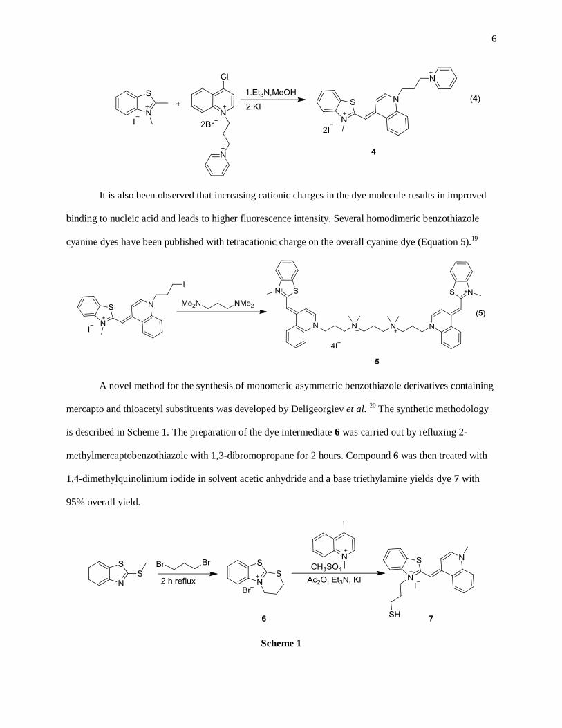
6
It is also been observed that increasing cationic charges in the dye molecule results in improved
binding to nucleic acid and leads to higher fluorescence intensity. Several homodimeric benzothiazole
cyanine dyes have been published with tetracationic charge on the overall cyanine dye (Equation 5).19
A novel method for the synthesis of monomeric asymmetric benzothiazole derivatives containing
mercapto and thioacetyl substituents was developed by Deligeorgiev et al. 20
The synthetic methodology
is described in Scheme 1. The preparation of the dye intermediate 6 was carried out by refluxing 2-
methylmercaptobenzothiazole with 1,3-dibromopropane for 2 hours. Compound 6 was then treated with
1,4-dimethylquinolinium iodide in solvent acetic anhydride and a base triethylamine yields dye 7 with
95% overall yield.
Scheme 1

7
A new method for the synthesis of benzothiazole monocyanines was developed making use of
solid phase resin support.21
The schematic representation of the solid phase synthesis is described in the
Scheme 2. The first step of this synthetic method involves the attachment of 2-mercaptobenzothiazole to
the merrifield resin. This merrifield resin attached benzothiazole was then reacted in the next step with 4-
methylbenzenesulfonate to yield N-methyl benzothiazolium salt 8. In the final step the condensation of
the N-methylbenzothiazolium salt 8 with carboxylic acid derivative of lepidine afforded cyanine dye 9.
Traceless cleavage of merrifield resin was carried out during the course of the synthesis of the cyanine
dye. Presence of Carboxylic acid side chain attached to the quinoline ring makes dye available for post
synthetic modification. Attaching amino acid to the carboxylic end group and modifying it with folic acid
can be used to identify cancer cells that have extra folacin acceptor on the cellular surface. The most im-
portant feature of the solid phase synthesis is the elimination of the lengthy purification step involved
with the conventional liquid phase method which allows the use of synthesized dyes directly for the bio-
logical screening and elimination of the use of toxic catalyst provides greener approach.
Scheme 2
8
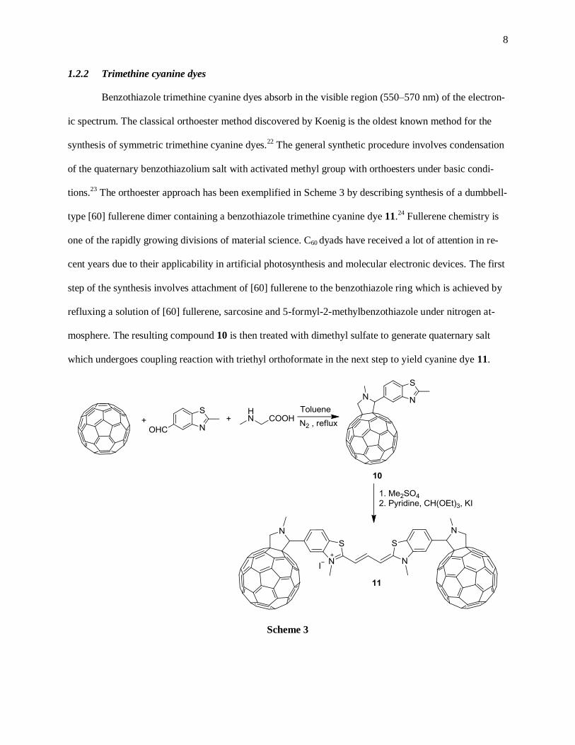
1.2.2 Trimethine cyanine dyes
Benzothiazole trimethine cyanine dyes absorb in the visible region (550–570 nm) of the electron-
ic spectrum. The classical orthoester method discovered by Koenig is the oldest known method for the
synthesis of symmetric trimethine cyanine dyes.22
The general synthetic procedure involves condensation
of the quaternary benzothiazolium salt with activated methyl group with orthoesters under basic condi-
tions.23
The orthoester approach has been exemplified in Scheme 3 by describing synthesis of a dumbbell-
type [60] fullerene dimer containing a benzothiazole trimethine cyanine dye 11.24
Fullerene chemistry is
one of the rapidly growing divisions of material science. C60 dyads have received a lot of attention in re-
cent years due to their applicability in artificial photosynthesis and molecular electronic devices. The first
step of the synthesis involves attachment of [60] fullerene to the benzothiazole ring which is achieved by
refluxing a solution of [60] fullerene, sarcosine and 5-formyl-2-methylbenzothiazole under nitrogen at-
mosphere. The resulting compound 10 is then treated with dimethyl sulfate to generate quaternary salt
which undergoes coupling reaction with triethyl orthoformate in the next step to yield cyanine dye 11.
Scheme 3

9
A new class of phosphonate labeled trimethine benzothiazole cyanine dyes has been published
using orthoester approach.25
Synthetic methodology for the phosphonate labeled cyanine dye starts with
the synthesis of compound 12 which is obtained by refluxing 2-methylbenzothiazole with diethyl 3-
bromopropylphosphonate in acetonitrile. Phosphonate salt 12 was then converted to cyanine dye 14 using
triethyl orthoformate under basic conditions. Conversion of phosphonate salt to phosphonic acid salt was
achieved by acidic hydrolysis which is used in the further step to synthesize dye 13. The presence of
phosphonic acid side chain improves water solubility and helps to increase complexing ability of the
benzothiazole cyanine dye.
Scheme 4
Another approach for the synthesis of benzothiazole trimethine cyanine dye involves usage of
N,N-diphenylformamidine or iodoform as a condensing agent. This synthetic method allows synthesis of
asymmetrical cyanine dyes as well.26
Scheme 5 describes the use of N,N-diphenylformamidine for the
synthesis of benzothiazole trimethine cyanine dye. Alkylated benzoxazole derivative is first treated with
N,N-diphenylformamidine in presence of acetic anhydride to obtain half dye 15. Compound 15 in the next
step is condensed with 2, 3-dimethylbenozthiazolium iodide to yield final asymmetric dye 16. Different
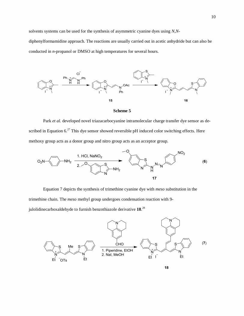
10
solvents systems can be used for the synthesis of asymmetric cyanine dyes using N,N-
diphenylformamidine approach. The reactions are usually carried out in acetic anhydride but can also be
conducted in n-propanol or DMSO at high temperatures for several hours.
Scheme 5
Park et al. developed novel triazacarbocyanine intramolecular charge transfer dye sensor as de-
scribed in Equation 6.27
This dye sensor showed reversible pH induced color switching effects. Here
methoxy group acts as a donor group and nitro group acts as an acceptor group.
Equation 7 depicts the synthesis of trimethine cyanine dye with meso substitution in the
trimethine chain. The meso methyl group undergoes condensation reaction with 9-
julolidinecarboxaldehyde to furnish benzothiazole derivative 18.28

11
1.2.3 Pentamethine cyanine dyes
Pentamethine cyanine dyes absorb generally in the visible region (650–670 nm) but fluoresce in
the near-infrared region (690–710 nm) of the electronic spectrum. The general synthetic procedure in-
volves use of malonaldehyde bis(phenylimine)monohydrochloride under basic conditions as exemplified
in Equation 8.29, 30
Mehranpour et al. modified malonaldehyde linker to form γ-substituted pentamethine cyanine
dyes as shown in Scheme 6.31
The new γ-substituted pentamethine cyanine dyes were synthesized using
three step procedure. First step involves synthesis of N-pyridinium acetic acid salt 20 by reaction between
3, 5-dimethylpyridine with bromoacetic acid. In the second step compound 20 was formylated in pres-
ence of N,N-dimethylformamide and phosphorus oxychloride to obtain compound 21. The synthesis of
cyanine dye 22 was carried out by condensation reaction between compound 21 and 2,3-dimethyl
benzothiazole tetrafluoroborate in presence of sterically hindered hunig base. The presence of the pyridine
ring in the pentamethine chain makes the dye more rigid and the cationic charge makes it water soluble.
Scheme 6
12
Yagupolskii et al. developed a benzothiazole cyanines with fluorine substituted pentamethine
chain as described in Scheme 7.32
Stille coupling reaction was employed in the first step for coupling of 2-
iodobenzothiazole with fluorinated polymethine chain in presence of copper iodide and
tetrakis(triphenylphosphine) palladium (0) which afforded compound 23. Compound 23 was alkylated by
reaction with methyl iodide in presence of silver tetrafluoroborate to obtain compound 24 which on reac-
tion with 2-fluoromethylbenzothiazole methylene base yields cyclic compound 25. Compound 25 was
subjected to alkylation reaction which on further reaction with p-dimethylaminotoluidine provides
pentamethine cyanine dye 26.
Scheme 7
The synthesis of asymmetrical pentamethine benzothiazole cyanine dyes can be furnished utiliz-
ing aldehyde analogue of the benzothiazole moiety.33
Meguellati et al. adapted aldehyde analogue strate-
gy to synthesize imino pentamethine cyanine dye as depicted in Scheme 8. These dyes remain stable in
solid state where as in solution within an hour regenerate aldehyde and imine.34
Compound 29 was syn-
thesized by reacting malonaldehyde bis(phenylimine)monohydrochloride with 1,2,3,3-
tetramethylindolenine iodide salt. Basic hydrolysis of the activated hemicyanine derivative 27 in sodium
hydroxide solution afforded compound 28 in excellent yield. In the last step compound 28 was treated

13
with N-methyl-2-amino benzothiazolium salt to yield cyanine dye 29. The introduction of the imine bond
in the polymethine chain makes dye formation reversible owing to the external reaction conditions.
Scheme 8
Losytskyy et al. synthesized a series of benzothiazole fluorophores with cyclohexene and
cyclopentene ring substitution in the pentamethine chain.35
These synthesized compounds were studied as
fluorescent probes for nucleic acid and proteins and hypothesized to act as nucleic acid groove binders.
Synthesis of pentamethine cyanine dyes 34, 35 containing cyclopentene group in the pentmethine chain
was furnished by condensing 2-methylcylcopentane-1,3-diones 30, 31 with quaternary salts of 2-
methylbenzothiazolium 32, 33 at 210 0C under basic conditions of triethylamine (Equation 9).
Synthesis of pentamethine dyes with cyclohexene ring in the pentamethine chain was achieved by con-
densation of quaternary salts of benzothiazole containing active methylene group with the 1,5-dimethoxy-
1,4-cyclohexadienes or with 1,3-diethoxy-5,5-dimethyl- or 1,3-diethoxy-2,5,5-trimethyl-1,3-

14
cyclohexadienes either by melting the two compounds or by heating in benzonitrile at 120-130 0C as
shown in Equation 10.
1.2.4 Heptamethine cyanine dyes
Heptamethine cyanine dyes typically absorb and fluoresce in the near-infrared region (750 – 1100
nm) of the electronic spectrum. The general synthetic method for the synthesis of benzothiazole
heptacyanines with flexible polymethine chain involves use of glutaconaldehyde dianil
monohydrochloride as a heptamethine linker.36
As described in Equation 11, various benzothiazole
heptamethine derivatives have been synthesized varying the substituent attached to the benzothiazole ni-
trogen.37, 38
(Equation 11)
Another class of benzothiazole heptamethine dyes has been published containing five- or six-
membered cyclic system as a part of heptamethine linker making use of Vilsmeier-Haack reagent.39, 40
The presence of carbocyclic ring in the heptamethine chain makes dye more rigid which helps to increase
fluorescence quantum yield and decrease the aggregation of the dye. Vilsmeier-Haack reagents are de-
rived from cyclopentanone or cyclohexanone in two step procedure as described in Equation 12.

15
General methodology for the synthesis of heptamethine cyanines with rigid polymethine chain
involves condensation of a Vilsmeier-Haack reagents 38 or 39 and a heterocycle with activated methyl
group in ethanol in presence of sodium acetate catalyst.41
Another approach for the synthesis of
heptamethine cyanine dyes involves refluxing a quaternary salt of benzothiazole and bis aldehyde in
butanol:benzene solvent mixture with continuous removal of the water from the reaction mixture using
Dean –Stark apparatus.42
The major advantage of using this method of synthesis is it eliminates usage of
catalyst and provides final dye 40 in high yields (Equation 13).
1.3 Applications of benzothiazole cyanine dyes
The field of cyanine dyes is ever expanding field of science. Since the discovery of cyanine dyes,
their application was limited to photographic sensitizers.43
Other than photography, cyanine dyes also
finds applications in several other fields such as recording media44
, laser materials45
, solar cells46
47
48
semiconductors.49
50
Cyanine dyes have always been regarded as good silver halide photography sensitizers. A silver
halide solution possesses limited sensitivity up to 550 nm but when it is doped with dye its sensitivity ex-
tends up to 650 nm and sometimes even into near-infrared region of the electronic spectrum. Sensitizer
dyes show bathochromic shift when added to the silver halide solution by up to 20-80 nm. Benzothiazole
dyes 41 and 42 are used as photographic sensitizers.51

16
Another important application of benzothiazole dyes is its use in recording media. Most of the
CD-R uses indolenine based cyanine dyes as recording dye. With the discovery of the DVD-R which re-
quires shorter wavelength laser beam, benzothiazole trimethine cyanine dye was considered as better dye
to get appropriate reflection and modulation of the DVD-R. Another advantage of using trimethine cya-
nine dye is it shows good chemical and photostability. Typically benzothiazole cyanine dyes such as
compound 43 are used for the purpose of recording media.52
Dye sensitized solar cells have attracted tremendous attention since last decade. Cyanine dyes
substituted heavy metal based complexes as sensitizers in these solar cells. Advantages of using cyanine
dyes as solar cell sensitizers include cost efficiency and easy recycling of solar cells. Several
benzothiazole cyanine dyes such as compound 44 have been synthesized and studied for their efficiency
towards sensitization of solar cell.53

17
Optical imaging is one of the widely recognized and emerging areas of research which focuses
mainly on studying molecular and metabolic functions in vivo. In the past few years, cyanine dyes have
emerged as optical contrast agents for image guided surgery owing to their appealing photophysical prop-
erties. Compared to alternate imaging methods such as positron emission tomography (PET), this optical
imaging technique using fluorescent probe is relatively harmless, highly sensitive and non-invasive. Dif-
ferent imaging modalities such as magnetic resonance imaging (MRI), computed tomography (CT) make
use of exogenously applied contrast agents and mainly produce signals particular to the disease tissue thus
allowing to monitor physiological and molecular conditions distinctive of specific disease state. The ma-
jor obstacles to optical imaging involves strong absorption by hemoglobin, deoxyhemoglobin, water, and
lipids in the mid-visible region of the electronic spectrum which eventually affects the penetration of the
light inside tissue and hence the obtained information reflects only the superficial characterization. Cya-
nine dyes used for optical imaging particularly absorb and fluoresce in the near-infrared region of the
electronic spectrum. Indocyanine green (ICG) was the first cyanine dye approved by FDA for imaging
techniques contains the benz[e]indolium heterocyclic moiety as a part of the dye structure.
Figure 3. Structure of Indocyanine Green (ICG)
There are certain drawbacks associated with ICG as a contrast agent such as lack of target speci-
ficity, low renal clearance of the dye (high tissue retention) and instability in the aqueous media.54
In the
past few years, more and more research has focused on correcting the problems associated with ICG. Re-
searchers chemically modified the indolenine moiety to increase hydrophilicity.55
In another approach,
ICG was bound to either peptides or antibodies to make the dye target specific.56
As most of the research

18
is associated with already developed contrast optic agent ICG, very few publications concerning in vivo
imaging are associated with other cyanine dye structures. Frangioni and Henary recently developed a
zwitterionic near-infrared fluorophore to be used as a probe for image-guided surgery.54
In this thesis we focus on developing hepta- and pentamethine benzothiazole cyanine dyes as im-
aging agents. The bio-distribution study was conducted in collaboration with Drs. Frangioni and Choi,
Beth Israel Deaconess Medical Center, Boston, MA, USA.
2 RESULTS AND DISCUSSIONS
2.1 Aim of the current study
Most of the published papers report radio labeled benzothiazole compounds as contrast agents
which are used in conjunction with Positron Emission Tomography (PET) imaging technology.57, 58
PET
provides good sensitivity for deep tissue imaging but it is bound by several limitations such as low spatial
resolution, patient exposure to radioactive substances, high instrumentation cost and data acquisition rate
is relatively low.59
Optical imaging is emerging as a quick, non-radioactive, cost efficient and high resolu-
tion imaging technique. In the standard imaging technique, the contrast signal is generated by irradiating
light on the tissue and collection of the emitted fluorescent light at a higher wavelength with optical sepa-
ration of the excitation wavelength. The near-infrared region of the electromagnetic spectrum furnishes
two basic advantages which include reduction in the background fluorescence arising from biological tis-
sues and an increase in the light penetration depth. As tissues generate very little near- infrared signal,
external near-infrared absorbing agent is administered for in vivo studies. The fundamental problems as-
sociated with this optical imaging technique include non-specificity of the contrast agents towards body
tissues and several physiological issues regarding tissue retention causing increased background signal.
Designing near-infrared absorbing optical contrast agents possessing organ selectivity and total
body clearance has always remained a challenge to scientists. There is not much literature available for
the applicability of the benzothiazole dyes towards near-infrared in vivo imaging studies; therefore, we

19
decided to design and synthesize various near-infrared dyes having the benzothiazole core structure. In
the first chapter, the synthesis of heptamethine dyes is discussed as a part of an ongoing project. The se-
cond chapter concerns structural optimization of the pentamethine cyanine fluorophores for brain target-
ing application.
2.1.1 Synthesis of novel heptamethine benzothiazole cyanine dyes
Benzothiazole heptamethine cyanine dyes generally show absorption and fluorescence in the
750–850 nm region of the electromagnetic spectrum. Previously, our group synthesized a series of
heptamethine cyanine dyes utilizing indolenine heterocylic moiety with a rigid cyclohexene ring as a part
of the heptamethine linker. This class of compound demonstrated promising in vivo near-infrared imaging
performance.54
Currently, our laboratory is investigating the synthesis of various heptamethine cyanine
dyes with flexible polymethine linker for in vivo imaging applicability, and additional analogs were pre-
pared using the benzothiazole heterocyclic structure. Commercially available 3-bromopropyltrimethyl
ammonium bromide has been extensively employed for the synthesis of various heptamethine cyanine
dyes with various heterocycles in our lab for the ongoing project. Additional bromopropyl salts with
quaternized nitrogen were synthesized with pyridinium, methylpyrrolidinium and methylmorpholinium
moieties with slight modification to published procedure23
as described in Scheme 9.
Scheme 9

20
Commercially available 1,3-dibromopropane was treated with pyridine at 45 0C in acetonitrile for
eight hours to afford compound 45. Synthesis of compound 46 was furnished by reacting 1,3-
dibromopropane with N-methylpyrrolidine for two hours in ice cold conditions. N-methylmorpholine on
stirring with 1,3-dibromopropane at room temperature for eight hours yielded compound 47. Compounds
45–47 were synthesized in 45-55 % overall yield. Bromopropyl quaternary salts 45–47 and commercially
available 3-bromopropyl trimethylammonium bromide were then melted with 2-methylbenzothiazole in
sealed tube to furnish compounds 48–51 as described in Scheme 10 with slight modification of the previ-
ously published procedure.23
The quaternized benzothiazole compounds 48–51 were condensed with
glutaconaldehyde dianil monohydrochloride under basic conditions to yield tricationic heptamethine cya-
nine dyes 52–55 which were purified on reversed phase column using acetone:water solvent system. The
overall yields of the purified dyes 52–55 ranged between 60 - 65%.
Scheme 10
The basic principle behind synthesizing cyanine dyes with multiple cationic charges was focused
on increasing the solubility of the cyanine dye in water which is expected to result in a more appealing

21
distribution of the heptamethine cyanine dyes in vivo and should help in obtaining increased renal clear-
ance. The predicted physiochemical properties for the compounds 52–55 are listed in Figure 4.
Figure 4. Distribution coefficient data of heptamethine benzothiazole cyanine dyes predicted using
ChemAxon software
In past few years, total polar surface area has been considered as a major factor for target-oriented
imaging studies.60
Modification of the end group on the propyl chain attached to the benzothiazole nitro-
gen showed simultaneous changes in the total polar surface area as well as in distribution coefficient (Log
D) values as shown in Figure 4. The Log D values of the synthesized cyanine dyes 52–55 were predicted
to be less than zero suggesting compounds possess high hydrophilicity. The imaging studies of the syn-
thesized heptamethine cyanine dyes are still in progress. Following is representation of the absorp-
tion/emission spectrum for the heptamethine cyanine dye 52. This dye absorbs at 760 nm in the electro-
magnetic spectrum and fluoresces at 791 nm in the emission spectrum (excitation wavelength 660 nm), as
shown below in Figure 5.

22
Figure 5. Absorption and emission spectrum of compound 52
The rest of the synthesized compounds 53–55 showed absorption/emission wavelength maxima within
the same range as that of the compound 52 in the electromagnetic spectrum. Compounds 52–55 were
characterized by 1H and
13C NMR, liquid chromatography and mass spectrometry analysis. Compound 54
was chosen for complete characterization and analysis.
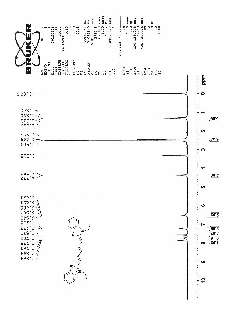
1H NMR spectrum of the compound 54 shows two doublets at 7.99 ppm and 7.88 ppm corre-
sponding to the benzothiazole aromatic protons nearer to the sulfur and nitrogen atoms Hb. A multiplet
formed between 7.58 ppm ~ 7.37 ppm corresponds to the protons from the heptamethine chain Hd, Hf and
the benzothiazole ring protons Ha. The protons Hc and He in the cationic polymethine chain produce a
characteristic doublet and a triplet at 6.92 ppm and 6.56 ppm respectively. J coupling constant of 12.8 Hz
for these protons suggest the all trans character of the heptamethine chain. Broad doublets at 4.39 ppm
and 3.73 ppm correspond to the four methylene protons from the propyl chain attached to the nitrogen
atom. The multiplets at 3.55 ppm and 2.12 ppm represent remaining protons from the propyl chain and
the protons from the pyrrolidine ring. Finally, a sharp singlet at 3.05 ppm corresponds to the N – methyl
group attached to the pyrrolidine ring.

23
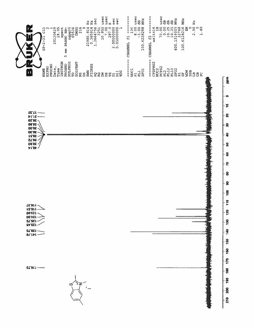
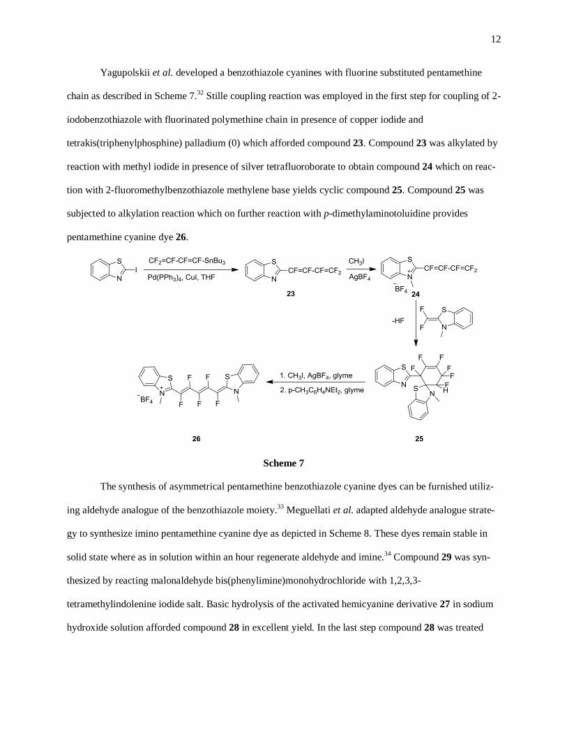
Figure 6. 1H NMR spectrum of compound 54 in DMSO-d6 at 25
0C
13C NMR of the compound 54 shows as expected well resolved 11 peaks in the aromatic region and 6
peaks in the aliphatic region of the spectrum.
24
Figure 7. 13
C NMR spectrum of compound 54 in DMSO-d6 at 25 0C
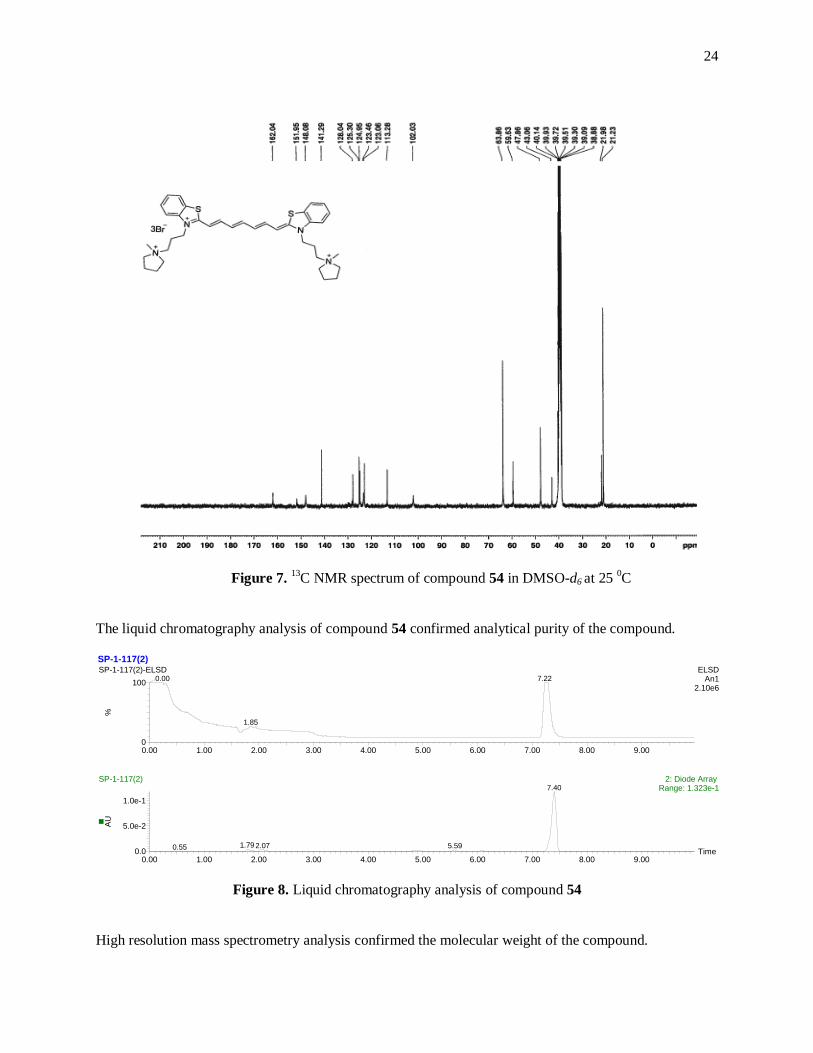
The liquid chromatography analysis of compound 54 confirmed analytical purity of the compound.
Figure 8. Liquid chromatography analysis of compound 54
High resolution mass spectrometry analysis confirmed the molecular weight of the compound.
SP-1-117(2)
Time0.00 1.00 2.00 3.00 4.00 5.00 6.00 7.00 8.00 9.00
AU
0.0
5.0e-2
1.0e-1
0.00 1.00 2.00 3.00 4.00 5.00 6.00 7.00 8.00 9.00
%
0
100
0.00 1.00 2.00 3.00 4.00 5.00 6.00 7.00 8.00 9.00
%
0
100
SP-1-117(2)-ELSD ELSDAn1
2.10e6
7.220.00
1.85
SP-1-117(2)-MS 1: TOF MS ES+ TIC
1.42e5
7.57
SP-1-117(2) 2: Diode Array Range: 1.323e-17.40
1.790.55 2.07 5.59

25
2.1.2 Design and synthesis of novel pentamethine benzothiazole cyanine dyes
Currently, there are not many references associated with cyanine dyes targeting brain tumors.
One of the major challenges associated with tumor identification is developing an agent that differentiates
between tumor and normal cells.61
Introduction of a benzothiazole cyanine dye showing brain tumor im-
aging capability will lay the foundation for synthesis of a new class of compound active towards targeting
the brain in vivo. The aim of the presented work was to synthesize various pentamethine benzothiazole
cyanine dyes and to study their in vivo imaging specificity towards targeting brain tumor.
Thioflavin-T is the benzothiazole containing imaging agent which shows fluorescence enhance-
ment upon binding to aggregates of protein “amyloids”.62, 63
Figure 9. Structure of Thioflavin-T
It has been observed that patients with Alzheimer’s disease have shown accumulation of β -
amyloid fibrils in the brain.64, 65
These fragments are then converted in to the hard plaques. In recent stud-
ies it has been suggested that by imaging these plaques, the severity of the Alzheimer’s disease can be
estimated.9 Thioflavin-T is structurally composed of 6-methylbenzothiazole heterocyclic ring quaternized
at the nitrogen atom by methyl substituent linked to a hydrophobic phenyl ring possessing a
dimethylamino end group. For the current research project, the benzothiazole structure of the Thioflavin-
T was kept as an integral part of the cyanine dye replacing the hydrophobic phenyl group with the
pentamethine chain. This feature allowed benzothiazole cyanine dyes to fluoresce in the near-infrared
region of the electronic spectrum. It was hypothesized that the structural similarity of the synthesized dyes
towards Thioflavin-T might promote benzothiazole dyes towards targeting the brain.
The blood brain barrier is the physical and biochemical barrier which prevents entry of specific
substances from blood into the cerebrospinal fluid and consequently into central nervous system. There

26
exists ambiguous factors regarding the passage of a substance through the blood brain barrier though it is
believed that compounds furnishing modified Lipinski’s rule of five have highest chances to surpass the
blood brain barrier.60
The Lipinski’s rule suggests that compounds should fulfill the following basic re-
quirements:
1. Partition coefficient should not be more than 5 (Log D < 5)
2. Molecular mass should be less than 500 g/mol
3. hydrogen bond donors ≤ 5
4. hydrogen bond acceptors ≤ 10
5. Total polar surface area should be less than 90 Å2
Considering Thioflavin-T as a lead compound, initial attempts were focused onto the synthesis of
cyanine dyes with methyl substituted benzothiazole aromatic ring in accordance with Lipinski’s rule of
five. Physiochemical predictions are a powerful tool to help expect in vivo efficacy without synthesizing
an extensive library of compounds; using ChemAxon software we designed benzothiazole pentamethine
cyanines and excluded those compounds which significantly deviate from Lipinski’s rule. Commercially
available 2,5-dimethylbenzothiazole was heated at 80 0C with methyl and ethyl iodides. The resulting
crude compounds were crystallized from methanol to afford quaternary benzothiazolium salts 56 and 57
in 76% and 65% yield, respectively. Various alkyl tosylates were heated at 120 0C with 2,6-dimethyl-
benzothiazole to furnish benzothiazolium quaternary salts 58–60 in greater than 60% yield. The
quaternized salts 56–60 in the next step were condensed with malonaldehyde bisphenylimine hydrochlo-
ride in the presence of triethylamine in acetonitrile yielding benzothiazole cyanine dyes 61–65, as depict-
ed in Scheme 11.

27
Scheme 11
The purification of the cyanine dyes 61–65 was performed using solvent precipitation. The crude
dye was dissolved in N,N–dimethylformamide (5 mL) and the pure dye was precipitated out of the solu-
tion by the dropwise addition of diethyl ether (15 mL) leaving all the soluble impurities in solution.
The yields obtained for the synthesized benzothiazole cyanine dyes 61–65 were more than 60% with the
remaining material being starting material, half-converted products and other non-target products.
The synthesized benzothiazole cyanine dyes 61–65 followed the desired parameters of the Lipinski’s rule
of five. The predicted total polar surface area of 7.12 Å2, hydrogen donor/acceptor capability of 0/1 and
the molecular weight less than 500 g/mol are well within range of the Lipinski’s rule. The only parameter
that differed in all these discussed compounds was distribution coefficient (Log D) value, as shown in
Figure 10.

28
Figure 10. Distribution coefficient data of pentamethine benzothiazole cyanine dyes 63-65 predicted us-
ing ChemAxon software
Although the predicted Log D values for the cyanine dyes 61-65 fall well within the Lipinski’s
range, results obtained from bio-distribution studies indicated that none of the dyes exhibited contrast sig-
nal in the brain. It has also been noted that as the length of the side chain attached to the benzothiazole
nitrogen increases, a marked increase in the accumulation of the dye in the liver was observed which can
be attributed to the increasing lipophilic nature of the dye referring to their elevated Log D values.
The next approach towards synthesizing benzothiazole dyes was focused on removing the methyl substit-
uent attached to the benzothiazole aromatic ring taking into account the lipophilicity factor associated
with the earlier discussed benzothiazole dyes. It was hypothesized that removal of the methyl substituent
from benzothiazole aromatic ring will help in decreasing the distribution coefficient (Log D) of the cya-
nine dye and this might result in directing the cyanine dye towards brain. It has already been published
that radio labeled benzothiazole compounds without substituent(s) on the benzothiazole aromatic ring can
serve as brain targeting agents.66
Scheme 12 represents the synthetic route used for the synthesis of the
benzothiazole cyanine dyes 71–75.

29
Scheme 12
Synthesis of quaternary salts 66–70 was furnished in more than 50% yield after precipitation from metha-
nol by heating different alkyl halides with 2-methylbenzothiazole under neat conditions. The utilization of
the neat conditions for the synthesis of benzothiazolium salts 66–70 helped to obtain excellent yields in
less reaction time than conventional solvent based chemistry.67, 68
Though compound 70 showed a single
spot on the TLC analysis, it had displayed multiple peaks in aromatic as well as in aliphatic region of the
1H NMR spectrum. This compound was then used as crude in the dye synthesis step. In the next step, qua-
ternary benzothiazolium salts 66–70 were treated with malonaldehyde bisphenylimine hydrochloride un-
der basic conditions to furnish benzothiazole dyes 71–75. The crude dyes were purified using the afore-
mentioned precipitation method from N,N-dimethylformamide by addition of diethyl ether (20 mL, 1:4)
affording pure cyanine dyes in more than 60% yield. The predicted Log D values for the cyanine dyes 71-
75 are as shown in Figure 11.
Figure 11. Distribution coefficient data of pentamethine benzothiazole cyanine dyes 71-73 predicted us-
ing ChemAxon software

30
By comparing the Log D values for the benzothiazole dyes from Figures 10 and 11, it can be observed
that the removal of the methyl substituent from the benzothiazole aromatic ring lowered the Log D value
substantially. Bio-distribution data revealed that compound 71 displayed no signal in the brain tumor
though a signal in the pituitary gland was remarkably bright. It should be noted that cyanine dye 71 has
the lowest Log D value as per the Figures 10 and 11. Benzothiazole cyanine dyes 72 and 73 displayed no
contrast signal in the brain. This bio-distribution pattern for compounds 72 and 73 can be attributed to
their higher Log D values as the predicted total polar surface area and hydrogen donor/acceptor capability
remained the same as that of the earlier discussed benzothiazole dyes 61–65. Bio-distribution results ob-
tained for benzothiazole cyanine dyes 61–65 and 71–75 suggested that lowering the Log D value would
definitely help to direct the benzothiazole dyes towards the brain.
Klunk et al. developed a radio labeled 6-hydroxybenzothiazole derivative of Thioflavin-T which acted as
better contrast agent that more effectively crossed the blood brain barrier.9
Figure 12. Pittsburgh compound B9
According to study reported by Chopra, 6-hydroxy derivatives of benzothiazole proved to be superior im-
aging agents over 4–,5– and 7–hydroxy derivatives.69
Further approach towards lowering the Log D value
of benzothiazole cyanine dyes was achieved by substituting hydroxy group in the benzothiazole aromatic
ring. Scheme 13 describes the synthetic procedure used for the synthesis of various hydroxy substituted
benzothiazole cyanine dyes 81–85. In order to achieve the synthesis of hydroxy substituted benzothiazole
cyanine dyes 81–83, 6-hydroxy-2-methylbenzothiazole was first heated with various alkyl tosylates in
neat conditions to furnish quaternary salts 76–78. This synthetic approach provided more than 60% yields
of the quaternary salts. These salts were then reacted with malonaldehyde bisphenylimine hydrochloride
under basic conditions to yield final hydroxyl-substituted benzothiazole dyes 81–83. The pure compounds
were isolated by using precipitation technique making use of the DMF:diethyl ether solvent system (20

31
mL, 1:4). Generally, hydroxy substituted cyanine dyes show limited solubility and the tosylate counter
anion was utilized to increase the overall solubility of the cyanine dye.
Scheme 13
It is evident from Figures 10, 11 and 13 that different substitutions on the benzothiazole ring affected the
Log D values significantly. The highest Log D values were observed for methyl substituted benzothiazole
cyanine dyes ranging between 2.8 to 4.5. The predicted Log D values of hydroxy substituted
benzothiazole cyanine dyes 81–83 however ranged in 1 to 3.
Figure 13. Distribution coefficient data of pentamethine benzothiazole cyanine dyes 81-83 predicted us-ing ChemAxon software
Bio-distribution data obtained for compounds 81–83 revealed that compound 81 displayed increased con-
trast signal in the brain. This improved imaging capability of the cyanine dye 81 can be attributed to low-
ering the Log D value of all of the earlier discussed benzothiazole compounds. The total polar surface
area and hydrogen donor/ acceptor values for dyes 81-83 predicted as 47.58 Å2 and 2/3 respectively. To
determine if lowering the Log D value further could increase the brain targeting efficacy of the dye, we
introduced the quaternary ammonium side chain on the benzothiazole cyanine dye 84 to increase the ap-

32
pealing imaging characteristics by synthesizing a more hydrophilic dye. The cyanine dye 84 was synthe-
sized in 2 steps beginning with fusing 6-hydroxy-2-methylbenzothiazole with 3-bromopropyltrimethyl
ammonium bromide at 140 0C for 4 hours. In the subsequent step, the benzothiazolium salt was subjected
for the dye synthesis as shown in Scheme 13. The yields obtained for compounds 79 and 84 were 52%
and 54%, respectively. The Log D value of compound 84 was predicted to be -6.28 and confirms its high-
ly hydrophilic nature. This dye failed to target the brain though hydrophilicity of the dye aided in better
renal clearance. The total polar surface area and hydrogen donor/acceptor capability remained the same
for this dye as of compounds 81–83. It was then believed that the better brain targeting benzothiazole
cyanine dye should possess Log D value less than +1.00 but it should not be as low as that for compound
84 keeping 6-hydroxybenzothiazole as a base structure. As described earlier, Thioflavin-T possesses a
tertiary amine group in the structure. It was then assumed that incorporating the tertiary amine substituent
in the existing benzothiazole model structure would definitely help to attain improved contrast signal in
the brain. This task was achieved by modifying the side chain attached to compound 85 to a tertiary
amine group. The quaternary salt 80 was synthesized by fusing 6-hydroxy-2-methylbenzothiazole with 2-
bromo-N,N-dimethylethanamine hydrobromide at 150 0C for 4 hours. The crude solid on precipitation
furnished compound 80 in a 47% yield as the hydrobromide salt. In the next step, compound 80 was
treated with malonaldehyde bisphenylimine hydrochloride and triethylamine to afford final dye 85. Cya-
nine dye 85 was purified by reversed phase column chromatography using acetone:water solvent system
in 53% yield. The final dye was obtained in the tertiary amine form which was confirmed by various
spectroscopic methods.

33
Figure 14. Distribution coefficient data of pentamethine benzothiazole cyanine dyes 84 and 85 predicted
ChemAxon software
Replacing the quaternary ammonium group to a tertiary amine and removing one carbon atom from the
propyl side chain resulted in lowering the Log D value to -0.35 as seen in compound 85(Figure 14). The
total polar surface area and hydrogen donor/acceptor capability for compound 85 predicted to be highest
when compared to all of the earlier discussed pentamethine benzothiazole cyanine dyes as 54.06 Å2 and
2/5 respectively. As hypothesized earlier, lowering the Log D value for compound 85 helped to observe a
significant change towards the brain targeting ability of the dye. The in vivo imaging behavior of the most
promising compounds (71, 81 and 85) out of the whole set of synthesized benzothiazole pentamethine
cyanine dyes has been summarized in the proceeding chapter concerning the bio-distribution and target-
ing study.
The comprehensive synthetic procedure for all the synthesized compounds 45–85 has been described in
the experimental section and respective spectral data has been reported in the appendix. Benzothiazole
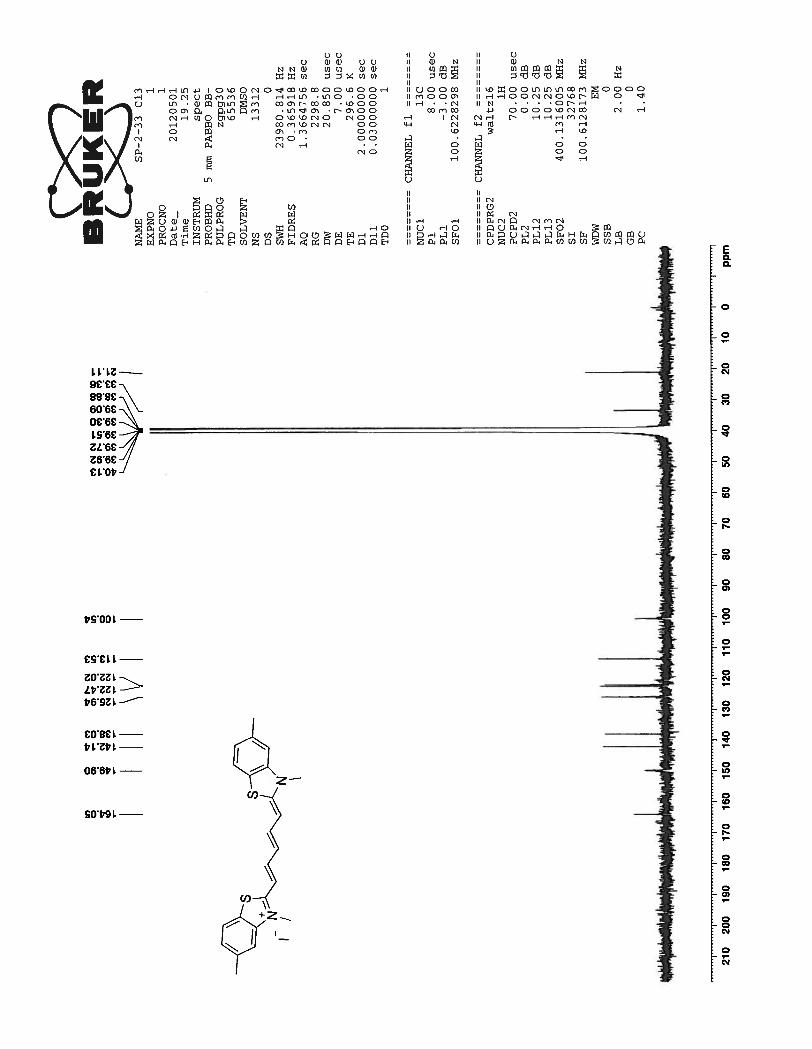
cyanine dye 73 was chosen for complete characterization analysis.
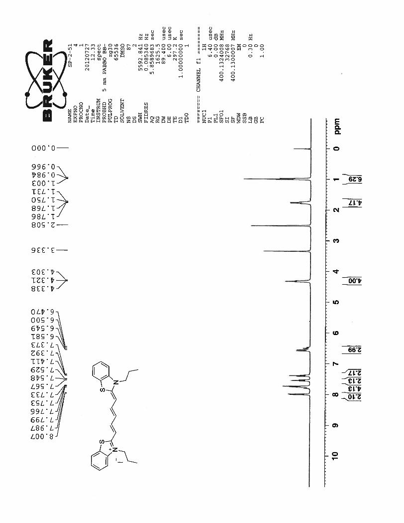
The 1H NMR of compound 73 depicts a total of 10 signals, seven in the aromatic region and three
in the aliphatic region. This suggests that compound 73 has a symmetric chromophoric structure. Dou-
blets at 7.99 ppm and 7.74 ppm correspond to the protons Hb of the benzothiazole aromatic ring. Two tri-
plets at 7.54 ppm and 7.39 ppm are related to benzothiazole aromatic ring protons Ha. The doublets of the
benzothiazole aromatic ring are more downfield than triplets due to the deshielding effect caused by ni-
trogen and sulfur atoms next to them. Three peaks were expected from the pentamethine chain. The signal

34
for protons Hd should be a triplet which is merged with the doublet formed by benzothiazole proton Hb at
7.79 ppm. The next multiplet formed between 6.58 ppm ~ 6.47 ppm can be attributed to the protons Hc
and He from pentamethine bridge. Although proton peaks in the above discussed multiplet region are not
well resolved, it still can be seen that J coupling constant for the doublet can be calculated as 12.8 Hz.
This high value of J coupling constant suggests that the pentamethine bridge adopts an entirely trans con-
formation. A triplet formed at 4.32 ppm corresponds to methylene proton of the propyl chain nearer to the
nitrogen atom of the benzothiazole ring. The next quartet at 1.76 ppm corresponds to other methylene
protons of the propyl chain. Finally triplet at 0.98 ppm corresponds to the methyl protons of the propyl
chain which are highly shielded protons in the molecule and hence shows the most upfield signal for the
compound 73.

35
Figure 15. 1H NMR spectrum of compound 73 in DMSO-d6 at 23
0C
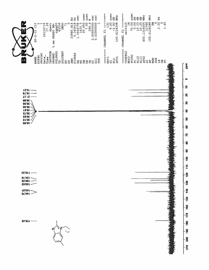
13C spectrum for the same compound showed well resolved 13 peaks as expected. Three peaks at
47.26 ppm, 20.76 ppm and 10.83 ppm correspond to the propyl side chain attached to the benzothiazole
nitrogen atom. All other remaining signals between 163.54 to 100.43 correspond to the carbon atoms
from the benzothiazole ring and the pentamethine bridge.

36
Figure 16. 13
C NMR spectrum of compound 73 in DMSO-d6 at 23 0C
Finally, compound purity was determined performing liquid chromatography-mass spectrometry analysis
using a reversed phase C18 column with water/acetonitrile gradient elution. The detectors used for liquid
chromatography were Sedex 75 ELSD and absorbance detector at wavelength 650 nm. The well resolved
single peaks at 10.40 min and 10.43 min on the either of the spectrum ensures that the sample is analyti-
cally pure as shown in Figure 17. High resolution mass spectrometry analysis confirmed the molecular
weight of the compound.

37
Figure 17. Liquid chromatography analysis of compound 73
2.2 Bio-distribution and targeting study
The biodistribution and targeting study of the benzothiazole cyanine dyes was conducted by Drs.
Hak Soo Choi and John Frangioni at Beth Israel Deaconess Medical Center (BIDMC), Harvard Medical
School in Boston, MA.
Brain Tumor Animal Models:
Neurosphere cells from S100ß-verbB;p53−/− or S100ß-verbB;p53+/− mouse brain tumors were obtained
from Dr. Kyuson Yun (Jackson Laboratory, Bar Harbor, ME) and cultured as tumorspheres in modified
DME/F-12 supplemented with 1% B27 (Invitrogen) and 1% Penicillin/Streptomycin. Animals were
housed in an AAALAC-certified facility, and all animal studies were performed under the supervision of
Beth Israel Deaconess Medical Center’s Institutional Animal Care and Use Committee (IACUC) in ac-
cordance with approved institutional protocol #155-2008. For the tumor targeting study, 6-week-old (20-
30 g) male C57BL/6 mice were purchased from Taconic Farms (Germantown, NY), and the mice were
anesthetized using ketamine/xylazine in a 100:10 mg/kg ratio. 3 × 10⁵ neurosphere cells resuspended in 3
µL of fresh medium were delivered 3 mm deep into the right side of midline slowly (0.2 µL/min) via
guidance from a stereotactic frame (Harvard Apparatus, Holliston, MA). Control mice were implanted
SP-2-51(2)
Time0.00 2.00 4.00 6.00 8.00 10.00 12.00 14.00 16.00 18.00
%
0
100
0.00 2.00 4.00 6.00 8.00 10.00 12.00 14.00 16.00 18.00
AU
0.0
5.0e-1
1.0
SP-2-51(2) 3: Diode Array Range: 1.34710.40
SP-2-51(2) ELSDAn1
1.69e6
10.43

38
with 3 µL of medium without cells to allow similar surgical procedures to be performed on the tumor-
bearing and non-tumor-bearing mice. Mice formed reasonable sized tumors in 1-2 weeks post-
inoculation.
Intraoperative NIR Fluorescence Imaging System:
The dual-NIR channel FLARE™ imaging system has been described in detail previously.70, 71
The
FLARE™ imaging system is capable of visualizing surgical anatomy along with two independent chan-
nels of NIR fluorescence, where the first NIR fluorescence channel can be used for highlighting brain tu-
mors, while the second NIR channel can be used for visualizing blood vessels and/or normal nervous tis-
sues. In this study, 670 nm excitation and 760 nm excitation fluence rates used were 4.0 and 11.0
mW/cm2, respectively, with white light (400 to 650 nm) at 40,000 lx. Color video and 2 independent
channels (700 nm and 800 nm) of NIR fluorescence images were acquired simultaneously with custom
FLARE software at rates up to 15 Hz over a 15 cm diameter field of view (FOV). The imaging system
was positioned at a distance of 18 inches from the surgical field. A custom filter set (Chroma Technology
Corporation, Brattleboro, VT) composed of a 650 ± 22 nm excitation filter, a 680 nm dichroic mirror, and
a 710 ± 25 nm emission filter were used.
Biodistribution and Targeting using Optical Fluorescence Imaging:
The in vivo behavior of the benzothiazole pentamethine cyanine dyes was significantly different
depending upon the physiochemical properties. Compound 71 showed high affinity for the pituitary gland
(arrowhead in Figure 18) 4 hr after a single IV injection, but almost no signal was detected in the glioma
(brain tumor) as shown resected in Figure 18 (bottom left). Starting with this lipophilic and cationic
fluorophore (Log D at pH 7.4 = 1.8), we modified the alkyl chain length and functional moieties to have
higher hydrophilicity and lowering lipophilicity and better blood-brain barrier (BBB) penetration. Alt-
hough compound 81 has lower Log D value (1.18) compared to compound 71, tumor targeting efficiency
was not remarkably improved (Figure 19). Finally compound 85 was synthesized with two hydroxyl
groups to improve hydrophilicity and lowering Log D value at pH 7.4 and two tertiary amine moieties to
improve blood brain barrier (BBB) penetration.

39
As shown in Figure 20, compound 85 efficiently penetrated BBB and targeted to the glioma with
high affinity. However, strong background signals were found in choroid plexus and lateral ventricles that
decrease the tumor specificity of this molecule. Table 2 summarizes the physiochemical properties of
compounds 71, 81 and 85.
Table 2. In silico physicochemical properties of NIR fluorophores calculated using Marvin and JChem calculator plugins (ChemAxon, Budapest, Hungary). Log D = partition coefficient at pH 7.4,
TPSA = total polar surface area.
Compound Mol Weight Formula Log D TPSA H bond
acceptors
H bond
donors
71 363.52 C21H19S2N2 1.80 7.12 1 0
81 395.52 C21H19S2N2O2 1.18 47.58 3 2
85 509.71 C27H33S2N4O2 -0.35 54.06 5 2
A 25 nM sample of targeted fluorophore (compounds 71, 81, 85) was injected intravenously 4 hours prior
to imaging. Shown in Figures 18, 19 and 20 are representative images of in situ biodistribution (top) and
resected consecutive brain tissue after targeting (bottom). Abbreviations used are: Bl, bladder; Du, duo-
denum; In, intestine; Ki, kidneys; Li, liver; Pa, pancreas; and Sp, spleen. Arrowhead = pituitary gland;
arrows = tumor. All NIR fluorescence images have identical exposure times and normalization. Scale bars
= 1 cm.
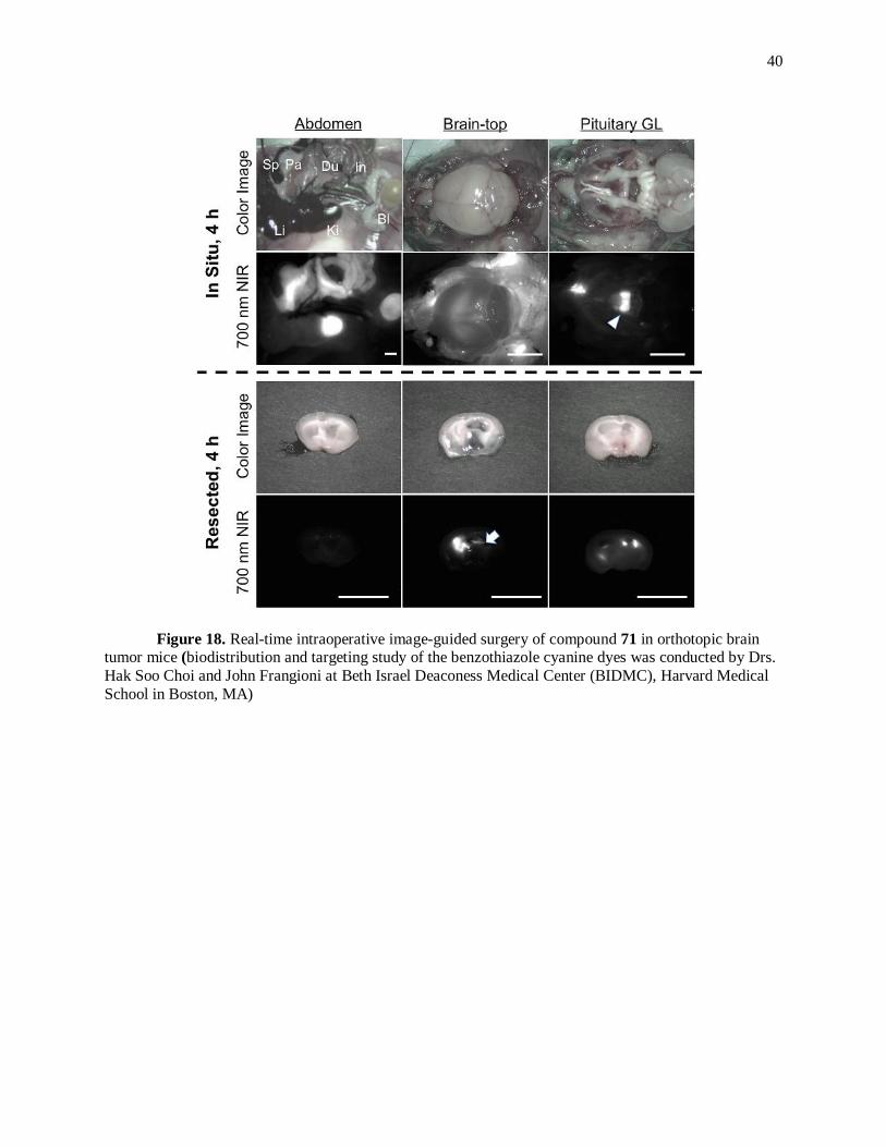
40
Figure 18. Real-time intraoperative image-guided surgery of compound 71 in orthotopic brain tumor mice (biodistribution and targeting study of the benzothiazole cyanine dyes was conducted by Drs.
Hak Soo Choi and John Frangioni at Beth Israel Deaconess Medical Center (BIDMC), Harvard Medical
School in Boston, MA)

41
Figure 19. Real-time intraoperative image-guided surgery of compound 81 in orthotopic brain
tumor mice (biodistribution and targeting study of the benzothiazole cyanine dyes was conducted by Drs.
Hak Soo Choi and John Frangioni at Beth Israel Deaconess Medical Center (BIDMC), Harvard Medical School in Boston, MA)

42
Figure 20. Real-time intraoperative image-guided surgery of compound 85 in orthotopic brain
tumor mice (biodistribution and targeting study of the benzothiazole cyanine dyes was conducted by Drs. Hak Soo Choi and John Frangioni at Beth Israel Deaconess Medical Center (BIDMC), Harvard Medical
School in Boston, MA)

43
3 CONCLUSION
In past few years, cyanine dyes have attracted interest as potential in vivo imaging agents for bi-
omedical applications.72, 73
The development of cyanine dyes containing heterocycles other than
indolenine and benz[e]indolenine would definitely facilitate a new approach towards development of new
class of imaging agents.
In summary, tricationic heptamethine benzothiazole cyanine dyes with various quaternary ammo-
nium side chains and three different sets of pentamethine benzothiazole cyanine dyes which fluoresce
between 690 nm and 790 nm were synthesized in high yield and characterized using various spectroscop-
ic techniques for their optical properties, structure confirmation and analytical purity. The synthesized
cyanine dyes were purified by either precipitation or by reversed phase column chromatography tech-
nique. Furthermore, benzothiazole pentamethine cyanine dyes showed promising in vivo imaging capabil-
ity towards targeting the brain tumor after successfully incorporating the tertiary amine side chain on the
benzothiazole nitrogen. These results might provide a lead for the further development of ideal in vivo
brain tumor targeting agent.

44
4 EXPERIMENTAL
All the chemicals were purchased from commercial sources and were used without further purifi-
cation. 1H and
13C NMR data was collected on a Bruker DPX-400 spectrometer at ambient temperature in
DMSO-d6 or Methanol-d4 with tetramethylsilane as internal standard. Absorption spectra were obtained
on a Cary 5 vis-NIR spectrophotometer. Fluorescence data was collected on Schimadzu RF 5301 PC
spectrofluorometer. Excitation wavelength was set at 660 nm for heptamethine cyanine dyes and 620 nm
for pentamethine cyanine dyes. Slit width was kept at 5 nm for excitation and emission mode. Light
source xenon arc lamp was utilized in the fluorometer. All absorption and emission measurements were
performed in spectrometric grade methanol. Liquid chromatography was operated using Waters 2487 sin-
gle wavelength absorption detector with wavelength set to either 650 or 750 nm. Sedex 75 ELSD was
used as alternate detector in liquid chromatography. Waters Delta-Pak 5 uM 100A 3.9 x 150 mm reversed
phase C18
column was used for the liquid chromatography. High resolution mass spectra were obtained on
either Waters Micromass LCT TOF ES+ Premier Mass Spectrometer or Georgia state mass spectrometry
facility using Waters Q-TOF micro (ESI-Q-TOF) mass spectrometer.
Physiochemical predictions of cyanine dyes:
Hydrogen bond donors and acceptors, total polar surface area and Log D were predicted using
MarvinSketch 5.9 (ChemAxon, Budapest, Hungary). The Log D predictions derived from the calculations
by Viswanadhan,74
Klopman,75
and the PHYSPROP© database at pH 7.4 were averaged. The total polar
surface area estimation is based on the method given by Ertl.76
Procedure for preparation of compound 45:
This compound was synthesized as per the previously published procedure.23
A mixture of pyridine (1eq.)
and 1, 3-dibromopropane (3 eq.) was stirred in acetonitrile at 45 0C for 8 hours. The resulting salt was
then filtered and washed with diethyl ether.
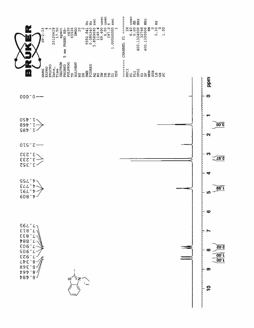
1-(3-bromopropyl)pyridin-1-ium bromide (45): Yield 45%; 1H NMR (DMSO-d6, 400 MHz): δ 9.23 (d,
J = 15.6 Hz, 2H), 8.66 (t, J = 7.6 Hz, 1H), 8.21 (t, J = 6.4 Hz, 2H), 4.81 (t, J = 7.2 Hz, 2H), 3.61 (t, J =

45
6.8 Hz, 2H), 2.55 (q, J = 6.8 Hz, 2H). 13
C NMR (DMSO-d6, 100 MHz): δ 145.72, 145.01, 128.10, 59.37,
33.13, 30.16.
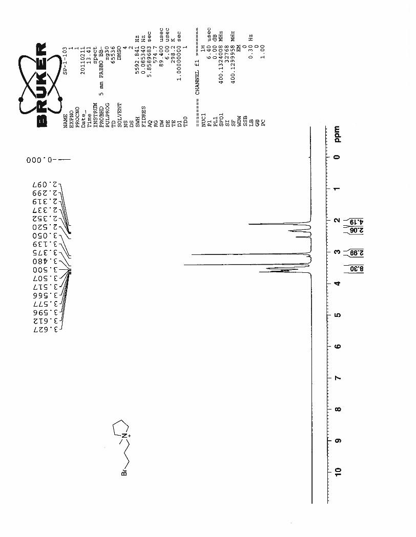
Procedure for preparation of compound 46:
This compound was synthesized with slight modification of the previously published procedure.23
A mix-
ture of N – methylpyrrolidine (1 eq.) and 1, 3-dibromopropane (3 eq.) was stirred at 0 0C for 2 hours. The
precipitate thus formed is filtered and washed with diethyl ether.
1-(3-bromopropyl)-1-methylpyrrolidin-1-ium bromide (46): Yield 55%; 1H NMR (DMSO-d6, 400
MHz): δ 3.62~3.48 (m, 8H), 3.05 (s, 3H), 2.35~2.29 (m, 2H), 2.09 (brs, 4H).13
C NMR (DMSO-d6, 100
MHz): δ 63.62, 61.57, 47.62, 30.75, 26.40, 21.01.
Procedure for preparation of compound 47:
This compound was synthesized with slight modification of the previously published procedure.23
A mix-
ture of N – methylmorpholine (1 eq.) and 1, 3-dibromopropane (3 eq.) was stirred at room temperature for
24 hours. The resulting precipitate was filtered and washed with diethyl ether.
4-(3-bromopropyl)-4-methylmorpholin-4-ium bromide (47): Yield 50%; 1H NMR (DMSO-d6, 400
MHz): δ 3.95 (s, 4H), 3.68 ~ 3.60 (m, 4H), 3.51~3.45 (m, 4H), 3.21 (s, 3H), 2.35~2.28 (m, 2H). 13
C NMR
(DMSO-d6, 100 MHz): δ 62.16, 59.78, 59.15, 46.42, 30.51, 24.37.
General procedure for preparation of compounds 48–51:
These compounds were synthesized with slight modification of the previously published procedure.23
A mixture of 2-methylbenzothiazole and either compounds 45–47 or 3-bromopropyltrimethyl ammonium
bromide was heated in a sealed tube at 130 0C for 18 hours. The resulting sticky solid was washed with
diethyl ether and crude compound was used as is in the further step.
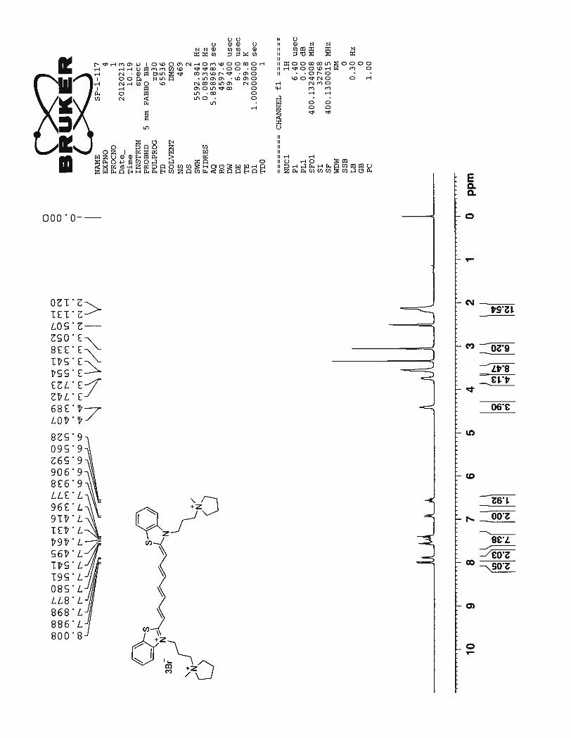
General procedure for synthesis of compounds 52–55:
A mixture of benzothiazolium salt 48–51 (1eq.) and glutaconaldehyde dianil monohydrochloride (0.5 eq.)
was heated in acetonitrile at 45 0C for 4 hours in presence of triethylamine (1eq.) under nitrogen atmos-
phere. The precipitate obtained was filtered and washed with diethyl ether. The target compounds were
isolated by purification using reverse phase column chromatography (acetone:water 5:1).

46
3-(3-(trimethylammonio)propyl)-2-((1E,3E,5E,7Z)-7-(3-(3-
(trimethylammonio)propyl)benzo[d]thiazol-2(3H)-ylidene)hepta-1,3,5-trien-1-yl)benzo[d]thiazol-3-
ium bromide (52): Yield 62% (0.18 g) ; m.p. 205- 207 0C;
1H NMR (DMSO-d6 , 400 MHz) : δ 8.00 (d, J
= 8 Hz, 2H), 7.86 (d, J = 8 Hz, 2H), 7.57~7.37 (m,7H), 6.88 (d, J = 13.2 Hz, 2H), 6.56 (t, J = 12.8 Hz,
2H), 4.39 (brs, 4H), 3.75 (brt, J1 = 6.8 Hz, J2 = 8 Hz, 4H), 3.13 (s, 18H), 2.17 (brs, 4H). 13
C NMR
(DMSO-d6, 100 MHz) : δ 162.05, 152.01, 148.16, 141.28, 128.09, 125.34, 125.00, 123.56, 123.10,
113.25, 102.00, 62.07, 52.55, 43.00, 21.25. ESI-HRMS m/z: calc. for (C33H45S2N4Br)2+
640.2269, found
640.2264. λabs = 760 nm λfluo = 791 nm
3-(3-(pyridin-1-ium-1-yl)propyl)-2-((1E,3E,5E,7Z)-7-(3-(3-(pyridin-1-ium-1-
yl)propyl)benzo[d]thiazol-2(3H)-ylidene)hepta-1,3,5-trien-1-yl)benzo[d]thiazol-3-ium bromide (53):
Yield 61% (0.15 g); m.p. 189 – 1910C;
1H NMR (DMSO-d6, 400 MHz): δ 9.38 (d, J = 5.6 Hz, 4H), 8.65
(t, J = 7.6 Hz, 2H), 8.21 (t, J = 6.8 Hz, 5H), 7.97 (d, J = 8 Hz, 2H), 7.90 (d, J = 8.4 Hz, 2H), 7.53~7.33
(m, 6H), 6.97 (d, J = 12.8 Hz, 2H), 6.54 (t, J = 12.4 Hz, 2H), 5.07 (brt, J = 7.2 Hz, 4H), 4.53 (brs, 4H),
2.42 (brs, 4H). 13
C NMR (DMSO-d6, 100 MHz): δ 161.88, 151.69, 147.85, 145.52, 144.75, 141.17,
127.98, 127.81, 125.23, 124.77, 123.31, 122.91, 113.20, 102.06, 57.35, 43.03, 29.24. ESI-HRMS m/z:
calc. for (C37H37S2N4Br2)+
759.0826, found 759.0848. λabs = 760 nm λfluo = 795 nm
SP-1-79-MS
Time0.00 1.00 2.00 3.00 4.00 5.00 6.00 7.00 8.00 9.00
AU
0.0
1.0e-1
2.0e-1
0.00 1.00 2.00 3.00 4.00 5.00 6.00 7.00 8.00 9.00
%
0
100
0.00 1.00 2.00 3.00 4.00 5.00 6.00 7.00 8.00 9.00
%
0
100
SP-1-79-ELSD ELSDAn1
2.10e6
7.05
1.92
4.88
SP-1-79-MS 1: TOF MS ES+ TIC
1.40e5
7.39
4.397.67 8.36
SP-1-79 2: Diode Array Range: 2.707e-17.64
1.820.57

47
3-(3-(1-methylpyrrolidin-1-ium-1-yl)propyl)-2-((1E,3E,5E,7Z)-7-(3-(3-(1-methylpyrrolidin-1-ium-1-
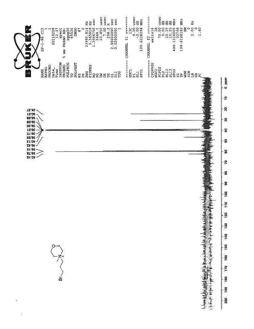
yl)propyl)benzo[d]thiazol-2(3H)-ylidene)hepta-1,3,5-trien-1-yl)benzo[d]thiazol-3-ium bromide (54):
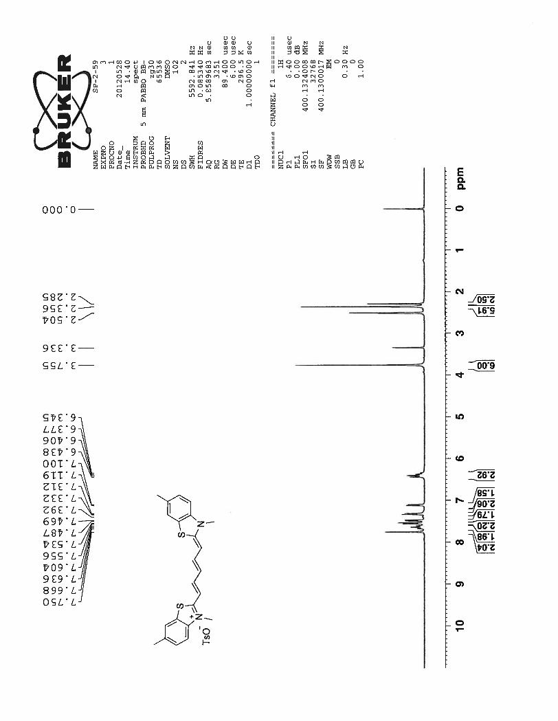
Yield 65% (0.17 g); m.p. 168 – 170 0 C;
1H NMR (DMSO-d6, 400 MHz): δ 7.99 (d, J = 8 Hz, 2H), 7.88
(d, J = 8.4 Hz, 2H), 7.58~7.58 (m, 7H), 6.92 (d, J = 12.8 Hz, 2H), 6.56 (t, J = 12.8 Hz , 2H), 4.39 (brd, J
= 7.2 Hz, 4H), 3.73 (brd, J = 7.6 Hz, 4H), 3.55 (brd, J = 5.2 Hz, 8H), 3.05 (s, 6H), 2.13 (m, 6H). 13
C NMR
(DMSO-d6, 100 MHz): δ 162.04, 151.95, 148.08, 141.29, 128.04, 125.30, 124.95, 123.46, 123.06,
113.28, 102.03, 63.86, 59.63, 47.86, 43.06, 21.98, 21.23. ESI-HRMS m/z: calc. for (C37H49S2N4Br2)+
771.1765, found 771.1734. λabs = 760 nm λfluo = 795 nm
4-methyl-4-(3-(2-((1E,3E,5E,7Z)-7-(3-(3-(4-methylmorpholino-4-ium)propyl)benzo[d]thiazol-2(3H)-
ylidene)hepta-1,3,5-trien-1-yl)benzo[d]thiazol-3-ium-3-yl)propyl)morpholin-4-ium bromide (55):
Yield 63% (0.18 g); m.p. 222 – 224 0C;
1H NMR (DMSO-d6, 400 MHz): δ 8.01 (d, J = 7.6 Hz, 2H), 7.87
(d, J = 8.4 Hz, 2H), 7.59~7.38 (m, 7H), 6.91 (d, J = 13.2 Hz, 2H), 6.57 (t, J = 12.4 Hz, 2H), 4.41 (brs,
SP-1-99-MS
Time0.00 1.00 2.00 3.00 4.00 5.00 6.00 7.00 8.00 9.00
AU
0.0
1.0e-1
2.0e-1
0.00 1.00 2.00 3.00 4.00 5.00 6.00 7.00 8.00 9.00
%
0
100
0.00 1.00 2.00 3.00 4.00 5.00 6.00 7.00 8.00 9.00
%
0
100
SP-1-99-ELSD ELSDAn1
2.10e6
7.27
0.40
1.81
SP-1-99-MS 1: TOF MS ES+ TIC
1.20e5
7.49
4.688.82
SP-1-99 2: Diode Array Range: 3.069e-17.30
1.790.40
SP-1-99-MS
Time0.00 1.00 2.00 3.00 4.00 5.00 6.00 7.00 8.00 9.00
AU
0.0
1.0e-1
2.0e-1
0.00 1.00 2.00 3.00 4.00 5.00 6.00 7.00 8.00 9.00
%
0
100
0.00 1.00 2.00 3.00 4.00 5.00 6.00 7.00 8.00 9.00
%0
100
SP-1-99-ELSD ELSDAn1
2.10e6
7.27
0.40
1.81
SP-1-99-MS 1: TOF MS ES+ TIC
1.20e5
7.49
4.688.82
SP-1-99 2: Diode Array Range: 3.069e-17.30
1.790.40
SP-1-117(2)
Time0.00 1.00 2.00 3.00 4.00 5.00 6.00 7.00 8.00 9.00
AU
0.0
5.0e-2
1.0e-1
0.00 1.00 2.00 3.00 4.00 5.00 6.00 7.00 8.00 9.00
%
0
100
0.00 1.00 2.00 3.00 4.00 5.00 6.00 7.00 8.00 9.00
%
0
100
SP-1-117(2)-ELSD ELSDAn1
2.10e6
7.220.00
1.85
SP-1-117(2)-MS 1: TOF MS ES+ TIC
1.42e5
7.57
SP-1-117(2) 2: Diode Array Range: 1.323e-17.40
1.790.55 2.07 5.59

48
4H), 4.04~3.88 (m, 12H), 3.48 (s, 8H), 3.19 (s, 6H), 2.17 (brs, 4H). 13
C NMR (DMSO-d6, 100 MHz): δ
161.95, 151.85, 148.02, 141.18, 127.95, 125.20, 124.87, 123.42, 122.97, 113.19, 101.96, 59.72, 59.27,
47.00, 42.94, 19.85. ESI-HRMS m/z: calc. for (C37H49S2N4O2Br2)+
803.1664, found 803.1663. Anal.
Calcd for C37H49S2N4O2Br2.7.3H2O: C, 48.40; H, 5.90; N, 6.19; Br, 23.28; Found C, 48.53; H, 6.05; N,
6.12; Br, 23.56. λabs = 760 nm λfluo = 790 nm
General procedure for preparation of compounds 56 and 57:
These compounds were synthesized with slight modification of the previously published procedure.26, 77
A
mixture of 2, 5-dimethylbenzothiazole (1 eq.) and alkyl halide (2 eq.) were heated at 800C for 6 hours.
After cooling the mixture to room temperature resulting crude solid was purified by precipitation from
methanol and diethyl ether. The precipitated solid was then filtered and dried in vacuo.
2, 3, 5-trimethylbenzo[d]thiazol-3-ium iodide (56): Yield 76%; 1H NMR (DMSO-d6, 400 MHz): δ 8.31
(d, J = 8.4 Hz, 1H), 8.15 (s, 1H), 7.62 (d, J = 8.4 Hz, 1H), 4.18 (s, 3H), 3.17 (s, 3H), 2.56 (s, 3H). 13
C
NMR (DMSO-d6, 100 MHz): δ 176.73, 141.79, 139.75, 129.45, 125.75, 124.00, 123.90, 116.51, 116.37,
36.29, 21.14, 17.32.
3-ethyl-2,5-dimethylbenzo[d]thiazol-3-ium iodide (57): Yield 65%; 1H NMR (DMSO-d6, 400 MHz): δ
8.32 (d, J = 8.4 Hz, 1H), 8.20 (s, 1H), 7.64 (d, J = 8.4 Hz, 1H), 4.73 (q, J = 7.2 Hz, 2H) , 3.21 (s, 3H),
2.58 (s., 3H), 1.46 (t, J = 7.2 Hz, 3H). 13
C NMR (DMSO-d6, 100 MHz): δ 176.45, 140.76, 140.01, 129.52,
126.25, 124.15, 116.33, 44.60, 21.17, 16.73, 13.21.
SP-1-116_2
Time0.50 1.00 1.50 2.00 2.50 3.00 3.50 4.00 4.50 5.00 5.50 6.00 6.50 7.00 7.50 8.00 8.50 9.00 9.50
%
0
100
0.50 1.00 1.50 2.00 2.50 3.00 3.50 4.00 4.50 5.00 5.50 6.00 6.50 7.00 7.50 8.00 8.50 9.00 9.50
AU
0.0
5.0e-1
1.0
0.50 1.00 1.50 2.00 2.50 3.00 3.50 4.00 4.50 5.00 5.50 6.00 6.50 7.00 7.50 8.00 8.50 9.00 9.50
%
0
100
SP-1-116_2 ELSDAn1
1.77e6
6.97
1.620.06
SP-1-116_2 2: Diode Array Range: 1.3327.27
5.50
SP-1-116 MS 1: TOF MS ES+ TIC
9.65e4
6.98
4.699.318.06

49
General procedure for synthesis of compounds 58–60:
A mixture of 2,6-dimethylbenzothiazole (1 eq.) and alkyl tosylate (2 eq.) were heated at 1200C for 2
hours. After cooling the mixture to room temperature acetone was added to the thick slurry. Resulting
sticky solid was purified by precipitation from methanol and diethyl ether and dried in vacuo.
2,3,6-trimethylbenzo[d]thiazol-3-ium 4-methylbenzenesulfonate (58): Yield 72%; 1H NMR (DMSO-
d6, 400 MHz): δ 8.18~8.13 (m, 2H), 7.67 (d, J = 8 Hz, 1H), 7.45 (d, J = 7.2 Hz, 2H), 7.07 (d, J = 7.2 Hz,
2H), 4.15 (s, 3H), 3.12 (s, 3H), 2.51 (s, 3H), 2.25 (s, 3H). 13
C NMR (DMSO-d6, 100 MHz): δ 176.06,
145.55, 139.77, 138.30, 137.68, 130.51, 128.77, 128.05, 125.42, 123.68, 116.35, 36.08, 20.97, 20.77,
16.85.
3-ethyl-2,6-dimethylbenzo[d]thiazol-3-ium 4-methylbenzenesulfonate (59): Yield 70%; 1H NMR
(DMSO-d6, 400 MHz): δ 8.22~8.20 (m, 2H), 7.68 (d, J = 8.4 Hz, 1H), 7.48 (d, J = 8 Hz, 2H), 7.08 (d, J =
8 Hz, 2H), 4.72 (q, J = 7.2 Hz, 2H), 3.17 (s, 3H), 2.51 (s, 3H), 2.26 (s, 3H), 1.42 (t, J = 7.6 Hz, 3H). 13
C
NMR (DMSO-d6, 100 MHz): δ 175.72, 145.56, 138.66, 138.36, 137.71, 130.72, 129.26, 128.06, 125.48,
123.95, 123.86, 116.27, 44.68, 20.97, 20.78, 16.47, 13.23.
2,6-dimethyl-3-propylbenzo[d]thiazol-3-ium 4-methylbenzenesulfonate (60): Yield 60%; 1H NMR
(Methanol-d4, 400 MHz): δ 7.94~7.86 (m, 2H), 7.60 ~ 7.50 (m, 4H), 7.01 (d, J = 7.6 Hz, 3H), 4.52 (t, J =
8 Hz, 2H), 3.02 (s, 3H), 2.30 (s, 3H), 2.17 (s, 3H), 1.82 (p, J = 7.6 Hz, 2H), 0.93 (t, J = 7.6 Hz, 3H). 13
C
NMR (Methanol-d4, 100 MHz): δ 176.68, 143.74, 141.65, 140.91, 140.72, 132.42, 130.81, 129.86,
126.93, 124.70, 117.57, 52.20, 22.95, 21.57, 21.44, 17.12, 11.33.
General procedure for synthesis of compounds 61–65:
A mixture of benzothiazole quaternary salt 56–60 (2 eq.) and malonaldehyde bis(phenylimine)
monohydrochloride (1 eq.) were heated to 45 0C in presence of triethylamine (2 eq.) in acetonitrile for 2
hours. The solution was cooled down to room temperature and resulting solid was filtered. The resulting
crude solid was purified by precipitation from DMF–diethyl ether (20 mL, 1:4).
2-((1E,3E,5Z)-5-(3,5-dimethylbenzo[d]thiazol-2(3H)-ylidene)penta-1,3-dien-1-yl)-3,5-
dimethylbenzo[d]thiazol-3-ium iodide (61): Yield 63% (0.15 g); m.p. >260 0C;
1H NMR (DMSO-d6,

50
400 MHz): δ 7.83 (d, J = 8 Hz, 2H), 7.70 (t, J = 12 Hz, 2H), 7.52 (s, 2H), 7.21 (d, J = 8 Hz, 2H),
6.50~6.42 (m, 3H), 3.77 (s, 6H), 2.43 (s, 6H). 13
C NMR (DMSO-d6, 100 MHz): δ 164.05, 149.90, 142.14,
138.03, 125.94, 122.47, 122.02, 113.53, 100.54, 33.36, 21.11. ESI-HRMS m/z: calc. for (C23H23N2S2)+
391.1297, found 391.1308. λabs = 654 nm λfluo = 678 nm
3-ethyl-2-((1E,3E,5Z)-5-(3-ethyl-5-methylbenzo[d]thiazol-2(3H)-ylidene)penta-1,3-dien-1-yl)-5-
methylbenzo[d]thiazol-3-ium iodide (62): Yield 56% (0.16 g); m.p. >260 0C;
1H NMR (DMSO-d6, 400
MHz): δ 7.85 (d, J =8 Hz, 2H), 7.73 (t, J = 12.4 Hz, 2H), 7.57 (s, 2H), 7.22 (d, J = 7.6 Hz, 2H),
6.54~6.42 (m, 3H), 4.36 (brd, J = 6.4 Hz, 4H), 2.44 (s, 6H), 1.31 (t, J = 6.8 Hz, 6H). 13
C NMR (DMSO-
d6, 100 MHz): δ 163.36, 150.14, 141.05, 138.14, 125.94, 125.53, 122.20, 113.22, 99.91, 41.10, 21.02,
12.56. ESI-HRMS m/z: calc. for (C25H27N2S2)+ 419.1610, found 419.1602. λabs = 660 nm λfluo = 679 nm
SP-2-33
Time0.00 2.00 4.00 6.00 8.00 10.00 12.00 14.00 16.00 18.00
%
0
100
0.00 2.00 4.00 6.00 8.00 10.00 12.00 14.00 16.00 18.00
AU
0.0
5.0e-1
1.0
SP-2-33 2: Diode Array Range: 1.359.99
SP-2-33 ELSDAn1
9.31e5
9.94
3.84

51
2-((1E,3E,5Z)-5-(3,6-dimethylbenzo[d]thiazol-2(3H)-ylidene)penta-1,3-dien-1-yl)-3,6-
dimethylbenzo[d]thiazol-3-ium 4-methylbenzenesulfonate (63): Yield 70% (0.18 g); m.p. 210 - 212
0C;
1H NMR (DMSO-d6 , 400 MHz) : δ 7.75 (s, 2H), 7.63 (t, J = 12.8 Hz, 2H), 7.54 (d, J = 8.8 Hz, 2H),
7.47 (d, J = 7.2 Hz, 2H), 7.32 (d, J = 8 Hz, 2H), 7.11 (d, J = 7.6 Hz, 3H), 6.43~6.34 (m, 3H), 3.75 (s, 6H),
2.35 (s, 6H), 2.28 (s, 3H). 13
C NMR (DMSO-d6, 100 MHz): δ 163.15, 149.59, 145.77, 139.84, 137.40,
134.61, 128.60, 127.91, 125.39, 125.01, 122.53, 120.23, 112.76, 100.16, 33.28, 20.67, 20.51. ESI-HRMS
m/z: calc. for (C23H23N2S2)+ 391.1297, found 391.1298. λabs = 655 nm λfluo = 676 nm
3-ethyl-2-((1E,3E,5Z)-5-(3-ethyl-6-methylbenzo[d]thiazol-2(3H)-ylidene)penta-1,3-dien-1-yl)-6-
methylbenzo[d]thiazol-3-ium 4-methylbenzenesulfonate (64): Yield 64% (0.14 g); m.p. 194 - 196 0C;
1H NMR (DMSO-d6 , 400 MHz) : δ 7.76 (s, 2H), 7.69~7.57 (m, 4H), 7.48 (d, J = 8 Hz, 2H), 7.33 (d, J =
8.4 Hz, 2H), 7.11 (d, J = 8 Hz, 3H), 6.47~6.36 (m, 3H), 4.33 (brd, J = 7.2 Hz, 4H), 2.36 (s, 6H), 2.28 (s,
3H), 1.28 (t, J = 6.8 Hz, 6H). 13
C NMR (DMSO-d6, 100 MHz): δ 162.63, 150.13, 145.89, 138.93, 137.48,
SP-2-34
Time4.00 6.00 8.00 10.00 12.00 14.00 16.00
%
0
100
4.00 6.00 8.00 10.00 12.00 14.00 16.00
AU
0.0
5.0e-1
1.0
SP-2-34 3: Diode Array Range: 1.32910.67
SP-2-34 ELSDAn1
7.82e5
10.55
SP-59a
Time4.00 6.00 8.00 10.00 12.00 14.00
%
83
4.00 6.00 8.00 10.00 12.00 14.00
AU
1.0e-2
2.0e-2
3.0e-2
SP-59a 2: Diode Array Range: 3.398e-28.10
SP-59a ELSDAn2
3.17e5
8.42

52
134.81, 128.88, 128.01, 125.49, 125.42, 122.82, 120.37, 112.74, 99.74, 41.24, 20.77, 20.62, 12.67. ESI-
HRMS m/z: calc. for (C25H27N2S2)+ 419.1610, found 419.1615. λabs = 660 nm λfluo = 679 nm
6-methyl-2-((1E,3E,5Z)-5-(6-methyl-3-propylbenzo[d]thiazol-2(3H)-ylidene)penta-1,3-dien-1-yl)-3-
propylbenzo[d]thiazol-3-ium 4-methylbenzenesulfonate (65): Yield 65% (0.13 g); m.p. 195 – 197 0C;
1H NMR (DMSO-d6 , 400 MHz) : δ 7.74 (s, 2H), 7.66~7.58 (m, 4H), 7.49 (d, J = 7.6 Hz, 2H), 7.31 (d, J =
8.4 Hz, 2H), 7.11 (d, J = 7.6 Hz, 3H), 6.46~6.38 (m, 3H), 4.24 (brt, J = 7.2 Hz, 4H), 2.35 (s, 6H), 2.28 (s,
3H), 1.71 (q, J = 7.2Hz, 4H), 0.95 (t, J = 7.2Hz, 6H). 13
C NMR (DMSO-d6, 100 MHz): δ 162.96, 149.92,
145.90, 139.40, 137.46, 134.76, 128.79, 127.99, 125.49, 125.23, 122.71, 120.45, 113.01, 100.08, 47.21,
20.76, 20.59, 10.80. ESI-HRMS m/z: calc. for (C27H31N2S2)+ 447.1923, found 447.1927. λabs = 660 nm
λfluo = 680 nm
SP-60a
Time2.00 4.00 6.00 8.00 10.00
%
61
2.00 4.00 6.00 8.00 10.00
AU
0.0
2.5e-2
5.0e-2
7.5e-2
SP-60a 2: Diode Array Range: 9.827e-28.72
SP-60a ELSDAn2
4.79e5
8.98
SP-2-66
Time0.00 2.00 4.00 6.00 8.00 10.00 12.00 14.00 16.00 18.00
AU
0.0
2.0e-1
4.0e-1
6.0e-1
8.0e-1
1.0
1.2
SP-2-67 3: Diode Array Range: 1.32611.44

53
General Procedure for preparation of compounds 66–70:
These compounds were synthesized with slight modification of the previously published procedure.67, 78
A
mixture of 2-methylbenzothiazole (1 eq.) and alkyl halide (3 eq.) was heated at 800C for 6 to 18 hours.
The mixture was cooled to room temperature and resulting crude solid was subjected for purification by
precipitation from methanol and diethyl ether (10 mL, 1:4). The precipitated solid was then filtered and
dried in vacuo. Compound 70 was used as crude in the next step.
2,3-dimethylbenzo[d]thiazol-3-ium iodide (66): Yield 81%; 1H NMR (DMSO-d6 , 400 MHz): δ 8.49 (d,
J = 8 Hz, 1H), 8.31 (d, J = 8.4 Hz, 1H), 7.89 (q, J1 = 7.2 Hz, J2 = 0.8Hz, 1H), 7.80 (t, J1 = 8.4 Hz, J2 = 7.2
Hz, 1H), 4.24 (s, 3H), 3.22 (s, 3H). 13
C NMR (DMSO-d6, 100 MHz): δ 177.04, 141.47, 129.15, 128.58,
127.93, 124.47, 116.70, 36.44, 17.48.
3-ethyl-2-methylbenzo[d]thiazol-3-ium iodide (67): Yield 72%; 1H NMR (DMSO-d6 , 400 MHz): δ
8.47 (d, J = 8 Hz, 1H), 8.35 (d, J = 8.4 Hz, 1H), 7.92 ~ 7.79 (m, 2H), 4.78 (q, J = 7.2 Hz, 2H), 3.23 (s,
3H), 1.46 (t, J = 7.2 Hz, 3H). 13
C NMR (DMSO-d6, 100 MHz): δ 176.87, 140.46, 129.37, 129.14, 128.05,
124.69, 116.68, 44.74, 16.79, 13.22.
2-methyl-3-propylbenzo[d]thiazol-3-ium iodide (68): Yield 60%; 1H NMR (DMSO-d6 , 400 MHz): δ
8.47 (d, J = 8 Hz, 1H), 8.38 (d, J = 8.4 Hz, 1H), 7.91~7.79 (m, 2H), 4.70 (t, J = 7.6 Hz, 2H), 3.23 (s, 3H),
1.90 (p, J = 7.6 Hz, 2H), 1.01 (t, J = 7.6 Hz, 3H). 13
C NMR (DMSO-d6, 100 MHz): δ 177.09, 140.08,
129.33, 129.04, 128.06, 124.63, 116.93, 50.44, 21.32, 17.01, 10.76.
3-butyl-2-methylbenzo[d]thiazol-3-ium iodide (69): Yield 50%; 1H NMR (DMSO-d6 , 400 MHz): δ
8.47 (d, J = 8 Hz, 1H), 8.35 (d, J = 8.8 Hz, 1H), 7.88~7.78 (m, 2H), 4.72 (t, J = 7.6 Hz, 2H), 3.23 (s, 3H),
1.89~1.78 (m, 2H), 1.50 ~ 1.41 (m, 2H), 0.93 (t, J = 7.2 Hz, 3H). 13
C NMR (DMSO-d6, 100 MHz): δ
176.98, 140.81, 129.33, 129.05, 128.05, 124.64, 116.88, 49.06, 29.77, 19.27, 16.99, 13.52.
General Procedure for synthesis of compounds 71–75:
A mixture of benzothiazole quaternary salt 66–70 (2 eq.) and malonaldehyde bis(phenylimine)
monohydrochloride (1 eq.) were heated to 45 0C in presence of triethylamine (2 eq.) in acetonitrile for 2

54
hours. The solution was cooled to room temperature and the resulting solid was filtered. It was then puri-
fied by precipitation from DMF–diethyl ether (20 mL, 1:4).
3-methyl-2-((1E,3E,5Z)-5-(3-methylbenzo[d]thiazol-2(3H)-ylidene)penta-1,3-dien-1-
yl)benzo[d]thiazol-3-ium iodide (71): Yield 68% (0.21 g); m.p. >260 0C;
1H NMR (DMSO-d6, 400
MHz): δ 7.98 (d, J = 8 Hz, 2H), 7.75~7.67 (m, 4H), 7.55 (t, J = 7.2 Hz, 2H), 7.38 (t, J = 7.6 Hz, 2H),
6.53~6.50 (m, 3H), 3.80 (s, 6H). 13
C NMR (DMSO-d6, 100 MHz): δ 164.01, 150.28, 142.02, 127.95,
125.14, 124.94, 122.93, 113.35, 100.62, 33.48. ESI-HRMS m/z: calc. for (C21H19N2S2)+ 363.0990, found
363.1004. λabs = 650 nm λfluo = 670 nm
3-ethyl-2-((1E,3E,5Z)-5-(3-ethylbenzo[d]thiazol-2(3H)-ylidene)penta-1,3-dien-1-yl)benzo[d]thiazol-
3-ium iodide (72): Yield 65% (0.19 g); m.p. >260 0C;
1H NMR (DMSO-d6, 400 MHz): δ 7.98 (d, J = 7.6
Hz, 2H), 7.77~7.69 (m, 4H), 7.53 (t, J = 7.6 Hz, 2H), 7.37 (t, J = 7.6 Hz, 2H), 6.55~6.46 (m, 3H), 4.38 (q,
J = 6.8 Hz, 4H), 1.30 (t, J = 7.2 Hz, 3H). 13
C NMR (DMSO-d6, 100 MHz): δ 163.22, 150.57, 140.93,
128.00, 125.39, 124.91, 123.05, 120.74, 113.09, 100.06, 41.28, 12.65. ESI-HRMS m/z: calc. for
(C23H23N2S2)+ 391.1297, found 391.1298. λabs = 650 nm λfluo = 673 nm
SP-2-28
Time2.00 4.00 6.00 8.00 10.00 12.00
%
0
100
2.00 4.00 6.00 8.00 10.00 12.00
AU
0.0
5.0e-1
1.0
SP-2-28 2: Diode Array Range: 1.3359.15
SP-2-28 ELSDAn1
1.15e6
9.04
0.29

55
3-propyl-2-((1E,3E,5Z)-5-(3-propylbenzo[d]thiazol-2(3H)-ylidene)penta-1,3-dien-1-
yl)benzo[d]thiazol-3-ium iodide (73): Yield 61% (0.16 g); m.p. 250 - 252 0C;
1H NMR (DMSO-d6, 400
MHz): δ 7.99 (d, J = 8 Hz, 2H), 7.79 ~ 7.73 (m, 4H), 7.54 (t, J = 7.6 Hz, 2H), 7.39 (t, J = 7.6 Hz, 2H),
6.58 ~ 6.47 (m, 3H), 4.32 (brt, J = 6.8 Hz, 4H), 1.76 (q, J = 7.2 Hz, 4H), 0.98 (t, J = 7.6 Hz, 6H). 13
C
NMR (DMSO-d6, 100 MHz): δ 163.54, 150.38, 141.39, 127.92, 125.22, 124.88, 123.04, 120.82, 113.38,
100.43, 47.26, 20.76, 10.83.0.38, 141.39, 127.92, 125.22, 124.88, 123.04, 120.82, 113.38, 100.43, 47.26,
20.76, 10.83. ESI-HRMS m/z: calc. for (C25H27N2S2)+ 419.1616, found 419.1626. λabs = 654 nm λfluo =
675 nm
SP-2-29
Time0.00 2.00 4.00 6.00 8.00 10.00 12.00 14.00 16.00 18.00
%
0
100
0.00 2.00 4.00 6.00 8.00 10.00 12.00 14.00 16.00 18.00
AU
0.0
5.0e-1
1.0
SP-2-29 2: Diode Array Range: 1.3210.00
SP-2-29 ELSDAn1
7.80e5
9.83
0.014.46 14.4113.11
SP-2-51(2)
Time0.00 2.00 4.00 6.00 8.00 10.00 12.00 14.00 16.00 18.00
%
0
100
0.00 2.00 4.00 6.00 8.00 10.00 12.00 14.00 16.00 18.00
AU
0.0
5.0e-1
1.0
SP-2-51(2) 3: Diode Array Range: 1.34710.40
SP-2-51(2) ELSDAn1
1.69e6
10.43

56
3-butyl-2-((1E,3E,5Z)-5-(3-butylbenzo[d]thiazol-2(3H)-ylidene)penta-1,3-dien-1-yl)benzo[d]thiazol-
3-ium iodide (74): Yield 62% (0.14 g); m.p. 240 - 242 0C;
1H NMR (DMSO-d6, 400 MHz): δ 7.99 (d, J =
8 Hz, 2H), 7.79~7.71 (m, 4H), 7.55 (t, J = 7.6 Hz, 2H), 7.39 (t, J = 7.6 Hz, 2H), 6.55~6.48 (m, 3H), 4.34
(brt, J = 7.2 Hz, 4H), 1.71 (m, 4H), 1.41 (m, 4H), 0.94 (t, J = 7.2 Hz, 6H). 13
C NMR (DMSO-d6, 100
MHz): δ 163.34, 150.26, 141.23, 127.89, 125.21, 124.82, 122.95, 120.91, 113.26, 100.36, 45.82, 29.38,
19.29, 13.63. ESI-HRMS m/z: calc. for (C27H31N2S2)+ 447.1923, found 447.1933. λabs = 655 nm λfluo =
673 nm
3-(3-phenylpropyl)-2-((1E,3E,5Z)-5-(3-(3-phenylpropyl)benzo[d]thiazol-2(3H)-ylidene)penta-1,3-
dien-1-yl)benzo[d]thiazol-3-ium bromide (75): Yield 63% (0.17 g); m.p. 130 - 132 0C;
1H NMR
(DMSO-d6, 400 MHz): δ 7.97 (d, J = 8 Hz, 2H), 7.73 ~ 7.52 (m, 6H), 7.39 ~ 7.21 (m, 10H), 6.42 ~ 6.39
(m, 3H), 4.35 (brt, J = 6.8 Hz, 4H), 2.76 (t, J = 7.6 Hz, 4H), 2.03 (m, 4H). 13
C NMR (DMSO-d6, 100
MHz): δ 163.56, 150.40, 141.31, 140.77, 128.40, 128.21, 127.94, 126.09, 125.31, 124.93, 123.01, 120.77,
113.21, 100.26, 45.65, 31.81, 28.79. ESI-HRMS m/z: calc. for (C37H35N2S2)+ 571.2236, found 571.2240.
λabs = 655 nm λfluo = 674 nm
SP-2-30
Time0.00 2.00 4.00 6.00 8.00 10.00 12.00 14.00 16.00 18.00
%
0
100
0.00 2.00 4.00 6.00 8.00 10.00 12.00 14.00 16.00 18.00
AU
0.0
5.0e-1
1.0
SP-2-30 2: Diode Array Range: 1.33911.25
SP-2-30 ELSDAn1
9.85e5
11.09

57
General Procedure for synthesis of compounds 76–78:
A mixture of 6-hydroxy-2-methylbenzothiazole (1 eq.) and alkyl tosylate (2 eq.) was heated at 1200C for
2 hours. After cooling the mixture to room temperature, acetone was added where hydroxy salts precipi-
tated out. Crude compounds were further purified by precipitation from methanol and diethyl ether (5
mL, 1:9)
6-hydroxy-2,3-dimethylbenzo[d]thiazol-3-ium 4-methylbenzenesulfonate (76): Yield 65%; 1H NMR
(DMSO-d6, 400 MHz): δ 10.62 (s, 1H), 8.06 (d, J = 9.2 Hz, 1H), 7.70 (d, J = 2.4 Hz, 1H), 7.46 (d, J = 8.4
Hz, 2H), 7.27 (dd, J1 = 2 Hz, J2 = 7.2 Hz, 1H), 7.08 (d, J = 7.6 Hz, 2H), 4.10 (s, 3H), 3.06 (s, 3H), 2.27 (s,
3H). 13
C NMR (DMSO-d6, 100 MHz): δ 173.24, 157.57, 145.68, 137.61, 134.81, 130.31, 128.02, 125.45,
118.54, 117.68, 117.54, 108.65, 36.05, 20.78, 16.59.
3-ethyl-6-hydroxy-2-methylbenzo[d]thiazol-3-ium 4-methylbenzenesulfonate (77): Yield 64%; 1H
NMR (DMSO-d6, 400 MHz): δ 10.67 (s, 1H), 8.09 (d, J = 8.8 Hz, 1H), 7.69 (d, J = 1.2 Hz, 1H), 7.49 (d, J
= 7.6 Hz, 2H), 7.27 (d, J = 7.6 Hz, 1H), 7.06 (d, J = 7.6 Hz, 2H), 4.64 (d, J = 6.8 Hz, 2H), 3.09 (s, 3H),
2.23 (s, 3H), 1.38 (t, J = 7.2 Hz, 3H). 13
C NMR (DMSO-d6, 100 MHz): δ 172.76, 157.60, 145.39, 137.88,
133.58, 130.78, 128.22, 128.07, 125.35, 118.72, 117.65, 117.47, 108.86, 44.61, 20.80, 16.25, 13.30.
6-hydroxy-2-methyl-3-propylbenzo[d]thiazol-3-ium 4-methylbenzenesulfonate (78): Yield 68%; 1H
NMR (DMSO-d6, 400 MHz): δ 10.63 (s, 1H), 8.14 (d, J = 9.2 Hz, 1H), 7.71 (s, 1H), 7.47 (d, J = 7.6 Hz,
SP-2-27
Time2.00 4.00 6.00 8.00 10.00 12.00
%
0
100
2.00 4.00 6.00 8.00 10.00 12.00
AU
0.0
5.0e-1
1.0
SP-2-27 2: Diode Array Range: 1.30910.49
SP-2-27 ELSDAn1
7.79e5
10.69
0.19

58
2H), 7.27 (d, J = 9.2 Hz, 1H), 7.10 (d, J = 7.6 Hz, 2H), 4.59 (t, J = 7.2 Hz, 2H), 3.17 (s, 3H), 2.27 (s,
3H), 1.84 (q, J = 7.2 Hz, 2H), 0.98 (t, J = 7.2 Hz, 3H). 13
C NMR (DMSO-d6, 100 MHz): δ 173.07, 157.56,
145.71, 137.58, 134.04, 130.70, 128.02, 125.46, 118.68, 117.80, 108.66, 50.30, 21.38, 20.76, 16.42,
10.70.
Procedure for synthesis of compound 79:
A mixture of 6-hydroxy-2-methylbenzothiazole (1eq.) and 3-bromopropyltrimethyl ammonium bromide
(2 eq.) was heated at 1200C for 4 hours. After cooling the mixture to room temperature methanol was
added and insoluble solid was filtered. The crude solid was purified by precipitation from DMSO-ethyl
acetate.
6-hydroxy-2-methyl-3-(3-(trimethylammonio)propyl)benzo[d]thiazol-3-ium bromide (79): Yield
52%; 1H NMR (DMSO-d6, 400 MHz): δ 10.58 (s, 1H), 8.35 (d, J = 8.8 Hz, 1H), 7.81 (s, 1H), 7.29 (d, J =
8 Hz, 1H), 4.73 (brs, 2H), 3.72 (brs, 2H), 3.24 (s, 3H), 3.13 (s, 9H), 2.34 (brs, 2H). 13
C NMR (DMSO-d6,
100 MHz): δ 174.03, 157.54, 133.92, 130.71, 118.67, 117.86, 108.89, 61.75, 52.54, 45.91, 21.84, 17.07.
Procedure for synthesis of compound 80:
A mixture of 6-hydroxy-2-methylbenzothiazole (1eq.) and 2-bromo-N,N-dimethylethanamine
hydrobromide (2 eq.) was heated at 1500C for 4 hours. After cooling the mixture to room temperature
methanol was added and insoluble solid was filtered. The crude solid was purified by precipitation from
DMSO-ethyl acetate.
3-(3-(dimethylammonio)propyl)-6-hydroxy-2-methylbenzo[d]thiazol-3-ium bromide (80): Yield
47%; 1H NMR (DMSO-d6, 400 MHz): δ 10.65 (brs, 1H), 10.33 (brs, 1H), 8.27 (d, J = 9.2 Hz, 1H), 7.81
(d, J = 2.4 Hz, 1H), 7.32 (q, J1 = 2 Hz, J2 = 7.2 Hz, 1H), 5.10 (brt, J = 7.6 Hz, 2H), 3.71 (brs, 2H), 3.25 (s,
3H), 2.98 (s, 6H). 13
C NMR (DMSO-d6, 100 MHz): δ 175.52, 157.60, 133.72, 130.76, 118.66, 117.64,
108.95, 51.85, 43.29, 42.59, 17.15.
General Procedure for synthesis of compounds 81–85:
A mixture of benzothiazole quaternary salt 76–80 (2 eq.) and malonaldehyde bis(phenylimine)
monohydrochloride (1 eq.) were heated to 45 0C in presence of triethylamine (3 eq.) in acetonitrile for 2
59
hours. The solution was cooled down to room temperature and resulting solid was filtered. Compounds
81–83 were purified by precipitation from DMF-diethyl ether (20 mL, 1:4). Compounds 84 and 85 were
purified using reverse phase column chromatography (acetone:water 5:1).
6-hydroxy-2-((1E,3E,5Z)-5-(6-hydroxy-3-methylbenzo[d]thiazol-2(3H)-ylidene)penta-1,3-dien-1-yl)-
3-methylbenzo[d]thiazol-3-ium 4-methylbenzenesulfonate (81): Yield 61% (0.16 g); m.p. >260 0C;
1H
NMR (DMSO-d6 , 400 MHz) : δ 10.06 (s, 2H), 7.61~7.46 (m, 5H), 7.35 (s, 2H), 7.10 (d, J = 7.6 Hz, 1H),
6.96 (d, J = 6.96 Hz, 2H), 6.40~6.34 (m, 3H), 3.73 (m, 6H), 2.28 (s, 1H). 13
C NMR (DMSO-d6, 100
MHz): δ 162.04, 155.25, 148.70, 134.80, 127.90, 126.32, 125.37, 115.69, 113.86, 108.63, 99.62, 33.28,
20.73. ESI-HRMS m/z: calc. for (C21H19N2O2S2)+ 395.0888, found 395.0893. λabs = 665 nm λfluo = 688 nm
3-ethyl-2-((1E,3E,5Z)-5-(3-ethyl-6-hydroxybenzo[d]thiazol-2(3H)-ylidene)penta-1,3-dien-1-yl)-6-
hydroxybenzo[d]thiazol-3-ium 4-methylbenzenesulfonate (82): Yield 56% (0.17 g); m.p. 234 - 236 0C;
1H NMR (DMSO-d6 , 400 MHz) : δ 10.45 (s, 2H), 7.58~7.48 (m, 5H), 7.31 (s, 2H), 7.11 (d, J = 6.8 Hz,
1H), 6.93 (d, J = 8 Hz, 2H), 6.41~6.34 (m, 3H), 4.30 (brs, 4H), 2.28 (s, 2H), 1.28 (s, 6H). 13
C NMR
(DMSO-d6, 100 MHz): δ 161.07, 156.14, 148.69, 145.81, 137.55, 133.43, 128.03, 126.75, 125.49, 119.53,
116.13, 113.76, 108.86, 99.18, 41.18, 20.77, 12.71. ESI-HRMS m/z: calc. for (C23H23N2O2S2)+ 423.1195,
found 423.1193. λabs = 665 nm λfluo = 689 nm
SP-2-43(5)
Time0.00 2.00 4.00 6.00 8.00 10.00 12.00 14.00 16.00 18.00
%
0
100
0.00 2.00 4.00 6.00 8.00 10.00 12.00 14.00 16.00 18.00
AU
0.0
5.0e-1
1.0
SP-2-43(5) 3: Diode Array Range: 1.3867.80
SP-2-43(5) ELSDAn1
2.05e6
7.75

60
6-hydroxy-2-((1E,3E,5Z)-5-(6-hydroxy-3-propylbenzo[d]thiazol-2(3H)-ylidene)penta-1,3-dien-1-yl)-
3-propylbenzo[d]thiazol-3-ium 4-methylbenzenesulfonate (83): Yield 62% (0.15 g); m.p. >260 0C;
1H
NMR (DMSO-d6 , 400 MHz) : δ 10.13 (s, 2H), 7.62 ~ 7.52 (m, 4H), 7.36 (s, 2H), 6.96 (d, J = 8.8 Hz,
2H), 6.44~6.35 (m, 3H), 4.23 (brs, 4H), 1.72 (brd, J = 6.8 Hz, 4H), 0.95 (t, J = 7.2 Hz, 6H). 13
C NMR
(DMSO-d6, 100 MHz): δ 161.83, 155.42, 148.87, 134.27, 126.55, 115.91, 114.11, 108.77, 95.54, 47.26,
20.82, 10.84. ESI-HRMS m/z: calc. for (C25H27N2O2S2)+ 451.1508, found 451.1514. λabs = 670 nm λfluo =
689 nm
6-hydroxy-2-((1E,3E,5Z)-5-(6-hydroxy-3-(2-(trimethylammonio)ethyl)benzo[d]thiazol-2(3H)-
ylidene)penta-1,3-dien-1-yl)-3-(2-(trimethylammonio)ethyl)benzo[d]thiazol-3-ium bromide (84):
Yield 54% (0.16 g); m.p. 242 - 244 0C;
1H NMR (DMSO-d6 , 400 MHz) : δ 10.12 (s, 2H), 7.67~7.62 (m,
4H), 7.39 (d, J = 2 Hz, 2H), 6.99 (dd, J1 = 2 Hz, , J2 = 7.2 Hz, 2H), 6.68 (d, J = 13.2 Hz, 2H), 6.50 (t, J =
12 Hz, 1H), 4.34 (brs, 4H), 3.65 (brs, 4H), 3.10 (s, 18H), 2.13 (brs, 2H). 13
C NMR (DMSO-d6, 100 MHz):
SP-2-49(3)
Time0.00 2.00 4.00 6.00 8.00 10.00 12.00 14.00 16.00 18.00
%
0
100
0.00 2.00 4.00 6.00 8.00 10.00 12.00 14.00 16.00 18.00
AU
0.0
5.0e-1
1.0
SP-2-49(3) 3: Diode Array Range: 1.3498.70
SP-2-49(3) ELSDAn1
7.26e5
8.69
9.80
SP-53a
Time6.00 7.00 8.00 9.00 10.00 11.00 12.00 13.00 14.00
%
87
6.00 7.00 8.00 9.00 10.00 11.00 12.00 13.00 14.00
AU
0.0
5.0e-2
1.0e-1
SP-53a 2: Diode Array Range: 1.422e-17.24
SP-53a ELSDAn2
2.99e5
7.50
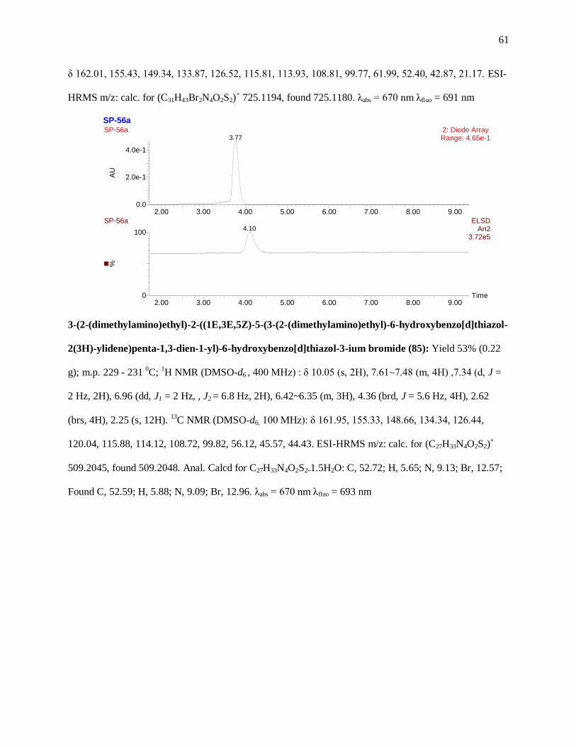
61
δ 162.01, 155.43, 149.34, 133.87, 126.52, 115.81, 113.93, 108.81, 99.77, 61.99, 52.40, 42.87, 21.17. ESI-
HRMS m/z: calc. for (C31H43Br2N4O2S2)+ 725.1194, found 725.1180. λabs = 670 nm λfluo = 691 nm
3-(2-(dimethylamino)ethyl)-2-((1E,3E,5Z)-5-(3-(2-(dimethylamino)ethyl)-6-hydroxybenzo[d]thiazol-
2(3H)-ylidene)penta-1,3-dien-1-yl)-6-hydroxybenzo[d]thiazol-3-ium bromide (85): Yield 53% (0.22
g); m.p. 229 - 231 0C;
1H NMR (DMSO-d6 , 400 MHz) : δ 10.05 (s, 2H), 7.61~7.48 (m, 4H) ,7.34 (d, J =
2 Hz, 2H), 6.96 (dd, J1 = 2 Hz, , J2 = 6.8 Hz, 2H), 6.42~6.35 (m, 3H), 4.36 (brd, J = 5.6 Hz, 4H), 2.62
(brs, 4H), 2.25 (s, 12H). 13
C NMR (DMSO-d6, 100 MHz): δ 161.95, 155.33, 148.66, 134.34, 126.44,
120.04, 115.88, 114.12, 108.72, 99.82, 56.12, 45.57, 44.43. ESI-HRMS m/z: calc. for (C27H33N4O2S2)+
509.2045, found 509.2048. Anal. Calcd for C27H33N4O2S2.1.5H2O: C, 52.72; H, 5.65; N, 9.13; Br, 12.57;
Found C, 52.59; H, 5.88; N, 9.09; Br, 12.96. λabs = 670 nm λfluo = 693 nm
SP-56a
Time2.00 3.00 4.00 5.00 6.00 7.00 8.00 9.00
%
0
100
2.00 3.00 4.00 5.00 6.00 7.00 8.00 9.00
AU
0.0
2.0e-1
4.0e-1
SP-56a 2: Diode Array Range: 4.65e-13.77
SP-56a ELSDAn2
3.72e5
4.10
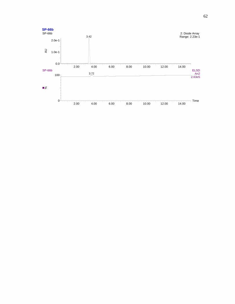
62
SP-66b
Time2.00 4.00 6.00 8.00 10.00 12.00 14.00
%
0
100
2.00 4.00 6.00 8.00 10.00 12.00 14.00
AU
0.0
1.0e-1
2.0e-1
SP-66b 2: Diode Array Range: 2.23e-13.42
SP-66b ELSDAn2
2.63e5
3.72
63
REFERENCES
1. Williams, C. H. G., Trans. R. Soc. Edinburg 1856, 21, 377.
2. Tolmachev, A. I.; Il’chenko, A. Y., Kirk-Othmer Encyclopedia of Chemical Technology 2005.
3. Hamer, F., Mainly Introductory. In Chemistry of Heterocyclic Compounds, John Wiley & Sons,
Inc.: 2008; pp 1-31.
4. Armitage, B., In Heterocyclic Polymethine Dyes, Strekowski, L., Ed. Springer Berlin /
Heidelberg: 2008; Vol. 14, pp 11-29.
5. Katritzky, A. R.; Fan, W.-Q.; Li, Q.-L., Bridged cyanine dyes. Part 1. 1-(N-methyl-4-pyridinio)-
3-(N-methylpyridylene)cyclopenta-1,4-dienes. Journal of heterocyclic chemistry 1988, 25, 1311-1314.
6. G.B. Behera, P. K. B., Cyanine dyes : Self aggregation and behaviour in surfactants A review.
J.surface Sci. Technol. 2007, 23, 1-31.
7. Kaloyanova, S.; Trusova, V. M.; Gorbenko, G. P.; Deligeorgiev, T., Synthesis and fluorescence
characteristics of novel asymmetric cyanine dyes for DNA detection. Journal of Photochemistry and
Photobiology A: Chemistry 2011, 217, 147-156.
8. Kovalska, V. B.; Volkova, K. D.; Losytskyy, M. Y.; Tolmachev, O. I.; Balanda, A. O.;
Yarmoluk, S. M., 6,6′-Disubstituted benzothiazole trimethine cyanines – new fluorescent dyes for DNA
detection. Spectrochimica Acta Part A: Molecular and Biomolecular Spectroscopy 2006, 65, 271-277.
9. Klunk, W. E.; Engler, H.; Nordberg, A.; Wang, Y.; Blomqvist, G.; Holt, D. P.; Bergström, M.;
Savitcheva, I.; Huang, G.-F.; Estrada, S.; Ausén, B.; Debnath, M. L.; Barletta, J.; Price, J. C.; Sandell, J.;
Lopresti, B. J.; Wall, A.; Koivisto, P.; Antoni, G.; Mathis, C. A.; Långström, B., Imaging brain amyloid
in Alzheimer's disease with Pittsburgh Compound-B. Annals of Neurology 2004, 55, 306-319.
10. Mills, W. H., LIV.-The cyanine dyes. Part IV. Cyanine dyes of the benzothiazole series. Journal
of the Chemical Society, Transactions 1922, 121, 455-466.
11. Todor G. Deligeorgiev, S. K. a. J. J. V., Intercalating Cyanine Dyes for Nucleic Acid Detection.
Recent Patents on Materials Science 2009, 2, 1-26.
64
12. Volkova, K. D.; Kovalska, V. B.; Balanda, A. O.; Losytskyy, M. Y.; Golub, A. G.; Vermeij, R. J.;
Subramaniam, V.; Tolmachev, O. I.; Yarmoluk, S. M., Specific fluorescent detection of fibrillar α-
synuclein using mono- and trimethine cyanine dyes. Bioorganic & Medicinal Chemistry 2008, 16,
1452-1459.
13. Ogul'chansky, T. Y.; Losytskyy, M. Y.; Kovalska, V. B.; Yashchuk, V. M.; Yarmoluk, S. M.,
Interactions of cyanine dyes with nucleic acids. XXIV. Aggregation of monomethine cyanine dyes in
presence of DNA and its manifestation in absorption and fluorescence spectra. Spectrochimica Acta Part
A: Molecular and Biomolecular Spectroscopy 2001, 57, 1525-1532.
14. Brooker, L. G. S.; Keyes, G. H.; Williams, W. W., Color and constitution. V. The absorption of
unsymmetrical cyanines. Resonance as a basis for a classification of dyes. J. Am. Chem. Soc. 1942, 64,
199-210.
15. Zhao, J.-L.; Lv, Y.; Ren, H.-J.; Sun, W.; Liu, Q.; Fu, Y.-L.; Wang, L.-Y., Synthesis, spectral
properties of cyanine dyes-β-cyclodextrin and their application as the supramolecular host with
spectroscopic probe. Dyes and Pigments 2013, 96, 180-188.
16. Deligeorgiev, T. G.; Gadjev, N. I.; Drexhage, K.-H.; Sabnis, R. W., Preparation of intercalating
dye thiazole orange and derivatives. Dyes and Pigments 1995, 29, 315-322.
17. Deligeorgiev, T. G.; Zaneva, D. A.; Katerinopoulos, H. E.; Kolev, V. N., A novel method for the
preparation of monomethine cyanine dyes. Dyes and Pigments 1999, 41, 49-54.
18. Deligeorgiev, T. G.; Zaneva, D. A.; Kim, S. H.; Sabnis, R. W., Preparation of monomethine
cyanine dyes for nucleic acid detection. Dyes and Pigments 1998, 37, 205-211.
19. Timtcheva, I.; Maximova, V.; Deligeorgiev, T.; Gadjev, N.; Drexhage, K. H.; Petkova, I.,
Homodimeric monomethine cyanine dyes as fluorescent probes of biopolymers. Journal of
Photochemistry and Photobiology B: Biology 2000, 58, 130-135.
20. Deligeorgiev, T.; Gadjev, N.; Vasilev, A.; Drexhage, K.-H.; Yarmoluk, S. M., Synthesis of novel
monomeric cyanine dyes containing mercapto and thioacetyl substituents for nucleic acid detection. Dyes
and Pigments 2006, 70, 185-191.

65
21. Fei, X.; Yang, S.; Zhang, B.; Liu, Z.; Gu, Y., Solid-Phase Synthesis and Modification of Thiazole
Orange and Its Derivatives and their Spectral Properties. Journal of Combinatorial Chemistry 2007, 9,
943-950.
22. W, K., Ber Dtsch Chem Ges 1924, 685
23. Kabatc, J.; Pączkowski, J., Three-cationic carbocyanine dyes as sensitizers in very efficient
photoinitiating systems for multifunctional monomer polymerization. Journal of Polymer Science Part A:
Polymer Chemistry 2009, 47, 4636-4654.
24. Xiao, S.; Xu, J.-H.; Li, Y.-S.; Du, C.-M.; Li, Y.-L.; Jiang, L.; Zhu, D., Preparation and
characterization of a novel dumbbell-type [60]fullerene dimer containing a cyanine dye. New Journal of
Chemistry 2001, 25, 1610-1612.
25. Mazières, M. R.; Duprat, C.; Bellan, J.; Wolf, J. G., Synthesis and characterisation of new
phosphonate labelled cyanines. Dyes and Pigments 2007, 74, 404-409.
26. Shi, Q.-Q.; Sun, R.; Ge, J.-F.; Xu, Q.-F.; Li, N.-J.; Lu, J.-M., A comparative study of symmetrical
and unsymmetrical trimethine cyanine dyes bearing benzoxazolyl and benzothiazolyl groups. Dyes and
Pigments 2012, 93, 1506-1511.
27. Park, J.-Y.; Gwon, S.-Y.; Yoon, N.-S.; Son, Y.-A.; Kim, S.-H., Novel triazacarbocyanine dye
sensor synthesis: pH switching effect. Fibers and Polymers 2011, 12, 976-978.
28. Mitekura, H.; No, T.; Suzuki, K.; Satake, K.; Kimura, M., Spectroscopic properties of meso-
substituted cyanine dyes: evidences for intramolecular charge transfer from a julolidine moiety as a meso-
substituent to the cyanine chromophore. Dyes and Pigments 2002, 54, 113-120.
29. Renikuntla, B. R.; Rose, H. C.; Eldo, J.; Waggoner, A. S.; Armitage, B. A., Improved
Photostability and Fluorescence Properties through Polyfluorination of a Cyanine Dye. Organic Letters
2004, 6, 909-912.
30. Kaur, Crosslinking of viral nanoparticles with “clickable” fluorescent crosslinkers at the
interface. SCIENCE CHINA Chemistry 2010, 53, 1287.

66
31. Mehranpour, A. M.; Hashemnia, S.; Maghamifar, R., Synthesis and Characterization of New γ-
Substituted Pentamethine Cyanine Dyes. Synthetic Communications 2010, 40, 3594-3602.
32. Yagupolskii, L. M.; Chernega, О. I.; Kondratenko, N. V.; Chernega, A. N.; Vlasenko, Y. G.;
Nedelkov, R. V.; Yagupolskii, Y. L., Synthesis of the first representative of dicarbothiacyanine dyes with
completely fluorinated polymethine chain. Journal of Fluorine Chemistry 2010, 131, 165-171.
33. Nesterenko, Y. A.; Briks, Y. L.; Kachkovskii, A. D.; Tolmachev, A. I., Synthesis and spectral
properties of pyran-based heterocyclic polyene vinylogs. Khim. Geterotsikl. Soedin. 1990, 252-5.
34. Meguellati, K.; Spichty, M.; Ladame, S., Reversible Synthesis and Characterization of Dynamic
Imino Analogues of Trimethine and Pentamethine Cyanine Dyes. Organic Letters 2009, 11, 1123-1126.
35. Losytskyy, M.; Volkova, K.; Kovalska, V.; Makovenko, I.; Slominskii, Y.; Tolmachev, O.;
Yarmoluk, S., Fluorescent Properties of Pentamethine Cyanine Dyes with Cyclopentene and Cyclohexene
Group in Presence of Biological Molecules. J Fluoresc 2005, 15, 849-857.
36. Kharitonova, Journal of organic chemistry of the USSR 1992, 28, 2159.
37. SMITH, G. S. L. B. A. L. 1,1'-diaralkyl-4,4'-carbocyanine salts 1941.
38. Vompe, A. F.; Ivanova, L. V.; Meskhi, L. M.; Monich, N. V.; Raikhina, R. D., Reactions of
polymethine dyes. I. Synthesis of pseudobases of polymethine dyes and their reactions. Zh. Org. Khim.
1985, 21, 584-94.
39. Miltsov, S.; Encinas, C.; Alonso, J., Novel synthesis of ketocyanine dyes. Tetrahedron Letters
2001, 42, 6129-6131.
40. Matsui, M.; Hashimoto, Y.; Funabiki, K.; Jin, J.-Y.; Yoshida, T.; Minoura, H., Application of
near-infrared absorbing heptamethine cyanine dyes as sensitizers for zinc oxide solar cell. Synthetic
Metals 2005, 148, 147-153.
41. Makin, Journal of organic chemistry of the USSR 1977, 13, 2440.
42. Narayanan, N.; Patonay, G., A New Method for the Synthesis of Heptamethine Cyanine Dyes:
Synthesis of New Near-Infrared Fluorescent Labels. The Journal of Organic Chemistry 1995, 60, 2391-
2395.

67
43. Tani, T., The present status and future prospects of silver halide photography. Journal of Imaging
Science and Technology 1995, 39, 31-40.
44. Dai, Z. F.; Qun, L.; Peng, B. X., Synthesis and characterization of some stabilized
indocarbocyanine dye complex colorants for optical recording media. Dyes and Pigments 1998, 36, 243-
248.
45. Reisfeld, R.; Weiss, A.; Saraidarov, T.; Yariv, E.; Ishchenko, A. A., Solid-state lasers based on
inorganic–organic hybrid materials obtained by combined sol–gel polymer technology. Polymers for
Advanced Technologies 2004, 15, 291-301.
46. Zhang, Q. F.; Dandeneau, C. S.; Zhou, X. Y.; Cao, G. Z., ZnO Nanostructures for Dye-Sensitized
Solar Cells. Advanced Materials 2009, 21, 4087-4108.
47. Liang, M.; Xu, W.; Cai, F. S.; Chen, P. Q.; Peng, B.; Chen, J.; Li, Z. M., New triphenylamine-
based organic dyes for efficient dye-sensitized solar cells. Journal of Physical Chemistry C 2007, 111,
4465-4472.
48. Ehret, A.; Stuhl, L.; Spitler, M. T., Spectral Sensitization of TiO2 Nanocrystalline Electrodes
with Aggregated Cyanine Dyes. Journal of Physical Chemistry B 2001, 105, 9960-9965.
49. Nasr, C.; Hotchandani, S.; Kamat, P. V.; Das, S.; Thomas, K. G.; George, M. V., Electrochemical
and Photoelectrochemical Properties of Monoaza-15-crown Ether Linked Cyanine Dyes:
Photosensitization of Nanocrystalline SnO2 Films. Langmuir 1995, 11, 1777-1783.
50. Tang, X.; Gan, F., Optical storage performance of cyanine-doped ormosil film. Proc. SPIE-Int.
Soc. Opt. Eng. 1997, 3136, 210.
51. Qun, L.; Guo-Liang, L.; Bi-Xian, P.; Zheng-Xin, L., Synthesis, characterization and photographic
properties of some new styryl cyanine dyes. Dyes and Pigments 1998, 38, 211-218.
52. Tian, H.; Meng, F., Chapter 2 - Cyanine dyes for solar cells and optical data storage. In
Functional Dyes, Sung-Hoon, K., Ed. Elsevier Science: Amsterdam, 2006; pp 47-84.

68
53. Ehret, A.; Stuhl, L.; Spitler, M. T., Variation of carboxylate-functionalized cyanine dyes to
produce efficient spectral sensitization of nanocrystalline solar cells. Electrochimica Acta 2000, 45, 4553-
4557.
54. Choi, H. S.; Nasr, K.; Alyabyev, S.; Feith, D.; Lee, J. H.; Kim, S. H.; Ashitate, Y.; Hyun, H.;
Patonay, G.; Strekowski, L.; Henary, M.; Frangioni, J. V., Synthesis and In Vivo Fate of Zwitterionic
Near-Infrared Fluorophores. Angewandte Chemie International Edition 2011, 50, 6258-6263.
55. Licha, K.; Riefke, B.; Ntziachristos, V.; Becker, A.; Chance, B.; Semmler, W., Hydrophilic
Cyanine Dyes as Contrast Agents for Near-infrared Tumor Imaging: Synthesis, Photophysical Properties
and Spectroscopic In vivo Characterization . Photochemistry and Photobiology 2000, 72, 392-398.
56. Licha, K., Contrast Agents for Optical Imaging
Contrast Agents II. In Krause, W., Ed. Springer Berlin / Heidelberg: 2002; Vol. 222, pp 1-29.
57. Neumaier, B.; Deisenhofer, S.; Sommer, C.; Solbach, C.; Reske, S. N.; Mottaghy, F., Synthesis
and evaluation of 18F-fluoroethylated benzothiazole derivatives for in vivo imaging of amyloid plaques
in Alzheimer's disease. Applied Radiation and Isotopes 2010, 68, 1066-1072.
58. Ribeiro Morais, G.; Paulo, A.; Santos, I., A Synthetic Overview of Radiolabeled Compounds for
β-Amyloid Targeting (Eur. J. Org. Chem. 7/2012). European Journal of Organic Chemistry 2012, 2012.
59. Ono, M.; Watanabe, H.; Kimura, H.; Saji, H., BODIPY-Based Molecular Probe for Imaging of
Cerebral β-Amyloid Plaques. ACS Chemical Neuroscience 2012, 3, 319-324.
60. Pajouhesh, H.; Lenz, G. R., Medicinal Chemical Properties of Successful Central Nervous
System Drugs. NeuroRX 2005, 2, 541-553.
61. Humblet, V.; Misra, P.; Bhushan, K. R.; Nasr, K.; Ko, Y.-S.; Tsukamoto, T.; Pannier, N.;
Frangioni, J. V.; Maison, W., Multivalent Scaffolds for Affinity Maturation of Small Molecule Cell
Surface Binders and Their Application to Prostate Tumor Targeting. Journal of Medicinal Chemistry
2008, 52, 544-550.

69
62. Robbins, K. J.; Liu, G.; Lin, G.; Lazo, N. D., Detection of Strongly Bound Thioflavin T Species
in Amyloid Fibrils by Ligand-Detected 1H NMR. The Journal of Physical Chemistry Letters 2011, 2,
735-740.
63. Khurana, R.; Coleman, C.; Ionescu-Zanetti, C.; Carter, S. A.; Krishna, V.; Grover, R. K.; Roy, R.;
Singh, S., Mechanism of thioflavin T binding to amyloid fibrils. Journal of Structural Biology 2005, 151,
229-238.
64. Yan, P.; Bero, A. W.; Cirrito, J. R.; Xiao, Q.; Hu, X.; Wang, Y.; Gonzales, E.; Holtzman, D. M.;
Lee, J.-M., Characterizing the Appearance and Growth of Amyloid Plaques in APP/PS1 Mice. The
Journal of Neuroscience 2009, 29, 10706-10714.
65. Sisodia, S. S.; Price, D. L., Role of the beta-amyloid protein in Alzheimer's disease. The FASEB
Journal 1995, 9, 366-70.
66. Klunk, W. E.; Wang, Y.; Huang, G.-f.; Debnath, M. L.; Holt, D. P.; Shao, L.; Hamilton, R. L.;
Ikonomovic, M. D.; DeKosky, S. T.; Mathis, C. A., The Journal of Neuroscience 2003, 23, 2086-2092.
67. Kuramoto, N.; Natsukawa, K.; Asao, K., Synthesis and characterization of deep-coloured
squarylium dyes for laser optical recording media. Dyes and Pigments 1989, 11, 21-35.
68. Elizalde, L. E.; Ledezma, R.; López, R. G., Synthesis of Photochromic Monomers Derived from
1′‐(2‐Methacryloxyethyl)‐3,3‐Dimethyl‐2‐[2H]‐Spirobenzopyran Indoline. Synthetic Communications
2005, 35, 603-610.
69. Chopra, A., In: Molecular Imaging and Contrast Agent Database (MICAD) [Internet]. Bethesda
(MD): National Center for Biotechnology Information (US). In 2004-2012 ed.; 2009 Dec 8 [Updated
2009 Jan 7].
70. Sylvain Gioux, H. S. C., John V. Frangioni, Image-Guided Surgery using Invisible Near-Infrared
Light: Fundamentals of Clinical Translation. Mol Imaging 2010, 9, 237-255.
71. Troyan, S.; Kianzad, V.; Gibbs-Strauss, S.; Gioux, S.; Matsui, A.; Oketokoun, R.; Ngo, L.;
Khamene, A.; Azar, F.; Frangioni, J., The FLARE™ Intraoperative Near-Infrared Fluorescence Imaging

70
System: A First-in-Human Clinical Trial in Breast Cancer Sentinel Lymph Node Mapping. Annals of
Surgical Oncology 2009, 16, 2943-2952.
72. Frangioni, J. V., In vivo near-infrared fluorescence imaging. Current Opinion in Chemical
Biology 2003, 7, 626-634.
73. Zhang, X.; Bloch, S.; Akers, W.; Achilefu, S., Near-Infrared Molecular Probes for In Vivo
Imaging. In Current Protocols in Cytometry, John Wiley & Sons, Inc.: 2001.
74. Viswanadhan, V. N.; Ghose, A. K.; Revankar, G. R.; Robins, R. K., Atomic physicochemical
parameters for three dimensional structure directed quantitative structure-activity relationships. 4.
Additional parameters for hydrophobic and dispersive interactions and their application for an automated
superposition of certain naturally occurring nucleoside antibiotics. Journal of Chemical Information and
Computer Sciences 1989, 29, 163-172.
75. Klopman, G.; Li, J.-Y.; Wang, S.; Dimayuga, M., Computer Automated log P Calculations Based
on an Extended Group Contribution Approach. Journal of Chemical Information and Computer Sciences
1994, 34, 752-781.
76. Ertl, P.; Rohde, B.; Selzer, P., Fast Calculation of Molecular Polar Surface Area as a Sum of
Fragment-Based Contributions and Its Application to the Prediction of Drug Transport Properties.
Journal of Medicinal Chemistry 2000, 43, 3714-3717.
77. Eibergen, N. R.; Im, I.; Patel, N. Y.; Hergenrother, P. J., Identification of a Novel Protein
Synthesis Inhibitor Active against Gram-Positive Bacteria. ChemBioChem 2012, 13, 574-583.
78. Poronik, Y. M.; Hugues, V.; Blanchard-Desce, M.; Gryko, D. T., Octupolar Merocyanine Dyes:
A New Class of Nonlinear Optical Chromophores. Chemistry – A European Journal 2012, 18, 9258-
9266.

71
APPENDIX
1H and
13C NMR