synthesizing topological structures containing rna
TRANSCRIPT
ARTICLE
Received 22 Aug 2016 | Accepted 15 Feb 2017 | Published 31 Mar 2017
Synthesizing topological structures containingRNADi Liu1, Yaming Shao2, Gang Chen1, Yuk-Ching Tse-Dinh3, Joseph A. Piccirilli1,2 & Yossi Weizmann1
Though knotting and entanglement have been observed in DNA and proteins, their existence
in RNA remains an enigma. Synthetic RNA topological structures are significant for
understanding the physical and biological properties pertaining to RNA topology, and these
properties in turn could facilitate identifying naturally occurring topologically nontrivial
RNA molecules. Here we show that topological structures containing single-stranded
RNA (ssRNA) free of strong base pairing interactions can be created either by configuring
RNA–DNA hybrid four-way junctions or by template-directed synthesis with a single-
stranded DNA (ssDNA) topological structure. By using a constructed ssRNA knot as a highly
sensitive topological probe, we find that Escherichia coli DNA topoisomerase I has low
RNA topoisomerase activity and that the R173A point mutation abolishes the unknotting
activity for ssRNA, but not for ssDNA. Furthermore, we discover the topological inhibition of
reverse transcription (RT) and obtain different RT–PCR patterns for an ssRNA knot and circle
of the same sequence.
DOI: 10.1038/ncomms14936 OPEN
1 Department of Chemistry, The University of Chicago, Chicago, Illinois 60637, USA. 2Department of Biochemistry and Molecular Biology, The University ofChicago, Chicago, Illinois 60637, USA. 3 Department of Chemistry and Biochemistry, Biomolecular Sciences Institute, Florida International University, Miami,Florida 33199, USA. Correspondence and requests for materials should be addressed to Y.W. (email: [email protected]).
NATURE COMMUNICATIONS | 8:14936 | DOI: 10.1038/ncomms14936 |www.nature.com/naturecommunications 1
Knotting and entanglement are not only common macro-scopic phenomena, but also present at the molecular level,via either random statistical threading1,2 or elegant rational
designs3–5. The occurrence of molecular topology is also frequentin biological context6, and two of the most important biologicalmacromolecules, DNA7,8 and proteins9,10, have been found toadopt nontrivial topologies. Whereas the functional implicationsof knotted proteins still remain unclear, DNA topology isa prominent and fundamental theme in modern biology, andlargely defines the structural, biological and functional principlesof DNA and most DNA-processing enzymes8,11. In fact, thebiological importance of DNA topology can partly be reflected bythe diversity of DNA topoisomerases (DNA Topos)12–14,which are enzymes evolved to solve the topological problemsof DNA.
An interesting question naturally arises regarding the impor-tance of RNA topology. Although the recent systematic screeningof the Protein Data Bank indicated the absence of genuinelinear knots in known RNA structures15,16, it is yet prematureto disclaim the existence of naturally occurring knotted (linear orclosed) RNA structures. On the one hand, the set of solvedRNA structures are not representative of all RNA molecules.There are many more RNAs with unknown structures, andRNAs that remain to be identified. It is likely that knottedRNA structures can be discovered as more and more RNAstructures are solved. On the other hand, RNA pseudoknotswith two sufficiently long (at least around 11 bp) helices canadopt knotted conformation and several likely knot-formingcandidates have been suggested16 based on their sequencesfrom an RNA pseudoknot database17. Instead of passively waitingfor RNA topological structures to emerge through theaccumulation of structural data, RNA topologies can becreated and investigated through synthesis. Importantly,synthetic RNA topological structures can help us understandthe physical and biological properties associated with RNAtopology. Based on these properties, new tools and methods toidentify the naturally occurring RNA topological structures canbe developed.
Synthetic DNA topology is an active field, where variousnanoscale DNA topological structures have been constructedand functionalized18–20. However, synthetic RNA topologyhas received far less attention. So far, the only method to accessit was described by Seeman and co-workers21. As a sequelto a series of their remarkable studies on single-strandedDNA (ssDNA) topologies22–25, a single-stranded RNA (ssRNA)trefoil knot was constructed by utilizing the intrinsictopological properties of an RNA duplex21. The realization ofsynthetic RNA topology essentially led to the discovery of the firstenzyme with RNA topoisomerase (RNA Topo) activity—Escherichia coli DNA Topo III (ref. 21). Based on this, RNATopo activity has also been recently found in other Type IADNA Topos26,27, including the human DNA Topo 3b (ref 26),which is crucial to neurodevelopment. RNA topology andRNA Topos, similar to their DNA counterparts, have thepotential to transform our understanding of fundamental RNAbiology.
Here we demonstrate that synthetic RNA topologies canbe accessed either by configuring the RNA–DNA hybridfour-way junction (4WJ), or by template-directed synthesis usinga ssDNA topological structure. The resulting RNA topologicalstructures are free of strong base-pairing interactions, enablingthe RNA Topo activity study of E. coli DNA Topo I and thediscovery of topological inhibition of reverse transcription (RT).We expect our work on synthetic RNA topological structureswill stimulate research in the essentially unexplored area of RNAtopology and RNA topoisomerase.
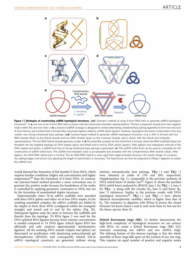
ResultsStrategies for the construction of ssRNA topological structures.The first ssRNA knot was constructed by Seeman’s helix-basedmethod21, the principle of which is that a half-turn (5 or 6 bp) ofRNA duplex generates a node (Fig. 1a,b). Recently, we haveexpanded the spectrum of synthetic DNA topologies with aversatile method based on the stacked X structure ofDNA 4WJ, in which the two helical strands (continuous alongthe stacked helices) are held by the two crossover strands(exchanged at the junction) to form a node for topologicalconstruction28. Compared with the previous helix-based method,this junction-based method has three major advantages. First, theresulting topological constructs contain no intrinsic strong basepairings. Second, the method enables the convenient generationof both positive and negative nodes. Finally, the methodcircumvents undesired braiding of the ssDNA linkers that isfrequently encountered in the helix-based method29. Thisjunction-based method can be further developed for syntheticRNA topologies by using the RNA–DNA hybrid 4WJ (Fig. 1c),which contains RNA helical strands and DNA crossover strands.Thus, RNA topological structures can be generated by foldingRNA scaffolds into hybrid 4WJs with DNA staples. Figure 1dillustrates the construction of an RNA trefoil knot using thehelix-based method. Alternatively, DNA-templated synthesis canbe used to construct an RNA topology, that is, a pre-preparedssDNA topological structure can be used as a template tosynthesize the corresponding RNA structure (Fig. 1e).
Right- and left-handed RNA trefoil knots with the same sequence.To produce closed RNA topological structures, the linearRNA strands can be ligated enzymatically. However, RNA strandssynthesized by in vitro runoff transcription, which is the mostconvenient and economic method to produce long RNA strands,contain both 50- and 30-heterogeneities. Therefore, theirenzymatic ligation poses a significant challenge. To solve theseproblems, a 50-end hammerhead ribozyme and a 30-end hepatitisdelta virus ribozyme30 were designed in each RNA transcriptto undergo self-cleavage to produce uniform ends (Fig. 2a).T4 polynucleotide kinase is then used to add a phosphate groupto the 50-hydroxyl group (in the presence of ATP) and removethe 20,30-cyclic phosphate31 of self-cleaved RNA transcripts.The resulting RNA molecules contain both ends proper forligation and can serve as the scaffolds for topologicalconstruction.
The RNA–DNA hybrid 4WJs presumably have structuralproperties similar to that of DNA 4WJs due to the fact thatthe former have been utilized in the construction of variousRNA–DNA hybrid nanostructures32,33, within which the 4WJsare composed of helical strands of RNA and crossover strands ofDNA. Therefore, the design principles of DNA nanostructuresbased on DNA 4WJs also hold for those containing theRNA–DNA hybrid 4WJs. The tensegrity triangle34, a structuralmotif containing three 4WJs, is a case in point and it is utilizedextensively in this work for topological construction. The numberof base pairs between the 4WJs dictates the tensegrity triangle tobe either right- or left-handed35, which results in positiveor negative nodes, respectively. The construction of topoisomersof both right- and left-handed trefoil knots can be achievedby folding the same RNA scaffolds into different tensegritytriangles with different sets of DNA staples (Fig. 2b). Aftergeometrical analysis according to previous work28,35 (see alsoSupplementary Table 1), we used a 17-bp-edged tensegritytriangle for the right-handed RNA trefoil knot 3þ
1ð Þ, TKj(þ )and 14-bp-edged for the left-handed trefoil knot 3�
1
� �, TKj(� ).
Notably, for the helix-based method, generating positive nodes
ARTICLE NATURE COMMUNICATIONS | DOI: 10.1038/ncomms14936
2 NATURE COMMUNICATIONS | 8:14936 | DOI: 10.1038/ncomms14936 | www.nature.com/naturecommunications
would demand the formation of left-handed Z-form RNA, whichrequires harsher conditions (higher salt concentration and highertemperature)36 than the formation of Z-form DNA. In contrast,our junction-based method provides a more convenient way togenerate the positive nodes because the handedness of the nodesis controlled by applying geometric constraints to 4WJs, but notby the formation of nonstandard duplex structures.
Experimentally, three 76-nt ssRNA scaffolds were annealedwith three DNA splints and either set of four DNA staples. In theresulting assembled complex, the ssRNA scaffolds are folded bythe staples to form three 4WJs (configured within the tensegritytriangle) and joined end to end by the splints (Fig. 2b).Subsequent ligation seals the nicks in between the scaffolds andthereby fixes the topology. T4 RNA ligase 2 was used for theDNA-splinted RNA ligation because lower enzyme concentrationis needed compared to T4 DNA ligase (which ligates RNA lessefficiently and only catalyses approximately stoichiometricligation). All the assisting DNA strands (staples and splints) aredissociated on purification with denaturing polyacrylamide gelelectrophoresis (dPAGE), and consequently relaxed 228-ntssRNA topological constructs are generated without strong
intrinsic intramolecular base pairings. TKj(þ ) and TKj(� )were obtained in yields of 13% and 28%, respectively(Supplementary Fig. 1), comparable to the previous synthesis ofDNA trefoil knots of similar size28. Figure 2c shows the purifiedRNA trefoil knots analysed by dPAGE: lane 1 for TKj(þ ), lane 3for TKj(� ), along with the circular (Cj, lane 5) and linear (Lj,lane 7) references. Similar to the previous results with DNAtopological structures28, TKj(þ ) and TKj(� ) have almostidentical electrophoretic mobility, which is higher than that ofCj. The resistance to digestion with RNase R proves the closedstructure for knots (lanes 2 and 4) and circle (lane 6), but not forthe linear species (lane 8).
Hybrid Borromean rings (BR). To further demonstrate thehigh-level complexity of topological structures we can achievewith RNA, we create a hybrid Borromean rings (BR) 632
� �
structure containing one ssRNA and two ssDNA rings.The defining feature of this famous topology is that the wholeassembly of three rings falls apart on the scission of any one ring.This requires an equal number of positive and negative nodes
ssDNA template DNA–RNA hybrid ssRNA knot
ComplementaryRNA strands DNase I
1. Annealing2. Ligation
Digestion
a b
c
e
d
Figure 1 | Strategies of constructing ssRNA topological structures. (a,b) Seeman’s method of using A-form RNA helix to generate ssRNA topological
structures21. In a, one turn of an A-form RNA helix is shown with the helical and schematic representations. The two component strands form two negative
nodes within this one-turn helix. In b, a strand of ssRNA (orange) is designed to contain alternating complementary pairing segments to form two one-turn
A-form helices, and a trefoil knot is formed after enzymatic ligation aided by a DNA splint (green). However, topological structures constructed in this way
contain very strong intrastrand base pairings. (c,d) Junction-based method to generate ssRNA topological structures. In c, a 4WJ is formed with two
RNA strands (blue) as the helical strands and two DNA strands (grey) as the crossover strands, and is shown with the helical and schematic
representations. The two RNA helical strands generate a node. In d, the assembly complex for the trefoil knot is formed, where the RNA scaffolds (blue) are
threaded into the targeted topology by DNA staples (grey) and linked end to end by DNA splints (green). After ligation and subsequent removal of the
DNA staples and splints, a ssRNA knot free of strong intrastrand base pairings is generated. (e) The ssDNA trefoil knot can be used as a template for the
construction of ssRNA trefoil knot. The ssDNA knot template (red) is pre-prepared and annealed with the complementary RNA strands (blue). After
ligation, the DNA–RNA hybrid knot is formed. The ds DNA–RNA hybrid is more rigid than single-stranded structure, the careful design of curvature
(by adding bulges) and torsion (by adjusting the length of hybrid helix) is necessary. The hybrid knot can then be subjected to DNase I digestion to obtain
the ssRNA knot.
NATURE COMMUNICATIONS | DOI: 10.1038/ncomms14936 ARTICLE
NATURE COMMUNICATIONS | 8:14936 | DOI: 10.1038/ncomms14936 |www.nature.com/naturecommunications 3

to ensure that no two rings are interlocked. In our previousconstruction of ssDNA BR28, two tensegrity triangles withdifferent handednesses were designed in the assembly complexto meet this requirement. Similarly, in the assembly complexfor this hybrid BR, a 95-nt circular RNA, a 105-nt circularDNA and two linear DNA (57- and 59-nt, respectively) scaffoldsare folded into a 17-edged right-handed tensegrity trianglefor the three positive nodes and a 14-edged left-handed onefor the three negative nodes (Fig. 2d). The two linearDNA scaffolds (precursors for the 116-nt DNA ring) are joinedby two splints and the hybrid BR structure is formed after ligationby T4 DNA ligase (Supplementary Fig. 2). To conclusively provethe topology of this hybrid BR, each DNA ring is installed witha unique restriction site. As shown in Fig. 2e, the hybridBR is disassembled by the cleavage of the ssRNA ring by RNaseH (lane 2), or either ssDNA ring by the corresponding nickase(lanes 3 and 4).
In the field of chemical topology, molecular BR is a well-knownchallenging target37, attracting researchers from differentdisciplines to develop various novel strategies to realizeit25,28,38–41. Here we further extend the chemical diversity ofthis topological target by creating this hybrid BR structure,
which is the first BR molecule to contain component rings ofdifferent materials. To the best of our knowledge, it is alsothe first topological structure composed of both DNA andRNA. Additionally, the successful construction of a topologicaltarget as complex as BR reasonably implies that our junction-based method would provide access to the ssRNA or ssRNA–ssDNA hybrid versions of any of our previous topological targetsrealized with ssDNA28.
ssRNA knot constructed via DNA-templated synthesis.Synthetic ssDNA topological structures constructed previously, inprinciple, can direct the synthesis of ssRNA structures of thesame topology (Fig. 1c). The intermediates would be thedouble-stranded (ds) RNA–DNA hybrid structures; however, theconversion of the structures from ssDNA to ds version is notalways straightforward. This is especially true for small ds knots,which are more difficult to synthesize than other ds topologicalstructures that contain only rings (such as rotaxanes5
or catenanes6). Because ds nucleic acid structure is more rigidand adopts a better-defined geometry compared to ssDNA orssRNA, constructing ds knot necessitates the careful designof curvature and torsion in 3D space28: (1) the total curvature
DNA template
PO O
O O-
5′HO3′
P O
O
-O
O-
OH
5′
3′
Run-off transcript
5′
3′HH ribozyme HDV ribozyme
Self-cleavage
Transcription
5′-phosphorylation &3′-dephosphorylation
3 Scaffolds
3 Splints
14 bp
17 bp
Staple set 1
Staple set 2
nt
766
500
350300
250
200
RNase R digestion
M1
CjTKj(+) TKj(–) Lj
2 3 4 5 6 7 8
– + – + – + – +
95-nt RNA ring
57- and 59-nt linear DNA
105-nt DNA ring
4 Scaffolds
RNA suitable for ligation
8 Staples
2 Splints
M 1 2 3 4 5 6 7 8
Hybrid BR
DNA C116
DNA L116
DNA C105
RNA C95
RNA L95
DNA L105
nt
766500300
200
150
100
75
a b c
d e
31+
31–
623
Figure 2 | Constructing ssRNA topological structures with the junction-based method. (a) Preparation of the ssRNA strand with uniform ends and
proper end groups for the DNA-splinted RNA ligation. (b) Both the positive (TKj(þ )) and negative (TKj(� )) RNA trefoil knots of the same sequence are
constructed by configuring tensegrity triangles with different handedness. The same scaffolds (blue) are threaded by different staple sets (grey or purple)
to form a 17-bp-edged right-handed tensegrity triangle for the positive trefoil knot, or a 14-bp-edged left-handed tensegrity triangle for the negative one,
respectively. Each topology is designated by the Alexander–Briggs notation ni or nCi , where n is the minimal number of nodes, C is the number of
components (for links), and i distinguishes different topologies with the same n and C. (c) dPAGE analysis of TKj(þ ) (lanes 1 and 2), TKj(� ) (lanes 3 and
4), and their circular (Cj, lanes 5 and 6) and linear (Lj, lanes 7 and 8) counterparts. Lanes 2, 4, 6 and 8 contain samples digested by RNase R. (d) The
assembly complex for the hybrid BR contains tensegrity triangles of both handedness to generate three positive nodes plus three negative nodes.
(e) Topological analyses of the hybrid BR. Lane 1, gel-purified BR; lanes 2–6, BR treated by RNase H, Nt.AlwI (for cleaving the red ring), Nt.BspQI
(for cleaving the green ring), DNase I, and E. coli DNA Topo I; lanes 7 and 8, DNA and RNA references of the three individual components. During the
purification of BR, the breaking down of the 95-nt circular RNA component is unavoidable, and a portion of BR falls apart as a result. The treatment of BR by
RNase H, Nt.AlwI and Nt.BspQI is conducted in the presence of an assisting DNA strand complementary to the corresponding ssRNA or ssDNA ring.
LX and CX represent X-nt linear and circular species, respectively. In all the gels, lane M contains the DNA size markers.
ARTICLE NATURE COMMUNICATIONS | DOI: 10.1038/ncomms14936
4 NATURE COMMUNICATIONS | 8:14936 | DOI: 10.1038/ncomms14936 | www.nature.com/naturecommunications
of a knot should be larger than 4p (more than two times thatrequired for a circle) according to the Fary–Milnor theorem;(2) the torsion of a knotted space curve should not be zeroeverywhere because a knot cannot be flattened onto the plane.
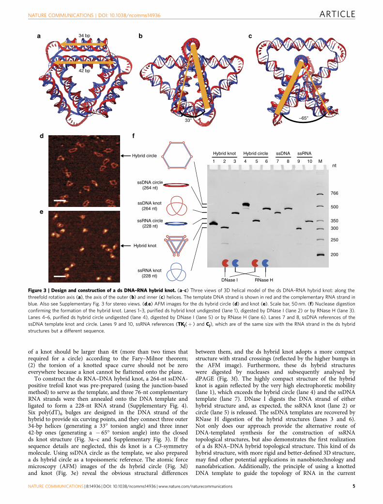
To construct the ds RNA–DNA hybrid knot, a 264-nt ssDNA-positive trefoil knot was pre-prepared (using the junction-basedmethod) to serve as the template, and three 76-nt complementaryRNA strands were then annealed onto the DNA template andligated to form a 228-nt RNA strand (Supplementary Fig. 4).Six poly(dT)6 bulges are designed in the DNA strand of thehybrid to provide six curving points, and they connect three outer34-bp helices (generating a 33� torsion angle) and three inner42-bp ones (generating a � 65� torsion angle) into the closedds knot structure (Fig. 3a–c and Supplementary Fig. 3). If thesequence details are neglected, this ds knot is a C3-symmetrymolecule. Using ssDNA circle as the template, we also prepareda ds hybrid circle as a topoisomeric reference. The atomic forcemicroscopy (AFM) images of the ds hybrid circle (Fig. 3d)and knot (Fig. 3e) reveal the obvious structural differences
between them, and the ds hybrid knot adopts a more compactstructure with strand crossings (reflected by the higher bumps inthe AFM image). Furthermore, these ds hybrid structureswere digested by nucleases and subsequently analysed bydPAGE (Fig. 3f). The highly compact structure of the hybridknot is again reflected by the very high electrophoretic mobility(lane 1), which exceeds the hybrid circle (lane 4) and the ssDNAtemplate (lane 7). DNase I digests the DNA strand of eitherhybrid structure and, as expected, the ssRNA knot (lane 2) orcircle (lane 5) is released. The ssDNA templates are recovered byRNase H digestion of the hybrid structures (lanes 3 and 6).Not only does our approach provide the alternative route ofDNA-templated synthesis for the construction of ssRNAtopological structures, but also demonstrates the first realizationof a ds RNA–DNA hybrid topological structure. This kind of dshybrid structure, with more rigid and better-defined 3D structure,may find other potential applications in nanobiotechnology andnanofabrication. Additionally, the principle of using a knottedDNA template to guide the topology of RNA in the current
34 bp
33° –65°
42 bp
nt
766
500
350
300
250
200
Hybrid knot
DNase I RNase H
Hybrid circle Hybrid knot Hybrid circle ssDNA ssRNA
ssRNA knot(228 nt)
ssRNA circle(228 nt)
ssDNA circle(264 nt)
ssDNA knot(264 nt)
M1 2 3 4 5 6 7 8 9 10
e
a b c
d
e
f
Figure 3 | Design and construction of a ds DNA–RNA hybrid knot. (a–c) Three views of 3D helical model of the ds DNA–RNA hybrid knot: along the
threefold rotation axis (a), the axis of the outer (b) and inner (c) helices. The template DNA strand is shown in red and the complementary RNA strand in
blue. Also see Supplementary Fig. 3 for stereo views. (d,e) AFM images for the ds hybrid circle (d) and knot (e). Scale bar, 50 nm. (f) Nuclease digestion
confirming the formation of the hybrid knot. Lanes 1–3, purified ds hybrid knot undigested (lane 1), digested by DNase I (lane 2) or by RNase H (lane 3).
Lanes 4–6, purified ds hybrid circle undigested (lane 4), digested by DNase I (lane 5) or by RNase H (lane 6). Lanes 7 and 8, ssDNA references of the
ssDNA template knot and circle. Lanes 9 and 10, ssRNA references (TKj(þ ) and Cj), which are of the same size with the RNA strand in the ds hybrid
structures but a different sequence.
NATURE COMMUNICATIONS | DOI: 10.1038/ncomms14936 ARTICLE
NATURE COMMUNICATIONS | 8:14936 | DOI: 10.1038/ncomms14936 |www.nature.com/naturecommunications 5
research reveals the possibility of synthesizing topologicalstructures of other non-nucleic acid materials with the moregeneral DNA-templated synthesis42.
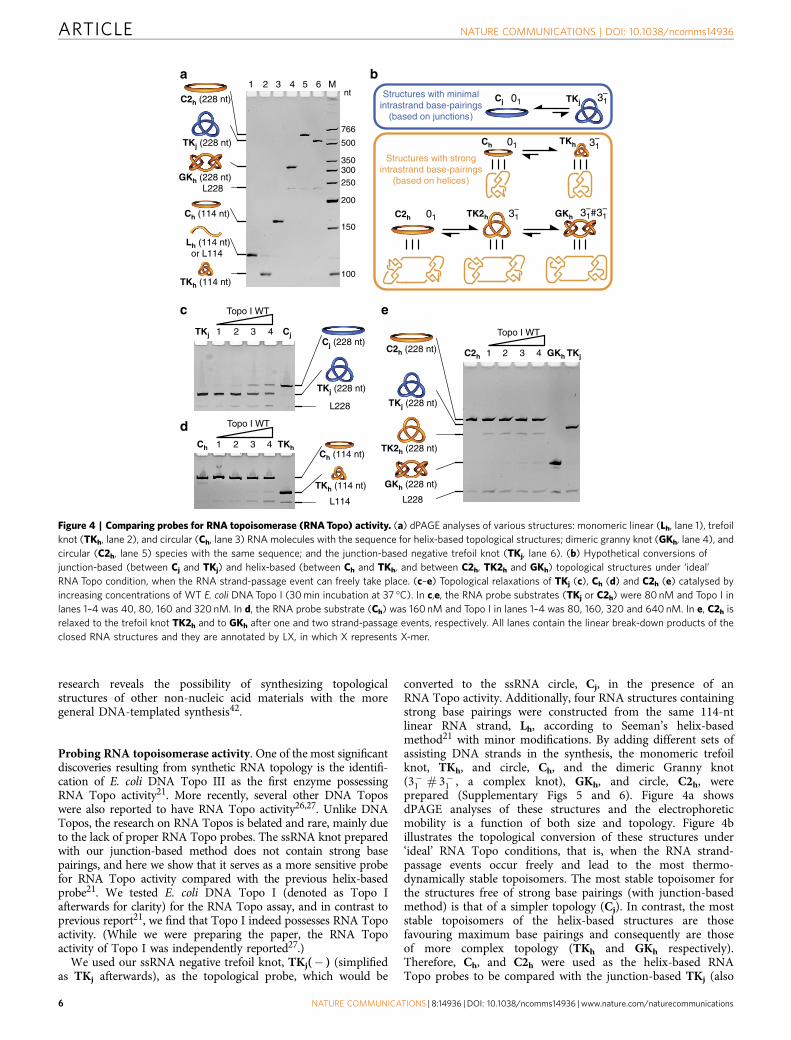
Probing RNA topoisomerase activity. One of the most significantdiscoveries resulting from synthetic RNA topology is the identifi-cation of E. coli DNA Topo III as the first enzyme possessingRNA Topo activity21. More recently, several other DNA Toposwere also reported to have RNA Topo activity26,27. Unlike DNATopos, the research on RNA Topos is belated and rare, mainly dueto the lack of proper RNA Topo probes. The ssRNA knot preparedwith our junction-based method does not contain strong basepairings, and here we show that it serves as a more sensitive probefor RNA Topo activity compared with the previous helix-basedprobe21. We tested E. coli DNA Topo I (denoted as Topo Iafterwards for clarity) for the RNA Topo assay, and in contrast toprevious report21, we find that Topo I indeed possesses RNA Topoactivity. (While we were preparing the paper, the RNA Topoactivity of Topo I was independently reported27.)
We used our ssRNA negative trefoil knot, TKj(� ) (simplifiedas TKj afterwards), as the topological probe, which would be
converted to the ssRNA circle, Cj, in the presence of anRNA Topo activity. Additionally, four RNA structures containingstrong base pairings were constructed from the same 114-ntlinear RNA strand, Lh, according to Seeman’s helix-basedmethod21 with minor modifications. By adding different sets ofassisting DNA strands in the synthesis, the monomeric trefoilknot, TKh, and circle, Ch, and the dimeric Granny knot(3�
1 # 3�1 , a complex knot), GKh, and circle, C2h, were
prepared (Supplementary Figs 5 and 6). Figure 4a showsdPAGE analyses of these structures and the electrophoreticmobility is a function of both size and topology. Figure 4billustrates the topological conversion of these structures under‘ideal’ RNA Topo conditions, that is, when the RNA strand-passage events occur freely and lead to the most thermo-dynamically stable topoisomers. The most stable topoisomer forthe structures free of strong base pairings (with junction-basedmethod) is that of a simpler topology (Cj). In contrast, the moststable topoisomers of the helix-based structures are thosefavouring maximum base pairings and consequently are thoseof more complex topology (TKh and GKh respectively).Therefore, Ch, and C2h were used as the helix-based RNATopo probes to be compared with the junction-based TKj (also
1 2 3 4 GKh TKjC2h
TK2h (228 nt)
Topo I WT
TKj (228 nt)
C2h (228 nt)
GKh (228 nt)
L228
1 2 3 4 TKhChCh (114 nt)
TKh (114 nt)
L114
Topo I WT
Topo I WT
TKj (228 nt)
L228
Cj (228 nt)1 2 3 4 CjTKj
Structures with minimalintrastrand base-pairings
(based on junctions)
Structures with strongintrastrand base-pairings
(based on helices)
Cj TKj
TKh
TK2h GKh
01
01
nt
766
500
350300
250
200
150
100
1 2 3 4 5 6 M
C2h (228 nt)
TKj (228 nt)
GKh (228 nt) L228
Ch (114 nt)
TKh (114 nt)
Lh (114 nt)or L114
a b
c
d
e
Ch
01C2h
31–
31–
31– 31#31
– –
Figure 4 | Comparing probes for RNA topoisomerase (RNATopo) activity. (a) dPAGE analyses of various structures: monomeric linear (Lh, lane 1), trefoil
knot (TKh, lane 2), and circular (Ch, lane 3) RNA molecules with the sequence for helix-based topological structures; dimeric granny knot (GKh, lane 4), and
circular (C2h, lane 5) species with the same sequence; and the junction-based negative trefoil knot (TKj, lane 6). (b) Hypothetical conversions of
junction-based (between Cj and TKj) and helix-based (between Ch and TKh, and between C2h, TK2h and GKh) topological structures under ‘ideal’
RNA Topo condition, when the RNA strand-passage event can freely take place. (c–e) Topological relaxations of TKj (c), Ch (d) and C2h (e) catalysed by
increasing concentrations of WT E. coli DNA Topo I (30min incubation at 37 �C). In c,e, the RNA probe substrates (TKj or C2h) were 80nM and Topo I in
lanes 1–4 was 40, 80, 160 and 320nM. In d, the RNA probe substrate (Ch) was 160 nM and Topo I in lanes 1–4 was 80, 160, 320 and 640 nM. In e, C2h is
relaxed to the trefoil knot TK2h and to GKh after one and two strand-passage events, respectively. All lanes contain the linear break-down products of the
closed RNA structures and they are annotated by LX, in which X represents X-mer.
ARTICLE NATURE COMMUNICATIONS | DOI: 10.1038/ncomms14936
6 NATURE COMMUNICATIONS | 8:14936 | DOI: 10.1038/ncomms14936 | www.nature.com/naturecommunications
see Supplementary Fig. 7 for more explanation of why Ch, but notTKh, is used as the helix-based probe).
Figure 4c shows the increasing conversion of TKj to Cj catalysedby increasing concentrations of wild-type (WT) Topo I. ThoughTopo I has RNA Topo activity, this activity is relatively low andB28–35% of conversion was observed when the molar ratio ofTopo I to RNA is 4:1 (lane 4). Based on our previous result thatTopo I catalyses the fast approximately stoichiometric unknottingof ssDNA knot within 30min28, the RNA Topo activity of Topo I isestimated to beB1/15–1/12 of the DNA Topo activity. Comparingwith TKj, Ch is a much less sensitive RNA Topo probe (Fig. 4d).Only B3% conversion was observed when the molar ratio of TopoI to RNA is 4:1 (lane 4). Figure 4e shows the RNA Topo assay usingprobe C2h. Though C2h is more sensitive than Ch, probably due tothe more severe topological stress, it is still not as sensitive as TKj athigh enzyme-to-RNA ratios. As expected, there are two products ofthe topological conversion of C2h, the larger-amount intermediatetrefoil knot TK2h (after one strand-passage event), and the smaller-amount final GKh (after two strand-passage events). We also findthat TK2h migrates slightly faster than TKj, though both are of thesame size and topology, probably due to the different sequences orthe formation of transient base pairings within TK2h even duringmigration in the dPAGE.
The observation that Ch and C2h (helix-based) are not assensitive probes as TKj (junction-based) can be explained by boththe Topo I binding to ssRNA and its low RNA Topo activity. TopoI binding is expected to inhibit the formation of base pairings,countering the thermodynamic driving force for the topologicalconversion illustrated in Fig. 4b for Ch and C2h. This problem isfurther exaggerated due to the low RNA Topo activity, whichnecessitates a higher concentration of Topo I and ultimately leadsto even more severe protein binding. The low sensitivity of thehelix-based probe may account for the previous failure of findingRNA Topo activity of Topo I (ref. 21). Besides, better sensitivitywould circumvent the inconvenience associated with the use ofautoradiography with 32P-labelled RNA as described in the recentstudy27.
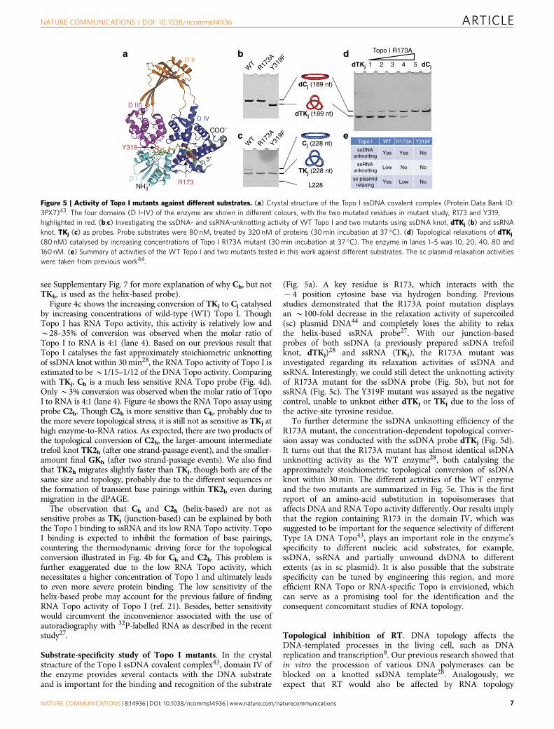
Substrate-specificity study of Topo I mutants. In the crystalstructure of the Topo I ssDNA covalent complex43, domain IV ofthe enzyme provides several contacts with the DNA substrateand is important for the binding and recognition of the substrate
(Fig. 5a). A key residue is R173, which interacts with the� 4 position cytosine base via hydrogen bonding. Previousstudies demonstrated that the R173A point mutation displaysan B100-fold decrease in the relaxation activity of supercoiled(sc) plasmid DNA44 and completely loses the ability to relaxthe helix-based ssRNA probe27. With our junction-basedprobes of both ssDNA (a previously prepared ssDNA trefoilknot, dTKj)28 and ssRNA (TKj), the R173A mutant wasinvestigated regarding its relaxation activities of ssDNA andssRNA. Interestingly, we could still detect the unknotting activityof R173A mutant for the ssDNA probe (Fig. 5b), but not forssRNA (Fig. 5c). The Y319F mutant was assayed as the negativecontrol, unable to unknot either dTKj or TKj due to the loss ofthe active-site tyrosine residue.
To further determine the ssDNA unknotting efficiency of theR173A mutant, the concentration-dependent topological conver-sion assay was conducted with the ssDNA probe dTKj (Fig. 5d).It turns out that the R173A mutant has almost identical ssDNAunknotting activity as the WT enzyme28, both catalysing theapproximately stoichiometric topological conversion of ssDNAknot within 30min. The different activities of the WT enzymeand the two mutants are summarized in Fig. 5e. This is the firstreport of an amino-acid substitution in topoisomerases thataffects DNA and RNA Topo activity differently. Our results implythat the region containing R173 in the domain IV, which wassuggested to be important for the sequence selectivity of differentType IA DNA Topo43, plays an important role in the enzyme’sspecificity to different nucleic acid substrates, for example,ssDNA, ssRNA and partially unwound dsDNA to differentextents (as in sc plasmid). It is also possible that the substratespecificity can be tuned by engineering this region, and moreefficient RNA Topo or RNA-specific Topo is envisioned, whichcan serve as a promising tool for the identification and theconsequent concomitant studies of RNA topology.
Topological inhibition of RT. DNA topology affects theDNA-templated processes in the living cell, such as DNAreplication and transcription8. Our previous research showed thatin vitro the procession of various DNA polymerases can beblocked on a knotted ssDNA template28. Analogously, weexpect that RT would also be affected by RNA topology
WT
R173A
Y319F
WT
R173A
Y319F
TKj (228 nt)
L228
Cj (228 nt)
dTKj (189 nt)
dCj (189 nt)
Topo I R173A
1 2 3 4 5 dCjdTKj
Topo I WT R173A Y319F
ssDNAunknotting
ssRNAunknotting
Yes Yes
Yes
No
Low No No
sc plasmidrelaxing
Low No
D II
D III
R173
Y319
D I
D IV
COO–
NH3+
5′3’
a b d
ec
Figure 5 | Activity of Topo I mutants against different substrates. (a) Crystal structure of the Topo I ssDNA covalent complex (Protein Data Bank ID:
3PX7)43. The four domains (D I–IV) of the enzyme are shown in different colours, with the two mutated residues in mutant study, R173 and Y319,
highlighted in red. (b,c) Investigating the ssDNA- and ssRNA-unknotting activity of WT Topo I and two mutants using ssDNA knot, dTKj (b) and ssRNA
knot, TKj (c) as probes. Probe substrates were 80 nM, treated by 320 nM of proteins (30min incubation at 37 �C). (d) Topological relaxations of dTKj
(80 nM) catalysed by increasing concentrations of Topo I R173A mutant (30min incubation at 37 �C). The enzyme in lanes 1–5 was 10, 20, 40, 80 and
160 nM. (e) Summary of activities of the WT Topo I and two mutants tested in this work against different substrates. The sc plasmid relaxation activities
were taken from previous work44.
NATURE COMMUNICATIONS | DOI: 10.1038/ncomms14936 ARTICLE
NATURE COMMUNICATIONS | 8:14936 | DOI: 10.1038/ncomms14936 |www.nature.com/naturecommunications 7
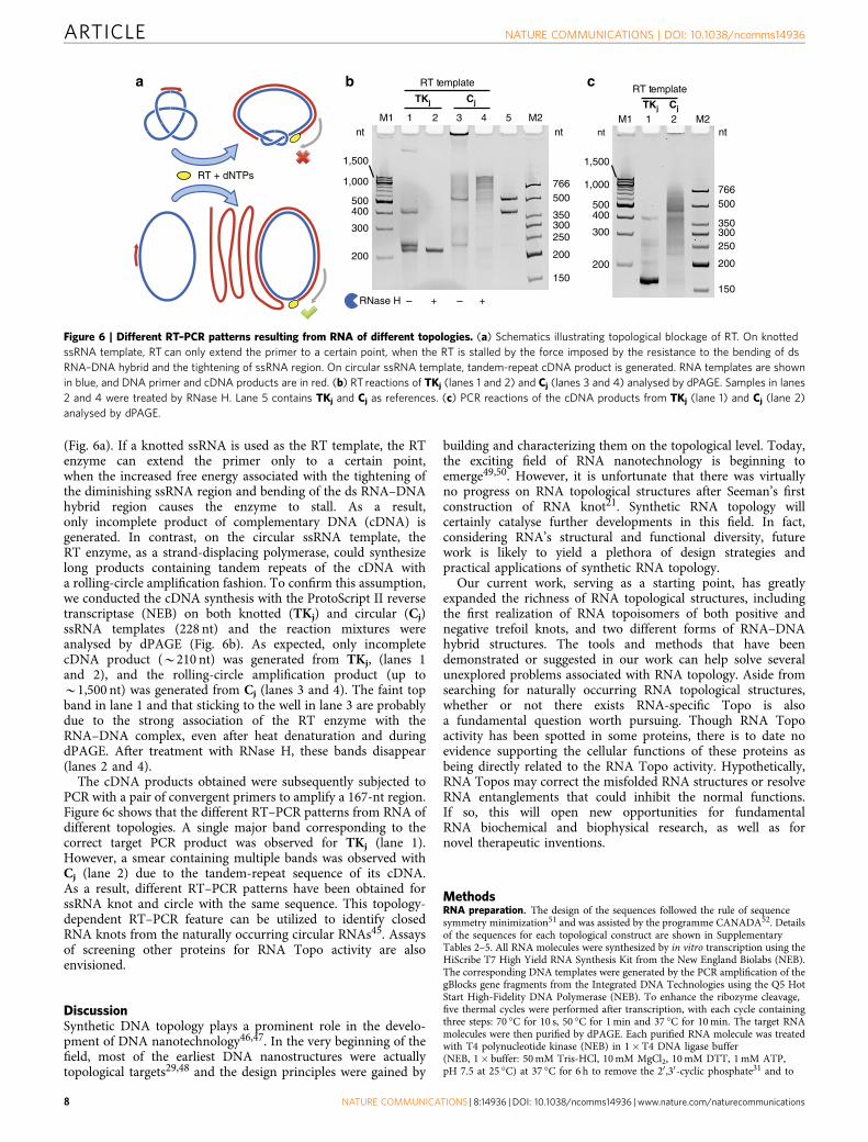
(Fig. 6a). If a knotted ssRNA is used as the RT template, the RTenzyme can extend the primer only to a certain point,when the increased free energy associated with the tightening ofthe diminishing ssRNA region and bending of the ds RNA–DNAhybrid region causes the enzyme to stall. As a result,only incomplete product of complementary DNA (cDNA) isgenerated. In contrast, on the circular ssRNA template, theRT enzyme, as a strand-displacing polymerase, could synthesizelong products containing tandem repeats of the cDNA witha rolling-circle amplification fashion. To confirm this assumption,we conducted the cDNA synthesis with the ProtoScript II reversetranscriptase (NEB) on both knotted (TKj) and circular (Cj)ssRNA templates (228 nt) and the reaction mixtures wereanalysed by dPAGE (Fig. 6b). As expected, only incompletecDNA product (B210 nt) was generated from TKj, (lanes 1and 2), and the rolling-circle amplification product (up toB1,500 nt) was generated from Cj (lanes 3 and 4). The faint topband in lane 1 and that sticking to the well in lane 3 are probablydue to the strong association of the RT enzyme with theRNA–DNA complex, even after heat denaturation and duringdPAGE. After treatment with RNase H, these bands disappear(lanes 2 and 4).
The cDNA products obtained were subsequently subjected toPCR with a pair of convergent primers to amplify a 167-nt region.Figure 6c shows that the different RT–PCR patterns from RNA ofdifferent topologies. A single major band corresponding to thecorrect target PCR product was observed for TKj (lane 1).However, a smear containing multiple bands was observed withCj (lane 2) due to the tandem-repeat sequence of its cDNA.As a result, different RT–PCR patterns have been obtained forssRNA knot and circle with the same sequence. This topology-dependent RT–PCR feature can be utilized to identify closedRNA knots from the naturally occurring circular RNAs45. Assaysof screening other proteins for RNA Topo activity are alsoenvisioned.
DiscussionSynthetic DNA topology plays a prominent role in the develo-pment of DNA nanotechnology46,47. In the very beginning of thefield, most of the earliest DNA nanostructures were actuallytopological targets29,48 and the design principles were gained by
building and characterizing them on the topological level. Today,the exciting field of RNA nanotechnology is beginning toemerge49,50. However, it is unfortunate that there was virtuallyno progress on RNA topological structures after Seeman’s firstconstruction of RNA knot21. Synthetic RNA topology willcertainly catalyse further developments in this field. In fact,considering RNA’s structural and functional diversity, futurework is likely to yield a plethora of design strategies andpractical applications of synthetic RNA topology.
Our current work, serving as a starting point, has greatlyexpanded the richness of RNA topological structures, includingthe first realization of RNA topoisomers of both positive andnegative trefoil knots, and two different forms of RNA–DNAhybrid structures. The tools and methods that have beendemonstrated or suggested in our work can help solve severalunexplored problems associated with RNA topology. Aside fromsearching for naturally occurring RNA topological structures,whether or not there exists RNA-specific Topo is alsoa fundamental question worth pursuing. Though RNA Topoactivity has been spotted in some proteins, there is to date noevidence supporting the cellular functions of these proteins asbeing directly related to the RNA Topo activity. Hypothetically,RNA Topos may correct the misfolded RNA structures or resolveRNA entanglements that could inhibit the normal functions.If so, this will open new opportunities for fundamentalRNA biochemical and biophysical research, as well as fornovel therapeutic inventions.
MethodsRNA preparation. The design of the sequences followed the rule of sequencesymmetry minimization51 and was assisted by the programme CANADA52. Detailsof the sequences for each topological construct are shown in SupplementaryTables 2–5. All RNA molecules were synthesized by in vitro transcription using theHiScribe T7 High Yield RNA Synthesis Kit from the New England Biolabs (NEB).The corresponding DNA templates were generated by the PCR amplification of thegBlocks gene fragments from the Integrated DNA Technologies using the Q5 HotStart High-Fidelity DNA Polymerase (NEB). To enhance the ribozyme cleavage,five thermal cycles were performed after transcription, with each cycle containingthree steps: 70 �C for 10 s, 50 �C for 1min and 37 �C for 10min. The target RNAmolecules were then purified by dPAGE. Each purified RNA molecule was treatedwith T4 polynucleotide kinase (NEB) in 1�T4 DNA ligase buffer(NEB, 1� buffer: 50mM Tris-HCl, 10mM MgCl2, 10mM DTT, 1mM ATP,pH 7.5 at 25 �C) at 37 �C for 6 h to remove the 20 ,30-cyclic phosphate31 and to
RT + dNTPs
nt
766500
300350
250
200
150
nt
1,000
1,500
500
300
400
200
RT template
TKj Cj
RNase H – + – +
M2M1 1 2 3 4 5nt
766500
300350
250
200
150
nt
1,000
1,500
500
300
400
200
M2TKj Cj
RT template
M1 1 2
a b c
Figure 6 | Different RT–PCR patterns resulting from RNA of different topologies. (a) Schematics illustrating topological blockage of RT. On knotted
ssRNA template, RT can only extend the primer to a certain point, when the RT is stalled by the force imposed by the resistance to the bending of ds
RNA–DNA hybrid and the tightening of ssRNA region. On circular ssRNA template, tandem-repeat cDNA product is generated. RNA templates are shown
in blue, and DNA primer and cDNA products are in red. (b) RT reactions of TKj (lanes 1 and 2) and Cj (lanes 3 and 4) analysed by dPAGE. Samples in lanes
2 and 4 were treated by RNase H. Lane 5 contains TKj and Cj as references. (c) PCR reactions of the cDNA products from TKj (lane 1) and Cj (lane 2)
analysed by dPAGE.
ARTICLE NATURE COMMUNICATIONS | DOI: 10.1038/ncomms14936
8 NATURE COMMUNICATIONS | 8:14936 | DOI: 10.1038/ncomms14936 | www.nature.com/naturecommunications

add a phosphate to 50-hydroxyl end. After the treatment, these RNA moleculeswere directly used as RNA scaffolds for the topological construction.
Topological construction. The ssRNA topological constructs were prepared withthe all-in-one protocol. It involves two steps: (1) Annealing to get the assemblycomplex. Equimolecular quantities (normally with a final concentration of 1 mMeach) of all strands (RNA scaffolds, DNA staples and splints) were mixed ina buffer with the ultimate concentration of 1�T4 DNA ligase buffer (by adding10�T4 DNA ligase buffer because the kinase-treated RNA scaffolds were in1� buffer) and annealed from 70 to 16 �C over 4 h. (2) Ligation to seal the nicks.To each 100 ml of reaction mixture, 4 ml of T4 RNA ligase 2 (NEB, 10U ml� 1),1.5 ml of 100mM fresh DTT (NEB) and 1.5 ml of 100mM fresh ATP (NEB) wereadded and incubated at 16 �C at least 16 h for the ligation. The ds RNA–DNAhybrid structures were prepared using the corresponding ssDNA knots astemplates, which were prepared according to previous work28. The complementaryRNA strands were designed as three substrands, which were annealed to thessDNA knot templates with a ratio of complementary:template¼ 1.2:1. Afterthe annealing, T4 RNA ligase 2 was used to seal the nicks in complementaryRNA strands.
dPAGE. Gels of different concentrations were prepared using 30% acrylamideand bis-acrylamide solution (Bio-Rad, 29:1) with 7M urea in 0.5�TBE buffer(Bio-Rad) and run on a PROTEAN II xi cell (Bio-Rad) or a Mini-PROTEAN Tetracell (Bio-Rad). Samples were mixed 1:1 with TBE-urea sample buffer (Bio-Rad) andheated at 70 �C for 5min before they were loaded into the wells. Gel concentrationswere carefully chosen to ensure the proper separations between different topologiesas well as the references. For imaging, gels were stained with GelRed (Biotium), andimages were taken by Gel Doc XRþ (Bio-Rad) imaging system and processed bysoftware Image Lab (v.4.0.1, Bio-Rad). For purification, gels (without staining)were visualized by UV shadowing against a fluorescent thin-layer chromatographyplate and bands of interest were cut. The bands were then eluted using thecrush-and-soak method and the eluent was purified and concentrated on3K Nanosep filters (Pall). The concentration of product was determined bymeasuring the OD260. Optionally, the ssRNA knots and circles can be digestedby RNase R (Epicentre) after the gel extraction to remove the unavoidablecleaved linear RNA during the purification. However, RNase R digestion is notuseful for the hybrid BR.
Digestion with various nucleases. Various nucleases were used in this work,including Nt.AlwI (NEB), Nt.BspQI (NEB), RNase H (NEB), DNase I (NEB) andRNase R (Epicentre). We used the reaction conditions for these enzymes asrecommended by the providers.
Topoisomerase assay. The E. coli Topo I WT and mutant proteins wereexpressed and purified as described in previous study53. Commercial productof E. coli Topo I (NEB) was also tested and its RNA Topo activity was found to beslightly higher than that of the in-house prepared WT enzyme, probably due to thecontamination of E. coli Topo III (Supplementary Fig. 8). The reaction buffercontained 1�NEBuffer 4 (NEB, 1� buffer: 50mM KOAc, 20mM Tris-acetate,10mM Mg(OAc)2, 1mM DTT, pH 7.9 at 25 �C) and 100mgml� 1 BSA. Theconcentration of substrates and proteins were described in the text. Reactionswere quenched by phenol–chloroform extraction followed by ethanol precipitation.The reactions were then analysed by dPAGE.
AFM imaging. For AFM imaging of the ds RNA–DNA hybrid structures, 30 ml of0.1mgml� 1 polyornithine (Sigma) solution was added to freshly cleaved mica andstand for 3min to increase the binding to the structures before applying thesamples. Then the mica was rinsed with 1ml water and dried with compressed air.An aliquot of 5 ml of each sample (about 5 nM) in 1�TAE-Mg buffer (11mMMgCl2, 40mM Tris, 20mM acetic acid, 1mM EDTA, pH 8.0) was applied to thetreated mica and stand for 1min. Then the mica was rinsed with 1ml water anddried with compressed air. AFM imaging was performed on a Veeco MultiMode 8AFM in the ScanAsyst in air mode using the scanayst-air tips (Veeco). The AFMimages were processed with the software Gwyddion.
RT–PCR. The ProtoScript II First Strand cDNA Synthesis Kit (NEB) was used forthe RT. Each reaction (20 ml) contained 100 nM ssRNA template (TKj or Cj) and200 nM primer (sequence is shown in Supplementary Table 6) and followed therecommended protocol. After RT, the enzyme was heat-inactivated at 80 �C for5min. Then the reactions were treated by RNase H. For the subsequent PCR,HotStart-IT FideliTaq DNA Polymerase (Affymetrix) was used and 1 ml of each RTmixture was added to each 50 ml of PCR reaction for amplification.
Data availability. Data supporting the findings of this study are available withinthe article and its Supplementary Information Files, and from the correspondingauthor on reasonable request.
References1. Wasserman, E. The preparation of interlocking rings: a catenane. J. Am. Chem.
Soc. 82, 4433–4434 (1960).2. Frank-Kamenetskii, M. D., Lukashin, A. V. & Vologodskii, A. V.
Statistical mechanics and topology of polymer chains. Nature 258,398–402 (1975).
3. Forgan, R. S., Sauvage, J. P. & Stoddart, J. F. Chemical topology: complexmolecular knots, links, and entanglements. Chem. Rev. 111, 5434–5464ð2011Þ:
4. Ayme, J. F., Beves, J. E., Campbell, C. J. & Leigh, D. A. Template synthesis ofmolecular knots. Chem. Soc. Rev. 42, 1700–1712 (2013).
5. Gil-Ramirez, G., Leigh, D. A. & Stephens, A. J. Catenanes: fifty years ofmolecular links. Angew. Chem. Int. Ed. 54, 6110–6150 (2015).
6. Lim, N. C. & Jackson, S. E. Molecular knots in biology and chemistry. J. Phys.Condens. Matter 27, 354101 (2015).
7. Droge, P. & Cozzarelli, N. R. Topological structure of DNA knots andcatenanes. Methods Enzymol. 212, 120–130 (1992).
8. Bates, A. D. & Maxwell, A. DNA Topology 2 edn (Oxford Univ. Press, 2005).9. Virnau, P., Mirny, L. A. & Kardar, M. Intricate knots in proteins: function and
evolution. PLoS Comput. Biol. 2, e122 (2006).10. Sulkowska, J. I., Rawdon, E. J., Millett, K. C., Onuchic, J. N. & Stasiak, A.
Conservation of complex knotting and slipknotting patterns in proteins.Proc. Natl Acad. Sci. USA 109, E1715–E1723 (2012).
11. Pommier, Y. DNA Topoisomerases and Cancer 1 edn (Springer-Verlag, 2012).12. Wang, J. C. DNA topoisomerases—why so many. J. Biol. Chem. 266,
6659–6662 (1991).13. Wang, J. C. Cellular roles of DNA topoisomerases: a molecular perspective.
Nat. Rev. Mol. Cell Biol. 3, 430–440 (2002).14. Chen, S. H., Chan, N. L. & Hsieh, T. S. New mechanistic and functional
insights into DNA topoisomerases. Annu. Rev. Biochem. 82, 139–170ð2013Þ:
15. Micheletti, C., Di Stefano, M. & Orland, H. Absence of knots inknown RNA structures. Proc. Natl Acad. Sci. USA 112, 2052–2057ð2015Þ:
16. Burton, A. S., Di Stefano, M., Lehman, N., Orland, H. & Micheletti, C. Theelusive quest for RNA knots. RNA Biol. 13, 134–139 (2016).
17. van Batenburg, F. H. D. PseudoBase: a database with RNA pseudoknots.Nucleic Acids Res. 28, 201–204 (2000).
18. Seeman, N. C. in Molecular Catenanes, Rotaxanes and Knots (eds Sauvage, J. P.& Dietrich-Buchecker, C.) 323–356 (Wiley, 2007).
19. Jester, S. S. & Famulok, M. Mechanically interlocked DNA nanostructures forfunctional devices. Acc. Chem. Res. 47, 1700–1709 (2014).
20. Lu, C. H., Cecconello, A. & Willner, I. Recent advances in the synthesis andfunctions of reconfigurable interlocked DNA nanostructures. J. Am. Chem. Soc.138, 5172–5185 (2016).
21. Wang, H., Di Gate, R. J. & Seeman, N. C. An RNA topoisomerase. Proc. NatlAcad. Sci. USA 93, 9477–9482 (1996).
22. Mueller, J. E., Du, S. M. & Seeman, N. C. Design and synthesis of a knot fromsingle-stranded DNA. J. Am. Chem. Soc. 113, 6306–6308 (1991).
23. Du, S. M. & Seeman, N. C. Synthesis of a DNA knot containing both positiveand negative nodes. J. Am. Chem. Soc. 114, 9652–9655 (1992).
24. Du, S. M., Stollar, B. D. & Seeman, N. C. A synthetic DNA molecule in threeknotted topologies. J. Am. Chem. Soc. 117, 1194–1200 (1995).
25. Mao, C., Sun, W. & Seeman, N. C. Assembly of Borromean rings from DNA.Nature 386, 137–138 (1997).
26. Xu, D. et al. Top3b is an RNA topoisomerase that works with fragile Xsyndrome protein to promote synapse formation. Nat. Neurosci. 16, 1238–1247(2013).
27. Ahmad, M. et al. RNA topoisomerase is prevalent in all domains of life andassociates with polyribosomes in animals. Nucleic Acids Res. 44, 6335–6349(2016).
28. Liu, D., Chen, G., Akhter, U., Cronin, T. M. & Weizmann, Y. Creating complexmolecular topologies by configuring DNA four-way junctions. Nat. Chem. 8,907–914 (2016).
29. Chen, J. H. & Seeman, N. C. Synthesis from DNA of a molecule with theconnectivity of a cube. Nature 350, 631–633 (1991).
30. Avis, J. M., Conn, G. L. & Walker, S. C. Cis-acting ribozymes for the productionof RNA in vitro transcripts with defined 50 and 30 ends.Methods Mol. Biol. 941,83–98 (2012).
31. Wichlacz, A., Legiewicz, M. & Ciesiolka, J. Generating in vitro transcripts withhomogenous 30 ends using trans-acting antigenomic delta ribozyme. NucleicAcids Res. 32, e39 (2004).
32. Ko, S. H. et al. Synergistic self-assembly of RNA and DNA molecules. Nat.Chem. 2, 1050–1055 (2010).
33. Wang, P., Ko, S. H., Tian, C., Hao, C. & Mao, C. RNA-DNA hybridorigami: folding of a long RNA single strand into complex nanostructuresusing short DNA helper strands. Chem. Commun. 49, 5462–5464ð2013Þ:
NATURE COMMUNICATIONS | DOI: 10.1038/ncomms14936 ARTICLE
NATURE COMMUNICATIONS | 8:14936 | DOI: 10.1038/ncomms14936 |www.nature.com/naturecommunications 9
34. Liu, D., Wang, M., Deng, Z., Walulu, R. & Mao, C. Tensegrity: construction ofrigid DNA triangles with flexible four-arm DNA junctions. J. Am. Chem. Soc.126, 2324–2325 (2004).
35. Birac, J. J., Sherman, W. B., Kopatsch, J., Constantinou, P. E. & Seeman, N. C.Architecture with GIDEON, a program for design in structural DNAnanotechnology. J. Mol. Graph. Model. 25, 470–480 (2006).
36. Hall, K., Cruz, P., Tinoco, I., Jovin, T. M. & van de Sande, J. H. ‘Z-RNA’—a left-handed RNA double helix. Nature 311, 584–586 (1984).
37. Cantrill, S. J., Chichak, K. S., Peters, A. J. & Stoddart, J. F. Nanoscale borromeanrings. Acc. Chem. Res. 38, 1–9 (2005).
38. Chichak, K. S. et al. Molecular Borromean rings. Science 304, 1308–1312(2004).
39. Huang, S. L., Lin, Y. J., Hor, T. S. & Jin, G. X. Cp*Rh-based heterometallicmetallarectangles: size-dependent Borromean link structures and catalytic acyltransfer. J. Am. Chem. Soc. 135, 8125–8128 (2013).
40. Thorp-Greenwood, F. L., Kulak, A. N. & Hardie, M. J. An infinite chainmail ofM6L6 metallacycles featuring multiple Borromean links. Nat. Chem. 7, 526–531(2015).
41. Kim, T. et al. Selective synthesis of molecular Borromean rings: engineering ofsupramolecular topology via coordination-driven self-assembly. J. Am. Chem.Soc. 138, 8368–8371 (2016).
42. Niu, J., Hili, R. & Liu, D. R. Enzyme-free translation of DNA into sequence-defined synthetic polymers structurally unrelated to nucleic acids. Nat. Chem.5, 282–292 (2013).
43. Zhang, Z., Cheng, B. & Tse-Dinh, Y. C. Crystal structure of a covalentintermediate in DNA cleavage and rejoining by Escherichia coli DNAtopoisomerase I. Proc. Natl Acad. Sci. USA 108, 6939–6944 (2011).
44. Narula, G. & Tse-Dinh, Y. C. Residues of E. coli topoisomerase I conserved forinteraction with a specific cytosine base to facilitate DNA cleavage. NucleicAcids Res. 40, 9233–9243 (2012).
45. Frank-Kamenetskii, M. D. RNA topology. Artif. DNA PNA XNA 4, 35–36(2014).
46. Seeman, N. C. Structural DNA nanotechnology: an overview. Methods Mol.Biol. 303, 143–166 (2005).
47. Seeman, N. C. Structural DNA Nanotechnology (Cambridge University Press,2016).
48. Zhang, Y. & Seeman, N. C. A solid-support methodology for the constructionof geometrical objects from DNA. J. Am. Chem. Soc. 114, 2656–2663 (1992).
49. Guo, P. The emerging field of RNA nanotechnology. Nat. Nanotechnol. 5,833–842 (2010).
50. Grabow, W. W. & Jaeger, L. RNA self-assembly and RNA nanotechnology. Acc.Chem. Res. 47, 1871–1880 (2014).
51. Seeman, N. C. Nucleic acid junctions and lattices. J. Theor. Biol. 99, 237–247(1982).
52. Feldkamp, U. CANADA: designing nucleic acid sequences fornanobiotechnology applications. J. Comput. Chem. 31, 660–663 (2010).
53. Narula, G. et al. The strictly conserved Arg-321 residue in the active site ofEscherichia coli topoisomerase I plays a critical role in DNA rejoining. J. Biol.Chem. 286, 18673–18680 (2011).
AcknowledgementsD.L. acknowledges the HHMI International Student Research Fellowship. This work issupported by the University of Chicago, the NSF CAREER Award (DMR-15,55,361) toY.W. and an NIH grant (R01GM102489) to J.A.P.
Author contributionsD.L. and Y.W. conceived the project. D.L. designed the experiments, performed theresearch and analysed the data. Y.S. and G.C. assisted the biochemical assays.Y.-C.T.-D. contributed the Topo I enzymes. Y.-C.T.-D. and J.A.P. advised the assays.All authors discussed the results and wrote the paper.
Additional informationSupplementary Information accompanies this paper at http://www.nature.com/naturecommunications
Competing interests: The authors declare no competing financial interests.
Reprints and permission information is available online at http://npg.nature.com/reprintsandpermissions/
How to cite this article: Liu, D. et al. Synthesizing topological structures containingRNA. Nat. Commun. 8, 14936 doi: 10.1038/ncomms14936 (2017).
Publisher’s note: Springer Nature remains neutral with regard to jurisdictional claims inpublished maps and institutional affiliations.
This work is licensed under a Creative Commons Attribution 4.0International License. The images or other third party material in this
article are included in the article’s Creative Commons license, unless indicated otherwisein the credit line; if the material is not included under the Creative Commons license,users will need to obtain permission from the license holder to reproduce the material.To view a copy of this license, visit http://creativecommons.org/licenses/by/4.0/
r The Author(s) 2017
ARTICLE NATURE COMMUNICATIONS | DOI: 10.1038/ncomms14936
10 NATURE COMMUNICATIONS | 8:14936 | DOI: 10.1038/ncomms14936 | www.nature.com/naturecommunications