synthetic lethality exploitation by an anti-trop-2-sn-38...
TRANSCRIPT

1
Synthetic lethality exploitation by an anti-Trop-2-SN-38 antibody-drug
conjugate, IMMU-132, plus PARP-inhibitors in BRCA1/2-wild-type triple-
negative breast cancer
Thomas M. Cardillo1,*, Robert M. Sharkey1, Diane L. Rossi1, Roberto Arrojo1, Ali A. Mostafa1, and
David M. Goldenberg1,*
1Immunomedics, Inc., Morris Plains, NJ 07950
Note: Supplemental data for this article are available at Clinical Cancer Research Online
(http://clincancerres.aacrjournals.org/).
Presented in part at the annual meetings of the American Association for Cancer Research
2016, and the San Antonio Breast Cancer Symposium 2015.
DISCLOSURE OF POTENTIAL CONFLICTS OF INTEREST All authors are employed and hold stock or stock options in Immunomedics, Inc. DMG holds
patents.
RUNNING TITLE
Synergy and efficacy of combined IMMU-132 plus PARPi in TNBC
KEYWORDS
Sacituzumab govitecan, IMMU-132, PARP-Inhibitors, Triple-Negative Breast Cancer, BRCA1/2
*Corresponding authors: Thomas M. Cardillo, Immunomedics, Inc., 300 American Rd, Morris
Plains, NJ 07950; Telephone 973-605-8200 Ext 179; Fax, 973-605-1340; E-mail,
[email protected]. David M. Goldenberg, Immunomedics, Inc., 300 American Rd,
Morris Plains, NJ 07950; Telephone, 973-605-8200 Ext. 128; Fax, 973-605-8282; E-mail,
Abstract: 250 words; Text: 4,807 words; 3 Tables; 3 Figures
Research. on June 18, 2018. © 2017 American Association for Cancerclincancerres.aacrjournals.org Downloaded from
Author manuscripts have been peer reviewed and accepted for publication but have not yet been edited. Author Manuscript Published OnlineFirst on January 9, 2017; DOI: 10.1158/1078-0432.CCR-16-2401

2
STATEMENT OF TRANSLATIONAL RELEVANCE
Synthetic lethality is an approach gaining interest as a therapy of tumors with defects in DNA
homologous recombination repair pathways. Poly(ADP-ribose) polymerase inhibitors (PARPi)
are currently under clinical evaluation based on their synthetic lethality potential in tumors
with BRCA1/2 mutations. Sacituzumab govitecan (IMMU-132), composed of a topoisomerase I
inhibitor, SN-38, conjugated to an anti-Trop-2 antibody, is also under clinical investigation and
has achieved objective responses in a range of solid tumors, including relapsed/refractory
triple-negative breast cancer (TNBC) (NCT01631552). We demonstrate preclinically that
combining IMMU-132 with PARPi in TNBC results in increased DNA damage above that
achieved with single agent exposure, regardless of BRCA1/2 status. Furthermore, IMMU-132
plus PARPi, at clinically relevant doses, produce significantly improved antitumor effects
compared to monotherapy in mice bearing both BRCA1/2-mutated and -wild-type TNBC
tumors. This combination is well tolerated by the animals. These data provide the rationale for
combining IMMU-132 and PARPi clinically against TNBC.
Research. on June 18, 2018. © 2017 American Association for Cancerclincancerres.aacrjournals.org Downloaded from
Author manuscripts have been peer reviewed and accepted for publication but have not yet been edited. Author Manuscript Published OnlineFirst on January 9, 2017; DOI: 10.1158/1078-0432.CCR-16-2401

3
ABSTRACT Purpose: Both Poly(ADP-ribose) polymerase inhibitors (PARPi) and sacituzumab govitecan
(IMMU-132) are currently under clinical evaluation in triple-negative breast cancer (TNBC). We
sought to investigate the combined DNA-damaging effects of the topoisomerase I (Topo I)-
inhibitory activity of IMMU-132 with PARPi disruption of DNA repair in TNBC.
Experimental Design: In vitro, human TNBC cell lines were incubated with IMMU-132 and
various PARPi (olaparib, rucaparib, or talazoparib) to determine the effect on growth, double-
stranded DNA (dsDNA) breaks, and cell-cycle arrest. Mice bearing BRCA1/2-mutated or -wild-
type human TNBC tumor xenografts were treated with the combination of IMMU-132 and
PARPi (olaparib or talazoparib). Study survival endpoint was tumor progression to >1.0 cm3
and tolerability assessed by hematological changes.
Results: Combining IMMU-132 in TNBC with all three different PARPi results in synergistic
growth inhibition, increased dsDNA breaks, and accumulation of cells in the S-phase of the cell
cycle, regardless of BRCA1/2 status. A combination of IMMU-132 plus olaparib or talazoparib
produces significantly improved antitumor effects and delay in time-to-tumor-progression
compared to monotherapy in mice bearing BRCA1/2-mutated HCC1806 TNBC tumors.
Furthermore, in mice bearing BRCA1/2-wild-type tumors (MDA-MB-468 or MDA-MB-231), the
combination of IMMU-132 plus olaparib imparts a significant antitumor effect and survival
benefit above that achieved with monotherapy. Most importantly, this combination was well
tolerated, with no substantial changes in hematologic parameters.
Research. on June 18, 2018. © 2017 American Association for Cancerclincancerres.aacrjournals.org Downloaded from
Author manuscripts have been peer reviewed and accepted for publication but have not yet been edited. Author Manuscript Published OnlineFirst on January 9, 2017; DOI: 10.1158/1078-0432.CCR-16-2401

4
Conclusions: These data demonstrate the added benefit of combining Topo I-inhibition
mediated by IMMU-132 with synthetic lethality provided by PARPi in TNBC, regardless of
BRCA1/2-status, thus supporting the rationale for such a combination clinically.
Research. on June 18, 2018. © 2017 American Association for Cancerclincancerres.aacrjournals.org Downloaded from
Author manuscripts have been peer reviewed and accepted for publication but have not yet been edited. Author Manuscript Published OnlineFirst on January 9, 2017; DOI: 10.1158/1078-0432.CCR-16-2401
5
INTRODUCTION
Synthetic lethality is a concept where the simultaneous mutational loss of function of two
different genes results in cell death, whereas loss of just one gene is still compatible with
cellular viability (1). This concept has been applied to cancer therapy in which a cell carrying a
genetic mutation is targeted with a chemotherapeutic that blocks the function of another gene
used by the cell to overcome this first mutation. In this context, the drug will be more potent in
cells carrying the mutation than it would be in others that are genetically intact. One class of
defects susceptible to synthetic lethality is those that affect homologous recombination repair
(HRR) of double-stranded DNA (dsDNA) breaks. BRCA1 and BRCA2 are two such genes involved
in HRR, and mutational loss of BRCA1/2 makes a cell more susceptible to drugs that block other
DNA repair mechanisms (2). Another protein involved in maintaining the integrity of DNA is
poly(ADP-ribose) polymerase (PARP) (3). PARP is a family of enzymes whose primary function is
to repair single-stranded DNA breaks before they advance to double-stranded breaks. PARP
inhibitors (PARPi) have been developed to treat multiple cancer types with BRCA1/2 mutations,
thereby creating synthetic lethality in the BRCA1/2-defective cells (4).
Clinically, therapy with PARPi has resulted in sustained antitumor responses in ovarian (5-8),
prostate (5), pancreatic (9), and triple-negative breast cancers (TNBC) (7, 10). In patients with
TNBC, approximately 25% carry germline mutations of BRCA1/2 (11). In one clinical trial, TNBC
patients with germline BRCA1/2 mutations were treated with the PARPi, olaparib. While this
therapy demonstrated a higher disease stabilization rate in BRCA1/2-mutant compared to non-
mutant patients, there were no sustained responses achieved in either cohort (7). This is in
Research. on June 18, 2018. © 2017 American Association for Cancerclincancerres.aacrjournals.org Downloaded from
Author manuscripts have been peer reviewed and accepted for publication but have not yet been edited. Author Manuscript Published OnlineFirst on January 9, 2017; DOI: 10.1158/1078-0432.CCR-16-2401
6
contrast to Tutt et al. (10), in which 60% of TNBC patients with germline BRCA1/2 mutations
had a partial response and 35% stable disease. This discrepancy is thought to be due, in part, to
the large heterogeneity of TNBC and BRCA mutations.
Efforts to improve the effect of PARPi focus mainly on stressing DNA repair pathways by
increasing dsDNA breaks with such agents as ionizing radiation or platinum-based therapeutics,
while these repair pathways are being blocked with PARPi (12, 13). In addition to agents that
directly interact with DNA to cause breaks, agents that inhibit topoisomerase I (Topo I),
including irinotecan, have been shown to synergize with PARPi to deter the growth of a range
of human tumor cell lines, including those of lung, ovarian, colon, and breast cancers (14, 15).
These efforts demonstrate that by combining a DNA-damaging agent with a HRR synthetic
lethality-based therapy, improved antitumor effects can be achieved compared to each single
modality.
Sacituzumab govitecan (IMMU-132) is an antibody drug-conjugate composed of the active
metabolite of irinotecan, SN-38, conjugated to an anti-Trop-2 antibody (drug:Ab ratio = 7.6)
(16). Preclinically, IMMU-132 has significant efficacy across a broad range of Trop-2-positive
human cancer xenograft models, including non-small-cell lung cancer (NSCLC), pancreatic,
colon, gastric cancers, and TNBC (16-18). In both gastric and TNBC tumor cells, IMMU-132
specifically mediated dsDNA breaks in Trop-2-expressing cells (16, 17). In a current clinical trial
(ClinicalTrials.gov, NCT01631552), IMMU-132 has achieved objective responses against a range
of solid tumors, including small-cell lung carcinoma (19), NSCLC (20), and TNBC (21). In TNBC
Research. on June 18, 2018. © 2017 American Association for Cancerclincancerres.aacrjournals.org Downloaded from
Author manuscripts have been peer reviewed and accepted for publication but have not yet been edited. Author Manuscript Published OnlineFirst on January 9, 2017; DOI: 10.1158/1078-0432.CCR-16-2401
7
patients, with a median of 5 prior therapies, IMMU-132 treatment achieved a confirmed
objective response rate of 31%. This therapy was well-tolerated over multiple cycles lasting as
long as 1 year, with neutropenia being the major adverse event (21).
Since PARPi can enhance the activity of DNA-damaging agents, such as SN-38, the current study
was undertaken to examine the effect of combining IMMU-132-mediated DNA damage with
PARPi in BRCA1/2-mutated and -wild-type human TNBC cell lines and xenografts. Three
different PARPi were assessed for their interaction with IMMU-132 in terms of cytotoxicity,
DNA stability, and in vivo efficacy. In addition, tolerability was determined in vivo for such
combinations. Results from these studies provide the rationale for using IMMU-132 and PARPi
combinations clinically for the treatment of TNBC, regardless, surprisingly, of BRCA1/2
mutational status.
MATERIALS AND METHODS
Cell lines, antibody-drug conjugates, and PARP-inhibitors
All human cancer cell lines were purchased from the American Type Culture Collection (ATCC;
Manassas, VA). Each cell line was maintained according to the recommendations of ATCC,
routinely tested for mycoplasma using MycoAlert® Mycoplasma Detection Kit (Lonza; Rockland,
ME), and authenticated by short tandem repeat (STR) assay by the ATCC. Cells were in culture
less than 6 months when employed in experiments. IMMU-132 and a control ADC (anti-CD20
hA20-SN-38) were prepared at Immunomedics, Inc., as described previously (18). PARPi
Research. on June 18, 2018. © 2017 American Association for Cancerclincancerres.aacrjournals.org Downloaded from
Author manuscripts have been peer reviewed and accepted for publication but have not yet been edited. Author Manuscript Published OnlineFirst on January 9, 2017; DOI: 10.1158/1078-0432.CCR-16-2401
8
(olaparib, talazoparib, and rucaparib) were purchased and dissolved in DMSO according to the
manufacturer’s recommendations (Selleck Chemicals, Houston, TX).
In vitro combination cytotoxicity assays, Western blotting, and cell cycle analysis
Cytotoxicity studies were conducted as described previously (18), and are presented in the
Supplemental Data. Cell lysates were prepared and immunoblotting for phospho-H2A.X,
poly(ADP-ribose) (PAR), PARP, FACCD2, BARD1, Rad51, ERCC1, and β-actin were done as
described in Supplemental Data. Concentrations, timing, and primary antibodies are shown in
the Figure legends. Asynchronistic cells were used for cell cycle analysis, as described in
Supplemental Data.
In vivo therapeutic studies
All animal studies were approved by Rutgers School of Biomedical and Health Sciences
Institutional Animal Care and Use Committee. NCr female athymic nude (nu/nu) mice, 4-8
weeks old, were purchased from Taconic Farms (Germantown, NY). Xenografts were
established by harvesting cells from tissue culture and mixing 1:1 with matrigel, such that each
mouse received a total of 1x10⁷ cells s.c. in the right flank. Tumor volume (TV) was determined
by measurements in two dimensions using calipers, with volumes defined as: L x w2/2, where L
is the longest dimension of the tumor and w the shortest. Mice were randomized into
treatment groups and therapy begun when tumor volumes were approximately 0.3 cm3.
Treatment regimens, dosages, and number of animals in each experiment are described in the
Results and in the Figure legends. The lyophilized IMMU-132 and control ADC (anti-CD20,
Research. on June 18, 2018. © 2017 American Association for Cancerclincancerres.aacrjournals.org Downloaded from
Author manuscripts have been peer reviewed and accepted for publication but have not yet been edited. Author Manuscript Published OnlineFirst on January 9, 2017; DOI: 10.1158/1078-0432.CCR-16-2401
9
hA20-CL2A-SN-38) were reconstituted and diluted as required in sterile saline. Olaparib was
diluted in 2-hydroxy-propyl-β-cyclodextran/PBS (10% w/v) and administered i.p. to the mice.
Talazoparib was diluted in 10% DMAc/5% Kolliphor HS15/85% PBS and administer p.o. to the
animals.
Mice were deemed to have succumbed to disease progression and euthanized once tumors
grew to greater than 1.0 cm3 in size. A partial response is defined as shrinking the tumor >30%
from initial size. Stable disease is when the tumor volume remains between 70% and 120% of
initial size. Time- to-tumor progression (TTP) was determined as time when tumor grew more
than 20% from its nadir.
Hematologic toxicity of combined IMMU-132 and olaparib was assessed in female BALB/c mice,
as described in Supplemental Data. Dosages and timing are described in the Results and in the
Figure legends.
Statistical analysis of in vivo data
A critical-Z test was performed on the survival data of treatment and control groups with
P≤0.05 for any mouse deemed an outlier. Such mice were removed from further statistical
analysis and are noted in the Results. Statistical analysis of tumor growth was based on area-
under-the-curve (AUC). Profiles of individual tumor growth were obtained through linear-curve
modeling. An F-test was employed to determine equality of variance between groups prior to
statistical analysis of growth curves. A two-tailed t-test was used to assess statistical
Research. on June 18, 2018. © 2017 American Association for Cancerclincancerres.aacrjournals.org Downloaded from
Author manuscripts have been peer reviewed and accepted for publication but have not yet been edited. Author Manuscript Published OnlineFirst on January 9, 2017; DOI: 10.1158/1078-0432.CCR-16-2401
10
significance between the various treatment groups and controls, except for the saline control,
where a one-tailed t-test was used in the comparison. Survival studies were analyzed using
Kaplan-Meier plots (log-rank analysis), using the Prism GraphPad Software (v6.05) package
(Advanced Graphics Software, Inc.; Encinitas, CA). Significance was set at P≤0.05.
RESULTS
Changes in expression of various HRR proteins mediated by IMMU-132
It had previously been found that up-regulation of several different proteins involved in HRR
upon DNA damage (FANCD2, BARD1, Rad51, and ERCC1) play an important role in resistance to
DNA-damaging treatment in TNBC, particularly in BRCA1/2 wild-type tumor cells (22-24). To
determine the effect IMMU-132-mediated DNA damage has on these other repair proteins,
BRCA1/2 wild-type TNBC cell lines (MDA-MB-231, MDA-MB-468, and BT-20) were exposed to
various concentrations of IMMU-132 (25, 50, and 100 nM SN-38 equivalents) for 24 h. In two of
the cell lines (MDA-MB-231 and BT-20) levels of all four HRR proteins examined were elevated,
suggesting that the cells were activating these DNA-repair pathways in response to IMMU-132-
medaited damage (Figure S1, Supplemental Data). In a BRCA1/2-defective cell line (HCC1806),
levels of these proteins remained unchanged in response to IMMU-132 exposure. Interestingly,
MDA-MB-468 (BRCA1/2 wild-type, PTEN defective), FANCD2, Rad51, and ERCC1 levels dropped
relative to basal levels upon IMMU-132 exposure. These data suggest that HCC1806 and MDA-
MB-468 TNBC tumors would be the most sensitive to the combination of IMMU-132 and PARPi,
since they do not activate these HRR rescue pathways.
Research. on June 18, 2018. © 2017 American Association for Cancerclincancerres.aacrjournals.org Downloaded from
Author manuscripts have been peer reviewed and accepted for publication but have not yet been edited. Author Manuscript Published OnlineFirst on January 9, 2017; DOI: 10.1158/1078-0432.CCR-16-2401
11
In vitro synergistic growth inhibition of TNBC when IMMU-132 is combined with various
PARPi
Changes in IC50-values for IMMU-132 when combined with various PARPi were determined in
human TNBC cell lines with BRCA1/2 mutations (HCC38 and HCC1806) and those with wild-type
BRCA1/2 (MDA-MB-468, MDA-MB-231, and BT-20). Isobolograms for IMMU-132 were clearly
enhanced when combined with olaparib, rucaparib, or talazoparib, demonstrating a synergistic
interaction in all four cell lines (Figure S2, Supplemental Data). IC50-values for IMMU-132 alone
were 1.9 to 82.5 nM, whereas olaparib and rucaparib IC50-values were 5.4 to 256.7 μM (Table
1). While talazoparib was more potent (4.7 to 322 nM) than either olaparib or rucaparib, IC50-
values for IMMU-132 indicated it was >2-fold more potent than talazoparib. Calculated
combinatorial index (C.I.) values demonstrate that even when combined with IC10-
concentrations, an additive effect was achievable (i.e., C.I. = 1.0), but this was further enhanced
to synergy (i.e., C.I. < 1.0) when as little as IC20-concentrations were used (Table 1).
Importantly, this synergy occurred in both BRCA1/2-mutated and -wild-type cell lines, despite
the up-regulation of HRR proteins shown in MDA-MB-231 and BT-20 in response to IMMU-132-
mediated DNA damage.
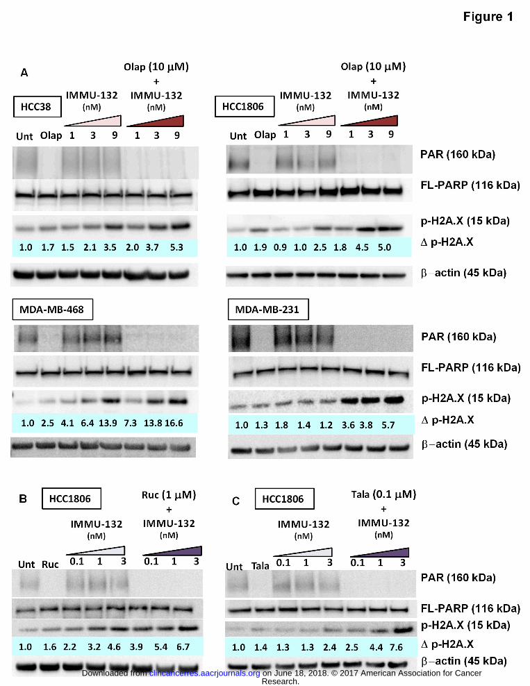
IMMU-132 plus PARPi mediated increases in dsDNA breaks
Assessment of dsDNA breaks was measured in TNBC cell lines for IMMU-132 and PARPi
combinations (24-h incubation; Figure 1). Four TNBC cell lines were tested with IMMU-132 and
olaparib (Fig. 1A). Not unexpectedly, poly(ADR-ribose) (PAR) was not detected in any of the
olaparib-treated cells, while IMMU-132 alone had only a minor effect on PAR levels. When
Research. on June 18, 2018. © 2017 American Association for Cancerclincancerres.aacrjournals.org Downloaded from
Author manuscripts have been peer reviewed and accepted for publication but have not yet been edited. Author Manuscript Published OnlineFirst on January 9, 2017; DOI: 10.1158/1078-0432.CCR-16-2401
12
combined, PAR was still not detected, which was due to inhibition, not loss of PARP, as
indicated by equivalent levels of full-length PARP (FL-PARP) present in all samples. Increased
dsDNA breaks, as evidenced by increases in p-H2A.X (25), were shown in all four cell lines
treated with olaparib alone, although the level in MDA-MB-231 was only 30% above untreated
controls, which were set to 1.0, whereas MDA-MB-468 cells showed a 2.5-fold increase (highest
response). In three of the four cell lines, IMMU-132 treatment alone also resulted in increased
p-H2A.X levels with 9 nM SN-38-equivalents of IMMU-132 (187 ng/mL of the antibody-drug
conjugate, or ADC), demonstrating a >2.5-fold increase (range 2.5- to 13.9-fold). Only the
BRCA1/2-wild-type MDA-MB-231 was seemingly resistant to single-agent IMMU-132 or
olaparib. However, when IMMU-132 (9 nM) was combined with olaparib, there was >5-fold
increase in p-H2A.X in all four cell lines, including MDA-MB-231, which exhibited a 5.7-fold
increase in p-H2A.X levels. Even at 3 nM SN-38-equivalents of IMMU-132 (62 ng/mL of the
ADC), when combined with olaparib, levels of p-H2A.X rose >76% above that achieved by each
agent alone in all four cell lines, with the highest response in MDA-MB-231 (171%), followed by
HCC1806 (136%), MDA-MB-468(116%), and HCC38 (76%).
Rucaparib and talazoparib were used at 10- and 100-fold lower concentrations, respectively,
when combined with IMMU-132 in HCC1806 (Fig. 1B, C, respectively). Likewise, the amount of
IMMU-132 required to demonstrate increases in dsDNA breaks, when combined with these two
PARPi, was lower (0.1, 1, and 3 nM SN-38-equivalents). Rucaparib alone resulted in a 1.6-fold
increase in dsDNA breaks, whereas 0.1 nM SN-38 equivalents of IMMU-132 (2 ng/mL of the
ADC) resulted in a 2.2-fold increase. Together, IMMU-132 plus rucaparib further increased the
Research. on June 18, 2018. © 2017 American Association for Cancerclincancerres.aacrjournals.org Downloaded from
Author manuscripts have been peer reviewed and accepted for publication but have not yet been edited. Author Manuscript Published OnlineFirst on January 9, 2017; DOI: 10.1158/1078-0432.CCR-16-2401

13
amount of dsDNA breaks 3.9-fold, which is 77% higher than either single agent. Talazoparib
and IMMU-132 combined demonstrated a dose-response with the amount of dsDNA breaks
increasing by 78% at the lowest IMMU-132 concentration to 216% at the highest concentration.
These data demonstrate that three different PARPi, when combined with IMMU-132, result in
increased DNA damage in the form of double-stranded breaks above that observed with single
agent treatments, which is consistent with the in vitro cytotoxicity assays demonstrating
synergistic growth-inhibition when IMMU-132 was combined with these PARPi.
Changes in cell cycle mediated by IMMU-132 plus olaparib
Cell cycle changes were assessed in all four TNBC cell lines when IMMU-132 and olaparib were
incubated with asynchronistic cells for 24 h (Fig. S3, Supplemental Data). Greater than 80% of
the cells in the untreated groups were in the G1-phase. Cells began to accumulate in S-phase
when exposed to either olaparib or IMMU-132 alone. However, the combination
demonstrated an even greater percentage of cells in S-phase, with a concomitant decrease in
the percentage of cells in G1-phase and little change in G2/M. For both BRCA1/2-mutated cells,
as well as BRCA1/2-wild-type MDA-MB-231, the combination of IMMU-132 and olaparib
resulted in a greater than 2-fold increase in the percentage of cells in the S-phase relative to
each agent alone. MDA-MB-468 demonstrated the least increase in the percentage of cells in
S-phase relative to single agent treatment, but this was due primarily to the greater effect each
agent alone had, with more than 32% and 50% of the cells in S-phase upon treatment with
olaparib or IMMU-132 alone, respectively. Even so, the combination increased the number of
cells in S-phase by 12% over IMMU-132 alone. Similar results were observed when IMMU-132
Research. on June 18, 2018. © 2017 American Association for Cancerclincancerres.aacrjournals.org Downloaded from
Author manuscripts have been peer reviewed and accepted for publication but have not yet been edited. Author Manuscript Published OnlineFirst on January 9, 2017; DOI: 10.1158/1078-0432.CCR-16-2401
14
was combined with rucaparib (1 μM) or talazoparib (0.2 μM), in which the combination of
IMMU-132 plus either PARPi increased the percentage of cells in S-phase for all four cell lines,
relative to single agent treatment (Table S1 and S2, respectively; Supplemental Data).
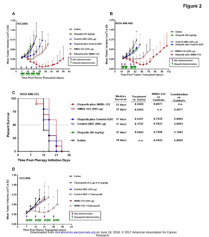
Improved efficacy of IMMU-132 in mice bearing TNBC tumors when combined with olaparib
or talazoparib
Therapeutic efficacy of combining IMMU-132 with olaparib was tested in HCC1806 and MDA-
MB-468 tumor xenografts (Figure 2). Importantly, in both experiments the dose and schedule
of IMMU-132 administered to the animals was chosen to give only a modest therapeutic effect
in order to detect a potential interaction between IMMU-132 and olaparib.
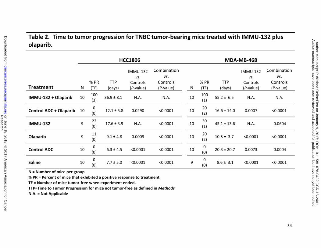
In mice bearing HCC1806 tumors (Figure 2A), the combination therapy resulted in a significant
antitumor effect when compared to all other treatment groups (P<0.0017, AUC; Table S3,
Supplemental Data). In this group, all of the mice exhibited a partial response, with 30% being
tumor-free (i.e., complete response) when the experiment ended on therapy day 108. Mean
time-to-tumor-progression (TTP) for this group was 36.9 ± 8.1 days (Table 2). In contrast,
tumors in mice given IMMU-132 or olaparib monotherapy progressed with mean TTP’s of 17.6
± 3.9 and 9.1 ± 4.8 days, respectively. Between the two monotherapy groups, IMMU-132
significantly delayed tumor progression when compared to olaparib therapy (P=0.0009).
Overall, the combination had a significantly greater delay in TTP when compared to either
single-agent therapy group (P<0.0001). Of note, this combination proved to be well-tolerated
by the mice, with no appreciable loss in body weight (Fig. S4A, Supplemental Data). Even mice
Research. on June 18, 2018. © 2017 American Association for Cancerclincancerres.aacrjournals.org Downloaded from
Author manuscripts have been peer reviewed and accepted for publication but have not yet been edited. Author Manuscript Published OnlineFirst on January 9, 2017; DOI: 10.1158/1078-0432.CCR-16-2401
15
treated with 4-fold more IMMU-132 (500 μg twice weekly x 4 weeks) combined with olaparib
demonstrated no significant loss in weight (Fig. S4B, Supplemental Data).
In mice bearing MDA-MB-468 tumors, the combination of IMMU-132 plus olaparib had a
significant antitumor effect when compared to all other treatment groups, including IMMU-132
monotherapy (Fig. 2B, P<0.0040; AUC, Table S4 in Supplementary Data). In terms of response
rate, 30% of mice treated with only IMMU-132 exhibited a partial response, as did 20% of the
animals treated with olaparib (Table 2). In contrast, all of the mice in the combination group
exhibited a partial response, with a TTP of 55.2 ± 6.5 days, which was significantly better than
all the other groups (P<0.0004) except IMMU-132 alone, which approached significance at a
TTP of 45.1 ± 13.6 days (P=0.0604). As in the HCC1806 tumor model, the mice tolerated the
combination of IMMU-132 and olaparib without loss of body weight (Fig. S4C, Supplemental
Data).
Given the ability of IMMU-132 and olaparib to improve efficacy significantly compared to
single-agent therapy in MDA-MB-468 tumors, which carry wild-type BRCA1/2, another therapy
study utilized mice bearing MDA-MB-231 tumors that likewise are wild-type for BRCA1/2, but
demonstrated synergy when IMMU-132 and PARPi were combined in vitro. MDA-MB-231
tumors progressed very rapidly, reaching experimental end-points (i.e., tumor volume >1.0 cm3)
within 14 days of starting therapy. Given this rapid disease progression, antitumor effects are
represented as the ability to provide survival benefit and not tumor regressions. As noted in
the previous two therapy experiments, olaparib alone had no antitumor effects in mice bearing
Research. on June 18, 2018. © 2017 American Association for Cancerclincancerres.aacrjournals.org Downloaded from
Author manuscripts have been peer reviewed and accepted for publication but have not yet been edited. Author Manuscript Published OnlineFirst on January 9, 2017; DOI: 10.1158/1078-0432.CCR-16-2401

16
MDA-MB-231 tumors, with median survival no different than saline control mice (Fig. 2C). Not
unexpectedly, IMMU-132 alone did not significantly improve the overall survival of the treated
animals, since it was shown previously to be resistant to this therapy (16). However, when mice
were treated with the combination of IMMU-132 plus olaparib, there was a significant increase
in median survival from 14 days for saline control animals to 21 days for those mice treated
with the combination (P=0.0005). Likewise, treatment with olaparib also resulted in a
significant survival benefit when combined with the non-specific control ADC (MST=17 days,
P=0.0257), suggesting that the olaparib does sensitize this resistant tumor to SN-38. These
results show, unexpectedly, that even in tumors resistant to PARPi and IMMU-132
monotherapies, the combination is able to overcome this resistance and impart a survival
benefit when compared to saline control animals.
Since talazoparib’s potency was greater than olaparib in vitro, both alone and when combined
with IMMU-132, mice bearing HCC1806 tumors were treated with the combination of IMMU-
132 and talazoparib (Fig. 2D). Whereas mice in the olaparib experiments were treated with 50
mg/kg, talazoparib-treated animals were given only 0.33 mg/kg. As observed in the olaparib
experiments, IMMU-132 plus talazoparib had a significant antitumor effect when compared to
all other treatments, including monotherapy with IMMU-132 or talazoparib (P<0.0088, AUC).
Mean TTP was 21.8 ± 9.6 days, which was >2.8-fold longer than any of the other treatment
groups (P<0.0021, Table 3). Similar to the combinations with olaparib, IMMU-132 plus
talazoparib did not result in any observable toxicity in the mice (Fig. S4D, Supplemental Data).
Research. on June 18, 2018. © 2017 American Association for Cancerclincancerres.aacrjournals.org Downloaded from
Author manuscripts have been peer reviewed and accepted for publication but have not yet been edited. Author Manuscript Published OnlineFirst on January 9, 2017; DOI: 10.1158/1078-0432.CCR-16-2401
17
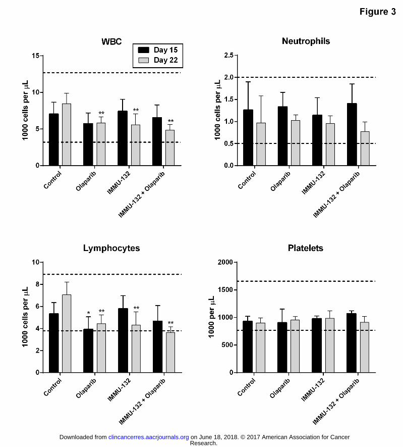
Hematologic tolerability of IMMU-132 plus olaparib in naïve BALB/c mice
Clinical studies indicate that there is no off-tumor, on-target toxicities in patients (19) and
therefore an experiment was performed to determine if the combination of a PARP inhibitor
with IMMU-132's SN-38 would have increased toxicities at therapeutically active doses. Since
normal tissues of mice are not targeted by the anti-Trop-2 antibody comprising IMMU-132,
toxicity studies were conducted in mice to determine possible off-target toxicities at doses of
IMMU-132 and olaparib shown to have therapeutic activity when combined in tumor-bearing
mice (i.e., 250 μg IMMU-132 plus 50 mg/kg olaparib). The schedule used mimics the clinical
regimen of IMMU-132 (i.e., weekly for 2 weeks with one week off per cycle) with olaparib
administered also over a two week period. This same doseschedule does demonstrate
antitumor activity over this two-week period, as shown in the efficacy studies described above
(Figure 2A-B). Naïve BALB/c mice were administered a two-week course of IMMU-132 plus
olaparib therapy to ascertain any possible myelotoxicity. On day 15 (i.e., 7 days after the last
IMMU-132 injection and 3 days after last dose of olaparib), there was no evidence of
hematological toxicity in any of the mice (Figure 3). In particular, the combination did not
result in any significant drop in total white blood cell (WBC) or lymphocyte counts. Most
importantly, there was no evidence of neutropenia or thrombocytopenia in any of these mice.
Only mice treated with olaparib monotherapy demonstrate a significant drop in lymphocytes
relative to vehicle control animals (P=0.0481), although the mean (3.92 ± 1.13 K/μL) remained
within the normal range (3.8 to 8.9 K/μL). After a further 7 days (i.e., day 22), the assessment
of toxicity was determined in the remaining mice. At this time-point, all three treatments
showed a significant drop in WBC and lymphocyte counts relative to control animals
Research. on June 18, 2018. © 2017 American Association for Cancerclincancerres.aacrjournals.org Downloaded from
Author manuscripts have been peer reviewed and accepted for publication but have not yet been edited. Author Manuscript Published OnlineFirst on January 9, 2017; DOI: 10.1158/1078-0432.CCR-16-2401
18
(P<0.0012). However, most of these values still remained within an acceptable range, with the
lowest values found in those mice treated with the combination, which fell just below the lower
end of the normal range (mean lymphocyte count of 3.62 ± 0.54 K/μL versus 7.04 ± 1.17 K/μL
for control, P=0.003). These results indicate that the combination of IMMU-132 and olaparib,
which has been shown to elicit antitumor effects in TNBC tumor-bearing mice, is not associated
with any significant myelotoxicity, and suggests a potentially high therapeutic window.
DISCUSSION
Exploitation of defects to a cell’s HRR pathways has gained widespread interest as a means to
induce synthetic lethality in various human cancers, including TNBC (1, 2). Of particular interest
are those drugs that target PARP (26, 27). While some clinical studies have suggested that use
of PARPi can provide meaningful responses, other trials have shown no significant
improvements in clinical outcome (7, 10). Given these mixed results, new approaches seek to
combine PARPi with other DNA-damaging agents, such as platinum-based drugs, microtubule
inhibitors, or irinotecan (14, 22, 28-31). Here, we examined the effect of combining several
PARPi (olaparib, rucaparib, and talazoparib) with our Topo I-inhibiting ADC, anti-Trop-2/SN-38-
ADC, IMMU-132. All three PARPi synergized with IMMU-132 to inhibit the growth of four
human TNBC cell lines. This occurred independently of BRCA1/2 status, as evidenced by the
synergistic growth-inhibition in-vitro of two cell lines with wild-type BRCA1/2. IMMU-132
combined with these PARPi increased the dsDNA breaks above that observed with each single
agent, and the accumulation of cells in the S-phase of the cell cycle. Most importantly, in mice
bearing either a BRCA1/2-mutated TNBC tumor (HCC1806) or ones with wild-type BRCA1/2
Research. on June 18, 2018. © 2017 American Association for Cancerclincancerres.aacrjournals.org Downloaded from
Author manuscripts have been peer reviewed and accepted for publication but have not yet been edited. Author Manuscript Published OnlineFirst on January 9, 2017; DOI: 10.1158/1078-0432.CCR-16-2401
19
(MDA-MB-468 and MDA-MB-231), the combination of IMMU-132 plus a PARPi resulted in
significant antitumor effects above those observed with monotherapy. These results clearly
illustrate the potential clinical benefit that may be derived by combining IMMU-132 with a
PARPi in TNBC.
Topo I is an enzyme utilized by the cell to allow unwinding of the DNA strand during
transcription and replication (32). PARP-1, the most abundant of the PARP proteins, has been
found to co-localize with Topo I throughout the cell cycle. However, upon DNA damage, PARP-
1 dissociates from Topo I, resulting in reduced activity of this enzyme (33). We hypothesized
that by combining the Topo I inhibitor carried by IMMU-132 (i.e., SN-38) with a PARPi, there
would be an accumulation of dsDNA breaks due to the inability of the remaining HRR pathways
in the cell to repair this damage with high fidelity, resulting in apoptosis and cell death.
Additionally, in cells lacking functional BRCA1/2 or are otherwise deficient in HRR, the more
error-prone, non-homologous end-joining (NHEJ) pathway is utilized, further compromising the
cell towards irreparable DNA damage and apoptosis (34). Others have found that in both
BRCA1-wild-type and -mutated TNBC cell lines, the combination of CPT-11 (the prodrug of SN-
38) and a PARPi could result in synergistic growth-inhibition in vitro (22). Here, too, we
demonstrated that IMMU-132, when combined with olaparib, rucaparib, or talazoparib,
mediates synergistic tumor cell growth-inhibition and increased dsDNA breaks in both
BRCA1/2-mutated and -wild-type cell lines. These data confirm the ability of IMMU-132-
mediated inhibition of Topo I, when combined with PARPi, to synergize growth-inhibition in
human TNBC cells regardless of BRCA1/2 mutational status.
Research. on June 18, 2018. © 2017 American Association for Cancerclincancerres.aacrjournals.org Downloaded from
Author manuscripts have been peer reviewed and accepted for publication but have not yet been edited. Author Manuscript Published OnlineFirst on January 9, 2017; DOI: 10.1158/1078-0432.CCR-16-2401

20
HRR pathways are more active during the late S/G2-phase of the cell cycle, whereas the more
error-prone NHEJ repair pathway is most evident throughout the S-phase (35). Cells with HRR
defects, as well as those exposed to PARPi, will arrest at G2/M-phase (23, 36). While we found
that PARPi (olaparib, rucaparib, and talazoparib) and IMMU-132 exposure resulted in small
increases in the number of cells in the G2/M-phase of the cell cycle, it was also evident that
there was greater accumulation in the S-phase. Most importantly, the combination
demonstrated an even greater percentage of cells in S-phase, with a concomitant decrease in
the percentage of cells in G1- and little change in the G2/M-phase. Interestingly, the interaction
of PARP-1 and Topo I was found to be most concentrated during the S-phase (33). Our data are
consistent with this and suggests that when using a PARPi combined with IMMU-132, we are
disrupting this PARP-1/Topo 1 interaction, resulting in accumulation in the S-phase and thus
towards the more error-prone NHEJ repair pathway that predominates during this phase of the
cell cycle.
Combining IMMU-132 therapy with a PARPi (olaparib or talazoparib) in mice bearing both
BRCA1/2-mutated and -wild-type tumors demonstrated significant antitumor effects greater
than that observed with monotherapy, thus extending our in vitro observations to in vivo TNBC
models. Likewise, others have demonstrated improved efficacy when a PARPi is combined with
a DNA-damaging chemotherapeutic in mice bearing human TNBC xenografts (22, 30). In one
example, CPT-11 was used in combination with veliparib to improve the therapeutic outcome
significantly in mice bearing s.c. MX-1 TNBC tumors (22). Similar to our experience, the PARPi
Research. on June 18, 2018. © 2017 American Association for Cancerclincancerres.aacrjournals.org Downloaded from
Author manuscripts have been peer reviewed and accepted for publication but have not yet been edited. Author Manuscript Published OnlineFirst on January 9, 2017; DOI: 10.1158/1078-0432.CCR-16-2401
21
alone had no effect on tumor growth, whereas the combination of CPT-11 and veliparib
demonstrated a significantly-enhanced antitumor effect. It should be noted that CPT-11 is
more effectively converted to its active SN-38 form in mice than in humans, and therefore
similar findings may not be reproducible in the clinical setting (37). However, by using IMMU-
132 with its SN-38 payload attached, we are not dependent on the efficiency of prodrug-to-
drug conversion by the patient, and therefore the antitumor effects we observed with the
combination of IMMU-132 plus PARPi are more clinically-relevant.
Approximately 80% of breast cancer patients carrying hereditary BRCA1 mutations present with
TNBC (38). While BRCA1/2 status is an important marker in TNBC, it is not the only one. Many
other BRCA1/2-wild-type TNBCs occur as sporadic tumors that share traits with germline BRCA-
mutated tumors and are termed to have BRCAness (39, 40). For example, analysis of germline
mutations in 158 TNBC patients demonstrated that in addition to BRCA1 mutations (17% of
patients), mutations of the CHEK2 checkpoint kinase 2 gene, nibrin NBN gene, and Ataxia-
Telangiectasia Mutated (ATM) genes also were found (3.9%), and are themselves involved in
various aspects of the HRR pathway (41-43). Overall, the evidence suggests that besides
BRCA1/2 status, many other genes contribute to the BRCAness of TNBC. In accordance with
these observations, our ability to synergize and demonstrate significantly improved efficacy in
the MDA-MB-468 tumor could be explained by the PTEN mutation it carries and its loss of
function. Loss of PTEN expression results in Rad51 dissociation from DNA replication forks, and
subsequent destabilization and stalled replication. Cells lacking PTEN have deficient HRR
functions, likely due to reduced Rad51 recruitment to the replicating DNA, culminating in loss of
Research. on June 18, 2018. © 2017 American Association for Cancerclincancerres.aacrjournals.org Downloaded from
Author manuscripts have been peer reviewed and accepted for publication but have not yet been edited. Author Manuscript Published OnlineFirst on January 9, 2017; DOI: 10.1158/1078-0432.CCR-16-2401

22
fidelity during DNA synthesis (44). As with BRCA1/2 mutations, cells lacking PTEN function are
prone to HRR deficiencies and, subsequently, have been shown to be sensitive to PARPi (45,
46). Our results demonstrate that IMMU-132 combined with a PARPi will significantly improve
efficacy in both traditional BRCA1/2-defective TNBC tumors as well as in tumors demonstrating
BRCAness in the form of deficiencies in other proteins involved in HRR pathways (e.g., PTEN).
Since not all TNBC tumors exhibit BRCAness, we evaluated the ability of IMMU-132 plus a PARPi
to inhibit tumor growth in such tumor types. MDA-MB-231 carries no mutations to PTEN,
BRCA1/2, or other known HHR proteins, and as such is expected to be resistant to PARPi
treatment (22, 46). Previous in-vivo studies demonstrated that mice bearing MDA-MB-231
tumor xenografts were resistant to IMMU-132 therapy (16). One reason for this resistance may
be over-expression of Rad51 in MDA-MB-231 (47). Cells exposed to SN-38 and olaparib have
been found to increase Rad51 recruitment in the HRR pathway (48). In fact, we found that
IMMU-132 exposure does result in an increase in several different proteins involved in HRR,
including Rad51, BARD1, FANCD2, and ERCC1 in BRCA1/2 wild-type cell lines. Of note was the
observation that while these proteins are up-regulated in some cell lines (MDA-MB-231 and BT-
20), they are actually down-regulated in MDA-MB-468, which may account for its sensitivity to
both IMMU-132 monotherapy as well as the combination therapy. At the same time, it is likely
that this increased expression of these HRR proteins in MDA-MB-231 allows for a more efficient
repair of damaged DNA caused by IMMU-132 than would otherwise occur in cells that had low
expression. A second possible reason for MDA-MB-231 resistance may be that it has low
expression of Trop-2 on its surface, which could limit how much SN-38 can be delivered to the
Research. on June 18, 2018. © 2017 American Association for Cancerclincancerres.aacrjournals.org Downloaded from
Author manuscripts have been peer reviewed and accepted for publication but have not yet been edited. Author Manuscript Published OnlineFirst on January 9, 2017; DOI: 10.1158/1078-0432.CCR-16-2401

23
tumor by IMMU-132 (17). However, when IMMU-132 and its SN-38 payload was combined
with olaparib, we achieved a significant survival benefit in MDA-MB-231 tumor-bearing
animals. Moreover, while the amount of SN-38 delivered by IMMU-132 alone may be
insufficient to retard tumor growth, enough must accrete in the tumor so that when combined
with olaparib, we are able to slow tumor progression enough to provide a survival benefit. We
are currently studying various MDA-MB-231 clones that were transfected with Trop-2 to
increase expression levels in order to better determine what role Trop-2 surface expression
plays in a tumor’s sensitivity to IMMU-132 therapy, either alone or when combined with PARPi.
In a current phase I/II clinical trial, IMMU-132 has demonstrated an objective response rate
(confirmed complete response + partial response) of 31% in 58 patients with metastatic TNBC
(21). At the maximum tolerated dose, grade 3-4 toxicities included neutropenia, febrile
neutropenia, diarrhea, fatigue, anemia, leukopenia, and dyspnea. All of these toxicities were
manageable, with the incidence of severe diarrhea being less than that reported for the
parental prodrug, irinotecan. Single agent olaparib toxicities include grade 3-4 fatigue and
thrombocytopenia (5). In clinical studies evaluating the combination of either olaparib or
veliparib with irinotecan, dose-limiting toxicities included fatigue, anorexia, diarrhea, nausea,
vomiting, febrile neutropenia, neutropenia, and thrombocytopenia (31, 49). A hallmark in
these clinical trials was that the amount of irinotecan and PARPi administered was reduced
below the single-agent maximum tolerated doses in order to gain acceptable tolerability.
Based on our experience with IMMU-132 in the clinic, as well as those reported with PARPi plus
irinotecan, we do not expect to observe any different dose-limiting toxicity with a combination
Research. on June 18, 2018. © 2017 American Association for Cancerclincancerres.aacrjournals.org Downloaded from
Author manuscripts have been peer reviewed and accepted for publication but have not yet been edited. Author Manuscript Published OnlineFirst on January 9, 2017; DOI: 10.1158/1078-0432.CCR-16-2401

24
of IMMU-132 plus PARPi’s. The doses of IMMU-132 administered to mice in all our preclinical
studies were chosen based on their ability to produce a minimal effect, so that we could better
determine if the combinations were beneficial and therefore indicated a good therapeutic
window when IMMU-132 was combined with a PARPi. Importantly, in all our combination
studies, the mice tolerated both the PARPi and IMMU-132 well, with no significant loss in body
weight or substantial hematologic toxicity during the course of treatment. Given such a
potential for a high therapeutic window, we speculate that even at lower IMMU-132 dosages,
combinations with a PARPi in this subset of breast cancer patients will achieve improved
therapeutic responses without unmanageable toxicity.
In summary, through the utilization of PARPi to target TNBC and create synthetic lethality
combined with Topo I inhibition mediated by IMMU-132, a synergistic growth-inhibitory
outcome was achieved in human TNBC tumor lines, regardless of BRCA1/2 status. While
BRCAness is an important element in this combined effect, it is not limiting. Indeed, PARPi are
under investigation in a variety of cancers involving breast, ovarian, pancreatic, non-small-cell
lung, gastric cancer, glioblastoma, melanoma, and others. Our results indicate that the
combination of PARPi and IMMU-132 could broaden the range of tumors that are usually
treated with the former. It is interesting that IMMU-132 does target all of these cancer types
(17), which may make this combination a rational approach. However, combination studies
with PARPi have been problematic because of hematological toxicity, thus requiring dose
modification. The studies described here have shown tolerability for the combination in terms
of hematological parameters and animal body weight, but the ultimate clinical experience will
Research. on June 18, 2018. © 2017 American Association for Cancerclincancerres.aacrjournals.org Downloaded from
Author manuscripts have been peer reviewed and accepted for publication but have not yet been edited. Author Manuscript Published OnlineFirst on January 9, 2017; DOI: 10.1158/1078-0432.CCR-16-2401
25
of course be critical. Given the promising results obtained thus far with IMMU-132 in patients
with TNBC, the logical next step is to combine this therapy with PARPi to further improve
clinical outcome.
AUTHOR CONTRIBUTIONS
•Conception and design: T.M. Cardillo, R.M. Sharkey, D.L. Rossi, and D.M. Goldenberg
•Acquisition of data: T.M. Cardillo, D.L. Rossi, A.A. Mostafa, and R. Arrojo
•Analysis and interpretation of data: T.M. Cardillo, R.M. Sharkey, D.L. Rossi, A.A. Mostafa, R.
Arrojo, and D.M. Goldenberg
•Writing, review and/or revision of the manuscript: T.M. Cardillo, R.M. Sharkey, D.L. Rossi, and
D.M. Goldenberg
•Administrative, technical, or material support: T.M. Cardillo, R.M. Sharkey, and D.M.
Goldenberg
•Study supervision: T.M. Cardillo and D.M. Goldenberg
ACKNOWLEDGEMENTS
We thank Serengulam V. Govindan, Nalini Sathyanarayan, Christine Mazza-Ferreira, Jing Xia,
and Jennifer Donnell for contributions to synthetic and conjugation chemistries, Maria Zalath
for assistance with the animal studies, and Rongxiu Li for technical assistance in performing the
cell cycle FACS analysis.
Research. on June 18, 2018. © 2017 American Association for Cancerclincancerres.aacrjournals.org Downloaded from
Author manuscripts have been peer reviewed and accepted for publication but have not yet been edited. Author Manuscript Published OnlineFirst on January 9, 2017; DOI: 10.1158/1078-0432.CCR-16-2401
26
REFERENCES
1. Kaelin WG Jr. The concept of synthetic lethality in the context of anticancer therapy. Nat
Rev Cancer 2005;5:689-98.
2. McLornan DP, List A, Mufti GJ. Applying synthetic lethality for the selective targeting of
cancer. N Engl J Med 2014;371:1725-35.
3. Satoh MS, Linhahl T. Role of poly(ADP-ribose) formation in DNA repair. Nature
1992;356:356-58.
4. Benafif S, Hall M. An update on PARP inhibitors for the treatment of cancer. Onco Targets
Ther 2015;8:519-28.
5. Fong PC, Boss DS, Yap TA, Tutt A, Wu P, Mergui-Roelvink M, et al. Inhibition of poly(ADP-
ribose) polymerase in tumors from BRCA mutation carriers. N Engl J Med 2009;361:123-34.
6. Fong PC, Yap TA, Boss DS, Carden CP, Mergui-Roelvink M, Gourley C, et al. Poly(ADP)-ribose
polymerase inhibition: frequent durable responses in BRCA carrier ovarian cancer
correlating with platinum-free interval. J Clin Oncol 2010;28:2512-19.
7. Gelmon KA, Tischkowitz M, Mackay H, Swenerton K, Robidoux A, Tonkin K, et al. Olaparib
in patients with recurrent high-grade serous or poorly differentiated ovarian carcinoma or
triple-negative breast cancer: a phase 2, multicentre, open-label, non-randomised study.
Lancet Oncol 2011;12:852-61.
8. Marulonis UA, Harter P, Gourley C, Friedlander M, Vergote I, Rustin G, et al. Olaparib
maintenance therapy in patients with platinum-sensitive, relapsed serous ovarian cancer
and a BRCA mutation: overall survival adjusted for postprogression poly(adenosine
diphosphate ribose) polymerase inhibitor therapy. Cancer 2016;122:1844-52.
Research. on June 18, 2018. © 2017 American Association for Cancerclincancerres.aacrjournals.org Downloaded from
Author manuscripts have been peer reviewed and accepted for publication but have not yet been edited. Author Manuscript Published OnlineFirst on January 9, 2017; DOI: 10.1158/1078-0432.CCR-16-2401

27
9. Lowery MA, Kelsen DP, Stadler ZK, Yu KH, Janjigian YY, D’Adamo DR, et al. An emerging
entity: pancreatic adenocarcinoma associated with a known BRCA mutation: clinical
descriptors, treatment implications, and future directions. Oncologist 2011;16:1397-402.
10. Tutt A, Robson M, Garber JE, Domchek SM, Audeh MW, Weitzel JN, et al. Oral poly(ADP-
ribose) polymerase inhibitor olaparib in patients with BRCA1 or BRCA2 mutations and
advanced breast cancer: a proof-of-concept trial. Lancet 2010;376:235-44.
11. Lips EH, Mulder L, Oonk A, van der Kolk LE, Hogervorst FB, Imholz AL, et al. Triple-negative
breast cancer: BRCAness and concordance of clinical features with BRCA1-mutation
carriers. Br J Cancer 2013;108:2172-77.
12. Albert JM, Cao C, Kim KW, Willey CD, Geng L, Xiao D, et al. Inhibition of poly(ADP-ribose)
polymerase enhances cell death and improves tumor growth delay in irradiated lung
cancer models. Clin Cancer Res 2007;13:3033-42.
13. Evers B, Drost R, Schut E, de Bruin M, van der Burg E, Derksen PW, et al. Selective inhibition
of BRCA2-deficient mammary tumor cell growth by AZD2281 and cisplatin. Clin Cancer Res
2008;14:3916-25.
14. Delaney CA, Wang LZ, Kyle S, White AW, Calvert AH, Curtin NJ, et al. Potentiation of
temozolomide and topotecan growth inhibition and cytotoxicity by novel poly(adenosine
diphosphoribose) polymerase inhibitors in a panel of human tumor cell lines. Clin Cancer
Res 2000;6:2860-7.
15. Genther Williams SM, Kuznicki AM, Andrade P, Dolinski BM, Elbi C, O'Hagan RC, et al.
Treatment with the PARP inhibitor, niraparib, sensitizes colorectal cancer cell lines to
irinotecan regardless of MSI/MSS status. Cancer Cell Int. 2015;15:14.
Research. on June 18, 2018. © 2017 American Association for Cancerclincancerres.aacrjournals.org Downloaded from
Author manuscripts have been peer reviewed and accepted for publication but have not yet been edited. Author Manuscript Published OnlineFirst on January 9, 2017; DOI: 10.1158/1078-0432.CCR-16-2401
28
16. Goldenberg DM, Cardillo TM, Govindan SV, Rossi EA, Sharkey RM. Trop-2 is a novel target
for solid cancer therapy with sacituzumab govitecan (IMMU-132), an antibody-drug
conjugate (ADC). Oncotarget 2015;6:22496-512.
17. Cardillo TM, Govindan SV, Sharkey RM, Trisal P, Arrojo R, Liu D, et al. Sacituzumab
govitecan (IMMU-132), an anti-Trop-2/SN-38 antibody-drug conjugate: characterization
and efficacy in pancreatic, gastric, and other cancers. Bioconjug Chem 2015;26:919-31.
18. Cardillo TM, Govindan SV, Sharkey RM, Trisal P, Goldenberg DM. Humanized anti-Trop-2
IgG-SN-38 conjugate for effective treatment of diverse epithelial cancers: preclinical
studies in human cancer xenograft models and monkeys. Clin Cancer Res 2011;17:3157-69.
19. Starodub AN, Camidge DR, Scheff RJ, Thomas SS, Guarino MJ, Masters G, et al. Trop-2 is a
therapeutic target for the antibody-drug conjugate (ADC), sacituzumab govitecan (IMMU-
132), in patients (pts) with previously treated metastatic small-cell lung cancer (mSCLC).
American Society of Clinical Oncology (ASCO) Annual Meeting, Chicago, IL, June 3-6, 2016,
Abstract no. 8559.
20. Camidge DR, Heist R, Masters G, Scheff RJ, Starodub AN, Messersmith WA, et al. Therapy
of metastatic, non-small-cell lung cancer with the anti-Trop-2-SN-38 antibody-drug
conjugate, sacituzumab govitecan (IMMU-132). American Society of Clinical Oncology
(ASCO) Annual Meeting, Chicago, IL, June 3-6, 2016, Abstract no. 9011.
21. Bardia A, Diamond JR, Mayer IA, Starodub AN, Moroose R, Isakoff S, et al. Safety and
efficacy of anti-Trop-2 antibody drug conjugate, sacituzumab govitecan (IMMU-132), in
heavily pretreated patients with TNBC. 2015 San Antonio Breast Cancer Symposium, San
Antonio, TX, December 8-12, 2015, Abstract no. PD3-06.
Research. on June 18, 2018. © 2017 American Association for Cancerclincancerres.aacrjournals.org Downloaded from
Author manuscripts have been peer reviewed and accepted for publication but have not yet been edited. Author Manuscript Published OnlineFirst on January 9, 2017; DOI: 10.1158/1078-0432.CCR-16-2401
29
22. Boerner JL, Nechiporchik N, Mueller KL, Polin L, Heilbrun L, Boerner SA, et al. Protein
expression of DNA damage repair proteins dictates response to topoisomerase and PARP
inhibitors in triple-negative breast cancer. PLoS One 2015;10:e0119614.
23. McCabe N, Turner NC, Lord CJ, Kluzek K, Bialkowska A, Swift S, et al. Deficiency in the
repair of DNA damage by homologous recombination and sensitivity to poly(ADP-ribose)
polymerase inhibition. Cancer Res 2006;66:8109-15.
24. Wiegmans AP, Yap P-Y, Ward A, Lim YC, Khanna KK. Differences in expression of key DNA
damage repair genes after epigenetic-induced BRCAness dictate synthetic lethality with
PARP1 inhibition. Mol Cancer Ther 2015;14:2321-31.
25. Bonner WM, Redon CE, Dickey JS, Nakamura AJ, Sedelnikova OA, Solier S, et al.
GammaH2AX and cancer. Nat Rev Cancer 2008;8:957-67.
26. Lord CJ, Tutt ANJ, Ashworth A. Synthetic lethality and cancer therapy: lessons learned
from the development of PARP inhibitors. Annu Rev Med 2015;66:455-70.
27. Audeh MW. Novel treatment strategies in triple-negative breast cancer: specific role of
poly(adenosine diphosphate-ribose) polymerase inhibition. Pharmgenomics Pers Med
2014;7:307-16.
28. Dent RA, Lindeman GJ, Clemons M, Wildiers H, Chan A, McCarthy NJ, et al. Phase I trial of
the oral PARP inhibitor olaparib in combination with paclitaxel for first- or second-line
treatment of patients with metastatic triple-negative breast cancer. Breast Cancer Res
2013;15:R88.
29. Davidson D, Wang Y, Aloyz R, Panasci L. The PARP inhibitor ABT-888 synergizes irinotecan
treatment of colon cancer cell lines. Invest New Drugs 2013;31:461-8.
Research. on June 18, 2018. © 2017 American Association for Cancerclincancerres.aacrjournals.org Downloaded from
Author manuscripts have been peer reviewed and accepted for publication but have not yet been edited. Author Manuscript Published OnlineFirst on January 9, 2017; DOI: 10.1158/1078-0432.CCR-16-2401

30
30. Karginova O, Siegel MB, Van Swearingen AE, Deal AM, Adamo B, Sambade MJ, et al.
Efficacy of carboplatin alone and in combination with ABT888 in intracranial murine
models of BRCA-mutated and BRCA-wild-type triple-negative breast cancer. Mol Cancer
Ther 2015;14:920-30.
31. Chen EX, Jonker DJ, Siu LL, McKeever K, Keller D, Wells J, et al. A Phase I study of olaparib
and irinotecan in patients with colorectal cancer: Canadian Cancer Trials Group IND 187.
Invest New Drugs 2016;34:450-7.
32. Wang JC. Cellular roles of DNA topoisomerases: a molecular perspective. Nat Rev Mol Cell
Biol 2002;3-430-40.
33. Yung TM, Sato S, Satoh MS. Poly(ADP-ribosyl)ation as a DNA damage-induced post-
translational modification regulating poly(ADP-ribose) polymerase-1-topoisomerase I
interaction. J Biol Chem 2004;279:39686-96.
34. Plummer R. Perspective on the pipeline of drugs being developed with modulation of DNA
damage as a target. Clin Cancer Res 2010;16:4527-31.
35. Rothkamm K, Krüger I, Thompson LH, Löbrich M. Pathways of DNA double-strand break
repair during the mammalian cell cycle. Mol Cell Biol 2003;23:5706-15.
36. Chuang HC, Kapuriya N, Kulp SK, Chen CS, Shapiro CL. Differential anti-proliferative
activities of poly(ADP-ribose) polymerase (PARP) inhibitors in triple-negative breast cancer
cells. Breast Cancer Res Treat 2012;134:649-59.
37. Zamboni WC, Stewart CF, Cheshire PJ, Richmond LB, Hanna SK, Luo X, et al. Studies of the
efficacy and pharmacology of irinotecan against human colon tumor xenograft models. Clin
Cancer Res 1998;4:743-53.
Research. on June 18, 2018. © 2017 American Association for Cancerclincancerres.aacrjournals.org Downloaded from
Author manuscripts have been peer reviewed and accepted for publication but have not yet been edited. Author Manuscript Published OnlineFirst on January 9, 2017; DOI: 10.1158/1078-0432.CCR-16-2401

31
38. Lakhani SR, Van De Vijver MJ, Jacquemier J, Anderson TJ, Osin PP, McGuffog L, et al. The
pathology of familial breast cancer: predictive value of immunohistochemical markers
estrogen receptor, progesterone receptor, HER-2, and p53 in patients with mutations in
BRCA1 and BRCA2. J Clin Oncol 2002;20:2310-18.
39. Turner N, Tutt A, Ashworth A. Hallmarks of 'BRCAness' in sporadic cancers. Nat Rev Cancer
2004;4:814-19.
40. Lips EH, Mulder L, Hannemann J, Laddach N, Vrancken Peeters MT, van de Vijver MJ, et al.
Indicators of homologous recombination deficiency in breast cancer and association with
response to neoadjuvant chemotherapy. Ann Oncol 2011;22:870-76.
41. Domagala P, Jakubowska A, Jaworska-Bieniek K, Kaczmarek K, Durda K, Kurlapska A, et al.
Prevalence of germline mutations in genes engaged in DNA damage repair by homologous
recombination in patients with triple-negative and hereditary non-triple-negative breast
cancers. PLoS One 2015;10:e0130393.
42. Williams RS, Williams JS, Tainer JA. Mre11-Rad50-Nbs1 is a keystone complex connecting
DNA repair machinery, double-strand break signaling, and the chromatin template.
Biochem Cell Biol 2007;85:509-20.
43. Reinhardt HC, Yaffe MB. Kinases that control the cell cycle in response to DNA damage:
Chk1, Chk2, and MK2. Curr Opin Cell Biol 2009;21:245-55.
44. He J, Kang X, Yin Y, Chao KS, Shen WH. PTEN regulates DNA replication progression and
stalled fork recovery. Nat Commun 2015;6:7620.
45. McEllin B, Camacho CV, Mukherjee B, Hahm B, Tomimatsu N, Bachoo RM, et al. PTEN loss
compromises homologous recombination repair in astrocytes: implications for
Research. on June 18, 2018. © 2017 American Association for Cancerclincancerres.aacrjournals.org Downloaded from
Author manuscripts have been peer reviewed and accepted for publication but have not yet been edited. Author Manuscript Published OnlineFirst on January 9, 2017; DOI: 10.1158/1078-0432.CCR-16-2401
32
glioblastoma therapy with temozolomide or poly(ADP-ribose) polymerase inhibitors.
Cancer Res 2010;70:5457-64.
46. Shen Y, Rehman FL, Feng Y, Boshuizen J, Bajrami I, Elliott R, et al. BMN 673, a novel and
highly potent PARP1/2 inhibitor for the treatment of human cancers with DNA repair
deficiency. Clin Cancer Res 2013;19:5003-15.
47. Koehn H, Magan N, Isaacs RJ, Stowell KM. Differential regulation of DNA repair protein
Rad51 in human tumour cell lines exposed to doxorubicin. Anticancer Drugs 2007;18:419-
25.
48. Tahara M, Inoue T, Sato F, Miyakura Y, Horie H, Yasuda Y, et al. The use of Olaparib
(AZD2281) potentiates SN-38 cytotoxicity in colon cancer cells by indirect inhibition of
Rad51-mediated repair of DNA double-strand breaks. Mol Cancer Res 2014;13:1170-80.
49. LoRusso PM, Li J, Burger A, Heilbrun LK, Sausville EA, Boerner SA, et al. Phase I safety,
pharmacokinetic, and pharmacodynamic study of the poly(ADP-ribose) polymerase (PARP)
inhibitor veliparib (ABT-888) in combination with irinotecan in patients with advanced solid
tumors. Clin Cancer Res 2016;22:3227-37.
Research. on June 18, 2018. © 2017 American Association for Cancerclincancerres.aacrjournals.org Downloaded from
Author manuscripts have been peer reviewed and accepted for publication but have not yet been edited. Author Manuscript Published OnlineFirst on January 9, 2017; DOI: 10.1158/1078-0432.CCR-16-2401

33
TABLES AND FIGURES
Table 1. Combination index values for IMMU-132 plus olaparib, rucaparib, or talazoparib in four TNBC cell lines.
Cell Line
Single Agent IC50-values
(mean ± S.D.)
C.I.-values
Olaparib + IMMU-132 IC10 IC20 IC30
Rucaparib + IMMU-132 IC10 IC20 IC30
Talazoparib + IMMU-132 IC10 IC20 IC30
IMMU-132 (nM)
Olaparib (μM)
Rucaparib(μM)
Talazoparib(nM)
HCC1806 1.9 ± 0.4 7.3 ± 1.4 5.4 ± 0.3 4.7 ± 1.0 0.81 0.59 0.29 1.09 0.80 0.46 1.21 0.80 0.50
HCC38 3.4 ± 0.5 28.5 ± 18.7 16.9 ± 0.7 104.9 ± 44.7 0.46 0.25 0.16 1.07 0.91 0.47 0.49 0.20 0.11
MDA-MB-468 3.5 ± 0.4 9.8 ± 1.0 10.3 ± 0.8 322.3 ± 27.7 0.87 0.49 0.22 1.01 0.63 0.34 0.37 0.15 0.07
MDA-MB-231 7.5 ± 1.2 21.7 ± 6.0 7.1 ± 1.0 36.3 ± 8.0 0.89 0.42 0.28 0.69 0.40 0.22 0.40 0.17 0.08
BT-20 82.5 ± 22.8 256.7 ± 40.4 n.d. n.d 0.91 0.54 0.27 n.d. n.d. n.d. n.d. n.d. n.d.
C.I.-values = Combination Index values determined as described in Methods. IC10, IC20, and IC30 = CI values determined when IMMU-132 or PARPi used at concentrations calculated to cause 10%, 20%, and 30% growth inhibition when used alone, respectively. n.d., Not Done
Research.
on June 18, 2018. © 2017 A
merican A
ssociation for Cancer
clincancerres.aacrjournals.org D
ownloaded from
Author m
anuscripts have been peer reviewed and accepted for publication but have not yet been edited.
Author M
anuscript Published O
nlineFirst on January 9, 2017; D
OI: 10.1158/1078-0432.C
CR
-16-2401
34
Table 2. Time to tumor progression for TNBC tumor-bearing mice treated with IMMU-132 plus olaparib.
Treatment
HCC1806 MDA-MB-468
N % PR (TF)
TTP (days)
IMMU-132 vs.
Controls (P-value)
Combination vs.
Controls (P-value)
N % PR (TF)
TTP (days)
IMMU-132 vs.
Controls (P-value)
Combination vs.
Controls (P-value)
IMMU-132 + Olaparib 10 100 (3) 36.9 ± 8.1 N.A. N.A. 10 100
(1) 55.2 ± 6.5 N.A. N.A.
Control ADC + Olaparib 10 0 (0) 12.1 ± 5.8 0.0290 <0.0001 10 20
(2) 16.6 ± 14.0 0.0007 <0.0001
IMMU-132 9 22 (0) 17.6 ± 3.9 N.A. <0.0001 10 30
(1) 45.1 ± 13.6 N.A. 0.0604
Olaparib 9 11 (0) 9.1 ± 4.8 0.0009 <0.0001 10 20
(2) 10.5 ± 3.7 <0.0001 <0.0001
Control ADC 10 0 (0) 6.3 ± 4.5 <0.0001 <0.0001 10 0
(0) 20.3 ± 20.7 0.0073 0.0004
Saline 10 0 (0) 7.7 ± 5.0 <0.0001 <0.0001 9 0
(0) 8.6 ± 3.1 <0.0001 <0.0001
N = Number of mice per group % PR = Percent of mice that exhibited a positive response to treatment TF = Number of mice tumor-free when experiment ended. TTP=Time to Tumor Progression for mice not tumor-free as defined in Methods N.A. = Not Applicable
Research.
on June 18, 2018. © 2017 A
merican A
ssociation for Cancer
clincancerres.aacrjournals.org D
ownloaded from
Author m
anuscripts have been peer reviewed and accepted for publication but have not yet been edited.
Author M
anuscript Published O
nlineFirst on January 9, 2017; D
OI: 10.1158/1078-0432.C
CR
-16-2401

35
Table 3. Time to tumor progression for HCC1806 tumor-bearing mice treated with IMMU-132 plus talazoparib.
Treatment N TTP
(days)
IMMU-132vs.
Control (P-value)
Combination vs.
Control (P-value)
IMMU-132 + Talazoparib 10 21.8 ± 9.6 N.A. N.A.
Control ADC + Talazoparib 10 6.8 ± 5.4 0.6979 0.0004
IMMU-132 9 7.9 ± 6.6 N.A. 0.0021
Talazoparib 9 5.9 ± 5.9 0.5099 0.0005
Control ADC 10 4.6 ± 2.1 0.1866 0.0003
Saline 10 4.2 ± 1.9 0.0709 0.0001 N = Number of mice per groups TTP=Time to Tumor Progression as defined in Methods N.A. = Not Applicable
Research. on June 18, 2018. © 2017 American Association for Cancerclincancerres.aacrjournals.org Downloaded from
Author manuscripts have been peer reviewed and accepted for publication but have not yet been edited. Author Manuscript Published OnlineFirst on January 9, 2017; DOI: 10.1158/1078-0432.CCR-16-2401

36
FIGURE LEGENDS
Figure 1. Western blot assessment of effects on PAR, PARP, and dsDNA breaks mediated by
IMMU-132 plus PARPi in TNBC tumor-lines. Cells were plated overnight in 6-well plates
before the addition of chemotherapeutics. After a 24-h incubation, cells were harvested and
cell lysates resolved and transferred for Western analysis as described in Methods. PAR and
full-length PARP (FL-PARP) levels were determined on the same gel. Assessment of dsDNA
breaks (p-H2A.X) was calculated as ratios relative to untreated control (Unt) normalized to β-
actin protein loading control (Δp-H2A.X). (A) All four TNBC cell lines (HCC38, HCC1806, MDA-
MB-468, and MDA-MB-231) were exposed to olaparib (Olap), IMMU-132, or the combination of
both. IMMU-132 concentrations are expressed in terms of SN-38 equivalents. (B) HCC1806
cells exposed to rucaparib (Ruc) and IMMU-132 or to (C) talazoparib (Tala) and IMMU-132.
Figure 2. Therapeutic efficacy of IMMU-132 plus PARPi in BRCA1/2-deficient and -wild-type
TNBC tumor-xenograft disease models. Tumors were established and disease end-points are
described in Methods. All the ADCs and controls were administered in the amounts indicated
(green arrows = PARPi injections, purple arrows = ADC injections). (A) Mice bearing HCC1806
tumors (N = 9-10) were injected with IMMU-132 weekly for 4 weeks and with olaparib daily
(Monday through Friday) for 4 weeks. (B) MDA-MB-468 tumor-bearing mice (N=9-10) were
injected weekly with IMMU-132 for two weeks with one week off before repeating. Likewise,
olaparib was administered daily (M-F) for two weeks with one week off before repeating. (C)
Mice bearing MDA-MB-231 tumors (N=10) were treated with IMMU-132 and olaparib under
the same schedule as the HCC1806 mice. (D) Mice bearing HCC1806 tumors (N=9-10) were
Research. on June 18, 2018. © 2017 American Association for Cancerclincancerres.aacrjournals.org Downloaded from
Author manuscripts have been peer reviewed and accepted for publication but have not yet been edited. Author Manuscript Published OnlineFirst on January 9, 2017; DOI: 10.1158/1078-0432.CCR-16-2401
37
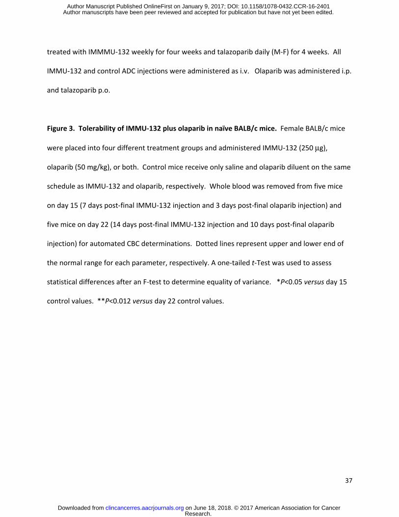
treated with IMMMU-132 weekly for four weeks and talazoparib daily (M-F) for 4 weeks. All
IMMU-132 and control ADC injections were administered as i.v. Olaparib was administered i.p.
and talazoparib p.o.
Figure 3. Tolerability of IMMU-132 plus olaparib in naïve BALB/c mice. Female BALB/c mice
were placed into four different treatment groups and administered IMMU-132 (250 μg),
olaparib (50 mg/kg), or both. Control mice receive only saline and olaparib diluent on the same
schedule as IMMU-132 and olaparib, respectively. Whole blood was removed from five mice
on day 15 (7 days post-final IMMU-132 injection and 3 days post-final olaparib injection) and
five mice on day 22 (14 days post-final IMMU-132 injection and 10 days post-final olaparib
injection) for automated CBC determinations. Dotted lines represent upper and lower end of
the normal range for each parameter, respectively. A one-tailed t-Test was used to assess
statistical differences after an F-test to determine equality of variance. *P<0.05 versus day 15
control values. **P<0.012 versus day 22 control values.
Research. on June 18, 2018. © 2017 American Association for Cancerclincancerres.aacrjournals.org Downloaded from
Author manuscripts have been peer reviewed and accepted for publication but have not yet been edited. Author Manuscript Published OnlineFirst on January 9, 2017; DOI: 10.1158/1078-0432.CCR-16-2401
Published OnlineFirst January 9, 2017.Clin Cancer Res Thomas M. Cardillo, Robert M. Sharkey, Diane L Rossi, et al. BRCA1/2-wild-type triple-negative breast cancerantibody-drug conjugate, IMMU-132, plus PARP-inhibitors in Synthetic lethality exploitation by an anti-Trop-2-SN-38
Updated version
10.1158/1078-0432.CCR-16-2401doi:
Access the most recent version of this article at:
Material
Supplementary
http://clincancerres.aacrjournals.org/content/suppl/2017/01/07/1078-0432.CCR-16-2401.DC1
Access the most recent supplemental material at:
Manuscript
Authoredited. Author manuscripts have been peer reviewed and accepted for publication but have not yet been
E-mail alerts related to this article or journal.Sign up to receive free email-alerts
Subscriptions
Reprints and
To order reprints of this article or to subscribe to the journal, contact the AACR Publications
Permissions
Rightslink site. Click on "Request Permissions" which will take you to the Copyright Clearance Center's (CCC)
.http://clincancerres.aacrjournals.org/content/early/2017/01/07/1078-0432.CCR-16-2401To request permission to re-use all or part of this article, use this link
Research. on June 18, 2018. © 2017 American Association for Cancerclincancerres.aacrjournals.org Downloaded from
Author manuscripts have been peer reviewed and accepted for publication but have not yet been edited. Author Manuscript Published OnlineFirst on January 9, 2017; DOI: 10.1158/1078-0432.CCR-16-2401