syrinx resolution after posterior fossa decompression in patients with scoliosis secondary to chiari...
TRANSCRIPT
CHINESE SECTION
Syrinx resolution after posterior fossa decompression in patientswith scoliosis secondary to Chiari malformation type I
Tao Wu • Zezhang Zhu • Jian Jiang •
Xin Zheng • Xu Sun • Bangping Qian •
Feng Zhu • Yong Qiu
Received: 7 September 2011 / Revised: 14 October 2011 / Accepted: 28 October 2011 / Published online: 16 November 2011
� Springer-Verlag 2011
Abstract
Introduction Description of syrinx resolution after
posterior fossa decompression (PFD) in patients with
scoliosis secondary to Chiari malformation type I (CMI)
and syringomyelia (SM) has been rarely reported in the
literature. This study was performed to investigate the
outcome of PFD in patients with scoliosis secondary to
CMI and to identify potential predictive factors for better
outcome after PFD.
Material and methods Patients with scoliosis secondary
to CMI and SM, who had undergone PFD during the period
2000 through 2009, were recruited. Inclusion criteria were
(1) age B 18 years, (2) diagnosis of SM associated with
CMI, (3) scoliosis as the first complaint, (4) having
undergone preoperative and follow-up magnetic resonance
imaging (MRI). Patients with acquired CMI anomalies or
who had received syringosubarachnoid shunting were
excluded. The maximal S/C ratio and syrinx length were
measured to evaluate syrinx resolution after PFD. A 20%
decrease in S/C ratio or length at the latest follow-up was
defined as a significant radiographic improvement and
complete resolution was used to describe the syrinx
disappearing after PFD.
Results 44 patients were recruited. Follow-up MRI was
conducted for all 44 patients at 6 ± 3 months postopera-
tively, for 37 patients at 2 years ± 3 months, for 26
patients at 4 years ± 3 months, and for 15 patients at
6 years ± 3 months. 97.7% (43 of 44) of patients showed
significant radiographic improvement by MRI. The dis-
tance of tonsillar descent (mm) was correlated significantly
with the surgical outcome (r = 0.116, P = 0.013). Sig-
nificant improvement was observed within 6 months
postoperatively, with continued slow improvement after
that.
Conclusion Syringes showed significant improvement
after PFD in most patients with scoliosis secondary to CMI.
Resolution generally occurred within 6 months follow-up
and continued at a slow rate for several years. In addition,
the severity of tonsillar descent is a potential predictor for
better improvement after standard PFD.
Keywords Resolution � Syrinx � Chiari malformation
type I � Syringomyelia � Scoliosis � Posterior fossa
decompression
Introduction
Understanding of the pathomechanism of syrinx formation
and progression is crucial for surgeons in their selection of
a surgical approach for syringomyelia (SM) secondary to
Chiari malformation type I (CMI). There is broad con-
sensus on the role of mechanical blockage of cerebrospinal
fluid flow at the craniocervical junction in the pathogenesis
of SM secondary to CMI [1, 2]. Posterior fossa decom-
pression (PFD) is currently preferred by most surgeons as
the standard surgical option in the treatment of SM
secondary to CMI [3–6]. This results in decompression of
T. Wu and Z. Zhu contributed equally to this work.
T. Wu � Z. Zhu � X. Zheng � X. Sun � B. Qian �F. Zhu � Y. Qiu (&)
Department of Spine Surgery, Affiliated Drum Tower Hospital
of Nanjing University Medical School,
Zhongshan Road No. 321, Nanjing 210008, China
e-mail: [email protected]
J. Jiang
Department of Neurosurgery,
Affiliated Drum Tower Hospital of Nanjing University Medical
School, Zhongshan Road No. 321, Nanjing 210008, China
123
Eur Spine J (2012) 21:1143–1150
DOI 10.1007/s00586-011-2064-3
the cerebellar tonsil restores the normal cerebrospinal fluid
flow [7]. Several studies have shown the effectiveness of
PFD for treatment of syrinx in these patients: 50–100%
experienced a decrease in size of spinal cavity [5, 8–11].
Recently, Wetjen et al. [12] performed a serial magnetic
resonance imaging (MRI) study to establish the time course
for syrinx resolution after PFD and found that the syrinx
began to resolve within 3.6 months after decompression
and continued to decrease for months to years. However,
these studies included patient populations with a broad age
range, resulting in lack of consideration of the age influ-
ence on syrinx resolution. In addition, a wide variety of
surgical procedures as adjuvants to standard PFD, includ-
ing syringosubarachnoid shunting, obex plugging and
resection of the cerebellar tonsil, have been advocated in
previous studies, which potentially induce bias with respect
to the evaluation of syrinx resolution. To date, few reports
have quantitatively evaluated syrinx resolution after PFD
or identified predictive factors for better improvement. In
addition to occipital pain and vertigo, scoliosis is one of the
most frequent clinical presentations in children and
adolescents with CMI; it is sometimes the first symptom
leading to clinical consultation. The present study focused
on pediatric patients with CMI and scoliosis as their chief
complaint. The aim was to perform a serial investigation of
the time course of syrinx resolution in the absence of syrinx
shunting and to identify potential predictive factors for
better improvement after standard PFD.
Patients and methods
Patients
This retrospective study included patients with CMI who
underwent PFD for SM at our center during the period
from September 2000 to April 2009. Patients were selected
according to the following inclusion criteria: (1) age
B 18 years, (2) diagnosed with SM secondary to CMI,
(3) scoliosis as the first complaint, (4) having undergone
preoperative and follow-up MRI. Patients with acquired
CMI anomalies, any form of syrinx shunting, congenital
spinal cord deformity or congenital spinal deformity were
excluded.
A total of 44 patients who met the above-mentioned
inclusion criteria were recruited, including 26 men and 18
women with a mean age of 12.1 years (range 6–18 years)
and a mean follow-up duration of 3.8 years (range
3 months–6.3 years) (Table 1). The mean Cobb angle of
the major curve at surgery was 47.1� (range 25�–92�).
A single thoracic curve was identified in 30 patients
(68.2%), in whom 15 were left-sided. There were one
patient with a left thoracolumbar curve, two with a left
lumbar curve, one with a right thoracolumbar curve, and
one with a right lumbar curve. The remaining nine patients
had double major curves.
Surgical procedure
The surgical technique for PFD, which consisted of sub-
occipital craniectomy, C-1 laminectomy and duraplasty,
was standardized for all recruited patients, as described by
Isu [13]. Patients were placed in the prone position with the
head fixed in a horseshoe head holder. The craniectomy
usually extended from the foramen magnum upward to the
inferior nuchal line of the occipital bone. This area typi-
cally measured 3 cm in width and 3 cm in height. It was
necessary to ensure adequate decompression of the rim of
the foramen magnum. The posterior arch of C-1 was also
resected until the maximal diameter of the C-1 arch was
exposed. The dura was opened in a Y-shaped fashion in a
cranial to spinal direction, keeping as much of the arach-
noid membrane intact as possible. The dura was subse-
quently closed with a piece of deep fascia, and the midline
incision was sutured to complete the surgical procedure.
Radiographic evaluation
All patients underwent preoperative MRI of the entire
spinal cord with a 1.5-T MRI system (Gyroscan Intera;
Philips Medical Systems, Best, The Netherlands). Sagittal
images were obtained using T1-weighted and T2-weighted
spin-echo techniques with 3-mm sections and 2.5-mm
intersectional gaps, which were set as a routine imaging
protocol. Follow-up MRI was conducted with the same
technique at 6 months (±3 months), 2 years (±3 months),
4 years (±3 months), and 6 years (±3 months) after
decompression surgery. The severity of cerebellar tonsillar
descent and features of the syrinx were evaluated via
measurement of the following indices on the T1-weighted
sagittal MRI: the distance of cerebellar tonsillar descent
(mm), the configuration of the syrinx, the maximal syrinx/
cord (S/C) ratio and length of the syrinx (Fig. 1). The
extent of cerebellar tonsillar descent was further classified
Table 1 Patient demographic and clinical characteristics
Variable Total (n)
Number 44 (male 26, female 18)
Age 6–18 years (mean 12.1 years)
Follow-up duration 3 months–6.3 years (mean 3.8 years)
Descent of cerebellar tonsil Grade I, 24; grade II, 18; grade III, 2
Preoperative S/C, length 0.61/8.1 vertebrae
Postoperative S/C, length 0.29/3.3 vertebrae
Complication incidence 34.1% (15/44)
1144 Eur Spine J (2012) 21:1143–1150
123
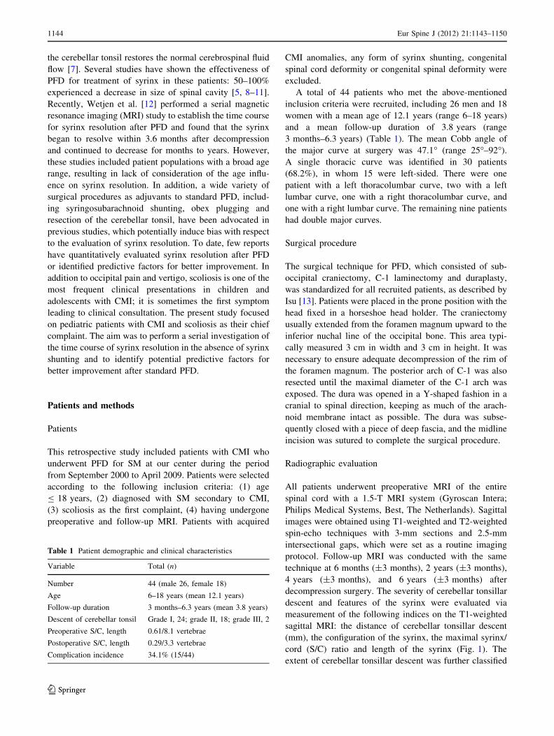
into three grades (Fig. 2) [14]: grade I, in which the tonsil
descended beyond the foramen magnum but did not reach
the C-1 arch; grade II, in which the tonsil reached the C-1
arch; and grade III, in which the tonsil descended beyond
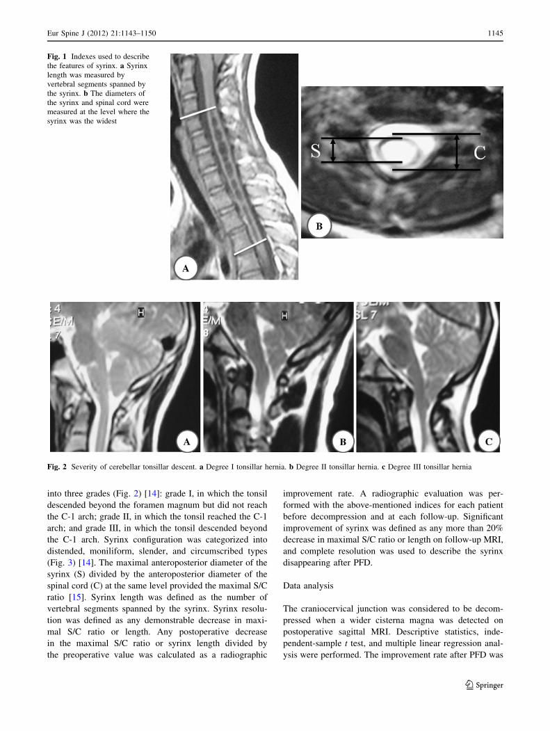
the C-1 arch. Syrinx configuration was categorized into
distended, moniliform, slender, and circumscribed types
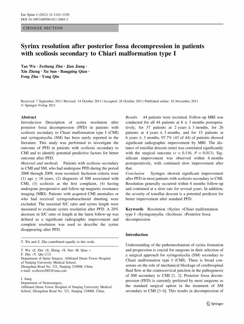
(Fig. 3) [14]. The maximal anteroposterior diameter of the
syrinx (S) divided by the anteroposterior diameter of the
spinal cord (C) at the same level provided the maximal S/C
ratio [15]. Syrinx length was defined as the number of
vertebral segments spanned by the syrinx. Syrinx resolu-
tion was defined as any demonstrable decrease in maxi-
mal S/C ratio or length. Any postoperative decrease
in the maximal S/C ratio or syrinx length divided by
the preoperative value was calculated as a radiographic
improvement rate. A radiographic evaluation was per-
formed with the above-mentioned indices for each patient
before decompression and at each follow-up. Significant
improvement of syrinx was defined as any more than 20%
decrease in maximal S/C ratio or length on follow-up MRI,
and complete resolution was used to describe the syrinx
disappearing after PFD.
Data analysis
The craniocervical junction was considered to be decom-
pressed when a wider cisterna magna was detected on
postoperative sagittal MRI. Descriptive statistics, inde-
pendent-sample t test, and multiple linear regression anal-
ysis were performed. The improvement rate after PFD was
Fig. 1 Indexes used to describe
the features of syrinx. a Syrinx
length was measured by
vertebral segments spanned by
the syrinx. b The diameters of
the syrinx and spinal cord were
measured at the level where the
syrinx was the widest
Fig. 2 Severity of cerebellar tonsillar descent. a Degree I tonsillar hernia. b Degree II tonsillar hernia. c Degree III tonsillar hernia
Eur Spine J (2012) 21:1143–1150 1145
123
calculated for each patient, and an independent-sample
t test was performed to investigate the influence of age and
sex on the outcome of PFD. Associations between
improvement rate and the distance of cerebellar tonsil
descent (mm), curve magnitude, and particularly, preop-
erative size of the syrinx were explored by multiple linear
regression. Statistical analyses were performed with SPSS
13 software (SPSS, Chicago, IL, USA). For all analyses,
statistical significance was set at P \ 0.05.
Results
Based on detailed history-taking and physical examination
at presentation, the following symptoms and signs were
identified: occipital pain in 10 patients (22.7%), vertigo in
5 (11.4%), unilateral absence of abdominal reflex in 14
(31.8%), paresthesia in 6 (13.0%) and extremity weakness
in 5 (11.4%). Relief of symptoms and physical signs was
achieved in 21 patients (47.7%) immediately after surgery
and in 37 patients (84.1%) at the final follow-up. No
neurologic complications occurred in this series of patients.
Superficial infection occurred in four cases (9.1%). In
addition to the above, nine patients (20.5%) experienced
transient headache, two (4.5%) experienced incisional
cerebrospinal fluid leakage, and one (2.3%) experienced
transient intracranial hypertension after surgery.
In our series, the Cobb angle of the main curve ranged
from 25� to 92�. There were 18 patients, in whom the Cobb
angle of main curve was more than 50�, receiving surgical
correction of scoliosis six months after PFD. The 26
remaining patients received a bracing treatment for their
scoliosis after PFD. After the mean follow-up duration of
2.3 years, 15 patients had no obvious progression of
scoliosis while 11 patients had a curve progression. Of
these 11 patients, 10 patients underwent surgical correction
of scoliosis when Cobb angle of scoliosis progressed
over 50�.
Preoperative MRI demonstrated tonsillar herniation of
grade I in 24 patients, grade II in 18, and grade III in 2
(Table 1). Syrinx configuration was identified as distended
type in 6 patients, moniliform type in 11, slender type in 11
and circumscribed type in 16.
Follow-up MRI was conducted for all 44 patients at
6 ± 3 months postoperatively, for 37 patients at 2 years ±
3 months, for 26 patients at 4 years ± 3 months, and for
15 patients at 6 years ± 3 months. 81.8% (36/44) of these
patients achieved the criteria of significant improvement
within 6 months after surgery, and 97.7% (43/44) achieved
significant improvement by the final follow-up. The time
course for syrinx resolution, in terms of syrinx size, among
these patients is shown in Figs. 4, 5 and 6. The syrinx
resolved substantially in most patients within 6 months.
17 patients showed complete syrinx resolution within
6 months after surgery, and 20 patients showed complete
resolution at 6 years. No significant syrinx improvement
was observed in some patients at 6 months after decom-
pression; however, this was followed by a significant
improvement at the next visit.
Correlation analyses were conducted to assess associa-
tions between syrinx improvement rate and potential
predictors including age, sex, syrinx configuration, cere-
bellar tonsillar descent, follow-up duration, and curve
magnitude. Multiple linear regression analysis revealed
that only the distance of cerebellar tonsillar descent (mm)
before surgery was positively correlated with improvement
Fig. 3 Syrinx was classified by shape. a Distended type. b Moniliform type. c Circumscribed type. d Slender type
1146 Eur Spine J (2012) 21:1143–1150
123
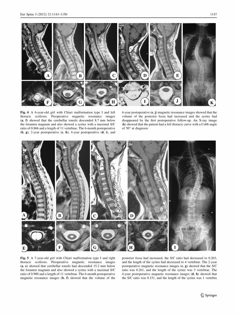
Fig. 4 A 6-year-old girl with Chiari malformation type I and left
thoracic scoliosis. Preoperative magnetic resonance images
(a, f) showed that the cerebellar tonsils descended 8.7 mm below
the foramen magnum and also showed a syrinx with a maximal S/C
ratio of 0.866 and a length of 11 vertebrae. The 6-month postoperative
(b, g), 2-year postoperative (c, h), 4-year postoperative (d, i), and
6-year postoperative (e, j) magnetic resonance images showed that the
volume of the posterior fossa had increased and the syrinx had
disappeared by the first postoperative follow-up. An X-ray image
(k) showed that the patient had a left thoracic curve with a Cobb angle
of 50� at diagnosis
Fig. 5 A 7-year-old girl with Chiari malformation type I and right
thoracic scoliosis. Preoperative magnetic resonance images
(a, e) showed that cerebellar tonsils had descended 15.2 mm below
the foramen magnum and also showed a syrinx with a maximal S/C
ratio of 0.900 and a length of 11 vertebrae. The 6-month postoperative
magnetic resonance images (b, f) showed that the volume of the
posterior fossa had increased, the S/C ratio had decreased to 0.263,
and the length of the syrinx had decreased to 4 vertebrae. The 2-year
postoperative magnetic resonance images (c, g) showed that the S/C
ratio was 0.261, and the length of the syrinx was 3 vertebrae. The
4-year postoperative magnetic resonance images (d, h) showed that
the S/C ratio was 0.151, and the length of the syrinx was 1 vertebra
Eur Spine J (2012) 21:1143–1150 1147
123
rate (P = 0.013), but it contributed only a little to the
improvement rate (rcerebellar tonsillar descent = 0.116) (Table 2).
Other factors, such as syrinx configuration, curve magni-
tude, and follow-up duration, were found to have no cor-
relation with improvement rate.
Discussion
The outcome of PFD for the treatment of SM secondary to
CMI has varied widely in previous studies in terms of
maximal S/C ratio and syrinx length. Hida et al. [8]
reported that 30 of 33 patients with SM secondary to CMI
exhibited obvious syrinx resolution after PFD. Depreitere
et al. [16] reported that 80% of patients (16/20) showed a
favorable result consisting of complete syrinx resolution in
8 patients and obvious syrinx reduction in another 8.
Wetjen et al. [12] documented the time course of syrinx
resolution after PFD and found that the syrinx resolved
significantly over a short period of time after decompres-
sion. Significant improvement was also reported in 7 of 7
patients in a study by Feldstein and Choudhri [17] and 11
of 12 in a study by Ghanem et al. [18]. However, low rates
of significant improvement have also been reported. In
studies by Caldarelli and colleagues and McGirt and col-
leagues, syrinx resolution after PFD was found only in 50
and 62% of patients, respectively [10, 19]. This discor-
dance may be related to broad age range, different surgical
procedures, or variability in the definition of significant
improvement. In general, pediatric patients have a better
response to PFD than adults [5], implying that the more
pediatric patients recruited for a study, the better the
surgical outcome. Different surgical procedures might
decompress the cerebellar tonsils to different extents or
even exert a direct effect on the syrinx, resulting in different
rates of significant syrinx improvement [4, 8, 20–23]. Var-
iability in the definition of significant improvement also
may have led to contrary results among previous reports.
To explore the course of syrinx resolution after PFD in a
scoliosis population, patients with a complaint of scoliosis
were recruited for the present study. To avoid the influence
of age and surgical procedure on syrinx resolution, we
recruited CMI patients with the age of less than or equal to
18 years and excluded patients with any form of syrinx
shunting to quantitatively evaluate the outcome of PFD.
In the present study, patients with a [20% decrease in
syrinx size were defined as having significant improve-
ment. Significant syrinx improvement was identified in 43
of 44 patients (97.7%) approximately 6 years after
decompression. This was relatively higher than that
reported by Munshi and colleagues and Depreitere and
colleagues [16, 24]. This is consistent with the fact that
pediatric patients experience less permanent neurologic
injury and higher improvement rates after PFD, owing to
their enhanced repair capability and short clinical history
[5, 25, 26].
The time course of syrinx resolution after PFD has not
been well documented, although it is known that syrinx can
resolve within weeks of this procedure [8]. Results of the
present study showed significant syrinx improvement in
81.8% (36/44) of patients, with stable syrinx in 18.2%
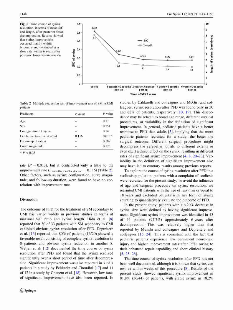
Fig. 6 Time course of syrinx
resolution, in terms of mean S/C
and length, after posterior fossa
decompression. Results showed
that syrinx improvement
occurred mainly within
6 months and continued at a
slow rate within 6 years after
posterior fossa decompression
Table 2 Multiple regression test of improvement rate of SM in CMI
patients
Predictors r value P value
Age – 0.77
Sex – 0.151
Configuration of syrinx – 0.14
Cerebellar tonsillar descent 0.116 0.013*
Follow-up duration – 0.189
Curve magnitude – 0.123
* P \ 0.05
1148 Eur Spine J (2012) 21:1143–1150
123

(8/44) within 6 months after surgery. Stable syringes
improved with time, with significant improvement in 97.7%
(43/44) of patients by 6 years after surgery. Interestingly, it
was noted that in most patients, syrinx size continued to
decrease slowly over time. This is consistent with the results
of the Wetjen et al. [12].
Several factors, such as sex, preoperative size and syrinx
length, have been reported as predictors of better surgical
outcome after PFD [4, 5, 20]. Badie et al. [27] reported that
female patients have a better response to PFD than male
patients because female patients have a lower posterior
fossa volume. In a study by Park et al. [20], it was found
that the longer the syrinx, the better the outcome. To
identify other potential predictive factors, we used multiple
linear regressions. With the exception of distance of cere-
bellar tonsil descent, which showed a small contribution to
the improvement rate at the last follow-up (P = 0.013,
rthe degree of cerebellar tonsil descending = 0.116), no association
between the factors assessed, including sex, syrinx con-
figuration, follow-up duration, curve magnitude, or
improvement rate at the final follow-up, was found.
Patients with more severe cerebellar tonsillar descent often
have a worse cerebrospinal fluid abnormality. After PFD,
the cerebellar tonsils are decompressed and cerebrospinal
fluid flow is restored, resulting in more benefit from PFD.
Considering that the patients in the present study were
relatively young, we suggest that extrapolation of the above
findings should be restricted to pediatric patients with CMI.
A potential limitation of the present study was the
retrospective nature, such that the follow-up duration
varied widely. This limitation would influence the power of
the study. Nonetheless, we believe that the present results
can document the time course of syrinx resolution in
patients with scoliosis secondary to CMI and SM. We
found significant syrinx improvement after PFD in most
pediatric patients with CMI, particularly within the initial
several months after surgery. Severe tonsillar descent
might be a predictor of better surgical outcome after PFD
Acknowledgments No benefits in any form have been or will be
received from a commercial party related directly or indirectly to the
subject of this manuscript. This work was supported by the National
Natural Science Foundation of China (Grant No. 81171672).
Conflict of interest None.
References
1. Milhorat TH, Nishikawa M, Kula RW, Dlugacz YD (2010)
Mechanisms of cerebellar tonsil herniation in patients with Chiari
malformations as guide to clinical management. Acta Neurochir
(Wien) 152:1117–1127. doi:10.1007/s00701-010-0636-3
2. McGirt MJ, Atiba A, Attenello FJ, Wasserman BA, Datoo G,
Gathinji M, Carson B, Weingart JD, Jallo GI (2008) Correlation
of hindbrain CSF flow and outcome after surgical decompression
for Chiari I malformation. Childs Nerv Syst 24:833–840. doi:
10.1007/s00381-007-0569-1
3. Tubbs RS, Lyerly MJ, Loukas M, Shoja MM, Oakes WJ (2007)
The pediatric Chiari I malformation: a review. Childs Nerv Syst
23:1239–1250. doi:10.1007/s00381-007-0428-0
4. Durham SR, Fjeld-Olenec K (2008) Comparison of posterior
fossa decompression with and without duraplasty for the surgical
treatment of Chiari malformation Type I in pediatric patients: a
meta-analysis. J Neurosurg Pediatr 2:42–49. doi:10.3171/PED/
2008/2/7/042
5. Navarro R, Olavarria G, Seshadri R, Gonzales-Portillo G,
McLone DG, Tomita T (2004) Surgical results of posterior fossa
decompression for patients with Chiari I malformation. Childs
Nerv Syst 20:349–356. doi:10.1007/s00381-003-0883-1
6. Xie J, Wang Y, Zhao Z, Zhang Y, Si Y, Yang Z, Liu L, Lu N
(2011) One-stage and posterior approach for correction of mod-
erate to severe scoliosis in adolescents associated with Chiari I
malformation: is a prior suboccipital decompression always
necessary? Eur Spine J 20:1106–1113. doi:10.1007/s00586-011-
1717-6
7. Hankinson TC, Klimo P Jr, Feldstein NA, Anderson RC,
Brockmeyer D (2007) Chiari malformations, syringohydromyelia
and scoliosis. Neurosurg Clin N Am 18:549–568. doi:10.1016/
j.nec.2007.04.002
8. Hida K, Iwasaki Y, Koyanagi I, Sawamura Y, Abe H (1995)
Surgical indication and results of foramen magnum decompres-
sion versus syringosubarachnoid shunting for syringomyelia
associated with Chiari I malformation. Neurosurgery 37:673–678
(discussion 678–679)
9. Alzate JC, Kothbauer KF, Jallo GI, Epstein FJ (2001) Treatment
of Chiari I malformation in patients with and without syringo-
myelia: a consecutive series of 66 cases. Neurosurg Focus
11:1–9. doi:10.3171/foc.2001.11.1.4
10. Caldarelli M, Novegno F, Vassimi L, Romani R, Tamburrini G,
Di Rocco C (2007) The role of limited posterior fossa craniec-
tomy in the surgical treatment of Chiari malformation type I:
experience with a pediatric series. J Neurosurg 106:187–195. doi:
10.3171/ped.2007.106.3.187
11. Takayasu M, Takagi T, Hara M, Anzai M (2004) A simple
technique for expansive suboccipital cranioplasty following
foramen magnum decompression for the treatment of syringo-
myelia associated with Chiari I malformation. Neurosurg Rev
27:173–177. doi:10.1007/s10143-004-0338-5
12. Wetjen NM, Heiss JD, Oldfield EH (2008) Time course of
syringomyelia resolution following decompression of Chiari
malformation type I. J Neurosurg Pediatr 1:118–123. doi:
10.3171/PED/2008/1/2/118
13. Isu T, Sasaki H, Takamura H, Kobayashi N (1993) Foramen
magnum decompression with removal of the outer layer of the
dura as treatment for syringomyelia occurring with Chiari I
malformation. Neurosurgery 33:844–849 (discussion 849–850)
14. Ono A, Ueyama K, Okada A, Echigoya N, Yokoyama T, Harata
S (2002) Adult scoliosis in syringomyelia associated with Chiari I
malformation. Spine (Phila Pa 1976) 27:E23–E28
15. Qiu Y, Zhu ZZ, Wang B, Yu Y, Qian BP, Zhu F (2008) Radio-
logical presentations in relation to curve severity in scoliosis
associated with syringomyelia. J Pediatr Orthoped 28:128–133
16. Depreitere B, Van Calenbergh F, van Loon J, Goffin J, Plets C
(2000) Posterior fossa decompression in syringomyelia associated
with a Chiari malformation: a retrospective analysis of 22
patients. Clin Neurol Neurosurg 102:91–96. doi:10.1016/S0303-
8467(00)00073-1
Eur Spine J (2012) 21:1143–1150 1149
123
17. Feldstein NA, Choudhri TF (1999) Management of Chiari I
malformations with holocord syringohydromyelia. Pediatr
Neurosurg 31:143–149. pii: pne31143
18. Ghanem IB, Londono C, Delalande O, Dubousset JF (1997)
Chiari I malformation associated with syringomyelia and scoli-
osis. Spine (Phila Pa 1976) 22:1313–1317 (discussion 1318)
19. McGirt MJ, Attenello FJ, Atiba A, Garces-Ambrossi G, Datoo G,
Weingart JD, Carson B, Jallo GI (2008) Symptom recurrence
after suboccipital decompression for pediatric Chiari I malfor-
mation: analysis of 256 consecutive cases. Childs Nerv Syst
24:1333–1339. doi:10.1007/s00381-008-0651-3
20. Park YS, Kim DS, Shim KW, Kim JH, Choi JU (2009) Factors
contributing improvement of syringomyelia and surgical outcome
in type I Chiari malformation. Childs Nerv Syst 25:453–459. doi:
10.1007/s00381-008-0763-9
21. Attenello FJ, McGirt MJ, Gathinji M, Datoo G, Atiba A,
Weingart J, Carson B, Jallo GI (2008) Outcome of Chiari-asso-
ciated syringomyelia after hindbrain decompression in children:
analysis of 49 consecutive cases. Neurosurgery 62:1307–1313
(discussion 1313). doi:10.1227/01.neu.0000333302.72307.3b
22. Nishikawa M, Ohata K, Baba M, Terakawa Y, Hara M (2004)
Chiari I malformation associated with ventral compression and
instability: one-stage posterior decompression and fusion with a
new instrumentation technique. Neurosurgery 54:1430–1434
(discussion 1434–1435)
23. Tubbs RS, McGirt MJ, Oakes WJ (2003) Surgical experience in
130 pediatric patients with Chiari I malformations. J Neurosurg
99:291–296. doi:10.3171/jns.2003.99.2.0291
24. Munshi I, Frim D, Stine-Reyes R, Weir BK, Hekmatpanah J,
Brown F (2000) Effects of posterior fossa decompression with
and without duraplasty on Chiari malformation-associated
hydromyelia. Neurosurgery 46:1384–1389 (discussion 1389–
1390)
25. Milhorat TH, Johnson WD, Miller JI, Bergland RM, Hollenberg-
Sher J (1992) Surgical treatment of syringomyelia based on
magnetic resonance imaging criteria. Neurosurgery 31:231–244
(discussion 244–235)
26. Cai C, Oakes WJ (1997) Hindbrain herniation syndromes: the
Chiari malformations (I and II). Semin Pediatr Neurol 4:179–191
27. Badie B, Mendoza D, Batzdorf U (1995) Posterior fossa volume
and response to suboccipital decompression in patients with
Chiari I malformation. Neurosurgery 37:214–218
1150 Eur Spine J (2012) 21:1143–1150
123