systematic review of the survival rate and incidence of
TRANSCRIPT

The International Journal of Oral & Maxillofacial Implants 99
Today, partially edentulous individuals represent the main group of patients requiring treatment in
daily dental practice. Therefore, oral implants are the
predominant treatment modality for the rehabilitation of these patients.1 Using implants, fixed partial den-tures can be applied in situations where removable dentures would previously have been necessary.2–4 In addition, more treatment options that preserve the tooth structure are possible by replacing missing single teeth with dental implants.5 Since most of the patients provided with oral implants are between 40 and 50 years of age, promising long-term survival rates for implants and prostheses are expected both by the clinician and the patient to ensure the longevity of the prosthesis.6–8 The definition “long-term” has been specified as a follow-up of at least 5 years.9 Thus, sur-vival rates and the incidence of biologic, technical, and esthetic events should be based on mean observation periods of at least 5 years.10
Several years ago, hierarchies of evidence were de-veloped as aid for the interpretation and evaluation of research findings.11 As evidence, systematic reviews were ranked to be excellent in terms of effectiveness, appropriateness, and feasibility. An evidence level of “excellent” equates with the strongest scientific basis
1 PhD Student, Department of Oral Implantology and Prosthetic Dentistry Academic Center for Dentistry Amsterdam (ACTA), Amsterdam, The Netherlands; Assistant Professor, Division of Fixed Prosthodontics, School of Dental Medicine, University of Bern, Bern, Switzerland.
2 Associate Professor, Department of Prosthodontics, College of Dentistry, Yonsei University, Seoul, Korea.
3 Professor, Institute of Social and Preventive Medicine, University of Bern, Bern, Switzerland.
4 Professor, Department of Reconstructive Sciences, Center for Biomaterials, University of Connecticut Health Center, Farmington, Connecticut, USA.
Correspondence to: Dr Anja Zembic, Department of Oral Implantology and Prosthetic Dentistry, Academic Center for Dentistry Amsterdam (ACTA), Gustav Mahlerlaan 3004, 1081 LA Amsterdam, The Netherlands. Fax: +31 020 598 03 33. Email: [email protected] ©2014 by Quintessence Publishing Co Inc.
Systematic Review of the Survival Rate and Incidence of Biologic, Technical, and Esthetic Complications of
Single Implant Abutments Supporting Fixed ProsthesesAnja Zembic, Dr Med Dent1/Sunjai Kim, DDS, MSD, PhD2/
Marcel Zwahlen, PhD, MSc3/J. Robert Kelly, DDS, MS, DMedSc4
Purpose: To assess the 5-year survival rate and number of technical, biologic, and esthetic complications
involving implant abutments. Materials and Methods: Electronic (Medline) and hand searches were
performed to assess studies on metal and ceramic implant abutments. Relevant data from a previous review
were included. Two reviewers independently extracted the data. Failure and complication rates were analyzed,
and estimates of 5-year survival proportions were calculated from the relationship between event rate and
survival function. Multivariable robust Poisson regression was used to compare abutment characteristics.
Results: The search yielded 1,558 titles and 274 abstracts. Twenty-four studies were selected for data
analysis. The survival rate for ceramic abutments was 97.5% (95% confidence interval [CI]): 89.6% to 99.4%)
and 97.6% (95% CI: 96.2% to 98.5%) for metal abutments. The overall 5-year rate for technical complications
was 11.8% (95% CI: 8.5% to 16.3%), 8.9% (95% CI: 4.3% to 17.7%) for ceramic and 12.0% (95% CI: 8.5% to
16.8%) for metal abutments. Biologic complications occurred with an overall rate of 6.4% (95% CI: 3.3% to
12.0%), 10.4% (95% CI: 1.9% to 46.7%) for ceramic, and 6.1% (95% CI: 3.1% to 12.0%) for metal abutments.
Conclusions: The present meta-analysis on single-implant prostheses presents high survival rates of single
implants, abutments, and prostheses after 5 years of function. No differences were found for the survival
and failure rates of ceramic and metal abutments. No significant differences were found for technical,
biologic, and esthetic complications of internally and externally connected abutments. Int J Oral MaxIllOfac IMplants 2014;29(suppl):99–116. doi: 10.11607/jomi.2014suppl.g2.2
Key words: biologic complications, ceramics, complication rates, esthetic complications, failures, implant abutments, implant prostheses, metal, survival, systematic review, technical complications, titanium, zirconia
© 2014 BY QUINTESSENCE PUBLISHING CO, INC. PRINTING OF THIS DOCUMENT IS RESTRICTED TO PERSONAL USE ONLY. NO PART MAY BE REPRODUCED OR TRANSMITTED IN ANY FORM WITHOUT WRITTEN PERMISSION FROM THE PUBLISHER.
Zembic et al
100 Volume 29, Supplement, 2014
for clinical practice along with the least risk of error.11 Consequently, systematic reviews are an optimal tool for the development of practice guidelines and clinical recommendations.
A recent systematic review confirmed single im-plants to be a successful treatment method with sur-vival rates of 97.2% at 5 years and 95.2% at 10 years.12 However, implant survival rates are not the only es-sential consideration when advising the patient on different treatment options. Prosthetic and implant abutment outcomes need to be considered as well. Dif-ferent kinds of abutments are available with respect to material (metal and ceramic) and shape (prefabricated and customized, both with various internal designs). At this time, metal abutments are classified as the “gold standard,” although high-strength zirconia abutments are being utilized more widely and may be an adequate alternative to metal abutments for the clinical use. The results of a previous systematic review showed similar outcomes for ceramic and metal abutments.13 However, the results need to be interpreted with caution due to a high variation in the number of analyzed abutments and differing numbers of studies and follow-up times.
Since the use of ceramic abutments has spread with-in the last few years, an increase in clinical studies might thus be expected. An update of the available most re-cent clinical data may help the clinician decide upon the most ideal abutment in each individual situation.
The aim was to systematically review the existing dental literature on the survival rates of metal and ce-ramic abutments supporting single implant crowns with a mean observation period of at least 3 years. In addition, the occurrence of negative biologic, techni-cal, and esthetic events was evaluated for metal and ceramic abutments.
Materials and Methods
The PICO (population, intervention, comparison, out-come) question was stated as follows: For single-tooth implant prostheses in anterior and posterior locations, are there differences in survival/performance based on technical, biologic, and esthetic outcomes as influ-enced by material and design?
search strategyThe present systematic review was performed as an update of a previously published systematic review with the same objectives.13
A Medline (PubMed) search was performed for clini-cal studies published in dental journals from January 1, 2009 up to April 30, 2012. The search was limited to English, German, French, Dutch, and Korean language publications (Table 1).
search terms The following search terms were grouped to the three main subjects (implants, abutments, and material) and linked with “and” as follows:
Implants"Dental Implants, Single-Tooth" [MeSH] AND "dental implants" AND “dental implant* single tooth” AND “sin-gle tooth implant*” AND "single implant" AND "dental implant" AND "single tooth implant" AND "single tooth implants" AND "single implants" AND “Denture, Partial, Fixed” [MeSH] AND “Dental Prosthesis Design” [MeSH] AND "fixed restoration" AND “Denture Design” [MeSH] AND “implant*” AND "fixed prosthodontic" AND "fixed partial denture" AND "fixed prosthodontics" AND "fixed partial dentures" AND “dental implants” [MeSH] AND “Dental Prosthesis, Implant-Supported” AND “fixed dental prosthesis” AND “fixed dental prostheses”.
Abutments“Dental Abutments” [MeSH] AND "implant abutment" AND “implant* reconstruct*” AND “implant* abut-ment*” AND "implant abutments" AND “abutment*” AND “dental abutment*”.
Material"Titanium" [MeSH] AND “Gold” [MeSH] AND “ceram-ics” [MeSH] AND “aluminum” [MeSH] AND “Zirconium” [MeSH] AND “ceramic*” AND “titan*” AND “metal*” AND “zirconi*” AND “gold*” AND “alumin*” AND “metals” [MeSH].
Thereafter, the search results from the three subject groups were combined with each other using “OR.” The electronic search was complemented by manual searching of the bibliographies of the most recent sys-tematic reviews12,14,15 and of all included publications.
inclusion CriteriaThe criteria for study inclusion were:
• Studies with at least 10 included patients • Clinical studies only • Studies with a mean follow-up of at least 3 years
(unless there was an immediate negative effect)• Studies reporting on details and outcomes of
implant abutments• Studies reporting on partially edentulous patients
receiving implant-supported single crowns
exclusion CriteriaReports based on patient chart reviews, question-naires, or interviews were excluded as were case re-ports and multiple publications on the same patient cohort.
© 2014 BY QUINTESSENCE PUBLISHING CO, INC. PRINTING OF THIS DOCUMENT IS RESTRICTED TO PERSONAL USE ONLY. NO PART MAY BE REPRODUCED OR TRANSMITTED IN ANY FORM WITHOUT WRITTEN PERMISSION FROM THE PUBLISHER.
Group 2
The International Journal of Oral & Maxillofacial Implants 101
study selectionAll obtained titles and abstracts were checked for in-clusion by two independent reviewers (SK and AZ). In case the abstract was not available, a full text article was acquired. On the basis of the chosen abstracts, full-text articles were selected for independent as-sessment by the reviewers. If the information in title and abstract was insufficient for inclusion or exclusion, full-text articles were also obtained. In case of any dis-agreement regarding inclusion, a decision was made by the three reviewers by consensus. The agreement among the three reviewers for the inclusion of full-text articles was subsequently calculated by Cohen kappa
coefficient. In addition, 16 publications on single im-plant prostheses were included for analysis from the previous review.13
data extractionA data extraction sheet was used by two reviewers (SK, AZ) to extract the relevant data from the included pa-pers. Information on several parameters was recorded including: author(s), study design, year of publication, mean follow-up time, implant system, number of abut-ments, abutment material, drop-outs, and survival rates, as well as the incidence of biologic, technical, and esthetic complications of abutments. Disagree-
table 1 systematic search strategy
Focus question For single-tooth implant reconstructions in anterior and posterior locations are there differences in survival/performance based on technical, biologic, and esthetic outcomes as influenced by material, design, and fabrication?
search strategy
Population Patients with single-implant reconstructions
Intervention or exposure Single implants with a mean follow-up of 3 y
Comparison Abutment material (metal vs ceramic)
Outcome Survival rate of implants, abutments, reconstructions
Search combination Implants:"Dental Implants, Single-Tooth" [MeSH] AND "dental implants" AND “dental implant* single tooth” AND “single tooth implant*” AND "single implant" AND "dental implant" AND "single tooth implant" AND "single tooth implants" AND "single implants" AND “Denture, Partial, Fixed” [MeSH] AND “Dental Prosthesis Design” [MeSH] AND "fixed restoration" AND “Denture Design” [MeSH] AND “im-plant*” AND "fixed prosthodontic" AND "fixed partial denture" AND "fixed prosthodontics" AND "fixed partial dentures" AND “dental implants” [MeSH] AND “Dental Prosthesis, Implant-Supported” AND “fixed dental prosthesis” AND “fixed dental prostheses”Abutments:“Dental Abutments” [MeSH] AND "implant abutment" AND “implant* reconstruct*” AND “implant* abutment*” AND "implant abutments" AND “abutment*” AND “dental abutment*”Material:"Titanium" [MeSH] AND “Gold” [MeSH] AND “ceramics” [MeSH] AND “aluminum” [MeSH] AND “Zirconium” [MeSH] AND “ceramic*” AND “titan*” AND “metal*” AND “zirconi*” AND “gold*” AND “alumin*” AND “metals” [MeSH]Thereafter, the search results from the three subject groups were combined with each other using “OR”
database search
Electronic PubMed, Cochrane Central Register of Controlled Trials (CENTRAL)
Journals Clinical Oral Implants Research, International Journal of Oral Maxillofacial Implants, International Journal of Oral and Maxillofacial Surgery, Clinical Implant Dentistry and Related Research, Implant Dentistry, Jour-nal of Implantology, Journal of Periodontology, Journal of Clinical Periodontology, Clinical Oral Investigation, Dental Materials, International Journal of Prosthodontics, European Journal of Oral Implantology
selection criteria
Inclusion criteria Studies with at least 10 included patients Clinical studies only Studies with a mean follow-up of at least 3 years; studies reporting on details and outcomes of implant abutmentsStudies reporting on partially edentulous patients receiving implant-supported single crowns
Exclusion criteria Reports based on patient chart reviews, questionnaires, or interviewsCase reports
CT, controlled trial; RCT, randomized controlled trial; NR, not reported.
© 2014 BY QUINTESSENCE PUBLISHING CO, INC. PRINTING OF THIS DOCUMENT IS RESTRICTED TO PERSONAL USE ONLY. NO PART MAY BE REPRODUCED OR TRANSMITTED IN ANY FORM WITHOUT WRITTEN PERMISSION FROM THE PUBLISHER.

Zembic et al
102 Volume 29, Supplement, 2014
First electronic search:1,558 titles
Independently selected and agreed upon by two reviewers:
274, abstracts obtained
Interreader agreementk = 0.88 ± 0.87
Independently selected and agreed upon by two reviewers:
192 abstracts, full text obtained
Articles from Sailer et al13
29
Reviews:15
Excluded:107
Included:5
Included:16
Excluded:13
Further handsearching3 articles (reference)
Final number of included studies:24
Fig 1 Search strategy.
ment regarding data extraction was resolved by consensus. The number of events and the corresponding total exposure time of the prostheses were calculated. In case the publication did not provide sufficient information, the corresponding authors of the respective publications were contacted via email. Additionally, the data from included studies on single implant crowns from the previous review were extracted.13
Survival was defined as the abutment/implant prosthesis re-maining in situ for the observation period with or without modi-fications.
Technical complications included abutment fracture, abut-ment screw fracture, abutment screw loosening, misfit at the im-plant-abutment junction (gap), fracture of the implant prosthesis, chipping of the veneering ceramic, and loosening of the implant prosthesis.
The analysis of biologic complications encompassed bone loss of more than 2 mm, soft tissue recession, and general soft tissue complications.
The analysis of the esthetic complications included soft tissue discoloration and other esthetic problems.
statistical analysisFailure and complication rates were calculated by dividing the
number of events (failures or complications) as the numerator by the total time of the prostheses being under observation as the denominator. The numerator could usually be extracted directly from the publication. If all patients/prostheses had a fixed follow-up time point, this was taken as the obser-vation period for all. Otherwise, the total observation time was calculated by taking the sum of the following: (1) exposure time of prostheses that could be followed for the full observation period; (2) exposure time up to failure of the prostheses that were lost due to failure; and (3) exposure time up to the end of observation time for prostheses that did not complete the observation pe-riod for reasons such as death, change of address, refusal to participate, nonresponse, chronic illnesses, missed appointments, and work commitments. If all three components for the calculation of the total exposure time were not available, the total exposure time was estimated by multiplying the mean fol-low-up time by the number of constructions under observation.
For each study, event rates for the abut-ments and the prostheses were calculated by dividing the total number of events by the total abutment exposure time in years. For additional analysis, the total number of events was considered to be Poisson distrib-uted for a given sum of abutment exposure years and robust Poisson regression with a logarithmic link-function and total expo-sure time per study as an offset variable was used.16 Robust Poisson regression allowed for the calculation of standard errors and 95% confidence intervals (CI), which incor-porated heterogeneity among studies.
Five-year survival proportions were cal-culated via the relationship between event rate and survival function S(T) by assuming constant event rates17:
S(T) = exp(–T × event rate)
For the 5-year survival, T was equal to 5.The 95% CIs for the survival proportions
were calculated by using the 95% CIs of the event rates. Multivariable robust Poisson re-gression was used to formally compare con-struction subtypes and to assess other study characteristics and to estimate event rate ratios and their 95% CIs. All analyses were performed using Stata, version 12.
© 2014 BY QUINTESSENCE PUBLISHING CO, INC. PRINTING OF THIS DOCUMENT IS RESTRICTED TO PERSONAL USE ONLY. NO PART MAY BE REPRODUCED OR TRANSMITTED IN ANY FORM WITHOUT WRITTEN PERMISSION FROM THE PUBLISHER.
Group 2
The International Journal of Oral & Maxillofacial Implants 103
results
The search strategy is presented in Fig 1. The Medline search provided a total of 1,558 titles. After screening of all titles, both reviewers agreed upon 274 abstracts. Finally, 24 full-text articles reporting on the clinical performance of implant abutments were selected (Table 2). Three out of 24 studies were gained through the hand search and 16 articles were retrieved from the previous review. The studies were published from 1996 until 2012. The inter-reviewer agreement for the inclusion of the studies was κ = 0.88 ± 0.87 (Cohen kappa coefficient).
excluded studiesOne hundred twenty-two studies were excluded due to the following reasons: mean observation period less than 3 years (n = 27), no detailed information on abut-ments (n = 42), no detailed results on abutments (n = 6), data obtained from patient chart reviews (n = 3), splint-ed crowns (n = 8), case reports (n = 19), reviews (n = 15), or mixed data on FPDs and single implant crowns (n = 2).
included studiesAmong the selected full-text articles, three studies25,31,41 were randomized clinical trials (RCTs) comparing differ-ent abutment materials (zirconia vs titanium, alumina vs
table 2 Characteristics of the included studies
studyYear of
publication study design
total no. of
included patients
age range
Mean age setting
Mean follow-up
(y)drop-out
(%)
Avivi-Arber and Zarb18
1996 Prospective CT 41 14.5–3.9 33.5 University 4 5
Henry et al19 1996 Prospective CT 92 NR NR Multicenter 5 8
Andersson et al20 1998 Prospective CT 57 NR 32 Specialist clinic 5 5
Scheller et al21 1998 Multicenter prospective CT
82 14–73 35 Multicenter 5 25
Levine et al22 1999 Retrospective 129 NR NR Multicenter 3.3 19
Wannfors and Smedberg23
1999 Prospective 69 17–72 26 Specialist clinic 3 3
Bianco et al24 2000 Retrospective CT 214 16–70 NR Multicenter 8 9
Andersson et al25 2001 RCT 15 17–49 32 Specialist clinic 3 0
Krennmair et al26 2002 Retrospective 112 NR 31.3 Private practice and university
3 NR
Muche et al27 2003 Retrospective 76 NR 45 University 3 NR
Glauser et al28 2004 Prospective CT 27 26–75 44 University 4.1 9
Romeo et al29 2004 Prospective CT 250 20–67 NR University 3.9 NR
Brägger et al30 2005 Prospective cohort study
127 19–78 49.3 University 10 NR
Vigolo et al31 2006 Prospective RCT 20 NR NR University 4 0
Canullo32 2007 Prospective cohort study
25 25–70 NR Private practice 3.3 NR
Cooper et al33 2007 Prospective cohort study
48 NR 30.6 University 3 9
MacDonald et al34 2009 Prospective 20 NR 43.5 University 8 3
Vigolo and Givani35 2009 Prospective 144 25–55 37 Private practice 5 0
Bonde et al36 2010 Retrospective 51 19–79 43 University 10 3
Urdaneta et al37 2010 Retrospective 81 28–92 58.7 Specialist clinic 5.9 27
Ekfeldt et al38 2011 Retrospective 25 NR NR Specialist clinic 3–5 NR
Visser et al39 2011 Prospective 93 18–63 33 University 5 1
Gotfredsen40 2012 Prospective 20 18–59 33 University 10 5
Zembic et al41 2013* Prospective RCT 22 23–59 41.3 University 5.6 4
*Available ahead of print in 2012.
© 2014 BY QUINTESSENCE PUBLISHING CO, INC. PRINTING OF THIS DOCUMENT IS RESTRICTED TO PERSONAL USE ONLY. NO PART MAY BE REPRODUCED OR TRANSMITTED IN ANY FORM WITHOUT WRITTEN PERMISSION FROM THE PUBLISHER.

Zembic et al
104 Volume 29, Supplement, 2014
table 3 Characteristics of abutment and Prostheses
studyYear of
publicationimplant system
implant diam-eter location
total abutments
abutment material
abutment type
Fixation torque
abutment connection Prosthesis material
Cemented implants
screw-retained implants
Avivi-Arber and Zarb18 1996 Nobel Biocare 3.75, 4.0 Incisor, canine, premolar, molar 42 Titanium NR NR External hexagon Metal-ceramic or metal-acrylic, 1 all-ceramic
NR NR
Henry et al19 1996 Nobel Biocare NR NR 96 Titanium NR NR External hexagon NR NR NR
Andersson et al20 1998 Nobel Biocare NR 51 incisors, 1 canine, 13 premolars 65 Titanium NR NR External hexagon 62 all-ceramic, 3 metal-ceramic 65 0
Scheller et al21 1998 Nobel Biocare 3.75, 4.0 87 maxilla, 12 mandible 65 Titanium Prefabricated 32 External hexagon 16 porcelain fused to metal, 81 full ceramic
97 0
Levine et al22 1999 Straumann 3.5, 4.1 22 anterior, 135 posterior 157 Titanium NR 32 Internal NR 76 81
Wannfors and Smed-berg23
1999 Nobel Biocare NR 40% max incisor, 20%–30% max lateral incisor, 15%–20% max canine, 5 implants in mandible
76 Gold Customized, prefabricated
32 External hexagon 36 gold-resin, 35 gold-ceramic, 9 all-ceramic
36 44
Bianco et al24 2000 Nobel Biocare NR anterior and posterior 229 Titanium NR NR External hexagon Metal, metal-ceramic, all-ceramic 203 31
Andersson et al25 2001 Nobel Biocare 3.75, 4.0 17 incisors, 2 canines, 1 premolar
10 Alumina NR NR External hexagon All-ceramic 10 0
Andersson et al25 2001 NR NR 10 Titanium NR 10–32 External hexagon All-ceramic 10 0
Krennmair et al26 2002 Frialit 2 NR NR 146 Titanium NR NR Internal Metal-ceramic, all-ceramic 93 53
Muche et al27 2003 3i NR NR 205 Metal NR 35 External hexagon Metal-ceramic 5 200
Glauser et al28 2004 Nobel Biocare 3.75, 4.0 25 incisors, 14 canines, 15 premolars
36 Zirconia NR 32 External hexagon All-ceramic 54 0
Romeo et al29 2004 Straumann Narrow, regular, wide
Anterior, posterior 121 Titanium NR NR Internal Metal-ceramic NR NR
Brägger et al30 2005 Straumann NR NR 69 Metal (titanium, gold-alloy)
NR 32 Internal NR 67 2
Vigolo et al31 2006 3i 3.75, 4.0 16 maxilla, 4 mandible, 0 anterior, 20 posterior 20 Titanium Customized 35 External hexagon Metal-ceramic 20 0
Vigolo et al31 2006 3i 3.75, 4.0 16 maxilla, 4 mandible, 0 anterior, 20 posterior 20 Gold Customized 35 External hexagon Metal-ceramic 20 0
Canullo32 2007 TSA implants NR Anterior and posterior 30 Zirconia NR 15 Internal All-ceramic 30 0
Cooper et al33 2007 Astra Tech NR Incisor, canine 43 Titanium NR 32 Internal Metal-ceramic, all-ceramic 54 0
MacDonald et al34 2009 Endopore 3.5, 4.1 13 posterior, 7 anterior 17 Titanium Prefabricated NR External hexagon Metal-ceramic 0 20
Vigolo and Givani35 2009 3i wide Only molars 182 Titanium Customized 32 External hexagon Metal-ceramic 182 0
Bonde et al36 2010 Nobel Biocare 3.3 (4), 3.75 (51)
42 anterior, 13 premolars, 49 maxilla, 6 mandible
52 Titanium Prefabricated NR External hexagon All-ceramic 55 0
Urdaneta et al37 2010 Bicon 3.3–6.0 NR 326 Titanium NR NR Internal 228 gold-resin, 82 metal-ceramic, 16 all-ceramic
326 0
Ekfeldt et al38 2011 Nobel Biocare 3.3–5.0 NR 40 Zirconia Customized 35 External hexagon 40 all-ceramic (25 one-piece) 15 25
Visser et al39 2011 Straumann 4.1 Anterior maxilla 92 Titanium abutment with gold coping screwed onto it
Customized 15 Internal All-ceramic 92 0
Gotfredsen40 2012 Astra Tech 4.5 18 anterior, 2 posterior 19 Titanium Prefabricated, customized
15 Internal Metal-ceramic 19 0
Zembic et al41 2013* Nobel Biocare 3.75 2 anterior, 16 posterior 18 Zirconia Customized 32 External hexagon All-ceramic 16 2
Zembic et al41 2013* Nobel Biocare 3.75 2 anterior, 8 posterior 10 Titanium Customized 32 External hexagon Metal-ceramic 10 0
*Available ahead of print in 2012. NR, not reported.
titanium, and titanium vs gold). Seventeen studies had a prospective design, seven studies were retrospective.
In total, 12 studies were performed at a university setting, 5 studies in a specialist clinic, 2 in private prac-tice, and 1 both at university and private practice. Four studies were multicenter studies.
Overall, 1,877 patients with 2,999 abutments were involved in the included studies. Out of these, 139 (7.4%) patients and 813 (27%) abutments were drop-outs and thus not followed. Six studies did not report the patient dropout rate. The mean age of all patients was 41 years, ranging from 14 to 92 years.
© 2014 BY QUINTESSENCE PUBLISHING CO, INC. PRINTING OF THIS DOCUMENT IS RESTRICTED TO PERSONAL USE ONLY. NO PART MAY BE REPRODUCED OR TRANSMITTED IN ANY FORM WITHOUT WRITTEN PERMISSION FROM THE PUBLISHER.

Group 2
The International Journal of Oral & Maxillofacial Implants 105
In the above-mentioned three RCTs, the outcomes were compared for 10 alumina and 10 titanium abut-ments, 20 gold and 20 titanium abutments, and 18 zir-conia and 10 titanium abutments.25,31,41
The majority of studies (13) reported on anterior and posterior abutment locations.18,20,22,24,25,28,29,31,32,34,36,40,41
Three studies reported on anterior abutment locations only.23,33,39 One study described posterior abutment locations only.35 Seven studies did not state the exact location of the abutments with regard to anterior or posterior.19,21,26,27,30,37,38
table 3 Characteristics of abutment and Prostheses
studyYear of
publicationimplant system
implant diam-eter location
total abutments
abutment material
abutment type
Fixation torque
abutment connection Prosthesis material
Cemented implants
screw-retained implants
Avivi-Arber and Zarb18 1996 Nobel Biocare 3.75, 4.0 Incisor, canine, premolar, molar 42 Titanium NR NR External hexagon Metal-ceramic or metal-acrylic, 1 all-ceramic
NR NR
Henry et al19 1996 Nobel Biocare NR NR 96 Titanium NR NR External hexagon NR NR NR
Andersson et al20 1998 Nobel Biocare NR 51 incisors, 1 canine, 13 premolars 65 Titanium NR NR External hexagon 62 all-ceramic, 3 metal-ceramic 65 0
Scheller et al21 1998 Nobel Biocare 3.75, 4.0 87 maxilla, 12 mandible 65 Titanium Prefabricated 32 External hexagon 16 porcelain fused to metal, 81 full ceramic
97 0
Levine et al22 1999 Straumann 3.5, 4.1 22 anterior, 135 posterior 157 Titanium NR 32 Internal NR 76 81
Wannfors and Smed-berg23
1999 Nobel Biocare NR 40% max incisor, 20%–30% max lateral incisor, 15%–20% max canine, 5 implants in mandible
76 Gold Customized, prefabricated
32 External hexagon 36 gold-resin, 35 gold-ceramic, 9 all-ceramic
36 44
Bianco et al24 2000 Nobel Biocare NR anterior and posterior 229 Titanium NR NR External hexagon Metal, metal-ceramic, all-ceramic 203 31
Andersson et al25 2001 Nobel Biocare 3.75, 4.0 17 incisors, 2 canines, 1 premolar
10 Alumina NR NR External hexagon All-ceramic 10 0
Andersson et al25 2001 NR NR 10 Titanium NR 10–32 External hexagon All-ceramic 10 0
Krennmair et al26 2002 Frialit 2 NR NR 146 Titanium NR NR Internal Metal-ceramic, all-ceramic 93 53
Muche et al27 2003 3i NR NR 205 Metal NR 35 External hexagon Metal-ceramic 5 200
Glauser et al28 2004 Nobel Biocare 3.75, 4.0 25 incisors, 14 canines, 15 premolars
36 Zirconia NR 32 External hexagon All-ceramic 54 0
Romeo et al29 2004 Straumann Narrow, regular, wide
Anterior, posterior 121 Titanium NR NR Internal Metal-ceramic NR NR
Brägger et al30 2005 Straumann NR NR 69 Metal (titanium, gold-alloy)
NR 32 Internal NR 67 2
Vigolo et al31 2006 3i 3.75, 4.0 16 maxilla, 4 mandible, 0 anterior, 20 posterior 20 Titanium Customized 35 External hexagon Metal-ceramic 20 0
Vigolo et al31 2006 3i 3.75, 4.0 16 maxilla, 4 mandible, 0 anterior, 20 posterior 20 Gold Customized 35 External hexagon Metal-ceramic 20 0
Canullo32 2007 TSA implants NR Anterior and posterior 30 Zirconia NR 15 Internal All-ceramic 30 0
Cooper et al33 2007 Astra Tech NR Incisor, canine 43 Titanium NR 32 Internal Metal-ceramic, all-ceramic 54 0
MacDonald et al34 2009 Endopore 3.5, 4.1 13 posterior, 7 anterior 17 Titanium Prefabricated NR External hexagon Metal-ceramic 0 20
Vigolo and Givani35 2009 3i wide Only molars 182 Titanium Customized 32 External hexagon Metal-ceramic 182 0
Bonde et al36 2010 Nobel Biocare 3.3 (4), 3.75 (51)
42 anterior, 13 premolars, 49 maxilla, 6 mandible
52 Titanium Prefabricated NR External hexagon All-ceramic 55 0
Urdaneta et al37 2010 Bicon 3.3–6.0 NR 326 Titanium NR NR Internal 228 gold-resin, 82 metal-ceramic, 16 all-ceramic
326 0
Ekfeldt et al38 2011 Nobel Biocare 3.3–5.0 NR 40 Zirconia Customized 35 External hexagon 40 all-ceramic (25 one-piece) 15 25
Visser et al39 2011 Straumann 4.1 Anterior maxilla 92 Titanium abutment with gold coping screwed onto it
Customized 15 Internal All-ceramic 92 0
Gotfredsen40 2012 Astra Tech 4.5 18 anterior, 2 posterior 19 Titanium Prefabricated, customized
15 Internal Metal-ceramic 19 0
Zembic et al41 2013* Nobel Biocare 3.75 2 anterior, 16 posterior 18 Zirconia Customized 32 External hexagon All-ceramic 16 2
Zembic et al41 2013* Nobel Biocare 3.75 2 anterior, 8 posterior 10 Titanium Customized 32 External hexagon Metal-ceramic 10 0
*Available ahead of print in 2012. NR, not reported.
© 2014 BY QUINTESSENCE PUBLISHING CO, INC. PRINTING OF THIS DOCUMENT IS RESTRICTED TO PERSONAL USE ONLY. NO PART MAY BE REPRODUCED OR TRANSMITTED IN ANY FORM WITHOUT WRITTEN PERMISSION FROM THE PUBLISHER.

Zembic et al
106 Volume 29, Supplement, 2014
table 4 Failed abutments and Prostheses
studyYear of
publication
total no. of abutments/prostheses
Mean follow-up abutment material Prosthesis material
no. of failures
(abutments)
no. of failures
(prostheses)
total abutment/prosthesis
exposure time
Avivi-Arber and Zarb18 1996 42 4 Titanium Metal-ceramic or metal-acrylic, 1 all-ceramic NR NR 168
Henry et al19 1996 96 5 Titanium NR 8 8 480
Andersson et al20 1998 55 5 Titanium 62 all-ceramic, 3 metal-ceramic 0 4 275
Scheller et al21 1998 65 5 Titanium 16 meta-ceramic, 81 all-ceramic 1 8 325
Levine et al22 1999 157 3.3 Titanium NR 0 4 518
Wannfors and Smedberg23
1999 76 3 Gold 36 gold-resin, 35 gold-ceramic, 9 all-ceramic 4 7 228
Bianco et al24 2000 229 8 Titanium Metal, metal-ceramic, all-ceramic 5 NR 1,832
Andersson et al25 2001 10 3 Alumina All-ceramic 2 NR 30
Andersson et al25 2001 10 3 Titanium All-ceramic 0 1 438
Krennmair et al26 2002 146 3 Titanium Metal-ceramic, all-ceramic 2 0 615
Muche et al27 2003 205 3 Metal Metal-ceramic 3 1 468
Glauser et al28 2004 36 4.1 Zirconia All-ceramic 0 0 690
Romeo et al29 2004 121 3.9 Titanium Metal-ceramic 5 5 80
Brägger et al30 2005 69 10 Metal (titanium, gold-alloy) NR 5 5 80
Vigolo et al31 2006 20 4 Titanium Metal-ceramic 0 0 129
Vigolo et al31 2006 20 4 Gold Metal-ceramic 0 0 136
Canullo32 2007 30 3.3 Zirconia All-ceramic 0 0 910
Cooper et al33 2007 43 3 Titanium Metal-ceramic, all-ceramic 3 3 520
MacDonald et al34 2009 17 8 Titanium Metal-ceramic 0 1 1,923
Vigolo and Givani35 2009 182 5 Titanium Metal-ceramic 0 0 460
Bonde et al36 2010 52 10 Titanium All-ceramic 3 3 190
Urdaneta et al37 2010 326 5.9 Titanium 228 gold-resin, 82 metal-ceramic, 16 all-ceramic 3 16 30
Ekfeldt et al38 2011 40 3-5 Zirconia 40 all-ceramic (25 one-piece) 0 0 148
Visser et al39 2011 92 5 Titanium abutment with gold coping
All-ceramic 3 11 99
Gotfredsen40 2012 19 10 Titanium Metal-ceramic 0 2 160
Zembic et al41 2013* 18 5.6 Zirconia All-ceramic 2 2 101
Zembic et al41 2013* 10 5.6 Titanium Metal-ceramic 1 1 56
*Available ahead of print in 2012. Total summary estimate (95% CI, random-effects Poisson regression) for total exposure time: 11,089; estimated abutment failure rate per 100 abutment years: 0.48 (0.30–0.77); estimated prosthesis failure rate per 100 prosthesis years: 0.91 (0.62–1.32); estimated 5-year abutment failure rate per 100 abutment years: 2.37% (1.49–3.77); estimated 5-year prosthesis failure rate per 100 prosthesis years: 4.42% (3.06–6.37).
The studies reported on eight commercially avail-able implant systems: Brånemark System (Nobel Biocare), Astra Tech Dental Implants System (Astra Tech), ITI Dental Implants System (Straumann), 3i Im-plants (Implant Innovations), Endopore Implants (In-nova Corporation), TSA Implants (Impladent), Frialit 2 Implants (Friatek), and Bicon Dental Implants (Bicon) (Table 3).
Thus, nine studies evaluated implant systems with internal implant-abutment connections (Astra Tech, Straumann, Bicon, Frialit 2, and TSA Implants), and the remaining 15 studies evaluated implants with external implant-abutment connections (Brånemark System, 3i, and Endopore Implants) (Table 3). In total, 1,003 inter-
nally connected abutments (30 zirconia and 973 metal abutments) were evaluated and 1,183 externally con-nected abutments (94 zirconia, 10 alumina, and 1,079 metal abutments).
abutment survivalA total of 2,186 abutments were followed with a mean observation period of 5.5 years. Altogether, 134 ceram-ic abutments and 2,052 metal abutments were evalu-ated at follow-up in the included studies (Table 4).
Only two studies did not report on abutment fail-ures.18,22 Out of the 22 studies reporting abutment failures, two ceramic abutments (1.5%) and 45 metal abutments (2.2%) were lost, resulting in an estimated
© 2014 BY QUINTESSENCE PUBLISHING CO, INC. PRINTING OF THIS DOCUMENT IS RESTRICTED TO PERSONAL USE ONLY. NO PART MAY BE REPRODUCED OR TRANSMITTED IN ANY FORM WITHOUT WRITTEN PERMISSION FROM THE PUBLISHER.

Group 2
The International Journal of Oral & Maxillofacial Implants 107
5-year failure rate of 2.5% (95% CI: 0.6% to 10.4%) for ceramic and 2.4% (95% CI: 1.5% to 3.8%) for metal abutments (Table 4). The failure rate of all abutments per 100 abutment years amounted to 0.48% (95% CI: 0.30% to 0.77%) (Table 4 and Fig 2). The overall esti-mated 5-year abutment survival rate was 97.6% (95% CI: 96.2% to 98.5%) (Table 4 and Fig 2).
Ceramic abutments showed survival of 97.5% (95% CI: 89.6% to 99.4%) at 5 years and did not differ sig-nificantly from metal abutments, which showed 97.6% survival (95% CI: 96.2% to 98.5%).
In total, six abutments fractured, two internally con-nected zirconia abutment (Replace Select, Nobel Bio-care), two externally connected alumina abutments
(Brånemark, Nobel Biocare), and three titanium abut-ments that were internally connected to Bicon im-plants.25,37,38
Sixty-eight abutments could not be evaluated due to implant loss as reported in 13 studies (2 ceramic, 66 metal abutments).19,21,23,24,26,27,29,30,33,36,37,39,41 For the remaining abutments, no reason for loss was men-tioned.
There was no difference in the occurrence of abut-ment failures for implants with internal compared to external implant-abutment connection (rate ratio = 1.0; 95% CI: 0.4 to 2.6).
implant survivalSince it is logical to assume that implant survival sig-nals abutment survival, it is reasonable to use implant survival as secondary measure.
All included studies except for two28,32 reported on the survival rates of implants. Overall, the esti-mated 5-year implant survival rate for single implants amounted to 96.9% (95% CI: 95.6% to 97.8%). Sixty-nine out of 2,186 followed-up implants were lost. The estimated 5-year failure rate for single implants amounted to 3.1% (95% CI: 2.2% to 4.4%).
The 5-year survival rate was similar for implants supporting metal abutments (96.9%; 95% CI: 95.6% to 97.8%) and implants supporting ceramic abutments (95.8%; 95% CI: 83.7% to 99.0%). Implants restored with ceramic abutments failed more often at 5 years (4.2%; 95% CI: 1.0% to 16.3%).
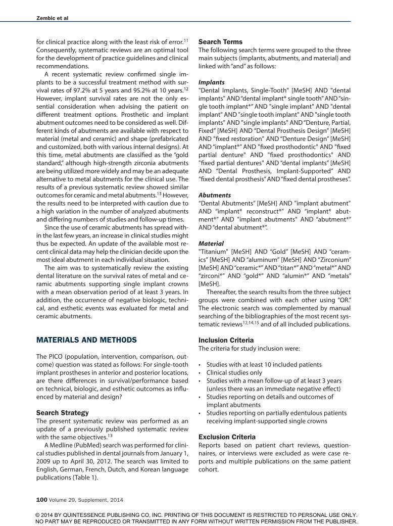
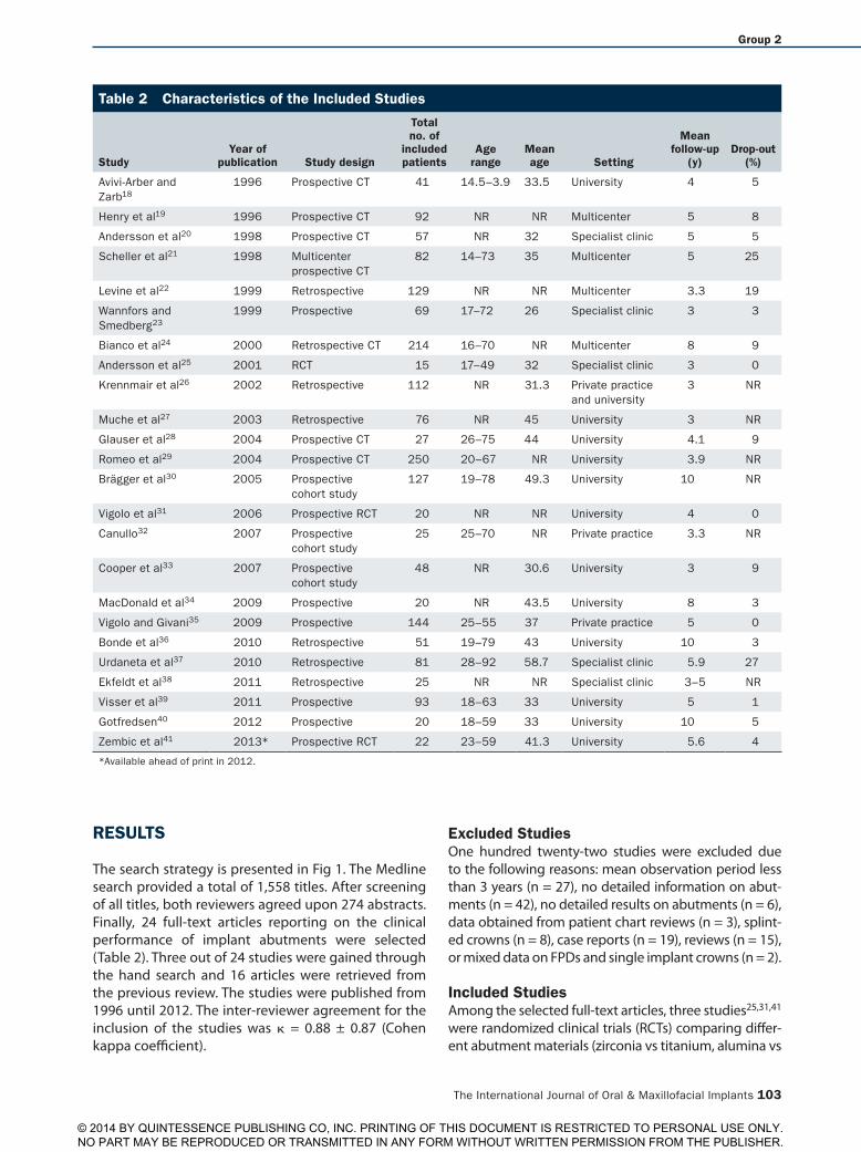
There was no difference in the occurrence of implant failures for implants with internal compared to external implant-abutment connection (rate ratio = 1.0; 95% CI: 0.5 to 2.0). The estimated implant failure per 100 im-plant years was 0.64% (95% CI: 0.5% to 0.9%) (Fig 3).
Prosthesis survivalAll studies reported on the survival rates of the prosthe-ses. The reasons for failure or refabrication, respective-ly, were mainly major fracture or insufficient esthetics.
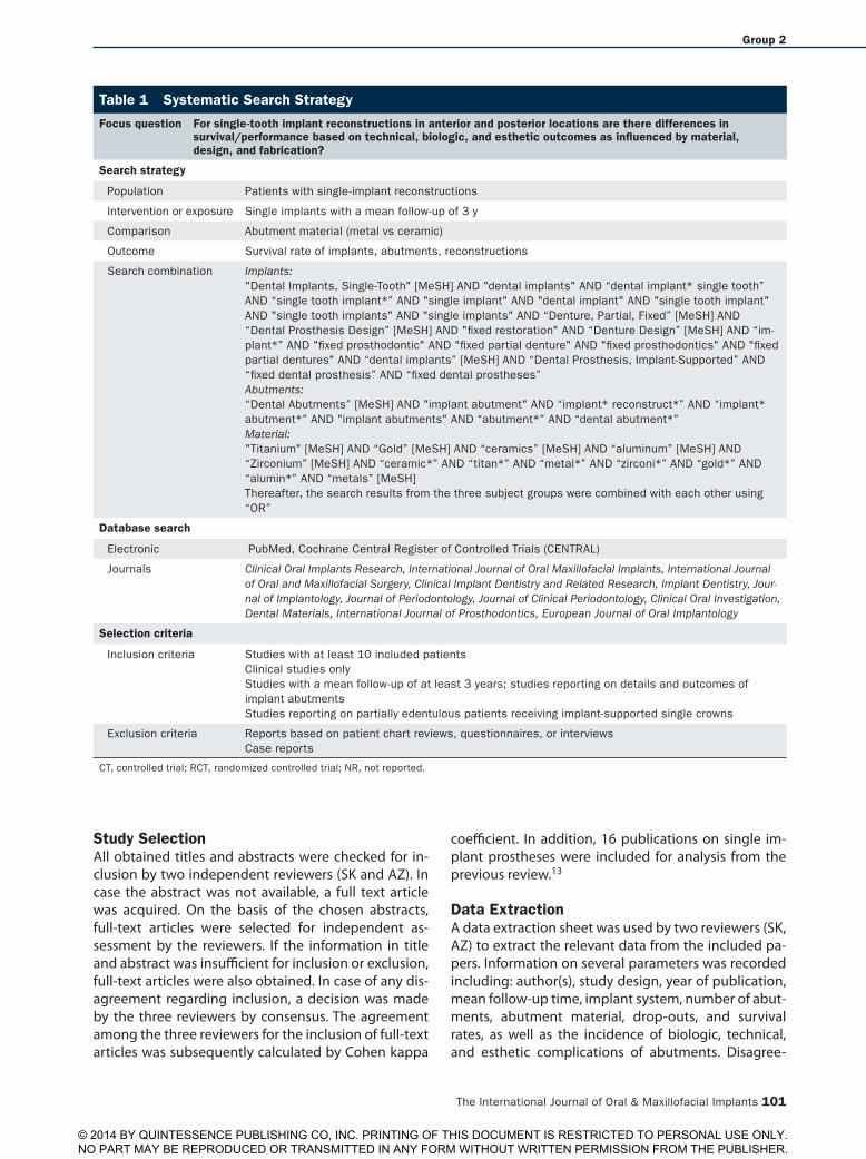
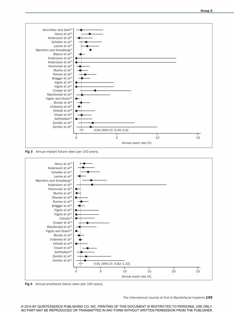
The estimated 5-year survival rate of single-implant prostheses was 95.6% (95% CI: 93.6% to 96.9%) (Fig 4). The failure rate for prostheses on ceramic abutments was less than for prostheses on metal abutments (2.6%; 95% CI: 0.6% to 11.3% vs 4.5%; 95% CI: 3.1% to 6.6%). This difference was not significant.
The rate of lost prostheses was similar for internal and external implant-abutment connections (rate ratio = 0.9; 95% CI: 0.4 to 2.1) (Table 4).
technical ComplicationsSeveral technical complications were reported in 21 studies. The overall estimated 5-year rate for techni-cal complications was 11.8% (95% CI: 8.5% to 16.3%) (Table 5; Fig 5).
table 4 Failed abutments and Prostheses
studyYear of
publication
total no. of abutments/prostheses
Mean follow-up abutment material Prosthesis material
no. of failures
(abutments)
no. of failures
(prostheses)
total abutment/prosthesis
exposure time
Avivi-Arber and Zarb18 1996 42 4 Titanium Metal-ceramic or metal-acrylic, 1 all-ceramic NR NR 168
Henry et al19 1996 96 5 Titanium NR 8 8 480
Andersson et al20 1998 55 5 Titanium 62 all-ceramic, 3 metal-ceramic 0 4 275
Scheller et al21 1998 65 5 Titanium 16 meta-ceramic, 81 all-ceramic 1 8 325
Levine et al22 1999 157 3.3 Titanium NR 0 4 518
Wannfors and Smedberg23
1999 76 3 Gold 36 gold-resin, 35 gold-ceramic, 9 all-ceramic 4 7 228
Bianco et al24 2000 229 8 Titanium Metal, metal-ceramic, all-ceramic 5 NR 1,832
Andersson et al25 2001 10 3 Alumina All-ceramic 2 NR 30
Andersson et al25 2001 10 3 Titanium All-ceramic 0 1 438
Krennmair et al26 2002 146 3 Titanium Metal-ceramic, all-ceramic 2 0 615
Muche et al27 2003 205 3 Metal Metal-ceramic 3 1 468
Glauser et al28 2004 36 4.1 Zirconia All-ceramic 0 0 690
Romeo et al29 2004 121 3.9 Titanium Metal-ceramic 5 5 80
Brägger et al30 2005 69 10 Metal (titanium, gold-alloy) NR 5 5 80
Vigolo et al31 2006 20 4 Titanium Metal-ceramic 0 0 129
Vigolo et al31 2006 20 4 Gold Metal-ceramic 0 0 136
Canullo32 2007 30 3.3 Zirconia All-ceramic 0 0 910
Cooper et al33 2007 43 3 Titanium Metal-ceramic, all-ceramic 3 3 520
MacDonald et al34 2009 17 8 Titanium Metal-ceramic 0 1 1,923
Vigolo and Givani35 2009 182 5 Titanium Metal-ceramic 0 0 460
Bonde et al36 2010 52 10 Titanium All-ceramic 3 3 190
Urdaneta et al37 2010 326 5.9 Titanium 228 gold-resin, 82 metal-ceramic, 16 all-ceramic 3 16 30
Ekfeldt et al38 2011 40 3-5 Zirconia 40 all-ceramic (25 one-piece) 0 0 148
Visser et al39 2011 92 5 Titanium abutment with gold coping
All-ceramic 3 11 99
Gotfredsen40 2012 19 10 Titanium Metal-ceramic 0 2 160
Zembic et al41 2013* 18 5.6 Zirconia All-ceramic 2 2 101
Zembic et al41 2013* 10 5.6 Titanium Metal-ceramic 1 1 56
*Available ahead of print in 2012. Total summary estimate (95% CI, random-effects Poisson regression) for total exposure time: 11,089; estimated abutment failure rate per 100 abutment years: 0.48 (0.30–0.77); estimated prosthesis failure rate per 100 prosthesis years: 0.91 (0.62–1.32); estimated 5-year abutment failure rate per 100 abutment years: 2.37% (1.49–3.77); estimated 5-year prosthesis failure rate per 100 prosthesis years: 4.42% (3.06–6.37).
© 2014 BY QUINTESSENCE PUBLISHING CO, INC. PRINTING OF THIS DOCUMENT IS RESTRICTED TO PERSONAL USE ONLY. NO PART MAY BE REPRODUCED OR TRANSMITTED IN ANY FORM WITHOUT WRITTEN PERMISSION FROM THE PUBLISHER.

Zembic et al
108 Volume 29, Supplement, 2014
Henry et al19
Andersson et al20
Scheller et al21
Wannfors and Smedberg23
Bianco et al24
Andersson et al25
Andersson et al25
Krennmair et al26
Muche et al27
Glauser et al28
Romeo et al29
Brägger et al30
Vigolo et al31
Vigolo et al31
Canullo32
Cooper et al33
MacDonald et al34
Vigolo and Givani35
Bonde et al36
Urdaneta et al37
Ekfeldt et al38
Visser et al39
Gotfredsen40
Zembic et al41
Zembic et al41
0.48 (95% CI: 0.3–0.77)
0 5 10 15
Annual event rate (%)
Fig 2 Annual abutment failure rates (per 100 years).
There was no significant difference with respect to the technical complication rate for ceramic and metal abutments. The estimated 5-year technical compli-cation rate for ceramic abutments added up to 8.9% (95% CI: 4.3% to 17.7%), whereas it was 12.0% (95% CI: 8.5% to 16.8%) for metal abutments. The rate of tech-nical complications was found to be 1.3 times (rate ratio = 1.3; 95% CI: 0.7 to 2.4) higher for implants with external implant-abutment connection than with in-ternal implant-abutment connection.
The most common technical complication was abut-ment screw loosening, which was reported for 4.6% of the abutments. In total, 99 abutment screws were found loose (2 ceramic and 97 metal abutments). One of the studies was an outlier with 29.1% abutment screw loos-ening.19 In that study, Brånemark gold abutment screws were used. The second most common technical compli-cation was crown loosening, reported in 13 studies with an incidence of 4.3% (93 loosened crowns out of 2,186 evaluated crowns). In total, 9 loosened crowns were metal-ceramic and 6 were all-ceramic crowns, while 8 studies did not specify the prosthesis material of loose crowns.18,19,22,24,26,30,33,37 Metal abutments supported all loosened crowns.The third most common complica-
tion was chipping of the veneering ceramic, which was evident in 2.7% of the abutments supporting single im-plant crowns (55 crowns supported by metal abutments and 4 crowns supported by ceramic abutments).
Misfit was reported in seven studies and occurred at 20 out of 2,186 implant-abutment connections (1 ceramic and 19 metal abutments).20,23,24,32,38,39,41 Abutment fractures were found in 0.2% of abutments reported from two studies.37,38 In one study, three abutment fractures occurred at internally connected titanium abutments with a narrow neck part connect-ing to Bicon implants.37 The other retrospective study described a broken customized CAD/CAM zirconia abutment after 2 months (Procera, Nobel Biocare).38 This abutment type is externally connected to the im-plant. The incidence of abutment screw fractures was low at 5 years with 0.2% and was reported at externally connected metal abutments only.18,19,27
Biologic ComplicationsBiologic complications (from a total of 2,186 abut-ments) affected both soft and hard tissue (Table 6). Fistulae (n = 5), general peri-implant soft tissue inflam-mations (n = 5), mucositis (n = 3), and bleeding (n = 2)
© 2014 BY QUINTESSENCE PUBLISHING CO, INC. PRINTING OF THIS DOCUMENT IS RESTRICTED TO PERSONAL USE ONLY. NO PART MAY BE REPRODUCED OR TRANSMITTED IN ANY FORM WITHOUT WRITTEN PERMISSION FROM THE PUBLISHER.
Group 2
The International Journal of Oral & Maxillofacial Implants 109
Avivi-Arber and Zarb18
Henry et al19
Andersson et al20
Scheller et al21
Levine et al22
Wannfors and Smedberg23
Bianco et al24
Andersson et al25
Andersson et al25
Krennmair et al26
Muche et al27
Romeo et al29
Brägger et al30
Vigolo et al31
Vigolo et al31
Cooper et al33
MacDonald et al34
Vigolo and Givani35
Bonde et al36
Urdaneta et al37
Ekfeldt et al38
Visser et al39
Gotfredsen40
Zembic et al41
Zembic et al41
0.64 (95% CI: 0.45–0.9)
0 5 10 15
Annual event rate (%)
Fig 3 Annual implant failure rates (per 100 years).
Henry et al19
Andersson et al20
Scheller et al21
Levine et al22
Wannfors and Smedberg23
Andersson et al25
Krennmair et al26
Muche et al27
Glauser et al28
Romeo et al29
Brägger et al30
Vigolo et al31
Vigolo et al31
Canullo32
Cooper et al33
MacDonald et al34
Vigolo and Givani35
Bonde et al36
Urdaneta et al37
Ekfeldt et al38
Visser et al39
Gotfredsen40
Zembic et al41
Zembic et al41
0.91 (95% CI: 0.62–1.32)
0 5 15 25
Annual event rate (%)
10 20
Fig 4 Annual prosthesis failure rates (per 100 years).
© 2014 BY QUINTESSENCE PUBLISHING CO, INC. PRINTING OF THIS DOCUMENT IS RESTRICTED TO PERSONAL USE ONLY. NO PART MAY BE REPRODUCED OR TRANSMITTED IN ANY FORM WITHOUT WRITTEN PERMISSION FROM THE PUBLISHER.

Zembic et al
110 Volume 29, Supplement, 2014
were described with regard to the soft tissue.19,20,31,36,38 With regard to hard tissue, peri-implantitis (n = 14), pocket probing depths ≥ 5 mm (n = 1), and bone loss of more than 2 mm was mentioned in nine stud-ies.19–21,24,30,34,38–40 A peri-implant abscess was a rare event and found only in one study.40
The estimated 5-year rate for biologic complica-tions was 6.4% (95% CI: 3.3% to 12.0%). The biologic failure rate per 100 abutment years ranged from 0.7% to 2.6% (Fig 6). The incidence of biologic events was almost twice as high for ceramic abutments compared to metal abutments (10.4%; 95% CI: 1.9% to 46.7% vs. 6.1%; 95% CI: 3.1% to 12.0%) (Table 6 and Fig 6). Even though, there was no significant difference (P > .05) between metal and ceramic abutments.
The rate of biologic complications was found to be two times (rate ratio = 2.0, 95%; CI: 0.4 to 8.9) higher for implants with external implant-abutment connec-tion than with internal implant-abutment connection. This difference did not reach statistical significance (P > .05).
esthetic ComplicationsEsthetic outcomes were reported in several studies in a nonstandardized way. Whereas some studies used questionnaires for patients to rate the esthetic out-come, other studies evaluated the esthetic outcome of the crowns by dentists and patients subjective-ly.20,23,26,38–40 In addition, some studies evaluated the papilla height and/or peri-implant mucosal color.34,42
table 5 technical Complications occurring in abutments and Prostheses
studyYear of
publication
total no. of abutments/prostheses
abutment fractures Misfit
screw fractures
abutment screw
loosening Chipping
Crown loosen-
ing
Avivi-Arber and Zarb18 1996 42 NR NR 2 NR 5 1
Henry et al19 1996 96 0 NR 1 28 NR 13
Andersson et al20 1998 55 NR 1 NR 1 NR NR
Scheller et al21 1998 65 NR NR NR 4 7 3
Levine et al22 1999 157 0 NR 0 4 NR 18
Wannfors and Smedberg23 1999 76 NR 8 NR 14 2 NR
Bianco et al24 2000 229 NR 9 NR 22 3 13
Andersson et al25 2001 10 2 NR 0 0 0 0
Andersson et al25 2001 10 0 NR 0 0 0 0
Krennmair et al26 2002 146 0 NR 0 5 1 12
Muche et al27 2003 205 0 NR 1 8 2 NR
Glauser et al28 2004 36 0 NR NR 2 3 NR
Romeo et al29 2004 121 NR NR 0 0 2 4
Brägger et al30 2005 69 0 NR 0 2 3 1
Vigolo et al31 2006 20 0 NR 0 0 0 0
Vigolo et al31 2006 20 0 NR 0 0 0 0
Canullo32 2007 30 0 0 0 0 1 0
Cooper et al33 2007 43 0 NR 0 0 3 2
MacDonald et al34 2009 17 0 NR 0 3 0 3
Vigolo and Givani35 2009 182 NR NR 0 0 0 0
Bonde et al36 2010 52 0 NR 0 3 3 3
Urdaneta et al37 2010 326 3 NR NR NR 18 18
Ekfeldt et al38 2011 40 1 1 0 NR NR 0
Visser et al39 2011 92 NR 1 NR 1 1 NR
Gotfredsen40 2012 19 0 NR 0 2 2 2
Zembic et al41 2013* 18 0 0 0 0 0 0
Zembic et al41 2013* 10 0 0 0 0 3 0
*Available ahead of print in 2012. Total summary estimate (95% CI, random-effects Poisson regression) for technical complications: 2.5 (1.8–3.6); estimated 5-year failure rate for technical complications: 11.8% (8.5–16.3). NR, not reported.
© 2014 BY QUINTESSENCE PUBLISHING CO, INC. PRINTING OF THIS DOCUMENT IS RESTRICTED TO PERSONAL USE ONLY. NO PART MAY BE REPRODUCED OR TRANSMITTED IN ANY FORM WITHOUT WRITTEN PERMISSION FROM THE PUBLISHER.

Group 2
The International Journal of Oral & Maxillofacial Implants 111
Avivi-Arber and Zarb18
Henry et al19
Andersson et al20
Scheller et al21
Levine et al22
Wannfors and Smedberg23
Bianco et al24
Andersson et al25
Andersson et al25
Krennmair et al26
Muche et al27
Glauser et al28
Romeo et al29
Brägger et al30
Vigolo et al31
Vigolo et al31
Canullo32
Cooper et al33
MacDonald et al34
Vigolo and Givani35
Bonde et al36
Urdaneta et al37
Ekfeldt et al38
Visser et al39
Gotfredsen40
Zembic et al41
Zembic et al41
2.52 (95% CI: 1.78–3.56)
0 5 20
Annual event rate (%)
10 15
Fig 5 Annual rates for technical complications at ceramic and metal abutments (per 100 years).
Avivi-Arber and Zarb18
Henry et al19
Andersson et al20
Scheller et al21
Levine et al22
Wannfors and Smedberg23
Bianco et al24
Andersson et al25
Andersson et al25
Krennmair et al26
Muche et al27
Glauser et al28
Romeo et al29
Brägger et al30
Vigolo et al31
Vigolo et al31
Canullo32
Cooper et al33
MacDonald et al34
Vigolo and Givani35
Bonde et al36
Urdaneta et al37
Ekfeldt et al38
Visser et al39
Gotfredsen40
Zembic et al41
Zembic et al41
1.32 (95% CI: 0.68–2.56)
0 5 20
Annual event rate (%)
10 15
Fig 6 Annual rates for biologic complications at ceramic and metal abutments (per 100 years).
© 2014 BY QUINTESSENCE PUBLISHING CO, INC. PRINTING OF THIS DOCUMENT IS RESTRICTED TO PERSONAL USE ONLY. NO PART MAY BE REPRODUCED OR TRANSMITTED IN ANY FORM WITHOUT WRITTEN PERMISSION FROM THE PUBLISHER.

Zembic et al
112 Volume 29, Supplement, 2014
The overall estimated 5-year esthetic complication rate for single-implant prostheses was 0.9% (95% CI: 0.4% to 2.3%) (Fig 7). Esthetic problems occurred in 1.0% (95% CI: 0.4% to 2.5%) of all implant prostheses supported by metal abutments. No esthetic complica-tions were reported in the five studies using ceramic abutments. The instrumented color analysis of mucosal tissues found a tissue color change both for metal and ceramic abutments.13,41 However, no perceivable dif-ference between titanium and zirconia abutments was visually observed when the thickness of the mucosa exceeded 2 mm.
The rate of negative esthetic events was found to be 1.3 times higher (rate ratio = 1.3; 95% CI: 0.2 to 8.1, P > .05) at prostheses with external implant-abutment connection than with internal. This difference did not reach statistical significance.
disCussion
The 5-year survival rate of single implant abutments was 98%. Thus, both ceramic and metal abutments survived at a rate of more than 95% at 5 years.
table 6 no. of Biological and esthetic Complications at abutments/Prostheses and estimated 5-Year Failure rrate
studyYear of
publication
total no. of abutments/prostheses
Bone loss (> 2 mm)
soft tissue complication recession
Biologic complications
esthetic complications
Avivi-Arber and Zarb18 1996 42 NR 7 5 12 NR
Henry et al19 1996 96 1 NR NR 1 NR
Andersson et al20 1998 55 11 1 NR 12 0
Scheller et al21 1998 65 4-8 5 NR 0 1
Levine et al22 1999 157 4 NR NR 4 NR
Wannfors and Smedberg23
1999 76 0 NR NR NR 7
Bianco et al24 2000 229 6 2 2 10 5
Andersson et al25 2001 10 0 0 0 0 0
Andersson et al25 2001 10 0 0 0 0 0
Krennmair et al26 2002 146 0 1 4 5 4
Muche et al27 2003 205 NR NR NR NR NR
Glauser et al28 2004 36 0 0 NR 0 NR
Romeo et al29 2004 121 NR NR NR NR NR
Brägger et al30 2005 69 13 NR NR 13 NR
Vigolo et al31 2006 20 0 0 0 1 NR
Vigolo et al31 2006 20 0 0 0 1 NR
Canullo32 2007 30 NR 0 NR 0 NR
Cooper et al33 2007 43 0 0 0 0 NR
MacDonald et al34 2009 17 1 NR NR 0 NR
Vigolo and Givani35 2009 182 0 NR NR NR NR
Bonde et al36 2010 52 0 NR NR 7 NR
Urdaneta et al37 2010 326 NR NR NR NR NR
Ekfeldt et al38 2011 40 3 NR 1 9 0
Visser et al39 2011 92 NR NR 1 1 4
Gotfredsen40 2012 19 1 1 NR 2 NR
Zembic et al41 2013* 18 0 0 0 0 0
Zembic et al41 2013* 10 0 0 0 0 0
*Available ahead of print in 2012. Total summary estimate (95% CI, random-effects Poisson regression) for biologic complications: 1.32 (0.68–2.56); total summary estimate (95% CI, random-effects Poisson regression) for esthetic complications: 0.19 (0.08–0.47); estimated 5-year failure rate for biologic complications: 6.4% (3.3–12.0); estimated 5-year failure rate for esthetic complications: 0.94% (0.38–2.30). NR, not reported.
© 2014 BY QUINTESSENCE PUBLISHING CO, INC. PRINTING OF THIS DOCUMENT IS RESTRICTED TO PERSONAL USE ONLY. NO PART MAY BE REPRODUCED OR TRANSMITTED IN ANY FORM WITHOUT WRITTEN PERMISSION FROM THE PUBLISHER.

Group 2
The International Journal of Oral & Maxillofacial Implants 113
Avivi-Arber and Zarb18
Henry et al19
Andersson et al20
Scheller et al21
Levine et al22
Wannfors and Smedberg23
Bianco et al24
Andersson et al25
Andersson et al25
Krennmair et al26
Muche et al27
Glauser et al28
Romeo et al29
Brägger et al30
Vigolo et al31
Vigolo et al31
Canullo32
Cooper et al33
MacDonald et al34
Vigolo and Givani35
Bonde et al36
Urdaneta et al37
Ekfeldt et al38
Visser et al39
Gotfredsen40
Zembic et al41
Zembic et al41
0.19 (95% CI: 0.08–0.47)
0 5
Annual event rate (%)
10 15
Fig 7 Annual rates for esthetic complications at ceramic and metal abutments (per 100 years).
The most common complications at 5 years were technical complications (11.8%), followed by biologic complications (6.4%). Esthetic complications were fewest and occurred in 0.9% at 5 years.
implant survivalThe overall 5-year implant survival rate of single im-plants amounted to 96.9% based on this systematic review. This result is in accordance with the results of two systematic reviews on single implants report-ing 5-year survival rates of 96.4% and 97.2%, respec-tively.12,13 With today’s optimized implant surfaces and configurations, most of the implant failures are likely to occur before loading.12 Thus, not many failures are expected to happen at 5 years of clinical function. This might explain the positive implant survival rates found in the existing studies.
abutment survivalThe present results correspond to the results of a pre-vious systematic review on implant abutments, also reporting an estimated 5-year abutment survival rate of 98%.13 Most of the evaluated abutments were metal abutments (n = 2,052). From the 134 ceramic abut-ments, mainly zirconia abutments were evaluated
(124 zirconia, 10 alumina abutments). In total, six abut-ments fractured, one externally connected zirconia abutment, two externally connected alumina abut-ments, and three titanium abutments being internally connected to Bicon implants.25,37,38
Alumina abutments were the first generation of ceramic abutments. Previous studies demonstrated a failure rate between 1.9% and 7% after 1 to 5 years of clinical use.25,43,44 In the above-mentioned RCT alumi-na abutments were compared with the “gold standard” titanium and showed a lower survival rate of 93% com-pared to 100% for titanium abutments.25 This explains the introduction of a stronger substitute material.
The subsequently developed high-strength ceramic zirconia showed superior mechanical properties with much higher bending strength and fracture toughness compared to alumina.45 Thus, a superior clinical behavior for zirconia might be expected and zirconia might even serve as an alternative to metal in various indications. However, clinical studies on zirconia abutments are scarce (only four studies in this review). When zirconia and titani-um abutments were compared in a RCT, the survival rate for both materials was 100% after 5 years of function.41 Other studies with a shorter follow-up confirm these posi-tive results for zirconia abutments.28,32,46–48
© 2014 BY QUINTESSENCE PUBLISHING CO, INC. PRINTING OF THIS DOCUMENT IS RESTRICTED TO PERSONAL USE ONLY. NO PART MAY BE REPRODUCED OR TRANSMITTED IN ANY FORM WITHOUT WRITTEN PERMISSION FROM THE PUBLISHER.

Zembic et al
114 Volume 29, Supplement, 2014
Two internally connected zirconia abutments fractured within 2 months in a retrospective study.38
Among several factors influencing the stability of zir-conia abutments, the abutment wall thickness is dis-cussed as being critical.49 A minimum abutment wall thickness of 0.5 mm was recommended for zirconia abutments, especially when using CAD/CAM tech-niques.50 The fractured abutment consisted of an in-ternal insert of titanium to adapt to the implant.38 The abutment wall thickness might have been insufficient, which might have caused the fractures after a short period of only 2 months.
No clinical long-term data are available for zirconia abutments. Taking the nature of ceramics into account, one might assume fatigue fractures over time.51 On the other hand, the fatigue performance of zirconia is likely increased through its behavior called “transfor-mation toughening” which causes a resistance to crack growth compared to other polycrystalline ceramics.52 This might explain the positive clinical results for zirco-nia abutments thus far.
Metal abutments are still considered the “gold stan-dard” due to high survival rates and excellent physical properties.20 When gold and titanium abutments were compared in a RCT there were no significant differenc-es with regard to survival and peri-implant bone and soft tissue parameters after 4 years of clinical service.31
Three internally connected titanium abutments frac-tured in one study.37 These abutments are constructed for specially configured locking-taper implants (Bicon) containing a thin neck part, which might be prone to fracture. Furthermore, the crown-to-implant ratio is in-creased in this implant-abutment configuration, which might increase the stress at the weakest point, ie, the thin abutment neck part, and thus contribute to its fracture.
Usually, fractures of metal abutment are a rare event and were estimated to occur in only 0.07% at 5 years.13 The only additional fractures were limited to one spe-cific implant system (Bicon implants).37
It has to be taken into account that the number of observed metal abutments (n = 2,052) was much higher than of ceramic abutments (n = 134). On one hand, this might explain why no significant difference between the outcomes of ceramic and metal abutments was cal-culated. On the other hand, the results have to be inter-preted with caution. It may be recommended that the application of ceramic abutments should be selective and not generalized for every situation.
There was no difference in the occurrence of abut-ment failures for implants with internal compared to external implant-abutment connection. In contrast, a tendency towards less risk for fracture was observed with abutments having an internal implant-abutment connection in the previous review.13
technical Complications The most common technical complication found was abutment screw loosening (4.6%), mostly observed with metal abutments. This finding is in agreement with several other studies.12,13,53,54 The high rate for abutment screw loosening in the present study might partly be explained by one study, which reported 29.1% of screw loosenings and used the first genera-tion of Brånemark gold abutment screws, known for this problem.19 The majority of the abutment screws loosened in externally connected abutments (n = 85) compared to internally connected ones (n = 14). The tendency of less screw loosening at internal implant-abutment connections is supported by other stud-ies.13,55,56 A recent systematic review on abutment screw loosening for single-implant restorations did not find a difference with internally compared to ex-ternally connected implants.57 The authors concluded that abutments screw loosening is irrespective of the implant-abutment geometry and occurs rarely, pro-vided that a proper antirotational torque is applied.57
The second most common technical complication was crown loosening (4.3%). Metal abutments sup-ported all loosened crowns. The cement used was not evaluated. Since in some parts of the world there is a preference for the use of provisional cement for im-plant prostheses, one might speculate that a high rate of crown loosening is plausible.
The chipping rate of veneering ceramics (2.7%) in the present study was less than reported in previous systematic reviews (4%) at 5 years.12,13
Biologic and esthetic ComplicationsThere is a lack of classification for the report of biologic complications. Consequently, negative events were re-ported in a non-standardized way and comparison of the studies was impeded. There was a trend for a high-er incidence of biologic complications with ceramic abutments (10.4%) compared to metal abutments (6.1%), but without statistical significance. This finding is rather unusual. Animal studies demonstrated a com-parable soft tissue integration of alumina, zirconia, and titanium.58–60 Other studies found even fewer inflam-matory cells in the epithelium around zirconia than ti-tanium and gold, and finally less bacterial adhesion at zirconia clinically.61–64
Another systematic review indicated a similar soft tissue complication rate of 7.1% after 5 years.12 Even though the proportion of biologic complications at externally connected abutments was found to be 1.7 times that of internally connected abutments, the type of connection did not have a significant influence on the estimated rate of biologic complications (P > .05).
In contrast to the results of a previous review, the incidence of recession in the present study was higher
© 2014 BY QUINTESSENCE PUBLISHING CO, INC. PRINTING OF THIS DOCUMENT IS RESTRICTED TO PERSONAL USE ONLY. NO PART MAY BE REPRODUCED OR TRANSMITTED IN ANY FORM WITHOUT WRITTEN PERMISSION FROM THE PUBLISHER.

Group 2
The International Journal of Oral & Maxillofacial Implants 115
at metal abutments.13 The reason for this observation remains unclear. The present review indicated no es-thetic failures with prostheses on ceramic abutments. This finding is in accordance with a previous review and RCT where less soft tissue discoloration was found for ceramic abutments.13,54
There is a large heterogeneity among the studies concerning the evaluation of the esthetics, due to a lack of standardization. The scientific value of the es-timated 5-year esthetic complication rate is rather low. Standardized esthetic parameters, such as the pink and white esthetic score65,66 are thus strongly advis-able and should be applied more often in future stud-ies on implant prostheses.
ConClusions
The present meta-analysis on single implant prosthe-ses presents high survival rates of single implants, abutments and prostheses after 5 years of function.
There are no performance differences in technical or biologic outcomes for ceramic and metal abutments. The only significant finding pertaining to esthetics was a difference in tissue color with both metal and ceram-ic abutments, which was greater for metal abutments up to 2 mm mucosal thickness.
Similarly, no differences were found for either ex-ternal or internal implant-abutment connections. The incidence of technical complications is higher than for either esthetic or biologic complications.
aCKnowledGMents
This consensus work was sponsored by the International Team for Implantology. The authors reported no conflicts of interest related to this study.
reFerenCes
1. Jokstad A, Brägger U, Brunski JB, Carr AB, Naert I, Wennerberg A. Quality of dental implants. Int Dent J 2003;53(Suppl 2):409–443.
2. Jemt T, Lekholm U. Oral implant treatment in posterior partially edentulous jaws: A 5-year follow-up report. Int J Oral Maxillofac Implants 1993;8:635–640.
3. Brägger U, Hammerle C, Weber HP. Fixed reconstructions in partially edentulous patients using two-part ITI implants (Bonefit) as abut-ments. Clin Oral Implants Res 1990;1:41–49.
4. Jemt T, Lekholm U, Adell R. Osseointegrated implants in the treatment of partially edentulous patients: A preliminary study on 876 consecu-tively placed fixtures. Int J Oral Maxillofac Implants 1989;4:211–217.
5. Brägger U, Burgin WB, Hammerle CH, Lang NP. Associations be-tween clinical parameters assessed around implants and teeth. Clin Oral Implants Res 1997;8:412–421.
6. Romeo E, Chiapasco M, Ghisolfi M, Vogel G. Long-term clinical effectiveness of oral implants in the treatment of partial edentu-lism. Seven-year life table analysis of a prospective study with iti dental implants system used for single-tooth restorations. Clin Oral Implants Res 2002;13:133–143.
7. Lekholm U, Gunne J, Henry P, Higuchi K, Linden U, Bergstrom C, van Steenberghe D. Survival of the Branemark implant in partially edentulous jaws: A 10-year prospective multicenter study. Int J Oral Maxillofac Implants 1999;14: 639–645.
8. Buser D, Mericske-Stern R, Bernard JP, et al Long-term evaluation of non-submerged ITI implants. Part 1: 8-year life table analysis of a prospective multi-center study with 2,359 implants. Clin Oral Implants Res 1997;8:161–172.
9. Wennström J, Palmer R. Consensus report: Clinical Trials. In: Lang NP, Karring T, Linde J (eds). Proceedings of the 3rd European workshop on periodontology. Berlin: Quintessence, 1999:255–259.
10. Pjetursson BE, Tan K, Lang NP, Brägger U, Egger M, Zwahlen M. A sys-tematic review of the survival and complication rates of fixed partial dentures (FPDs) after an observation period of at least 5 years. Clin Oral Implants Res 2004;15:625–642.
11. Evans D. Hierarchy of evidence: A framework for ranking evidence evaluating healthcare interventions. J Clin Nurs 2003;12:77–84.
12. Jung RE, Zembic A, Pjetursson BE, Zwahlen M, Thoma DS. Systematic review of the survival rate and the incidence of biological, technical, and aesthetic complications of single crowns on implants reported in longitudinal studies with a mean follow-up of 5 years. Clin Oral Implants Res 2012;23(suppl 6):2–21.
13. Sailer I, Philipp A, Zembic A, Pjetursson BE, Hammerle CH, Zwahlen M. A systematic review of the performance of ceramic and metal implant abutments supporting fixed implant reconstructions. Clin Oral Implants Res 2009;20(suppl 4):4–31.
14. Abt E, Carr AB, Worthington HV. Interventions for replacing miss-ing teeth: Partially absent dentition. Cochrane Database Syst Rev 2012;2:CD003814.
15. Anusavice KJ. Standardizing failure, success, and survival decisions in clinical studies of ceramic and metal-ceramic fixed dental pros-theses. Dent Mater 2012;28:102–111.
16. Kirkwood BR, Sterne JAC. Poisson Regression. Essential Medical Statistics. Oxford: Blackwell Science Ltd, 2003:249–262.
17. Kirkwood BR, Sterne JAC. Survival Analysis: Displaying and Compar-ing Survival Patterns. In: Essential Medical Statistics. Oxford: Black-well Science Ltd, 2003:272–286.
18. Avivi-Arber L, Zarb GA. Clinical effectiveness of implant-supported single-tooth replacement: The Toronto study. Int J Oral Maxillofac Implants 1996;11:311–321.
19. Henry PJ, Laney WR, Jemt T, et al. Osseointegrated implants for single-tooth replacement: A prospective 5-year multicenter study. Int J Oral Maxillofac Implants 1996;11:450–455.
20. Andersson B, Odman P, Lindvall AM, Brånemark PI. Cemented single crowns on osseointegrated implants after 5 years: Results from a prospective study on CeraOne. Int J Prosthodont 1998;11:212–218.
21. Scheller H, Urgell JP, Kultje C, et al. A 5-year multicenter study on implant-supported single crown restorations. Int J Oral Maxillofac Implants 1998;13:212–218.
22. Levine RA, Clem DS 3rd, Wilson TG Jr, Higginbottom F, Solnit G. Multicenter retrospective analysis of the ITI implant system used for single-tooth replacements: Results of loading for 2 or more years. Int J Oral Maxillofac Implants 1999;14:516–520.
23. Wannfors K, Smedberg JI. A prospective clinical evaluation of differ-ent single-tooth restoration designs on osseointegrated implants. A 3-year follow-up of Branemark implants. Clin Oral Implants Res 1999;10:453–458.
24. Bianco G, Di Raimondo R, Luongo G, et al. Osseointegrated implant for single-tooth replacement: A retrospective multicenter study on routine use in private practice. Clinical implant dentistry and related research 2000;2:152–158.
25. Andersson B, Taylor A, Lang BR, et al. Alumina ceramic implant abut-ments used for single-tooth replacement: A prospective 1- to 3-year multicenter study. Int J Prosthodont 2001;14:432–438.
26. Krennmair G, Schmidinger S, Waldenberger O. Single-tooth replace-ment with the Frialit-2 system: A retrospective clinical analysis of 146 implants. Int J Oral Maxillofac Implants 2002;17:78–85.
© 2014 BY QUINTESSENCE PUBLISHING CO, INC. PRINTING OF THIS DOCUMENT IS RESTRICTED TO PERSONAL USE ONLY. NO PART MAY BE REPRODUCED OR TRANSMITTED IN ANY FORM WITHOUT WRITTEN PERMISSION FROM THE PUBLISHER.

Zembic et al
116 Volume 29, Supplement, 2014
27. Muche R, Krausse A, Strub JR. [Success rates of implant supported prostheses in partially edentulous patients—Part II]. Schweiz Monatsschr Zahnmed 2003;113:404–410.
28. Glauser R, Sailer I, Wohlwend A, Studer S, Schibli M, Scharer P. Experi-mental zirconia abutments for implant-supported single-tooth restorations in esthetically demanding regions: 4-year results of a prospective clinical study. Int J Prosthodont 2004;17:285–290.
29. Romeo E, Lops D, Margutti E, Ghisolfi M, Chiapasco M, Vogel G. Long-term survival and success of oral implants in the treatment of full and partial arches: A 7-year prospective study with the ITI dental implant system. Int J Oral Maxillofac Implants 2004;19:247–259.
30. Brägger U, Karoussis I, Persson R, Pjetursson B, Salvi G, Lang N. Technical and biological complications/failures with single crowns and fixed partial dentures on implants: A 10-year prospective cohort study. Clin Oral Implants Res 2005;16:326–334.
31. Vigolo P, Givani A, Majzoub Z, Cordioli G. A 4-year prospective study to assess peri-implant hard and soft tissues adjacent to titanium versus gold-alloy abutments in cemented single implant crowns. J Prosthodont 2006;15:250–256.
32. Canullo L. Clinical outcome study of customized zirconia abutments for single-implant restorations. Int J Prosthodont 2007;20:489–493.
33. Cooper LF, Ellner S, Moriarty J, et al. Three-year evaluation of single-tooth implants restored 3 weeks after 1-stage surgery. Int J Oral Maxillofac Implants 2007;22:791–800.
34. MacDonald K, Pharoah M, Todescan R, Deporter D. Use of sintered porous-surfaced dental implants to restore single teeth in the maxilla: A 7- to 9-year follow-up. Int J Periodontics Restorative Dent 2009;29:191–199.
35. Vigolo P, Givani A. Platform-switched restorations on wide-diameter implants: A 5-year clinical prospective study. Int J Oral Maxillofac Implants 2009;24:103–109.
36. Bonde MJ, Stokholm R, Isidor F, Schou S. Outcome of implant-sup-ported single-tooth replacements performed by dental students. A 10-year clinical and radiographic retrospective study. Eur J Oral Implantol 2010:3:37–46.
37. Urdaneta RA, Rodriguez S, McNeil DC, Weed M, Chuang SK. The effect of increased crown-to-implant ratio on single-tooth locking-taper implants. Int J Oral Maxillofac Implants 2010;25:729–743.
38. Ekfeldt A, Furst B, Carlsson GE. Zirconia abutments for single-tooth implant restorations: A retrospective and clinical follow-up study. Clin Oral Implants Res 2011;22:1308–1314.
39. Visser A, Raghoebar GM, Meijer HJ, Meijndert L, Vissink A. Care and aftercare related to implant-retained dental crowns in the maxillary aesthetic region: A 5-year prospective randomized clinical trial. Clinical implant dentistry and related research 2011;13:157–167.
40. Gotfredsen K. A 10-year prospective study of single tooth implants placed in the anterior maxilla. Clinical Implant Dent Relat Res 2012; 14:80–87.
41. Zembic A, Bosch A, Jung RE, Hammerle CH, Sailer I. Five-year results of a randomized controlled clinical trial comparing zirconia and titanium abutments supporting single-implant crowns in canine and posterior regions. Clin Oral Implants Res 2013;24:384–390.
42. Zembic A, Sailer I, Jung RE, Hammerle CH. Randomized-controlled clinical trial of customized zirconia and titanium implant abutments for single-tooth implants in canine and posterior regions: 3-year results. Clin Oral Implants Res 2009;20:802-808
43. Andersson B, Glauser R, Maglione M, Taylor A. Ceramic implant abut-ments for short-span FPDs: A prospective 5-year multicenter study. Int J Prosthodont 2003;16:640–646.
44. Andersson B, Schärer P, Simion M, Bergström C. Ceramic implant abutments used for short-span fixed partial dentures: A prospective 2-year multicenter study. Int J Prosthodont 1999;12:318–324.
45. Lüthy H. Strength and toughness of dental ceramics. In: Möhrmann, WH, ed. CAD/ CIM in Aesthetic Dentistry. Cerec 10 Year Anniversary Symposium. Chicago: Quintessence, 1996:229–240.
46. Nakamura K, Kanno T, Milleding P, Ortengren U. Zirconia as a dental implant abutment material: A systematic review. Int J Prosthodont 2010;23:299–309.
47. Sailer I, Zembic A, Jung RE, Siegenthaler D, Holderegger C, Ham-merle CH. Randomized controlled clinical trial of customized zirconia and titanium implant abutments for canine and posterior single-tooth implant reconstructions: Preliminary results at 1 year of function. Clin Oral Implants Res 2009;20:219–225.
48. Nothdurft F, Pospiech P. Prefabricated zirconium dioxide implant abutments for single-tooth replacement in the posterior region: Evaluation of peri-implant tissues and superstructures after 12 months of function. Clin Oral Implants Res 2010;21:857–865.
49. Luthardt RG, Holzhuter MS, Rudolph H, Herold V, Walter MH. CAD/CAM-machining effects on y-TZP zirconia. Dent Mater 2004;20: 655–662.
50. Denry I, Kelly JR. State of the art of zirconia for dental applications. Dent Mater 2008;24:299–307.
51. Rekow D, Thompson VP. Engineering long term clinical success of advanced ceramic prostheses. J Mater Sci Mater Med 2007;18:47–56.
52. Piconi C, Maccauro G. Zirconia as a ceramic biomaterial. Biomaterials 1999;20:1–25.
53. Berglundh T, Persson L, Klinge B. A systematic review of the inci-dence of biological and technical complications in implant dentistry reported in prospective longitudinal studies of at least 5 years. J Clin Periodontol 2002;29(suppl 3):197–212, 232–233.
54. Jung RE, Pjetursson BE, Glauser R, Zembic A, Zwahlen M, Lang NP. A systematic review of the 5-year survival and complication rates of implant-supported single crowns. Clin Oral Implants Res 2008;19: 119–130.
55. Norton MR. The Astra Tech single-tooth implant system: A report on 27 consecutively placed and restored implants. Int J Periodontics Restorative Dent 1997;17:574–583.
56. Khraisat A, Stegaroiu R, Nomura S, Miyakawa O. Fatigue resistance of two implant/abutment joint designs. J Prosthet Dent 2002;88: 604–610.
57. Theoharidou A, Petridis HP, Tzannas K, Garefis, P. Abutment screw loosening in single-implant restorations: A systematic review. Int J Oral Maxillofac Implants 2008;23:681–690.
58. Hashimoto M, Akagawa Y, Nikai H, Tsuru H. Single-crystal sapphire endosseous dental implant loaded with functional stress—Clinical and histological evaluation of peri-implant tissues. J Oral Rehabil 1988;15:65–76.
59. Abrahamsson I, Berglundh T, Glantz P-O, Lindhe J. The mucosal at-tachment at different abutments. An experimental study in dogs. J Clin Periodontology 1998;25:721–727.
60. Kohal RJ, Weng D, Bachle M, Strub JR Loaded custom-made zirconia and titanium implants show similar osseointegration: An animal experiment. J Periodontol 2004;75:1262–1268.
61. Welander M, Abrahamsson I, Berglundh T. The mucosal barrier at implant abutments of different materials. Clin Oral Implants Res 2008;19:635–641.
62. Degidi M, Artese L, Scarano A, Perrotti V, Gehrke P, Piattelli A. Inflammatory infiltrate, microvessel density, nitric oxide synthase expression, vascular endothelial growth factor expression, and proliferative activity in peri-implant soft tissues around titanium and zirconium oxide healing caps. J Periodontol 2006;77:73–80.
63. Rimondini L, Cerroni L, Carrassi A, Torricelli P. Bacterial colonization of zirconia ceramic surfaces: An in vitro and in vivo study. Int J Oral Maxillofac Implants 2002;17:793–798.
64. Scarano A, Piattelli M, Caputi S, Favero GA, Piattelli A. Bacterial adhe-sion on commercially pure titanium and zirconium oxide disks: An in vivo human study. J Periodontol 2004;75:292–296.
65. Furhauser R, Florescu D, Benesch T, Haas R, Mailath G, Watzek G. Evaluation of soft tissue around single-tooth implant crowns: The pink esthetic score. Clin Oral Implants Res 2005;16:639–644.
66. Belser UC, Grütter L, Vailati F, Bornstein MM, Weber HP, Buser D. Outcome evaluation of early placed maxillary anterior single-tooth implants using objective esthetic criteria: A cross-sectional, retro-spective study in 45 patients with a 2- to 4-year follow-up using pink and white esthetic scores. J Periodontol 2009;80:140–151.
© 2014 BY QUINTESSENCE PUBLISHING CO, INC. PRINTING OF THIS DOCUMENT IS RESTRICTED TO PERSONAL USE ONLY. NO PART MAY BE REPRODUCED OR TRANSMITTED IN ANY FORM WITHOUT WRITTEN PERMISSION FROM THE PUBLISHER.