systematic revision of the oriental planthopper genus miasa distant ... · pdf filesystematic...
TRANSCRIPT

© Senckenberg Gesellschaft für Naturforschung, 2014.
137
72 (2): 137 – 164
25.7.2014
ISSN 1863-7221 (print) | eISSN 1864-8312 (online)
Systematic revision of the Oriental planthopper genus Miasa Distant (Hemiptera: Fulgoromorpha: Dictyophari-dae), with description of a new genus from southern India
Zhi-Shun Song 1, 2, Michael D. Webb 3 & Ai-Ping Liang *, 1
1 Key Laboratory of Zoological Systematics and Evolution, Institute of Zoology, Chinese Academy of Sciences, Beijing, 100101, China; Zhi-Shun Song [[email protected]]; Ai-Ping Liang * [[email protected]] — 2 Museum für Naturkunde, Leibniz Institute for Research on Evolution and Biodiversity, Humboldt University Berlin, Invalidenstrasse 43, Berlin 10115, Germany — 3 Department of Entomology, Natural History Museum, Cromwell Road, South Kensington, London, U.K.; Michael D. Webb [[email protected]] — * Corresponding author
Accepted 30.v.2014. Published online at www.senckenberg.de/arthropod-systematics on 18.vii.2014.
AbstractThe dictyopharid planthopper genus Miasa Distant, 1906 (Orthopagini) is revised to include three known species, M. producta (Lethierry, 1888), M. smaragdilinea (Walker, 1857), and M. wallacei Muir, 1923, and two new species M. borneensis sp.n. and M. nigromaculata sp.n., all distributed in Southeast Asia. The confusion regarding the taxonomy of Miasa species is clarified on the basis of a critical review and examination of historical material. Male genitalia of all species and fifth-instar nymph of M. smaragdilinea are described and illustrated for the first time. A new genus Indomiasa gen.n. which is closely related to Miasa, is also established for a single new species, I. distanti sp.n., from southern India. A phylogenetic analysis based on morphological characters of adults was conducted to reconstruct the species-level phylogenetic relationships of Miasa and Indomiasa. The results show that the monophyly of Miasa and Miasa + Indomiasa is well supported. Keys to the genera of Orthopagini in the Oriental region and to the species of Miasa are provided.
Key wordsNew species, redescription, male genitalia, fifth-instar nymph, phylogeny, biogeography, taxonomy.
1. Introduction
The planthopper family Dictyopharidae is a moderately large family of Fulgoromorpha, containing more than 730 described species in 167 genera (Metcalf 1946; Song & liang 2011a; liang & Song 2012; Bourgoin 2014). Members of Dictyopharidae (Fig. 1) often can be recognized by their variably anteriorly produced head, although this character is not unique to Dictyopharidae within Fulgoromorpha (o’Brien 2002). It is widely ac-cepted that this family is a sister clade of the lanternfly family Fulgoridae in the hypotheses of Fulgoromorpha
phylogeny based on either morphological characters or DNA sequence data (aSche 1988; eMeljanov 1990; Bour goin 1993; Yeh et al. 2005; urBan & crYan 2007, 2009; Song & liang 2013). Dictyopharidae is widely distributed in most parts of the world, especially in tropical and subtropical regions such as South America, the Oriental region and the East Indies (Metcalf 1946). Members of this group are pre-dominantly monocot-feeders, and a few are major agri-cultural pests on Poaceae (grasses), such as rice, maize,

Song et al.: Revision of Miasa planthoppers
138
and sugarcane (WilSon & o’Brien 1987; WilSon et al. 1994). Traditionally, the family was separated into two sub-families Dictyopharinae and Orgeriinae (Muir 1930; Met calf 1946; eMeljanov 1980; SzWedo 2008). Dic tyo-pha rinae, distributed worldwide, comprises eleven extant tribes and two fossil tribes (Melichar 1912; Muir 1923, 1930; Metcalf 1946; eMeljanov 1979, 1983, 1997, 2008, 2011; SzWedo 2008). As in many other groups of Ful go -ro morpha, quite a number of genera and higher taxa with-in Dictyopharinae still lack standard revisionary studies, and their monophyly has never been tested cla disti cally as well (liang & Song 2012). Within Dictyopharinae, the tribe Orthopagini Emel-janov is a relatively large group with 19 genera and more than 50 species mainly distributed in the Afrotropic and Oriental regions. Although there have been a continuing series of works emphasizing taxonomic revision of ge-neric level within Orthopagini which were published (liang & Song 2006; Song & liang 2006a,b, 2007, 2011b, 2012a,b; eMeljanov 2008, 2011; Song et al. 2012), the phylogenetic relationships of most genera within Ortho-pagini are poorly understood (Song et al. in prep.). The Oriental genus Miasa was established by diStant (1906) for a single species – Elidiptera smaragdilinea
Walker, 1857, from Malacca (Malay Peninsula) – on the basis of the distinctly slender, straight and linear cephalic process, and narrow and long forewings. When describ-ing Miasa, diStant (1906) stated that “this genus is allied to Dictyopharoides Fowl., from which it may be at once separated by the non-serrate anterior femora, a character omitted in Fowler’s diagnosis.” It was farther from the Neotropical genus Elidiptera Spinola, 1839 because the latter was moved into the family Achilidae. diStant (1906) redescribed M. smaragdilinea (Walk-er) based on material from Myanmar (Burma) instead of Walker’s type specimen. In the same year, SchMidt (1906) claimed that diStant’s (1906) description was inaccurate and incomplete, giving as his reason that Dis-tant’s character ‘green face with the central ridge testa-ceous’ for M. smaragdilinea was not mentioned in Walker’s (1857) description, and such an obvious feature, if present, could not possibly have been ignored by Walker (1857). Consequently, SchMidt (1906) described a new species, M. rubrovittata, for Distant’s smaragdilinea, based on his own material from Sumatra and Java, which matched Distant’s description of specimens with green frons. In response to Schmidt’s judgment, diStant (1916) declared that his description and illustration of M. sma
Fig. 1. Miasa specimens. A: M. borneensis sp.n., photographed by Dr. Arthur Anker at Danum Valley, Sabah, Borneo, Malaysia on 3 Feb. 2011. B: M. wallacei, photographed by Dennis Sim at Negeri Sembilan, Malaysia on 21 Apr. 2013.
A
B

139
ARTHROPOD SYSTEMATICS & PHYLOGENY — 72 (2) 2014
ragdilinea from Burma agreed entirely with Walker’s type in the British Museum, and proposed that M. rubrovittata from Sumatra and Java was a junior synonym of M. smaragdilinea. However, this action was not adopted by subsequent workers, such as Baker (1927), SchMidt (1928) and Metcalf (1946), although SchMidt (1928) did admit that he had misidentified his specimens as M. smaragdilinea and re-named them as M. producta (Lethierry) but at the same time, unaccountably retain-ing M. rubrovittata as a valid species. During the above period a fourth species, Miasa wallacei Muir, 1923, was described from southern Thailand (Muir 1923). Based on an examination of most of the Miasa type specimens in the current work we not only confirm the synonymy of M. rubrovittata Schmidt with M. smaragdilinea (Walker), but also point out that diStant (1906) misidentified his specimens from Burma as M. smaragdilinea as they may belong to M. wallacei Muir. Although there is a strong resemblance among the Miasa species, they can be distinguished by a combina-tion of differences in body color and in the male genitalia (Figs. 15, 17), and they also differ in their distribution (Fig. 18). We accordingly revise the genus Miasa to in-clude three known species, M. producta, M. smaragdilinea, and M. wallacei, and two new species, M. borneensis sp.n. and M. nigromaculata sp.n., from Borneo. In addi-tion, one new species, representing a new closely related genus, Indomasia distanti gen.n., sp.n., is described from southern India. A phylogenetic analysis based on morphological characters of adults was conducted to re-construct the phylogeny of Miasa and Indomasia.
2. Materials and methods
2.1. Specimens and museums
Specimens examined were deposited in the following institutions whose names are abbreviated in the text as follows: BMNH Natural History Museum, London, UK; BPBM Bernice P. Bishop Museum, Honolulu, HI, USA; IZCAS Institute of Zoology, Chinese Academy of Scienc-es, Beijing, P.R. China; MFNB Museum für Naturkunde der Humboldt Universität, Berlin, Germany; MIZPAS Museum and Institute of Zoology, Polish Academy of Sciences, Warsaw, Poland; MMBC Moravian Museum, Brno, Czech Republic; MZLU Museum of Zoology, Lund University, Lund, Sweden; SDEI Senckenberg Deutsches Entomologisches Institut, Müncheberg, Ger-many; SNSD Senckenberg Naturhistorische Sammlun-gen Dresden, Dresden, Germany; and USNM National Museum of Natural History, Washington, DC, USA.
2.2. Terminology and techniques
The morphological terminology used in this study fol-lows anufriev & eMeljanov (1988) for general mor-phology (see Fig. 2), Bourgoin & huang (1990) for
Fig. 2. Some measurements and terms regarding Miasa. A – C: Lateral, dorsal and ventral views of head, pronotum and mesonotum of M. nigromaculata sp.n. – Abbreviations: ac – anteclypeus; bsp – black spot on lateral area behind eyes of pronotum; bspf – black spot on preocular field; cp – cephalic process; cpf – carina in preocular field; f – frons; fcs – frontoclypeal suture; FHL – the former portion of head length; HW – head width; icf – intermediate carina of frons; lcf – lateral carina of frons; lcm – lateral carina of mesonotum; lcv –lateral carina of vertex; LHL – the latter portion of head length; llcp – lower lateral carina of pronotum; m – mesonotum; mcc – median carina of clypeus; mcf – median carina of frons; mcm – median carina of mesonotum; mcp – median carina of pronotum; o – ocellus; p – pronotum; pa – pedicel of antenna; pc – postclypeus; t – tegula; ulcp – upper lateral carina of pronotum; v – vertex; VW – vertex width.
A B C
Song et al.: Revision of Miasa planthoppers
140
male genitalia, and Bourgoin (1993) for female geni-talia. The following abbreviations are used in the text. BL: body length (from apex of cephalic process to tip of forewings); HL: head length (including two portions: the former is from apex of cephalic process to constricted and curved part, and the latter is from curved part to base of eyes); HW: head width (including eyes); VW: vertex width; FWL: forewing length. The postabdomina of specimens used for dissection were cleared in 10% KOH at room temperature for ca. 6 – 12 hours, rinsed in distilled H2O, then transferred to H2O with 10% glycerol for examination. Observations, measurements and photography were made under a com-pound optical stereomicroscope (Zeiss Discovery V12) equipped with a Nikon D7000 digital camera. Final im-ages were compiled from multiple photographs using CombineZM Image Stacking Software, and improved with the Adobe Photoshop CS5 software. Morphologi-cal characters were observed and illustrated by a Zeiss (Stemi SV II) optical stereomicroscope with a drawing tube attached to the microscope. We also compiled the species distribution data (geo-graphic locations with longitudes and latitudes) associ-ated with specimens or taken from the literature, which were then transferred into a geographic information sys-tem program (ArcView ver. 3.3) for the mapping process.
2.3. Taxon sampling
A cladistic analysis of morphological characters of adults was conducted, including all species of Miasa and Indomiasa and six species representing its putatively related genera within Orthopagini in the Oriental region. Three Dictyopharini species, Dictyophara europaea (L., 1767), Putala rostrata Melichar, 1903, and Raivuna micida Fen-nah, 1978, were chosen as outgroup taxa (Table 1).
3. Taxonomy
3.1. Tribe Orthopagini Emeljanov, 1983
Orthopagini Emeljanov, 1983: 306; eMeljanov 2011: 1024. Type genus. Orthopagus Uhler, 1896.
Distribution. The Old World.
Remarks. A total of 12 genera within Orthopagini are distributed in the Oriental region (Saigona Matsumura is also distributed in the eastern Palaearctic region). Most of them were revised or are in preparation by the first author and his collaborators in recent years (liang & Song 2006; Song & liang 2006a,b, 2007, 2011b, 2012a,b; Song et al. 2012; Song et al. in prep.), but a comprehensive systemat-ic revision for this tribe in the world is still lacking. Here is a first provisional key to the genera of Orthopagini in the Oriental region provided in this paper.
3.2. Key to the genera of Orthopagini in the Oriental region
1 Forewings with M vein first bifurcating M1 + 2 and M3 + 4 near basal 1/3, which is branched in succession to a dozen accessory veins on apical 2/3; numerous netted crossveins among Sc + R, M and CuA on api-cal 2/3; aedeagus with a pair of branched endosomal processes .......................................... Metaurus Stål
1’ Forewings with M vein first bifurcating near or be-yond middle; fewer netted crossveins among Sc + R, M and CuA on apical 2/3; aedeagus with endosomal processes not bifurcate .......................................... 2
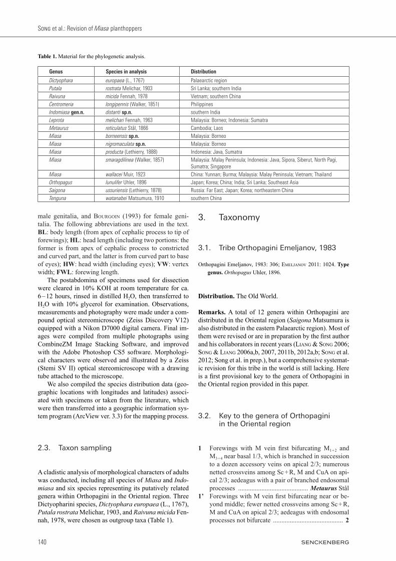
Table 1. Material for the phylogenetic analysis.
Genus Species in analysis Distribution
Dictyophara europaea (L., 1767) Palaearctic regionPutala rostrata Melichar, 1903 Sri Lanka; southern IndiaRaivuna micida Fennah, 1978 Vietnam; southern ChinaCentromeria longipennis (Walker, 1851) PhilippinesIndomiasa gen.n. distanti sp.n. southern IndiaLeprota melichari Fennah, 1963 Malaysia: Borneo; Indonesia: SumatraMetaurus reticulatus Stål, 1866 Cambodia; LaosMiasa borneensis sp.n. Malaysia: BorneoMiasa nigromaculata sp.n. Malaysia: BorneoMiasa producta (Lethierry, 1888) Indonesia: Java, SumatraMiasa smaragdilinea (Walker, 1857) Malaysia: Malay Peninsula; Indonesia: Java, Sipora, Siberut, North Pagi,
Sumatra; SingaporeMiasa wallacei Muir, 1923 China: Yunnan; Burma; Malaysia: Malay Peninsula; Vietnam; ThailandOrthopagus lunulifer Uhler, 1896 Japan; Korea; China; India; Sri Lanka; Southeast AsiaSaigona ussuriensis (Lethierry, 1878) Russia: Far East; Japan; Korea; northeastern ChinaTenguna watanabei Matsumura, 1910 southern China
141
ARTHROPOD SYSTEMATICS & PHYLOGENY — 72 (2) 2014
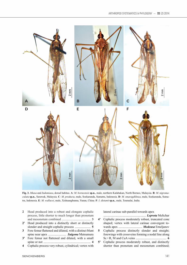
2 Head produced into a robust and elongate cephalic process, little shorter to much longer than pronotum and mesonotum combined ..................................... 3
2’ Head produced into a distinctly short or distinctly slender and straight cephalic process .................... 5
3 Fore femur flattened and dilated, with a distinct blunt spine near apex ....................... Saigona Matsumura
3’ Fore femur not flattened and dilated, with a small spine or not ............................................................ 4
4 Cephalic process very robust, cylindrical; vertex with
lateral carinae sub-parallel towards apex ...................................................... Leprota Melichar4’ Cephalic process moderately robust, truncated cone
shaped; vertex with lateral carinae convergent to-wards apex .............................. Medeusa Emeljanov
5 Cephalic process distinctly slender and straight; forewings with crossveins forming a nodal line along Sc + R, M and CuA veins ....................................... 6
5’ Cephalic process moderately robust, and distinctly shorter than pronotum and mesonotum combined;
Fig. 3. Miasa and Indomiasa, dorsal habitus. A: M. borneensis sp.n., male, northern Kalabakan, North Borneo, Malaysia. B: M. nigromaculata sp.n., Sarawak, Malaysia. C: M. producta, male, Soekaranda, Sumatra, Indonesia. D: M. smaragdilinea, male, Soekaranda, Suma-tra, Indonesia. E: M. wallacei, male, Xishuangbanna, Yunan, China. F: I. distanti sp.n., male, Tenmalai, India.
A
D
B
E
C
F

Song et al.: Revision of Miasa planthoppers
142
forewings without nodal line ................................. 76 Cephalic process distinctly slender and elongate;
frons with median carina moderately ridged; fore femora with a distinct blunt spine near apex; hind tibiae with 6 apical teeth .................. Miasa Distant
6’ Cephalic process more robust and shorter; frons with median carina robust and strongly produced; fore femora with a short small spine near apex; hind tibiae with 7 apical teeth ............ Indomiasa gen.n.
7 Forewings with a large crescent-shaped fuscous streak on posterior margin of apical part; fore femora flattened and dilated, with a distinct blunt spine near apex ........................................... Orthopagus Uhler
7’ Forewings without above streak on posterior margin of apical part; fore femora not flattened and dilated, with a minute spine near apex or not .................... 8
8 Frons with median carina robust and strongly pro-duced ..................................................................... 9
8’ Frons with median carina moderately ridged ...... 119 Head gradually narrowed and acuminated to apex;
hind tibiae with 6 apical teeth ..... Centromeria Stål9’ Head more or less truncate at apex; hind tibiae with
8 apical spines ..................................................... 1010 Frons with media carina broadly purplish-red, inter-
mediate carinae approaching frontoclypeal suture
.................................. Truncatomeria Song & Liang10’ Frons with media carina virescent or ochraceous, in-
termediate carinae approaching posterior margin of eyes .......................... Dictyotenguna Song & Liang
11 Vertex with lateral carinae before eyes gradually convergent and acuminated to apex
................................................. Tenguna Matsumura 11’ Vertex with lateral carinae more or less truncate api-
cally .................................. Dictyopharina Melichar
3.3. Miasa Distant, 1906
Miasa Distant, 1906: 247; SchMidt 1906: 280; Melichar 1912: 37; SchMidt 1915: 348; diStant 1916: 28; SchMidt 1928: 129; Metcalf 1946: 34. Type species. Elidiptera smaragdilinea Wal ker, 1857; by original designation.
Putalamorpha Bierman, 1910: 9. Type species. Stenocranus productus Lethierry, 1888; by original designation. Synonymized by Melichar 1912: 79.
Diagnosis. Head in front of eyes distinctly upturned, pro-duced into a laterally compressed, distinctly slender and
Fig. 4. Miasa and Indomiasa, lateral habitus. A: M. borneensis sp.n. B: M. nigromaculata sp.n. C: M. producta. D: M. smaragdilinea. E: M. wallacei. F: I. distanti sp.n.
A
C
E
B
D
F

143
ARTHROPOD SYSTEMATICS & PHYLOGENY — 72 (2) 2014
straight linear process; vertex without media carina, lat-eral carinae parallel at base, abruptly strongly constricted and curved upwardly before eyes, very narrow and medi-ally sulcate in remaining part; frons with lateral carinae distinctly expanded outwards below antennae, more or less convergent towards and abruptly strongly constrict-ed before eyes, intermediate carinae beneath cephalic process very narrow and sulcate, approaching to anterior margin of eyes; rostrum very slender and long, reaching to apex of hind femora; pronotum centrally angularly convex and hood-like anteriorly, lateral anterior angles rounded, median carina sharp and high; mesonotum tri-carinate with median carina generally too indistinct to be visible, lateral carinae incurved anteriorly towards me-dian carina; forewings with dark streak on distal third of wing; crossveins very scarce, forming a nodal line along Sc + R, M and CuA veins at apical third; stigmal area clear, with 2 – 4 cells; fore femora with a distinct blunt spine near apex; hind tibiae with 6 black-tipped apical teeth; aedeagus with paired membranous inflated apical lobes, without spines.
Description of adults. Coloration: General color in dried specimens ferruginous-brown, marked with pale green and black in dorsal habitus. Females slightly
darker than males. Head with cephalic process black above, pale green or ochraceous beneath. Eyes fuscous or reddish brown. Postclypeus, anteclypeus and thorax beneath, black and white. Forewings hyaline, with vena-tion and streak on distal third of wing dull ochraceous or fuscous. Legs ochraceous; coxae, trochanters and base of hind femora black; fore tibiae with a prominent subapical creamy-white annulation. Head (Figs. 1, 2, 3A – E, 4A – E, 5A – E, 6A – E) produced in front of eyes and extended into a distinctly upturned and laterally compressed slender and straight linear pro-cess, which is medially sulcate above and beneath. Ver-tex (Figs. 2B, 3A – E) basally slightly convex medially, nearly rectangular, posterior surface in relation to pro-notum elevated; lateral carinae parallel, strongly ridged at base, abruptly and strongly constricted and curved upwardly before eyes, very narrow and medially sulcate in remaining part, acuminate apically; posterior margin arcuate; median carina absent. Frons (Fig. 5A – E) elon-gate, anterior portion with intermediate carinae strongly narrowed and protruded anteriorly in ventral and lateral views (Figs. 5A – E, 6A – E), so apical part of frons dis-tinctly visible in dorsal view (Figs. 2B, 3A – E); lateral carinae ridged, distinctly expanded outwards below an-tennae, more or less convergent towards and abruptly
Fig. 5. Miasa and Indomiasa, head, pronotum and mesonotum, lateral view. A: M. borneensis sp.n. B: M. nigromaculata sp.n. C: M. producta. D: M. smaragdilinea. E: M. wallacei Muir, 1923. F: I. distanti sp.n.
A
D
B
E
C
F
Song et al.: Revision of Miasa planthoppers
144
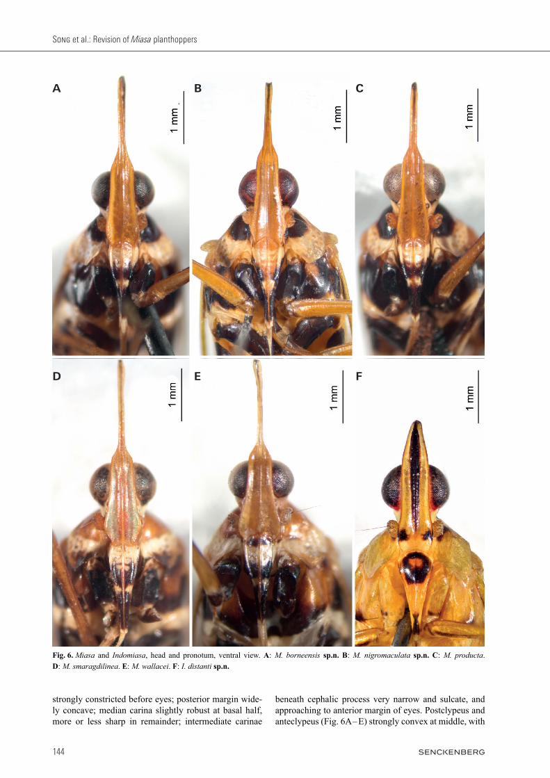
strongly constricted before eyes; posterior margin wide-ly concave; median carina slightly robust at basal half, more or less sharp in remainder; intermediate carinae
beneath cephalic process very narrow and sulcate, and approaching to anterior margin of eyes. Postclypeus and anteclypeus (Fig. 6A – E) strongly convex at middle, with
Fig. 6. Miasa and Indomiasa, head and pronotum, ventral view. A: M. borneensis sp.n. B: M. nigromaculata sp.n. C: M. producta. D: M. smaragdilinea. E: M. wallacei. F: I. distanti sp.n.
A
D
B
E
C
F
145
ARTHROPOD SYSTEMATICS & PHYLOGENY — 72 (2) 2014
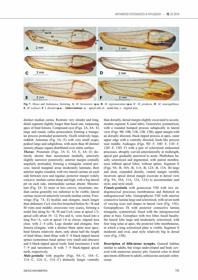
distinct median carina. Rostrum very slender and long, distal segment slightly longer than basal one, surpassing apex of hind femora. Compound eyes (Figs. 2A, 4A – E) large and round, callus postocularis forming a triangu-lar process protruded posteriorly. Ocelli relatively large, reddish. Antennae (Fig. 5A – E) with very small scape; pedicel large and subglobose, with more than 40 distinct sensory plaque organs distributed over entire surface. Thorax: Pronotum (Figs. 3A – E, 5A – E, 6A – E) dis-tinctly shorter than mesonotum medially, anteriorly slightly narrower posteriorly; anterior margin centrally angularly protruded, forming a triangular central pro-cess; lateral marginal areas moderately laminate, their anterior angles rounded, with two lateral carinae on each side between eyes and tegulae; posterior margin widely concave; median carina sharp and high, with a big lateral pit on each side, intermediate carinae absent. Mesono-tum (Fig. 3A – E) more or less convex, tricarinate; me-dian carina generally too indistinct to be visible, lateral carinae incurved anteriorly towards median carina. Fore-wings (Fig. 7A – E) hyaline and elongate, much longer than abdomen; CuA vein fi rst branched before Sc + R and M veins near middle; crossveins very scarce, forming a nodal line along Sc + R, M and CuA veins at apical 1/3; apical cells about 10 – 12; Pcu and A1 veins fused into a long Pcu + A1 vein at apical 1/6 in clavus; stigmal area clear, with 2 – 4 cells. Legs slender and elongate, fore femora elongate, with a distinct blunt spine near apex; hind femora relatively short, only about half the length of hind tibiae; hind tibiae with 5 – 8 black-tipped lateral spines (sometimes basal spines very small and obscure) and 6 black-tipped apical teeth; hind tarsomeres I with 7 – 9 and tarsomeres II with 7 – 9 black-tipped apical teeth, respectively.Male genitalia with pygofer (Figs. 9A – C, 10A – C, 11A – C, 12A – C, 13A – C) distinctly longer ventrally
than dorsally, dorsal margin slightly excavated to accom-modate segment X ( anal tube). Gonostyles symmetrical, with a rounded humped process subapically in lateral view (Figs. 9B, 10B, 11B, 12B, 13B); upper margin with an dorsally directed, black-tipped process at apex, outer upper edge with a ventrally directed, hook-like process near middle. Aedeagus (Figs. 9D – F, 10D – F, 11D – F, 12D – F, 13D – F) with a pair of sclerotized endosomal processes, abruptly curved anterolaterally at midlength, apical part gradually narrowed to acute. Phallobase ba-sally sclerotized and pigmented, with paired membra-nous infl ated apical lobes, without spines. Segment X (Figs. 9A – B, 10A – B, 11A – B, 12A – B, 13A – B) large and stout, expanded distally, ventral margin variably incurved; apical dorsal margin excavate in dorsal view (Fig. 9A, 10A, 11A, 12A, 13A) to accommodate anal style; anal style small.Female genitalia with gonocoxae VIII with two en-dogonocoxal processes membranous and fl attened on endogonocoxal lobe. Gonopophyses VIII with anterior connective lamina large and sclerotized, with seven teeth of varying sizes and shapes in lateral view (Fig. 13G). Gonopophyses IX with posterior connective lamina triangular, symmetrical, fused with the intergonocoxal plate at base. Gonoplacs with two lobes fused basally: the lateral lobe large and moderately sclerotized, with four long setae at apex; the posterior lobe membranous, in which a long sclerotized plate is visible. Segment X moderate and oval, anal style relatively big in dorsal view (Fig. 13H).
Description of fi fth- instar nymphs. General habitus similar to adults, but wings undeveloped and body cov-ered with numerous sensory pits. General color in dried specimens different to adults, ochraceous and pale ochra-ceous.
Fig. 7. Miasa and Indomiasa, forewing. A: M. borneensis sp.n. B: M. nigromaculata sp.n. C: M. producta. D: M. smaragdilinea. E: M. wallacei. F: I. distanti sp.n. – Abbreviations: ac – apical cell; nl – nodal line; s – stigmal area.
A
C
E
B
D
F

Song et al.: Revision of Miasa planthoppers
146
Cephalic process (Fig. 8A – D) before eyes distinctly upturned, broader and more robust than adults, more or less compressed laterally. Vertex (Fig. 8C) basally
arched, lateral margins strongly carinate, slightly con-vergent, abruptly constricted and curved upwardly be-fore eyes, and medially sulcate in remainder; median
Fig. 8. Fifth-instar nymph of Miasa smaragdilinea. A: Dorsal habitus. B: Head and pronotum, ventral view. C: Head, dorsal view. D: Head, lateral view. E: Thorax, dorsal view. F: Abdomen, dorsal view. G: Abdominal tergites VI – VIII, caudal view, showing wax-secreting plates. – Abbreviations: sp – sensory pit; wsp – wax secreting plate.
A
E G
F
B C D

147
ARTHROPOD SYSTEMATICS & PHYLOGENY — 72 (2) 2014
carina more or less obvious between eyes; anterior mar-gin acuminate and posterior margin indistinct. Frons (Fig. 8B) elongate, expanded outwards below antennae, more or less convergent towards and slightly constrict-ed before eyes; lateral areas (Fig. 8D) between lateral carinae and intermediate carinae distinctly broad, with more than 70 sensory pits from apex to base; intermedi-ate carinae reaching to frontoclypeal suture, and median carina distinct and complete. Clypeus, rostrum and eyes (Fig. 8D) similar to adults, but ocelli absent. Pronotum (Fig. 8E) with anterior margin centrally angularly convex, posterior margin widely concave; disc with about 18 sensory pits between median carina and upper lateral carinae, with 5 – 6 sensory pits be-tween upper lateral carinae and lower lateral carinae, with 6 sensory pits on 6 – 7 on apical margin of ventral lobes, respectively. Mesonotum (Fig. 8E) with 5 sen-sory pits outside lateral carinae. Metanotum (Fig. 8E) with 7 – 8 sensory pits outside lateral carinae. Forewing pads (Fig. 8E) with 2 indistinct sensory pits in middle. Legs similar to adults, very elongate and slender. Abdomen (Fig. 8F) 9-segmented, slender and elon-gate. Tergites II – V (Fig. 8A) with distinct median ca-rina and intermediate carinae; tergites III – VI much longer than others; tergites IV – VII (Fig. 8F) with 0, 2, 5, and 4 sensory pits between median carina and inter-mediate carinae, 4, 4, 0, and 0 sensory pits between in-termediate carinae and lateral carinae, and 14, 11, 5, and 0 pits on ventrolateral areas; tergites VII – IX very short and nearly covered by the former tergite, so the terminal more or less truncate; tergites VI – VII (Fig. 8G) with each pair of wax-secreting plates on posterolateral areas separated from rest of tergite by carina and directed pos-teriorly.
Distribution. Burma; Indonesia; Malaysia; China; Thai-land; Vietnam.
Remarks. When describing the genus Miasa, diStant (1906) stated that “this genus is allied to Dictyopharoides Fowl., from which it may be at once separated by the non-serrate anterior femora, a character omitted in Fow-ler’s diagnosis.” Actually, Dictyopharoides Fowler, 1900 belongs to the tribe Nersiini Emeljanov for its forewing veins Sc + R and M with a long common stem (eMeljanov 2011) and Miasa belongs in the tribe Orthopagini based on the following characters: forewings with Sc + R and M originated from basal cell without common stem; fore femora usually with a spine on ventral subapex; fore and middle tarsomeres I and II with a pair of acutellae; apical lobes of phallobase spineless or just with very short small spines; and structure of female genitalia. Miasa can be easily distinguished from other gen-era in Orthopagini by the distinctly slender, straight and linear cephalic process, and forewings with a nodal line along Sc + R, M and CuA veins at apical 1/3. See also discussion under Indomiasa gen.n.
3.4. Key to the species of Miasa Distant
1 Frons below eyes including median carina uniformly dull ochraceous (Fig. 6A – C,E); pronotum with pos-terolateral corner pale yellow to ochraceous with or without dark spot behind eye (Figs. 5A – C,E, 6A – C,E); forewings posterior margin broadly dull ochraceous (Fig. 7A – C,E); aedeagus with two pairs of ventral lobes and a pair of dorsolateral lobes (Figs. 9D – F, 10D – F, 11D – F, 13D – F) .......................... 2
1’ Frons below eyes emerald green, with median carina testaceous (Fig. 6D) or if uniformly dull ochraceous medial carina darker; pronotum with lateroventral corner brown, without dark spot behind eye (Figs. 5D, 6D); forewings with inner margin of clavus nar-rowly dark brown (Fig. 7D); aedeagus with two pairs of ventral lobes, but without dorsolateral lobes (Fig. 12D – F); Southern Malay Peninsula, Sumatra and Java .............................. M. smaragdilinea (Walker)
2 Male segment X broad basally, not hatchet-shaped in lateral view (Figs. 9B, 10B, 11B) .......................... 3
2’ Male segment X narrow basally, hatchet-shaped in lateral view (Fig. 13B); southwestern China, South-east Asia to North Malay Peninsula
...................................................... M. wallacei Muir3 Preocular field with a blackish brown spot (Fig.
5B,C); male segment X with ventral margin weakly incurved in lateral view (Figs. 10B, 11B) ............. 4
3’ Preocular field without blackish brown spot (Fig. 5A); male segment X with ventral margin distinctly incurved sub-basally in lateral view (Fig. 9B); Bor-neo ........................................... M. borneensis sp.n.
4 Upper process of gonostlyes distinctly broad at apex (Fig. 10B); basal ventral lobes of aedeagus distinctly short and small (Fig. 10E); male segment X with api-cal ventral margin distinctly produced in a long pro-cess in lateral view (Fig. 10B); Borneo
............................................ M. nigromaculata sp.n.4’ Upper process of gonostlyes not broad at apex (Fig.
11B); basal ventral lobes of aedeagus distinctly long (Fig. 11E); male segment X with ventral margin not protruded in lateral view (Fig. 11B); Sumatra and Java ................................... M. producta (Lethierry)
3.5. Miasa borneensis sp.n.
Figs. 1A, 3A, 4A, 5A, 6A, 7A, 9
Miasa smaragdilinea (Walker): kirkaldY 1913: 13 [error].
Material examined. Holotype ♂, MALAYSIA, Sabah: North Borneo (SE), Forest Camp, 19 km, N. of Kalabakan, 60 m, light trap, 29.x.1962, Y. Hirashima leg. (BPBM). — Paratypes. MA-

Song et al.: Revision of Miasa planthoppers
148
LAYSIA, Sabah: 1♂, North Borneo (SE), Forest Camp, 19 km, N. of Kalabakan, light trap, 27.x.1962, Y. Hirashima leg.; 1♂, Tenompok, 1460 m, Jesselton, 48 km E., 26 – 31.i.1959, T. C. Maa leg. (BPBM); 2♂♂, Sandakan, Baker leg. (USNM); Sarawak: 1♂ (MIZ 313185), 1♀ (MIZ 313186), Mt. Matang, 1012 ft., 10.xii.1909; Miasa producta Leth. [Schmidt’s handwriting], Edm. Schmidt, determ. 1926 (MIZPAS); 2♂♂, Kuching, 21.ii.1899, 2. xi.1899 (BMNH); ‘BORNEO’: 1♀ (abdomen missing), S.O. Bor-neo, Wahnes, V. Wolf. v. Schönberg leg.; Miasa smaragdilinea Walk.; 1♂, Mindai, 6.82 (MFNB).
Etymology. The species is named for its occurrence in Borneo.
Description. BL: ♂ 11.6 – 12.4 mm, ♀ 13.1 mm; HL: ♂ (1.6 + 1.0) – (1.8 + 1.0) mm, ♀ (1.6 + 1.0) mm; HW: ♂
1.3 mm, ♀ 1.2 mm; VW: ♂ 0.3 – 0.4 mm, ♀ 0.4 mm; FWL: ♂ 9.1 – 9.9 mm, ♀ 10.0 mm. General color ferruginous marked with pale green and blackish brown in dorsal habitus. Vertex between eyes and genae pale green or ochraceous, preocular field without blackish brown spot, frons below eyes includ-ing median carina and basal postclypeus uniformly dull ochraceous. Anterolateral marginal areas of pronotum behind eyes and lateral areas of mesonotum glossy black-ish brown, sometimes just dull ochraceous on mesono-tum (see Remarks); median carina, apical marginal areas of ventral lobes and posterior lateral angles of pronotum, and a broad median fascia to mesonotum pale green or pale ochraceous. Forewings with stigmal area and poste-rior margin broadly dull ochraceous, a large oblique tri-angular apical streak and a narrow streak along nodal line
Fig. 9. Miasa borneensis sp.n. A: Male pygofer and segment X, dorsal view. B: Male postabdomen, lateral view. C: Male pygofer and gonostyles, ventral view. D: Aedeagus, dorsal view. E: Aedeagus, lateral view. F: Aedeagus, ventral view. – Abbreviations: as – anal style; at – anal tube (segment X); avm – apical ventral margin of segment X; bvm – basal ventral margin of segment X; dll – dorsolateral lobe on phallotheca; dmp – dorsal margin of pygofer in profile; ep – endosomal processes; g – gonostyle; hpg – hook-like process of gonostyle; p – pygofer; pt – phallotheca; rhp – rounded humped process of gonostyle; upg – upper process of gonostyle; vl – ventral lobe on phal-lotheca; vmp – ventral margin of pygofer in profile.
A
D
B
E
C
F
149
ARTHROPOD SYSTEMATICS & PHYLOGENY — 72 (2) 2014
fuscous; hind wings with an apical fuscous spot. Abdo-men above and beneath testaceous mixed with fuscous, a broad central stripe and a lateral stripe on each side above pale green or ochraceous. Cephalic process (Fig. 5A) in front of eyes relatively long, with the ratio of its length to basal length of vertex (from curved part to base of vertex) about (1.9 – 2.0) : 1. Frons (Fig. 6A) elongate. Forewings (Fig. 7A) with ra-tio of length to width about 3.7 : 1. Hind tibiae with 5 – 7 (mainly 6) lateral spines; hind tarsomeres I with 6 – 7 and tarsomeres II with 6 – 8 black-tipped apical teeth, respec-tively. Male genitalia with pygofer (Fig. 9A – C) ventral to dorsal length (about 1.9 : 1); posterior margin dorsally more or less protruded in lateral view (Fig. 9B). Gono-styles relatively large, more or less expanded towards apex, broadest subapically in lateral view (Fig. 9B), apex rounded; upper process elongate, acute apically. Aedea-gus (Fig. 9D – F) relatively small, with a pair of long endosomal processes extended posteriorly and curved dorsally; phallobase sclerotized and pigmented at lateral sides, membranous and moderately inflated dorsally and ventrally, with a pair of dorsolateral lobes directed poste-riorly, and two pairs of ventral lobes, directed posteriorly and ventrally, respectively. Segment X in lateral view with base ventral margin protruded ventrally at base and concaved in middle, so base is slightly narrower than apex and looks like broad in lateral view (Fig. 9B), with ratio of length to width near middle about 1.7 : 1 in dorsal view (Fig. 9A).
Distribution. Malaysia (Borneo).
Remarks. The new species is very similar to M. producta, but it can be distinguished from the latter by the preocular field usually without blackish brown spot, and male segment X with ventral margin strongly incurved sub-basally in profile. M. smaragdilinea described by kirkaldY (1913) and two M. producta specimens men-tioned by SchMidt (1928) from Borneo should belong to the new species. Specimens from northwestern Borneo lack the black-ish brown spots on the lateral areas of mesonotum having this area just dull ochraceous. However, there is no obvi-ous difference of male genitalia between them.
3.6. Miasa nigromaculata sp.n.
Figs. 2, 3B, 4B, 5B, 6B, 7B, 10
Material examined. Holotype ♂, MALAYSIA, Sarawak: Gu-nong Mulu National Park, v – viii.1978, P.M. Hammond & J.E. Mar-shall leg., R.G.S. Expedition 1977 – 8, B.M. 1978 – 49 (BMNH). — Paratypes. MALAYSIA, Sarawak: 1♂, Gunong Mulu National Park, Long Pala base camp, ii – vii.1978, V.F. Eastop, R.G.S. Ex-pedition 1977 – 8, B.M. 1978 – 411; 1♀, Gunong Mulu National
Park, near base camp, 50 – 100m., v – viii.1978, P.M. Hammond & J.E. Marshall leg., R.G.S. Expedition 1977 – 8, B.M. 1978 – 49; 1♂, Baram, 18.vii.1920, J.C. Moulton leg.; Sabah: 1♂, 1♀, Jesselton, 4000’, 9.iii.1968, P.J.L. Roche leg. (BMNH).
Etymology. This new species name is derived from the Latin combination of the prefix “nigro-” plus “maculate”, referring to its black macula on the forewings.
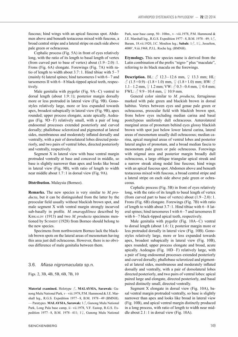
Description. BL: ♂ 12.3 – 12.6 mm, ♀ 13.3 mm; HL: ♂ (1.5 + 0.9) – (1.8 + 1.0) mm, ♀ (1.8 + 1.0) mm; HW: ♂ 1.1 – 1.2 mm, ♀ 1.2 mm; VW: ♂ 0.3 – 0.4 mm, ♀ 0.4 mm; FWL: ♂ 9.9 – 10.4 mm, ♀ 10.9 mm. General color similar to M. producta, ferruginous marked with pale green and blackish brown in dorsal habitus. Vertex between eyes and genae pale green or ochraceous, preocular field with blackish brown spot, frons below eyes including median carina and basal postclypeus uniformly dull ochraceous. Anterolateral marginal areas of pronotum behind eyes glossy blackish brown with spot just below lower lateral carina, lateral areas of mesonotum usually dull ochraceous; median ca-rina, apical marginal areas of ventral lobes and posterior lateral angles of pronotum, and a broad median fascia to mesonotum pale green or pale ochraceous. Forewings with stigmal area and posterior margin broadly dull ochraceous, a large oblique triangular apical streak and a narrow streak along nodal line fuscous; hind wings with an apical fuscous spot. Abdomen above and beneath testaceous mixed with fuscous, a broad central stripe and a lateral stripe on each side above pale green or ochra-ceous. Cephalic process (Fig. 5B) in front of eyes relatively long, with the ratio of its length to basal length of vertex (from curved part to base of vertex) about (1.9 – 2.0) : 1. Frons (Fig. 6B) elongate. Forewings (Fig. 7B) with ratio of length to width about 3.7 : 1. Hind tibiae with 6 – 8 lat-eral spines; hind tarsomeres I with 6 – 7 and tarsomeres II with 6 – 7 black-tipped apical teeth, respectively. Male genitalia with pygofer (Fig. 10A – C) ventral to dorsal length (about 1.6 : 1); posterior margin more or less protruded dorsally in lateral view (Fig. 10B). Gono-styles relatively large, more or less expanded towards apex, broadest subapically in lateral view (Fig. 10B), apex rounded; upper process elongate and broad, acute apically. Aedeagus (Fig. 10D – F) relatively large, with a pair of long endosomal processes extended posteriorly and curved dorsally; phallobase sclerotized and pigment-ed at lateral sides, membranous and moderately inflated dorsally and ventrally, with a pair of dorsolateral lobes directed posteriorly, and two pairs of ventral lobes: apical paired large and elongate, directed posteriorly, and basal paired distinctly small, directed ventrally. Segment X elongate in dorsal view (Fig. 10A), ba-sal ventral margin protruded ventrally, so base is slightly narrower than apex and looks like broad in lateral view (Fig. 10B), and apical ventral margin distinctly produced in a long process, with ratio of length to width near mid-dle about 2.1 : 1 in dorsal view (Fig. 10A).
Song et al.: Revision of Miasa planthoppers
150
Distribution. Malaysia (Borneo).
Remarks. The new species is similar to M. producta in having a preocular spot but can be distinguished from the latter by the smaller spot just below the lateral carina of the pronotum, the upper process of gonostlyes distinctly broad at apex, the basal ventral lobes of aedeagus dis-tinctly short and small and the male segment X with api-cal ventral margin distinctly produced in a long process in profile.
3.7. Miasa producta (Lethierry, 1888)
Figs. 3C, 4C, 5C, 6C, 7C, 11
Stenocranus productus Lethierry, 1888: 468.
Miasa smaragdilinea [nec Walker]: Schmidt, 1906: 280; Melichar 1912: 38.
Putalamorpha producta (Lethierry): BierMan 1910: 10.Miasa producta (Lethierry): SchMidt 1928: 129; Metcalf 1946: 35.
Material examined. INDONESIA, Sumatra: 1♀, Soekaranda, H. Dohrn leg.; Miasa smaragdilinea [Schmidt’s handwriting], Edm. Schmidt, determ. 1906 (MFNB); 2♂♂ (MIZ 313169 – 313170), 3♀♀ (MIZ 313171 – 313173), Soekaranda, i.1894, Dohrn leg.; Miasa producta Leth. [Schmidt’s handwriting], Edm. Schmidt, de-term. 1926; 1♂ (MIZ 313174), Liangagas, H. Dohrn leg.; Miasa producta Leth. [Schmidt’s handwriting], Edm. Schmidt, determ. 1926; 3♂♂ (MIZ 313175 – 313177), 6♀♀ (MIZ 313178 – 313183), Soekaranda, H. Dohrn leg.; Miasa producta Leth. [Schmidt’s hand-writing], Edm. Schmidt, determ. 1926 ♀ (Mus. Zool. Polonicum, Warszawa, 12/45; MIZPAS); 2♀♀, Soekaranda, H. Dohrn leg.; Miasa smaragdilinea [Schmidt’s handwriting], Edm. Schmidt,
Fig. 10. Miasa nigromaculata sp.n. A: Male pygofer and segment X, dorsal view. B: Male postabdomen, lateral view. C: Male pygofer and gonostyles, ventral view. D: Aedeagus, dorsal view. E: Aedeagus, lateral view. F: Aedeagus, ventral view.
A
D
B
E
C
F
151
ARTHROPOD SYSTEMATICS & PHYLOGENY — 72 (2) 2014
determ. 1906 (MMBC); 2♀♀, Soekaranda, H. Dohrn leg.; Miasa smaragdilinea [Schmidt’s handwriting], Edm. Schmidt, determ. 1906 (SDEI); Java: 1♀ (MIZ 313184), K. Fruhstorfer leg.; Miasa producta Leth. [Schmidt’s handwriting], Edm. Schmidt, determ. 1926 (Mus. Zool. Polonicum, Warszawa, 12/45; MIZPAS); 4♀♀, Tjimerang, Djampang, iii.1939, M. E. Walsh leg. (MZLU).
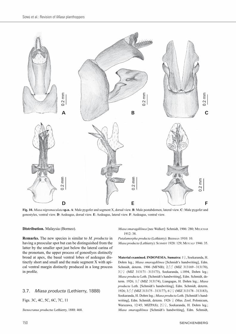
Redescription of adults. BL: ♂ 11.6 – 11.9 mm, ♀ 12.9 – 13.1 mm; HL: ♂ (1.4 + 0.9) – (1.6 + 1.1) mm, ♀ (1.6 + 1.0) – (1.7 + 1.1) mm; HW: ♂ 1.1 – 1.2 mm, ♀ 1.2 – 1.3 mm; VW: ♂ 0.3 – 0.4 mm, ♀ 0.4 – 0.5 mm; FWL: ♂ 9.0 – 9.2 mm, ♀ 9.9 – 10.1 mm. Color as in M. nigromaculata (see also SchMidt 1906). Cephalic process (Fig. 5C) in front of eyes rela-tively short, with the ratio of its length to basal length of vertex (from curved part to base of vertex) about (1.8 – 1.9) : 1. Frons (Fig. 6C) elongate. Forewings (Fig. 7C) with ratio of length to width about 3.9 : 1. Hind tibiae
with 5 – 7 (mainly 6) lateral spines; hind tarsomeres I with 7 – 9 and tarsomeres II with 7 – 9 black-tipped apical teeth, respectively. Male genitalia with pygofer (Fig. 11A – C) ventral to dorsal length (about 1.9 : 1); posterior margin more or less protruded dorsally in lateral view (Fig. 11B). Gon-ostyles relatively large, more or less expanded towards apex, broadest subapically in lateral view (Fig. 11B), apex rounded; upper process elongate, acute apically. Ae-deagus (Fig. 11D – F) relatively small, with a pair of long endosomal processes extended posteriorly and curved dorsally; phallobase sclerotized and pigmented at lateral sides, membranous and moderately inflated dorsally and ventrally, with a pair of dorsolateral lobes directed poste-riorly, and two pairs of ventral lobes, directed posteriorly and ventrally, respectively. Segment X large and stout, basal ventral margin pro-truded ventrally, so base is slightly narrower than apex
Fig. 11. Miasa producta. A: Male pygofer and segment X, dorsal view. B: Male postabdomen, lateral view. C: Male pygofer and gono-styles, ventral view. D: Aedeagus, dorsal view. E: Aedeagus, lateral view. F: Aedeagus, ventral view.
A
D
B
E
C
F

Song et al.: Revision of Miasa planthoppers
152
and looks like broad in lateral view (Fig. 11B), with ratio of length to width near middle about 1.7 : 1 in dorsal view (Fig. 11A).
Distribution. Indonesia (Sumatra, Java).
Remarks. The species is very similar to M. nigromaculatus both species having a preocular spot (see Remarks under nigromaculatus). It can be easily distinguished from other Miasa species by the relatively short cephalic process and shape of the male segment X.
3.8. Miasa smaragdilinea (Walker, 1857)
Figs. 3D, 4D, 5D, 6D, 7D, 12
Elidiptera smaragdilinea Walker, 1857: 86.Dictyophora [sic] smaragdilinea (Walker): Walker 1858: 318.Helicoptera (?) smaragdilinea (Walker): atkinSon 1886: 37. Miasa smaragdilinea (Walker): diStant 1906: 248; SchMidt 1906:
280 [error]; Melichar 1912: 38 [error]; kirkaldY 1913: 13; SchMidt 1915: 34; diStant 1916: 28; Muir 1923: 561; SchMidt 1928: 129; Metcalf 1946: 36.
Fig. 12. Miasa smaragdilinea. A: Male pygofer and segment X, dorsal view. B: Male postabdomen, lateral view. C: Male pygofer and gonostyles, ventral view. D: Aedeagus, dorsal view. E: Aedeagus, lateral view. F: Aedeagus, ventral view.
A
D
B
E
C
F

153
ARTHROPOD SYSTEMATICS & PHYLOGENY — 72 (2) 2014
Miasa rubrovittata Schmidt, 1906: 284. Synonymised by diStant, 1916: 28.
Miasa rubrovittata (Schmidt): Melichar 1912: 38; SchMidt 1915: 34; diStant 1916: 28; Baker 1927: 32; SchMidt 1928: 129; Metcalf 1946: 35.
Type material examined. Syntype ♂ of Elidiptera smaragdilinea Walker, MALAYSIA, Malay Peninsula: Mt Ophir, Wal-lace (BMNH). — Holotype ♂ of Miasa rubrovittata Schmidt, 1906 (MIZ 313187), INDONESIA, Sumatra: Soekaranda, H. Dohrn leg., i.1894; Type [red label]; Miasa rubrovittata Schmidt [Schmidt’s handwriting], Edm. Schmidt, determ. 1906 (Mus. Zool. Polonicum, Warszawa, 12/45; MIZPAS). — Allotype ♀ of Miasa rubrovittata Schmidt, 1906 (MIZ 313188), INDONESIA, Suma-tra: Soekaranda, H. Dohrn leg. i.1894; Type [red label]; Miasa rubrovittata Schmidt [Schmidt’s handwriting], Edm. Schmidt, determ. 1906 (Mus. Zool. Polonicum, Warszawa, 12/45; MIZ-PAS). — Paratypes of Miasa rubrovittata Schmidt, 1906: 3 males (MIZ 313189 – 313191), INDONESIA, Sumatra: Soekaranda, H. Dohrn leg. i.1894; Cotype [yellow label]; Miasa rubrovittata Schmidt [Schmidt’s handwriting], ♂, Edm. Schmidt, determ. 1906; 3♂♂ (MIZ 313192, 313195), 4♀♀ (MIZ 313193, 313194, 313196, 313197) and 2 fifth instar nymphs (MIZ 313198, 313199), Soekaranda, H. Dohrn leg.; Cotype [yellow label]; Miasa rubrovittata [Schmidt’s handwriting], Edm. Schmidt, determ. 1906 (Mus. Zool. Polonicum, Warszawa, 12/45; all MIZPAS); 1♀, Soekaranda, H. Dohrn leg.; Miasa rubrovittata [Schmidt’s handwriting], Edm. Schmidt, determ. 1906; Type (MFNB). 1♀ (MIZ 313200); 1♂, 1♀, Soekaranda, H. Dohrn leg.; type [red label]; Miasa rubrovittata [Schmidt’s handwriting], Edm. Schmidt, determ. 1906 (MMBC); 1♂, 1♀, Soekaranda, H. Dohrn leg.; type [red label]; Miasa rubrovittata [Schmidt’s handwriting], Edm. Schmidt, determ. 1906 (SNSD); 1♀, Soekaranda, i.1894, H. Dohrn leg.; type [red label]; Miasa rubrovittata [Schmidt’s handwriting], Edm. Schmidt, de-term. 1906 (SDEI); Java: K. Fruhstorfer leg.; Cotype [yellow la-bel]; Miasa rubrovittata Schmidt [Schmidt’s handwriting], Edm. Schmidt, determ. 1906 (Mus. Zool. Polonicum, Warszawa, 12/45; MIZPAS). Other material examined. MALAYSIA, Malay Peninsula: 1♀, Perak, Larut Hills, 500 – 2000’, 22.vi.1938; 1♀, Perak, Batang Pa-dang, Jor camp, 11.iii.1925, H.M. Pendlebury leg.; 1♂, 1♀, Sel-angor, 29.xii.1939 (BMNH). INDONESIA, Sumatra: 1♀, Ober Langkat, Deli, 1894, M. Ude. leg. (MFNB); Mentawai Islands: 1♀, Sipora I., 27.x.1924; 1♂, 4♀♀, Siberut I leg. (BMNH). 2♂♂, 1♀, Soekaranda, H. Dohrn leg.; Miasa rubrovittata [Schmidt’s handwriting], Edm. Schmidt, determ. 1906 (SNSD).
Redescription of adults. BL: ♂ 14.7 – 15.1 mm, ♀ 15.7 – 15.9 mm; HL: ♂ (2.0 + 1.0) – (2.0 + 1.1) mm, ♀ (2.0 + 1.0) – (2.1 + 1.1) mm; HW: ♂ 1.3 – 1.4 mm, ♀ 1.2 – 1.3 mm; VW: ♂ 0.4 mm, ♀ 0.4 – 0.5 mm; FWL: ♂ 11.3 – 11.5 mm, ♀ 12.3 – 12.4 mm. Frons below eyes emerald green with median carina broadly testaceous or dull ochraceous with median ca-rina darker. Forewings with posterior margin of clavus, a long and broad stripe along the terminal part of posterior margin and a narrow streak along nodal line, fuscous. Ce-phalic process (Fig. 5D) in front of eyes relatively long, with the ratio of its length to basal length of vertex (from
curved part to base of vertex) about 2.0 : 1. Frons (Fig. 6D) relatively short and broad. Forewings (Fig. 7D) elon-gate, with ratio of length to width about 4.3 : 1. Hind tib-iae with 6 – 8 (mainly 7) lateral spines; hind tarsomeres I with 8 – 9 and tarsomeres II with 8 – 9 black-tipped apical teeth, respectively. Male genitalia with pygofer (Fig. 12A – C) distinctly longer ventrally than dorsally (about 2.6 : 1); posterior margin distinctly protruded dorsally in lateral view (Fig. 12B). Gonostyles relatively large, more or less expanded towards apex, broadest subapically in lateral view (Fig. 12B), apex rounded; upper process elongate, acute api-cally. Aedeagus (Fig. 12D – F) distinctly elongate, with a pair of long endosomal processes extended posteriorly and curved laterally; phallobase elongate, sclerotized and pigmented at lateral sides, membranous and moderately inflated dorsally and ventrally, with two pairs of ventral lobes: upper pair robust, directed dorsally; lower pair relatively small, directed anterolaterally. Segment X with basal ventral margin not protruded, gradually widening towards apex with base much nar-rower than apex in lateral view (Fig. 12B), with ratio of length to width near middle about 1.7 : 1 in dorsal view (Fig. 12A).
Description of fifth-instar nymphs. Body length (from apex of cephalic process to tip of abdomen: 8.6 – 9.1 mm; HL: (1.7 + 0.9) – (1.8 + 1.0) mm; HW: male 1.0 – 1.1 mm; VW: 0.4 mm.
Distribution. Indonesia (Sumatra, Java, Sipora, Siberut, and North Pagi), Malaysia (Malay Peninsula), Singa-pore.
Remarks. This species was originally described by Walker (1857) from an unknown number of specimens from ‘Mt Ophir’ collected by ‘Wallace’. A single speci-men (syntype) is present in the BMNH bearing this data and the registration number 68.4 referring to an entry in the museums register for 1868.4. The species can be easily distinguished from other Miasa species by the relatively larger body size, the emerald green frons with broadly testaceous median carina, the longer forewings with a broader stripe along the terminal part of posterior margin and the shape of the male genitalia.
3.9. Miasa wallacei Muir, 1923
Figs. 3E, 4E, 5E, 6E, 7E, 13
Miasa wallacei Muir, 1923: 561.? Miasa smaragdilinea (Muir): diStant 1906: 248, fig. 108. Miasa wallacei (Muir): Metcalf 1946: 36.
Type material examined. Holotype ♂, THAILAND: S. Thailand, Biserat, 24.x.1901 (BMNH).

Song et al.: Revision of Miasa planthoppers
154
Other material examined. CHINA, Yunnan: 3♂♂, 3♀♀, Xish-uangbanna, Mengzhe, 870 m, 6.ix.1958, S.Y. Wang leg.; 1♂, Xishuangbanna, Xiaomengyang, 850 m, 4.ix.1957, S.Y. Wang leg.; 1♀, Mengla, 20.ix.1979, Y. Shang & G.Q. Liu leg.; 1 ♂, Mengla, 25.ix.1979, S.L. Liu leg.; 1 ♂, 1♀, Xishuangbanna, Menglun, 650 m, 25.vii.1959, S.F. Li leg. (IZCAS); VIET-NAM: 1 ♂, Dilinh (Djiring), 27.ix – 14.x.1960, C.M. Yoshi-moto leg.; 1 ♂, Fyan, 900 – 1000 m, 11.vii – 9.viii.1961, N.R. Spencer leg.; 1♀, Ap. Hung-Lam, 21 km, NW of Dilinh, 1100 m, 29.ix – 5.x.1960, C.M. Yoshimoto leg. (BPBM); BURMA: 2♂♂, 1♀, Mergue, Distant leg.; MALAYSIA, Malay Penin-sula: 8♂♂, 8♀♀, Pahang, various localities and dates; 1♂, 3♀♀, Selangor, various dates; THAILAND: 4♂♂, 3♀♀, same data as holotype (BMNH).
Redescription of adults. BL: ♂ 13.3 – 13.8 mm, ♀ 14.5 – 14.8 mm; HL: ♂ (1.8 + 0.9) – (1.8 + 1.0) mm, ♀ (1.8 + 1.0) – (2.0 + 1.0) mm; HW: ♂ 1.1 – 1.2 mm, ♀ 1.2 – 1.3 mm; VW: males 0.3 – 0.4 mm, ♀ 0.4 – 0.5 mm; FWL: ♂ 10.2 – 10.4 mm, ♀ 10.7 – 11.0 mm. General color similar to M. smaragdilinea, ferrugi-nous brown marked with pale green in dorsal habitus. Vertex between eyes, preocular field and genae pale green or ochraceous, frons below eyes including median carina uniformly dull ochraceous, base of postclypeus glossy blackish brown. Anterolateral marginal areas of pronotum behind eyes ferruginous brown or blackish brown, lateral areas of mesonotum ferruginous brown; median carina, apical marginal areas of ventral lobes and
Fig. 13. Miasa wallacei. A: Male pygofer and segment X, dorsal view. B: Male postabdomen, lateral view. C: Male pygofer and gono-styles, ventral view. D: Aedeagus, dorsal view. E: Aedeagus, lateral view. F: Aedeagus, ventral view. G: Anterior connective lamina of female gonopophyses VIII. H: Female segment X, dorsal view.
A
D
B
E
C
H
GF

155
ARTHROPOD SYSTEMATICS & PHYLOGENY — 72 (2) 2014
posterior lateral angles of pronotum, and a broad medi-an fascia to mesonotum pale green or pale ochraceous. Forewings and hindwings similar to M. producta. Abdo-men above and beneath testaceous mixed with fuscous, a broad central stripe and a lateral stripe on each side above pale green or ochraceous. Cephalic process (Fig. 5E) in front of eyes relatively short, with the ratio of its length to basal length of vertex (from curved part to base of vertex) about (2.3 – 2.4) : 1. Forewings (Fig. 7E) with ratio of length to width about 3.6 : 1. Hind tibiae with 5 – 6 (mainly 5) lateral spines; hind tarsomeres I with 8 – 9 and tarsomeres II with 8 – 9 black-tipped apical teeth, respectively. Male genitalia with pygofer (Fig. 13A – C) much longer ventrally than dorsally (about 4.6 : 1); posterior margin distinctly protruded dorsally in lateral view (Fig. 13B); a pair of large rounded humped process on vent-rolateral area (Fig. 13B,C). Gonostyles relatively large, more or less expanded towards apex, broadest subapi-cally in lateral view (Fig. 13B), apex straight; upper process more elongate, acute apically. Aedeagus (Fig. 13D – F) relatively large, with a pair of long endosomal processes extended posteriorly and curved anteriorly; phallobase sclerotized and pigmented at lateral sides, membranous and moderately inflated dorsally and ven-trally, with a pair of dorsal lobes directed posteriorly, and two pairs of ventral lobes: upper pair large and elon-gate, directed dorsally; lower pair relatively small and rounded. Segment X steeply tectiform, with sides very long on apical half, ratio of length to width near middle about 1.4 : 1 in dorsal view (Fig. 13A); basal ventral margin not protruded, rapidly widening to apex beyond middle, so in lateral view (Fig. 13B) somewhat hatchet-shape, base much narrower than apex.
Distribution. China, Vietnam, Burma, Thailand.
Remarks. This species was originally described from a single male specimen from ‘Biserat, Siam, Malay States’. The male specimen bearing this data and labeled ‘holo-type’ is presumed to be the type, there being four other males and three females with the same data present in the BMNH collection (see Material examined). The spe-cies can be easily separated from other Miasa species by the hatchet-shaped male segment X in lateral view (Fig. 13B), and also by the postclypeus basally being glossy blackish brown (Fig. 6E) and the male pygofer with a pair of large rounded humped processes on ventrolateral area (Fig. 13B – C). As in M. borneensis, some variation of color pattern occurs in M. wallacei. The large glossy blackish brown spot on the pronotum behind the eye of the holotype and some other specimens is paler in some other topotypi-cal specimens and the area is just ferruginous brown in specimens from Burma (3 specimens), Yunnan, China (13 specimens) and Vietnam (3 specimens). However, as all specimens have similar male genitalia they are con-sidered conspecific.
The specimen from Burma (Tenasserim: Myitta) de-scribed and figured by diStant (1906) as M. smaragdulinea may be this species based on the three other M. wallacei specimens from Myanmar examined.
3.10. Indomiasa gen.n.
Type species. Indomiasa distanti sp.n., by present designation and monotypy.
Etymology. The new generic name is a combination of the prefix “Indo-” (India) plus the generic name of its related group “Miasa”, gender: feminine.
Diagnosis. Cephalic process before eyes strongly up-turned, gradually convergent anteriorly, more or less acuminate apically; vertex without media carina, lateral carinae parallel at base, abruptly strongly constricted and curved upwardly before eyes, more or less parallel anteri-orly, acuminate apically; frons with median carina robust and strongly convex, intermediate carinae reaching mid-dle margin of eyes; pronotum centrally angularly convex and hood-like anteriorly, lateral anterior angles rounded, median carina sharp and high; mesonotum tricarinate with median carina generally too indistinct to be visible, lateral carinae incurved anteriorly towards median ca-rina; forewings with dark streak on distal third of wing; crossveins very scarce, forming a nodal line along Sc + R, M and CuA veins at apical third; stigmal area clear, with 2 cells; fore femora with a small spine near apex; hind tibiae with 7 apical black-tipped teeth; aedeagus with paired membranous inflated apical lobes, without spines.
Description. Head (Figs. 3F, 4F, 5F) produced in a short and slightly slender cephalic process. Vertex (Fig. 3F) basally slightly convex medially, slightly narrower than transverse diameter of eyes in dorsal view, posterior sur-face in relation to pronotum elevated; lateral margins sub-parallel at base, abruptly constricted and curved upwardly before eyes, more or less parallel anteriorly, acuminate apically; posterior margin arcuate; median carina absent. Frons (Fig. 6F) elongate, lateral margins carinate and nearly parallel, more or less expanded out-wards below antennae, posterior margin widely concave; median carina robust and strongly produced, intermedi-ate carinae obsoletely developed, slightly converging posteriorly and approaching to anterior margin of eyes. Postclypeus and anteclypeus (Fig. 6F) strongly convex at middle, median carina indistinct. Rostrum relatively short, distal segment slightly longer than basal one, reaching to middle of hind femora. Compound eyes (Fig. 6F) large and round, callus postocularis forming a small triangular process protruded posteriorly. Ocelli relatively large, reddish. Antennae (Fig. 6F) with very small scape; pedicel large and subglobose, with more than 40 distinct sensory plaque organs distributed over entire surface.

Song et al.: Revision of Miasa planthoppers
156
Thorax: Pronotum (Figs. 3F, 5F, 6F) distinctly shorter than mesonotum medially, anteriorly slightly narrower posteriorly; anterior margin centrally angularly convex, forming a long triangular central process; lateral margin-al areas moderately laminate, their anterior angles round-ed and distinctly convex, with two lateral carinae on each side between eyes and tegulae, but both carinae too ob-scure to be visible; posterior margin widely concave; me-dian carina sharp and high, with a big lateral pit on each side, intermediate carinae absent. Mesonotum (Fig. 3F) more or less convex, tricarinate; median carina distinct, lateral carinae incurved anteriorly towards median carina. Forewings (Fig. 7F) hyaline and elongate, much longer than abdomen; CuA vein first branched before Sc + R and M veins near middle; crossveins very scarce, formed a nodal line along Sc + R, M and CuA veins at apical 1/3; apical cells about 10; Pcu and A1 veins fused into a long Pcu + A1 vein at apical 1/5 in clavus; stigmal area clear, with 2 cells. Legs moderately elongate, fore femora rela-tively short, with a small spine near apex; hind femora relatively short, only about half the length of hind tibiae; hind tibiae with 5 – 6 lateral black-tipped spines and 7 apical black-tipped apical teeth; hind tarsomeres I with 16 – 17 and tarsomeres II with 13 – 14 black-tipped apical teeth, respectively.Male genitalia with pygofer (Fig. 14A – C) distinctly longer ventrally than dorsally, dorso-lateral margins strongly and angularly produced posteriorly in dorsal view (Fig. 14A). Gonostyles large and symmetrical, with a rounded humped process subapically in lateral view (Figs. 14B); upper margin with a long black-tipped pro-cess at apex, directed dorsally; outer upper edge with a ventrally directed, hook-like process near middle in lat-eral view (Fig. 14B). Aedeagus (Fig. 14D – F) with a pair of long sclerotized endosomal processes, abruptly curved laterally at midlength, apical part gradually narrowed distally to acute apex; phallobase basally sclerotized and pigmented, dorsal part mostly sclerotized and pigmented, with paired membranous lobes covering numerous small spines at apex. Segment X narrow and elongate, anal style small. Female genitalia unknown.
Distribution. India.
Remarks. The new genus is externally similar to Miasa, but can be distinguished from the latter by the cephalic process more robust and shorter; the frons with median carina robust and strongly produced; the fore femora relatively short, with a short small spine near apex; the hind tibiae with 7 apical teeth; and hind tarsomeres I with 16 – 17 and tarsomeres II with 13 – 14 black-tipped apical teeth, respectively. It can be distinguished from Centromeria Stål with a similar shaped cephalic process, by its more slender and elongate forewings with crossveins very scarce forming a nodal line along Sc + R, M and CuA veins at apical third and the hind tibiae with 7 apical teeth.
3.11. Indomiasa distanti sp.n.
Figs. 3F, 4F, 5F, 6F, 7F, 14
Material examined. Holotype ♂, INDIA: Tenmalai, 500 – 800’, Travancore, S. India, 11 – 17.x.[19]38 (BMNH).
Etymology. This new species is named after William Lucas Distant, an excellent English entomologist in the Natural History Museum, London.
Description of adults. ♂, length (from apex of cephalic process to tip of forewings) 12.9 mm; HL (0.6 + 1.0) mm; HW 1.4 mm; VW 0.2 mm; FWL 10.4 mm. General color greenish-ochraceous. Lateral carinae of vertex before eyes, median carina and area between lateral carinae, two big spots of frons below antennae, a large spot on postclypeus and apex of anteclypeus black-ish brown. Compound eyes fuscous with posterior mar-gin purplish-red; ocelli purplish-red. Pronotum with a small area behind eyes blackish brown; anterior central area with median carina, pale green. Mesonotum with median carina and apex, pale green. Forewings with stigmal area and a large oblique triangular apical streak fuscous, an obscure streak along nodal line, posterior margin of clavus, surrounding area of streak dull, and all crossveins pale ochraceous. Abdomen above and be-neath greenish-ochraceous. Male genitalia with pygofer relatively small, dis-tinctly longer ventrally than dorsally (about 2.8 : 1), dor-sal margin slightly excavated to accommodate segment X, dorso-lateral margins strongly and angularly pro-duced posteriorly in dorsal view (Fig. 14A); posterior margin with two processes, directed posteriorly, near upper middle in lateral view (Fig. 14B), upper process rounded, lower process angular. Gonostyles with poste-rior margin nearly straight, with a rounded humped pro-cess subapically (Fig. 14B); upper margin with a long black-tipped process at apex, directed dorsally; outer upper edge with a ventrally directed, hook-like process near middle (Fig. 14B). Aedeagus (Fig. 14D – F) moder-ately stout, phallobase with a pair of small membranous lobes near subapex, directed laterally in dorsal view (Fig. 14D); ventral part with two pairs of large lobes: ventrolateral pair moderately large, directed posteriorly at apex in lateral view (Fig. 14E), ventral pair very elon-gate and large, protruded posteriorly and curved anteri-orly, with numerous small spines at apex. Segment X narrow and elongate, with ratio of length to width near middle about 2.2 : 1 in dorsal view (Fig. 14A); basal ventral margin not protruded, rapidly wid-ening to apex beyond middle, so base much narrower than apex in lateral view (Fig. 14B); apical dorsal mar-gin deeply excavate in dorsal view (Fig. 14A) to accom-modate anal style; anal style small.
Distribution. Southern India.

157
ARTHROPOD SYSTEMATICS & PHYLOGENY — 72 (2) 2014
4. Phylogenetic analysis of Miasa and Indomiasa
4.1. Characters
The data matrix (Table 2) was composed of 64 morpho-logical characters from the coloration, head, thorax, and male postabdomen (including genitalia) of adults. All characters were equally weighted, and in all characters, states were treated as unordered. A dash ( – ) was entered in a matrix position if a character is not applicable to a taxon, and a question mark (?) was entered if the charac-ter condition was ambiguous or unexamined.
Coloration0 Vertex, lateral carinae before eyes: [0] dull ochra-
ceous; [1] black (Fig. 2A,B).
1 Frons, median carina: [0] ochraceous; [1] purplish-red (Fig. 6D); [2] black (Fig. 6F).
2 Frons, areas between intermediate carinae and lateral carinae: [0] ochraceous; [1] emerald green (Fig. 6D); [2] reddish ochraceous.
3 Base of postclypeus: [0] dull ochraceous (Fig. 6A – D); [1] blackish brown (Fig. 6E,F).
4 Cephalic process, preocular field (lateral area before eyes): [0] without spot; [1] with a black spot (Fig. 2A,B).
5 Pronotum, dark brown spots on lateral areas behind eyes: [0] absent; [1] present (Fig. 2C).
6 Forewings, fuscous streak on apical region: [0] ab-sent; [1] present (Fig. 7).
7 Forewings, apical streak: [0] narrow, not exceeding CuA1 posteriorly (Fig. 7A); [1] broad, exceeding CuA2 posteriorly (Fig. 7D).
8 Forewings, apical streak: [0] approaching to nodal line anteriorly (Fig. 7A); [1] not approaching to nod-al line anteriorly (Fig. 7E).
Fig. 14. Indomiasa distanti sp.n. A: Male pygofer and segment X, dorsal view. B: Male postabdomen, lateral view. C: Male pygofer and gonostyles, ventral view. D: Aedeagus, dorsal view. E: Aedeagus, lateral view. F: Aedeagus, ventral view. – Abbreviation: ppp – posterior process of pygofer.
A
D
B
E
C
F

Song et al.: Revision of Miasa planthoppers
158
9 Forewings, dull ochraceous posterior margin: [0] nar-row, not exceeding Pcu + A1 (Fig. 7D, F); [1] broad, exceeding CuP (Fig. 7A – C,E).
10 Legs: [0] pale green, without spots; [1] brown with whitish stripes or spots (Fig. 1).
11 Abdominal segments III – VI: [0] pale green without spots or stripes; [1] dark brown with whitish spots or stripes (Fig. 1).
Head12 Cephalic process before eyes: [0] robust; [1] distinct-
ly slender (Fig. 5F); [2] very slender (Fig. 5A – E).13 Cephalic process before eyes: [0] short, less or little
longer than longitudinal diameter of eyes (Fig. 5F); [1] much longer (Fig. 5A – E).
14 Cephalic process before eyes: [0] unbent or slightly bent; [1] strongly bent (Fig. 5).
15 Cephalic process with a short oblique carina in pre-ocular field: [0] absent; [1] present (Fig. 2A, B).
16 Vertex, width of posterior margin in relation to trans-verse diameter of eyes: [0] narrower (Fig. 2B); [1] nearly equal or little wider; [2] much wider.
17 Vertex, median carina: [0] absent; [1] present.18 Vertex, median carina: [0] only distinct in base; [1]
nearly completely distinct.19 Vertex, apex: [0] broadly and angulately convex; [1]
nearly acuminate.20 Vertex, posterior margin: [0] broadly concave (Fig.
2B); [1] angularly concave (no more than 100°).21 Vertex, posterior surface in relation to pronotum: [0]
in the same plane; [1] in an elevated plane.22 An apical carina between anterior margins of frons
and vertex: [0] absent; [1] present.23 Frons, median carina: [0] ridged; [1] robust and
strong ly convex (Fig. 6F). 24 Frons, intermediate carinae approaching to: [0] ante-
rior margin or middle of eyes (Fig. 2C); [1] fronto-clypeal suture.
25 Frons, lateral margins below the antennae: [0] nearly straight; [1] angularly convex (Fig. 2C).
26 Rostrum, apex: [0] reaching base to middle of hind femora; [1] reaching or surpassing apex of hind fem-ora.
27 Rostrum, basal segment relative to distal one: [0] dis tinctly longer (no less than 1.2 × as long as distal one); [1] nearly equal or slightly longer.
Thorax28 Pronotum, lateral carinae: [0] absent; [1] present in
basal third to half.29 Pronotum, ventral lobes with a carina: [0] absent; [1]
present.30 Pronotum, anterior central margin: [0] arcuately con-
vex; [1] angularly convex (Fig. 2B).31 Pronotum, posterior margin: [0] broadly arcuately
concave (Fig. 2B); [1] angularly concave (no more than 120°).
32 Pronotum, anterolateral angles: [0] angular; [1] round-ed (Fig. 2B).
33 Mesonotum, lateral carinae: [0] gradually incurved; [1] nearly straight.
34 Mesonotum, lateral carinae: [0] parallel; [1] conver-gent.
35 Forewings, veins: [0] without setae; [1] with short setae.
36 Forewings, numbers of apical cells of M: [0] 4 – 5 (Fig. 7A); [1] ≥ 6.
37 Forewings, shape of stigmal area: [0] quadrangular; [1] elongate (Fig. 7A).
38 Forewings, nodal line: [0] absent; [1] present (Fig. 7A).
39 Forewings, ratio of length to width: [0] 3.0 – 3.5 ×; [1] 3.6 – 4.0 ×; [2] > 4.0 ×.
40 Fore and middle tarsomeres I and II, acutellae: [0] 2; [1] > 2.
41 Fore femora: [0] neither slender nor dilated; [1] slen-der and elongate; [2] flattened and dilated.
42 Fore femora, a small spine on ventral subapex: [0] absent; [1] present.
43 Fore femora, subapical spine: [0] small and acute; [1] large and blunt.
44 Hind femora and tibiae: [0] not slender and elongate; [1] slender and elongate.
45 Hind tibiae, number of apical teeth: [0] 6; [1] 7; [2] 8.46 Hind tarsomeres I and II, number of apical teeth: [0]
7 – 13; [1] 14 – 20.
Male postabdomen47 Pygofer (lateral view), ventral margin in relation to
dorsal one: [0] < 1.5 × (Fig. 9B); [1] 1.5 – 3.0 × (Fig. 12B); [2] > 3.0 × (Fig. 13B).
48 Pygofer (lateral view), posterior process: [0] absent; [1] present (Fig. 14B).
49 Pygofer, numbers of posterior process: [0] 1; [1] 2 (Fig. 14B).
50 Pygofer, poster margin if posterior process absent: [0] slightly protruded posteriorly (Fig. 9B); [1] dis-tinctly protruded posteriorly (Fig. 12B).
51 Pygofer, a pair of rounded humped process on vent-rolateral area: [0] absent; [1] present (Fig. 9B).
52 Gonostyles, upper process: [0] neither elongate nor broad; [1] elongate (Fig. 14B); [2] broad (Fig. 10B).
53 Aedeagus, apical membranous lobes of phallotheca: [0] with long spines; [1] with very short small spines or not.
54 Aedeagus, apical membranous lobes of phallotheca: [0] without spine; [1] with very short small spines.
55 Aedeagus, phallotheca with dorsolateral lobes: [0] present (Fig. 9D,E); [1] absent.
56 Aedeagus, lower ventral lobes of phallotheca: [0] short (Fig. 10E); [1] elongate (Fig. 14E).
57 Aedeagus, lower ventral lobes of phallotheca: [0] rounded (Fig. 9E); [1] pointed (Fig. 10E).
58 Aedeagus, endosomal processes: [0] short, just ex-tended from phallotheca; [1] long, much extended from phallotheca (Fig. 9D,E).
59 Aedeagus, apex of endosomal processes: [0] acute (Fig. 9D,E); [1] obtuse.

159
ARTHROPOD SYSTEMATICS & PHYLOGENY — 72 (2) 2014
60 Aedeagus, endosomal processes: [0] nearly straight; [1] curved in middle (Fig. 9D,E).
61 Segment X, shape (dorsal view): [0] oval; [1] trun-cate; [2] distinctly elongate (Fig. 14A); [3] irregular (Fig. 13A).
62 Segment X (lateral view), apical ventral margins: [0] not protruded; [1] protruded ventrally (Fig. 13A, B).
63 Segment X (lateral view), basal ventral margins: [0] not protruded; [1] protruded ventrally (Fig. 9A,B).
4.2. Character analysis
The cladistic analysis was conducted with WinClada ver. 1.00.08 (nixon 1999, 2002) and implemented in NONA ver. 2.0 (goloBoff 2000). Phylogenetic relationships were reconstructed with a heuristic analysis by search-ing for the most parsimonious trees (MPTs) with 1000 maximum trees, 1000 replications and 10 starting trees per replication. Characters were selected to map the homoplasy/homology in the homoplasy setting dialog with any extra steps making it homoplasious. Bootstrap (felSenStein 1985) and jackknife (farriS et al. 1996) values were used as support measures, and calculated in NONA for the hypothesised clades with 1000 replica-tions.
4.3. Results
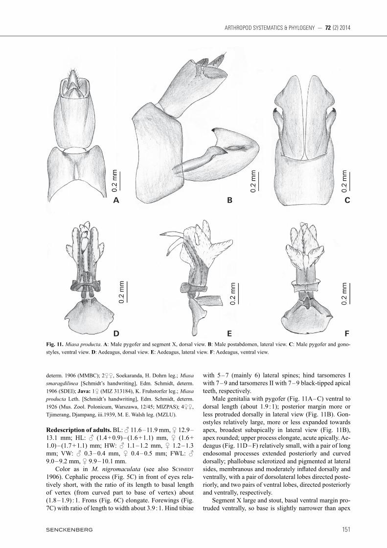
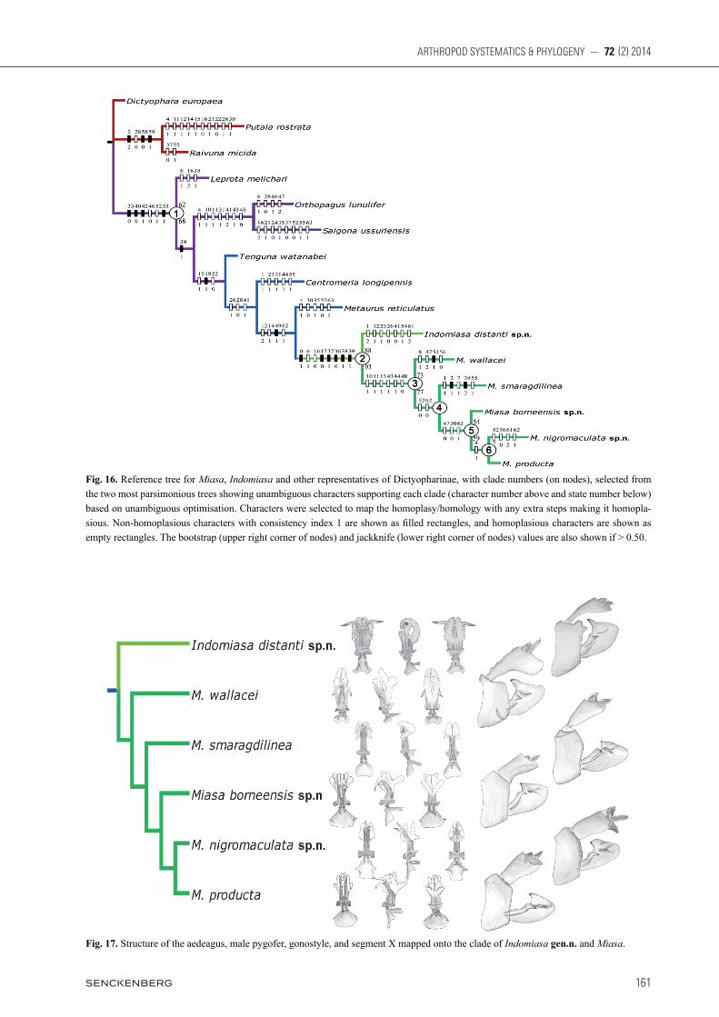
Analysis of the dataset (see Table 2) resulted in two MPTs with tree length = 162 steps, consistency index (CI) = 0.46, and retention index (RI) = 0.61. A strict con-
sensus of both trees is shown in Fig. 15 and a reference tree selected from the two MPTs is presented in Fig. 16 with the more robust bootstrap/jackknife support values mapped onto it. According to the result of phylogenetic analysis, Miasa species make up a good monophylum, and the new genus Indomiasa can be considered as the sister group of it. Clade 1 in Fig. 16 supports the monophyly of Ortho-pagini represented by Miasa and its seven putatively re-lated genera all distributed in the Oriental region, which is defined phylogenetically by four unique synapomor-phies with consistency index 1: 33-0 (character 33, state 0), 40-0, 42-1, and 53-1. This clade is further defined by two unambiguous character changes: 46-0 and 52-1. It has 62% bootstrap and 68% jackknife values. The phy-logenetic relationships among three genera Leprota, Orthopagus, and Saigona differ between the two MPTs, but the remaining four taxa form a series of successive sister taxa to Miasa. The new genus Indomiasa and all Miasa species con-stitute clade 2 (also see Fig. 17), which is supported by six synapomorphies (0-1, 17-0, 32-1, 36-0, 38-1, and 39-1), and two homoplasious characters (6-1 and 16-0). Indomiasa gen.n. can be considered as the sister group of Miasa, which is supported by 88% bootstrap and 93% jackknife values. The monophyly of Miasa (clade 3) is supported by 73% bootstrap and 77% jackknife values. It is supported by six unambiguous character changes: 10-1, 11-1, 13-1, 43-1, 44-1, and 48-0. Within Miasa, M. walla cei has the following one autapomorphy not present in other Miasa species: a pair of rounded humped process on ventrolat-eral area of pygofer (51-1). It can be regarded as the sister group of the remaining Miasa species (clade 4). Clade 4 is grouped on two unambiguous character changes: 52-0 and 62-0. Within the clade, M. smaragdilinea is the sister group of remaining species (clade 5) although clade 4 has low bootstrap and jackknife values.
Table 2. Character state matrix.
Characters →Taxa ↓
00000000000123456789
11111111110123456789
22222222220123456789
33333333330123456789
44444444440123456789
55555555550123456789
66660123
Dictyophara europaea 0000000--- 0001001110 0010100111 0001001100 100-01110- 0000-0--10 1111Putala rostrata 0020100--- 0111110100 0100101000 1001001100 100-011110 -000-0--01 0110Raivuna micida 0020000--- 0001001110 0010100101 0001001000 100-011110 -000-11101 0000Centromeria longipennis 0100--0--- 0000011101 1101011000 1100101100 0110100110 -01101--10 1000Indomiasa distanti sp.n. 1201011000 00101100-1 0101010100 1010100111 0010011111 -011101010 1210Leprota melichari 0000001110 0000100210 0010100010 0000011100 0010020110 -011101010 0000Metaurus reticulatus 0000110--- 0020111101 0000111000 0000111100 0110011111 -00100111- -311Miasa borneensis sp.n. 1000011001 11211100-1 0100011100 1010100111 011110000- 0001001010 1001Miasa nigromaculata sp.n. 1000111001 11211100-1 0100011100 1010100111 011110000- 0021000110 1211Miasa producta 1000111001 11211100-1 0100011100 1010100111 011110000- 0001001110 1001Miasa smaragdilinea 1110001100 11211100-1 0100011100 1010100112 011110010- 1001011110 1000Miasa wallacei 1001?01011 11211100-1 0100011100 1010100111 011110020- 1111000010 1310Orthopagus lunulifer 0000111100 1100001110 1010100100 1100101100 021101120- 101100--10 1000Saigona ussuriensis 0000110--- 1101002100 0110010010 0100111000 021102010- 100111--10 1001Tenguna watanabei 0000000--- 0000011111 1000110110 1000101100 0010020110 -01100--10 1000
SONG et al.: Revision of Miasa planthoppers
160
Clade 5 is supported by three unambiguous character changes: pygofer with ventral margin in relation to dorsal one < 1.5 × in lateral view (47-0); pygofer with posterior margin slightly protruded posteriorly (50-0), and basal ventral margins of segment X protruded (63-1). This clade has 54% bootstrap and 59% jackknife values. Finally, M. nigromaculata sp.n. and M. producta share an unambiguous character change: preocular fi eld of cephalic process with a black spot (4-1); this supports that they form a monophyletic group (clade 6).
5. Discussion
The range of Miasa includes the Indo-Chinese and In-do-Malayan subregions in the Oriental region, showing a typical sympatric distribution pattern (Fig. 18). The northernmost distribution of the genus is southern Yun-nan, China, the easternmost reaches northeastern Borneo, and the southernmost extends to northern Java. Within
Fig. 15. Strict consensus of the two most parsimonious trees (tree length 171) for Miasa, Indomiasa and other representatives of Dictyo-pharinae. The head and pronotum (lateral view), and forewing are mapped onto the tree.
161
ARTHROPOD SYSTEMATICS & PHYLOGENY — 72 (2) 2014
Fig. 16. Reference tree for Miasa, Indomiasa and other representatives of Dictyopharinae, with clade numbers (on nodes), selected from the two most parsimonious trees showing unambiguous characters supporting each clade (character number above and state number below) based on unambiguous optimisation. Characters were selected to map the homoplasy/homology with any extra steps making it homopla-sious. Non-homoplasious characters with consistency index 1 are shown as fi lled rectangles, and homoplasious characters are shown as empty rectangles. The bootstrap (upper right corner of nodes) and jackknife (lower right corner of nodes) values are also shown if > 0.50.
Fig. 17. Structure of the aedeagus, male pygofer, gonostyle, and segment X mapped onto the clade of Indomiasa gen.n. and Miasa.

Song et al.: Revision of Miasa planthoppers
162
Miasa, M. wallacei is widespread in Southeast Asia from southwestern China via Indochina to Malay Peninsula. M. smaragdilinea is widely distributed in Malay Penin-sula, Sumatra, Mentawei Islands, and Java. The distribu-tion ranges of the other species are relatively limited. M. nigromaculata sp.n. and M. borneensis sp.n. are restrict-ed to northern Borneo, whereas M. producta occurs in Sumatra and Java. In contrast, the new monotypic genus Indomiasa is only found in southern India. As a relatively small group, the distribution pattern displayed by Miasa is largely concordant with other dictyopharid groups, for example Centromeria and Dictyomorpha Melichar, 1912, but the species diversity in Miasa is not high. More new species and more potential distribution areas such as Lesser Sundas, the Philippines and the Moluccas, are expected to be found in future. As fossils of Miasa and its related groups have never been found, it is difficult to trace the origin of this genus. Nevertheless, liang (1998) assumed that many tropical cercopoid and fulgoriod groups in the Oriental region might have a Gondwanan origin followed by dispersal into Southeast Asia along with the collision of the Indian subcontinent with southern Eurasia at the beginning of Tertiary. Similarly, Miasa is likely to have an Indian ori-gin from Gondwana because its sister group, Indomiasa gen.n., occurs in southern India. The situation is similar in many dictyopharid groups in the Oriental region, such as Centromeria and Dictyomorpha, which possess simi-lar distribution pattern and evolutionary history (liang & Song 2012; Song et al. in prep.).
The diversification center of Miasa is possibly lo-cated in Sundaland, which has been considered as one of the eight hottest hotspots of global biodiversity (MYerS et al. 2000). There have been masses of dictyopharid spe-cies possibly diversified in Sundaland, such as the genera Centromeria, Leprota and Metaurus within Orthopagini, suggesting this region could be considered as a center of speciation and diversification of Orthopagini (Song et al. 2012; Song & liang 2012a; Song et al. in prep.). A future test or confirmation for the present phylogenetic and bio-geographic inferences is expected to be developed and improved by more material, especially fossil and molec-ular data, allowing more accurate understanding of the origin and diversification of Miasa and other taxa.
6. Acknowledgments
We thank the following individuals and institutions for loans of speci-mens or access to collections: Jürgen Deckert (MFNB), Tomasz Hu-flejt (MIZPAS), Igor Malenovský and Pavel Lauterer (MMBC), Keith Arakaki and David Preston (BPBM), Christian Schmidt and Klaus-Dieter Klass (SNSD), Stephan Blank (SDEI), Richard C. Froeschner and Thomas J. Henry (USNM), and Rune Bygebjerg (MZLU). We are grateful to Arthur Anker and Dennis Sim, National University of Singapore, Singapore, for providing the photographs of Fig. 1 from Malaysia. We also wish to thank Dr. Klaus-Dieter Klass, Senckenberg Naturhistorische Sammlungen Dresden, Germany, for his very kind
Fig. 18. Geographic distribution of the species of Miasa and Indomiasa gen.n.

163
ARTHROPOD SYSTEMATICS & PHYLOGENY — 72 (2) 2014
editorial help with this paper. Two anonymous reviewers are greatly appreciated for their efforts in improving this paper. The work on which this paper is based was supported by the following sources: National Natural Science Foundation of China (No. 31101657, 31172128), Scientific Survey on the Middle- and Lower-reaches of Lancang (Mekong) River and Grand Shangri-La Area (No. 2008FY110300), National Science Fund for Fostering Tal-ents in Basic Research (Special subjects in animal taxonomy, NSFC-J1210002) and a grant (No. Y229YX5105) from the Key Laboratory of Zoological Systematics and Evolution of Chinese Academy of Sciences.
7. References
anufriev G.A., eMeljanov A.F. 1988. Order Homoptera. In: lehr P.A. (ed.), Keys to Insects of Soviet Far East. Vol. 2: Homo-ptera and Heteroptera. – Nauka Publishing House, Leningrad. 496 pp. [English translation by U.S. Department of Agricul-ture, 2001]
aSche M. 1988. Preliminary thoughts on the phylogeny of Fulgo-romorpha (Homoptera, Auchenorrhyncha). Pp. 47 – 53 in: vidano C., arzone A. (eds), Proceedings of the 6th Auchenor-rhyncha Meeting (Turin, Italy, 1987).
Baker C.F. 1927. Spolia Mentawiensia: Homoptera-Fulgoroidea, with an introduction by C. Boden Kloss. – Philippine Journal of Science 32(3): 391 – 411.
BierMan C.J.H. 1910. Homopteren aus Niederländisch Ost-Indien. II; herausgegeben von D. Mac Gillavry und K.W. Dammer-man. – Notes Leyden Museum 33: 1 – 68.
Bourgoin T. 1993. Female genitalia in Hemiptera Fulgoromorpha, morphological and phylogenetic data. – Annales de la Société Entomologique de France (Nouvelle Série) 29(3): 225 – 244.
Bourgoin T. 2014. FLOW (Fulgoromorpha Lists On the Web): a world knowledge base dedicated to Fulgoromorpha. Version 8. – Available at: http://hemiptera-databases.org/flow/ (accessed 30 January, 2014).
Bourgoin T., huang J. 1990. Morphologie comparée de l’appareil génitalia mâles des Tropiduchidae Trypetimorphini et remar-ques phylogénétiques (Hemiptera, Fulgoromorpha). – Annales de la Société Entomologique de France (Nouvelle Série) 26(4): 555 – 564.
diStant W.L. 1906. The Fauna of British India, including Ceylon and Burma. Rhynchota Vol. III (Heteroptera-Homoptera). – Taylor & Francis, London. 503 pp.
diStant W.L. 1916. The Fauna of British India, including Ceylon and Burma. Rhynchota Vol. VI (Heteroptera-Homoptera). – Taylor & Francis, London. 248 pp.
eMeljanov a.f. 1980. Phylogeny and evolution of subfamily Or-geriinae (Homoptera, Dictyopharidae). – Tshtenija pamjati Cholodkovskovo 32: 3 – 96.
eMeljanov a.f. 1990. An attempt of construction of phylogenetic tree of the planthoppers (Hemiptera, Cicadina). – Entomolo-gi cheskoye Obozreniye 69: 353 – 356. [English translation in En to mological Review 70: 24 – 28, 1990]
eMeljanov A.F. 2008. New genera and species of the family Dic-tyo pharidae (Homoptera), with notes on the systematics of the
subfamily Dictyopharinae. – Entomologicheskoye Obozreniye 87: 360 – 396. [English translation in Entomological Review 88: 296 – 328, 2008]
eMeljanov A.F. 2011. Improved tribal delimitation of the subfam-ily Dictyopharinae and description of new genera and new species (Homoptera, Fulgoroidea, Dictyopharidae). – Entomo-logicheskoe Obozrenie 90: 299 – 328. [English translation in Entomological Review 91: 1122 – 1145, 2011]
farriS j.S., alBert v.a., kallerSjö M., lipScoMB d., kluge a.g. 1996. Parsimony jackknifing outperforms neighbor joining. – Cladistics 12: 99 – 124.
goloBoff p. 2000. ‘NONA: A Tree Searching Program. Program and Documentation. Ver.2.0.’ – Published by the author, Tu cu-mán, Argentina; available at http://www.cladistics.com (veri-fied August 2012).
kirkaldY G.W. 1913. On some new species of leafhoppers. Part 1. – Bulletin Hawaiian Sugar Planters’ Association Experiment Station, Division of Entomology, Honolulu 12: 7 – 27.
lethierrY L.F. 1888. Liste des Hemiptères recueillis à Sumatra et dans l’ile de Nias par M. E. Modigliani. – Annali del Museo Civico di Storia Naturale di Genova, Genova (Ser. 2) 6: 460 – 470.
liang a.p. 1998. Cladistic biogeography of Cercopoidea and Ful-goroidea (Insecta: Homoptera) in China and adjacent regions. – Acta Zootaxonomica Sinica 23: 132 – 164.
liang a.p., Song z.S. 2006. Revision of the Oriental and eastern Palaearctic planthopper genus Saigona Matsumura, 1910 (He-miptera: Fulgoroidea: Dictyopharidae), with descriptions of five new species. – Zootaxa 1333: 25 – 54.
liang a.p., Song z.S. 2012. Revision of the Austro-Oriental plant-hopper genus Dictyomorpha Melichar, with description of a new genus Indodictyophara gen. nov. from south India (He-miptera: Fulgoroidea: Dictyopharidae). – Annals of the Ento-mological Society of America 105: 403 – 421.
Melichar L. 1912. Monographie der Dictyophorinen (Homopte-ra). – Abhandlungen der K. K. Zoologisch-Botanischen Ge-sellschaft in Wien 7(1): 1 – 221.
Metcalf Z.P. 1946. General catalogue of the Hemiptera, Fasci. IV. Fulgoroidea, Part 8 Dictyopharidae. – Smith College, North-ampton, Mass., USA. 245 pp.
Muir F. 1923. New species of fulgorids (Homoptera). – Annals and Magazine of Natural History (9) 11: 553 – 561.
Muir f. 1930. On the classification of the Fulgoroidea. – The An-nals and Magazine of Nature History 80: 461 – 473.
MYerS n., MitterMeier r.a., MitterMeier c.g., fonSeca g.a.B., kent j. 2000. Biodiversity hotspots for conservation priori-ties. – Nature 403: 853 – 858.
nixon k.c. 1999. The parsimony ratchet, a new method for rapid parsimony analysis. – Cladistics 15: 407 – 414.
nixon k.c. 2002. ‘Winclada, Version 1.00.08.’ – Published by the author, Ithaca, NY. USA; available at: http://www.cladistics.com/ (verified August 2012).
o’Brien l.B. 2002. The wild wonderful world of Fulgoromorpha. Pp. 83 – 102 in: holzinger W.E. (ed.), Zikaden: Leafhoppers, Planthoppers, and Cicadas (Insecta: Hemiptera: Auchenorrhyn-cha). – Oberösterreichisches Landesmuseum, Linz, Austria.
SchMidt E. 1906. Beitrag zur Kenntnis der Fulgoriden. Die Gat-tung Miasa Distant. – Stettiner Entomologische Zeitung 67: 280 – 286.
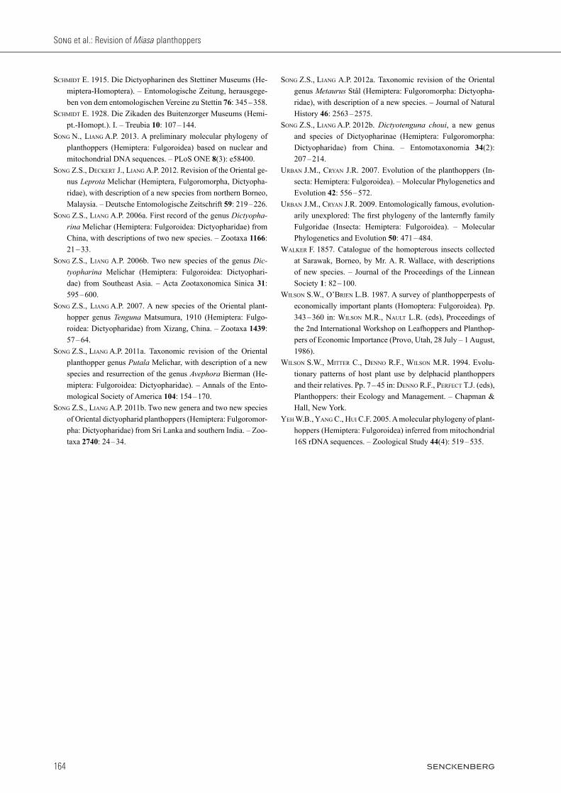
Song et al.: Revision of Miasa planthoppers
164
SchMidt E. 1915. Die Dictyopharinen des Stettiner Museums (He-miptera-Homoptera). – Entomologische Zeitung, herausgege-ben von dem entomologischen Vereine zu Stettin 76: 345 – 358.
SchMidt E. 1928. Die Zikaden des Buitenzorger Museums (Hemi-pt.-Homopt.). I. – Treubia 10: 107 – 144.
Song n., liang a.p. 2013. A preliminary molecular phylogeny of planthoppers (Hemiptera: Fulgoroidea) based on nuclear and mitochondrial DNA sequences. – PLoS ONE 8(3): e58400.
Song z.S., deckert j., liang a.p. 2012. Revision of the Oriental ge-nus Leprota Melichar (Hemiptera, Fulgoromorpha, Dictyo pha-ridae), with description of a new species from northern Bor neo, Malaysia. – Deutsche Entomologische Zeitschrift 59: 219 – 226.
Song z.S., liang a.p. 2006a. First record of the genus Dictyopharina Melichar (Hemiptera: Fulgoroidea: Dictyopharidae) from China, with descriptions of two new species. – Zootaxa 1166: 21 – 33.
Song z.S., liang a.p. 2006b. Two new species of the genus Dictyopharina Melichar (Hemiptera: Fulgoroidea: Dictyophari-dae) from Southeast Asia. – Acta Zootaxonomica Sinica 31: 595 – 600.
Song z.S., liang a.p. 2007. A new species of the Oriental plan t-hopper genus Tenguna Matsumura, 1910 (Hemiptera: Fulgo-roidea: Dictyopharidae) from Xizang, China. – Zootaxa 1439: 57 – 64.
Song z.S., liang a.p. 2011a. Taxonomic revision of the Oriental planthopper genus Putala Melichar, with description of a new species and resurrection of the genus Avephora Bierman (He-miptera: Fulgoroidea: Dictyopharidae). – Annals of the Ento-mological Society of America 104: 154 – 170.
Song z.S., liang a.p. 2011b. Two new genera and two new species of Oriental dictyopharid planthoppers (Hemiptera: Fulgoromor-pha: Dictyopharidae) from Sri Lanka and southern In dia. – Zoo-taxa 2740: 24 – 34.
Song z.S., liang a.p. 2012a. Taxonomic revision of the Oriental genus Metaurus Stål (Hemiptera: Fulgoromorpha: Dictyopha-ridae), with description of a new species. – Journal of Natural History 46: 2563 – 2575.
Song z.S., liang a.p. 2012b. Dictyotenguna choui, a new genus and species of Dictyopharinae (Hemiptera: Fulgoromorpha: Dictyopharidae) from China. – Entomotaxonomia 34(2): 207 – 214.
urBan j.M., crYan j.r. 2007. Evolution of the planthoppers (In-secta: Hemiptera: Fulgoroidea). – Molecular Phylogenetics and Evolution 42: 556 – 572.
urBan j.M., crYan j.r. 2009. Entomologically famous, evolution-arily unexplored: The first phylogeny of the lanternfly family Fulgoridae (Insecta: Hemiptera: Fulgoroidea). – Molecular Phylogenetics and Evolution 50: 471 – 484.
Walker F. 1857. Catalogue of the homopterous insects collected at Sarawak, Borneo, by Mr. A. R. Wallace, with descriptions of new species. – Journal of the Proceedings of the Linnean So ciety 1: 82 – 100.
WilSon S.W., o’Brien l.B. 1987. A survey of planthopperpests of economically important plants (Homoptera: Fulgoroidea). Pp. 343 – 360 in: WilSon M.R., nault L.R. (eds), Proceedings of the 2nd International Workshop on Leafhoppers and Planthop-pers of Economic Importance (Provo, Utah, 28 July – 1 August, 1986).
WilSon S.W., Mitter c., denno r.f., WilSon M.r. 1994. Evolu-tionary patterns of host plant use by delphacid planthoppers and their relatives. Pp. 7 – 45 in: denno R.F., perfect T.J. (eds), Planthoppers: their Ecology and Management. – Chapman & Hall, New York.
Yeh W.B., Yang c., hui c.f. 2005. A molecular phylogeny of plant-hoppers (Hemiptera: Fulgoroidea) inferred from mitochondrial 16S rDNA sequences. – Zoological Study 44(4): 519 – 535.