systems/circuits ... · tal area (vta) neurons in the ventral portion of the midbrain (taylor et...
TRANSCRIPT

Systems/Circuits
Sleep and Wakefulness Are Controlled by Ventral MedialMidbrain/Pons GABAergic Neurons in Mice
Yohko Takata, X Yo Oishi, X Xu-Zhao Zhou, Emi Hasegawa, Koji Takahashi, X Yoan Cherasse, X Takeshi Sakurai,and X Michael LazarusInternational Institute for Integrative Sleep Medicine (WPI-IIIS), University of Tsukuba, Tsukuba, Ibaraki 305-8575, Japan
Sleep–wake behavior is controlled by a wide range of neuronal populations in the mammalian brain. Although the ventral midbrain/pons(VMP) area is suggested to participate in sleep–wake regulation, the neuronal mechanisms have remained unclear. Here, we found thatnonspecific cell ablation or selective ablation of GABAergic neurons by expressing diphtheria toxin fragment A in the VMP in male miceinduced a large increase in wakefulness that lasted at least 4 weeks. In contrast, selective ablation of dopaminergic neurons in the VMPhad little effect on wakefulness. Chemogenetic inhibition of VMP GABAergic neurons also markedly increased wakefulness. The wake-promoting effect of the VMP GABAergic neuron ablation or inhibition was attenuated to varying degrees by the administration ofdopamine D1 or D2/3 receptor antagonists and abolished by the administration of both antagonists together. In contrast, chemogeneticactivation of VMP GABAergic neurons very strongly increased slow-wave sleep and reduced wakefulness. These findings suggest thatVMP GABAergic neurons regulate dopaminergic actions in the sleep–wake behavior of mice.
Key words: AAV; arousal; DREADD; EEG; neural circuits; REM sleep
IntroductionOne potential function of sleep is to conserve energy by enforcinginactivity when activity is not beneficial (Siegel, 2009). Con-versely, when varying ecological demands favor wakefulness, an-imals may have the ability to dispense with sleep. The circuitmechanisms underlying the control of wakefulness to adapt an
animal’s amount of sleep to its behavior, however, are largelyunknown.
We recently identified indirect pathway neurons in the nu-cleus accumbens as sleep-controlling neurons (Oishi et al.,2017b). The nucleus accumbens is innervated by ventral tegmen-tal area (VTA) neurons in the ventral portion of the midbrain(Taylor et al., 2014; Oishi and Lazarus, 2017). Although we andothers have reported that activation of the VTA dopaminergicneurons strongly induces wakefulness (Eban-Rothschild et al.,2016; Taylor et al., 2016; Oishi et al., 2017a), the role of thismidbrain area in sleep–wake regulation remains unclear becausethe findings are conflicting. In rats, neurotoxic lesions of the cellsin this area with hypocretin-2-saporin have no effect on sleep–wake behavior (Gerashchenko et al., 2006). In cats, electrolyticlesions of the ventral midbrain area, including the VTA, has nosignificant effect on sleep–wake behavior (Jones et al., 1973),whereas N-methyl-D-aspartate lesions of cells in an area contain-ing the VTA produce insomnia (Lai et al., 1999).
In the present study, we used an ablation technique based onthe viral expression of diphtheria toxin fragment A (DTA) andfound that nonspecific or GABAergic neuron-specific ablations
Received March 4, 2018; revised Sept. 20, 2018; accepted Sept. 24, 2018.Author contributions: Y.O. and M.L. designed research; Y.T., X.-Z.Z., E.H., and K.T. performed research; Y.C.
contributed unpublished reagents/analytic tools; Y.T., Y.O., X.-Z.Z., E.H., and T.S. analyzed data; Y.T., Y.O., E.H., andM.L. wrote the paper.
This work was supported by the Japan Society for the Promotion of Science [Grant-in-Aid for Young Scientists (B)JP15K18359 and Grant-in-Aid for Scientific Research (B) JP18H02534 to Y.O. and Grant-in-Aid for Scientific Research(B) JP17H02215 to M.L.]; the Japan Science and Technology Agency (CREST Grant JPMJCR1655 to M.L.); the Ministryof Education, Culture, Sports, Science, and Technology (MEXT) of Japan (Grants-in-Aid for Scientific Research onInnovative Areas “Living in Space” JP15H05935, JP15K21745, JP18H04966, and “Willdynamics” JP17H06047 toM.L.); the World Premier International Research Center Initiative from MEXT (Y.T., Y.O., Y.C., and M.L.); the JapanFoundation for Applied Enzymology (Y.O.); and the Naito Foundation (M.L.).
The authors declare no competing financial interests.Correspondence should be addressed to either Michael Lazarus or Yo Oishi, International Institute for Integrative
Sleep Medicine (WPI-IIIS), University of Tsukuba, 1-1-1 Tennodai, Tsukuba, Ibaraki 305-8575, Japan, E-mail:[email protected] or [email protected].
https://doi.org/10.1523/JNEUROSCI.0598-18.2018Copyright © 2018 the authors 0270-6474/18/3810080-13$15.00/0
Significance Statement
Current understanding of the neuronal mechanisms and populations that regulate sleep–wake behavior is incomplete. Here, weidentified a GABAergic ventral midbrain/pons area that is necessary for controlling the daily amount of sleep and wakefulness inmice. We also found that these inhibitory neurons control wakefulness by suppressing dopaminergic systems. Surprisingly,activation of these neurons strongly induced slow-wave sleep while suppressing wakefulness. Our study reveals a new brainmechanism critical for sleep–wake regulation.
10080 • The Journal of Neuroscience, November 21, 2018 • 38(47):10080 –10092

in the ventral medial midbrain/pons area (VMP) at the junctionof the ventral medial midbrain and pons largely increased wake-fulness in mice. The increased wakefulness was also observedafter chemogenetic inhibition of the VMP GABAergic neurons.Using pharmacologic tools, we further demonstrated that thisarousal effect was mediated by dopaminergic systems. Thesefindings indicate that the VMP GABAergic cells are critical for thecontrol of sleep–wake behavior.
Materials and MethodsMice and chemicals. We used the following mouse lines: C57BL/6J wild-type (WT, Charles River Laboratories), dopamine transporter (DAT)-Cre (Backman et al., 2006) (The Jackson Laboratory 006660), andvesicular GABA transporter (VGAT)-Cre (Vong et al., 2011) (kindlyprovided by Dr. Bradford Lowell, Harvard Medical School). Male mice(8 –20 weeks, 25–35 g) were used for the behavioral studies and housed ininsulated sound-proof recording chambers maintained at an ambienttemperature of 23 � 0.5°C with 55 � 3% humidity on a 12 h light/darkcycle (lights on at 8:00) and provided food and water ad libitum. Allexperiments were performed in accordance with the Animal Care Com-mittee of the University of Tsukuba and every effort was made to mini-mize the number of animals used as well as any pain or discomfort.
Clozapine-N-oxide (CNO, Millipore-Sigma), the selective D1 recep-tor antagonist SCH23390 (Millipore-Sigma), the selective D2/D3 recep-tor antagonist raclopride (Millipore-Sigma), and histamine H1 receptorantagonist ketotifen (Tokyo Chemical) were dissolved in saline.
Plasmids and adeno-associated viral vectors (AAVs). To generate thepAAV-CMV-FLEX-DTA plasmid, a FLEX-DTA cassette (see Fig. 1A)was synthesized by Eurofins Genomics K.K. and inserted between theClaI and BamHI restriction sites in a pAAV-MCS vector (Stratagene). Togenerate the pAAV-CMV-FLEX-hrGFP plasmid, the DTA fragment be-tween the AscI and NheI restriction sites in pAAV-CMV-FLEX-DTA wasreplaced with the hrGFP coding sequence. The plasmids pAAV-hSyn-FLEX-hM4Di-mCherry and pAAV-hSyn-FLEX-hM3Dq-mCherry(Krashes et al., 2011) were kindly provided by Dr. Bryan Roth (Universityof North Carolina School of Medicine, Chapel Hill, NC). The plasmidsgenerated in this study will be deposited in Addgene.
pAAV-CMV-FLEX-DTA, pAAV-CMV-FLEX-hrGFP, pAAV-CMV-Cre (Lazarus et al., 2011), pAAV-hSyn-FLEX-hM4Di-mCherry, orpAAV-hSyn-FLEX-hM3Dq-mCherry were packaged in the AAV of se-rotype rh10 using 293A cells as described previously (Kaur et al., 2017;Oishi et al., 2017a,b).
Surgery. For brain microinjections, mice anesthetized with pentobar-bital (60 mg kg �1, i.p.) were injected bilaterally into the VMP (3.4 mmposterior and 0.2 mm lateral to bregma, 4.4 mm below the dura) with 60nl per injection of AAV using a glass micropipette and an air pressureinjector system. To generate mice with nonselective cell-type ablation,WT mice were injected with a mixture (1:1) of AAV-FLEX-DTA (8.6 �10 10 particles ml �1) and AAV-Cre (1.5 � 10 11 particles ml �1) or AAV-FLEX-DTA alone. To generate mice with dopaminergic or GABAergicneuron ablation, DAT-Cre mice or VGAT-Cre mice, respectively, wereinjected with AAV-FLEX-DTA or AAV-FLEX-hrGFP (1.5 � 10 11 parti-cles ml �1). For the chemogenetic experiments, the VGAT-Cre mice wereinjected with AAV-FLEX-hM4Di-mCherry (1.1 � 10 11 particles ml �1)or AAV-FLEX-hM3Dq-mCherry (1.1 � 10 11 particles ml �1).
Mice were also chronically implanted with electroencephalography(EEG) and electromyography (EMG) electrodes for polysomnography,as described previously (Oishi et al., 2016). Briefly, the implant com-prised two stainless steel screws serving as EEG electrodes and two insu-lated, Teflon-coated silver wires were placed bilaterally into the trapeziusmuscles to serve as EMG electrodes. The electrodes were fixed to the skullwith dental acrylic.
EEG/EMG recordings. After allowing 1–2 weeks for postoperative re-covery and transgene expression, the mice were connected with EEG/EMG recording cables. The EEG/EMG signals were amplified andfiltered (EEG: 0.5– 64 Hz, EMG: 16 – 64 Hz), digitized at a sampling rateof 128 Hz, and recorded using SLEEPSIGN software (Kissei Comtec).Vigilance states were scored offline by characterizing 10 s epochs into
three stages: (1) awake, (2) slow-wave sleep (SWS), and (3) rapid eyemovement sleep (REMS) according to standard criteria (Oishi et al.,2016).
Pharmacologic and chemogenetic experiments. After baseline recordingsfor 24 h, VGAT-Cre DTA/VMP mice were administered saline, 0.03 mgkg �1 SCH23390, 2 mg kg �1 raclopride, or 10 mg kg �1 ketotifen at 10:00.Each mouse received all combinations of dopamine receptor antagonistsin a random order with at least 3 d intervals between drug administra-tions. A separate mouse group received ketotifen. Doses of the drugs wereselected based on previous studies (Qu et al., 2008; Unno et al., 2012;Oishi et al., 2017a). For chemogenetic inhibition, VGAT-Cre M4/VMP
mice were injected intraperitoneally with saline or 3 mg kg �1 CNO at10:00 on 2 consecutive days. For chemogenetic activation, each VGAT-Cre M3/VMP mouse received intraperitoneally saline or different doses(0.3, 0.9, and 2.7 mg kg �1) of CNO at 20:00 with at least 3 d intervalsbetween drug administrations.
Histology. Under deep anesthesia with an overdose of chloral hydrate(500 mg kg �1, i.p.), animals were perfused intracardially with salinefollowed by neutral buffered 10% formalin. Brains were then placed in20% sucrose overnight at 4°C to reduce freezing artifact. The brains weresectioned at 40 �m on a freezing microtome.
For immunohistochemistry, sections were incubated in 0.3% hydro-gen peroxide and then overnight with rabbit anti-DsRed (1:10,000, Clon-tech, catalog #632496, RRID:AB_10013483), mouse anti-NeuN (1:2000,Millipore, catalog #MAB377, RRID:AB_2298772), or rabbit anti-tyrosine hydroxylase (TH; 1:20,000, Millipore, catalog #AB152, RRID:AB_390204). Sections were incubated for 2 h in biotinylated antibody(1:1000, Jackson ImmunoResearch Laboratories) and treated withavidin– biotin complex (1:1000; Vectastain ABC Elite kit, Vector Labo-ratories) for 1 h. Immunoreactive cells were visualized by reaction with3,3�-diaminobenzidine and 0.01% hydrogen peroxide.
For in situ hybridization, we generated a 918-bp digoxigenin-labeledriboprobe for VGAT mRNA by PCR amplification (primers: forward,gcatgttcgtgctgggcctacc; reverse, cagcgcagcgtcagcccccag conjugated withthe T7 promoter) using mouse tail genomic DNA as a template, followedby in vitro transcription. Sections were then incubated with a 1 �g ml �1
concentration of the VGAT probe in 5� sodium citrate buffer containing50% formamide at 50°C overnight, washed in 1� saline sodium citratebuffer at 50°C, incubated with alkaline phosphatase conjugated anti-digoxigenin antibody (1:500, Roche) overnight, and visualized by reac-tion with 5-bromo-4-chloro-3-indolyl phosphate/nitro blue tetrazolium(BCIP/NBT, Millipore-Sigma).
For double-labeling of c-Fos and mCherry, we immunostained c-Foswith rabbit anti-c-Fos antibody (1:2000, Millipore, catalog #ABE457,RRID:AB_2631318), peroxidase-conjugated anti-rabbit antibody (1:250, Jackson ImmunoResearch Laboratories), and tyramide-conjugatedfluorescein (PerkinElmer). mCherry-positive cells were identified by thenative red fluorescence of mCherry. Images were analyzed with a confo-cal laser scanning microscope LSM800 (Carl Zeiss).
For quantitative histologic analysis, we counted cells positive for TH,VGAT mRNA, c-Fos, or mCherry using a counting box placed bilaterallyin the VMP. We placed a 500 � 650 �m box with the ventral medial edgeat the center of the interpeduncular nucleus and the medial border alongthe midline 3.8 mm posterior to bregma. The anterior–posterior coordi-nates were obtained from the Paxinos and Franklin mouse brain atlas(Paxinos and Franklin, 2001). The cell counts were adjusted using theAbercrombie correction factor (Guillery, 2002).
Electrophysiology. Five-week-old VGAT-Cre mice were injected inthe VMP with AAV-FLEX-hM4Di-mCherry or AAV-FLEX-hM3Dq-mCherry. After 1–2 weeks, the mice were decapitated under deep anes-thesia with isoflurane (Pfizer). Brains were extracted and cooled inice-cold cutting solution containing the following (in mM): 125 cholinechloride, 25 NaHCO3, 10 D(�)-glucose, 7 MgCl2, 2.5 KCl, 1.25NaH2PO4, and 0.5 CaCl2 bubbled with O2 (95%) and CO2 (5%). Hori-zontal brain slices (250 �m thick) including the VMP were prepared witha vibratome (VT1200S, Leica) and maintained for 1 h at room tempera-ture in artificial CSF (ACSF) containing the following (in mM): 125 NaCl,26 NaHCO3, 10 D(�)-glucose, 2.5 KCl, 2 CaCl2, and 1 MgSO4 bubbledwith O2 (95%) and CO2 (5%). The electrodes (5– 8 M�) were filled with
Takata et al. • Sleep–Wake Control by Mesopontine GABA J. Neurosci., November 21, 2018 • 38(47):10080 –10092 • 10081

Figure 1. Ablation of the VMP increases wakefulness. A, WT mice were injected with AAV-FLEX-DTA and AAV-Cre into the VMP. B, C, Brain sections were stained against NeuN to confirm that theVMP neurons were ablated in the mice injected with AAV-FLEX-DTA and AAV-Cre (WT DTA/VMP mice; B). No deficiency in VMP neurons was detected in mice injected with only AAV-FLEX-DTA(WT Ctrl/VMP mice; C). Dashed line delineates the area with neuronal loss in a WT DTA/VMP mouse. Scale bar, 500 �m. D, Superimposed neuronal loss areas in the (Figure legend continues.)
10082 • J. Neurosci., November 21, 2018 • 38(47):10080 –10092 Takata et al. • Sleep–Wake Control by Mesopontine GABA

an internal solution containing the following (in mM): 125 K-gluconate,10 HEPES, 10 phosphocreatine, 4 NaCl, 4 ATP, 2 MgCl2, 0.4 GTP, and0.2 EGTA, pH 7.3, adjusted with KOH). Firing of hM4Di-mCherry orhM3Dq-mCherry-expressing neurons was recorded in the current-clamp mode at a temperature of 30°C, which was maintained by perfu-sion of preheated ACSF before and after 5 �M CNO application. Thecombination of a MultiClamp 700B amplifier, a Digidata 1440A A/Dconverter, and Clampex 10.3 software (Molecular Devices) was used tocontrol membrane voltage and data acquisition.
Experimental design and statistical analysis. For behavioral experi-ments, the numbers of mice used were chosen based on expected varia-tions between animals and variability of AAV microinjections.
Data are presented as the mean and SEM. Statistical analyses wereperformed using GraphPad Prism software. Statistical comparisons be-tween two groups were performed using the unpaired (see Figs. 1H–M;2 F, J–O; 3 F, J, K, M–O, and 8C,D) or paired (see Figs. 4C,D; 5 D, F–H, and7C) two-tailed Student’s t test. For t tests, the normality of each datasetwas established using the Kolmogorov–Smirnov test. Comparisonsamong multiple parameters were performed by a mixed-model ANOVAfollowed by Sidak’s post hoc comparisons (see Figs. 1E–G,N–P; 2G–I, P–R, and 3G– I, P–S), one-way repeated-measures ANOVA followed byTukey’s post hoc comparisons (see Figs. 4 A, B and 7G–O), or two-wayrepeated-measures ANOVA followed by Sidak’s post hoc comparisons(Figs. 5 E, I; 6A–E, and 7D– F, P,Q). In all cases, p � 0.05 was consideredsignificant.
ResultsVMP cell deficiency increases wakefulness in miceTo examine the role of ventral midbrain neurons in sleep–wakeregulation, we first nonselectively ablated cells in this brain areain WT mice by stereotaxic co-microinjection of AAV carryingCre-dependent DTA (AAV-FLEX-DTA) and Cre recombinase(AAV-Cre; WT DTA/VMP mice; Fig. 1A,B). Because the ablationwas observed not only in the ventral medial portion of the mid-brain, but also in a part of the pons as confirmed by immuno-staining for the neuronal marker NeuN (Fig. 1B–D), the ablatedarea is hereafter referred to as the VMP. In all cases, the ablatedarea largely included the VTA and almost none of the substantianigra. Control mice were only injected with AAV-FLEX-DTAinto the VMP and did not express DTA in the VMP due to theabsence of Cre recombinase (WT Ctrl/VMP mice; Fig. 1C). At 1week after the AAV injections, EEG and EMG recordings of themice were analyzed to assess the baseline sleep–wake behavior ofthe animals. Compared with WT Ctrl/VMP mice, WT DTA/VMP miceexhibited largely increased wakefulness, especially in the first halfof the light period, when mice typically show high levels of sleep,but also in the second half of the dark period (F(1,10) 66.7, p �0.0001, ANOVA; Fig. 1E). Concomitantly, WT DTA/VMP mice hada lower amount of SWS (F(1,10) 60.9, p � 0.0001, ANOVA; Fig.1F) and REMS (F(1,10) 73.4, p � 0.0001, ANOVA; Fig. 1G). Wethen examined the pattern of waking in WTDTA/VMP and WTCtrl/VMP
mice. The mean duration of wake episodes in the WT DTA/VMP
mice was 3.1 (t(10) 3.05, p 0.012, unpaired t test; light period)and 7.7 (t(10) 4.03, p 0.0024, unpaired t test; dark period)times longer than that of wake episodes in the WT Ctrl/VMP mice
(Fig. 1H), whereas the number of wake episodes was lower by37% (t(10) 2.85, p 0.0172, unpaired t test) and 85% (t(10) 5.44, p 0.0003, unpaired t test) in the light and dark periods,respectively (Fig. 1K), suggesting that ablation of VMP cells effi-ciently consolidates wakefulness. The mean duration of SWS andREMS was unchanged (light period SWS: t(10) 0.57, p 0.58;dark period SWS: t(10) 1.46, p 0.17; light period REMS:t(10) 0.38, p 0.71; dark period REMS: t(9) 0.14, p 0.89,unpaired t test; Fig. 1 I, J), whereas the episode number was de-creased in WT DTA/VMP mice (light period SWS: t(10) 3.42, p 0.0066; dark period SWS: t(10) 5.71, p 0.0002; light periodREMS: t(10) 4.42, p 0.0013; dark period REMS: t(10) 6.89,p � 0.0001, unpaired t test; Fig. 1L,M). Animals have the abilityto adapt their sleep–wake behavior after the loss of brain cellsover the course of several weeks (Lai et al., 1999; Gerashchenko etal., 2006), so we measured EEG activity for up to 4 weeks aftersurgery. Although the wake amount of WT DTA/VMP mice in thedark period reached the same level as that of the WT Ctrl/VMP micebetween 2 and 4 weeks after surgery, the WT DTA/VMP mice main-tained significantly higher amounts of wakefulness in the lightperiod, even at 4 weeks after surgery (light period: F(1,10) 21.4,p 0.0009; dark period: F(1,10) 15.5, p 0.0028, ANOVA; Fig.1N). SWS and REMS amount were concomitantly lower inWT DTA/VMP mice especially during the light period (light periodSWS: F(1,10) 18.1, p 0.0017; dark period SWS: F(1,10) 16.2,p 0.0024; light period REMS: F(1,10) 26.0, p 0.0005; darkperiod REMS: F(1,10) 6.50, p 0.0289, ANOVA; Fig. 1O,P).These findings suggest that VMP cells contribute to the sleep–wake behavior of mice and that the absence of these cells cannotbe compensated for by other brain areas, at least in the lightperiod.
VMP dopaminergic neuron deficiency has little to no effecton wakefulnessThe VMP includes the VTA, which contains dopaminergic(60%) and GABAergic (35%) neurons (Oishi and Lazarus,2017). Because VTA dopaminergic neurons are implicated in theregulation of wakefulness (Eban-Rothschild et al., 2016; Oishi etal., 2017a), we next selectively ablated these neurons in the VMPto determine whether they contribute to baseline wakefulness inmice. To generate mice with VMP dopaminergic neuron defi-ciency (DAT-Cre DTA/VMP mice; Fig. 2A–C), AAV-FLEX-DTAwas microinjected into the VMP of DAT-Cre mice expressing theCre protein exclusively in dopaminergic neurons (Backman et al.,2006). Mice expressing humanized Renilla reniformis-derivedgreen fluorescent protein (hrGFP) in VMP dopaminergic neu-rons were generated as a control (DAT-Cre hrGFP/VMP mice; Fig.2D,E). The number of cells positive for TH, a marker of dopa-minergic neurons, was decreased by 94% in the VMP of DAT-Cre DTA/VMP mice compared with DAT-Cre hrGFP/VMP mice(t(10) 9.30, p � 0.0001, unpaired t test; Fig. 2F). Compared withthe DAT-Cre hrGFP/VMP mice, DAT-Cre DTA/VMP mice exhibitedno significant differences in the time course of wakefulness(F(1,10) 0.0135, p 0.91, ANOVA; Fig. 2G), SWS (F(1,10) 0.0013, p 0.97, ANOVA; Fig. 2H), or REMS (F(1,10) 0.137,p 0.72, ANOVA; Fig. 2I) or in the mean duration (light periodwakefulness: t(10) 1.02, p 0.33; dark period wakefulness:t(10) 0.957, p 0.36; light period SWS: t(10) 0.851, p 0.41;dark period SWS: t(10) 0.307, p 0.77; light period REMS:t(10) 1.09, p 0.30; dark period REMS: t(10) 1.72, p 0.12,unpaired t test; Fig. 2J–L) and number (light period wakefulness:t(10) � 0.01, p � 0.99; dark period wakefulness: t(10) 0.727, p 0.48; light period SWS: t(10) 0.026, p 0.98; dark period
4
(Figure legend continued.) VMP (n 6). E–G, Time course of hourly wakefulness (E), SWS(F), and REMS (G) at 1 week after surgery. H–M, Ablation of the VMP resulted in a longer meanduration of wake episodes (H–J), whereas the mean number of each episode was smaller(K–M). N–P, Ablation of the VMP resulted in higher amounts of wakefulness (N) and loweramounts of SWS (O) and REMS (P) for at least 4 weeks after surgery. White and gray backgroundin each panel indicate light and dark periods, respectively. *p � 0.05, **p � 0.01 comparedwith WT Ctrl/VMP mice. Data are presented as the mean � SEM (n 6). RN, Red nucleus; SN,substantia nigra; IP, interpeduncular nucleus.
Takata et al. • Sleep–Wake Control by Mesopontine GABA J. Neurosci., November 21, 2018 • 38(47):10080 –10092 • 10083

Figure 2. Ablation of the VMP dopaminergic neurons had little to no effect on wakefulness. A, DAT-Cre mice were injected with AAV-FLEX-DTA into the VMP. B–E, Brain sections stained against TH (B, D) orVGAT mRNA (C, E) from mice injected with AAV-FLEX-DTA (DAT-Cre DTA/VMP mice; B, C) or AAV-FLEX-hrGFP (DAT-Cre hrGFP/VMP mice; D, E) to confirm that the VMP dopaminergic neurons were selectively ablatedin DAT-Cre mice. Dashed line delineates the area with loss of dopaminergic neurons in a DAT-Cre DTA/VMP mouse. Scale bar, 500 �m. F, Number of neurons expressing TH in the VMP of mice injected with AAV.G–I, Time course of hourly wakefulness (G), SWS (H), and REMS (I) at 1 week after surgery. J–O, Ablation of the VMP dopaminergic neurons changed neither the mean duration (J–L) nor the number (M–O) ofeach episode. P–R, Total amount of wakefulness (P), SWS (Q), and REMS (R) for 12 h. White and gray background in each panel indicate light and dark periods, respectively. *p � 0.05, **p � 0.01 comparedwith DAT-Cre hrGFP/VMP mice. Data are presented as the mean � SEM (n 6). RN, Red nucleus; SN, substantia nigra; IP, interpeduncular nucleus.
10084 • J. Neurosci., November 21, 2018 • 38(47):10080 –10092 Takata et al. • Sleep–Wake Control by Mesopontine GABA
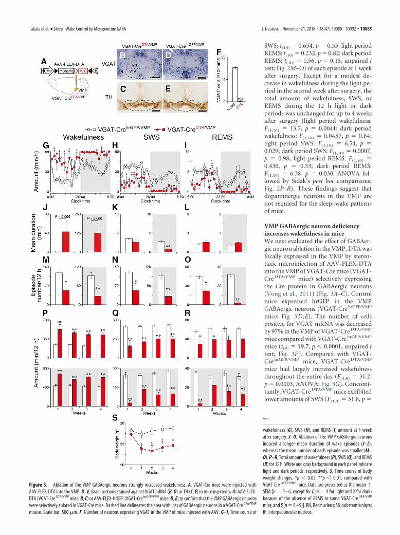
SWS: t(10) 0.654, p 0.53; light periodREMS: t(10) 0.232, p 0.82; dark periodREMS: t(10) 1.56, p 0.15, unpaired ttest; Fig. 2M–O) of each episode at 1 weekafter surgery. Except for a modest de-crease in wakefulness during the light pe-riod in the second week after surgery, thetotal amount of wakefulness, SWS, orREMS during the 12 h light or darkperiods was unchanged for up to 4 weeksafter surgery (light period wakefulness:F(1,10) 13.7, p 0.0041; dark periodwakefulness: F(1,10) 0.0457, p 0.84;light period SWS: F(1,10) 6.54, p 0.029; dark period SWS: F(1,10) 0.0007,p 0.98; light period REMS: F(1,10) 0.430, p 0.53; dark period REMS:F(1,10) 6.38, p 0.030, ANOVA fol-lowed by Sidak’s post hoc comparisons;Fig. 2P–R). These findings suggest thatdopaminergic neurons in the VMP arenot required for the sleep–wake patternsof mice.
VMP GABAergic neuron deficiencyincreases wakefulness in miceWe next evaluated the effect of GABAer-gic neuron ablation in the VMP. DTA wasfocally expressed in the VMP by stereo-taxic microinjection of AAV-FLEX-DTAinto the VMP of VGAT-Cre mice (VGAT-Cre DTA/VMP mice) selectively expressingthe Cre protein in GABAergic neurons(Vong et al., 2011) (Fig. 3A–C). Controlmice expressed hrGFP in the VMPGABAergic neurons (VGAT-CrehrGFP/VMP
mice; Fig. 3D,E). The number of cellspositive for VGAT mRNA was decreasedby 97% in the VMP of VGAT-Cre DTA/VMP
mice compared with VGAT-Cre hrGFP/VMP
mice (t(9) 19.7, p � 0.0001, unpaired ttest; Fig. 3F). Compared with VGAT-Cre hrGFP/VMP mice, VGAT-Cre DTA/VMP
mice had largely increased wakefulnessthroughout the entire day (F(1,9) 31.2,p 0.0003, ANOVA; Fig. 3G). Concomi-tantly, VGAT-Cre DTA/VMP mice exhibitedlower amounts of SWS (F(1,9) 31.8, p
Figure 3. Ablation of the VMP GABAergic neurons strongly increased wakefulness. A, VGAT-Cre mice were injected withAAV-FLEX-DTA into the VMP. B–E, Brain sections stained against VGAT mRNA (B, D) or TH (C, E) in mice injected with AAV-FLEX-DTA (VGAT-Cre DTA/VMP mice; B, C) or AAV-FLEX-hrGFP (VGAT-Cre hrGFP/VMP mice; D, E) to confirm that the VMP GABAergic neuronswere selectively ablated in VGAT-Cre mice. Dashed line delineates the area with loss of GABAergic neurons in a VGAT-Cre DTA/VMP
mouse. Scale bar, 500 �m. F, Number of neurons expressing VGAT in the VMP of mice injected with AAV. G–I, Time course of
4
wakefulness (G), SWS (H), and REMS (I) amount at 1 weekafter surgery. J–O, Ablation of the VMP GABAergic neuronsinduced a longer mean duration of wake episodes (J–L),whereas the mean number of each episode was smaller (M–O). P–R, Total amount of wakefulness (P), SWS (Q), and REMS(R) for 12 h. White and gray background in each panel indicatelight and dark periods, respectively. S, Time course of bodyweight changes. *p � 0.05, **p � 0.01, compared withVGAT-Cre hrGFP/VMP mice. Data are presented as the mean �SEM [n 5– 6, except for L (n 4 for light and 2 for dark)because of the absence of REMS in some VGAT-Cre DTA/VMP
mice, and S (n 8 –9)]. RN, Red nucleus; SN, substantia nigra;IP, interpeduncular nucleus.
Takata et al. • Sleep–Wake Control by Mesopontine GABA J. Neurosci., November 21, 2018 • 38(47):10080 –10092 • 10085

0.0003, ANOVA; Fig. 3H) and REMS(F(1,9) 18.0, p 0.0022, ANOVA; Fig.3I). The mean duration of wake episodesin VGAT-Cre DTA/VMP mice in the lightand dark periods was 20.2 and 24.6 timeslonger, respectively, than that in VGAT-Cre hrGFP/VMP mice, although the differ-ence was not statistically significant (lightperiod: t(9) 2.10, p 0.065, unpaired ttest; dark period: t(9) 2.10, p 0.066,unpaired t test; Fig. 3J), whereas the meannumber of wake episodes in the lightand dark period was 56% (t(9) 2.84,p 0.019, unpaired t test) and 70%(t(9) 3.59, p 0.0059, unpaired t test)lower, respectively (Fig. 3M ). These re-sults suggest that ablating VMP GABAe-rgic neurons efficiently consolidateswakefulness, similar to the effects ofnonselective VMP ablation in animals(Fig. 1 H, K ). For the mean duration,only SWS during the dark period wassignificantly reduced (light period SWS:t(9) 1.41, p 0.19; dark period SWS:t(9) 7.25, p � 0.0001; unpaired t test;Fig. 3K ), whereas the episode numbersof both SWS and REMS were decreasedin VGAT-Cre DTA/VMP mice during bothlight conditions (light period SWS:t(9) 2.80, p 0.021; dark period SWS:t(9) 3.56, p 0.0061; light periodREMS: t(9) 2.27, p 0.049; dark pe-riod REMS: t(9) 10.9, p � 0.0001, un-paired t test; Fig. 3 N, O). The increasedwakefulness in the VGAT-Cre DTA/VMP
mice was maintained in both the lightand dark periods for at least 4 weeks(light period: F(1,9) 47.1, p � 0.0001;dark period: F(1,9) 98.1, p � 0.0001,ANOVA; Fig. 3P). The SWS and REMSamounts were concomitantly lower inVGAT-CreDTA/VMP mice (light periodSWS: F(1,9) 50.4, p � 0.0001; dark periodSWS: F(1,9) 86.5, p � 0.0001; light period REMS: F(1,9) 11.9, p 0.0072; dark period REMS: F(1,9) 55.3, p � 0.0001, ANOVA; Fig.3Q,R). Despite the long-lasting wake time, the VGAT-CreDTA/VMP
mice appeared healthy although the body weight was slightly, butsignificantly, lower than that of VGAT-CrehrGFP/VMP mice (F(1,15) 8.42, p 0.011, ANOVA; Fig. 3S). These findings suggest that theVMP GABAergic neurons are essential for maintaining sleep–wakepatterns in mice.
Dopamine mediates the increased wakefulness in mice withVMP GABAergic neuron deficiencyWe next investigated the molecular mechanisms of increasedwakefulness in the VGAT-Cre DTA/VMP mice using phar-macologic methods. Because the VMP comprises the VTA androstromedial tegmental nucleus GABAergic neurons projectingto midbrain dopaminergic neurons (Jhou et al., 2009b; Om-elchenko and Sesack, 2009), we tested whether the dopaminergicsystem is involved in the increased wakefulness in VGAT-Cre DTA/VMP mice. Previous studies suggested that dopamineproduces wakefulness via D1 and D2 receptors, so we treated
VGAT-Cre DTA/VMP or VGAT-Cre hrGFP/VMP mice at 10:00 witheither the selective dopamine D1 receptor antagonist SCH23390at 0.03 mg kg�1, the selective dopamine D2/D3 receptor antago-nist raclopride at 2 mg kg�1, a mixture of SCH23390 and raclo-pride, or saline as a vehicle control. The mixture of SCH23390and raclopride decreased wakefulness for 6 h in VGAT-Cre DTA/
VMP and VGAT-Cre hrGFP/VMP mice after the drug injection, whileneither SCH23390 nor raclopride alone significantly affectedwakefulness in the VGAT-Cre DTA/VMP and control mice, suggest-ing that dopamine receptors mediate wakefulness under baselineconditions and in cases of VMP GABAergic neuron deficiency(Fig. 4A: F(2.06,14.4) 8.34, p 0.0037; Fig. 4B: F(2.09,10.4) 58.3,p 0.0057, ANOVA). In contrast, the histamine H1 receptorantagonist ketotifen at 10 mg kg�1 decreased wakefulness inVGAT-Cre hrGFP/VMP mice (t(4) 3.50, p 0.025, paired t test;Fig. 4D), but did not affect wakefulness in VGAT-Cre DTA/VMP
mice (t(5) 0.18, p 0.86, paired t test; Fig. 4C), suggesting thathistamine H1 receptors do not play an important role in theincreased wakefulness in mice with an absence of GABAergicneurons in the VMP.
Figure 4. Dopamine receptor antagonists suppressed wakefulness in mice with an absence of GABAergic neurons in the VMP.Total amount of wakefulness for 6 h in VGAT-Cre DTA/VMP (A, C) or VGAT-Cre hrGFP/VMP mice (B, D) treated with dopamine receptorantagonists (SCH23390 and/or raclopride; A, B) or the histamine H1 receptor antagonist ketotifen (C, D). Data are presented as themean � SEM (n 5– 8).
10086 • J. Neurosci., November 21, 2018 • 38(47):10080 –10092 Takata et al. • Sleep–Wake Control by Mesopontine GABA

Chemogenetic inhibition of VMP GABAergic neuronsincreases wakefulnessTo determine whether inhibition of VMP GABAergic neuronsalso affects sleep–wake regulation in mice, we chemogenetically
inhibited these neurons in the VMP of VGAT-Cre mice (VGAT-Cre M4/VMP mice; Fig. 5A,B) using inhibitory designer receptorsexclusively activated by designer drugs (DREADD) hM4Di,which suppress neuronal activity when the ligand CNO is applied
Figure 5. Chemogenetic inhibition of the VMP GABAergic neurons increased wakefulness. A, VGAT-Cre mice were injected with AAV-FLEX-hM4Di-mCherry into the VMP (VGAT-Cre M4/VMP mice).B, Brain sections were stained against mCherry to confirm that the hM4Di-mCherry protein was expressed in the VMP. Scale bar, 500 �m. C, Representative trace of current-clamp recording froma hM4Di-expressing neuron. The solid bar indicates the duration of 5 �M CNO application. The dotted line denotes 0 mV. D, Mean firing rate of hM4Di-expressing neurons before and after CNOapplication (n 8 cells from 3 mice). Data obtained from the same cell are connected with a line. E, Time course of hourly wakefulness. F–H, Chemogenetic inhibition increased the mean wakeepisode duration (F) and total amount of wakefulness (H), whereas the number of wake episodes (G) was reduced for 4 h after 3 mg kg �1 CNO injection. I, CNO application did not affect sleep–wakeamounts in naive VGAT-Cre mice. *p � 0.05, **p � 0.01 compared with saline. Data are presented as the mean � SEM (n 7– 8). RN, Red nucleus; SN, substantia nigra; IP, interpeduncularnucleus.
Takata et al. • Sleep–Wake Control by Mesopontine GABA J. Neurosci., November 21, 2018 • 38(47):10080 –10092 • 10087

(Urban and Roth, 2015). Whole-cellpatch-clamp recordings in acute sliceswere performed to test the response of asingle hM4Di-expressing VMP neuron toCNO application in a VGAT-Cre M4/VMP
mouse. We bath-applied 5 �M CNO to thebrain slices and found that CNO reducedthe firing rate of VMP neurons expressinghM4Di from 7.4 � 2.1 to 0.04 � 0.03spikes/s (t(7) 3.55, p 0.0094, paired ttest; Fig. 5C,D). Intraperitoneal adminis-tration of 3 mg kg�1 CNO to VGAT-Cre M4/VMP mice at 10:00, when micespend most of their time asleep, increasedwakefulness for 4 h after the injection (Fig.5E: F(1,7) 5.99, p 0.044, ANOVA; Fig.5H: t(7) 7.19, p 0.0002, paired t test).CNO increased the mean wake episodeduration by 4.6-fold (t(7) 5.49, p 0.0009, paired t test) and decreased themean episode number by 58% (t(7) 4.26, p 0.0038, paired t test) (Fig. 5F,G),similar to that in VGAT-Cre DTA/VMP mice(Fig. 3 J,M). CNO administration at 10:00did not affect the sleep–wake behavior ofnaive VGAT-Cre mice (wakefulness:F(1,6) 0.14, p 0.72; SWS: F(1,6) 1.16,p 0.32; REMS: F(1,6) 0.65, p 0.45,ANOVA; Fig. 5I). These results suggestthat wakefulness is also increased by tran-sient inhibition of VMP GABAergicneurons.
Blocking dopamine receptors abolishesthe increased wakefulness by inhibitionof VMP GABAergic neuronsNext, we investigated whether dopaminereceptors contribute to the increasedwakefulness after chemogenetic inhibi-tion of VMP GABAergic neurons usingSCH23390andraclopride.VGAT-CreM4/VMP
mice were pretreated with saline, 0.03 mgkg�1 SCH23390, 2 mg kg�1 raclopride, ora mixture of SCH23390 and raclopride 30min before the CNO injection at 10:00. InVGAT-Cre M4/VMP mice pretreated withSCH23390 or raclopride, CNO still in-creased wakefulness for several hours,although the total amount of wakeful-ness for 4 h after CNO injection was sig-nificantly decreased by both antagonists(Fig. 6A: F(1,6) 29.4, p 0.0016; Fig.6B: F(1,6) 38.6, p 0.0008; Fig. 6C:F(1,6) 25.2, p 0.0024; Fig. 6E:F(3,18) 72.9, p � 0.0001, ANOVA).Conversely, CNO injection did not in-duce wakefulness when mice were pre-treated with the mixture of SCH23390and raclopride (Fig. 6D: F(1,6) 22.9,p 0.003, ANOVA; Fig. 6E), indicatingthat dopamine receptors mediate for thewake effect after inhibition of VMPGABAergic neurons.
Figure 6. Dopamine receptor antagonists blocked the wake effect caused by inhibition of the VMP GABAergic system. A–D,Time course of wakefulness in VGAT-Cre M4/VMP mice administered CNO after pretreatment with saline (A), SCH23390 (B), raclo-pride (C), or SCH23390 and raclopride (D). E, Total amount of wakefulness for 4 h after injecting CNO in VGAT-Cre M4/VMP micepretreated with dopamine receptor antagonists. *p � 0.05, **p � 0.01 compared with saline. n.s., Not significant. Data arepresented as the mean � SEM (n 7).
10088 • J. Neurosci., November 21, 2018 • 38(47):10080 –10092 Takata et al. • Sleep–Wake Control by Mesopontine GABA

Chemogenetic activation of VMPGABAergic neurons induces SWSTo further examine the role of VMPGABAergic neurons in sleep–wake regu-lation, we introduced an excitatory DRE-ADD hM3Dq that evokes neuronalexcitation by administering CNO toVGAT-Cre mice (VGAT-Cre M3/VMP
mice; Fig. 7A). Whole-cell patch-clamprecordings in acute slices containing theVMP revealed that the application of 5 �M
CNO increased the firing rate of hM3Dq-expressing VMP neurons in VGAT-Cre M3/VMP mice from 5.6 � 1.3 to 13.4 �3.0 spikes/s (t(6) 2.82, p 0.030, pairedt test; Fig. 7B,C). Intraperitoneal admin-istration of CNO to VGAT-Cre M3/VMP
mice at 20:00 drastically decreased wake-fulness (F(3,9) 259.9, p � 0.0001,ANOVA) and increased SWS (F(3,9) 337.9, p � 0.0001, ANOVA) for 7 h afterthe injection (Fig. 7D,E). Interestingly,CNO also decreased the amount of REMS(F(3,9) 22.4, p 0.0002, ANOVA) forup to 10 h after the injection (Fig. 7F).CNO dose-dependently decreased themean wake episode duration (F(1.02,3.05) 41.9, p 0.0071, ANOVA; Fig. 7G) for12 h after the injection, but did not changethe sleep episode duration (Fig. 7H:F(1.39,4.16) 1.93, p 0.246; Fig. 7I:F(1.60,4.81) 1.80, p 0.255, ANOVA).Conversely, CNO increased the numberof both wake (F(1.67,5.02) 73.2, p 0.0002, ANOVA; Fig. 7J) and SWS(F(1.69,5.07) 76.2, p 0.0002, ANOVA;Fig. 7K) episodes, whereas the number ofREMS episodes was reduced (F(1.74,5.21) 9.98, p 0.018, ANOVA; Fig. 7L). Thetotal amount of each state during the 12 hafter injection indicated that CNO dose-dependently decreased wakefulness(F(1.91,5.72) 259.6, p � 0.0001, ANOVA;Fig. 7M) and REMS (F(2.00,5.99) 22.5,p 0.016, ANOVA; Fig. 7O), and in-creased SWS (F(1.98,5.95) 337.4, p �0.0001, ANOVA; Fig. 7N). CNO did notchange the EEG power spectrum duringSWS (F(3,9) 1.317, p 0.33, ANOVA;Fig. 7P). CNO administration at 20:00 didnot affect sleep–wake behavior in naiveVGAT-Cre mice (wakefulness: F(1,5) 3.28, p 0.13; SWS: F(1,5) 3.33, p 0.13; REMS: F(1,5) 3.34, p 0.13,
Figure 7. Chemogenetic activation of the VMP GABAergic neurons reduced wakefulness and induced SWS. A, VGAT-Cre micewere injected with AAV-FLEX-hM3Dq-mCherry into the VMP (VGAT-Cre M3/VMP mice). B, Representative trace of current-clamprecording from a hM3Dq-expressing neuron. The solid bar indicates the duration of 5 �M CNO application. The dotted line denotes0 mV. C, Mean firing rate of hM3Dq-expressing neurons before and after CNO application (n 7 cells from 3 mice). Data obtainedfrom the same cell are connected with a line. D–F, Time course of hourly wakefulness (D), SWS (E), and REMS (F). G–O, Mean
4
episode duration (G–I), number of episodes (J–L), and totalamount (M–O) of wakefulness (G, J, M), SWS (H, K, N), andREMS (I, L, O) for 12 h after CNO injection. P, EEG power densityduring SWS. Q, CNO (3 mg kg �1) application did not affectsleep–wake amounts in naive VGAT-Cre mice. *p � 0.05,**p � 0.01 compared with saline. Data are presented as themean � SEM (n 4).
Takata et al. • Sleep–Wake Control by Mesopontine GABA J. Neurosci., November 21, 2018 • 38(47):10080 –10092 • 10089
ANOVA; Fig. 7Q). These results suggest that chemogenetic acti-vation of VMP GABAergic neurons decreased wakefulness bydisrupting the maintenance of wake episodes, likely by inducingSWS.
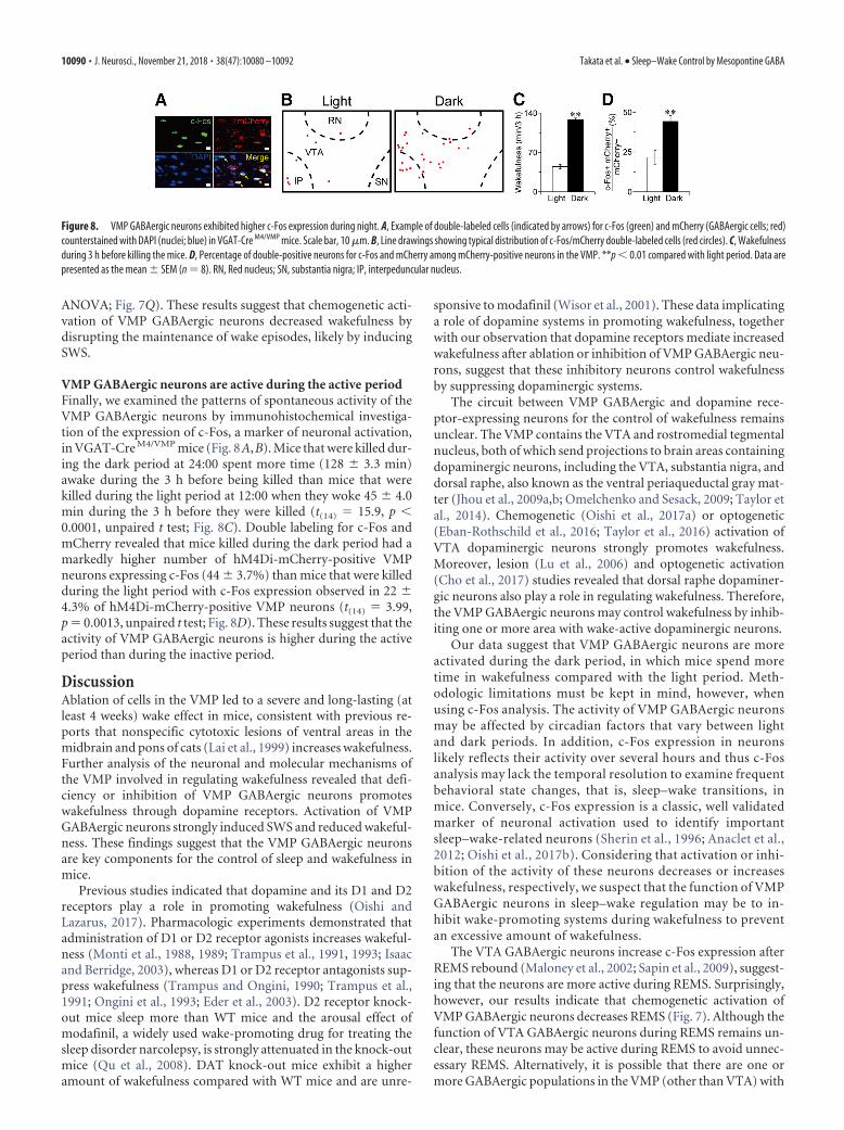
VMP GABAergic neurons are active during the active periodFinally, we examined the patterns of spontaneous activity of theVMP GABAergic neurons by immunohistochemical investiga-tion of the expression of c-Fos, a marker of neuronal activation,in VGAT-Cre M4/VMP mice (Fig. 8A,B). Mice that were killed dur-ing the dark period at 24:00 spent more time (128 � 3.3 min)awake during the 3 h before being killed than mice that werekilled during the light period at 12:00 when they woke 45 � 4.0min during the 3 h before they were killed (t(14) 15.9, p �0.0001, unpaired t test; Fig. 8C). Double labeling for c-Fos andmCherry revealed that mice killed during the dark period had amarkedly higher number of hM4Di-mCherry-positive VMPneurons expressing c-Fos (44 � 3.7%) than mice that were killedduring the light period with c-Fos expression observed in 22 �4.3% of hM4Di-mCherry-positive VMP neurons (t(14) 3.99,p 0.0013, unpaired t test; Fig. 8D). These results suggest that theactivity of VMP GABAergic neurons is higher during the activeperiod than during the inactive period.
DiscussionAblation of cells in the VMP led to a severe and long-lasting (atleast 4 weeks) wake effect in mice, consistent with previous re-ports that nonspecific cytotoxic lesions of ventral areas in themidbrain and pons of cats (Lai et al., 1999) increases wakefulness.Further analysis of the neuronal and molecular mechanisms ofthe VMP involved in regulating wakefulness revealed that defi-ciency or inhibition of VMP GABAergic neurons promoteswakefulness through dopamine receptors. Activation of VMPGABAergic neurons strongly induced SWS and reduced wakeful-ness. These findings suggest that the VMP GABAergic neuronsare key components for the control of sleep and wakefulness inmice.
Previous studies indicated that dopamine and its D1 and D2receptors play a role in promoting wakefulness (Oishi andLazarus, 2017). Pharmacologic experiments demonstrated thatadministration of D1 or D2 receptor agonists increases wakeful-ness (Monti et al., 1988, 1989; Trampus et al., 1991, 1993; Isaacand Berridge, 2003), whereas D1 or D2 receptor antagonists sup-press wakefulness (Trampus and Ongini, 1990; Trampus et al.,1991; Ongini et al., 1993; Eder et al., 2003). D2 receptor knock-out mice sleep more than WT mice and the arousal effect ofmodafinil, a widely used wake-promoting drug for treating thesleep disorder narcolepsy, is strongly attenuated in the knock-outmice (Qu et al., 2008). DAT knock-out mice exhibit a higheramount of wakefulness compared with WT mice and are unre-
sponsive to modafinil (Wisor et al., 2001). These data implicatinga role of dopamine systems in promoting wakefulness, togetherwith our observation that dopamine receptors mediate increasedwakefulness after ablation or inhibition of VMP GABAergic neu-rons, suggest that these inhibitory neurons control wakefulnessby suppressing dopaminergic systems.
The circuit between VMP GABAergic and dopamine rece-ptor-expressing neurons for the control of wakefulness remainsunclear. The VMP contains the VTA and rostromedial tegmentalnucleus, both of which send projections to brain areas containingdopaminergic neurons, including the VTA, substantia nigra, anddorsal raphe, also known as the ventral periaqueductal gray mat-ter (Jhou et al., 2009a,b; Omelchenko and Sesack, 2009; Taylor etal., 2014). Chemogenetic (Oishi et al., 2017a) or optogenetic(Eban-Rothschild et al., 2016; Taylor et al., 2016) activation ofVTA dopaminergic neurons strongly promotes wakefulness.Moreover, lesion (Lu et al., 2006) and optogenetic activation(Cho et al., 2017) studies revealed that dorsal raphe dopaminer-gic neurons also play a role in regulating wakefulness. Therefore,the VMP GABAergic neurons may control wakefulness by inhib-iting one or more area with wake-active dopaminergic neurons.
Our data suggest that VMP GABAergic neurons are moreactivated during the dark period, in which mice spend moretime in wakefulness compared with the light period. Meth-odologic limitations must be kept in mind, however, whenusing c-Fos analysis. The activity of VMP GABAergic neuronsmay be affected by circadian factors that vary between lightand dark periods. In addition, c-Fos expression in neuronslikely reflects their activity over several hours and thus c-Fosanalysis may lack the temporal resolution to examine frequentbehavioral state changes, that is, sleep–wake transitions, inmice. Conversely, c-Fos expression is a classic, well validatedmarker of neuronal activation used to identify importantsleep–wake-related neurons (Sherin et al., 1996; Anaclet et al.,2012; Oishi et al., 2017b). Considering that activation or inhi-bition of the activity of these neurons decreases or increaseswakefulness, respectively, we suspect that the function of VMPGABAergic neurons in sleep–wake regulation may be to in-hibit wake-promoting systems during wakefulness to preventan excessive amount of wakefulness.
The VTA GABAergic neurons increase c-Fos expression afterREMS rebound (Maloney et al., 2002; Sapin et al., 2009), suggest-ing that the neurons are more active during REMS. Surprisingly,however, our results indicate that chemogenetic activation ofVMP GABAergic neurons decreases REMS (Fig. 7). Although thefunction of VTA GABAergic neurons during REMS remains un-clear, these neurons may be active during REMS to avoid unnec-essary REMS. Alternatively, it is possible that there are one ormore GABAergic populations in the VMP (other than VTA) with
Figure 8. VMP GABAergic neurons exhibited higher c-Fos expression during night. A, Example of double-labeled cells (indicated by arrows) for c-Fos (green) and mCherry (GABAergic cells; red)counterstained with DAPI (nuclei; blue) in VGAT-Cre M4/VMP mice. Scale bar, 10 �m. B, Line drawings showing typical distribution of c-Fos/mCherry double-labeled cells (red circles). C, Wakefulnessduring 3 h before killing the mice. D, Percentage of double-positive neurons for c-Fos and mCherry among mCherry-positive neurons in the VMP. **p � 0.01 compared with light period. Data arepresented as the mean � SEM (n 8). RN, Red nucleus; SN, substantia nigra; IP, interpeduncular nucleus.
10090 • J. Neurosci., November 21, 2018 • 38(47):10080 –10092 Takata et al. • Sleep–Wake Control by Mesopontine GABA

strong REMS-suppressing ability. More detailed neuroanatomicstudies are required to clarify the roles of VMP GABAergic neu-rons in REMS regulation.
VTA dopaminergic neurons have a strong ability to promotewakefulness (Eban-Rothschild et al., 2016; Oishi et al., 2017a),but ablation of VTA dopaminergic neurons in the VMP in micedid not result in obvious changes in wakefulness (Fig. 3). Al-though this is not consistent with the decreased wakefulnessinduced by chemogenetic inhibition of VTA dopaminergic neu-rons reported by another group using a different Cre mouse line(Eban-Rothschild et al., 2016), it is consistent with findings fromour previous study that chemogenetic inhibition of VTA dopa-minergic neurons did not alter the amount of wakefulness inDAT-Cre mice (Oishi et al., 2017a). A potential reduction inwakefulness caused by a deficiency of VTA dopaminergic neu-rons may be compensated for by other wake-promoting systems.It is also plausible, however, that VTA dopaminergic neurons aretonically silenced by VMP GABAergic neurons and require sa-lient stimuli to become wake-active (Bromberg-Martin et al.,2010; Eban-Rothschild et al., 2016).
In conclusion, our study revealed a GABAergic ventral mid-brain/pons area that regulates sleep–wakefulness through dopa-minergic systems. Further studies to examine the neuroanatomicor neurochemical dissociation of the VMP are required to clarifyhow the level of wakefulness is regulated by VMP GABAergicneurons.
ReferencesAnaclet C, Lin JS, Vetrivelan R, Krenzer M, Vong L, Fuller PM, Lu J (2012)
Identification and characterization of a sleep-active cell group in the ros-tral medullary brainstem. J Neurosci 32:17970 –17976. CrossRef Medline
Backman CM, Malik N, Zhang Y, Shan L, Grinberg A, Hoffer BJ, Westphal H,Tomac AC (2006) Characterization of a mouse strain expressing cre re-combinase from the 3� untranslated region of the dopamine transporterlocus. Genesis 44:383–390. CrossRef Medline
Bromberg-Martin ES, Matsumoto M, Hikosaka O (2010) Dopamine in mo-tivational control: rewarding, aversive, and alerting. Neuron 68:815– 834.CrossRef Medline
Cho JR, Treweek JB, Robinson JE, Xiao C, Bremner LR, Greenbaum A, Gra-dinaru V (2017) Dorsal raphe dopamine neurons modulate arousaland promote wakefulness by salient stimuli. Neuron 94:1205–1219.e8.CrossRef Medline
Eban-Rothschild A, Rothschild G, Giardino WJ, Jones JR, de Lecea L (2016)VTA dopaminergic neurons regulate ethologically relevant sleep–wakebehaviors. Nat Neurosci 19:1356 –1366. CrossRef Medline
Eder DN, Zdravkovic M, Wildschiødtz G (2003) Selective alterations of thefirst NREM sleep cycle in humans by a dopamine D1 receptor antagonist(NNC-687). J Psychiatr Res 37:305–312. CrossRef Medline
Gerashchenko D, Blanco-Centurion CA, Miller JD, Shiromani PJ (2006) In-somnia following hypocretin2-saporin lesions of the substantia nigra.Neuroscience 137:29 –36. CrossRef Medline
Guillery RW (2002) On counting and counting errors. J Comp Neurol 447:1–7. CrossRef Medline
Isaac SO, Berridge CW (2003) Wake-promoting actions of dopamine D1and D2 receptor stimulation. J Pharmacol Exp Ther 307:386 –394.CrossRef Medline
Jhou TC, Geisler S, Marinelli M, Degarmo BA, Zahm DS (2009a) The meso-pontine rostromedial tegmental nucleus: a structure targeted by the lat-eral habenula that projects to the ventral tegmental area of Tsai andsubstantia nigra compacta. J Comp Neurol 513:566 –596. CrossRefMedline
Jhou TC, Fields HL, Baxter MG, Saper CB, Holland PC (2009b) The rostro-medial tegmental nucleus (RMTg), a GABAergic afferent to midbraindopamine neurons, encodes aversive stimuli and inhibits motor re-sponses. Neuron 61:786 – 800. CrossRef Medline
Jones BE, Bobillier P, Pin C, Jouvet M (1973) The effect of lesions ofcatecholamine-containing neurons upon monoamine content of thebrain and EEG and behavioral waking in the cat. Brain Res 58:157–177.CrossRef Medline
Kaur S, Wang JL, Ferrari L, Thankachan S, Kroeger D, Venner A, Lazarus M,Wellman A, Arrigoni E, Fuller PM, Saper CB (2017) A genetically de-fined circuit for arousal from sleep during hypercapnia. Neuron 96:1153–1167.e5. CrossRef Medline
Krashes MJ, Koda S, Ye C, Rogan SC, Adams AC, Cusher DS, Maratos-Flier E,Roth BL, Lowell BB (2011) Rapid, reversible activation of AgRP neuronsdrives feeding behavior in mice. J Clin Invest 121:1424 –1428. CrossRefMedline
Lai YY, Shalita T, Hajnik T, Wu JP, Kuo JS, Chia LG, Siegel JM (1999)Neurotoxic N-methyl-D-aspartate lesion of the ventral midbrain andmesopontine junction alters sleep–wake organization. Neuroscience 90:469 – 483. CrossRef Medline
Lazarus M, Shen HY, Cherasse Y, Qu WM, Huang ZL, Bass CE, Winsky-Sommerer R, Semba K, Fredholm BB, Boison D, Hayaishi O, Urade Y,Chen JF (2011) Arousal effect of caffeine depends on adenosine A2Areceptors in the shell of the nucleus accumbens. J Neurosci 31:10067–10075. CrossRef Medline
Lu J, Jhou TC, Saper CB (2006) Identification of wake-active dopaminergicneurons in the ventral periaqueductal gray matter. J Neurosci 26:193–202.CrossRef Medline
Maloney KJ, Mainville L, Jones BE (2002) c-fos expression in dopaminergicand GABAergic neurons of the ventral mesencephalic tegmentum afterparadoxical sleep deprivation and recovery. Eur J Neurosci 15:774 –778.CrossRef Medline
Monti JM, Hawkins M, Jantos H, D’Angelo L, Fernandez M (1988) Biphasiceffects of dopamine D-2 receptor agonists on sleep and wakefulness in therat. Psychopharmacology (Berl) 95:395– 400. Medline
Monti JM, Jantos H, Fernandez M (1989) Effects of the selective dopamineD-2 receptor agonist, quinpirole on sleep and wakefulness in the rat. EurJ Pharmacol 169:61– 66. CrossRef Medline
Oishi Y, Lazarus M (2017) The control of sleep and wakefulness by me-solimbic dopamine systems. Neurosci Res 118:66 –73. CrossRef Medline
Oishi Y, Takata Y, Taguchi Y, Kohtoh S, Urade Y, Lazarus M (2016) Poly-graphic recording procedure for measuring sleep in mice. J Vis Exp 107:e53678. CrossRef Medline
Oishi Y, Suzuki Y, Takahashi K, Yonezawa T, Kanda T, Takata Y, Cherasse Y,Lazarus M (2017a) Activation of ventral tegmental area dopamine neu-rons produces wakefulness through dopamine D2-like receptors in mice.Brain Struct Funct 222:2907–2915. CrossRef Medline
Oishi Y, Xu Q, Wang L, Zhang BJ, Takahashi K, Takata Y, Luo YJ, Cherasse Y,Schiffmann SN, de Kerchove d’Exaerde A, Urade Y, Qu WM, Huang ZL,Lazarus M (2017b) Slow-wave sleep is controlled by a subset of nucleusaccumbens core neurons in mice. Nat Commun 8:734. CrossRef Medline
Omelchenko N, Sesack SR (2009) Ultrastructural analysis of local collateralsof rat ventral tegmental area neurons: GABA phenotype and synapsesonto dopamine and GABA cells. Synapse 63:895–906. CrossRef Medline
Ongini E, Bonizzoni E, Ferri N, Milani S, Trampus M (1993) Differentialeffects of dopamine D-1 and D-2 receptor antagonist antipsychotics onsleep–wake patterns in the rat. J Pharmacol Exp Ther 266:726 –731.Medline
Paxinos G, Franklin K (2001) The mouse brain in stereotaxic coordinates.San Diego, CA: Academic.
Qu WM, Huang ZL, Xu XH, Matsumoto N, Urade Y (2008) DopaminergicD1 and D2 receptors are essential for the arousal effect of modafinil.J Neurosci 28:8462– 8469. CrossRef Medline
Sapin E, Lapray D, Berod A, Goutagny R, Leger L, Ravassard P, Clement O,Hanriot L, Fort P, Luppi PH (2009) Localization of the brainstemGABAergic neurons controlling paradoxical (REM) sleep. PLoS One4:e4272. CrossRef Medline
Sherin JE, Shiromani PJ, McCarley RW, Saper CB (1996) Activation of ven-trolateral preoptic neurons during sleep. Science 271:216 –219. CrossRefMedline
Siegel JM (2009) Sleep viewed as a state of adaptive inactivity. Nat Rev Neu-rosci 10:747–753. CrossRef Medline
Taylor NE, Van Dort CJ, Kenny JD, Pei J, Guidera JA, Vlasov KY, Lee JT,Boyden ES, Brown EN, Solt K (2016) Optogenetic activation of dopa-mine neurons in the ventral tegmental area induces reanimation fromgeneral anesthesia. Proc Natl Acad Sci U S A 113:12826 –12831. CrossRefMedline
Taylor SR, Badurek S, Dileone RJ, Nashmi R, Minichiello L, Picciotto MR
Takata et al. • Sleep–Wake Control by Mesopontine GABA J. Neurosci., November 21, 2018 • 38(47):10080 –10092 • 10091

(2014) GABAergic and glutamatergic efferents of the mouse ventral teg-mental area. J Comp Neurol 522:3308 –3334. CrossRef Medline
Trampus M, Ongini E (1990) The D1 dopamine receptor antagonist SCH23390 enhances REM sleep in the rat. Neuropharmacology 29:889 – 893.CrossRef Medline
Trampus M, Ferri N, Monopoli A, Ongini E (1991) The dopamine D1 re-ceptor is involved in the regulation of REM sleep in the rat. Eur J Phar-macol 194:189 –194. CrossRef Medline
Trampus M, Ferri N, Adami M, Ongini E (1993) The dopamine D1 receptoragonists, A68930 and SKF 38393, induce arousal and suppress REM sleepin the rat. Eur J Pharmacol 235:83– 87. CrossRef Medline
Unno K, Ozaki T, Mohammad S, Tsuno S, Ikeda-Sagara M, Honda K, Ikeda
M (2012) First and second generation H(1) histamine receptor antago-nists produce different sleep-inducing profiles in rats. Eur J Pharmacol683:179 –185. CrossRef Medline
Urban DJ, Roth BL (2015) DREADDs (designer receptors exclusively acti-vated by designer drugs): chemogenetic tools with therapeutic utility.Annu Rev Pharmacol Toxicol 55:399 – 417. CrossRef Medline
Vong L, Ye C, Yang Z, Choi B, Chua S Jr, Lowell BB (2011) Leptin action onGABAergic neurons prevents obesity and reduces inhibitory tone toPOMC neurons. Neuron 71:142–154. CrossRef Medline
Wisor JP, Nishino S, Sora I, Uhl GH, Mignot E, Edgar DM (2001) Dopami-nergic role in stimulant-induced wakefulness. J Neurosci 21:1787–1794.CrossRef Medline
10092 • J. Neurosci., November 21, 2018 • 38(47):10080 –10092 Takata et al. • Sleep–Wake Control by Mesopontine GABA