t ourse of neuro a mage ecovery induced by mdma
TRANSCRIPT

SEMMELLWEIS UNIIVERSITY
T
IN
S
PROG
TÉM
SZIG
SZIG
HIVA
JANO
TIME CO
NDUCED
SER
Semmelweis
L
GRAMVEZE
MAVEZETŐ:
GORLATI BI
GORLATI BI
ATALOS BÍ
OS SZENT
OURSE O
D BY MDM
ROTONIN
s University
Semm
Departm
Laboratory
Nation
ETŐ:
IZOTTSÁG
IZOTTSÁG
ÍRÁLÓK:
TAGOTHA
OF NEURO
MA: EXP
N TRANS
ESZT
Program: C
y, Faculty of
melweis Uni
ment of Phar
of Neuroche
nal Institute
ELNÖKE:
TAGJAI:
AI NEURO
ONAL DA
PRESSIO
PORTER
PhD thesis
TER KIR
Clinical neu
f Pharmacy,
and
iversity, Fac
macology an
and
emistry and
e of Psychiat
Dr. N
Dr. Ba
Dr. Tö
Dr. Lé
Dr. Ri
Dr. Fe
Dr. Tí
Budapest
2009
OSCIENCEE PhD SCHHOOL
AMAGE
ON AND D
R IN THE
AND RE
DISTRIBU
E RAT BR
ECOVERY
UTION O
RAIN
Y
OF
s
RILLY
urosciences
Departmennt of Pharmaacodynamiccs
culty of Meddicine,
nd Pharmaccotherapy
d Experimenntal Medicinne
try and Neuurology
agy Zoltán
agdy Györgyy
örök Tamás
évay Györgyy
iba Pál
ekete Mártonn
ímár Júlia

TABLE OF CONTENTS 1. ABBREVIATIONS ............................................................................... 5
2. INTRODUCTION ................................................................................. 8 2.1. MDMA pharmacology and history ..................................................................... 8
2.2. Serotonergic system .......................................................................................... 10
2.2.1. Serotonin .................................................................................................... 10
2.2.2. Serotonin synthesis in the central nervous system .................................... 11
2.2.3. Distribution of serotonergic cells .............................................................. 14
2.2.4. Main 5-HT pathways ................................................................................. 20
2.2.5. Serotonin transporter (5-HTT) ................................................................... 21
2.2.6. Serotonin receptors .................................................................................... 24
2.3. The role of serotonin in depression ................................................................... 25
2.4. The role of serotonin in sleep ............................................................................ 26
2.5. The role of serotonin in aggression ................................................................... 27
2.6. Acute neuropharmacological effects of MDMA in rats ................................... 28
2.6.1. Aggregation toxicity .................................................................................. 31
2.7. Neurotoxicity - long-term neuropharmacological effects of MDMA in rats .... 32
2.7.1. Proposed mechanism of MDMA induced neurotoxicity ........................... 32
2.7.1.1. Role of serotonin ................................................................................ 32
2.7.1.2. Role of dopamine ............................................................................... 35
2.7.1.3. Role of GABA .................................................................................... 36
2.7.1.4. Role of glutamate and nitric oxide ..................................................... 36
2.7.1.5. Role of metabolites ............................................................................. 37
2.7.1.6. Role of hyperthermia .......................................................................... 38
2.7.2. Neuronal recovery ..................................................................................... 39
2.8. Problems in relating animal and human data - interspecies scaling, dose, age
and gender differences ...................................................................................... 40
2.9. Metabolism of MDMA ..................................................................................... 45
2.10. Human studies ................................................................................................... 46
2.10.1. Acute pharmacological effects in humans ................................................. 47
2.10.2. Long-term pharmacological effects in humans ......................................... 48
2

3. OBJECTIVES ...................................................................................... 51
4. MATERIALS AND METHODS ........................................................ 52 4.1. Animals ............................................................................................................. 52
4.2. Drugs ................................................................................................................. 52
4.3. Treatment protocol ............................................................................................ 52
4.4. Experimental procedures .................................................................................. 53
4.4.1. In situ hybridization histochemistry .......................................................... 53
4.4.1.1. Section preparation ............................................................................. 53
4.4.1.2. mRNA probes ..................................................................................... 53
4.4.1.3. Hybridization protocol and quantitative analysis ............................... 54
4.4.2. Immunohistochemistry .............................................................................. 56
4.4.2.1. Quantitative analysis .......................................................................... 57
4.4.3. Resident-intruder test ................................................................................. 58
4.4.4. Vigilance studies ........................................................................................ 59
4.4.4.1. Surgery ............................................................................................... 59
4.4.4.2. Sleep recording and scoring ............................................................... 60
4.4.5. Statistical methods ..................................................................................... 61
5. RESULTS ............................................................................................. 62 5.1. In situ hybridization .......................................................................................... 62
5.1.1. 5-HTT mRNA expression in the dorsal raphe nucleus .............................. 62
5.1.2. 5-HTT mRNA expression in the median raphe nucleus ............................ 66
5.1.3. 5-HTT mRNA expression in the nucleus raphe pallidus, obscurus and
magnus ....................................................................................................... 66
5.2. Density of 5-HTT immunoreactive fibres in the brain ..................................... 70
5.2.1. Cerebral cortex and limbic cortical areas .................................................. 70
5.2.2. Hippocampus ............................................................................................. 74
5.2.3. Hypothalamus ............................................................................................ 76
5.2.4. Brainstem ................................................................................................... 78
5.2.5. Other investigated brain areas ................................................................... 79
5.3. Resident intruder test ........................................................................................ 80
5.3.1. Acute effects of MDMA in drug naïve rats and in rats previously exposed
to MDMA .................................................................................................. 80
3
5.3.2. Long-term effects of MDMA .................................................................... 80
5.4. Effects of MDMA on sleep parameters ............................................................ 82
5.4.1. REM sleep ................................................................................................. 82
5.4.2. NREM sleep .............................................................................................. 85
5.4.3. Wake parameters around activity onset ..................................................... 86
5.4.4. Sleep continuity ......................................................................................... 87
6. DISCUSSION ....................................................................................... 87 6.1. Morphological effects of MDMA ..................................................................... 89
6.2. Effects of MDMA on sleep ............................................................................... 94
6.3. Acute and long-term effects of MDMA on aggression .................................... 97
7. CONCLUSIONS ................................................................................ 101
8. SUMMARY ........................................................................................ 104 8.1. Summary ......................................................................................................... 104
8.2. Összefoglalás .................................................................................................. 106
9. REFERENCES .................................................................................. 108
10. PUBLICATIONS ............................................................................... 156 10.1. Publications relevant to the dissertation ......................................................... 156
10.1.1. Journal articles ......................................................................................... 156
10.1.2. Posters and presentations ......................................................................... 157
10.2. Other publications ........................................................................................... 159
10.2.1. Journal articles ......................................................................................... 159
10.2.2. Posters and presentations ......................................................................... 159
10.3. Book chapters .................................................................................................. 162
11. ACKNOWLEDGEMENT ................................................................ 162
4

1. ABBREVIATIONS
4V fourth ventricle
5-HIAA 5-hydroxyindole acetic acid
5-HIAL 5-hydroxyindole acetaldehyde
5-HT 5-hydroxytryptamine, serotonin
5-HTOL 5-hydroxytryptophol
5-HTT serotonin transporter
AANAT serotonin N-acetyltransferase
Ach acethylcholine
ANOVA analysis of variance
AW active wake
Aq aqueduct
BMR basal metabolic rate
BSA bovine serum albumine
cAMP cyclic adenosine monophosphate
CNS central nervous system
CSF cerebrospinal fluid
CYP cytochrome P450
DA dopamine
DAB 3,3’-diaminobenzidine
DAG diacyl-glycerol
DHMA 3,4-dihydroxymethamphetamine (N-methyl- α-methyldopamine)
DHT dihydroxytryptamine
DNA deoxyribonucleic acid
dNTP deoxyribonucleotide- triphosphate
DR dorsal raphe nucleus
EDTA ethylenediaminetetraacetic acid
EEG electroencephalogram
EMG electromyogram
GABA gamma-aminobutyric acid
5

GD dentate gyrus
HHA 3,4-dihydroxyamphetamine
HMA 4-hydroxy-3-methoxyamphetamine
HMMA 4-hydroxy-3-methoxy-methamphetamine
HPLC-ED high-performance liquid chromatography with electrochemical
detection
IP3 inositol-triphosphate
IR immunoreactive
LH lateral hypothalamic area
L-DOPA 3,4-dihydroxy-L-phenylalanine
L-NAME N-nitro-L-arginine methylester
MAO monoamine- oxidase
MDA 3,4-methylenedioxyamphetamine
MDAE 3,4-methylenedioxyethamphetamine
MDMA 3,4-methylendioxymethamphetamine
MFB medial forebrain bundle
MHPG 3-methoxy-4-hydroxy-phenylglycol
ml medial lemniscus
mPRF medial pontine reticular formation
MPTP 1-methyl-4-phenyl-1,2,3,6-tetrahydropyridine
MR median raphe nucleus
mRNA messenger ribonucleic acid
MRS magnetic resonance spectroscopy
NAA N-acetylaspartate
NE norepinephrine
NMDA N-methyl-D-aspartic acid
NO nitric oxide
NOS nitric oxide synthase
NREM non- rapid eye movement
PAGvl periaqueductal grey, ventrolateral part
PAGdm periaqueductal grey, dorsomedial part
PB phosphate buffer
6
PBS phosphate buffered saline
PCA parachloroamphetamine
PET positron emission tomogrpahy
PH posterior hypothalamic nucleus
PLC phospholipase C
PPTG peripeduncular tegmental nucleus
PVN paraventricular nucleus
PW passive wake
py pyramidal tract
REM rapid eye movement
RMg raphe magnus nucleus
RNAse ribonuclease
ROb raphe obscurus nucleus
RPa raphe pallidus nucleus
SAL saline
SCN suprachiasmatic nucleus
SEM standard error of the mean
SMC somatomotor cortex
SNRI serotonin- norepinephrine reuptake inhibitor
SPECT single photon emission computed tomography
SSC saline- sodium citrate
SSCX somatosensory cortex
SSRI selective serotonin reuptake inhibitor
SWS slow wave sleep
TBST Tris-Buffered Saline Tween-20
TMN tuberomamillary nucleus
TPH tryptophan- hydroxylase
VLPO posteroventral preoptic areas
VMAT vesicular monoamine transporter
xscp decussation of superior cerebellar peduncles
7
2. INTRODUCTION
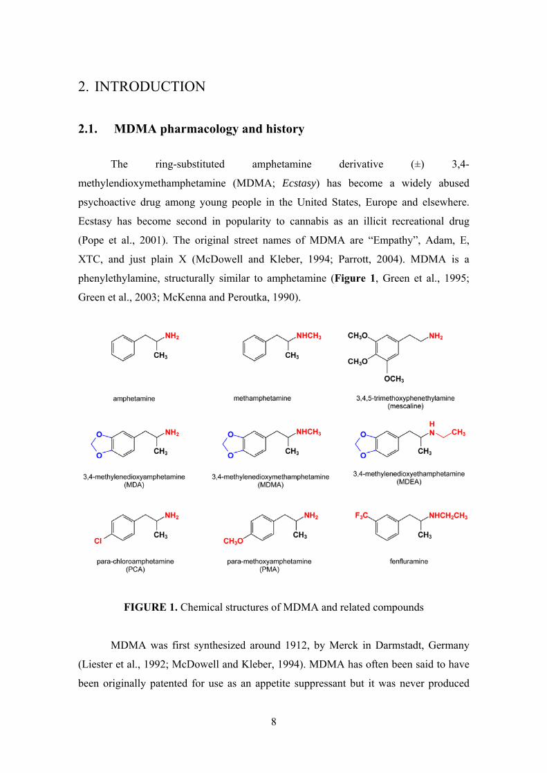
2.1. MDMA pharmacology and history
The ring-substituted amphetamine derivative (±) 3,4-
methylendioxymethamphetamine (MDMA; Ecstasy) has become a widely abused
psychoactive drug among young people in the United States, Europe and elsewhere.
Ecstasy has become second in popularity to cannabis as an illicit recreational drug
(Pope et al., 2001). The original street names of MDMA are “Empathy”, Adam, E,
XTC, and just plain X (McDowell and Kleber, 1994; Parrott, 2004). MDMA is a
phenylethylamine, structurally similar to amphetamine (Figure 1, Green et al., 1995;
Green et al., 2003; McKenna and Peroutka, 1990).
FIGURE 1. Chemical structures of MDMA and related compounds
MDMA was first synthesized around 1912, by Merck in Darmstadt, Germany
(Liester et al., 1992; McDowell and Kleber, 1994). MDMA has often been said to have
been originally patented for use as an appetite suppressant but it was never produced
8

commercially nor did it achieve clinical use for this indication. In the original patent
specification there were no indications for plans to develop an anorectic drug
(Freudenmann et al., 2006). Probably it was a precursor agent in a new chemical
pathway which was patented in order to avoid an existing patent for the synthesis of the
clotting agent hydrastatine. MDMA was mentioned only casually and without a name as
one of many chemical intermediates, and appeared only as a chemical formula
(methylsafrylamin). MDMA was neither studied in animals nor humans at Merck
around 1912. The first basic pharmacological tests using MDMA were performed by the
company’s chemists decades later (1927, 1952) (Bernschneider-Reif et al., 2006). The
United States Army experimented with the compound during the 1950s (Hardman et al.,
1973). MDMA was first used by humans in the late 1960s. MDMA remained on the
laboratory shelves until it was re-synthesised in the early 1970’s by Alexander Shulgin,
a research chemist employed to develop structural variants of mescaline and several
synthetic amphetamine derivatives (Shulgin, 1986).
The ability of MDMA to enhance feelings of openness and trust in its users was
the basis for the use of the drug as an adjunct to psychotherapy during the 1980s
(Downing, 1986; Greer and Tolbert, 1986; Grinspoon and Bakalar, 1986; Naranjo et al.,
1967).
MDMA has been illegal since it was classified as a Schedule I drug by the United States
Drug Enforcement Administration in July 1985 (Liester et al., 1992), due to its high
abuse potential, lack of clinical\ medical application, lack of accepted safety for use
under medical supervision and evidence that MDA (3,4-methylenedioxyamphetamine),
a related compound and major MDMA metabolite, induced serotonergic nerve terminal
degeneration in the rat brain (Ricaurte et al., 1985; Schmidt et al., 1986; Stone et al.,
1986).
In spite of its illegal status, since the mid 1980s it has become popular as a
recreational drug, often being taken at “rave” or “techno” parties, particularly in large
dancing clubs (Green et al., 1995).
Ecstasy comes in a variety of colours, shapes, and pill sizes, decorated with a
wide variety of designs or logos. As with any illicitly prepared and obtained recreational
drug, both doses and purity vary greatly (Baggott et al., 2000; Renfroe, 1986), but
tablets have regularly been found to contain between 80 and 150 mg of MDMA. Most
9

Ecstasy pills contain MDMA but studies also found several other constituents e.g.
MDA, MDAE (3,4-methylenedioxyethamphetamine), ketamine, amphetamine, codeine,
caffeine, ephedrine, salicylates or placebo (Baggott et al., 2000; Parrott, 2001; Schifano
et al., 1998).
Globally, after cannabis, amphetamines and ecstasy are among the most
commonly consumed illicit drugs. Results of a 2003 general Hungarian population
survey reveal that lifetime prevalence is 3.1% for ecstasy use (European Monitoring
Centre for Drugs and Drug Addiction, 2008). The 2006 survey showed that 20.3% of
15–17-year-old students have used an illicit substance at least once. Lifetime prevalence
of ecstasy use was 6.1%. The use of all drugs was found to be influenced by gender:
prevalence of drug use among male students was higher than among female students.
Available data for the 18–34-year age group showed that 20.1% reported lifetime
experience with illegal drugs. Lifetime experience with ecstasy was in second place, at
5.6% (European Monitoring Centre for Drugs and Drug Addiction, 2008).
2.2. Serotonergic system
2.2.1. Serotonin
Serotonin, 5-hydroxytryptamine (5-HT) is a biogenic monoamine that is widely
distributed in the body (Figure 2). Serotonin was first isolated in the 1950s from the
blood as a serum factor that increased smooth muscle tone (Amin et al., 1954; Twarog
and Page, 1953). 5-HT is present in significant concentrations in the central nervous
system (CNS) and in the periphery.
Serotonin in the CNS acts as a neurotransmitter and modulates a variety of
functions (Hoyer et al., 2002; Zifa and Fillion, 1992) such as mood (Meltzer, 1989;
Meltzer, 1990), anxiety (Nutt et al., 1990), aggression, impulsiveness (Berman et al.,
1997; Morgan, 1998), pain (Richardson, 1990), cognition, memory (Altman and
Normile, 1988), sleep (Wauquier and Dugovic, 1990), appetite (Curzon, 1990), emesis
(Bunce et al., 1991), motor activity (Jacobs and Fornal, 1997), body temperature
(Schwartz et al., 1995), epilepsy (Bagdy et al., 2007), endocrine function (Jorgensen,
10

2007), circadian rhythms (Wesemann and Weiner, 1990), social and reproductive
behaviour (Charnay et al., 1996; Gorzalka et al., 1990).
An important pool of 5-HT outside the CNS can be found in the
enterochromaffin cells of the gut and in blood platelets (Mitro et al., 2006). The greatest
concentration of 5-HT (80- 90%) is found in the enterochromaffin cells. 5-HT
participates in numerous physiological and homeostatic processes, such as
gastrointestinal peristalsis, blood coagulation and the maintenance of blood pressure
(Awabdy et al., 2003; Cote et al., 2004). The peripherally synthetized serotonin does not
effect activity of the CNS, because is not able to cross blood-brain barrier.
FIGURE 2. The chemical structure of serotonin
2.2.2. Serotonin synthesis in the central nervous system
Serotonin is synthesized from the amino acid precursor tryptophan through a
two-step process involving the tryptophan-hydroxylase (TPH) and then a
decarboxylation catalyzed by the L-aromatic acid decarboxylase (Figure 4, Tohyama
and Takatsuji, 1998). The TPH- mediated reaction is the rate-limiting step in the
pathway. Serotonin is packaged into synaptic vesicles and released into the synapse in
response to depolarizing stimuli (Figure 3). Once in the synapse, serotonin can interact
with both pre- and postsynaptic receptors. However, immediately after reacting with
pre- and postsynaptic receptors, it is critically important that serotonin be removed from
the synaptic cleft. Presynaptic plasma membrane transporters control synaptic actions of
5-HT by rapidly clearing the released 5-HT (Barker and Blakely, 1995; Jayanthi and
Ramamoorthy, 2005; Lesch, 1997). Once 5-HT is in the serotonergic neuron, it can be
enzimatically degraded by MAO (monoamine- oxidase) or taken back into the storage
11

vesicles via vesicular monoamine transporter (VMAT) for reuse (Figure 3, Lesch,
1997; Liu and Edwards, 1997).
Metabolism of 5-HT mainly occurs in the intestine, kidney, liver and lung by the
action of MAO to form the intermediate 5-hydroxyindole-3-acetaldehyde (5-HIAL).
This intermediate is either oxidized by aldehyde dehydrogenase to 5-
hydroxyindoleacetic acid (5-HIAA), or reduced by alcohol dehydrogenase or alcohol
reductase to 5-hydroxytryptophol (5-HTOL) (Beck and Helander, 2003; Musshoff,
2002).
Melatonin is biosynthesized from serotonin in the pineal gland, although there
are some additional pineal sites such as the retina and gastrointestinal tract. The rate-
limiting step in the synthesis of melatonin from serotonin is the one catalyzed by the
cytoplasmic enzyme serotonin N-acetyltransferase (AANAT) because its level and
activity also shows daily rhythm, with nighttime levels being higher than daytime
values. In contrast, the other enzyme, hydroxyindole-O-methyltransferase displays no
such day-night changes. Therefore, it is the AANAT rhythm that underlies the rhythmic
synthesis of melatonin, and AANAT has been called the melatonin rhythm enzyme
(Zheng and Cole, 2002).
FIGURE 3. Serotonergic synapse (from Lesch et al. 1997)
12

FIGURE 4. Synthesis and metabolism of serotonin
(the rate-limiting step of serotonin synthesis is highlighted in red colour and the rate-
limiting step of melatonin synthesis is highlighted in green colour)
Abbreviations: 5-HIAL - 5-hydroxyindole acetaldehyde; 5-HIAA - 5-
hydroxyindoleacetic acid, 5-HTOL - 5-hydroxytryptophol
13
2.2.3. Distribution of serotonergic cells
The serotonin-producing neurons in the brainstem raphe nuclei have the largest
and most complex efferent system in the brain. The special nature of the raphe
brainstem neurons was recognized by the classical neuroanatomist Ramón y Cajal, who
described these giant neurons in the brainstem midline but was unable to follow their
projections (Ramón y Cajal, 1911). Dahlström and Fuxe's work with histochemical
fluorescence provided details concerning the anatomical architecture of brain 5-HT
(Dahlström and Fuxe, 1964). Serotonergic neurons were first demonstrated by the
histofluorescence method (Falck et al., 1962). The original histofluorescence studies
were limited by the weak and rapidly falling fluorescence of 5-HT. The low sensitivity
of histofluorescence did not permit detection of fine axons in the forebrain, so the
density of innervation was initially underestimated. It was not possible to visualize the
distribution of 5-HT containing neurons until the advent of immunohistochemistry using
5-HT (Steinbusch, 1981), its precursor, 5-hydroxytryptophan (Touret et al., 1987), and
its synthetizing enzyme, tryptophan hydroxylase (Weissmann et al., 1987) antibodies.
5-HT neurons develop in the early fetal life as two separate clusters within the
brainstem: one rostral and one caudal group (Aitken and Törk, 1988). The rostral group
gives rise to almost all ascending 5-HT fibres, whereas the caudal group gives rise to the
majority of descending fibres (Aitken and Törk, 1988; Halliday et al., 1995; Wallace
and Lauder, 1983).
The cells which produce 5-HT are found only in restricted areas of the brain,
namely in the raphe nuclei groups which exist from the midbrain to the medulla
oblongata (Dahlström and Fuxe, 1964; Taber et al., 1960). These areas are classified
into nine regions (B1-B9) (Dahlström and Fuxe, 1964). The B1, B2, B3 and B4 groups
develop from 5-HT cells born in the caudal group, whereas the B5, B6, B7, B8 and B9
groups develop from 5-HT cells born in the rostral group (Törk, 1990). Serotonergic
axons that innervate the forebrain arise from neurons within the mesencephalic raphe
nuclei.
14
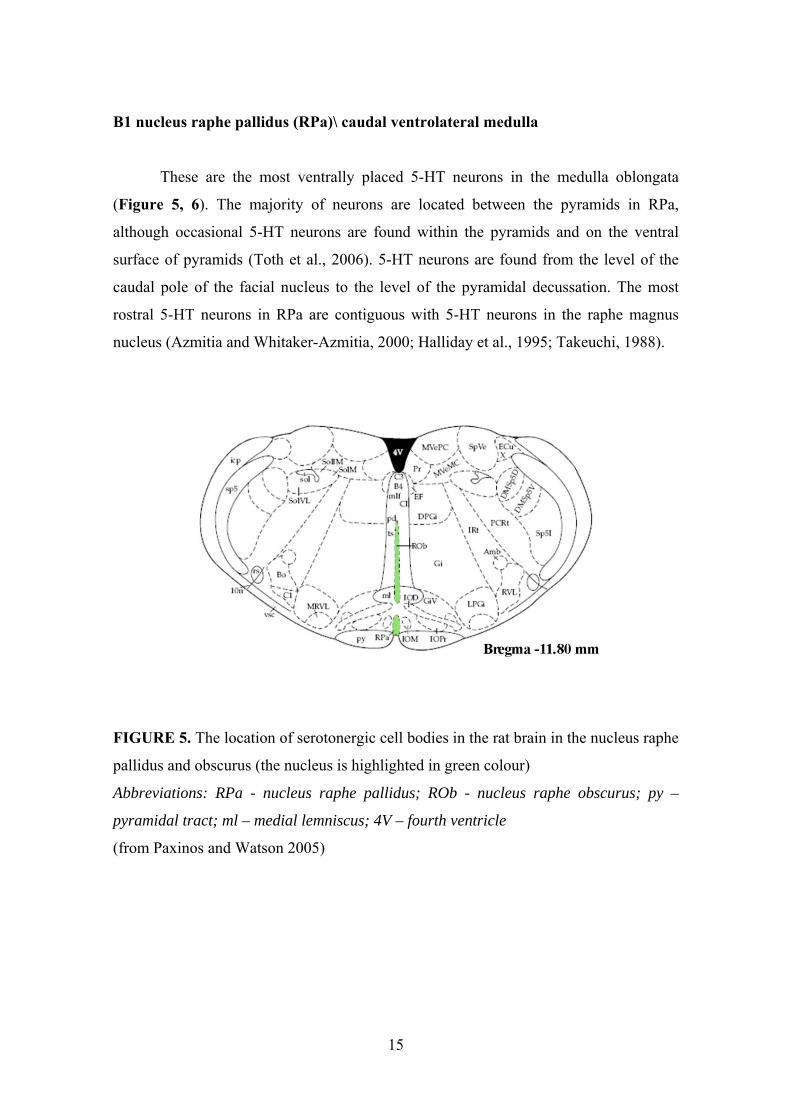
B1 nucleus raphe pallidus (RPa)\ caudal ventrolateral medulla
These are the most ventrally placed 5-HT neurons in the medulla oblongata
(Figure 5, 6). The majority of neurons are located between the pyramids in RPa,
although occasional 5-HT neurons are found within the pyramids and on the ventral
surface of pyramids (Toth et al., 2006). 5-HT neurons are found from the level of the
caudal pole of the facial nucleus to the level of the pyramidal decussation. The most
rostral 5-HT neurons in RPa are contiguous with 5-HT neurons in the raphe magnus
nucleus (Azmitia and Whitaker-Azmitia, 2000; Halliday et al., 1995; Takeuchi, 1988).
FIGURE 5. The location of serotonergic cell bodies in the rat brain in the nucleus raphe
pallidus and obscurus (the nucleus is highlighted in green colour)
Abbreviations: RPa - nucleus raphe pallidus; ROb - nucleus raphe obscurus; py –
pyramidal tract; ml – medial lemniscus; 4V – fourth ventricle
(from Paxinos and Watson 2005)
15
B2 nucleus raphe obscurus\ B4 central grey matter of the medulla oblongata
5-HT neurons in the nucleus raphe obscurus (ROb) are found along the midline
throughout the medulla oblongata as two vertical parallel sheets of neurons dorsal to the
inferior olive and pyramids (Figure 5). The majority of neurons are located in the
ventral half of the medulla oblongata, although occasional 5-HT neurons are located
within the central gray structures. 5-HT neurons are found from the level of the rostral
inferior olive to the level of the pyramidal decussation. The most rostral 5-HT neurons
in the ROb are contiguous with 5-HT neurons in the raphe magnus nucleus (Azmitia
and Whitaker-Azmitia, 2000; Halliday et al., 1995; Takeuchi, 1988).
B3 nucleus raphe magnus (RMg)\ rostral ventrolateral medulla
The RMg is located in the rostral ventromedial medulla of the brainstem. The
majority of neurons are found dorsal to the pyramids in the ventral half of the
tegmentum of the rostral medulla oblongata and caudal pons (Figure 6). The
distribution of B3 neurons resembles a triangle in transverse section. The most caudal 5-
HT neurons in RMg are contiguous with the midline 5-HT neurons in RPa and ROb
(Azmitia and Whitaker-Azmitia, 2000; Halliday et al., 1995; Takeuchi, 1988). Axons of
the RMg project to the spinal cord, terminating primarily in the dorsal horn (Jones and
Light, 1990).
16

FIGURE 6. The location of serotonergic cell bodies in the rat brain in the nucleus raphe
magnus (the nucleus is highlighted in green colour)
Abbreviations: RMg – nucleus raphe magnus; RPa – nucleus raphe pallidus; py –
pyramidal tract; ml – medial lemniscus; 4V – fourth ventricle
(from Paxinos and Watson 2005)
B5 pontine median raphe nucleus\ B8 midbrain median raphe nucleus\ caudal
linear nucleus
5-HT neurons in the B5 group are located in the pontine raphe nucleus. The
median raphe nucleus (MR, central superior, B8) lies within the central portion of the
reticular formation in the midbrain tegmentum (Figure 7). The larger B8 group of 5-HT
neurons is found throughout the midline raphe of the rostral pons and caudal midbrain.
17

Dorsal 5-HT neurons are contiguous with 5-HT cells in the (DR) raphe nucleus,
whereas the ventrally placed 5-HT neurons are contiguous with the B9 group. MR fibres
descend along the midline within the brainstem and mainly ascend within the MFB
(medial forebrain bundle) in the forebrain (Azmitia and Whitaker-Azmitia, 2000;
Halliday et al., 1995; Palkovits et al., 1977; Takeuchi, 1988).
B6 pontine dorsal raphe nucleus\ B7 midbrain dorsal raphe nucleus
The largest and most dense group of serotonergic neurons is the dorsal raphe
nucleus (DR, B7) which lies in the periaqueductal grey matter (Figure 7). It contains
235.000 5-HT immunoreactive (IR) neurons (Baker et al., 1990) in the human brain.
This nucleus extends from the levels just caudal to the oculomotor nucleus down to the
rostral portion of the fourth ventricle. The DR is continuous caudally with a smaller
group of 5-HT cells (B6) that lies along the midline and the floor of the fourth ventricle
(Azmitia and Whitaker-Azmitia, 2000; Halliday et al., 1995; Takeuchi, 1988).
B9 medial lemniscus
5-HT neurons in the B9 group are associated with the medial lemniscus (Figure
7). The B9 cell group consist of a scattered group of 5-HT neurons that lies along the
dorsal surface of the medial lemniscus in the ventrolateral tegmentum (Azmitia and
Whitaker-Azmitia, 2000; Halliday et al., 1995; Takeuchi, 1988).
18
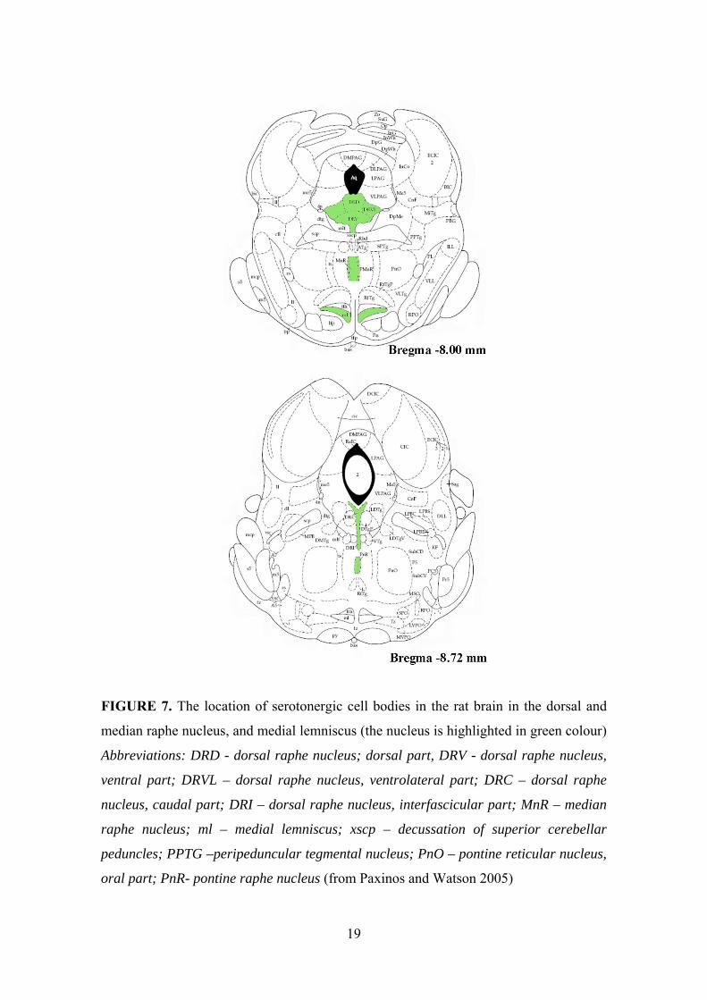
FIGURE 7. The location of serotonergic cell bodies in the rat brain in the dorsal and
median raphe nucleus, and medial lemniscus (the nucleus is highlighted in green colour)
Abbreviations: DRD - dorsal raphe nucleus; dorsal part, DRV - dorsal raphe nucleus,
ventral part; DRVL – dorsal raphe nucleus, ventrolateral part; DRC – dorsal raphe
nucleus, caudal part; DRI – dorsal raphe nucleus, interfascicular part; MnR – median
raphe nucleus; ml – medial lemniscus; xscp – decussation of superior cerebellar
peduncles; PPTG –peripeduncular tegmental nucleus; PnO – pontine reticular nucleus,
oral part; PnR- pontine raphe nucleus (from Paxinos and Watson 2005)
19

2.2.4. Main 5-HT pathways
Serotonergic axon terminals have been found in widespread areas of the
forebrain and throughout the brainstem and spinal cord (Brownstein and Palkovits,
1984; Palkovits et al., 1977; Steinbusch, 1981; Törk, 1990). The extent to which
neurons of each raphe nucleus project to various forebrain nuclei has been studied with
a variety of techniques, including selective lesions (Conrad and Pfaff, 1976; Meyer-
Bernstein and Morin, 1996; Palkovits et al., 1977) and both retrograde or anterograde
label transport (Datiche et al., 1995; De Olmos and Heimer, 1980; Kohler et al., 1982;
Li et al., 1993).
A series of studies employing small intracerebral lesions (Anden et al., 1971;
Ungerstedt, 1971) indicated that most 5-HT nerve terminals in the forebrain arise from
raphe nuclei in the midbrain and that the axons ascend through the lateral hypothalamus
within the medial forebrain bundle (Azmitia and Segal, 1978; Conrad et al., 1974;
Moore and Heller, 1967). Some 5-HT cells lie outside the raphe nuclei (e.g. solitary
nuclear complex and dorsomedial hypothalamus) (Brownstein and Palkovits, 1984;
Hoffman et al., 1998).
Anatomical evidence indicates that, in contrast to classical 5-HT neurotoxins,
MDMA destroys only axon terminals whilst cell bodies are left apparently intact
(Molliver et al., 1990; Molliver et al., 1989; Wilson et al., 1989). Furthermore, there is
evidence that MDMA causes a selective loss of 5-HT axons of a particular
morphological subtype, the so called “fine” 5-HT fibres, whilst sparing “beaded” 5-HT
fibres (Molliver et al., 1989; O'Hearn et al., 1988). It has also been proposed that
MDMA is more toxic to 5-HT neurons projecting from the DR than to those from the
MR. 5-HT axon terminals arising from the MR nucleus have large, spherical varicosities
(typically 3 μm in diameter), giving these axons a characteristic beaded appearance
(Figure 8). In contrast, axons that arise from the DR nucleus are of very fine caliber and
typically have minute, pleomorphic varicosities that are often granular or fusiform in
shape.
20

FIGURE 8. A schematic representation showing the two classes of axons
(from Molliver et al. 1989)
The first axon transport studies suggested that the DR nucleus projects
preferentially upon the cerebral cortex and the striatum while the MR innervates
primarily the hippocampus and the hypothalamus. Several studies applying more
sensitive methodology demonstrated that the projections were far more complex and
that there is considerable overlap in raphe projections to the forebrain. Azmitia and
Segal showed that the DR and MR project directly to the forebrain and give multiple,
anatomically distinct ascending fibre bundles (Azmitia and Segal, 1978). The terminal
distributions of DR and MR ascending projections converge, so that most areas of
cerebral cortex are innervated by both nuclei, with regional differences in the relative
contribution from each nucleus. Topographical studies which used highly sensitive
retrograde transport methods have shown that the cells within different regions of the
raphe nuclei project to separate areas of cortex (Kohler and Steinbusch, 1982; O'Hearn
and Molliver, 1984; Waterhouse et al., 1986).
2.2.5. Serotonin transporter (5-HTT)
The serotonin transporter (Figure 9) is similar to other biogenic amine
transporters (e.g. norepinephrine (NE) and dopamine (DA) transporters). 5-HTT is a
member of the family of sodium (Na+) and chloride (Cl-) dependent transporters
(Amara and Kuhar, 1993; Borowsky and Hoffman, 1995; Brownstein and Hoffman,
1994; Ramamoorthy et al., 1993). The transporter gene has been cloned from rat brain,
21
22
human brain, and human placenta (Blakely et al., 1991; Hoffman et al., 1991; Lesch et
al., 1993). Molecular weight of 5-HTT is 60-80 kDa. 5-HTT cDNAs are highly
conserved among species. In the rat and human species, each 5-HTT cDNA codes
approximately 630 amino acids and shares more than 90% common sequences (Gregor
et al., 1993; Ramamoorthy et al., 1993). 5-HTT proteins are commonly featured with 12
hydrophobic transmembrane segments, intracellular N- and C-terminals, glycosylated
sites in the second external loop, a potential kinase C phosphorilation site in the N- and
C-terminals, and a leucine zipper motif in the second transmembrane domain (Lesch,
1997). The N- and C-terminals and the third external loop are the least conserved
regions and have been used for discrimination among 5-HTTs. Serotonin transporter,
like serotonin, is seen most often in the raphe nuclear complex (Lesch et al., 1993;
Rattray et al., 1999). 5-HTTs have also been seen in the amygdala, thalamus,
hypothalamus, substantia nigra, and locus coeruleus. In addition, the 5-HTT is
expressed in the gut, placenta, lungs, lymphocytes and blood platelets.
Serotonin transporter, the mechanism for 5-HT high affinity uptake, is the essential
component for the termination of 5-HT transmission. It plays an important role in the
reuptake of 5-HT and subsequently terminates 5-HT transmission. 5-HTT plays a key
role in the presynaptic regulation of 5-HT transmission. 5-HTT is itself a target for
many drugs such as antidepressants, several neurotoxic agents including the
amphetamine derivative MDMA, fenfluramine, a neurotoxic metabolit of MPTP (1-
methyl-4-phenyl-1,2,3,6-tetrahydropyridine), and its analogues (Charnay et al., 1996;
Zhou et al., 2000). MDMA is a substrate of the 5-HTT, displacing 5-HT from its storage
vesicle, and ultimately inducing 5-HT efflux by a carrier-dependent process. MDMA
binds to all three presynaptic monoamine transporters, with highest affinity for the 5-HT
transporter. Affinities for the NE and DA transporters are at least 10-fold less (Battaglia
et al., 1988; Steele et al., 1987). Binding at both the 5-HT and DA transporters is
stereoselective, the S(+)-isomer is more potent, whereas no stereoselectivity is evident
at the NE transporter (Steele et al., 1987).

FIGURE 9. The structure of 5-HTT in the rat (from Hoffman et al. 1998)
23

2.2.6. Serotonin receptors
Serotonin receptors represent one of the most complex families of
neurotransmitter receptors and are classified into 7 types (Table 1), 5-HT1, 5-HT2, 5-
HT3, 5-HT4, 5-HT5, 5-HT6 and 5-HT7 and each type has subtypes (Barnes and Sharp,
1999; Hoyer et al., 1994; Hoyer et al., 2002). These receptors are localized in the brain
and in the peripheral organs (e.g. gut, cardiovascular system and blood) (Hoyer et al.,
2002), but their distribution is not homogeneous. The majority of 5-HT receptors are
postsynaptic but receptors such as 5-HT1A and 5-HT1B are mainly presynaptic and
modulate serotonin release. Except for the 5-HT3 receptor, which is a ligand-gated
cation channel, other 5-HT receptors belong to the G-protein coupled seven
transmembrane receptor superfamily (Aghajanian and Sanders-Bush, 2002; Barnes and
Sharp, 1999; Pauwels, 2000)
TABLE 1. Classification of serotonin receptors
Abbreviations: cAMP - cyclic adenosine monophosphate; IP3 - inositol-triphosphate;
DAG - diacyl-glycerol; PLC - phospholipase C
5-HT1 5-HT2 5-HT3 5-HT4 5-HT5 5-HT6 5-HT7
Subtypes 5-HT1A
5-HT1B
5-HT1D
5-HT1E
5-HT1F
5-HT2A
5-HT2B
5-HT2C
5-HT3A
5-HT3B
5-HT5A
5-HT5B
Major
signalling
pathway
Gi/G0
cAMP
Gq\11
IP3\ DAG
PLC
ion
channel
Gs
cAMP
5-HT5A: Gs
cAMP
5-HT5B: ?
Gs
cAMP
Gs
cAMP
24
2.3. The role of serotonin in depression
The 5-HTT plays a critical role in the regulation of 5-HT neurotransmission,
which has been linked to mood and personality disorders (Delgado et al., 1994).
Furthermore, serotonin transport has been implicated in the mechanism of widely used
antidepressants (Bengel et al., 1997; Lesch, 1997; Lesch et al., 1996). Tricyclics, such
as imipramine and amitriptyline, and the selective serotonin reuptake inhibitors (SSRIs)
fluvoxamine, fluoxetine, sertraline, citalopram, and paroxetine are widely used in the
treatment of depression. Serotonin-norepinephrine reuptake inhibitors (SNRIs) such as
venlafaxine inhibits the neuronal uptake of both serotonin and norepinepherine.
Tianeptine, an atypical tricyclic antidepressant is enhancing serotonin reuptake.
It has also been described that the incidence of depression is higher among previous
ecstasy users (Curran and Travill, 1997; Morgan, 2000; Parrott, 2001; Schifano et al.,
1998). One of the most typical symptoms of depression is the modification of sleep
patterns (Adrien et al., 1991; Akiskal et al., 1982). The most characteristic alteration is a
shortened latency of the first episode of rapid eye movement (REM) sleep, the so-called
reduced REM latency, and it has been shown that electroencephalogram (EEG) sleep
measurements can yield significant data to aid in differential diagnosis in psychiatry
(Akiskal et al., 1997; Kupfer et al., 1978). Strong diagnostic value of REM latency for
primary depression versus secondary depression, nonaffective psychiatric disorders and
nonpsychiatric controls have been proved, namely, 59% in sensitivity, 81% in
specificity, 0% in false positives in controls, and 83% confidence level has been found
(Akiskal et al., 1982). Shortened REM latency has also been described in subthreshold
depression and so-called ‘borderline’ personality disorders (Akiskal et al., 1997). An
association of the s allele of the functional polymorphism of the 5-HTT gene has also
been associated with subthreshold depression, and interestingly, this association was
primarily carried by the physical symptoms of depression, including sleep disturbance
(Gonda et al., 2005).
25
2.4. The role of serotonin in sleep
5-HT plays a key role in the regulation of sleep, especially REM sleep (Portas et
al., 1996). Initiation and duration of REM sleep is modulated by the serotonergic system
(Adrien, 2002; Portas et al., 1996). Increase in extracellular 5-HT concentration by 5-
HT releasers, 5-HT reuptake blockers or several 5-HT receptor agonists may increase
REM latency (Monti and Monti, 1999; Sommerfelt and Ursin, 1991; Ursin, 2002).
Physiologic role of 5-HT2 receptors in sleep regulation have been shown by the use of
neutral antagonists (Kitka and Bagdy, 2008). Preclinical studies involving
pharmacological depletion or anatomical lesions of 5-HT neurons have generally shown
changes in REM and non-rapid eye movement (NREM) sleep. However, most of these
preclinical studies involving massive depletions of 5-HT with sleep studies were
generally performed shortly after lesioning (Allen et al., 1993; Koella et al., 1968;
Touret et al., 1987).
Psychostimulants increase arousal and suppress sleep (McCann and Ricaurte,
2007), therefore a consequence of stimulant abuse is the disruption of normal sleep-
wake patterns. MDMA may produce lasting disruptions of sleep and circadian rhythms
because it has a neurotoxic potential towards the brain monoaminergic neurons known
to modulate normal sleep-wake patterns (Stenberg, 2007).
Although the functional consequences of MDMA-induced 5-HT neurotoxicity
are not clear, there have been reports of abstinent MDMA users who developed
disorders of mood, anxiety, cognition, impulsivity and sleep following MDMA
exposure. Alterations in sleep and circadian rhythm modulation alone can lead to
alterations in cognitive function, mood, impulsivity and endocrine function, creating the
possibility that alterations in the sleep-wake cycle could play a role in other
abnormalities seen in abstinent MDMA users (McCann and Ricaurte, 2007). Therefore,
humans who sustain 5-HT injury as a result of their MDMA use may be at a risk for
developing altered sleep architecture and disruptions in circadian rhythms.
MDMA abusers have also been reported to suffer from sleep disturbances
(Parrott et al., 2000; Williamson et al., 1997) although only a few studies have analyzed
the effects of MDMA on sleep patterns in humans and animals in a very controlled
fashion (Allen et al., 1993; Biello and Dafters, 2001). Balogh et al. evaluated the effects
26

of single acute doses (15 mg\kg) of systematically administered MDMA on sleep in
Dark Agouti rats (Balogh et al., 2004). Activity and wakefulness increased at least 6 h
after MDMA treatment in drug-naïve animals and also in animals that had received a
previous single dose of MDMA 21 days earlier. Robust circadian patterns of motor
activity and sleep were altered for approximately 5 days after drug treatment, and
alterations in motor activity, wakefulness, and “deep” slow wave sleep were still evident
21 days after MDMA administration.
2.5. The role of serotonin in aggression
Serotonin plays a crucial role in the regulation of impulsiveness and aggression
(Berman et al., 1997; Morgan, 1998), although other neurotransmitters, notably DA and
GABA (gamma-aminobutyric acid) have also been linked to human and animal
aggression (Miczek et al., 2002; Nelson and Chiavegatto, 2001). Serotonin release, or
high 5-HT levels are generally correlated with low aggressiveness (Nelson and
Chiavegatto, 2001; van der Vegt et al., 2003). In contrast, serotonergic dysfunction, as
defined by low CSF (cerebrospinal fluid) levels of 5-HT and 5-HIAA (5-hydroxyindole
acetic acid), has been associated with increased aggression (Clark and Neumaier, 2001;
Scearce-Levie et al., 1999). There is evidence that disorders of central serotonergic
neurotransmission, as reflected by low levels of 5-HIAA, are associated with impulsive
and aggressive personality traits (Coccaro, 1989; Linnoila et al., 1993).
Acute administration of MDMA has anti-aggressive effects in humans, rats and
mice (Curran et al., 2004; Miczek and Haney, 1994), presumably due to serotonin
release, but increased impulsiveness and aggression has been found amongst humans
who report previous chronic heavy ecstasy use (Bond et al., 2004; Gerra et al., 1998;
Morgan, 1998; Parrott et al., 2000). Although it is tempting to speculate that increased
aggression in human users is a symptom of MDMA-induced neurotoxicity, the
supporting evidence, whilst growing (Curran, 2000; Reneman et al., 2001b; Ricaurte et
al., 2000b), is not yet totally compelling. It is possible that symptomatic neurotoxicity
may be limited to heavy users, and/ or to particularly susceptible individuals (McCann
27

and Ricaurte, 1991), in whom vulnerability may be genetically or environmentally
determined (Roiser et al., 2005).
2.6. Acute neuropharmacological effects of MDMA in rats
MDMA is a potent indirect monoaminergic agonist and substrate-type 5-HT
releasing agent. MDMA acts as a substrate for the 5-HTT, binds to transporter proteins
and is subsequently transported into the cytoplasm of nerve terminals (Berger et al.,
1992; Rudnick and Wall, 1992). MDMA elevates extracellular 5-HT concentration by a
two-pronged mechanism: (1) it promotes efflux of 5-HT by a process of transporter-
mediated exchange and (2) it increases cytoplasmic levels of the transmitter by
disrupting the storage of 5-HT in vesicles (Rothman and Baumann, 2002). 5-HTTs, in
turn, mediate the translocation of drug molecules into the cytoplasm in exchange for 5-
HT molecules that flow out into the synaptic cleft. Note that MDMA is also a substrate
for the vesicular monoamine transporter (VMAT) present on intracellular vesicle
membranes. By disrupting compartmentalization of 5-HT into vesicles, MDMA
increases the pool of cytoplasmic transmitter available for drug-induced release
(Rothman and Baumann, 2002).
In the rat central nervous system MDMA has characteristic and well-
documented biphasic effects upon the serotonergic systems. First, shortly after
administration, there is an acute and rapid release of 5-HT (Green et al., 2003; Mechan
et al., 2002; Shankaran and Gudelsky, 1998). Second, in the brains of rodents and non-
human primates, MDMA causes a long-term reduction in 5-HT and 5-HIAA
concentration and 5-HT transporter density. The first phase lasts less than 24 h followed
immediately by the second phase which lasts approximately 12 months (or possibly
longer) (Schmidt, 1987; Stone et al., 1987b).
Acute effects of MDMA involve several neurotransmitters. Its main effects
concern 5-HT, but it also boost dopamine, norepinephrine and acethylcholine (Ach)
(Battaglia et al., 1991; Battaglia et al., 1987; Colado et al., 1999; Fitzgerald and Reid,
1990; Johnson et al., 1986; Schmidt et al., 1987; Yamamoto and Spanos, 1988).
28
MDMA has been shown to increase the release of 5-HT in both in vivo
microdyalisis (Gudelsky and Nash, 1996; Mechan et al., 2002; Sabol and Seiden, 1998)
and in vitro studies. Under in vitro conditions, the release of 5-HT is increased from
brain slices, synaptosomes and cultured neurons following exposure to MDMA (Crespi
et al., 1997; Johnson et al., 1986; Schmidt et al., 1987). Data from microdialysis studies
also indicate that MDMA enhances the release of 5-HT in vivo in numerous brain
regions. The administration of MDMA has been reported to result in a dose dependent
increase in the extracellular concentration of 5-HT in the striatum, hippocampus and
cortex (Gudelsky and Nash, 1996). Consistent with results from in vitro studies,
inhibition of the 5-HT transporter with fluoxetine suppresses MDMA-induced 5-HT
release in vivo (Gudelsky and Nash, 1996; Mechan et al., 2002), thereby further
supporting the conclusion that MDMA facilitates the transporter-mediated release of 5-
HT. MDMA induced 5-HT release is partially Ca2+-dependent (exocytotic-like) and
partially Ca2+-independent presumably through the neurotransporter (Crespi et al.,
1997).
Under in vitro conditions MDMA has been shown to promote the release of DA
from superfused brain slices, as well as to prevent the reuptake of DA into brain
synaptosomes (Johnson et al., 1991; Johnson et al., 1986; Schmidt et al., 1987; Steele et
al., 1987). Yamamoto and Spanos used in vivo voltammetry to demonstrate that
MDMA increases the release of striatal DA in vivo (Yamamoto and Spanos, 1988).
Numerous other investigators have confirmed these findings using in vivo microdialysis
(Esteban et al., 2001; Gudelsky et al., 1994; Hiramatsu and Cho, 1990; Nash and
Nichols, 1991; Sabol and Seiden, 1998).
In vitro MDMA has been shown to induce the release of norepinephrine from
brain tissue. Induction of both basal and stimulated [3H]NE release from preloaded rat
brain slices was blocked by desipramine (Fitzgerald and Reid, 1990). In a synaptosomal
preparation, MDMA induced NE release with similar potency to 5-HT and greater than
that for DA (Rothman et al., 2001).
Relatively few studies examined the effects of MDMA on central cholinergic
neurons. Fischer et al. observed that MDMA enhances the release of ACh in vitro from
striatal slices (Fischer et al., 2000). Acquas et al used in vivo microdialysis to
demonstrate that MDMA increases the extracellular concentration of ACh in the
29
prefrontal cortex and striatum (Acquas et al., 2001). Nair and Gudelsky used in vivo
microdialysis and high-performance liquid chromatography with electrochemical
detection (HPLC-ED) to assess the stimulatory effect of MDMA on the extracellular
concentration of ACh in the prefrontal cortex and dorsal hippocampus of the rat (Nair
and Gudelsky, 2006a; Nair and Gudelsky, 2006b).
In the acute phase, one of the most characteristic effects of MDMA is a rapid
and pronounced decrease in brain levels of 5-HT a few hours following drug
administration. For example, Schmidt demonstrated that both stereoisomers of the drug
could produce acute depletion of cortical serotonin measured 3 h after MDMA
administration (Schmidt, 1987). Connor et al. have reported a significant depletion in
cortical 5-HT concentrations both 30 min and 6 h after MDMA administration (Connor
et al., 1998). Acute administration of MDMA in rats also results in a rapid loss of TPH
activity in several brain regions (Che et al., 1995; Schmidt and Taylor, 1987). The
MDMA-induced decrease in TPH activity is influenced by body temperature. Che et al.
demonstrated that lowering body temperature protects against the immediate decrease in
TPH activity induced by MDMA (Che et al., 1995).
MDMA competitively inhibited 5-HT catabolism by rat brain MAO-A. Such
inhibition reduces the metabolism of 5-HT and dopamine within the nerve terminal and
therefore contributes to the increased release of the active neurotransmitter by MDMA
(Leonardi and Azmitia, 1994).
Acute treatment with MDMA results in a characteristic behavioural syndrome
which includes hyperthermia, hyperactivity, and the serotonin behavioural syndrome
(enhanced locomotor activity, reciprocal forepaw treading, head weaving, piloerection,
hind limb abduction, proptosis, ataxia and subsequent dose-dependent convulsions and
death (Green et al., 2003; Spanos and Yamamoto, 1989).
Data on the effects of MDMA on anxiety in laboratory animals are
contradictory. Recent experimental findings indicate that acute treatment with MDMA
may cause both anxiogenic (Bhattacharya et al., 1998; Gurtman et al., 2002; Lin et al.,
1999; Navarro and Maldonado, 1999; Navarro and Maldonado, 2002), and anxiolytic
effects (Lin et al., 1999; Morley and McGregor, 2000; Winslow and Insel, 1990)
depending upon the test situation (Morley and McGregor, 2000). Furthermore, the dose
30

range employed (Lin et al., 1999) or the basal anxiety level of the strain used (Green
and McGregor, 2002) might also explain the conflicting results observed.
Administration of MDMA produces a significant elevation of rat serum
corticosterone and prolactin concentrations 30 min post-injection (Connor et al., 1998;
Nash et al., 1988). Aldosterone and renin secretion have also been shown to increase
following MDMA treatment of rats (Burns et al., 1996). MDMA and some of its
metabolites can stimulate release of both oxytocin and vasopressin, the response being
dose-dependent (Forsling et al., 2001; Forsling et al., 2002).
MDMA has cardiac stimulant effects (Gordon et al., 1991), resulting in
tachycardia, and arrhythmia and also facilitates vasoconstriction (Fitzgerald and Reid,
1994).
Several studies used a [14C]2-deoxyglucose ([14C]2-DG) utilization technique to
identify brain region specific metabolic changes. Five minutes after a single dose of
MDMA (5–30 mg/kg, i.p.), glucose utilization was stimulated in components of the
extrapyramidal system and the limbic system showed decrements (Wilkerson and
London, 1989). Quate et al. revealed that acute exposure of Dark Agouti rats to a single
dose of MDMA (15 mg/kg, i.p.) elicited significant increases in local cerebral glucose
utilization that were most pronounced in the motor system and which were uncoupled
with acute changes in local cerebral blood flow, suggesting that MDMA might cause
acute cerebrovascular dysfunction (Quate et al., 2004).
MDMA binds with less affinity to 5-HT1, 5-HT2, α1, α2, β-adrenergic, M1 and
M2 muscarinic, histamine H1, dopamine D1 and D2 and opioid receptors (Morton, 2005).
MDMA has weak agonist actions at α2-adrenoceptors and 5-HT2 receptors, which might
influence its cardiac and pressor effects (Battaglia and De Souza, 1989; Lavelle et al.,
1999; Lyon et al., 1986).
2.6.1. Aggregation toxicity
The term “aggregate toxicity” first described by Gunn and Gurd, who found that
both behavioural and toxic effects of amphetamine were increased by some factors
(Gunn and Gurd, 1940). Later studies noted that toxicity was enhanced by elevated
ambient temperature, poor hydration, water deprivation and loud noise (Chance, 1946;
31

Chance, 1947; Dafters, 1995; Morton et al., 2001). Aggregate toxicity in rodents has
been noted for many other drugs, including caffeine, amiphenizole, pipradrol, β-
phenylisopropylhydrazine, methylphenidate and methamphetamine (Greenblatt and
Osterberg, 1961), isocarboxazide, desmethylimipramine, naloxone, and pethidine
(Doggett et al., 1977); morphine (Mohrland and Craigmill, 1980); racemic MDA (Davis
and Borne, 1984). MDMA-induced lethality is also increased by grouping animals or
housing them in hot conditions (Fantegrossi et al., 2003; Malberg and Seiden, 1998).
Investigations into the aggregate toxicity of MDMA may be particularly relevant given
the typical “rave” environment in which the drug is often abused by humans. At these
all-night dance parties, conditions of crowding and high ambient temperature are
common (Green et al., 1995) and may act to potentiate the toxic effects of MDMA.
Indeed, an initial report on the lethal effects of MDMA in humans noted that “In almost
every case, a recreational dose of the drug had been taken at a dance club or party where
crowds danced vigorously” (Randall, 1992).
2.7. Neurotoxicity - long-term neuropharmacological effects of
MDMA in rats
2.7.1. Proposed mechanism of MDMA induced neurotoxicity
There are several hypotheses that attempt to explain the mechanism underlying
serotonergic neurotoxicity.
2.7.1.1. Role of serotonin
The drug’s neurotoxocity is the most important question about its use. The
damage has been shown both histologically and biochemically using a variety of
experimental techniques in numerous animal species (rats, guinea pigs, squirrel
monkeys, cynomolgus monkeys, rhesus monkeys, baboons) and there have been many
studies on the mechanisms involved in the neurodegenerative change. Neurotoxicity has
been reported in laboratory animals (Commins et al., 1987; Schmidt, 1987; Schmidt and
32

Taylor, 1987), including axonal changes in serotonergic neurons of nonhuman primates
given relatively low dosages (Ricaurte et al., 1988b).
Twenty-four hours after MDMA, tissue 5-HT recovers to normal levels but
hydroxylase activity remains diminished. In the long-term phase, which begins within 7
days and lasts for months, MDMA causes a marked depletion of 5-HT and 5-HIAA
concentration and loss of 5-HTT binding and function (Battaglia 1988, Scanzello 1993).
Most investigators interpret this latter as a neurotoxic effect that involves the
serotonergic axons and depletion of terminals (Battaglia et al., 1987; Colado et al.,
1993; Green et al., 2003; Ricaurte et al., 2000b; Schmidt, 1987). There is also
histological evidence that MDMA produces morphological changes of the 5-HT
neurones (Green et al., 1995; Hegadoren et al., 1999; McKenna and Peroutka, 1990;
Seiden and Sabol, 1996; Seiden et al., 1993; Steele et al., 1994).
Many drugs used clinically produce effects that are similar to those produced by
MDMA. For instance, reserpine causes sustained depletions of brain tissue 5-HT, yet
reserpine in not considered a neurotoxin (Carlsson, 1976). Chronic administration of
SSRIs, like paroxetine and sertraline, leads to a marked loss of 5-HTT binding and
function comparable to MDMA, but these agents are therapeutic drugs rather than
neurotoxins (Benmansour et al., 1999; Frazer and Benmansour, 2002). The potent
selective serotonin releaser parabromomethamphetamine (V-111) was developed by
Knoll et al. and this agent may also be potentially toxic to serotonergic neurons, in a
mechanism similar to the action of MDMA (Knoll and Vizi, 1970; Knoll et al., 1970).
The persistent neurotoxic effects on 5-HT neurons following MDMA are similar to
those observed following parachloroamphetamine (PCA) in which marked reductions in
5-HT have been observed up to 4 months after a single injection (Kohler et al., 1978;
Sanders-Bush and Steranka, 1978). Studies in rats and nonhuman primates show that
systemic MDMA administration produces reductions in the levels of 5-HT, 5-HIAA, 5-
HT uptake sites or transporters (Battaglia et al., 1988; Lyles and Cadet, 2003; Sprague
et al., 1998) and the activity of TPH (Schmidt, 1987; Stone et al., 1987b) which last for
months or years after the cessation of drug exposure. Earlier studies also demonstrated
these lasting effects on brain 5-HT neurons, using immunocytochemical methods for
visualizing 5-HT containing neurons (Axt et al., 1994; Molliver et al., 1990; O'Hearn et
al., 1988; Wilson et al., 1989). Many papers reported that [3H]paroxetine binding is
33
reduced following MDMA administration (Ando et al., 2006; Balogh et al., 2004;
Battaglia et al., 1987; Colado and Green, 1995; Ferrington et al., 2006; Scanzello et al.,
1993).
MDMA toxicity is affected by doses, routes of administration, as well as by
treatment regimens. In addition, age, gender and species used can affect the
manifestations of MDMA neurotoxicity. Both the dose and number of injections of
MDMA affect the degree of neurotoxicity on 5-HT axons and terminals. Long-term
neurotoxic effects of MDMA require either a large single dose (20 mg/kg or more) or
several lower doses, typically 5 mg/kg twice daily for 4 consecutive days (Battaglia et
al., 1988; Colado et al., 1993; O'Shea et al., 1998; Ricaurte et al., 1988a). O’Shea et al.
found that the administration of a single dose of 4 mg\kg MDMA or daily for 4 days
had no effect on the serotonergic markers. When this dose was given twice daily for 4
days produced a marked damage (O'Shea et al., 1998).
The neurotoxic effects are evident up to a year after drug administration in rats
(Battaglia et al., 1987), and have been observed up to 7 years after drug administration
in non-human primates (Hatzidimitriou et al., 1999).
There is also preclinical evidence of regional differences in sensitivity to the
neurotoxic effects of MDMA. Areas rich in 5-HT terminals, such as the cerebral cortex
show more severe deficits than brain regions containing fibres of passage (e.g.
hypothalamus) or cell bodies (brainstem) (Commins et al., 1987; O'Hearn et al., 1988;
Steele et al., 1994). In particular, repeated administration of MDMA has been found to
produce especially long-lasting degeneration of serotonergic axons, and decreases in
brain serotonergic markers in the neocortex, hippocampus, caudate nucleus, putamen
and many thalamic nuclei (Aguirre et al., 1997; Fischer et al., 1995; Frederick et al.,
1995; Hatzidimitriou et al., 1999; Kleven et al., 1989; Lew et al., 1996; Ricaurte et al.,
1992; Sabol et al., 1996).
The results of O’Hearn et al. showed that repeated high-dose MDMA
administration has no effect on 5-HT cell bodies in the dorsal raphe, despite a profound
loss of 5-HT in forebrain projection areas 2 weeks after MDMA treatment (O'Hearn et
al., 1988).
Callahan et al. used anterograde transport of [3H]proline from the raphe nuclei to
trace ascending 5-HT axonal pathways in the rat forebrain 3 weeks after MDMA
34
treatment and observed a marked reductions in the labelled material to various forebrain
regions (Callahan et al., 2001). These results were similar to animals previously
lesioned with 5,7- dihydroxytryptamine (5,7- DHT), a documented 5-HT neurotoxin.
Gartside et al. investigated 5-HT neuronal activity in the dorsal raphe nuclei of rats
administered repeated doses of MDMA (Gartside et al., 1996). There were no
observable differences in the mean firing rate or regularity of firing of 5-HT neurons
between MDMA treated and control animals. Furthermore, electrical stimulation of the
dorsal raphe nucleus evoked a threefold increase in cortical and hippocampal dialysate
5-HT levels in both treatment groups. The apparent lack of effect of MDMA
administration on electrical activity in the dorsal raphe nucleus is consistent with the
observed lack of damage to dorsal raphe nuclei 5-HT cell bodies (Gartside et al., 1996;
O'Hearn et al., 1988).
The acute release of 5-HT is proposed to stimulate the 5-HT2A receptor, because
5-HT2A antagonists attenuate MDMA induced neurotoxicity (Nash, 1990; Schmidt et
al., 1990a).
2.7.1.2. Role of dopamine
MDMA can selectively destroy serotonergic axons with almost no effect on the
dopaminergic system in rats. MDMA increases the extracellular concentration of
dopamine in brain regions such as the striatum and nucleus accumbens (Gudelsky and
Nash, 1996; Nash, 1990; White et al., 1996; Yamamoto and Spanos, 1988). Several
studies hypothesized that the mechanism through which MDMA produces 5-HT
neurotoxicity is a dopamine-dependent process in which an MDMA-induced increase in
dopamine release results in the uptake of dopamine into 5-HT axon terminals (Sprague
and Nichols, 1995) and an induction of oxidative damage (Stone et al., 1989). 5-HT
activates 5-HT2A receptors, which enhances dopamine synthesis and release. Blockade
of 5-HT2A receptors diminishes MDMA-induced dopamine release (Nash, 1990). 5-HT2
receptor agonists administered together with MDMA potentiated MDMA-induced DA
release (Gudelsky et al., 1994). Moreover, prior administration of 6-hydroxydopamine,
which destroys DA terminals, also blocks MDMA neurotoxicity, while pretreatment
with L-DOPA (3,4-dihydroxy-L-phenylalanine), a dopamine precursor increases
35

MDMA neurotoxicity (Schmidt et al., 1990b). Sprague and Nichols suggest that MAO-
B metabolizes DA in the 5-HT terminal, producing hydrogen peroxide which could lead
to lipid peroxidation and oxidative stress (Sprague and Nichols, 1995).
2.7.1.3. Role of GABA
The role of the GABAergic system in the neurotoxic process appears to be
predominantly as a modulator of dopaminergic activity. Colado et al. reported that the
GABA agonist chlormethiazole attenuated serotonergic toxicity induced by MDMA
(Colado et al., 1993). Furthermore, Yamamoto et al. demonstrated an MDMA-induced
increase in extracellular DA coupled with a decrease in extracellular GABA levels in
the striatum. GABA might enhance its effects on DA synthesis and release, thus
potentiating its neurotoxic effects (Yamamoto et al., 1995).
2.7.1.4. Role of glutamate and nitric oxide
Glutamate, the most abundant excitatory amino acid in the CNS is known to
cause neuronal damage and this has led several laboratories to investigate the role of
glutamate in MDMA-induced neurotoxicity (Lyles and Cadet, 2003; Sprague and
Nichols, 1995).
White et al. demonstrated that locally applied MDMA has inhibitory effects on
glutamate-evoked neuronal firing in the nucleus accumbens and suggested that the
inhibition is mediated by increased extracellular dopamine and serotonin (White et al.,
1994).
One study showed that the coadministration of the NMDA-receptor antagonist,
MK-801 and MDMA caused a dose-related attenuation of the 5-HT depletion 7 days
after treatment (Colado et al., 1993; Finnegan and Taraska, 1996). In contrast, another
study found that coadministration of MK-801 attenuated only the methamphetamine-
induced decrease in TPH activity, but did not alter the effects of MDMA (Johnson et al.,
1989)
The role of NO (nitric oxide) in the neurotoxicity of MDMA has also been
studied in rats. Taraska et al. concluded that NO plays little or no role in the toxic
36
mechanism of action of MDMA because NGnitro-L-arginine methyl ester (L-NAME)
was the only NO synthase inhibitor that antagonized the dopamine- and serotonin-
depleting effects of MDMA and induced hypothermic response (Taraska and Finnegan,
1997). Two other NOS inhibitors, NG-monomethyl-L-arginine and NG-nitro-L-arginine
neither blocked the neurotoxic effects of MDMA nor caused hypothermia. However,
another study (Simantov and Tauber, 1997) found that MDMA can cause death of
human serotonergic cells and that these cells could be protected by L-NAME in the
culture.
2.7.1.5. Role of metabolites
Previous studies proposed that MDMA metabolites are the ultimate pathway of
serotonergic neurotoxicity. Schmidt and Taylor reported that acute cerebroventricular
injections of MDMA into the rat brain do not produce 5-HT axon destruction, therefore
any toxic metabolite produced would most likely be formed in the periphery (Schmidt
and Taylor, 1988).
Subcutan injection of MDA, MDMA, HMA (4-hydroxy-3-methoxyamphetamine) and
α-methyldopamine decreased the concentration of 5-HT in the frontal cortex (Yeh and
Hsu, 1991). In contrast, systemic administration of the major metabolites of MDA,
HHA (3,4- dihydroxyamphetamine) and of MDMA, DHMA (3,4-
dihydroxymethamphetamine) did not produce neurotoxicity (McKenna and Peroutka,
1990). DHMA is metabolized to quinone-like structures that were thought to be
involved in MDMA-induced neurotoxicity (Hiramatsu et al., 1990). MDMA metabolites
generate free radicals through redox cycling and these reactive species could inactivate
TPH and cause damage to protein and lipid components of the neuron terminal.
Catechols, hydroquinones and quinines can undergo spontaneous oxidation by oxygen,
to generate superoxides and hydrogen peroxide and these by-products could lead to
membrane damage and 5-HT terminal loss (Colado and Green, 1995; Paris and
Cunningham, 1992).
It has been supported that both oxidative and bioenergetic stress can underlie the
mechanism of MDMA induced neurotoxicity (Darvesh and Gudelsky, 2005; Gudelsky
and Yamamoto, 2003). Microdyalisis studies showing that MDMA induces oxidative
37

stress include the findings that MDMA increases the formation of reactive oxygen
species (e.g. hydroxyl radicals) and nitrogen species (e.g. nitric oxide) (Colado et al.,
1997; Shankaran and Gudelsky, 1999). Shankaran et al. demonstrated that concomitant
treatment of rats with ascorbic acid and a neurotoxic regimen of MDMA not only
prevents MDMA induced depletion of brain 5-HT but also the loss in ability of MDMA
to evoke 5-HT release (Shankaran et al., 2001).
It has also been suggested that 5-HT or a metabolite of 5-HT (e.g. 5,6- or 5,7-
dihydroxytriptamine) may be responsible for the toxicity. Blockade of 5-HT uptake by
fluoxetine or citalopram antagonizes MDMA induced 5-HT depletion and facilitates the
recovery of TPH activity (Lyles and Cadet, 2003; Schmidt, 1987; Schmidt and Taylor,
1987).
2.7.1.6. Role of hyperthermia
Several laboratories have shown that MDMA produce elevated body
temperature of rats at ambient temperature (Nash et al., 1988). Fatal hyperthermia is
associated with MDMA use in humans (Chadwick et al., 1991; Dar and McBrien,
1996).
Malberg et al. has found a close correlation between temperature response and
degree of neurotoxicity (Malberg and Seiden, 1998). Maintenance of animals at low
ambient temperature (10oC) before and after treatment with MDMA prevents the
hyperthermic response, producing hypothermia in some cases, and either attenuates or
eliminates MDMA-induced neurotoxicity (Broening et al., 1995; Schmidt et al., 1990a).
At elevated room temperatures (26-33oC), both the hyperthermic response and the
neurotoxicity are potentiated (Broening et al., 1995; Sanchez et al., 2004). Despite this
close relationship between acute hyperthermia and subsequent neurotoxicity the exact
role of hyperthermia in the development of the neurotoxic response is difficult to define
since it is possible to observe neurotoxicity also in the absence of hyperthermia
(Broening et al., 1995; O'Shea et al., 1998). In contrast, haloperidol, which prevents
MDMA-induced hyperthermia (Colado et al., 1999), pentobarbitone, NMDA
antagonists such as dizocilpine or the 5-HT2A receptor antagonist ketanserin which
produce hypothermia (Colado et al., 1999; Farfel and Seiden, 1995; Malberg et al.,
38
1996) all appear to have no specific protective effect against MDMA-induced
neurotoxicity. Furthermore, a few agents e.g. fluoxetine and clomethiazole have been
found to protect against the neurotoxicity of MDMA with no modification of
temperature (Colado et al., 1999; Colado et al., 1997; Sanchez et al., 2001). Several
studies conclude that neurotoxicity and hyperthermia are separable (Broening et al.,
1995; Colado et al., 1997; O'Shea et al., 1998). For example, MDMA administration
produced a hyperthermic response after every dose when the drug was given twice
weekly but no neurodegeneration was observed. Taken together, this evidence indicates
that hyperthermia has an important role but is not an essential factor in MDMA-induced
neurotoxicity.
2.7.2. Neuronal recovery
Björklund et al. reported that the serotonergic neurons show high capacity for
axonal sprouting and regeneration in the mature mammalian brain (Björklund et al.,
1981). Several studies demonstrated that the administration of 5,6-DHT, a neurotoxic
analogue of serotonin leads to axotomy and widespread terminal degeneration of
serotonergic neurons. The lesioned serotonergic neurons have the capacity to regrow
and re-establish some of their original connections (Björklund et al., 1973; Björklund
and Wiklund, 1980; Nobin et al., 1973; Wiklund and Bjorklund, 1980).
Previous studies report 5-HT neuronal recovery after PCA (Sanders-Bush et al.,
1972) and fenfluramine (Harvey and McMaster, 1975; Kleven et al., 1988). In contrast,
two studies suggest that 5-HT recovery in rats exposed to toxic amphetamine
derivatives may not be sustained. Zaczek et al. found an initial recovery of 5-HT
neuronal markers after fenfluramine treatment, but recovery is not maintained because
severe 5-HT deficits are apparent 6 to 8 months after drug administration (Zaczek et al.,
1990). Mamounas et al. also demonstrated an initial regenerative sprouting of 5-HT
axons after PCA injury, 6-12 months after PCA 5-HT axons were reduced in number
and abnormal (Mamounas et al., 1991).
Rats treated with MDMA show partial recovery of tissue 5-HT content, and
complete recovery of [3H]paroxetine-labeled 5-HT uptake sites 1 year after drug
administration (Battaglia et al., 1988; De Souza and Battaglia, 1989; De Souza et al.,
39

1990). MDMA-induced reductions in 5-HT levels and 5-HTT binding eventually
recover (Battaglia et al., 1988; Scanzello et al., 1993), suggesting the possibility that 5-
HT terminals are not destroyed. Similarly, Molliver et al. observed a regenerative
sprouting of 5-HT axons during 6 to 8 months after drug treatment (Molliver et al.,
1990; Molliver et al., 1989).
Scanzello et al. observed a recovery of 5-HT content 16 weeks after MDMA
treatment in some brain areas (e.g. hypothalamus, striatum) and at 32 weeks the
recovery was nearly complete in most brain regions (Scanzello et al., 1993).
[3H]paroxetine-binding values in hippocampus were 29% below control values at 52
weeks post-treatment. Recovery over the 52 week interval was noted for all depleted
regions but the rate and degree of recovery was found to be region dependent (Lew et
al., 1996; Sabol et al., 1996).
52 weeks after MDMA treatment there is evidence of a lasting reorganisation of
ascending 5-HT axon projections, with projections to distant forebrain sites (e.g. dorsal
neocortex) exhibiting little or no evidence of recovery, while projections to some more
proximal targets (e.g. hypothalamus) recover fully or in excess (Fischer et al., 1995).
These results indicate that altered reinnervation patterns develop much more frequently
in MDMA-treated primates than in MDMA-treated rodents. Other studies also observed
that in primates MDMA-induced 5-HT neural damage is persistent, and possibly
permanent (Insel et al., 1989; Ricaurte et al., 1992). MDMA-treated monkeys show
evidence of a “reorganization” of ascending 5-HT projections that is remarkably similar
to the “pruning effect” observed after a variety of neuron lesioning techniques,
including some using 5,6- and 5,7-DHT (Björklund et al., 1981; Frankfurt and Azmitia,
1984; Wiklund et al., 1978).
2.8. Problems in relating animal and human data - interspecies
scaling, dose, age and gender differences
The extrapolation of animal data to human recreational use is very difficult and
several critical factors must be considered, including species differences, route and
schedule of drug administration and drug dosages. A major point of controversy is
related to the relevance of MDMA doses administered to rats when compared to doses
40
taken by humans (Cole and Sumnall, 2003). The pharmacokinetics of the drug in rats
and humans may be similar. For example a dose of 1.5 mg/kg MDMA administered to
humans resulted in a peak plasma concentration of 331 ng/ml. Extrapolation from data
obtained with a dose of 10 mg/kg administered to female Dark Agouti rats suggests that
administration of a dose of 1.5 mg\kg would produce a very similar plasma
concentration. There is a view among some recreational ecstasy users that the doses
required to induce damage in experimental animals are so high that they have little
relevance to those taken by humans and that the drug is therefore „safe”. This view is
weakened by the observation that a single dose of MDMA of 10 mg/kg produced drug
levels similar to those involved in acute adverse reactions and that this dose caused
considerable neurotoxic damage to 5-HT nerve terminals (Colado and Green, 1995;
Green et al., 2003). It is undoubtedly true that many experimental studies have used
high doses, and it now appears that different strains exhibit a different sensitivity to the
drug (Malpass et al., 1999) and the long-term neurotoxic effects of MDMA.
The most obvious example is the Dark Agouti strain, which requires a single
dose (10–15 mg/ kg) of MDMA to produce a clear 30 to 50% or greater loss in cerebral
5-HT content (Colado and Green, 1995; O'Shea et al., 1998). This contrasts with the
several doses of MDMA, often a cumulative dose of 80 mg/kg within a few hours, that
are usually required to produce a similar loss in Sprague-Dawley, Hooded Lister and
Wistar rats (Aguirre et al., 1998; Colado et al., 1993; Shankaran and Gudelsky, 1999).
The dose required to induce neurotoxicity is strain-dependent. Humans typically
use MDMA orally, and although they may use up to 10 dosages per night, usually do
not use it during 4 consecutive days. The levels of neurotoxicity produced by oral
versus parenteral MDMA do not differ (Finnegan et al., 1988). However, De Souza et
al. reported that in rats, oral administration was less effective than intraperitoneal (De
Souza et al., 1997).
The neurodegenerative effects of MDMA on 5-HT neurons exhibited some
species specificity. The mouse is the one animal species that is relatively resistant to
MDMA-induced 5-HT injury, but at high doses mice also develop persistent loss in
brain 5-HT markers (Stone et al., 1987a). MDMA is neurotoxic to DA axons rather than
5-HT axons in mice (Cadet et al., 1995; Cadet et al., 2001; Colado et al., 2004). Cadet et
41

al. reported that a high dose of MDMA (50 mg\kg) produced dopamine depletion in the
striatum of non-transgenic mice (Cadet et al., 1995).
MDMA is self-administered by both rhesus monkeys and baboons suggesting
that MDMA may have a high abuse potential in man. Long lasting effects similar to
those observed in rats have also been reported in nonhuman primates. In nonhuman
primates MDMA causes dose-dependent reductions in 5-HT in the cortex, caudate
nucleus, putamen, hippocampus, hypothalamus and the thalamus. Reduced 5-HT level
was evident for up to 7 years following MDMA administration (Hatzidimitriou et al.,
1999). Immunohistochemical studies have also revealed marked decreases in axon
density in the forebrain of nonhuman primates treated with MDMA.
Studies also reported that altered reinnervation patterns develop much more
frequently in MDMA-treated primates than in MDMA-treated rodents. Baboons treated
with MDMA (5 mg/kg MDMA, twice daily for 4 consecutive days) also show marked
and prolonged decreases in 5-HT transporter density measured by PET with radioligand
(McN-5652), which selectively labels the 5-HT transporter (Scheffel et al., 1998). In
agreement with the results of Fischer et al., PET scans 9 and 13 months post-MDMA
showed regional differences in the apparent recovery of the 5-HT transporter, with
increases in some brain regions (e.g. hypothalamus) and persistent decreases in others
(e.g. neocortex) (Fischer et al., 1995).
Dosages of MDMA that produce neurotoxicity in rodents are higher than those
needed to produce neurotoxicity in primates (Battaglia et al., 1987; Ricaurte et al.,
1988a; Stone et al., 1986). The observation that smaller animal species require higher
doses of the drug to achieve equivalent drug effects is predicted by the principles of
interspecies scaling. The concept of interspecies scaling is based upon shared
biochemical mechanisms among eukaryotic cells (e.g. aerobic respiration), and it was
initially developed to describe variations in basal metabolic rate (BMR) between animal
species of different sizes (White and Seymour, 2005). This method utilizes known
relations between body mass/surface area and accounts for differences in drug
clearance. Similarly, the same technique can be applied to predict dosages of MDMA
that would be neurotoxic in humans. It is known that the neurotoxic dose in 1 kg
squirrel monkeys is 5 mg/kg and the equivalent dose in humans is found to be 1,28
mg/kg or approximately 96 mg in a 75 kg individual. The typical amount of MDMA
42

abused by humans is one or two tablets of 80-150 mg or 1-3 mg/kg administered orally
(Green et al., 2003; Schifano, 2004). This dose falls squarely in the neurotoxic range
predicted by the interspecies scaling method.
It is also important to consider the influence of age and sex differences on the
effects of MDMA. The role of age is known to cause variability in the sensitivity of
animals and humans to MDMA (Easton and Marsden, 2006). Principal users are
adolescents and young adults. Coincidentally, these are also peak reproductive ages.
There are few clinical reports on the effects on offspring development after intrauterine
exposure. One group has suggested that there may be an increase in congenital
malformations after prenatal MDMA exposure, including defects of the heart and feet
(McElhatton et al., 1999), but no clinical studies have appeared on possible effects on
brain development and function.
It has been demonstarted that the persistent effects of MDMA on the
serotonergic system in the rat are modulated by developmental age (Broening et al.,
1994). Results from animal studies suggest that fetal and neonatal rat brains are not
vulnerable to MDMA-induced serotonergic neurotoxicity (Allen et al., 1993; Broening
et al., 1994; Colado et al., 1997; Kelly et al., 2002), however other studies have found
effects (Koprich et al., 2003; Won et al., 2002). For example, Winslow and Insel found
that single or repeated doses of MDMA administered on P1–4 result in decreased
ultrasonic calling in rat pups and reduced cortical 5-HT and 5-HTT binding on P21
(Winslow and Insel, 1990). Meyer and Ali found that rats exposed to MDMA on P1– 4
and evaluated at P25 or P60 showed reduced hippocampal 5-HT concentrations and 5-
HTT binding at P25, and hippocampal 5-HTT binding remained reduced at P60 (Meyer
and Ali, 2002).
MDMA administration does not produce long-term 5-HT loss in immature rats
(PND 10) when compared to more mature rats (PND 40, 70) where damage does occur
(Broening et al., 1994). Piper and Meyer demonstrated that intermittent MDMA
exposure to rats during the adolescent period of development can produce memory
deficits and reduce anxiety in the absence of severe serotonergic damage (Piper and
Meyer, 2004). Additionally, long-term deficits in reference memory are produced
following MDMA exposure to rats of PND 11–20 (Vorhees et al., 2004) and a
43

combination of early and late exposure of MDMA in rats can produce disruption of
spatial acquisition and reversal learning (Cohen et al., 2005).
There are very little available data in humans which compare the extent of behavioural,
biochemical or physiological effects between adolescent and adult user groups of
MDMA. Deficits in verbal and visual memory have been most frequently observed in
adults (Gouzoulis-Mayfrank et al., 2000; Morgan, 1999; Parrott et al., 1998). Jacobsen
et al. found that MDMA use in adolescence may be associated with cognitive
impairments and dysfunction of inhibitory circuits within the hippocampus. These data
indicate that similar behavioural changes occur in both age group (Jacobsen et al.,
2004).
There have been few studies which examine the role of gender differences
related to the effects of MDMA. Although more men than women are involved in
alcohol, tobacco or illicit drug use, there is growing evidence based on animal and
clinical studies that females are more susceptible to the effects of drugs of abuse. Two
recent behavioral studies report that females show enhanced locomotion, less startle
response, and comparable effects in the plus maze (Bubenikova et al., 2005; Palenicek
et al., 2005) in Wistar rats after MDMA administration. Female Sprague-Dawley rats
show lower brain concentrations of MDA compared to males, suggesting a slower
metabolism of MDMA to MDA in females (Chu et al., 1996). Fitzgerald et al. showed a
higher level of MDMA in the plasma of female rats after a single drug injection
(Fitzgerald et al., 1989). Walker et al. found that MDMA elicited much more robust
locomotor activation in female than in male rats, but caused similar serotonin depletion
and persistent anxiety-like behaviors 2 weeks after the end of treatment (Walker et al.,
2007). A previous study in rodents has shown that male and female rats have
comparable brain levels of MDMA after acute treatment (Chu et al., 1996). Similar
results of comparable 5-HT depletion in males and females were also reported by
Slikker et al. (Slikker et al., 1989). Colado et al. demonstrated that female Dark Agouti
rats are more susceptible to the acute hyperthermic effects of MDMA (Colado et al.,
1995). The concentration of 5-HT and 5-HIAA in the cortex and hippocampus of male
and female rats were similar 7 days after injection of MDMA. Female Dark Agouti rats
having markedly decreased debrisoquine 4-hydroxylase activity than males (Gonzalez
et al., 1987). However, several investigators reported that in mice and rats, MDMA is
44

more toxic in males than females (Cadet et al., 1994; Koenig et al., 2005; Miller and
O'Callaghan, 1995). Koenig et al. observed that the male Long–Evans rats appear to be
more sensitive to locomotor and hyperpyretic effects of MDMA. Presumably the effects
of MDMA in rats depends on the used rat strain (Koenig et al., 2005).
Veryheyden et al. observed that women are more susceptible than men to mid-
week low mood following weekend use of MDMA (Verheyden et al., 2002). Liechti et
al. reported greater subjective responses to MDMA in women than in men, including
greater thought disturbances, perceptual changes, and fear of loss of body control. In
contrast, men experience greater increases in blood pressure (Liechti et al., 2001). Other
studies using SPECT or PET also indicate that women might be more susceptible than
men (Buchert et al., 2004; Reneman et al., 2001a). Biochemical evidence supports this
suggestion as analysis of CSF data revealed that reductions of 5-HIAA CSF were
greater in females than in males (46% vs 20%) (McCann et al., 1994). Buchert et al.
found reduction of serotonin transporter availability in current MDMA users and this
was more pronounced in female than in male subjects (Buchert et al., 2004). Schifano
reports that the MDMA lethality ratio between males and females is 4:1 (Schifano,
2004). Although these reports suggest that the effects of MDMA are not equivalent in
men and women, they could be related to differences in innate hormonal profiles,
volume and morphology of certain brain structures, or monoaminergic
neurotransmission.
2.9. Metabolism of MDMA
In humans, MDMA is metabolised principally by cytochrome P450 (CYP) 2D6
or debrisoquine hydroxylase (Tucker et al., 1994). Because of the polymorphic variants
of this isoenzyme, 5- 9% of the Caucasian population is considered to be a poor
metabolizer phenotype (Gonzalez and Meyer, 1991). There is evidence that “poor
metabolizers” of debrisoquine have lower metabolic capacity and higher plasma levels
of MDMA (de la Torre et al., 2005). In rats, MDMA is metabolized by an analogous
isoenzyme, CYP2D1. The Dark Agouti rat strain possesses decreased microsomal
CYP2D1 isoenzyme activity, and thus, the Dark Agouti rats are thought to be similar to
vulnerable human users, the “poor metabolizers”. The Dark Agouti rat is a useful
45
experimental tool to model MDMA exposures that are pertinent to the human
experience of the drug (Balogh et al., 2004; Kirilly et al., 2006; O'Shea et al., 1998;
Quate et al., 2004). This rat strain provides a model of a genetically- defined human
sub- population in which clinical complications may be more likely to occur.
MDMA displays nonlinear kinetics (de la Torre et al., 2000), which has
implications for the acute toxic response to the drug when a high dose, or more than one
dose, is taken in a single session (Farre et al., 2004). Approximately 8% of the MDMA
dose is excreted unchanged in rats but this rises to 50% in humans (de la Torre et al.,
2000).
Ecstasy tablets ingested by humans contain a racemic mixture of (+) and (-)
isomers of MDMA, and both stereoisomers are known to be bioactive (Johnson et al.,
1986; Schmidt et al., 1987).
Several pathways are involved in MDMA metabolism in the rat: N-
demethylation, O-dealkylation, deamination, and conjugation (O-methylation, O-
glucoronidation, and/or O-sulfation), resulting in the formation of 14 in vivo
metabolites (Lim and Foltz, 1988; Lim and Foltz, 1991a; Lim and Foltz, 1991b). Firstly,
MDMA is metabolized into MDA by N-demethylation, DHMA by demethylenation,
and 2-hydroxy-4,5-(methylenedioxy)methamphetamine by ring hydroxylation (Cho et
al., 1990; Lim and Foltz, 1988; Lim and Foltz, 1991a; Tucker et al., 1994). These
undergo further metabolism (possibly via catechol-O-methyl transferase mediated
reactions) to form HMMA (4-hydroxy-3-methoxy-methamphetamine) and HMA,
respectively.
2.10. Human studies
MDMA exhibits both psychostimulant and hallucinogenic properties (Shulgin,
1986). MDMA is often described as an entactogen producing a sense of touching within
(Gudelsky and Yamamoto, 2008; Nichols, 1986). The subjective effects of MDMA in
humans include a sense of well being, connectedness and diminished aggression
(Davison and Parrott, 1997; McDowell and Kleber, 1994). MDMA use is generally
associated with happy young people enjoying themselves in the social situation of dance
clubs or “raves”.
46

2.10.1. Acute pharmacological effects in humans
MDMA is rapidly absorbed following oral administration. The onset of effects
can take 20 to 60 min to occur, reaches its peak in 60 to 90 min after ingestion, and the
primary effects last for 3 to 5 h. Acute adverse effects are related to the dose ingested,
the frequency of dosing, the ingestion of other drugs (e.g. alcohol, nicotine,
amphetamine, cocaine, ketamine, and many other psychoactive drugs (Huxster et al.,
2006; Soar et al., 2006; Thomasius et al., 2006) and the environmental conditions in
which the user was present.
The acute psychological effects of MDMA include feelings of euphoria,
increased closeness to others, elevated self-confidence, heightened arousal and sensory
awareness (Downing, 1986; Greer and Tolbert, 1986; Liechti and Vollenweider, 2000),
increased sensory sensitivity (Cohen, 1995; Peroutka et al., 1988; Solowij et al., 1992).
More recent studies reported several acute side effects e.g. moderate derealisation and
depersonalisation, cognitive disturbances, elevated anxiety, tachycardia, panic attacks
(Cohen, 1995; Downing, 1986; Liechti and Vollenweider, 2000; Liester et al., 1992;
Whitaker-Azmitia and Aronson, 1989). Prospective and cross-sectional studies have
shown evidence for psychiatric and/or psychobiological problems prior to drug use
(Huxster et al., 2006; Soar et al., 2006; Thomasius et al., 2006).
Acute adverse physical effects of MDMA include suppressed appetite, bruxism,
papillary dilatation, trismus (jaw-clenching), dry mouth, palpitation, nausea, impaired
judgement and gait (ataxia). Subjects also exhibit symptoms of sympathetic arousal
including tremors and tachycardia (Downing, 1986; Green et al., 2003; McCann et al.,
1996; Peroutka et al., 1988).
After-effects may be quite pronounced, sometimes lasting 24- to 48 hours. The
most dramatic “hangover” effect is a sometimes profound anhedonia, depression,
irritability, difficulty in concentrating, headache, muscle aches, fatigue, but the users
may experience lethargy, anorexia, and decreased motivation (Cohen, 1995; Davison
and Parrott, 1997; Peroutka et al., 1988; Solowij et al., 1992).
Serious adverse effects of MDMA intoxication include cardiac arrhythmias,
hypertension, hyperthermia (body temperatures over 43oC), acute renal failure,
rhabdomyolisis, disseminated intravascular coagulation, serotonin syndrome,
47

hyponatremia, liver problems (centrilobular necrosis and microvascular steatosis, acute
cholestatic hepatitis) (Devlin and Henry, 2008; Milroy et al., 1996), seizures, coma, and,
in rare cases, death. MDMA impairs temperature control, so that in the hot and crowded
conditions at raves many dancers become overheated or hyperthermic (Dafters, 1994).
MDMA deaths are comparatively rare, but unfortunately they show a year-by
year increase, which may be facilitated by the decreasing cost of MDMA (Schifano et
al., 2006).
2.10.2. Long-term pharmacological effects in humans
The most important question is whether MDMA abuse causes neurotoxic
damage in humans. There is a growing consensus that MDMA might indeed be toxic to
humans. The neurotoxic potential of MDMA in humans has been evaluated indirectly
by measuring the concentration of 5-HIAA in cerebrospinal fluid in recreational ecstasy
users. The earliest study employing this technique failed to find evidence of reduced
levels of 5-HIAA in recreational users (Peroutka, 1987). However, other studies
(McCann et al., 1999; McCann et al., 1994; Ricaurte et al., 1990) reported significantly
lower levels of CSF 5-HIAA in recreational ecstasy users compared to polydrug users
who had never used ecstasy. There were no changes in the levels of the DA metabolite
homovanillic acid nor in the concentration of the NE metabolite 3-methoxy-4-hydroxy-
phenylglycol (MHPG). Kish et al. demonstrated severe depletion (50-80%) of striatal 5-
HT and 5-HIAA in the brain of a 26-year-old male who had taken MDMA regularly for
9 years (Kish et al., 2000).
Accumulating evidence indicates that chronic, heavy, recreational use of ecstasy
is associated with sleep disorders, depressed mood, persistent elevation of anxiety,
impulsiveness and hostility, and selective impairment of episodic memory, working
memory and attention, which can last for months after cessation of drug intake
(Morgan, 2000; Parrott, 2002; Parrott et al., 2002).
Recently, more direct evidence that MDMA produces long-term neurotoxic
effects on brain 5-HT systems has emerged from neuroimaging studies: decrease in 5-
HTT detected by positron emission tomography (PET) and single photon emission
computed tomography (SPECT), and a decrease in the neurometabolite NAA, a marker
48

for non-specific neuronal loss, detected by proton magnetic resonance spectroscopy
(1H-MRS) (Reneman et al., 2006). Daumann et al. found no differences in neocortical
regions, only a tendency towards lower NAA/Cr ratios in the hippocampus of MDMA
users, and therefore concluded that 1H-MRS is a less sensitive marker of potential
neurotoxic damage in ecstasy users (Daumann et al., 2004).
McCann et al. used positron emission tomography (PET) with the first- and
second- generation radioligand, carbon-11-labelled McN-5652 and DASB, which
selectively labels the 5-HT transporter. MDMA users showed decreased global and
regional brain 5-HT transporter binding with both PET ligands (McCann et al., 1998;
McCann et al., 2005). Ricaurte et al. observed decreased 5-HT transporter binding
which positively correlated with the extent of previous MDMA use (Ricaurte et al.,
2000a). Buchert et al. observed similar changes and found that women are more
susceptible than men to MDMA-induced alterations of the serotonergic system (Buchert
et al., 2003; Buchert et al., 2004).
Imaging studies used SPECT with the 5-HT transporter radioligand ([123I] b-CIT)
to investigate heavy ecstasy users and controls. The ecstasy users showed a widespread
reduction of 5-HT transporter binding (Haddad et al., 2002; Reneman et al., 2001b;
Semple et al., 1999).
The status of 5-HT markers in MDMA users has also been assessed with SPECT
using [123I]-I-R91150 a radioligand, which binds with a high affinity to 5-HT2A receptors.
5-HT2A receptor densities appeared to be significantly increased in the occipital cortex
of ecstasy users compared to controls, possibly as a compensatory response to 5-HT
depletion (Reneman et al., 2000).
There are a number of case reports of chronic psychiatric symptoms following
regular use of the drug including reports of depersonalisation (McGuire et al., 1994),
obsessive-compulsive symptoms (Marchesi et al., 2007), flashbacks (Creighton et al.,
1991; McGuire and Fahy, 1992; Schifano and Magni, 1994), panic attacks (Cohen,
1996; McCann and Ricaurte, 1991; McGuire et al., 1994; Schifano and Magni, 1994;
Series et al., 1994), psychosis (Creighton et al., 1991; McCann and Ricaurte, 1991;
McGuire and Fahy, 1991; McGuire et al., 1994; Schifano, 1991) and depression
(Benazzi and Mazzoli, 1991; McCann and Ricaurte, 1991; McGuire et al., 1994;
Schifano and Magni, 1994). Other studies have also shown that MDMA abuse can
49

negatively impact the performance of memory and psychomotor tasks (Gouzoulis-
Mayfrank et al., 2000; Morgan, 1999; Wareing et al., 2000). Memory was the first area
of cognition shown to be impaired in recreational ecstasy users (Krystal et al., 1992) and
this had been confirmed in several laboratories (McCann et al., 2000; Parrott et al.,
2000). Other psychobiological deficits include eating disorders (Curran and Robjant,
2006), impaired social and emotional judgement (Reay et al., 2006), and selective
changes in auditory evoked potentials (Daumann et al., 2006).
Clinical studies designed to critically evaluate the long-term effects of MDMA
are hampered by a number of factors, including comorbid psychopathology and
polydrug abuse among MDMA users. There are many variables that could not be
controlled, e.g. dose and purity of MDMA, number of times MDMA was used, set and
setting, and the time between interview and the last use of MDMA. Animal models
afford the unique opportunity to evaluate the effects of MDMA without many
complicating factors. The existing preclinical data strongly suggest that MDMA can
produce neurotoxic damage in the brain. Therefore, any prospective clinical study
involving MDMA administration is constrained by ethical considerations. Even
administration of low doses of MDMA (1.7 mg/kg) has been publicly questioned and
discussed (McCann et al., 2001; Vollenweider et al., 2001). All studies can be criticized
for the fact that accurate information on both the doses taken and the duration of drug
use is not available. This is because the drug has been synthesized and supplied illicitly
and most subjects are unreliable sources of information on use. In addition, many
subjects are poly-drug users so that it cannot be stated unequivocally that any event seen
results solely from MDMA use. Alternatively, the problem may relate to the
combination of MDMA with another recreational compound. This problem is
compounded by the fact that a significant percentage of ecstasy tablets contain
psychoactive compounds other than MDMA.
With regard to the possible therapeutic use, one group in particular has long
advocated a trial for the use of MDMA in post-traumatic stress disorder (Bouso et al.,
2008), and other trials for the use of MDMA to help rape or cancer victims have also
been proposed.
50

3. OBJECTIVES
The recreational drug ecstasy, 3,4-methylenedioxymethamphetamine (MDMA),
has been found to selectively damage brain serotonin neurons in experimental animals,
and probably in human MDMA users, but detailed morphometric analyses and parallel
functional measures during damage and recovery are missing. We specifically
emphasized the long-term morphological effects of a single dose of MDMA from the
brainstem up to the cerebral cortex. We hypothesized that long-lasting damage of the
serotonergic system will be manifested in changes of several sleep parameters.
Furthermore, we investigated the effects of MDMA on aggressive behaviour. These
studies were carried out in Dark Agouti rats, a special strain that may serve as a genetic
model for poor or moderate metabolizer ecstasy users.
The main objectives in our studies were:
1. How does 5-HTT mRNA expression change in the different raphe nuclei
during 6 months after MDMA treatment?
2. Can differences be observed in the changes concerning dorsal and median
raphe nuclei?
3. How does 5-HTT-immunoreactive (IR) fibre density change in the different
brain areas?
4. Do the examined brain areas show differences in sensitivity after MDMA
treatment?
5. How much regeneration can be observed 6 months after MDMA treatment?
6. How do sleep parameters change after MDMA treatment?
7. What sleep parameters change similarly and the opposite way compared to
what has previously been observed during depression?
8. How does MDMA treatment alter agressive behaviour?
51

4. MATERIALS AND METHODS
4.1. Animals
All animal experiments were carried out in accordance with the European
Communities Council Directive of 24 November 1986 (86/609/EEC) and National
Institutes of Health “Principles of laboratory animal care” (NIH Publications No. 85-23,
revised 1985). In addition, specific national laws were adhered to (Hungarian
Governmental Regulation about animal studies, December 31, 1998 and United
Kingdom Animals (Scientific Procedures) Act, 1986) and permission was obtained from
local ethical committees (22.1/2655/001/2007). Six to seven-week old male Dark
Agouti rats (Harlan, Olac Ltd, Shaw’s Farm, Blackthorn, Bicester, Oxon, UK, aged 4-5
weeks and weighing 50-80 g upon arrival) were used in the experiments. The animals (4
per cage) were kept under controlled environmental conditions (temperature at 21±1 oC,
and a 12 hour light-dark cycle starting at 10.00 a.m), with standard rat chow and water
freely available. To establish the resident status of the rats, these animals were housed
singly for the final two weeks prior to the test.
4.2. Drugs
(±)3,4-methylenedioxymethamphetamine hydrochloride (MDMA, certified
reference compound, purity >99.5%) was provided by Sanofi-Synthelabo-Chinoin,
Budapest, Hungary. The drug was dissolved in 0.9% NaCl at dose equivalent to 15
mg/kg free base and was injected intraperitoneally (i.p.) in a volume of 1 ml/kg. Control
animals received an injection of 0.9% NaCl (SAL, saline) in a volume of 1 ml/kg.
4.3. Treatment protocol
MDMA or saline pretreatment was administered at the age of 7 weeks and the
resident-intruder test was carried out 3 weeks later when the resident animals were 10
weeks old (weight 215±2.50 g) and the intruders were 5-6 weeks old (weight
52

80.65±1.35 g). Resident rats were pretreated with MDMA (15 mg/kg) or saline on a
single occasion, 3 weeks prior to the acute drug administration (MDMA), which
occurred twenty minutes prior to the introduction of the intruder to the home cage of the
resident (Fish et al., 1999). Treatment groups consisted of 7 animals in the resident-
intruder test. Each rat was tested only once.
To measure the time course of serotonin transporter mRNA expression after
MDMA treatment, in situ hybridization histochemistry was performed 3, 7, 21 and 180
days after drug treatment (n = 48: 6 animals in each control and treated group at the 4
time points). Paralelly, in separate subgroups of rats, the density of serotonergic fibres
was assessed using 5-HTT immunohistochemistry (n = 5- 6 per groups). Separate
subgroups of animals were used for vigilance studies (n = 7- 9 per groups) and for the
behavioural studies (n= 7).
4.4. Experimental procedures
4.4.1. In situ hybridization histochemistry
4.4.1.1. Section preparation
The animals were decapitated 3, 7, 21 and 180 days after MDMA treatment. The
brains were removed, frozen on dry ice and stored at -70oC. Coronal serial sections
(12μm) were cut throughout the lower brainstem at -20oC using cryostat (Reichert-Jung
Cryocut 1800) and thaw-mounted onto silanized slides, dried on a slide warmer and
stored at -70oC until hybridization and further quantitative analysis.
4.4.1.2. mRNA probes
A 3’-untranslated and C-terminal portion probe complementary to rat 5-HTT
cDNA sequences 1717-2201 (Genbank accession No. M79450) was used (Hansson et
al., 1998; Hoffman et al., 1998). DNA template was generated by PCR from transporter
specific cDNA using T7 RNA promoter and a downstream gene-specific sequence
(anti-sense). Polymerase chain reaction using 1μl rat 5-HTT cDNA, 2μl primers, 10mM
53

dNTPs, 25mM MgCl2 and 2.5 units Taq polymerase were amplified at 95oC 1 min,
55oC for 2 min and 72oC for 2.5 min for 35 cycles with a final extension at 72oC for 7
min. DNA templates were purified from agarose gels (Figure 10) using Fermentas
DNA Extraction Kit (Fermentas Inc., Hanover, MD). Complementary RNA probes were
transcribed from gel-purified DNA template using [35S]-UTP and T7 polymerase to
generate anti-sense probe.
FIGURE 10. Agarose gel electrophoresis of 5-HTT DNA
4.4.1.3. Hybridization protocol and quantitative analysis
Tissue sections were fixed in 4% buffered formaldehyde for 10 min followed by
5 min incubations in phosphate-buffered-saline (0.1M PBS pH 7.40). Sections were
then acetylated (0.1M triethanolamine and 0.25% acetic anhidride), delipidated,
dehydrated through a graded series of ethanol and air-dried prior to hybridization.
Sections were hybridized (overnight, 20-24 h, 55oC) with [35S]-labeled riboprobe per 80
μl hybridization buffer consisting of 1M Tris-HCl (pH 7.4), 0.5M
ethylenediaminetetraacetic acid (EDTA) (pH8.0), 5M NaCl, 100% formamide, 50%
dextran-sulphate, 100x Denhardt’s solution and RNAse free water. Following
incubation, slides were rinsed 4x in standard saline citrate (SSC). Non-hybridized
54
probes were digested by incubation with RNAse A (Sigma 20 μg/ml) for 30 min. After
a final wash in 0.1xSSC sections were dehydrated (Young, 1990). After drying, sections
were exposed to BAS-MS 2340 Imaging Plate (FujiPhotoFilm Co. Ltd., Tokyo, Japan)
for 6-13 days, depending on the intensity of the hybridization signal and scanned in a
FLA-3000 phosphoimager with 16bit/pixel and 50 μm resolution, converted to “TIFF”
file format by Aida Image Analyzer 3.10 software. The hybridization signals were
quantified with ImageJ (NIH) imaging software.
Quantitative analysis of 5-HTT mRNA expression was carried out on coronal
sections of the brains, averaging 2– 2 sections for the RPa (between -13.80 and -11.60
mm from the level of the bregma) and ROb (between -13.80 and -11.60 mm from the
level of the bregma) per animal. In RPa and ROb the number of cells and number of
silver grains overlying cell bodies were counted manually with light-field microscopy
using an objective x40 (Olympus BX51). The average of background grain density was
found less than 3 grains/cell.
For quantification we averaged (Figure 11) 9-9 sections for the dorsal raphe
nucleus (between -9.30 and -7.30 mm from the level of the bregma), 8-8 for the median
raphe nucleus (between -8.30 and -7.30 mm from the level of the bregma) and 6- 6 for
the raphe magnus nucleus per animal. We have measured three rostro-caudal portions of
the RMg: rostral (9.8–10.3 mm caudal to the level of the bregma), middle (10.3–10.8
mm) and caudal (10.8–11.3 mm).
The exact topography of these regions was determined according to a
conventional rat brain atlas (Paxinos and Watson, 2005). The mean grey values over the
raphe magnus nucleus, dorsal or median raphe nuclei were measured on the film
autoradiography, and also over a similar size of the surrounding area that did not
contain serotonergic cells. Differences measured in grey densities were used for the
evaluation and statistical analysis. To obtain more precise measures for the effects of
MDMA, 5-HTT expression was measured in individual cells. The sections were coated
with nuclear track emulsion (NTB-3, Kodak, Rochester, NY). After a 4-week exposure
at 4oC, slides were developed in Kodak Dektol (Kodak, Rochester, NY), fixed and
counterstained with a Giemsa stain. High magnification digital photographs were taken
on the raphe magnus nucleus, dorsal and median raphe nuclei with light- and dark-field
microscopy and a Qimaging, QiCAM Mono camera. For each experimental animal, 4-4
55

sectio
chose
respe
ons were an
en serotone
ectively). In
nalysed, an
ergic cell b
n the case of
nd the numb
bodies (25 o
f RMg we h
ber of silver
or 10 cells
have measur
r grains wer
in the DR
red all cell b
re measured
R and MR,
bodies.
d over rand
in each se
domly
ction,
intere
to th
expre
The thre
est was out
e surface ar
ession of th
eshold was
tlined and th
rea of outlin
he cell.
s adjusted
he mean gre
ned section
to backgro
ey density w
. The mean
ound signal
was automa
n grey densi
l level, the
atically calc
ty values re
e subdivisio
culated in re
eflect the m
on of
espect
mRNA
FIGU
dose
Abbr
4.4.2
perfu
15%
days
overn
solut
using
(30%
immu
URE 11. C
of MDMA
reviations: D
2. Immunoh
The anim
used transca
saturated p
after the s
night at 4 oC
tion one day
g a freezing
% ethylene
unohistoche
Coronal sect
A (B) and in
DR- dorsal
histochemis
mals were
ardially of Z
picric acid (v
single-dose
C in the fixe
y before sec
g microtom
e glycol,
emical proc
tions with ra
a control gr
raphe nucle
stry
deeply an
Zamboni fix
v/v%) in ph
of MDMA
ed solution.
ctioning. Fr
e, then the
20% glyce
cedures.
56
adiolabeled
roup (A)
eus, MR- m
nesthetized
xative soluti
hosphate bu
A treatments
. After fixat
ree-floating
sections w
erine in 0
d antisense p
edian raphe
with nemb
ion [4% par
uffer (0.1M
s. Brains w
tion, brains
40 μm-thic
were stored i
0.1 M PB
probe 21 daays after a ssingle
e nucleus
butal, thora
raformaldeh
PB pH 7.40
were remove
were stored
ck coronal s
in a cryopro
B) at -20
acotomized,
hyde (w/v%
0)] 7, 21 and
ed and post
d in 20% glu
sections wer
otectant sol
0 oC until
, and
%) and
d 180
tfixed
ucose
re cut
lution
l the

For 5-HTT immunostaining, peroxidase/DAB kit (EnVision TM, DAKO, Glostrup,
Denmark) was used. The sections were brought to room temperature and washed 3x in
0.1M TBST (0.05M Tris, 0.3M NaCl containing 0.1% Tween20, pH 7.2-7.6).
Endogenous peroxidase activity was blocked by application of TBST containing 0.03%
H2O2. After rinsing with 3x TBST the slides were blocked in 1% bovine serum
albumine (BSA) through 1 hour. The presence of 5-HTT was detected using a 1:3000
dilution of a rabbit polyclonal anti-5-HTT antibody (Oncogene, SanDiego, CA). The
primary antiserum was diluted in antibody diluent (DAKO, Glostrup, Denmark).
Sections were incubated 4 hours at room temperature. After rinsing in TBST,
peroxidase labelled polymer was used for one hour. After final rinse in TBST, immune
complexes were visualized in 3,3’-diaminobenzidine (DAB substrate-chromogen
solution). After washing in TBST, all sections were mounted on gelatine-coated slides,
air dried overnight and coverslipped with Depex.
4.4.2.1. Quantitative analysis
Monochrome digital images were made using similar light microscopic settings
(Olympus BX51, objective, aperture, exposure time) at magnification of x40. Four to
six representative, non-overlapping photographs were taken in each anatomical region
(Paxinos and Watson, 2005): cerebral cortex layer I-II [somatosensory (SSCX),
somatomotor (SMC) (3.30 - 2.30 mm caudal to the bregma) and parietal cortex (3.90 –
2.10 mm)], hippocampus (dentate gyrus (GD), CA1, CA2, CA3) (3.60 - 3.20 mm),
hypothalamus [posteroventral preoptic areas (VLPO) (0.30 - 0.40 mm), lateral
hypothalamic area (LH) (3.60 - 3.20 mm), posterior hypothalamic nucleus (PH) (3.60 -
4.30 mm), paraventricular nucleus (PVN) (1.50 - 2.10 mm), suprachiasmatic nucleus
(SCN) (0.90 – 1.40 mm), tuberomamillary nucleus (TMN) (3.80 - 4.00 mm)],
periaqueductal grey [ventrolateral part (PAGvl) (7.80 - 8.30 mm), dorsomedial part
(PAGdm)], peripeduncular tegmental nucleus (PPTG) (7.80 - 8.30 mm), medial pontine
reticular formation (mPRF) (8.80 - 7.80 mm), substantia nigra (5.80 – 4.80 mm), limbic
cortical areas [cingulate (1.20 - +0.30 mm) and piriform cortex (3.00 – 1.80 mm)],
striatum [caudate-putamen (1.80 – 0.30 mm), globus pallidus (1.80 – 0.30 mm),
dorsomedial striatum (1.80 – 0.30 mm) and ventrolateral striatum (1.80 – 0.30 mm)],
57

bed nucleus of stria terminalis (0.30 - +0.30 mm), amygdala [central (3.30 – 1.80 mm)
and medial (3.30 – 1.80 mm)], mediodorsal thalamic nucleus (2.80 – 2.30 mm), arcuate
nucleus (3.80 – 2.30 mm), anterior hypothalamic area (1.80 – 1.30 mm), medial
preoptic area (0.80 – 0.30 mm), lateral septal nucleus (0.30 - +1.20 mm).
To measure the density of serotonergic fibres, raw images were processed using Adobe
Photoshop 7.0 (Adobe Systems Inc., San Jose, CA, USA; identical contrast, sharpness
adjustment on each image). Quantitative analysis of serotonergic fibre density was
performed using the image software analySIS (Soft Imaging System GmbH.,
Lakewood, CO). The positively stained fibres were distinguished from background by
means of density thresholding, and the area occupied by immunostained fibre has been
determined and recorded in percentage of the total image area.
4.4.3. Resident-intruder test
The resident-intruder aggression test was conducted by introducing a small
(intruder) rat into the cage of a larger (resident) rat (Muehlenkamp et al., 1995). The
behaviour of the animals was recorded on videotape for 15 minutes immediately after
the introduction of the smaller animal into the cage (Holmes et al., 2002). The record
was scored manually at a later time using a computer program designed for the purpose,
as described by us previously (Kantor et al., 2000). The data were calculated from the
raw scores made by a trained observer and these were included in the results.
The following behavioural elements were scored:
Biting, boxing, kicking were scored and classified as impulsive aggressive behaviours,
and the incidence of each parameter, as well as their sum were calculated. The latency
of each behaviour (time elapsed until the first event) was also recorded. An additional
parameter, namely the latency of first impulsive aggressive behaviour (biting, boxing,
and kicking) was also recorded. This was defined as the time elapsed until the
appearance of the first element irrespective of whether this constituted biting or boxing
or kicking. Duration and latency of wrestling were also scored and calculated. Within
this parameter pushing and dominant posture were also included.
58

Duration of anogenital sniffing, grooming of the intruder, over-crawling and under-
crossing were included and summarized within the social behaviours. From these
behaviours, grooming the intruder was also analysed separately, scored and calculated.
The motor activity of each resident animal was observed and scored in each of the three
5-minute periods and a single numerical score was assigned to each rat, for each period.
Scoring was based on a five point scale (0-4); 0 indicating lack of movement, and 4
indicating continuous motor activity. The three scores from each of the periods were
summed, yielding a score in the range 0 to 12 for each resident animal. Exploratory
behaviour was scored in a similar way to locomotor activity, using a five point (0-4)
scale. Scoring recorded the frequency of the following events: rearing, sniffing,
exploration of the home cage.
4.4.4. Vigilance studies
4.4.4.1. Surgery
Animals were chronically equipped with EEG and EMG (electromyogram)
electrodes, as described previously (Filakovszky et al., 2001; Kantor et al., 2002).
Briefly, stainless steel screw electrodes were implanted epidurally over the left frontal
(L: 2.0 mm and A: 2.0 mm to bregma) and the left parietal cortex (L: 2.0 mm and A:
2.0 mm to lambda) for fronto-parietal EEG recordings. The ground electrode was
placed over the cerebellum. In addition EMG electrodes (stainless steel spring
electrodes embedded in silicon rubber, Plastics One Inc., Roanoke, VA) were placed in
the muscles of the neck. Surgery was performed under halothane (2%) anaesthesia
(Fluotec 3, Cyprane Limited, Keighley Yorkshire, UK) using a Kopf stereotaxic
device. After a 10-day recovery period, the rats were attached to the polygraph by a
flexible recording cable and an electric swivel, fixed above the cages, permitting free
movement of the animals. To habituate the animals to the recording conditions, the rats
were attached to the polygraph and received i.p. injections of physiological saline daily
for 5 days before the experiments. The animals remained connected to the recording
cables throughout the experiment.
59

4.4.4.2. Sleep recording and scoring
EEG, EMG and motor activity were recorded for 24 h periods, starting at light
onset as described earlier (Gottesmann, 1992; Kantor et al., 2004; Kantor et al., 2005).
The vigilance states were scored visually for 4 sec periods for first 2 hours after light
onset using conventional criteria (Kantor et al., 2004; Kantor et al., 2005). The
polygraphic recordings were classified by sleep analysis software for the 24 h
recordings (SleepSign for Animal; Kissei Comtec America Inc.). Results of manual and
automatic analysis showed strong correlations for all vigilance states (Kantor et al.,
2004). EEG power spectra were computed for consecutive 4 sec epochs on the
frequency range of 0.25 to 30 Hz (fast Fourier transformation routine, Hanning
window; frequency resolution, 0.25 Hz (Kantor et al., 2005).
The sleep variables were divided into four groups: REM and NREM [light slow
wave sleep (SWS-1) plus deep slow wave sleep (SWS-2)] sleep indexes, wake
parameters before and at around activity (dark) onset and the sleep continuity. The
REM sleep parameters included REM latency [the time elapsed from sleep onset until
the first 7 consecutive epochs of REM sleep (Mendelson, 1996)], duration of REM in
the first hour (calculated from the beginning of light onset), time of REM maximum
(the hour in which the most REM sleep occured) and the sum of REM in the light
period, the passive phase (sum of duration of all REM sleep periods during 0-12 h).
NREM sleep indexes included NREM latency (the time elapsed between light onset and
the first consecutive NREM sleep episode lasting at least 3 min, and not interrupted by
more than 14 consecutive 4 sec epochs, or a cumulated total of 240 sec not scored as
NREM sleep (Deboer et al., 2003; Huber et al., 1998). NREM sleep in the first and
second hour (duration of NREM phase were calculated from light onset), SWS-1 and
SWS-2 latencies (the time between light onset and the first cumulated total of minimum
28 seconds of SWS-1 or SWS-2), sum of SWS-1 and SWS-2 in the light period
(duration of all SWS-1 and SWS-2 sleep periods during 0-12 h). Quantitative analysis
of NREM sleep, namely EEG power spectra of SWS-1 and SWS-2 were also performed
as described above (Kantor et al., 2004).
The wake parameters around activity onset were the activity onset (defined as
the hour when the duration of active wake (AW) increased by a minimum 50%
60

compared to the average in the passive phase) and the passive wake (PW) before
activity onset (duration of passive wake during the hour preceding the activity onset).
The sleep continuity was defined as the sum of the number of awakenings (either AW
or PW) that disconnected any sleep periods [SWS-1, SWS-2, intermediate stage of sleep
(IS), or REM] during light period, the passive phase (0-12 h).
4.4.5. Statistical methods
For statistical analysis, STATISTICA 7.0 (Statsoft Inc., Tulsa, OK) and
GraphPad Prism (GraphPad Software Inc., San Diego, CA, USA) softwares were used.
Tests for homogeneity of variances in the case of the resident- intruder test
indicated that variances for duration of wrestling, social behaviour and grooming were
not homogenous and thus nonparametric test were performed for these parameters. Thus
results for numbers and latencies of events, motor activity and exploratory behaviour
and duration of all behaviours were analysed using Kruskal-Wallis nonparametric test.
Where statistical differences were detected, further comparisons were performed by
Mann-Whitney U-tests. For the analysis of pretreatment-treatment interactions two-way
analysis of variance (ANOVA) was used for all parameters.
Data from the 5-HTT immunohistochemistry, density of 5-HTT mRNA
expressions were analyzed using two-way (treatment and time) analysis of variance and
Newman-Keuls test for post-hoc comparisons. The REM, NREM, SWS-1 and SWS-2
latencies, the sum of REM, SWS-1 and SWS-2 in the light period, passive wake before
activity onset and the fragmentation of sleep were analyzed using the same method. REM
and NREM in the first and second hour were evaluated also by multivariate analysis of
variance (MANOVA) for repeated measures. In case of time of REM maximum and
activity onset Mann-Whitney U test was performed. Log-transformed values were used
for the statistical analysis of the EEG power spectra data.
61

5. RESULTS
5.1. In situ hybridization
5.1.1. 5-HTT mRNA expression in the dorsal raphe nucleus
The changes in 5-HTT mRNA expression densities were quantified by film
autoradiography at each timepoint studied (3, 7, 21 and 180 days after MDMA
treatment) (Figure 12A and 12B). Alterations in 5-HTT mRNA expressions were
further analysed on slides dipped into autoradiographic emulsion (Figure 13A).
Autoradiographs illustrating the levels of 5-HTT mRNA in the DR between groups are
shown in Figure 12A. In general, significant treatment x time interaction (F3, 40= 6.369,
p= 0.001) was found in the DR. Three days after a single injection of MDMA in the DR
there was no alteration in the levels of 5-HTT mRNA densities. A significant increase
was observed 7 days after drug administration (+24%, p = 0.008) (Figure 12B).
To obtain more sensitive measures for the effects of MDMA, 5-HTT mRNA
expressions of individual cells were counted 7 and 21 days after drug or saline treatment
(Figure 14). We noted significantly elevated 5-HTT mRNA expressions 7 days (+53%,
p = 0.033) (Figure 13A, 15A and 15C) and significantly reduced 5-HTT mRNA
expressions 21 days after MDMA treatment (-22%, p= 0.025) (Figure 13A, 15E and
15G). MDMA did not affect levels of 5-HTT mRNA in the dorsal raphe nucleus
compared to the control group 3 and 180 days after the drug exposure (Figure 12A and
12B).
62

FIGURE 12. Expression of 5-HTT mRNA in control (saline) rats, and in rats at 3, 7, 21
and 180 days after treatment with a single dose of MDMA (i.p. 15 mg/kg). (A)
Representative autoradiographic coronal sections through the dorsal raphe nucleus
(black arrowhead, ) and median raphe nucleus (white arrowhead, ) after in-situ
hybridization. Effects of MDMA on 5-HTT mRNA expression density in the dorsal
raphe nucleus (B) and the median raphe nucleus (C) in rats exposed to MDMA 3, 7, 21,
90 and 180 days earlier. 5-HTT mRNA is expressed as a relative ratio compared to
controls (%). Data are mean± S.E.M. (n=6). * Significantly different compared to the
saline group (p<0.05).
63
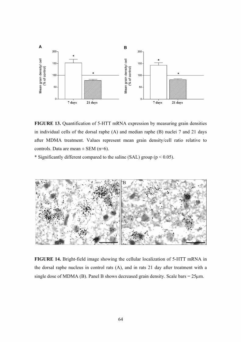
FIGURE 13. Quantification of 5-HTT mRNA expression by measuring grain densities
in individual cells of the dorsal raphe (A) and median raphe (B) nuclei 7 and 21 days
after MDMA treatment. Values represent mean grain density/cell ratio relative to
controls. Data are mean ± SEM (n=6).
* Significantly different compared to the saline (SAL) group (p < 0.05).
FIGURE 14. Bright-field image showing the cellular localization of 5-HTT mRNA in
the dorsal raphe nucleus in control rats (A), and in rats 21 day after treatment with a
single dose of MDMA (B). Panel B shows decreased grain density. Scale bars = 25μm.
64
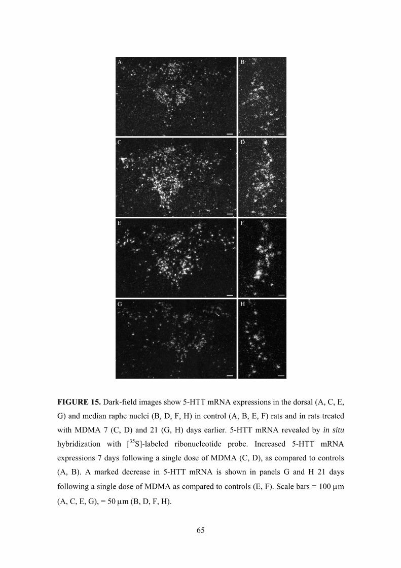
FIGURE 15. Dark-field images show 5-HTT mRNA expressions in the dorsal (A, C, E,
G) and median raphe nuclei (B, D, F, H) in control (A, B, E, F) rats and in rats treated
with MDMA 7 (C, D) and 21 (G, H) days earlier. 5-HTT mRNA revealed by in situ
hybridization with [35S]-labeled ribonucleotide probe. Increased 5-HTT mRNA
expressions 7 days following a single dose of MDMA (C, D), as compared to controls
(A, B). A marked decrease in 5-HTT mRNA is shown in panels G and H 21 days
following a single dose of MDMA as compared to controls (E, F). Scale bars = 100 μm
(A, C, E, G), = 50 μm (B, D, F, H).
65
5.1.2. 5-HTT mRNA expression in the median raphe nucleus
In the median raphe nucleus, 5-HTT mRNA densities were quantified by film
autoradiography at each survival time (3, 7, 21 and 180 days after MDMA treatment) by
treatment with MDMA (Figure 12A and 12C), and the changes in 5-HTT mRNA
expressions were further compared by analysis of the signal from the slides dipped into
autoradiographic emulsion, similarly to the measurements on the dorsal raphe nucleus
(Figure 13B). In general, significant treatment x time interaction (F3,39= 4.790, p
=0.006) was found in the MR. As shown in Figure 12C, 5-HTT mRNA expression
significantly decreased 21 days (-27%, p= 0.016) after drug exposure in the MR, but not
3 and 180 days after MDMA administration. To support these results by a more
sensitive method, 5-HTT mRNA expressions of individual cells were counted 7 and 21
days after MDMA administration. We found significant increase in the levels of 5-HTT
mRNA 7 days (+43%, p= 0.049) (Figure 13B, 15B and 15D) and significant decrease
21 days after MDMA treatment (-18%, p= 0.016) (13B, 15F and 15H). No differences
between controls and treated animals were observed 3 and 180 days after MDMA
administration (Figure 12A and 12C). Qualitatively, all of these effects were similar to
changes observed in the DR.
5.1.3. 5-HTT mRNA expression in the nucleus raphe pallidus, obscurus and magnus
Seven days after a single dose MDMA injection elevated 5-HTT mRNA
expressions were measured in the RMg (Figure 16, 17A, B, C and D), RPa and ROb
(Figure 19, 18A and 18B). The average increases of 5-HTT per cell were 24% (p=
0.101) in the RMg, 35% (p= 0.003) in the RPa and 10% (p= 0.012) in the ROb (Figure
16A, 19A and 19C). The values of the mean grey density were different in the three
different subregions of the RMg (rostral, middle and caudal). Significantly higher
values were found only in the middle portion of the nucleus (46%, p= 0.019). No
alterations in the levels of 5-HTT mRNA expressions or in cell numbers were detectable
in raphe nuclei examined 180 days after the exposure to MDMA (Figure 16B, 19 B and
19D) in the investigated nuclei, although values were measured only in three saline-
66

treated animals. The cell density (cell numbers/nuclei) did not show any alterations in
the raphe nuclei investigated 7 and 180 days after MDMA injections (Figure 20).
FIGURE 16. Mean grey density/cell in the raphe magnus (RMg) 7 days, (A) and 180
FIGURE 17. Low (A, B) and hig
days, (B) after MDMA treatment.
h (B, D) magnification (silver grains overlying a cell
bars: C, E= 100 μm; B, D= 20 μm.
body) bright-field microphotographs show 5-HTT mRNA expressions in raphe magnus
neurones 7 days after treatment with saline (A, B) or single dose of MDMA (C, D). 5-
HTT mRNA is revealed by in situ hybridization. Arrows indicate the midline. Scale
67
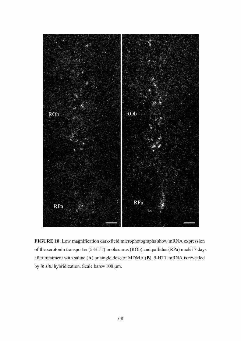
FIGURE 18. Low magnification dark-field microphotographs show mRNA expression
f the serotonin transporter (5-HTT) in obscurus (ROb) and pallidus (RPa) nuclei 7 days
o
after treatment with saline (A) or single dose of MDMA (B). 5-HTT mRNA is revealed
by in situ hybridization. Scale bars= 100 μm.
68

FIGURE 19. Number of grains/cell ratio in the raphe pallidus (RPa, A, B) and obscurus
group (p < 0.05).
(ROb, C, D) 7 days [saline (SAL): n = 6, MDMA: n = 7] and 180 days (SAL n = 3,
MDMA n = 6) after treatment. Data are mean ± SEM.
* Significantly different compared to the saline (SAL)
69
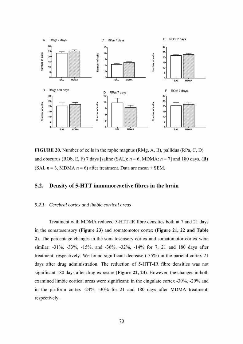
FIGURE 20. Number of cells in the raphe magnus (RMg, A, B), pallidus (RPa, C, D)
and obscurus (ROb, E, F) 7 days [saline (SAL): n = 6, MDMA: n = 7] and 180 days, (B)
(SAL n = 3, MDMA n = 6) after treatment. Data are mean ± SEM.
5.2. Density of 5-HTT immunoreactive fibres in the brain
5.2.1. Cerebral cortex and limbic cortical areas
Treatment with MDMA reduced 5-HTT-IR fibre densities both at 7 and 21 days
in the somatosensory (Figure 23) and somatomotor cortex (Figure 21, 22 and Table
2). The percentage changes in the somatosensory cortex and somatomotor cortex were
similar: -31%, -33%, -15%, and -36%, -32%, -14% for 7, 21 and 180 days after
treatment, respectively. We found significant decrease (-35%) in the parietal cortex 21
days after drug administration. The reduction of 5-HTT-IR fibre densities was not
significant 180 days after drug exposure (Figure 22, 23). However, the changes in both
examined limbic cortical areas were significant: in the cingulate cortex -39%, -29% and
in the piriform cortex -24%, -30% for 21 and 180 days after MDMA treatment,
respectively.
70
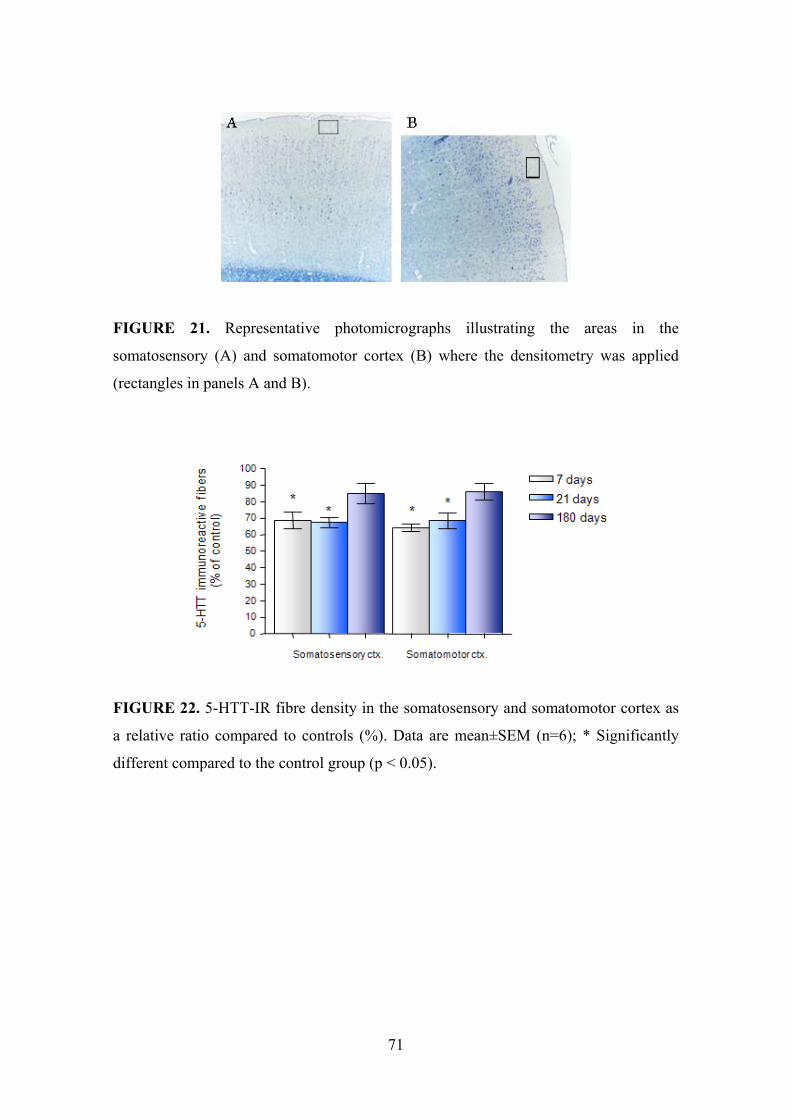
FIGURE 21. Representative photomicrographs illustrating the areas in the
somatosensory (A) and somatomotor cortex (B) where the densitometry was applied
(rectangles in panels A and B).
FIGURE 22. 5-HTT-IR fibre density in the somatosensory and somatomotor cortex as
a relative ratio compared to controls (%). Data are mean±SEM (n=6); * Significantly
different compared to the control group (p < 0.05).
71

TABLE 2.
5-HTT-IR fibre densities 7, 21 and 180 days after MDMA (15 mg/kg) treatment in brain areas of Dark Agouti rats
Brain area 7days 21days 180days SAL ± SEM MDMA ± SEM SAL ± SEM MDMA ± SEM SAL ± SEM MDMA ± SEM Cerebral cortex
Somatosensory cortex 6.92 ± 0.26 4.76 ± 0.35 * 6.02 ± 0.33 4.05 ± 0.18 * 6.31 ± 0.27 5.36 ± 0.40 Somatomotor cortex 7.17 ± 0.38 4.61 ± 0.16 * 6.27 ± 0.31 4.29 ± 0.28 * 6.28 ± 0.18 5.41 ± 0.32 *
Parietal cortex nm 5.90 ± 0.38 3.83 ± 0.18 * 6.44 ± 0.31 5.97 ± 0.16 Hippocampus
CA1 6.33 ± 0.47 4.67 ± 0.19 * 4.91 ± 0.52 3.09 ± 0.23 * 4.48 ± 0.55 2.87 ± 0.28 * CA2 7.08 ± 0.16 4.64 ± 0.36 * 5.79 ± 0.20 3.43 ± 0.27 * 5.37 ± 0.67 3.65 ± 0.28 * CA3 6.07 ± 0.25 4.09 ± 0.25 * 6.09 ± 0.28 3.30 ± 0.60 * 4.79 ± 0.43 3.31 ± 0.33 *
Dentate gyrus 2.78 ± 0.18 2.10 ± 0.20 2.81 ± 0.20 1.81 ± 0.11 2.80 ± 0.36 2.28 ± 0.24 Hypothalamus
Posteroventral preoptic areas 5.88 ± 0.49 5.23 ± 0.48 5.52 ± 0.44 4.57 ± 0.19 4.91 ± 0.58 4.80 ± 0.25 Lateral hypothalamic area 6.38 ± 0.48 5.60 ± 0.43 4.96 ± 0.18 4.03 ± 0.36 5.47 ± 0.29 5.04 ± 0.32
Posterior hypothalamic nucleus 6.71 ± 0.30 5.24 ± 0.31 * 6.25 ± 0.51 3.89 ± 0.40 * 4.52 ± 0.39 3.18 ± 0.30 * Paraventricular nucleus 5.43 ± 0.27 5.29 ± 0.46 6.08 ± 0.38 5.72 ± 0.52 5.84 ± 0.29 5.68 ± 0.27
Suprachiasmatic nucleus 6.78 ± 0.33 4.76 ± 0.34 * 5.53 ± 0.46 3.60 ± 0.19 * 4.84 ± 0.43 4.01 ± 0.28 Tuberomamillary nucleus 5.90 ± 0.19 4.78 ± 0.31 * 5.95 ± 0.36 3.86 ± 0.33 * 5.21 ± 0.37 4.02 ± 0.22 *
Brainstem Peripeduncular tegmental nucleus 4.70 ± 0.12 4.38 ± 0.28 4.12 ± 0.25 3.63 ± 0.33 3.93 ± 0.17 3.65 ± 0.46
Medial pontine reticular formation 4.23 ± 0.32 4.09 ± 0.35 2.93 ± 0.29 2.52 ± 0.32 3.22 ± 0.30 3.07 ± 0.38 Periaqueductal central grey
Ventrolateral part 10.34 ± 0.29 8.82 ± 0.39 8.28 ± 0.43 5.83 ± 0.42 * 6.66 ± 0.54 5.64 ± 0.71 Dorsomedial part 9.53 ± 0.71 7.92 ± 0.58 8.74 ± 0.60 5.61 ± 0.33 * 7.73 ± 0.70 6.47 ± 0.77
72

73
Brain area 7days 21days 180days SAL ± SEM MDMA ± SEM SAL ± SEM MDMA ± SEM SAL ± SEM MDMA ± SEM Substantia nigra 6.44 ± 0.36 5.16 ± 0.38 * nm 7.53 ± 0.21 6.76 ± 0.47 Limbic cortical areas
Cingulate cortex nm 5.88 ± 0.34 3.59 ± 0.07 * 5.48 ± 0.42 4.14 ± 0.28 * Piriform cortex nm 8.27 ± 0.15 5.87 ± 0.36 * 8.57 ± 0.81 5.98 ± 0.57 *
Striatum and pallidum nm Dorsomedial striatum nm 4.59 ± 0.37 2.62 ± 0.18 * 5.93 ± 0.39 4.83 ± 0.25 * Ventrolateral striatum nm 8.23 ± 0.54 5.93 ± 0.34 * 6.88 ± 0.34 5.79 ± 0.36
Caudate-putamen nm 7.44 ± 0.14 5.32 ± 0.26 * 6.50 ± 0.38 5.28 ± 0.39 * Globus pallidus nm 4.71 ± 0.43 3.03 ± 0.21 * 5.18 ± 0.33 4.15 ± 0.38
Bed nucleus of stria terminalis nm 5.92 ± 0.26 5.16 ± 0.26 5.40 ± 0.33 4.98 ± 0.26 Amygdala nm
Central nm 6.37 ± 0.36 5.51 ± 0.56 5.91 ± 0.39 5.54 ± 0.26 Medial nm 6.86 ± 0.51 5.27 ± 0.46 * 5.63 ± 0.37 5.05 ± 0.38
Mediodorsal thalamic nucleus nm 3.87 ± 0.42 2.91 ± 0.30 nm Arcuate nucleus nm 4.57 ± 0.55 5.25 ± 0.09 nm Anterior hypothalamic area nm 5.02 ± 0.56 4.70 ± 0.42 nm Medial preoptic area nm 4.45 ± 0.20 4.12 ± 0.69 nm Lateral septal nucleus nm 4.15 ± 0.38 3.55 ± 0.20 nm
Data are presented as mean ± SEM of 5-6 animals per group.
* Significantly different compared to the relevant control (SAL) group (p < 0.05).
(nm – not measured)
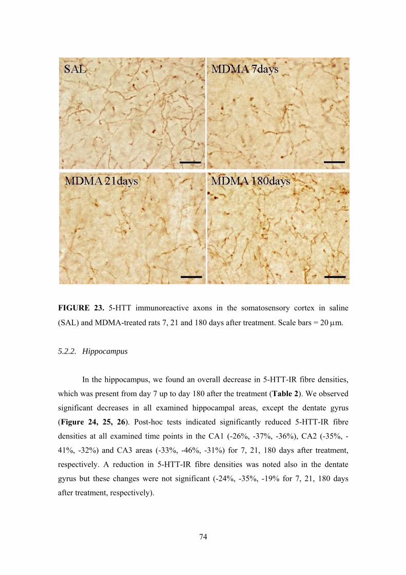
FIGURE 23. 5-HTT immunoreactive axons in the somatosensory cortex in saline
(SAL) and MDMA-treated rats 7, 21 and 180 days after treatment. Scale bars = 20 μm.
5.2.2. Hippocampus
In the hippocampus, we found an overall decrease in 5-HTT-IR fibre densities,
which was present from day 7 up to day 180 after the treatment (Table 2). We observed
significant decreases in all examined hippocampal areas, except the dentate gyrus
(Figure 24, 25, 26). Post-hoc tests indicated significantly reduced 5-HTT-IR fibre
densities at all examined time points in the CA1 (-26%, -37%, -36%), CA2 (-35%, -
41%, -32%) and CA3 areas (-33%, -46%, -31%) for 7, 21, 180 days after treatment,
respectively. A reduction in 5-HTT-IR fibre densities was noted also in the dentate
gyrus but these changes were not significant (-24%, -35%, -19% for 7, 21, 180 days
after treatment, respectively).
74

FIGURE 24. Representative photomicrographs illustrating the areas [oriens layer of
CA1, CA2 and CA3; molecular layer of GD (dentate gyrus)] in the hippocampus where
the densitometry was applied (rectangles in panels A - C).
FIGURE 25. 5-HTT-IR fibre density in the hippocampus CA1, CA2, CA2 and GD
(dentate gyrus) region as a relative ratio compared to controls (%).
Data are mean±SEM (n=6).
* Significantly different compared to the control group (p < 0.05).
75

FIGURE 26. 5-HTT immunoreactive axons in the hippocampal CA1 region in saline
(SAL) and MDMA-treated rats 7, 21 and 180 days after treatment. Scale bars = 20 μm.
5.2.3. Hypothalamus
The effects of MDMA on 5-HTT-IR fibre density were different in different
hypothalamic nuclei and areas (Table 2). Reduced 5-HTT-IR fibre densities were
measured in the posterior hypothalamic (PH), suprachiasmatic (SCN) and
tuberomamillary (TMN) nuclei, while the lateral (LH), posteroventral preoptic areas
(VLPO) and the paraventricular nucleus (PVN) were resistant to MDMA, at all time
points measured (Figure 27, 28). Alterations in 5-HTT-IR fibre densities were the
follows: LH -12%, -19%, -8%; PH -22%, -38%, -30%; SCN -30%, -35%, -17%; TMN -
19%, -35%, -23%; VLPO -11%, -17%, -2%; PVN -2%, -6%, -3%, for 7, 21 and 180
days after MDMA, respectively. The changes in 5-HTT-IR fibre density were not
76

significant at 21 days in the hypothalamic regions which connected to the aggressive
behaviour: arcuate nucleus (-14%) and anterior hypothalamic area (-7%).
FIGURE 27. Representative photomicrographs illustrating the areas in the
hypothalamus where the densitometry was applied (rectangles in panels A - F).
Abbreviations: f- fornix, mt- mammilothalamic tract, Ahy – anterior hypothalamic area,
3V – third ventricle
FIGURE 28. 5-HTT-IR fibre density in the different hypothalamic nuclei (VLPO, LH,
PH, PVN, SCN and TMN) as a relative ratio compared to controls (%). Data are
mean±SEM (n=6); * Significantly different compared to the control group (p < 0.05).
77

5.2.4. Brainstem
In general, changes in the brainstem were smaller (Figure 29, 30), and
significant alterations were observed only in two regions, in the ventrolateral (PAGvl)
and dorsomedial (PAGdm) cell groups of the periaqueductal gray matter in the
midbrain, but only at 21 days (Table 2). Effects of MDMA on the 5-HTT-IR fibre
densities in peripeduncular tegmental nucleus (PPTG) and the medial part of the pontine
reticular formation (mPRF) were not significant at any time points studied. Post-hoc
comparison revealed that the changes in 5-HTT-IR fibre densities were in PAGvl -15%,
-30%, -15%, PAGdm -17%, -36%, -16%, PPTG -7%, -12%, -7% and mPRF -3%, -14%,
-5%, for 7, 21, 180 days after treatment, respectively.
FIGURE 29.
Representative photomicrographs illustrating the areas in the brainstem where the
densitometry was applied (rectangles in panels A - C).
Abbreviations: DR – dorsal raphe nucleus; xscp – decussation of superior cerebellar
peduncles; MR – median raphe nucleus; Aq – aqueduct
78

FIGURE 30. 5-HTT-IR fibre density in the brainstem as a relative ratio compared to
controls (%). Data are mean±SEM (n=6); * Significantly different compared to the
control group (p < 0.05).
5.2.5. Other investigated brain areas
In the striatum and pallidum, we observed reduced 5-HTT-IR fibre density at 21
days. However, at 180 days we found significant decrease only in the dorsomedial
striatum and caudate-putamen. The percentage changes in the dorsomedial striatum,
ventrolateral striatum caudate-putamen and globus pallidus were follows: -43%, -19%
and -28%, -16% and -28%, -19% and -36%, -20% for 21 and 180 days after treatment,
respectively (Table 2). Effects of MDMA on 5-HTT-IR fibre densities in the substantia
nigra were significant (-20%) at 7 days (Table 2). In other examined brain areas (bed
nucleus of stria terminalis, central amygdala, mediodorsal thalamic nucleus, medial
preoptic area and lateral septal nucleus) the changes were not significant except in the
medial amygdala where we observed a 23% decrease at 21 days (Table 2).
79
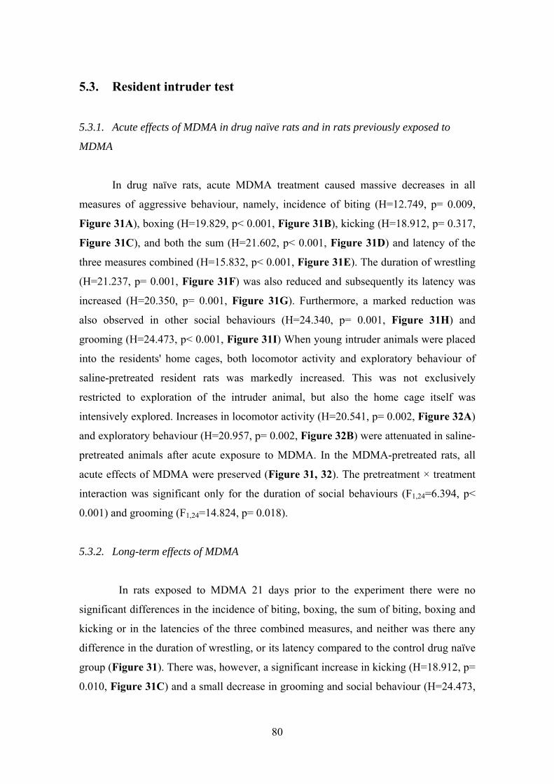
5.3. Resident intruder test
5.3.1. Acute effects of MDMA in drug naïve rats and in rats previously exposed to
MDMA
In drug naïve rats, acute MDMA treatment caused massive decreases in all
measures of aggressive behaviour, namely, incidence of biting (H=12.749, p= 0.009,
Figure 31A), boxing (H=19.829, p< 0.001, Figure 31B), kicking (H=18.912, p= 0.317,
Figure 31C), and both the sum (H=21.602, p< 0.001, Figure 31D) and latency of the
three measures combined (H=15.832, p< 0.001, Figure 31E). The duration of wrestling
(H=21.237, p= 0.001, Figure 31F) was also reduced and subsequently its latency was
increased (H=20.350, p= 0.001, Figure 31G). Furthermore, a marked reduction was
also observed in other social behaviours (H=24.340, p= 0.001, Figure 31H) and
grooming (H=24.473, p< 0.001, Figure 31I) When young intruder animals were placed
into the residents' home cages, both locomotor activity and exploratory behaviour of
saline-pretreated resident rats was markedly increased. This was not exclusively
restricted to exploration of the intruder animal, but also the home cage itself was
intensively explored. Increases in locomotor activity (H=20.541, p= 0.002, Figure 32A)
and exploratory behaviour (H=20.957, p= 0.002, Figure 32B) were attenuated in saline-
pretreated animals after acute exposure to MDMA. In the MDMA-pretreated rats, all
acute effects of MDMA were preserved (Figure 31, 32). The pretreatment × treatment
interaction was significant only for the duration of social behaviours (F1,24=6.394, p<
0.001) and grooming (F1,24=14.824, p= 0.018).
5.3.2. Long-term effects of MDMA
In rats exposed to MDMA 21 days prior to the experiment there were no
significant differences in the incidence of biting, boxing, the sum of biting, boxing and
kicking or in the latencies of the three combined measures, and neither was there any
difference in the duration of wrestling, or its latency compared to the control drug naïve
group (Figure 31). There was, however, a significant increase in kicking (H=18.912, p=
0.010, Figure 31C) and a small decrease in grooming and social behaviour (H=24.473,
80
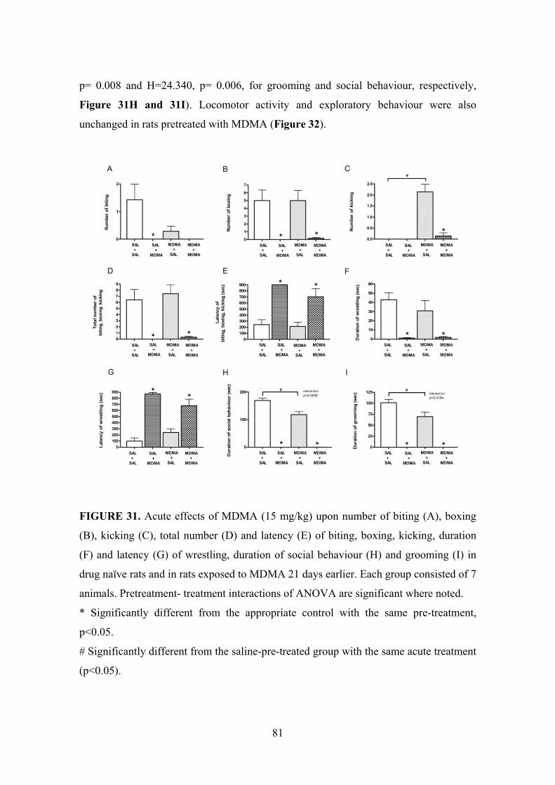
p= 0.008 and H=24.340, p= 0.006, for grooming and social behaviour, respectively,
Figure 31H and 31I). Locomotor activity and exploratory behaviour were also
unchanged in rats pretreated with MDMA (Figure 32).
FIGURE 31. Acute effects of MDMA (15 mg/kg) upon number of biting (A), boxing
(B), kicking (C), total number (D) and latency (E) of biting, boxing, kicking, duration
(F) and latency (G) of wrestling, duration of social behaviour (H) and grooming (I) in
drug naïve rats and in rats exposed to MDMA 21 days earlier. Each group consisted of 7
animals. Pretreatment- treatment interactions of ANOVA are significant where noted.
* Significantly different from the appropriate control with the same pre-treatment,
p<0.05.
# Significantly different from the saline-pre-treated group with the same acute treatment
(p<0.05).
81

FIGURE 32. Acute effects of MDMA (15 mg/kg) on motor activity and exploratory
behaviour in drug naïve rats and in rats exposed to MDMA 21 days earlier. Each group
consisted of 7 animals. * Significantly different from the appropriate control with the
same pre-treatment (p<0.05).
5.4. Effects of MDMA on sleep parameters
5.4.1. REM sleep
Complete 24 h recordings of REM sleep are shown in Figure 33A–C. In
addition, four parameters, REM latency, REM in the first hour of passive phase, time of
REM maximum and the sum of REM in the light period (passive phase) were measured
and calculated for the characterization of onset and duration of REM sleep (Table 3).
Significant decreases in REM latency were detected 7 and 21 days after MDMA
administration (treatment effect F1,36= 22.880, p< 0.0001). Changes in REM latency
were –55%, –53%, and –29%, 7, 21 and 180 days after MDMA, respectively. In
parallel, increased duration of REM (+245%) was found in the first hour of the passive
phase 21 days after MDMA (treatment effect F1,46= 7.689, p= 0.0079). Duration of
REM sleep analyzed during the whole light period (–15%, –34%, –15% at 7, 21 and 180
days after treatment, respectively) was not significantly affected by MDMA treatment.
At 7 days, some MDMA-treated animals showed an extreme time course of REM
during the passive phase; REM activity showed a biphasic pattern, with the higher peak
occurring very late, close to the dark phase (Figure 33A).
82

FIGURE 33. Twenty-four hour recordings of rapid eye movement (REM) sleep in
MDMA and saline (SAL)-treated animals 7 (A), 21 (B) and 180 (C) days after
treatment. Each point represents mean values ± SEM (n= 6-8) of the time spent in REM
sleep within each hour.
* Significantly different compared to the SAL group (p < 0.05).
83
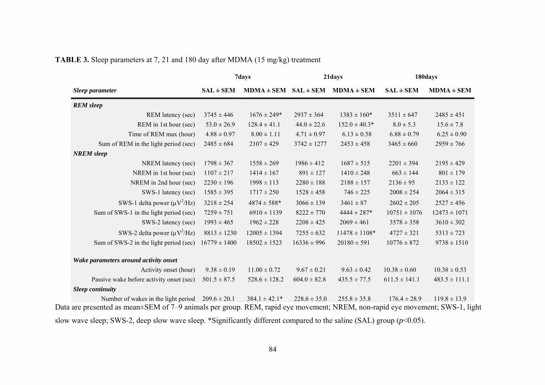
TABLE 3. Sleep parameters at 7, 21 and 180 day after MDMA (15 mg/kg) treatment
Data are presented as m me
slow wave sleep; SWS-2, deep slow wave sleep. *Significantly different compared to the saline (SAL) group (p<0.05).
ean±SEM of 7–9 animals per group. REM, rapid eye movement; NREM, non-rapid eye move nt; SWS-1, light
7days 21days 180days
Sleep parameter SAL ± SEM MDMA ± SEM SAL ± SEM MDMA ± SEM SAL ± SEM MDMA ± SEM
REM sleep REM latency (sec) 3745 ± 446 1676 ± 249* 2937 ± 364 1383 ± 160* 3511 ± 647 2485 ± 451
REM in 1st hour (sec) 53.0 ± 26.9 128.4 ± 41.1 44.0 ± 22.6 152.0 ± 40.3* 8.0 ± 5.3 15.6 ± 7.8 Time of REM max (hour) 4.88 ± 0.97 8.00 ± 1.11 4.71 ± 0.97 6.13 ± 0.58 6.88 ± 0.79 6.25 ± 0.90
Sum of REM in the light period (sec) 2485 ± 684 2107 ± 429 3742 ± 1277 2453 ± 458 3465 ± 660 2959 ± 766 NREM sleep
NREM latency (sec) 1798 ± 367 1558 ± 269 1986 ± 412 1687 ± 515 2201 ± 394 2195 ± 429 NREM in 1st hour (sec) 1107 ± 217 1414 ± 167 891 ± 127 1410 ± 248 663 ± 144 801 ± 179
NREM in 2nd hour (sec) 2230 ± 196 1998 ± 113 2280 ± 188 2188 ± 157 2136 ± 95 2133 ± 122 SWS-1 latency (sec) 1585 ± 395 1717 ± 250 1528 ± 458 746 ± 225 2008 ± 254 2064 ± 315
SWS-1 delta power (μV2/Hz) 3218 ± 254 4874 ± 588* 3066 ± 139 3461 ± 87 2602 ± 205 2527 ± 456 Sum of SWS-1 in the light period (sec) 7259 ± 751 6910 ± 1139 8222 ± 770 4444 ± 287* 10751 ± 1076 12473 ± 1071
SWS-2 latency (sec) 1993 ± 465 1962 ± 228 2208 ± 425 2069 ± 461 3578 ± 358 3610 ± 302
SWS-2 delta power (μV2/Hz) 8813 ± 1230 12005 ± 1394 7255 ± 632 11478 ± 1108* 4727 ± 321 5313 ± 723 Sum of SWS-2 in the light period (sec) 16779 ± 1400 18502 ± 1523 16336 ± 996 20180 ± 591 10776 ± 872 9738 ± 1510
Wake parameters around activity onset Activity onset (hour) 9.38 ± 0.19 11.00 ± 0.72 9.67 ± 0.21 9.63 ± 0.42 10.38 ± 0.60 10.38 ± 0.53
Passive wake before activity onset (sec) 501.5 ± 87.5 528.6 ± 128.2 604.0 ± 82.8 435.5 ± 77.5 611.5 ± 141.1 483.5 ± 111.1 Sleep continuity
Number of wakes in the light period 209.6 ± 20.1 384.1 ± 42.1* 228.6 ± 35.0 255.8 ± 35.8 176.4 ± 28.9 119.8 ± 13.9
84
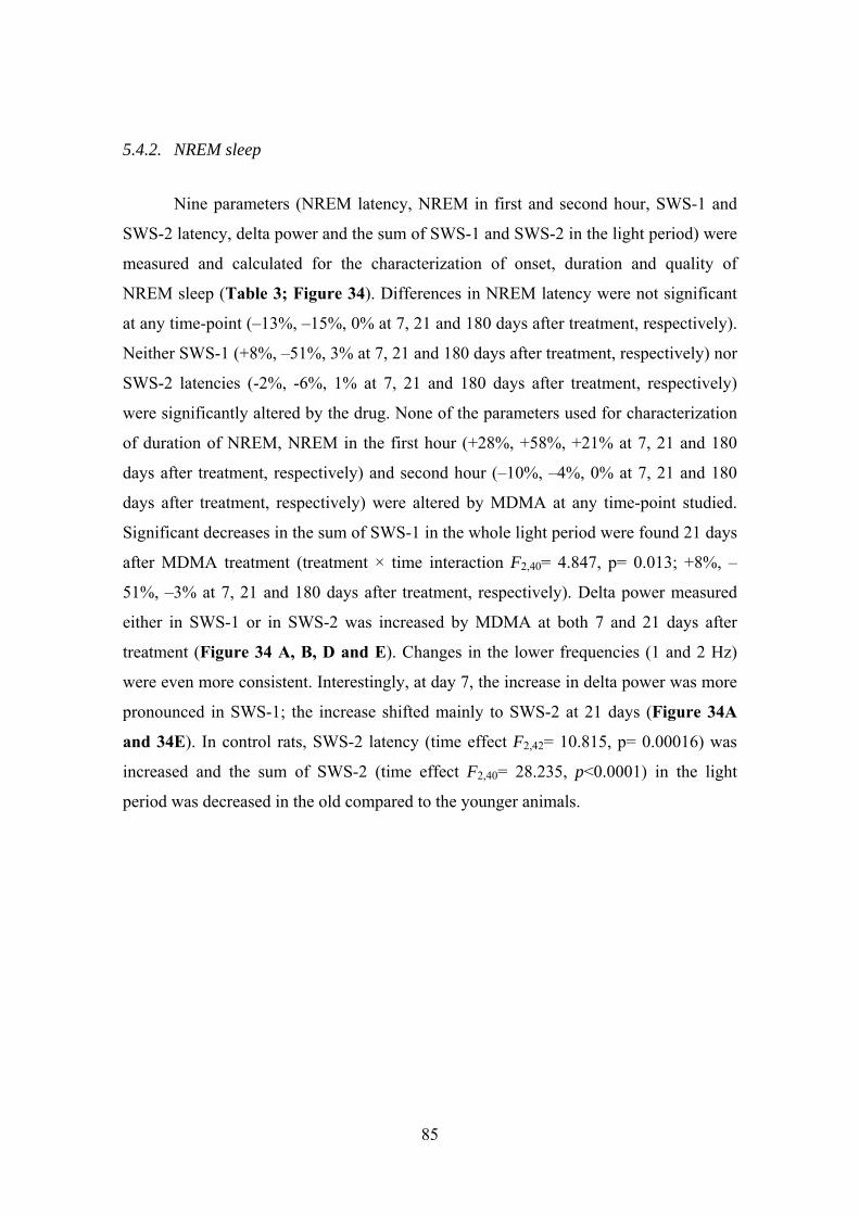
5.4.2. NREM sleep
Nine parameters (NREM latency, NREM in first and second hour, SWS-1 and
SWS-2 latency, delta power and the sum of SWS-1 and SWS-2 in the light period) were
measured and calculated for the characterization of onset, duration and quality of
NREM sleep (Table 3; Figure 34). Differences in NREM latency were not significant
at any time-point (–13%, –15%, 0% at 7, 21 and 180 days after treatment, respectively).
Neither SWS-1 (+8%, –51%, 3% at 7, 21 and 180 days after treatment, respectively) nor
SWS-2 latencies (-2%, -6%, 1% at 7, 21 and 180 days after treatment, respectively)
were significantly altered by the drug. None of the parameters used for characterization
of duration of NREM, NREM in the first hour (+28%, +58%, +21% at 7, 21 and 180
days after treatment, respectively) and second hour (–10%, –4%, 0% at 7, 21 and 180
days after treatment, respectively) were altered by MDMA at any time-point studied.
Significant decreases in the sum of SWS-1 in the whole light period were found 21 days
after MDMA treatment (treatment × time interaction F2,40= 4.847, p= 0.013; +8%, –
51%, –3% at 7, 21 and 180 days after treatment, respectively). Delta power measured
either in SWS-1 or in SWS-2 was increased by MDMA at both 7 and 21 days after
treatment (Figure 34 A, B, D and E). Changes in the lower frequencies (1 and 2 Hz)
were even more consistent. Interestingly, at day 7, the increase in delta power was more
pronounced in SWS-1; the increase shifted mainly to SWS-2 at 21 days (Figure 34A
and 34E). In control rats, SWS-2 latency (time effect F2,42= 10.815, p= 0.00016) was
increased and the sum of SWS-2 (time effect F2,40= 28.235, p<0.0001) in the light
period was decreased in the old compared to the younger animals.
85
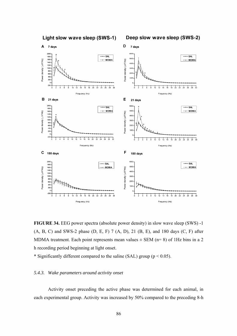
FIGURE 34. EEG power spectra (absolute power density) in slow wave sleep (SWS) -1
(A, B, C) and SWS-2 phase (D, E, F) 7 (A, D), 21 (B, E), and 180 days (C, F) after
MDMA treatment. Each point represents mean values ± SEM (n= 8) of 1Hz bins in a 2
h recording period beginning at light onset.
* Significantly different compared to the saline (SAL) group (p < 0.05).
5.4.3. Wake parameters around activity onset
Activity onset preceding the active phase was determined for each animal, in
each experimental group. Activity was increased by 50% compared to the preceding 8-h
86
resting period 2–3 h before lights off. No significant MDMA effect was found for this
parameter (+17%, 0%, 0% at 7, 21 and 180 days after treatment, respectively). Duration
of passive wake during the hour preceding the activity alteration was also calculated.
Changes in the duration of passive wake before activity onset were +5%, –28%, –21%
at 7, 21 and 180 days after treatment, respectively. None of these parameters were
significantly altered by MDMA treatment (Table 3).
5.4.4. Sleep continuity
The number of wake periods during the 12 h passive phase was used to characterize
sleep continuity. MDMA caused a significant increase in this parameter but only at the
7th day (+83%, +12%, –32% at 7, 21 and 180 days after treatment, respectively) (Table
3). The treatment × time interaction (F2,41= 7.306, p= 0.0019) was significant.
6. DISCUSSION
Parallel measurements of 5-HTT mRNA expression in five raphe nuclei, 5-HTT-
IR fibre density in several brain areas, and more than a dozen functional measures of
sleep at three time-points within 180 days after MDMA administration allowed us to
compare sleep disturbance with morphometric data and functions of raphe nuclei during
a long period of time when the brain serotonergic system was damaged or partially
recovered. Furthermore, we performed resident-intruder test to study the aggressive
behaviour in drug-naïve rats and also in rats previously exposed to MDMA 21 days
earlier.
In our study, MDMA was administered to Dark Agouti rats at a dose of 15
mg/kg. Although there are considerable strain differences in the susceptibility to
MDMA-induced long-term serotonergic depletion, many authors suggests that a single,
15 mg/kg injection of MDMA in rats may more accurately model the possible exposure
to the drug in humans, particularly when pharmacokinetic scaling is applied to body size
and basal metabolic rate (Colado and Green, 1995; Malpass et al., 1999). In contrast, it
is less likely that previously used models in which, typically, rats are treated with a
higher dose (20 mg/kg) twice a day for four consecutive days adequately reflect human
87
experience. Moreover, Dark Agouti rats show decreased microsomal CYP2D6
isoenzyme activity, which plays an important role in the metabolism of MDMA. As a
consequence, the Dark Agouti rat is considered to be a poor or moderate metaboliser of
the drug with MDMA pharmacokinetics that is similar to those which may be found in
particularly vulnerable human users (Balogh et al., 2004; O'Shea et al., 1998; Quate et
al., 2004). Such users might represent a genetically-defined human sub-population in
which clinical complications are more likely to occur.
Human studies are retrospective and are undermined by several factors that are
impossible to control (Curran et al., 2004; Morgan, 2000; Parrott et al., 2000),
furthermore, the variation in the metabolic rate that is crucial in the effect of MDMA is
very high. These complications include inaccurate reporting of the doses of MDMA
taken, the duration and frequency of drug use, and non-reporting of the concurrent use
of other substances, licit or illicit (Green et al., 1995; Morgan, 2000). Interindividual
variability of serotonergic damage measured by imaging techniques in humans is also
very high (McCann et al., 1998; McCann et al., 2005), but the results of these studies
are in many cases not significant. Therefore we assumed that a single dose treatment
with significant axotomy may cause changes in the functional parameters. Some other
data suggest that this dose in rats is in the higher dose range (O'Shea et al., 1998).
Indeed, the lesion is significant in our rats; and significant decrease by brain imaging
was found only in heavy ecstasy users (Ricaurte et al., 2000a). This suggests that our
dose regimen is relevant to heavy ecstasy use in humans or at least it mimics some
effect of heavy ecstasy use.
Under room temperature (20–22°C), MDMA administration to rats has generally
been reported to produce a marked hyperthermic response of approximately +1–2°C
(Broening et al., 1995; Colado et al., 1993; Dafters, 1994; Nash et al., 1988). In our
other study we found that the average body temperature was 1.4°C higher in MDMA-
treated rats than in saline-treated rats (Kovacs et al., 2007). In contrast, this change
cannot be considered great compared to results from humans where hyperthermia was
found to be one of the major symptoms of acute MDMA-induced toxicity, and body
temperatures of over 43°C have been reported.
All the timepoints we selected are after the acute effect period, however, the
majority of studies carried out in humans are based on weekend drug consumption
88

(Curran and Travill, 1997; Parrott, 2001). Effects such as fatigue, low mood,
concentration and memory difficulties, reduction of appetite, etc. last for about a week
or months (Morgan, 2000; Parrott, 2001). At the third week morphological studies
indicate significant axon damage which has been stabilised, however, regeneration has
not started.
6.1. Morphological effects of MDMA
In the present study, 5-HTT mRNA expression was measured in the two largest
group of ascending projecting serotonergic neurons, the DR and the MR. A transient
increase followed by a transient decrease and a complete recovery was found during the
180-day period in dorsal and median raphe nuclei. The effects of MDMA on 5-HTT
mRNA expression in DR and MR did not differ in time or extent.
In the examined three descending raphe nuclei, RMg, ROb and RPa, we
observed up-regulation of 5-HTT mRNA expressions at 7 days and also a complete
recovery at 180 days. The up-regulation of 5-HTT mRNA expressions 7 days after
MDMA treatment in all studied raphe nuclei may be explained by a compensatory
phenomenon following decreased axonal transport or a possible distal down-regulation
of the expression of 5-HTT (Kovacs et al., 2007).
In addition, 5-HTT-IR fibre density was also measured in a number of brain
regions. Decreases were detected in several brain areas, from the brainstem up to the
cerebral cortex at 7 and 21 days. Evidence for partial recovery was found in most
affected brain regions by 180 days. Our earlier results show diminished TPH- fibre
density in several brain regions 3 days, 1 and 3 weeks after the administration of
MDMA (Adori et al., 2006; Ando et al., 2006).
It is well established for several species (Hornung et al., 1990; Kosofsky and
Molliver, 1987; Mulligan and Tork, 1988) that the cerebral cortex contains at least two
major types of 5-HT fibres. It was suggested that the fine fibres are derived from the
DR, while thicker fibres are derived from the MR (Kosofsky and Molliver, 1987;
Mulligan and Tork, 1988). It has been reported previously that the fine serotonergic
fibres are selectively vulnerable to a number of amphetamine derivatives (Mamounas
and Molliver, 1988; O'Hearn et al., 1988). However, more recent neuroanatomical and
89
biochemical studies suggest that DR selectivity of these amphetamines may not be
exclusive (Haring et al., 1992; Hensler et al., 1994; Mamounas and Molliver, 1988;
McQuade and Sharp, 1995; O'Hearn et al., 1988). Data obtained from these studies are
in accordance with the observation that fine fibres arise from both raphe nuclei (Morin
and Meyer-Bernstein, 1999). Our immunohistochemical data support the conclusion
that axons of MR origin are also affected by MDMA. The origin of serotonergic
projections to the SCN is almost exclusively the MR (Glass et al., 2003; Meyer-
Bernstein et al., 1999), and MR fibres project heavily throughout the entire
hippocampus (Azmitia and Segal, 1978; Conrad et al., 1974; Vertes and Martin, 1988).
The SCN and the three CA areas of the hippocampus were strongly affected by MDMA
in our experiment. Furthermore, changes in 5-HTT mRNA expression in the DR and
MR were similar in the present study. These data support the view that axons of both
the DR and MR origin are temporarily damaged by the drug.
The spinal cord receives a strong and highly organized 5-HT projection
(Anderson et al., 1989). The major source of 5-HT innervation to the spinal cord arises
from the caudal groups of 5-HT cells (Halliday et al., 1995; Hökfelt et al., 1978;
Skagerberg and Bjorklund, 1985). The RMg innervates mainly the dorsal horn. The 5-
HT cells in ROb and RPa project to the ventral horn and the intermediolateral column
(Bacon et al., 1990; Törk, 1990). Few studies have examined the effect of MDMA or
other amphetamine analougues (e.g. PCA) in the rat spinal cord. Ridet et al. found that
PCA treatment caused a marked and selective elimination of fine 5-HT-IR fibres in the
dorsal horn and spared all other larger axons (Ridet et al., 1994). Several studies
reported that 5-HT plays an important role in pain perception (Basbaum and Fields,
1984; Hamon, 1997; LeBars, 1988) and the rostral ventromedial medulla, which
includes the RMg, is a key neural structure for pain modulation (Marinelli et al., 2002;
Mason, 2001; Taylor and Basbaum, 1995) with ON and OFF cells modulating pain
inhibition and facilitation (Yang et al., 2006). Besides serotonin there is also a number
of neuropeptides coexisting in the RMg, including endogenous opiate peptides,
substance P, somatostatin, galanin, vasoactive intestinal polypeptide, neuropeptide Y
and calcitonin gene-related peptide (Fields and Basbaum, 1994; Huang et al., 2000).
It is known that the neurotoxins selective for 5-HT neurons (e.g. 5,6-DHT and
5,7-DHT) prevent morphine-induced antinociception (Romandini et al., 1986). In
90

contrast, one study observed the enhancement of morphine-induced analgesia after
repeated injections of MDMA in rats. Furthermore, two weeks after drug treatment the
5-HT content decreased mostly in the cervical portion of the spinal cord and the
reduction was smaller in the thoracic segment (Nencini et al., 1988). Our group (Kovacs
et al., 2007) found decreased TPH fibre densities in all spinal cord regions 1 week after
MDMA administration, although the dorsal horn was the most vulnerable where the
fibre density was still lower, albeit statistically not significant at 180 days. Ricaurte et
al. demonstrated decreased 5-HT and 5-HIAA concentration in the monkey cervical
spinal cord two weeks after MDMA treatment (Ricaurte et al., 1988a). These results
indicated that MDMA is toxic to serotonergic fibres not only arising from cells in the
DR but also in the descending raphe nuclei. Our data indicate that MDMA treatment
influences the descending serotonergic cells and perhaps several physiological functions
which are connected to the spinal cord e.g. pain, autonomic function and motor activity
as well.
It is well known that serotonergic neurons show a high capacity for axonal
sprouting and regeneration after a variety of neuron lesioning techniques, including
some using 5,6- and 5,7-DHT (Björklund et al., 1981; Frankfurt and Azmitia, 1984;
Wiklund and Mollgard, 1979) in the brain. The serotonergic damage occurring after
very high single doses or after successive moderate doses of MDMA has been
confirmed across a variety of animal species, including rats, monkeys and other
primates. Neuronal recovery is most apparent in rats, whereas monkeys show only
partial recovery even after a prolonged period (Ricaurte et al., 2000a). Recovery of
serotonergic neurons in most areas examined was observed in animals treated with a
single dose of MDMA 180 days previously. Among the exceptions hippocampus should
be noted because we found a significant reduction (31-36%) in almost all hippocampal
areas 6 months after MDMA treatment. Our results support previous observations that 1
year following MDMA treatment the hippocampus is the only brain region where none
of the presynaptic 5-HT neuronal markers recovered (Scanzello et al., 1993). This
observation supports previous results that the rate and degree of recovery is region
dependent (Lew et al., 1996; Sabol et al., 1996). Our results support previous data
showing that the hippocampus is more resistant to recovery compared to other brain
regions. The explanation for this might be the specific hierarchy of hippocampal
91

neurons and/or alteration of synthesis or metabolism of neurotrophic factors (Martinez-
Turrillas et al., 2006).
In a previous study (Lew et al., 1996) it was found that 2 weeks after MDMA
administration the density of serotonin uptake sites in the cingulate cortex was 20% of
that in saline-treated animals, but had significantly increased to 43% of the density of
controls by 8 weeks and was completely recovered by 16 weeks. Our data partially
support these results because 5-HTT-IR fibre density in the limbic cortical areas was
decreased at 21 days but not normalized at 6 months after MDMA.
Among the consequences of these findings might be long-term changes in
memory functions and/or anxiety. MDMA-treated rats also displayed impaired memory
3 months after drug administration in an object recognition test (Morley et al., 2001).
Indeed, human studies most consistently point to cognitive and memory disturbances in
current and previous ecstasy users (Morgan, 2000; Parrott, 2001).
At 180 days we observed a significant reduction in 5-HTT-IR fibre density in the
dorsomedial striatum, caudate-putamen, and a trend towards decrease was found in the
globus pallidus (p= 0.068) and in the ventrolateral striatum (p=0.053). In one study
(Callaway and Geyer, 1992) investigating 5-HT and 5-HIAA levels in the striatum 36
days after MDMA treatment a significant reduction was observed. Similar results were
described earlier (Gurtman et al., 2002; McGregor et al., 2003) 10 and 12 weeks after
MDMA administration. Our study supports these results because we found significant
decrease in all examined striatal brain areas 21 days after MDMA treatment. Balogh et
al. demonstrated a significant decrease (16–20%) in the local cerebral metabolic glucose
utilisation in the medial striatum and globus pallidus three weeks after a single MDMA
administration in Dark Agouti rats, although change in [3H]paroxetine binding was not
significant in this study (Balogh et al., 2004).
High densities of 5-HT1B receptor were found in motor control centers such as
the basal ganglia (Sari, 2004). Several studies have suggested that 5-HT1B receptors are
involved in several physiological functions, behaviours and psychiatric diseases
including control of locomotor activity, drug abuse reinforcement, migraine, anxiety
states (Castanon et al., 1997; Green et al., 1984; Griebel et al., 1990) and aggressive
behaviour (Flannelly et al., 1985; Kirilly et al., 2006). Previous studies suggested that
some of the psychotropic and behavioural effects of MDMA may be mediated by
92

altered 5-HT1B receptor function. Sexton et al. examined the regulation of rat 5-HT1B
receptors after acute and chronic MDMA treatment and observed a transient alteration
in striatal 5-HT1B binding sites (Sexton et al., 1999). This study found no change in 5-
HT1B mRNA immediately or after 2 weeks, suggesting a posttranslational mechanism of
regulation. 5-HT1B receptors appear to mediate MDMA-induced motor hyperactivity:
hyperactivity is reduced by antagonists that block 5-HT1B receptors and it is attenuated
in 5-HT1B knockout mice (Clark and Neumaier, 2001). Gyongyosi et al. demonstrated a
reduced or eliminated AW and diurnal rhythm of this parameter after the administration
of the 5-HT1B agonist CP-94,253, in animals treated with MDMA 6 months earlier
(Gyongyosi et al., 2008). Parallel MDMA treatment decreased 5-HTT-IR fibre density
in several brain areas that are involved in sleep regulation and motor control. In our
study the loss of 5-HT nerve terminals in the striatum 6 months after MDMA
administration support earlier data that MDMA may cause long-term changes in 5-HT1B
receptor function.
Some authors described an increase in serotonergic markers in certain areas
(globus pallidus, hypothalamus) several months after the decrease of the axon density
which they explained as a „pruning effect”. One study provided evidence that 3 of the
examined 16 rats showed an altered pattern of distribution of 5-HT axon markers in the
hypothalamus in animals treated with MDMA 52 weeks previously (Fischer et al.,
1995). In our experiments, we used 6 animals per group, and we did not find any sign of
hyperinnervation in any brain areas examined. One possible explanation for the lack of
this phenomenon is that the relatively small single dose (15 mg\kg) applied in our study
caused only moderate destruction of axons and terminals. Even in the most affected
areas the changes did not exceed 30-40%. In previous studies where prunning effect has
been shown, higher doses of MDMA were used. Either rats or rhesus monkeys were
treated twice a day for 4 consecutive days, thus e.g. for rats a cumulative dose of 160
mg\kg\animal was used (Fischer et al., 1995; Hatzidimitriou et al., 1999).
93

6.2. Effects of MDMA on sleep
Acute and subacute effects of MDMA on sleep have been described by our
group earlier (Balogh et al., 2004). Here we concentrated on chronic and persistent
morphological and functional effects of the drug. A selective decrease in REM latency
was found 7 and 21 days after MDMA administration. Similarly, an increase in the
amount of REM was found in the first hour. None of the other parameters characterizing
REM sleep were altered. Our data support the view that initiation of REM sleep is
modulated by the serotonergic system (Adrien, 2002; Bhatti et al., 1998; Portas et al.,
1996). In contrast, none of the other REM sleep parameters nor most of the NREM
sleep parameters were affected by the temporary lesion of the serotonergic system in
our study. Sleep disturbances e.g., higher amounts of REM sleep, altered transition from
NREMs to REMs have been described in 5-HTT knockout mice (Alexandre et al., 2006;
Wisor et al., 2003). It is, however, not known whether a transient decrease of 5-HTT
expression together with decreased 5-HT availability in adulthood might cause any
significant effect in sleep parameters. It has been suggested that higher amounts of REM
sleep, described in 5-HTT knockout mice (Alexandre et al., 2006; Wisor et al., 2003)
result from excessive serotonergic tone during early life (Alexandre et al., 2006). Our
data support this notion, because a transient decrease of 5-HTT expression together with
decreased 5-HT availability observed 21 days after MDMA treatment failed to cause a
general enhancement in the amount of REM sleep. Increase in REM sleep was restricted
to the first hour of passive phase suggesting that it was caused solely by a decrease in
REM latency.
MDMA administration is followed by depletions in 5-HTT-IR fibre density in
some, but not all, brain areas involved in the regulation of REM sleep examined in this
study. Significant damage was found in the SCN and tuberomamillary nucleus, parallel
with reduced REM latency 7 and 21 days after MDMA treatment. In contrast, we could
not find significant changes in 5-HTT density around the “effector” neurons located in
the medial pontine reticular formation (Sinton and McCarley, 2004) and the
peripeduncular tegmental nucleus (Adrien, 2002) in the brainstem, thus, it is unlikely
that the observed decrease in REM latency could be a consequence of changes in the
serotonergic system of these areas.
94
The 5-HTT-IR fibre densities were also diminished in the tuberomamillary
nucleus which is also an important part of the arousal system. The histaminergic TMN,
parallel with the dorsal raphe serotonergic neurons, fire fastest during wakefullness,
slow down with EEG during NREM sleep, and nearly stop firing during REM sleep
(Saper et al., 2001; Steininger et al., 1999; Strecker et al., 2002). There is evidence that
the serotonergic system activates the histaminergic neurons (Eriksson et al., 2001; Saper
et al., 2001) thus the decrease of 5-HTT-IR fibre densities in the TMN may indicate a
reduced 5-HT excitation on histaminergic TMN neurons which may result in decreased
NREM and possibly REM latencies.
The decrease in REM latency induced by MDMA may be explained by a
decreased inhibition of the SCN by 5-HT, due to an earlier activation of SCN. Serotonin
inhibits the spontaneous discharge of SCN neurons in vivo and in hypothalamic slices in
vitro (Yu et al., 2001). This effect may be mediated via the 5-HT7 and 5-HT1B receptors
(Yannielli and Harrington, 2004; Yu et al., 2001). The SCN strongly promotes REM
sleep tendency during the rest phase, and the SCN neurons show increased activity
during REM sleep compared to NREM sleep, the peak of the firing rate of SCN is at the
middle of the passive phase when the amount of REM sleep is the highest during the
day (Deboer et al., 2003; Wurts and Edgar, 2000). In conclusion, the decrease in 5-
HTT-IR fibre density in the SCN could be one possible explanation for the decreased
REM latency.
MDMA has been shown to disrupt the circadian cycle in vivo and the firing
patterns of SCN in hypothalamic slices (Balogh et al., 2004; Colbron et al., 2002;
Dafters and Biello, 2003). Furthermore, disruption of the regular cycle has been
connected to serotonergic mechanisms (Biello and Dafters, 2001; Colbron et al., 2002).
Our findings of altered 5-HTT densities in the SCN after MDMA treatment further
support the role of SCN in the sleep disturbances caused by MDMA.
The importance of our findings is further supported by the fact that disturbances
of sleep are typical for most depressed patients and belong to the core symptoms of
depression according to DSM-IV (Adrien et al., 1991; Akiskal et al., 1982; Riemann et
al., 2001). More than 90% of depressed patients complain about disturbances in sleep
quality (Mendelson et al., 1977; Riemann et al., 2001). Difficulties in falling asleep,
frequent nocturnal awakenings and early morning awakening are the most frequent
95

complaints, and reduced REM latency and increased sleep fragmentation are the most
characteristic alterations described in depression using EEG sleep polygraphy (Riemann
et al., 2001). Kupfer et al. also reported the changes in sleep quality in NREM and
problems with sleep continuity (Kupfer et al., 1984). Based on these data the
hypofunction of the serotonergic system in depression has been suggested (Adrien,
1999). However, it is not known whether these alterations are also connected to
decreased availability of 5-HT in the brain.
Despite the reduced REM latency, we could not find significant changes in the
amount of REM sleep after REM onset. The firing rate of the DR neurons is almost
absent and is also decreasing in the MR during REM sleep (McGinty and Harper, 1976;
Portas et al., 1996; Urbain et al., 2006). It is more likely that during REM latency,
before the onset of REM sleep, the MDMA-induced damage elicits a faster reduction of
the 5-HT input, thus, the threshold will be reached earlier. The amount of REM sleep
does not change after that since the 5-HT input is absent. Furthermore, the amount of
REM sleep, once initiated, is determined primarily by homeostatic mechanisms (Wurts
and Edgar, 2000).
Among the nine parameters of NREM sleep, only three were altered by MDMA
treatment and even these alterations failed to follow the time-course of serotonergic
damage. The most consistent effect was an increase in delta power during the first hours
of the passive phase in NREM sleep. In addition, SWS-1 in the passive phase was
decreased at 21 days. This finding is consistent with the increase in delta power caused
by 5-HT2A receptor antagonists (Dugovic et al., 1989; Monti and Monti, 1999; Sebban
et al., 2002; Ursin, 2002). Interestingly, 5-HT2B and 5-HT2C receptor antagonists fail to
cause a similar effect (Kantor et al., 2002; Popa et al., 2005). The present experiments
provide evidence that a partial lesion of the serotonergic system depletes the
endogenous 5-HT tone that physiologically inhibits delta activity during NREM sleep.
The role of tonic inhibition of delta power by 5-HT through 5-HT2A receptors has been
suggested on the basis of pharmacological studies (Kantor et al., 2002; Kitka and
Bagdy, 2008; Monti and Monti, 1999; Sebban et al., 2002; Ursin, 2002).
A decrease in spontaneously expressed NREM sleep was found in knock out
mice lacking 5-HT2A receptors (Popa et al., 2005). The decrease in SWS-1 found in our
study is consistent with these data. Since absolutely no decrease in SWS-1 was found at
96
day 7 despite of an extensive decrease in serotonergic axon density, this latter effect
could be considered an adaptive change due to the lack of appropriate tonic activation of
5-HT2A receptors.
NREM disturbances, like increased sleep latency, diminished deep slow wave
sleep and changes in wake parameters described in depression (Kupfer et al., 1990;
Naylor et al., 1990; Thase et al., 1995) were not found in MDMA-treated animals.
Furthermore, the increase in delta power in MDMA-treated rats was just opposite to
attenuation of the same parameter reported in some depressed patients (Borbely et al.,
1984; Kupfer et al., 1984). Thus, NREM disturbances observed after partial lesion of
serotonergic axons are markedly different compared to those described in depressed
patients.
Sleep fragmentation, an inverse measure of sleep continuity, was increased by 7
days after MDMA administration. Interestingly, this disturbance was normalized by day
21, despite of the continuous presence of serotonergic lesions in all brain areas.
In one study, sleep alterations were compared in MDMA users and control
persons. Significant differences were found only in NREM sleep parameters. REM
latency was lower by 20% in MDMA users, but the difference was not significant. The
lack of significantly decreased REM latency in MDMA users may be explained by the
fact that the time elapsed since the last ecstasy use was very heterogeneous in the
sample (Allen et al., 1993).
6.3. Acute and long-term effects of MDMA on aggression
Acute MDMA treatment decreased aggressive-type behaviour as measured by
the resident-intruder test. Significant long term decreases in 5-HTT-IR fibre density was
found in many brain areas including those involved in the regulation of aggression 21
days after MDMA administration, although at this time-point aggressive-type behaviour
was unaltered.
The data from the present study support previous observations that acute
MDMA treatment decreases aggressive-type behaviour in rats, as measured by the
resident-intruder test (Miczek and Haney, 1994; Navarro and Maldonado, 1999;
Verheyden et al., 2002), as well as in human abusers (Curran et al., 2004). However, in
97

the present study there were some differences in the effects of MDMA upon separate
parameters of aggression, for example in the frequency of biting, boxing and kicking.
Closer examination of the data reveals that this is mainly due to differences in the
manifestation of attack between individuals, with some animals preferring to kick, while
others showing a greater tendency to box or bite.
Interestingly, changes were also found in other parameters not closely related to
aggression. Introducing an intruder to the home cage usually results in marked increases
in locomotor activity and exploratory behaviour from the resident animal, but this
hyperlocomotion was significantly attenuated by acute MDMA treatment (Figure 32).
Similarly, significant decreases were found in social behaviour (Figure 31). Thus it
appears that if the resident animal is treated with MDMA in advance of the introduction
of the intruder, it is less affected by the presence of the intruder animal. In parallel
studies (Kirilly et al., 2006) in animals without the behavioural paradigm, that is where
no stimulus or target for aggressive behaviour is available, MDMA-induced increases in
local cerebral metabolic rate were found in several brain regions that regulate (either
activate or inhibit) aggression, but also in other regions that regulate many other
physiological functions. This is perhaps not surprising given the widespread anatomical
distribution of serotonergic projections, and the possible involvement of both
dopaminergic and noradrenergic systems in the pharmacological response to MDMA.
Significant decreases in 5-HTT-IR fibre density between 30 and 40% were
found in several brain areas 21 days after a single exposure to MDMA. This finding is
totally in agreement with previous data, which showed that a similar MDMA treatment
caused long term serotonergic depletion in the forebrain of Dark Agouti rats (Colado et
al., 1998; O'Shea et al., 1998). Of particular interest in the context of our behavioural
data is the observation of decreased density in some areas of the brain known to be
involved in the regulation of aggression and impulsivity. However, despite the
deleterious effect of MDMA on the serotonergic system, and previous evidence that
depletion of serotonin in the amygdala facilitates aggressive behaviour (Morgan, 2000),
the present study indicates that manifestations of aggressive behaviour was not
abnormal in rats pre-treated with MDMA 21 days earlier. For example the animals did
not exhibit attacks to vulnerable areas on the target intruder, which is considered a
pathological form of aggression (Halasz et al., 2002a; Halasz et al., 2002b). There was,
98
however, evidence from metabolic mapping studies (Ando et al., 2006; Kirilly et al.,
2006) that, in the resting state at least, MDMA pre-treatment did result in altered
functional activity in some elements of the amygdala, indicating that the treatment was
not without effect.
The lack of evidence for increased aggression in this test may be explained in
many ways. First, the level of aggression exhibited by rats in this test is normally
relatively high. It could be difficult to detect further small increases in an already high
aggressive state, which is described as an essential factor in this test paradigm (Navarro
and Maldonado, 1999). Furthermore, chronic social isolation, which is necessary to
confer resident status, has been reported to decrease serotonin levels in forebrain areas
in the long-term (Dalley et al., 2002; Fulford and Marsden, 1998; Robbins et al., 1996),
and indeed this could be one explanation for the increased baseline aggression in the
resident-intruder test. A further decrease in 5-HT content as a result of MDMA pre-
treatment may not elicit any further increase in aggression.
Moreover, in keeping with these behavioural findings, LCMRglu was
significantly decreased (Kirilly et al., 2006) 3 weeks after MDMA administration, albeit
in the absence of aggression-inducing stimuli. A decrease in metabolism was found in
several brain areas, including also those thought to mediate aggressive behaviour, but
also in those that inhibit aggression. It is conceivable that the integration of these
inhibitory and disinhibitory effects could confound our measures of aggression, or
alternatively represent a compensatory mechanism to normalise function.
Some studies have demonstrated an increase in aggression and impulsivity in
heavy ecstasy users (Bond et al., 2004; Gerra et al., 2000; Gerra et al., 1998) which may
suggest that in the long term MDMA might increase aggression in humans. However,
no significant increase was found in light/moderate users (Morgan, 1998; Parrott et al.,
2000) and earlier studies in humans have found that, although aggression increases 4
days after exposure to MDMA, there is no significant increase in aggressive behaviour 7
days after the exposure (Curran et al., 2004). Only McCann et al. reported that heavy
ecstasy users had lower aggression than control participants (McCann et al., 1994).
Our results indicate that all of the acute behavioural effects of MDMA were
preserved when a neurotoxic MDMA pretreatment was applied 3 weeks before the test.
The MDMA-induced depletion of serotonergic terminals, for which our
99

immunohistochemistry data provide positive evidence in our study, could be assumed to
reduce the availability of releasable 5-HT, and might be expected to result in an
attenuated anti-aggressive response to acute MDMA treatment. However, these anti-
aggressive effects or any possible changes caused by serotonin depletion, may possibly
be masked by the general inhibitory behaviour which is unaffected by neurotoxic
MDMA pretreatment.
In our other parallel study (Kirilly et al., 2006) we investigated the effects of two
5-HT1B agonists (CGS-12066A and CP-94,253, both 5 mg/kg i.p.) on aggressive
behaviour in drug-naive rats and also in rats exposed to MDMA. We found that the anti-
aggressive effects of 5-HT1B-agonists in MDMA-pretreated rats were, in general,
preserved and only modest changes were observed. CGS-12066A, CP-94,253 produced
marked decreases in aggressive behaviours, especially in biting, boxing and kicking
found in drug-naive rats. These studies provide evidence that aggressive behaviour and
acute anti-aggressive effects of 5-HT1B receptor agonists remain intact 3 weeks after a
single dose of the drug despite significant damage to the serotonergic system. These
data do not, therefore, indicate a significant sensitization of 5-HT1B receptor function as
a consequence of a long-term serotonergic depletion. 5-HT1B receptors involved in this
action may be located post-synaptically (Barnes and Sharp, 1999; Clark and Neumaier,
2001; Scearce-Levie et al., 1999) or are heteroreceptors, located not only on
serotonergic terminals, but also on terminals where they might inhibit the release of
other neurotransmitters including GABA, acetylcholine or glutamate (Clark and
Neumaier, 2001; Sexton et al., 1999). Furthermore, there is the possibility that non-
serotonergic mechanisms are also involved in the acute effects of MDMA upon
aggression, including dopamine, glutamate or GABA (Sexton et al., 1999). It is also
possible that anxiety, reactivity and impulsivity confound the overt behavioural
expression of MDMA-induced effects, as has previously been described after partial 5-
HT depletion (Green et al., 2003; Harro, 2002). There was, however, a difference in
drug-induced motor activation. The increase in locomotor activity and exploration was
significantly enhanced in MDMA pretreated rats, suggesting that the involvement of 5-
HT1B receptors in locomotor activation differs from that in the regulation of aggression
and impulsivity.
100

7. CONCLUSIONS
The results of our study, which has focused on the long-term neurotoxic effects and
recovery after a single dose of MDMA, lead us to the following conclusions:
1. Our findings concerning the changes detected in 5-HTT mRNA expression and
fibre density both indicate lasting impairment of the serotonergic system and
suggest that a single use of MDMA may be associated with long-lasting
cognitive, learning, memory and mood deficits and sleep disturbances.
2. Previous studies found that several amphetamine derivatives are more toxic to
“fine” 5-HT fibres derived from the DR than “beaded” 5-HT fibres derived from
the MR. However, later neuroanatomical and biochemical studies suggest that
DR-selectivity of these amphetamines may not be exclusive. In our study the
effects of MDMA on 5-HTT mRNA expression in the DR and MR did not differ
in time or extent. Our immunohistochemical data also support the conclusion
that axons of median raphe origin are also affected by MDMA. These results do
not support the view that only „fine” axons derived from the DR are damaged by
the drug.
3. Our results indicate that different brain areas are damaged to differing degrees
after MDMA administration. This observation supports previous data that the
rate and degree of recovery is region-dependent and the hippocampus is more
resistant to recovery compared to other brain regions. The explanation for this
might be the specific hierarchy of hippocampal neurons and/or alteration of
synthesis or metabolism of neurotrophic factors. Among the consequences of
these findings may be long-term alterations and disturbances in cognitive and
memory functions in previous ecstasy users.
4. Our results support the view that initiation of REM sleep and delta power
NREM sleep are modulated by the serotonergic system. MDMA administration
is followed by depletions in 5-HTT-IR fibre density in some, but not all, brain
areas involved in the regulation of REM sleep examined in this study.
101

5. Altered sleep functions were found after a single dose of MDMA. Some of these
(e.g. reductions in REM latency and sleep fragmentation) were consistent with
those described in depression. Effects on other (e.g., NREM) parameters failed
to resemble disturbances described in depression. We may conclude that all
sleep disturbances described in depression can not be explained by this type of
serotonergic damage or the changes in the serotonergic system in depression are
only partially similar to the effects of MDMA, and thus point to the involvement
of other alterations or the involvement of additional neurotransmitter systems in
the pathophysiology of depression.
6. Acute MDMA administration abolished aggressive behaviour in the resident-
intruder test as expected. Unexpectedly, MDMA pretreatment and the
consequent partial depletion of serotonergic terminals did not increase
aggression.
7. Our results draw attention to the long-term dangers of ecstasy use particularly in
poor metabolizer persons.
102

NEW ORIGINAL OBSERVATIONS
1. To our knowledge, our group was the first to examine 5-HTT mRNA expression
in the brainstem and medullary raphe nuclei after a single dose of MDMA. We
were the first to show that the effects of MDMA on 5-HTT mRNA expression in
the dorsal and median raphe nucleus do not differ in time or extent.
2. Our group was the first to publish that after MDMA administration the 5-HTT
mRNA expression shows a transient increase followed by a transient decrease
and a complete recovery during the 180-day period in the examined raphe
nuclei.
3. Our group was the first to carry out such a detailed and long term
immunohystochemical study concerning the effects of MDMA. Our studies
included almost all brain areas. Based on the results from the examined 32 brain
areas we conclude that 7 and 21 days after MDMA treatment the 5-HTT-IR fibre
density decreases and at 180 days there is regeneration, except for a few brain
areas.
4. In the literature we were first to characterize the chronic sleep effects of
MDMA. We measured 16 sleep parameters in 3 different time-points. The most
important results are reduced REM latency, increased sleep fragmentation and
the changes in the EEG delta power density.
5. We were first to examine the effects of MDMA on sleep and aggression parallel with the changes in 5-HTT-IR fibre density in brain areas relevant to sleep and
aggression.
103

8. SUMMARY
8.1. Summary
“Ecstasy”, 3,4-methylenedioxymethamphetamine (MDMA), an amphetamine
analogue is one of the most widely used recreational drugs. In spite of the fact that
neurotoxic effects of MDMA has been found in several species from rodents to non-
human primates, and results increasingly point to damage also in human MDMA users,
data about the sensitivity of different brain areas and the recovery after neuronal
damage are scarce. Serotonin transporter (5-HTT) mRNA in the raphe nuclei also has
not been examined; consequently, direct comparisons of responses of dorsal and other
raphe nuclei have not been measured.
We specifically emphasized the effects of MDMA on 5-HTT protein in our
research because previous studies have shown that MDMA selectively destroyed the
serotonergic system both in experimental animals and humans. This protein is target of
MDMA and also a marker of the established damage.
Humans with genetic predisposition for the slow metabolism of MDMA, the so-
called “poor metabolizers” of debrisoquin are at higher risk. Five- 9% of the Caucasian
population is considered to carry this phenotype. These studies were carried out in Dark
Agouti rats, a special strain that show decreased microsomal CYP2D1 isoenzyme
activity, and thus may serve as a model of vulnerable human users.
These works were designed to characterize MDMA-induced damage and
recovery of the serotonergic system including sleep and morphological changes within
180 days. In our experiments we investigated the 5-HTT mRNA expression in the
brainstem (dorsal and median raphe nucleus) and medullary (nucleus raphe magnus,
obscurus and pallidus) raphe nuclei, 5-HTT immunoreactive (IR) fibre densities in
several brain areas, and 16 functional measures of sleep in response to a single dose of
+/- MDMA (15 mg\kg) which is relevant to ecstasy use in humans. Studies were carried
out at least at three time-points within 180 days. Furthermore, behavioural experiments
were performed 21 days after MDMA treatment, because at this timepoint
morphological studies indicated significant axonal damage without any sign of
regeneration.
104
We found similar changes in 5-HTT mRNA expression in the examined raphe
nuclei, namely transient increases 7 days after MDMA treatment followed by transient
decreases at 21 days. Significant (20–40%), widespread reductions in 5-HTT-IR fibre
density were detected in most brain areas at 7 and 21 days after MDMA administration.
All cortical, but only some brainstem areas were damaged.
Parallel to the neuronal damage we observed significant reductions in rapid eye
movement (REM) sleep latency, increased fragmentation of sleep and increases in delta
power spectra in non-REM sleep. Some of these (reductions in REM latency and
increased sleep fragmentation) were consistent with those described in depression. At
180 days almost all functional changes in sleep were normalized together with 5-HTT
mRNA expression in the examined raphe nuclei and the recovery of 5-HTT-IR fibre
density in most brain areas. Among the exceptions hippocampus should be noted
because we found a significant reduction even 180 days after MDMA treatment.
Disturbances in cognitive and memory functions in previous ecstasy users could be
likely consequences of the long-term damage of the hippocampus. Our results also
suggest that the acute MDMA administration abolished aggressive behaviour but
MDMA pretreatment and the consequent depletion of serotonergic terminals did not
affect aggression.
Our findings concerning the changes detected in 5-HTT mRNA expression and
fibre density indicate lasting impairment of the serotonergic system and suggest that a
single use of MDMA may be associated with long-lasting cognitive, learning, memory
and mood deficits and sleep disturbances particularly when a constellation of genetic
vulnerability and certain environmental factors are present. Our data provide further
evidence for the connection between altered serotonergic functions and sleep
disturbance.
105

8.2. Összefoglalás
A rekreációs drogok körébe tartozó 3,4-metiléndioxi-metamfetamin (MDMA),
közismert nevén az „ecstasy” használata a hazai és európai fiatalok körében egyre nő.
Kutatásom témája az MDMA neuronkárosító hatásának részletes leírása. Annak
ellenére, hogy az MDMA neuronkárosító hatását a rágcsálóktól a majmokig számos
fajban kimutatták, és egyre több adat szól amellett, hogy ennek veszélye a drogot
fogyasztó személyek között is fennáll, kevés adatot találunk a különböző agyterületek
eltérő érzékenységéről, szinte alig rendelkezünk információval a neuronkárosodást
követő regenerációról, és semmit sem tudunk a raphe magokban található szerotonerg
sejtek válaszáról.
Kísérleteimben különös hangsúlyt kapott a szerotonin transzporter (5-HTT)
fehérjére gyakorolt hatások vizsgálata, mert az irodalom alapján az MDMA mind
állatkísérletekben mind humán vizsgálatokban jelentősen és szelektíven károsítja a
szerotonerg rendszert és ez a fehérje egyrészt célpontja is az MDMA-nak, másfelől
markere is a létrejött károsodásnak.
Az MDMA lebontásáért a debrizoquin-4-hidroxiláz vagy CYP2D6 enzim
felelős. Az európai népesség 5-9%-ában ennek az enzimnek genetikailag
meghatározottan csökkent a működése, melynek következtében ebben a populációban
feltehetőleg nagyobb az MDMA toxicitása. Kutatásaink során Dark Agouti
patkánytörzset alkalmaztuk, mely mintegy modellje a genetikai okok miatt lassan
metabolizáló humán fenotípusnak.
Vizsgálatainkban a humán hatások szempontjából releváns egyszeri dózis +/-
MDMA (15 mg/kg) kezelést követően párhuzamosan vizsgáltuk a 5-HTT mRNS
expresszióját az agytörzsi (nucleus raphe dorsalis és medianus) és nyúltvelői (nucleus
raphe magnus, obscurus és pallidus) raphe magokban, a 5-HTT tartalmú rostok
sűrűségét számos agyterületen és 16 alvásparamétert a kezelést követő fél éves időszak
alatt, míg a szerotonerg rendszer károsodik, majd részben regenerálódik. Mivel a
vizsgált morfológiai paraméterekben 21 napnál igen markáns változást találtunk,
ugyanakkor a regenerációnak még semmi jelét sem láttuk, ebben az időpontban
magatartás vizsgálatot is végeztünk.
106

Kísérleteink alapján az agytörzsi, valamint a nyúltvelői raphe magokban hasonló
változásokat találtunk a 5-HTT mRNS expressziójában, nevezetesen a 7 nappal a
beadást követően mért átmeneti növekedést egy átmeneti csökkenés követi 21 nappal az
MDMA kezelés után. A 5-HTT fehérje mennyisége a legtöbb általunk vizsgált
agyterületen jelentős (20-40%) csökkenést mutatott. A kérgi területek mind, az
agytörzsi területek közül azonban csak némelyik sérült.
Ezzel párhuzamosan a vizsgált alvásparaméterek közül szignifikánsan csökkent
a REM (paradox alvás) latencia, fokozódott az alvástöredezettség, míg a non-REM
alvás idején az ún. delta EEG teljesítmény-sűrűség fokozódott. Ezek közül az első két
hatás egybeesik azzal, amit depressziós betegeken leírtak. Hat hónappal a kezelés után
az 5-HTT mRNS expressziója az összes vizsgált raphe magban, valamint az 5-HTT
rostdenzitás szinte az összes általunk mért agyterületen normalizálódott, párhuzamosan
az alvásparaméterekkel. A néhány kivétel közül kiemelendő a hippocampus, ahol még
fél év után is megmaradt a csökkenés. A hippocampus tartós károsodása valószínűleg
összefügg azzal az adattal, hogy a krónikus ecstasy fogyasztókban kognitív- és
memóriaproblémák jelentkezhetnek. A magatartás vizsgálatban kapott eredményeink
szerint az MDMA akut hatása, hogy csökkenti az agressziót. Ugyanakkor a később
kialakult neuronkárosodás nem okozott jelentős változást az agresszió mértékében.
Mivel az 5-HTT mRNS expressziójában és a rostdenzitásban talált változások
egyaránt a szerotonerg rendszer tartós károsodására utalnak, a genetikailag vagy az
egyéb körülmények miatt lassan metabolizáló személyek már egyszeri fogyasztás után
is komoly veszélynek vannak kitéve, amely számos funkció - pl. a tanulás, memória,
hangulat, alvás-ébrenlét ciklus - zavarához, egyensúlyának felbomlásához vezethet.
Eredményeink alátámasztják az összefüggést a szerotonerg rendszer változása és az
alvásproblémák között.
107

9. REFERENCES 1. Acquas, E, Marrocu, P, Pisanu, A, Cadoni, C, Zernig, G, Saria, A, Di Chiara,
G (2001) Intravenous administration of ecstasy (3,4-
methylendioxymethamphetamine) enhances cortical and striatal acetylcholine
release in vivo. Eur J Pharmacol, 418(3): 207-211.
2. Adori, C, Ando, RD, Kovacs, GG, Bagdy, G (2006) Damage of serotonergic
axons and immunolocalization of Hsp27, Hsp72, and Hsp90 molecular
chaperones after a single dose of MDMA administration in Dark Agouti rat:
temporal, spatial, and cellular patterns. J Comp Neurol, 497(2): 251-269.
3. Adrien, J (1999) The serotoninergic regulation of sleep by 5-HT and its
relationship to endogenous depression. In: Borbely, AA (Ed.), HFSP Workshop
VIII. The regulation of sleep. (pp. 131-139) Strasbourg.
4. Adrien, J (2002) Neurobiological bases for the relation between sleep and
depression. Sleep Med Rev, 6(5): 341-351.
5. Adrien, J, Dugovic, C, Martin, P (1991) Sleep-wakefulness patterns in the
helpless rat. Physiol Behav, 49(2): 257-262.
6. Aghajanian, GK, Sanders-Bush, E (2002) Serotonin. Chapter 2. In: KL. Davis,
C, D., Coyle, J. T., Nemeroff C. (Ed.), Neuropsychopharmacology: The Fifth
Generation of Progress.: American College of Neuropsychopharmacology.
7. Aguirre, N, Ballaz, S, Lasheras, B, Del Rio, J (1998) MDMA ('Ecstasy')
enhances 5-HT1A receptor density and 8-OH-DPAT-induced hypothermia:
blockade by drugs preventing 5-hydroxytryptamine depletion. Eur J Pharmacol,
346(2-3): 181-188.
8. Aguirre, N, Frechilla, D, Garcia-Osta, A, Lasheras, B, Del Rio, J (1997)
Differential regulation by methylenedioxymethamphetamine of 5-
hydroxytryptamine1A receptor density and mRNA expression in rat hippocampus,
frontal cortex, and brainstem: the role of corticosteroids. J Neurochem, 68(3):
1099-1105.
9. Aitken, AR, Törk, I (1988) Early development of serotonin-containing neurons
and pathways as seen in wholemount preparations of the fetal rat brain. J Comp
Neurol, 274(1): 32-47.
108
10. Akiskal, HS, Judd, LL, Gillin, JC, Lemmi, H (1997) Subthreshold depressions:
clinical and polysomnographic validation of dysthymic, residual and masked
forms. J Affect Disord, 45(1-2): 53-63.
11. Akiskal, HS, Lemmi, H, Yerevanian, B, King, D, Belluomini, J (1982) The
utility of the REM latency test in psychiatric diagnosis: a study of 81 depressed
outpatients. Psychiatry Res, 7(1): 101-110.
12. Alexandre, C, Popa, D, Fabre, V, Bouali, S, Venault, P, Lesch, KP, Hamon,
M, Adrien, J (2006) Early life blockade of 5-hydroxytryptamine 1A receptors
normalizes sleep and depression-like behavior in adult knock-out mice lacking the
serotonin transporter. J Neurosci, 26(20): 5554-5564.
13. Allen, RP, McCann, UD, Ricaurte, GA (1993) Persistent effects of (+/- )3,4-
methylenedioxymethamphetamine (MDMA, "ecstasy") on human sleep. Sleep,
16(6): 560-564.
14. Altman, HJ, Normile, HJ (1988) What is the nature of the role of the
serotonergic nervous system in learning and memory: prospects for development
of an effective treatment strategy for senile dementia. Neurobiol Aging, 9(5-6):
627-638.
15. Amara, SG, Kuhar, MJ (1993) Neurotransmitter transporters: recent progress.
Annu Rev Neurosci, 16: 73-93.
16. Amin, AH, Crawford, TB, Gaddum, JH (1954) The distribution of substance P
and 5-hydroxytryptamine in the central nervous system of the dog. J Physiol,
126(3): 596-618.
17. Anden, NE, Corrodi, H, Fuxe, K (1971) Hallucinogenic drugs of the
indolealkylamine type and central monoamine neurons. J Pharmacol Exp Ther,
179(2): 236-249.
18. Anderson, CR, McLachlan, EM, Srb-Christie, O (1989) Distribution of
sympathetic preganglionic neurons and monoaminergic nerve terminals in the
spinal cord of the rat. J Comp Neurol, 283(2): 269-284.
19. Ando, RD, Benko, A, Ferrington, L, Kirilly, E, Kelly, PA, Bagdy, G (2006)
Partial lesion of the serotonergic system by a single dose of MDMA results in
behavioural disinhibition and enhances acute MDMA-induced social behaviour on
the social interaction test. Neuropharmacology, 50(7): 884-896.
109

20. Awabdy, D, Bryan-Lluka, LJ, Wanstall, JC (2003) 5-Hydroxytryptamine and
platelets: uptake and aggregation in hypoxic pulmonary hypertensive rats. Eur J
Pharmacol, 459(1): 1-7.
21. Axt, KJ, Mamounas, LA, Molliver, ME (1994) Structural Features of
Amphetamine Neurotoxicity in the Brain. Amphetamine and its analogs:
psychopharmacology, toxicology, and abuse. (pp. 315-367) San Diego, CA:
Academic Press.
22. Azmitia, EC, Segal, M (1978) An autoradiographic analysis of the differential
ascending projections of the dorsal and median raphe nuclei in the rat. J Comp
Neurol, 179(3): 641-667.
23. Azmitia, EC, Whitaker-Azmitia, PM (2000) Anatomy, cell biology, and
maturation of the serotonergic system: neurotrophic implications for the actions of
psychotropic drugs. In: Watson, SJ (Ed.), Psychopharmacology: The Fourth
Generation in Progress, Part I., Chapter 42, New York: Lippincott Williams &
Wilkins, Online Edition.
24. Bacon, SJ, Zagon, A, Smith, AD (1990) Electron microscopic evidence of a
monosynaptic pathway between cells in the caudal raphe nuclei and sympathetic
preganglionic neurons in the rat spinal cord. Exp Brain Res, 79(3): 589-602.
25. Bagdy, G, Kecskemeti, V, Riba, P, Jakus, R (2007) Serotonin and epilepsy. J
Neurochem, 100(4): 857-873.
26. Baggott, M, Heifets, B, Jones, RT, Mendelson, J, Sferios, E, Zehnder, J (2000)
Chemical analysis of ecstasy pills. JAMA, 284(17): 2190.
27. Baker, KG, Halliday, GM, Tork, I (1990) Cytoarchitecture of the human dorsal
raphe nucleus. J Comp Neurol, 301(2): 147-161.
28. Balogh, B, Molnar, E, Jakus, R, Quate, L, Olverman, HJ, Kelly, PA, Kantor,
S, Bagdy, G (2004) Effects of a single dose of 3,4-
methylenedioxymethamphetamine on circadian patterns, motor activity and sleep
in drug-naive rats and rats previously exposed to MDMA. Psychopharmacology
(Berl), 173(3-4): 296-309.
29. Barker, EL, Blakely, RD (1995) Norepinephrine and serotonin transporters:
molecular targets of antidepressant drugs. In: Bloom, FE, Kupfer, DJ (Eds.)
110
Psychopharmacology: the fourth generation of progress (pp. 321-333) New York:
Raven.
30. Barnes, NM, Sharp, T (1999) A review of central 5-HT receptors and their
function. Neuropharmacology, 38(8): 1083-1152.
31. Basbaum, AI, Fields, HL (1984) Endogenous pain control systems: brainstem
spinal pathways and endorphin circuitry. Annu Rev Neurosci, 7: 309-338.
32. Battaglia, G, De Souza, EB (1989) Pharmacologic profile of amphetamine
derivatives at various brain recognition sites: selective effects on serotonergic
systems. NIDA Res Monogr, 94: 240-258.
33. Battaglia, G, Sharkey, J, Kuhar, MJ, de Souza, EB (1991) Neuroanatomic
specificity and time course of alterations in rat brain serotonergic pathways
induced by MDMA (3,4-methylenedioxymethamphetamine): assessment using
quantitative autoradiography. Synapse, 8(4): 249-260.
34. Battaglia, G, Yeh, SY, De Souza, EB (1988) MDMA-induced neurotoxicity:
parameters of degeneration and recovery of brain serotonin neurons. Pharmacol
Biochem Behav, 29(2): 269-274.
35. Battaglia, G, Yeh, SY, O'Hearn, E, Molliver, ME, Kuhar, MJ, De Souza, EB
(1987) 3,4-Methylenedioxymethamphetamine and 3,4-
methylenedioxyamphetamine destroy serotonin terminals in rat brain:
quantification of neurodegeneration by measurement of [3H]paroxetine-labeled
serotonin uptake sites. The Journal of Pharmacology and Experimental
Therapeutics, 242(3): 911-916.
36. Beck, O, Helander, A (2003) 5-hydroxytryptophol as a marker for recent alcohol
intake. Addiction, 98 Suppl 2: 63-72.
37. Benazzi, F, Mazzoli, M (1991) Psychiatric illness associated with "ecstasy".
Lancet, 338(8781): 1520.
38. Bengel, D, Johren, O, Andrews, AM, Heils, A, Mossner, R, Sanvitto, GL,
Saavedra, JM, Lesch, KP, Murphy, DL (1997) Cellular localization and
expression of the serotonin transporter in mouse brain. Brain Res, 778(2): 338-
345.
111

39. Benmansour, S, Cecchi, M, Morilak, DA, Gerhardt, GA, Javors, MA, Gould,
GG, Frazer, A (1999) Effects of chronic antidepressant treatments on serotonin
transporter function, density, and mRNA level. J Neurosci, 19(23): 10494-10501.
40. Berger, UV, Gu, XF, Azmitia, EC (1992) The substituted amphetamines 3,4-
methylenedioxymethamphetamine, methamphetamine, p-chloroamphetamine and
fenfluramine induce 5-hydroxytryptamine release via a common mechanism
blocked by fluoxetine and cocaine. Eur J Pharmacol, 215(2-3): 153-160.
41. Berman, ME, Tracy, JI, Coccaro, EF (1997) The serotonin hypothesis of
aggression revisited. Clin Psychol Rev, 17(6): 651-665.
42. Bernschneider-Reif, S, Oxler, F, Freudenmann, RW (2006) The origin of
MDMA ("ecstasy")--separating the facts from the myth. Pharmazie, 61(11): 966-
972.
43. Bhattacharya, S, Bhattacharya, A, Ghosal, S (1998) Anxiogenic activity of
methylenedioxymethamphetamine (Ecstasy): an experimental study. Biog.
Amines, (14): 217-237.
44. Bhatti, T, Gillin, JC, Seifritz, E, Moore, P, Clark, C, Golshan, S, Stahl, S,
Rapaport, M, Kelsoe, J (1998) Effects of a tryptophan-free amino acid drink
challenge on normal human sleep electroencephalogram and mood. Biol
Psychiatry, 43(1): 52-59.
45. Biello, SM, Dafters, RI (2001) MDMA and fenfluramine alter the response of the
circadian clock to a serotonin agonist in vitro. Brain Res, 920(1-2): 202-209.
46. Björklund, A, Nobin, A, Stenevi, U (1973) Regeneration of central serotonin
neurons after axonal degeneration induced by 5,6-dihydroxytryptamine. Brain
Res, 50(1): 214-220.
47. Björklund, A, Wiklund, L (1980) Mechanisms of regrowth of the bulbospinal
serotonin system following 5,6-dihydroxytryptamine induced axotomy. I.
Biochemical correlates. Brain Res, 191(1): 109-127.
48. Björklund, A, Wiklund, L, Descarries, L (1981) Regeneration and plasticity of
central serotoninergic neurons: a review. J Physiol (Paris), 77(2-3): 247-255.
49. Blakely, RD, Berson, HE, Fremeau, RT, Jr., Caron, MG, Peek, MM, Prince,
HK, Bradley, CC (1991) Cloning and expression of a functional serotonin
transporter from rat brain. Nature, 354(6348): 66-70.
112
50. Bond, AJ, Verheyden, SL, Wingrove, J, Curran, HV (2004) Angry cognitive
bias, trait aggression and impulsivity in substance users. Psychopharmacology
(Berl), 171(3): 331-339.
51. Borbely, AA, Tobler, I, Loepfe, M, Kupfer, DJ, Ulrich, RF, Grochocinski, V,
Doman, J, Matthews, G (1984) All-night spectral analysis of the sleep EEG in
untreated depressives and normal controls. Psychiatry Res, 12(1): 27-33.
52. Borowsky, B, Hoffman, BJ (1995) Neurotransmitter transporters: molecular
biology, function, and regulation. Int Rev Neurobiol, 38: 139-199.
53. Bouso, JC, Doblin, R, Farre, M, Alcazar, MA, Gomez-Jarabo, G (2008)
MDMA-assisted psychotherapy using low doses in a small sample of women with
chronic posttraumatic stress disorder. J Psychoactive Drugs, 40(3): 225-236.
54. Broening, HW, Bacon, L, Slikker, W, Jr. (1994) Age modulates the long-term
but not the acute effects of the serotonergic neurotoxicant 3,4-
methylenedioxymethamphetamine. J Pharmacol Exp Ther, 271(1): 285-293.
55. Broening, HW, Bowyer, JF, Slikker, W, Jr. (1995) Age-dependent sensitivity
of rats to the long-term effects of the serotonergic neurotoxicant (+/-)-3,4-
methylenedioxymethamphetamine (MDMA) correlates with the magnitude of the
MDMA-induced thermal response. J Pharmacol Exp Ther, 275(1): 325-333.
56. Brownstein, MJ, Hoffman, BJ (1994) Neurotransmitter transporters. Recent
Prog Horm Res, 49: 27-42.
57. Brownstein, MJ, Palkovits, M (1984) Catecholamines, serotonin, acethylcholine
and γ-aminobutyric acid in the rat brain: biochemical studies. In: Björklund, A,
Hökfelt, T (Eds.) Handbook of chemical neuroanatomy (pp. 23-27) Amsterdam -
New York - Oxford: Elsevier.
58. Bubenikova, V, Votava, M, Horacek, J, Palenicek, T (2005) Relation of sex
and estrous phase to deficits in prepulse inhibition of the startle response induced
by ecstasy (MDMA). Behav Pharmacol, 16(2): 127-130.
59. Buchert, R, Thomasius, R, Nebeling, B, Petersen, K, Obrocki, J, Jenicke, L,
Wilke, F, Wartberg, L, Zapletalova, P, Clausen, M (2003) Long-term effects of
"ecstasy" use on serotonin transporters of the brain investigated by PET. J Nucl
Med, 44(3): 375-384.
113
60. Buchert, R, Thomasius, R, Wilke, F, Petersen, K, Nebeling, B, Obrocki, J,
Schulze, O, Schmidt, U, Clausen, M (2004) A voxel-based PET investigation of
the long-term effects of "Ecstasy" consumption on brain serotonin transporters.
Am J Psychiatry, 161(7): 1181-1189.
61. Bunce, K, Tyers, M, Beranek, P (1991) Clinical evaluation of 5-HT3 receptor
antagonists as anti-emetics. Trends Pharmacol Sci, 12(2): 46-48.
62. Burns, N, Olverman, HJ, Kelly, PA, Williams, BC (1996) Effects of ecstasy on
aldosterone secretion in the rat in vivo and in vitro. Endocr Res, 22(4): 601-606.
63. Cadet, JL, Ladenheim, B, Baum, I, Carlson, E, Epstein, C (1994) CuZn-
superoxide dismutase (CuZnSOD) transgenic mice show resistance to the lethal
effects of methylenedioxyamphetamine (MDA) and of
methylenedioxymethamphetamine (MDMA). Brain Res, 655(1-2): 259-262.
64. Cadet, JL, Ladenheim, B, Hirata, H, Rothman, RB, Ali, S, Carlson, E,
Epstein, C, Moran, TH (1995) Superoxide radicals mediate the biochemical
effects of methylenedioxymethamphetamine (MDMA): evidence from using
CuZn-superoxide dismutase transgenic mice. Synapse, 21(2): 169-176.
65. Cadet, JL, Thiriet, N, Jayanthi, S (2001) Involvement of free radicals in
MDMA-induced neurotoxicity in mice. Ann Med Interne (Paris), 152 Suppl 3:
IS57-59.
66. Callahan, BT, Cord, BJ, Ricaurte, GA (2001) Long-term impairment of
anterograde axonal transport along fiber projections originating in the rostral
raphe nuclei after treatment with fenfluramine or
methylenedioxymethamphetamine. Synapse, 40(2): 113-121.
67. Callaway, CW, Geyer, MA (1992) Tolerance and cross-tolerance to the
activating effects of 3,4-methylenedioxymethamphetamine and a 5-
hydroxytryptamine1B agonist. J Pharmacol Exp Ther, 263(1): 318-326.
68. Carlsson, A (1976) The contribution of drug research to investigating the nature
of endogenous depression. Pharmakopsychiatr Neuropsychopharmakol, 9(1): 2-
10.
69. Castanon, N, Ramboz, S, Saudou, F, Hen, R (1997) Behavioral consequences
of 5-HT1B Receptor Gene Deletion. In: Gothert, HGBaM (Ed.), Handbook of
114

Experimental Pharmacology: Serotoninergic Neurons and 5-HT Receptors in the
CNS (pp. 351-365) Berlin: Springer-Verlag.
70. Chadwick, IS, Curry, PD, Linsley, A, Freemont, AJ, Doran, B (1991) Ecstasy,
3-4 methylenedioxymethamphetamine (MDMA), a fatality associated with
coagulopathy and hyperthermia. J R Soc Med, 84(6): 371.
71. Chance, M (1946) Aggregation as a factor influencing the toxicity of
sympathomimetic amines in mice. J Pharmacol Exp Ther, (87): 214-219.
72. Chance, M (1947) Factors influencing the toxicity of sympathomimetic amines to
solitary mice. J Pharmacol Exp Ther, (89): 289-296.
73. Charnay, Y, Leger, L, Vallet, PG, Greggio, B, Hof, PR, Jouvet, M, Bouras, C
(1996) Mapping of serotonin transporter messenger RNA-containing nerve cell
populations in the cat brainstem. J Chem Neuroanat, 10(2): 93-100.
74. Che, S, Johnson, M, Hanson, GR, Gibb, JW (1995) Body temperature effect on
methylenedioxymethamphetamine-induced acute decrease in tryptophan
hydroxylase activity. Eur J Pharmacol, 293(4): 447-453.
75. Cho, AK, Hiramatsu, M, Distefano, EW, Chang, AS, Jenden, DJ (1990)
Stereochemical differences in the metabolism of 3,4-
methylenedioxymethamphetamine in vivo and in vitro: a pharmacokinetic
analysis. Drug Metab Dispos, 18(5): 686-691.
76. Chu, T, Kumagai, Y, DiStefano, EW, Cho, AK (1996) Disposition of
methylenedioxymethamphetamine and three metabolites in the brains of different
rat strains and their possible roles in acute serotonin depletion. Biochem
Pharmacol, 51(6): 789-796.
77. Clark, MS, Neumaier, JF (2001) The 5-HT1B receptor: behavioral implications.
Psychopharmacol Bull, 35(4): 170-185.
78. Coccaro, EF (1989) Central serotonin and impulsive aggression. Br J Psychiatry
Suppl, (8): 52-62.
79. Cohen, MA, Skelton, MR, Schaefer, TL, Gudelsky, GA, Vorhees, CV,
Williams, MT (2005) Learning and memory after neonatal exposure to 3,4-
methylenedioxymethamphetamine (ecstasy) in rats: interaction with exposure in
adulthood. Synapse, 57(3): 148-159.
115
80. Cohen, RS (1995) Subjective reports on the effects of the MDMA ('ecstasy')
experience in humans. Prog Neuropsychopharmacol Biol Psychiatry, 19(7): 1137-
1145.
81. Cohen, RS (1996) Adverse symptomatology and suicide associated with the use
of methylenedioxymethamphetamine (MDMA; "Ecstasy"). Biol Psychiatry, 39(9):
819-820.
82. Colado, MI, Granados, R, O'Shea, E, Esteban, B, Green, AR (1998) Role of
hyperthermia in the protective action of clomethiazole against MDMA ('ecstasy')-
induced neurodegeneration, comparison with the novel NMDA channel blocker
AR-R15896AR. Br J Pharmacol, 124(3): 479-484.
83. Colado, MI, Green, AR (1995) The spin trap reagent alpha-phenyl-N-tert-butyl
nitrone prevents 'ecstasy'-induced neurodegeneration of 5-hydroxytryptamine
neurones. European Journal of Pharmacology, 280(3): 343-346.
84. Colado, MI, Murray, TK, Green, AR (1993) 5-HT loss in rat brain following
3,4-methylenedioxymethamphetamine (MDMA), p-chloroamphetamine and
fenfluramine administration and effects of chlormethiazole and dizocilpine. Br J
Pharmacol, 108(3): 583-589.
85. Colado, MI, O'Shea, E, Granados, R, Esteban, B, Martin, AB, Green, AR
(1999) Studies on the role of dopamine in the degeneration of 5-HT nerve endings
in the brain of Dark Agouti rats following 3,4-methylenedioxymethamphetamine
(MDMA or 'ecstasy') administration. British Journal of Pharmacology, 126(4):
911-924.
86. Colado, MI, O'Shea, E, Granados, R, Murray, TK, Green, AR (1997) In vivo
evidence for free radical involvement in the degeneration of rat brain 5-HT
following administration of MDMA ('ecstasy') and p-chloroamphetamine but not
the degeneration following fenfluramine. Br J Pharmacol, 121(5): 889-900.
87. Colado, MI, O'Shea, E, Green, AR (2004) Acute and long-term effects of
MDMA on cerebral dopamine biochemistry and function. Psychopharmacology
(Berl), 173(3-4): 249-263.
88. Colado, MI, Williams, JL, Green, AR (1995) The hyperthermic and neurotoxic
effects of 'Ecstasy' (MDMA) and 3,4 methylenedioxyamphetamine (MDA) in the
116
Dark Agouti (DA) rat, a model of the CYP2D6 poor metabolizer phenotype. Br J
Pharmacol, 115(7): 1281-1289.
89. Colbron, S, Jones, M, Biello, SM (2002) MDMA alters the response of the
circadian clock to a photic and non-photic stimulus. Brain Res, 956(1): 45-52.
90. Cole, JC, Sumnall, HR (2003) Altered states: the clinical effects of Ecstasy.
Pharmacol Ther, 98(1): 35-58.
91. Commins, DL, Vosmer, G, Virus, RM, Woolverton, WL, Schuster, CR,
Seiden, LS (1987) Biochemical and histological evidence that
methylenedioxymethylamphetamine (MDMA) is toxic to neurons in the rat brain.
J Pharmacol Exp Ther, 241(1): 338-345.
92. Connor, TJ, McNamara, MG, Finn, D, Currid, A, O'Malley, M, Redmond,
AM, Kelly, JP, Leonard, BE (1998) Acute 3,4-
methylenedioxymethamphetamine(MDMA) administration produces a rapid and
sustained suppression of immune function in the rat. Immunopharmacology,
38(3): 253-260.
93. Conrad, LC, Leonard, CM, Pfaff, DW (1974) Connections of the median and
dorsal raphe nuclei in the rat: an autoradiographic and degeneration study. J Comp
Neurol, 156(2): 179-205.
94. Conrad, LC, Pfaff, DW (1976) Efferents from medial basal forebrain and
hypothalamus in the rat. II. An autoradiographic study of the anterior
hypothalamus. J Comp Neurol, 169(2): 221-261.
95. Cote, F, Fligny, C, Fromes, Y, Mallet, J, Vodjdani, G (2004) Recent advances
in understanding serotonin regulation of cardiovascular function. Trends Mol
Med, 10(5): 232-238.
96. Creighton, FJ, Black, DL, Hyde, CE (1991) 'Ecstasy' psychosis and flashbacks.
Br J Psychiatry, 159: 713-715.
97. Crespi, D, Mennini, T, Gobbi, M (1997) Carrier-dependent and Ca(2+)-
dependent 5-HT and dopamine release induced by (+)-amphetamine, 3,4-
methylendioxymethamphetamine, p-chloroamphetamine and (+)-fenfluramine. Br
J Pharmacol, 121(8): 1735-1743.
117

98. Curran, HV (2000) Is MDMA ('Ecstasy') neurotoxic in humans? An overview of
evidence and of methodological problems in research. Neuropsychobiology,
42(1): 34-41.
99. Curran, HV, Rees, H, Hoare, T, Hoshi, R, Bond, A (2004) Empathy and
aggression: two faces of ecstasy? A study of interpretative cognitive bias and
mood change in ecstasy users. Psychopharmacology (Berl), 173(3-4): 425-433.
100. Curran, HV, Robjant, K (2006) Eating attitudes, weight concerns and beliefs
about drug effects in women who use ecstasy. J Psychopharmacol, 20(3): 425-
431.
101. Curran, HV, Travill, RA (1997) Mood and cognitive effects of +/-3,4-
methylenedioxymethamphetamine (MDMA, 'ecstasy'): week-end 'high' followed
by mid-week low. Addiction, 92(7): 821-831.
102. Curzon, G (1990) Serotonin and appetite. Ann N Y Acad Sci, 600: 521-530;
discussion 530-521.
103. Dafters, RI (1994) Effect of ambient temperature on hyperthermia and
hyperkinesis induced by 3,4-methylenedioxymethamphetamine (MDMA or
"ecstasy") in rats. Psychopharmacology (Berl), 114(3): 505-508.
104. Dafters, RI (1995) Hyperthermia following MDMA administration in rats: effects
of ambient temperature, water consumption, and chronic dosing. Physiol Behav,
58(5): 877-882.
105. Dafters, RI, Biello, SM (2003) The effect of 3,4-
methylenedioxymethamphetamine ('Ecstasy') on serotonergic regulation of the
mammalian circadian clock mechanism in rats: the role of dopamine and
hyperthermia. Neurosci Lett, 350(2): 117-121.
106. Dahlström, A, Fuxe, K (1964) Localization of monoamines in the lower brain
stem. Experientia, 20(7): 398-399.
107. Dalley, JW, Theobald, DE, Pereira, EA, Li, PM, Robbins, TW (2002) Specific
abnormalities in serotonin release in the prefrontal cortex of isolation-reared rats
measured during behavioural performance of a task assessing visuospatial
attention and impulsivity. Psychopharmacology (Berl), 164(3): 329-340.
108. Dar, KJ, McBrien, ME (1996) MDMA induced hyperthermia: report of a fatality
and review of current therapy. Intensive Care Med, 22(9): 995-996.
118

109. Darvesh, AS, Gudelsky, GA (2005) Evidence for a role of energy dysregulation
in the MDMA-induced depletion of brain 5-HT. Brain Res, 1056(2): 168-175.
110. Datiche, F, Luppi, PH, Cattarelli, M (1995) Serotonergic and non-serotonergic
projections from the raphe nuclei to the piriform cortex in the rat: a cholera toxin
B subunit (CTb) and 5-HT immunohistochemical study. Brain Res, 671(1): 27-37.
111. Daumann, J, Fischermann, T, Pilatus, U, Thron, A, Moeller-Hartmann, W,
Gouzoulis-Mayfrank, E (2004) Proton magnetic resonance spectroscopy in
ecstasy (MDMA) users. Neurosci Lett, 362(2): 113-116.
112. Daumann, J, Till, B, Fischermann, T, Rezk, M, Gouzoulis-Mayfrank, E
(2006) Intensity dependence of auditory evoked dipole source activity in polydrug
ecstasy users: evidence from an 18 months longitudinal study. J
Psychopharmacol, 20(2): 236-244.
113. Davis, WM, Borne, RF (1984) Pharmacologic investigation of compounds
related to 3,4-methylenedioxyamphetamine (MDA). Subst Alcohol Actions
Misuse, 5(2): 105-110.
114. Davison, D, Parrott, AC (1997) Ecstasy (MDMA) in Recreational Users: Self-
Reported Psychological and Physiological Effects. Human Psychopharmacology
Clinical & Experimental, 12(3): 221-226.
115. de la Torre, R, Farre, M, Mathuna, BO, Roset, PN, Pizarro, N, Segura, M,
Torrens, M, Ortuno, J, Pujadas, M, Cami, J (2005) MDMA (ecstasy)
pharmacokinetics in a CYP2D6 poor metaboliser and in nine CYP2D6 extensive
metabolisers. Eur J Clin Pharmacol, 61(7): 551-554.
116. de la Torre, R, Farre, M, Ortuno, J, Mas, M, Brenneisen, R, Roset, PN,
Segura, J, Cami, J (2000) Non-linear pharmacokinetics of MDMA ('ecstasy') in
humans. Br J Clin Pharmacol, 49(2): 104-109.
117. De Olmos, J, Heimer, L (1980) Double and triple labeling of neurons with
fluorescent substances; the study of collateral pathways in the ascending raphe
system. Neurosci Lett, 19(1): 7-12.
118. De Souza, EB, Battaglia, G (1989) Effects of MDMA and MDA on brain
serotonin neurons: evidence from neurochemical and autoradiographic studies.
NIDA Res Monogr, 94: 196-222.
119

119. De Souza, EB, Battaglia, G, Insel, TR (1990) Neurotoxic effect of MDMA on
brain serotonin neurons: evidence from neurochemical and radioligand binding
studies. Ann N Y Acad Sci, 600: 682-697; discussion 697-688.
120. De Souza, I, Kelly, JP, Harkin, AJ, Leonard, BE (1997) An appraisal of the
pharmacological and toxicological effects of a single oral administration of 3,4-
methylenedioxymethamphetamine (MDMA) in the rat. Pharmacol Toxicol, 80(5):
207-210.
121. Deboer, T, Vansteensel, MJ, Detari, L, Meijer, JH (2003) Sleep states alter
activity of suprachiasmatic nucleus neurons. Nat Neurosci, 6(10): 1086-1090.
122. Delgado, PL, Price, LH, Miller, HL, Salomon, RM, Aghajanian, GK,
Heninger, GR, Charney, DS (1994) Serotonin and the neurobiology of
depression. Effects of tryptophan depletion in drug-free depressed patients. Arch
Gen Psychiatry, 51(11): 865-874.
123. Devlin, RJ, Henry, JA (2008) Clinical review: Major consequences of illicit drug
consumption. Crit Care, 12(1): 202.
124. Doggett, NS, Reno, H, Spencer, PS (1977) A comparison of the acute toxicity of
some centrally acting drugs measured under crowded and uncrowded conditions.
Toxicol Appl Pharmacol, 39(1): 141-148.
125. Downing, J (1986) The psychological and physiological effects of MDMA on
normal volunteers. J Psychoactive Drugs, 18(4): 335-340.
126. Dugovic, C, Wauquier, A, Leysen, JE, Marrannes, R, Janssen, PA (1989)
Functional role of 5-HT2 receptors in the regulation of sleep and wakefulness in
the rat. Psychopharmacology (Berl), 97(4): 436-442.
127. Easton, N, Marsden, CA (2006) Ecstasy: are animal data consistent between
species and can they translate to humans? J Psychopharmacol, 20(2): 194-210.
128. Eriksson, KS, Stevens, DR, Haas, HL (2001) Serotonin excites
tuberomammillary neurons by activation of Na(+)/Ca(2+)-exchange.
Neuropharmacology, 40(3): 345-351.
129. Esteban, B, O'Shea, E, Camarero, J, Sanchez, V, Green, AR, Colado, MI
(2001) 3,4-Methylenedioxymethamphetamine induces monoamine release, but not
toxicity, when administered centrally at a concentration occurring following a
120

peripherally injected neurotoxic dose. Psychopharmacology (Berl), 154(3): 251-
260.
130. EuropeanMonitoringCentreforDrugsandDrugAddiction (2008) The state of
the drugs problem in Europe. National Report, Hungary (pp. 51-58) Luxembourg:
Annual Report.
131. Falck, B, Hillarp, NA, Thieme, G, Torp, A (1962) Fluorescence of
catecholamines and related compounds with formaldehyde. J. Histochem.
Cytochem., (10): 348-354.
132. Fantegrossi, WE, Godlewski, T, Karabenick, RL, Stephens, JM, Ullrich, T,
Rice, KC, Woods, JH (2003) Pharmacological characterization of the effects of
3,4-methylenedioxymethamphetamine ("ecstasy") and its enantiomers on lethality,
core temperature, and locomotor activity in singly housed and crowded mice.
Psychopharmacology (Berl), 166(3): 202-211.
133. Farfel, GM, Seiden, LS (1995) Role of hypothermia in the mechanism of
protection against serotonergic toxicity. I. Experiments using 3,4-
methylenedioxymethamphetamine, dizocilpine, CGS 19755 and NBQX. J
Pharmacol Exp Ther, 272(2): 860-867.
134. Farre, M, de la Torre, R, Mathuna, BO, Roset, PN, Peiro, AM, Torrens, M,
Ortuno, J, Pujadas, M, Cami, J (2004) Repeated doses administration of
MDMA in humans: pharmacological effects and pharmacokinetics.
Psychopharmacology (Berl), 173(3-4): 364-375.
135. Ferrington, L, Kirilly, E, McBean, DE, Olverman, HJ, Bagdy, G, Kelly, PA
(2006) Persistent cerebrovascular effects of MDMA and acute responses to the
drug. Eur J Neurosci, 24(2): 509-519.
136. Fields, HL, Basbaum, AI (Eds.) (1994) Central nervous system mechanisms of
pain modulation. Edinburgh: Churchill Livingstone.
137. Filakovszky, J, Kantor, S, Halasz, P, Bagdy, G (2001) 8-OH-DPAT and MK-
801 affect epileptic activity independently of vigilance. Neurochem Int, 38(7):
551-556.
138. Finnegan, KT, Ricaurte, GA, Ritchie, LD, Irwin, I, Peroutka, SJ, Langston,
JW (1988) Orally administered MDMA causes a long-term depletion of serotonin
in rat brain. Brain Res, 447(1): 141-144.
121

139. Finnegan, KT, Taraska, T (1996) Effects of glutamate antagonists on
methamphetamine and 3,4-methylenedioxymethamphetamine-induced striatal
dopamine release in vivo. J Neurochem, 66(5): 1949-1958.
140. Fischer, C, Hatzidimitriou, G, Wlos, J, Katz, J, Ricaurte, G (1995)
Reorganization of ascending 5-HT axon projections in animals previously
exposed to the recreational drug (+/-)3,4-methylenedioxymethamphetamine
(MDMA, "ecstasy"). J Neurosci, 15(8): 5476-5485.
141. Fischer, HS, Zernig, G, Schatz, DS, Humpel, C, Saria, A (2000) MDMA
('ecstasy') enhances basal acetylcholine release in brain slices of the rat striatum.
Eur J Neurosci, 12(4): 1385-1390.
142. Fish, EW, Faccidomo, S, Miczek, KA (1999) Aggression heightened by alcohol
or social instigation in mice: reduction by the 5-HT(1B) receptor agonist CP-
94,253. Psychopharmacology (Berl), 146(4): 391-399.
143. Fitzgerald, JL, Reid, JJ (1990) Effects of methylenedioxymethamphetamine on
the release of monoamines from rat brain slices. European Journal of
Pharmacology, 191(2): 217-220.
144. Fitzgerald, JL, Reid, JJ (1994) Sympathomimetic actions of
methylenedioxymethamphetamine in rat and rabbit isolated cardiovascular tissues.
J Pharm Pharmacol, 46(10): 826-832.
145. Fitzgerald, RL, Blanke, RV, Rosecrans, JA, Glennon, RA (1989)
Stereochemistry of the metabolism of MDMA to MDA. Life Sci, 45(4): 295-301.
146. Flannelly, KJ, Muraoka, MY, Blanchard, DC, Blanchard, RJ (1985) Specific
anti-aggressive effects of fluprazine hydrochloride. Psychopharmacology (Berl),
87(1): 86-89.
147. Forsling, M, Fallon, JK, Kicman, AT, Hutt, AJ, Cowan, DA, Henry, JA
(2001) Arginine vasopressin release in response to the administration of 3,4-
methylenedioxymethamphetamine ("ecstasy"): is metabolism a contributory
factor? J Pharm Pharmacol, 53(10): 1357-1363.
148. Forsling, ML, Fallon, JK, Shah, D, Tilbrook, GS, Cowan, DA, Kicman, AT,
Hutt, AJ (2002) The effect of 3,4-methylenedioxymethamphetamine (MDMA,
'ecstasy') and its metabolites on neurohypophysial hormone release from the
isolated rat hypothalamus. Br J Pharmacol, 135(3): 649-656.
122

149. Frankfurt, M, Azmitia, E (1984) Regeneration of serotonergic fibers in the rat
hypothalamus following unilateral 5,7-dihydroxytryptamine injection. Brain Res,
298(2): 273-282.
150. Frazer, A, Benmansour, S (2002) Delayed pharmacological effects of
antidepressants. Mol Psychiatry, 7 Suppl 1: S23-28.
151. Frederick, DL, Ali, SF, Slikker, W, Jr., Gillam, MP, Allen, RR, Paule, MG
(1995) Behavioral and neurochemical effects of chronic
methylenedioxymethamphetamine (MDMA) treatment in rhesus monkeys.
Neurotoxicol Teratol, 17(5): 531-543.
152. Freudenmann, RW, Oxler, F, Bernschneider-Reif, S (2006) The origin of
MDMA (ecstasy) revisited: the true story reconstructed from the original
documents. Addiction, 101(9): 1241-1245.
153. Fulford, AJ, Marsden, CA (1998) Conditioned release of 5-hydroxytryptamine
in vivo in the nucleus accumbens following isolation-rearing in the rat.
Neuroscience, 83(2): 481-487.
154. Gartside, SE, McQuade, R, Sharp, T (1996) Effects of repeated administration
of 3,4-methylenedioxymethamphetamine on 5-hydroxytryptamine neuronal
activity and release in the rat brain in vivo. J Pharmacol Exp Ther, 279(1): 277-
283.
155. Gerra, G, Zaimovic, A, Ferri, M, Zambelli, U, Timpano, M, Neri, E,
Marzocchi, GF, Delsignore, R, Brambilla, F (2000) Long-lasting effects of (+/-
)3,4-methylenedioxymethamphetamine (ecstasy) on serotonin system function in
humans. Biol Psychiatry, 47(2): 127-136.
156. Gerra, G, Zaimovic, A, Giucastro, G, Maestri, D, Monica, C, Sartori, R,
Caccavari, R, Delsignore, R (1998) Serotonergic function after (+/-)3,4-
methylene-dioxymethamphetamine ('Ecstasy') in humans. Int Clin
Psychopharmacol, 13(1): 1-9.
157. Glass, JD, Grossman, GH, Farnbauch, L, DiNardo, L (2003) Midbrain raphe
modulation of nonphotic circadian clock resetting and 5-HT release in the
mammalian suprachiasmatic nucleus. J Neurosci, 23(20): 7451-7460.
123

158. Gonda, X, Juhasz, G, Laszik, A, Rihmer, Z, Bagdy, G (2005) Subthreshold
depression is linked to the functional polymorphism of the 5HT transporter gene.
J Affect Disord, 87(2-3): 291-297.
159. Gonzalez, FJ, Matsunaga, T, Nagata, K, Meyer, UA, Nebert, DW, Pastewka,
J, Kozak, CA, Gillette, J, Gelboin, HV, Hardwick, JP (1987) Debrisoquine 4-
hydroxylase: characterization of a new P450 gene subfamily, regulation,
chromosomal mapping, and molecular analysis of the DA rat polymorphism.
DNA, 6(2): 149-161.
160. Gonzalez, FJ, Meyer, UA (1991) Molecular genetics of the debrisoquin-sparteine
polymorphism. Clin Pharmacol Ther, 50(3): 233-238.
161. Gordon, CJ, Watkinson, WP, O'Callaghan, JP, Miller, DB (1991) Effects of
3,4-methylenedioxymethamphetamine on autonomic thermoregulatory responses
of the rat. Pharmacol Biochem Behav, 38(2): 339-344.
162. Gorzalka, BB, Mendelson, SD, Watson, NV (1990) Serotonin receptor subtypes
and sexual behavior. Ann N Y Acad Sci, 600: 435-444; discussion 445-436.
163. Gottesmann, C (1992) Detection of seven sleep-waking stages in the rat.
Neurosci Biobehav Rev, 16(1): 31-38.
164. Gouzoulis-Mayfrank, E, Daumann, J, Tuchtenhagen, F, Pelz, S, Becker, S,
Kunert, HJ, Fimm, B, Sass, H (2000) Impaired cognitive performance in drug
free users of recreational ecstasy (MDMA). J Neurol Neurosurg Psychiatry,
68(6): 719-725.
165. Green, AR, Cross, AJ, Goodwin, GM (1995) Review of the pharmacology and
clinical pharmacology of 3,4-methylenedioxymethamphetamine (MDMA or
"Ecstasy"). Psychopharmacology (Berl), 119(3): 247-260.
166. Green, AR, Guy, AP, Gardner, CR (1984) The behavioural effects of RU
24969, a suggested 5-HT1 receptor agonist in rodents and the effect on the
behaviour of treatment with antidepressants. Neuropharmacology, 23(6): 655-661.
167. Green, AR, McGregor, IS (2002) On the anxiogenic and anxiolytic nature of
long-term cerebral 5-HT depletion following MDMA. Psychopharmacology
(Berl), 162(4): 448-450.
168. Green, AR, Mechan, AO, Elliott, JM, O'Shea, E, Colado, MI (2003) The
pharmacology and clinical pharmacology of 3,4-
124

methylenedioxymethamphetamine (MDMA, "ecstasy"). Pharmacol Rev, 55(3):
463-508.
169. Greenblatt, EN, Osterberg, AC (1961) Correlations of activating and lethal
effects of excitatory drugs in grouped and isolated mice. J Pharmacol Exp Ther,
131: 115-119.
170. Greer, G, Tolbert, R (1986) Subjective reports of the effects of MDMA in a
clinical setting. J Psychoactive Drugs, 18(4): 319-327.
171. Gregor, P, Patel, A, Shimada, S, Lin, CL, Rochelle, JM, Kitayama, S, Seldin,
MF, Uhl, GR (1993) Murine serotonin transporter: sequence and localization to
chromosome 11. Mamm Genome, 4(5): 283-284.
172. Griebel, G, Saffroy-Spittler, M, Misslin, R, Vogel, E, Martin, JR (1990)
Serenics fluprazine (DU 27716) and eltoprazine (DU 28853) enhance neophobic
and emotional behaviour in mice. Psychopharmacology (Berl), 102(4): 498-502.
173. Grinspoon, L, Bakalar, JB (1986) Can drugs be used to enhance the
psychotherapeutic process? Am J Psychother, 40(3): 393-404.
174. Gudelsky, GA, Nash, JF (1996) Carrier-mediated release of serotonin by 3,4-
methylenedioxymethamphetamine: implications for serotonin-dopamine
interactions. J Neurochem, 66(1): 243-249.
175. Gudelsky, GA, Yamamoto, BK (2003) Neuropharmacology and neurotoxicity of
3,4-methylenedioxymethamphetamine. Methods Mol Med, 79: 55-73.
176. Gudelsky, GA, Yamamoto, BK (2008) Actions of 3,4-
methylenedioxymethamphetamine (MDMA) on cerebral dopaminergic,
serotonergic and cholinergic neurons. Pharmacol Biochem Behav, 90(2): 198-207.
177. Gudelsky, GA, Yamamoto, BK, Nash, JF (1994) Potentiation of 3,4-
methylenedioxymethamphetamine-induced dopamine release and serotonin
neurotoxicity by 5-HT2 receptor agonists. Eur J Pharmacol, 264(3): 325-330.
178. Gunn, JA, Gurd, MR (1940) The action of some amines related to adrenaline:
phenylallylamine, phenylbutenylamine, diphenylethylamine. J Physiol, 98(4):
424-441.
179. Gurtman, CG, Morley, KC, Li, KM, Hunt, GE, McGregor, IS (2002)
Increased anxiety in rats after 3,4-methylenedioxymethamphetamine: association
with serotonin depletion. Eur J Pharmacol, 446(1-3): 89-96.
125

180. Gyongyosi, N, Balogh, B, Kirilly, E, Kitka, T, Kantor, S, Bagdy, G (2008)
MDMA treatment 6 months earlier attenuates the effects of CP-94,253, a 5-HT1B
receptor agonist, on motor control but not sleep inhibition. Brain Res, 1231: 34-
46.
181. Haddad, PM, Strickland, P, Anderson, I, Deakin, JF, Dursun, SM (2002)
Effects of MDMA (ecstasy) use and abstention on serotonin neurons. Lancet,
359(9317): 1616-1617; author reply 1617-1618.
182. Halasz, J, Liposits, Z, Kruk, MR, Haller, J (2002a) Neural background of
glucocorticoid dysfunction-induced abnormal aggression in rats: involvement of
fear- and stress-related structures. Eur J Neurosci, 15(3): 561-569.
183. Halasz, J, Liposits, Z, Meelis, W, Kruk, MR, Haller, J (2002b) Hypothalamic
attack area-mediated activation of the forebrain in aggression. Neuroreport,
13(10): 1267-1270.
184. Halliday, G, Harding, A, Paxinos, G (1995) Serotonin and Tachykinin System.
In: Paxinos, G (Ed.), The Rat Nervous System (pp. 929-960): Academic Press.
185. Hamon, M (1997) The Main Features of Central 5-HT1A Receptor. In: Gothert,
HGBaM (Ed.), Handbook of Experimental Pharmacology, Serotoninergic
Neurons and 5-HT Receptors in the CNS (pp. 239-268) Berlin: Springer-Verlag.
186. Hansson, SR, Cabrera-Vera, TM, Hoffman, BJ (1998) Infraorbital nerve
transection alters serotonin transporter expression in sensory pathways in early
postnatal rat development. Brain Res Dev Brain Res, 111(2): 305-314.
187. Hardman, HF, Haavik, CO, Seevers, MH (1973) Relationship of the structure
of mescaline and seven analogs to toxicity and behavior in five species of
laboratory animals. Toxicol Appl Pharmacol, 25(2): 299-309.
188. Haring, JH, Meyerson, L, Hoffman, TL (1992) Effects of para-
chloroamphetamine upon the serotonergic innervation of the rat hippocampus.
Brain Res, 577(2): 253-260.
189. Harro, J (2002) Long-term partial 5-HT depletion: interference of anxiety and
impulsivity? Psychopharmacology (Berl), 164(4): 433-434.
190. Harvey, JA, McMaster, SE (1975) Fenfluramine: evidence for a neurotoxic
action on midbrain and a long-term depletion of serotonin. Psychopharmacol
Commun, 1(2): 217-228.
126

191. Hatzidimitriou, G, McCann, UD, Ricaurte, GA (1999) Altered serotonin
innervation patterns in the forebrain of monkeys treated with (+/-)3,4-
methylenedioxymethamphetamine seven years previously: factors influencing
abnormal recovery. J Neurosci, 19(12): 5096-5107.
192. Hegadoren, KM, Baker, GB, Bourin, M (1999) 3,4-Methylenedioxy analogues
of amphetamine: defining the risks to humans. Neurosci Biobehav Rev, 23(4):
539-553.
193. Hensler, JG, Ferry, RC, Labow, DM, Kovachich, GB, Frazer, A (1994)
Quantitative autoradiography of the serotonin transporter to assess the distribution
of serotonergic projections from the dorsal raphe nucleus. Synapse, 17(1): 1-15.
194. Hiramatsu, M, Cho, AK (1990) Enantiomeric differences in the effects of 3,4-
methylenedioxymethamphetamine on extracellular monoamines and metabolites
in the striatum of freely-moving rats: an in vivo microdialysis study.
Neuropharmacology, 29(3): 269-275.
195. Hiramatsu, M, Kumagai, Y, Unger, SE, Cho, AK (1990) Metabolism of
methylenedioxymethamphetamine: formation of dihydroxymethamphetamine and
a quinone identified as its glutathione adduct. J Pharmacol Exp Ther, 254(2): 521-
527.
196. Hoffman, BJ, Hansson, SR, Mezey, E, Palkovits, M (1998) Localization and
dynamic regulation of biogenic amine transporters in the mammalian central
nervous system. Front Neuroendocrinol, 19(3): 187-231.
197. Hoffman, BJ, Mezey, E, Brownstein, MJ (1991) Cloning of a serotonin
transporter affected by antidepressants. Science, 254(5031): 579-580.
198. Hökfelt, T, Ljungdahl, A, Steinbusch, H, Verhofstad, A, Nilsson, G, Brodin,
E, Pernow, B, Goldstein, M (1978) Immunohistochemical evidence of substance
P-like immunoreactivity in some 5-hydroxytryptamine-containing neurons in the
rat central nervous system. Neuroscience, 3(6): 517-538.
199. Holmes, A, Murphy, DL, Crawley, JN (2002) Reduced aggression in mice
lacking the serotonin transporter. Psychopharmacology (Berl), 161(2): 160-167.
200. Hornung, JP, Fritschy, JM, Tork, I (1990) Distribution of two morphologically
distinct subsets of serotoninergic axons in the cerebral cortex of the marmoset. J
Comp Neurol, 297(2): 165-181.
127

201. Hoyer, D, Clarke, DE, Fozard, JR, Hartig, PR, Martin, GR, Mylecharane,
EJ, Saxena, PR, Humphrey, PP (1994) International Union of Pharmacology
classification of receptors for 5-hydroxytryptamine (Serotonin). Pharmacol Rev,
46(2): 157-203.
202. Hoyer, D, Hannon, JP, Martin, GR (2002) Molecular, pharmacological and
functional diversity of 5-HT receptors. Pharmacol Biochem Behav, 71(4): 533-
554.
203. Huang, Y, Brodda-Jansen, G, Lundeberg, T, Yu, LC (2000) Anti-nociceptive
effects of calcitonin gene-related peptide in nucleus raphe magnus of rats: an
effect attenuated by naloxone. Brain Res, 873(1): 54-59.
204. Huber, R, Deboer, T, Schwierin, B, Tobler, I (1998) Effect of melatonin on
sleep and brain temperature in the Djungarian hamster and the rat. Physiol Behav,
65(1): 77-82.
205. Huxster, JK, Pirona, A, Morgan, MJ (2006) The sub-acute effects of
recreational ecstasy (MDMA) use: a controlled study in humans. J
Psychopharmacol, 20(2): 281-290.
206. Insel, TR, Battaglia, G, Johannessen, JN, Marra, S, De Souza, EB (1989) 3,4-
Methylenedioxymethamphetamine ("ecstasy") selectively destroys brain serotonin
terminals in rhesus monkeys. J Pharmacol Exp Ther, 249(3): 713-720.
207. Jacobs, BL, Fornal, CA (1997) Serotonin and motor activity. Curr Opin
Neurobiol, 7(6): 820-825.
208. Jacobsen, LK, Mencl, WE, Pugh, KR, Skudlarski, P, Krystal, JH (2004)
Preliminary evidence of hippocampal dysfunction in adolescent MDMA
("ecstasy") users: possible relationship to neurotoxic effects. Psychopharmacology
(Berl), 173(3-4): 383-390.
209. Jayanthi, LD, Ramamoorthy, S (2005) Regulation of monoamine transporters:
influence of psychostimulants and therapeutic antidepressants. AAPS J, 7(3):
E728-738.
210. Johnson, M, Hanson, GR, Gibb, JW (1989) Effect of MK-801 on the decrease
in tryptophan hydroxylase induced by methamphetamine and its methylenedioxy
analog. Eur J Pharmacol, 165(2-3): 315-318.
128

211. Johnson, MP, Conarty, PF, Nichols, DE (1991) [3H]monoamine releasing and
uptake inhibition properties of 3,4-methylenedioxymethamphetamine and p-
chloroamphetamine analogues. Eur J Pharmacol, 200(1): 9-16.
212. Johnson, MP, Hoffman, AJ, Nichols, DE (1986) Effects of the enantiomers of
MDA, MDMA and related analogues on [3H]serotonin and [3H]dopamine release
from superfused rat brain slices. Eur J Pharmacol, 132(2-3): 269-276.
213. Jones, SL, Light, AR (1990) Electrical stimulation in the medullary nucleus
raphe magnus inhibits noxious heat-evoked fos protein-like immunoreactivity in
the rat lumbar spinal cord. Brain Res, 530(2): 335-338.
214. Jorgensen, HS (2007) Studies on the neuroendocrine role of serotonin. Dan Med
Bull, 54(4): 266-288.
215. Kantor, S, Anheuer, ZE, Bagdy, G (2000) High social anxiety and low
aggression in Fawn-Hooded rats. Physiol Behav, 71(5): 551-557.
216. Kantor, S, Jakus, R, Balogh, B, Benko, A, Bagdy, G (2004) Increased
wakefulness, motor activity and decreased theta activity after blockade of the 5-
HT2B receptor by the subtype-selective antagonist SB-215505. Br J Pharmacol,
142(8): 1332-1342.
217. Kantor, S, Jakus, R, Bodizs, R, Halasz, P, Bagdy, G (2002) Acute and long-
term effects of the 5-HT2 receptor antagonist ritanserin on EEG power spectra,
motor activity, and sleep: changes at the light-dark phase shift. Brain Res, 943(1):
105-111.
218. Kantor, S, Jakus, R, Molnar, E, Gyongyosi, N, Toth, A, Detari, L, Bagdy, G
(2005) Despite similar anxiolytic potential, the 5-hydroxytryptamine 2C receptor
antagonist SB-242084 [6-chloro-5-methyl-1-[2-(2-methylpyrid-3-yloxy)-pyrid-5-
yl carbamoyl] Indoline] and chlordiazepoxide produced differential effects on
electroencephalogram power spectra. J Pharmacol Exp Ther, 315(2): 921-930.
219. Kelly, PA, Ritchie, IM, Quate, L, McBean, DE, Olverman, HJ (2002)
Functional consequences of perinatal exposure to 3,4-
methylenedioxymethamphetamine in rat brain. Br J Pharmacol, 137(7): 963-970.
220. Kirilly, E, Benko, A, Ferrington, L, Ando, RD, Kelly, PA, Bagdy, G (2006)
Acute and long-term effects of a single dose of MDMA on aggression in Dark
Agouti rats. Int J Neuropsychopharmacol, 9(1): 63-76.
129
221. Kish, SJ, Furukawa, Y, Ang, L, Vorce, SP, Kalasinsky, KS (2000) Striatal
serotonin is depleted in brain of a human MDMA (Ecstasy) user. Neurology,
55(2): 294-296.
222. Kitka, T, Bagdy, G (Eds.) (2008) Effect of 5-HT2A/2B/2C receptor agonists and
antagonists on sleep and waking in laboratory animals and humans. Basel:
Birkhäuser.
223. Kleven, MS, Schuster, CR, Seiden, LS (1988) Effect of depletion of brain
serotonin by repeated fenfluramine on neurochemical and anorectic effects of
acute fenfluramine. J Pharmacol Exp Ther, 246(3): 822-828.
224. Kleven, MS, Woolverton, WL, Seiden, LS (1989) Evidence that both
intragastric and subcutaneous administration of
methylenedioxymethylamphetamine (MDMA) produce serotonin neurotoxicity in
rhesus monkeys. Brain Res, 488(1-2): 121-125.
225. Knoll, J, Vizi, ES (1970) Cross-tolerance between para-bromo-methamphetamine
(V-111) and LSD-25. Pharmacology, 4(5): 278-286.
226. Knoll, J, Vizi, ES, Knoll, B (1970) Pharmacological studies on para-bromo-
methamphetamine (V-111) and LSD. Acta Physiol Acad Sci Hung, 37(1): 151-
170.
227. Koella, WP, Feldstein, A, Czicman, JS (1968) The effect of para-
chlorophenylalanine on the sleep of cats. Electroencephalogr Clin Neurophysiol,
25(5): 481-490.
228. Koenig, J, Lazarus, C, Jeltsch, H, Ben Hamida, S, Riegert, C, Kelche, C,
Jones, BC, Cassel, JC (2005) MDMA (ecstasy) effects in pubescent rats: Males
are more sensitive than females. Pharmacol Biochem Behav, 81(3): 635-644.
229. Kohler, C, Chan-Palay, V, Steinbusch, H (1982) The distribution and origin of
serotonin-containing fibers in the septal area: a combined immunohistochemical
and fluorescent retrograde tracing study in the rat. J Comp Neurol, 209(1): 91-
111.
230. Kohler, C, Ross, SB, Srebro, B, Ogren, SO (1978) Long-term biochemical and
behavioral effects of p-chloroamphetamine in the rat. Ann N Y Acad Sci, 305: 645-
663.
130

231. Kohler, C, Steinbusch, H (1982) Identification of serotonin and non-serotonin-
containing neurons of the mid-brain raphe projecting to the entorhinal area and the
hippocampal formation. A combined immunohistochemical and fluorescent
retrograde tracing study in the rat brain. Neuroscience, 7(4): 951-975.
232. Koprich, JB, Chen, EY, Kanaan, NM, Campbell, NG, Kordower, JH, Lipton,
JW (2003) Prenatal 3,4-methylenedioxymethamphetamine (ecstasy) alters
exploratory behavior, reduces monoamine metabolism, and increases forebrain
tyrosine hydroxylase fiber density of juvenile rats. Neurotoxicol Teratol, 25(5):
509-517.
233. Kosofsky, BE, Molliver, ME (1987) The serotoninergic innervation of cerebral
cortex: different classes of axon terminals arise from dorsal and median raphe
nuclei. Synapse, 1(2): 153-168.
234. Kovacs, GG, Ando, RD, Adori, C, Kirilly, E, Benedek, A, Palkovits, M,
Bagdy, G (2007) Single dose of MDMA causes extensive decrement of
serotoninergic fibre density without blockage of the fast axonal transport in Dark
Agouti rat brain and spinal cord. Neuropathol Appl Neurobiol, 33(2): 193-203.
235. Krystal, JH, Price, LH, Opsahl, C, Ricaurte, GA, Heninger, GR (1992)
Chronic 3,4-methylenedioxymethamphetamine (MDMA) use: effects on mood
and neuropsychological function? Am J Drug Alcohol Abuse, 18(3): 331-341.
236. Kupfer, DJ, Foster, FG, Coble, P, McPartland, RJ, Ulrich, RF (1978) The
application of EEG sleep for the differential diagnosis of affective disorders. Am J
Psychiatry, 135(1): 69-74.
237. Kupfer, DJ, Frank, E, McEachran, AB, Grochocinski, VJ (1990) Delta sleep
ratio. A biological correlate of early recurrence in unipolar affective disorder.
Arch Gen Psychiatry, 47(12): 1100-1105.
238. Kupfer, DJ, Ulrich, RF, Coble, PA, Jarrett, DB, Grochocinski, V, Doman, J,
Matthews, G, Borbely, AA (1984) Application of automated REM and slow
wave sleep analysis: II. Testing the assumptions of the two-process model of sleep
regulation in normal and depressed subjects. Psychiatry Res, 13(4): 335-343.
239. Lavelle, A, Honner, V, Docherty, JR (1999) Investigation of the prejunctional
alpha2-adrenoceptor mediated actions of MDMA in rat atrium and vas deferens.
Br J Pharmacol, 128(5): 975-980.
131

240. LeBars (1988) Serotonin and pain. In: N.N. Osborne, MH (Ed.), Neuronal
Serotonin (pp. 171-229) Chichester: Wiley.
241. Leonardi, ET, Azmitia, EC (1994) MDMA (ecstasy) inhibition of MAO type A
and type B: comparisons with fenfluramine and fluoxetine (Prozac).
Neuropsychopharmacology, 10(4): 231-238.
242. Lesch, KP (1997) Molecular Biology, Pharmacology, and Genetics of the
Serotonin Transporter: Psychobiological and Clinical Implications. In: Gothert,
HGBaM (Ed.), Handbook of Experimental Pharmacology, Serotoninergic
Neurons and 5-HT Receptors in the CNS (pp. 671-705 (Chapter 626)) Berlin:
Springer.
243. Lesch, KP, Heils, A, Riederer, P (1996) The role of neurotransporters in
excitotoxicity, neuronal cell death, and other neurodegenerative processes. J Mol
Med, 74(7): 365-378.
244. Lesch, KP, Wolozin, BL, Murphy, DL, Reiderer, P (1993) Primary structure of
the human platelet serotonin uptake site: identity with the brain serotonin
transporter. J Neurochem, 60(6): 2319-2322.
245. Lew, R, Sabol, KE, Chou, C, Vosmer, GL, Richards, J, Seiden, LS (1996)
Methylenedioxymethamphetamine-induced serotonin deficits are followed by
partial recovery over a 52-week period. Part II: Radioligand binding and
autoradiography studies. J Pharmacol Exp Ther, 276(2): 855-865.
246. Li, YQ, Takada, M, Matsuzaki, S, Shinonaga, Y, Mizuno, N (1993)
Identification of periaqueductal gray and dorsal raphe nucleus neurons projecting
to both the trigeminal sensory complex and forebrain structures: a fluorescent
retrograde double-labeling study in the rat. Brain Res, 623(2): 267-277.
247. Liechti, ME, Gamma, A, Vollenweider, FX (2001) Gender differences in the
subjective effects of MDMA. Psychopharmacology (Berl), 154(2): 161-168.
248. Liechti, ME, Vollenweider, FX (2000) Acute psychological and physiological
effects of MDMA ("Ecstasy") after haloperidol pretreatment in healthy humans.
Eur Neuropsychopharmacol, 10(4): 289-295.
249. Liester, MB, Grob, CS, Bravo, GL, Walsh, RN (1992) Phenomenology and
sequelae of 3,4-methylenedioxymethamphetamine use. J Nerv Ment Dis, 180(6):
345-352; discussion 353-344.
132

250. Lim, HK, Foltz, RL (1988) In vivo and in vitro metabolism of 3,4-
(methylenedioxy)methamphetamine in the rat: identification of metabolites using
an ion trap detector. Chem Res Toxicol, 1(6): 370-378.
251. Lim, HK, Foltz, RL (1991a) In vivo formation of aromatic hydroxylated
metabolites of 3,4-(methylenedioxy)methamphetamine in the rat: identification by
ion trap tandem mass spectrometric (MS/MS and MS/MS/MS) techniques. Biol
Mass Spectrom, 20(11): 677-686.
252. Lim, HK, Foltz, RL (1991b) Ion trap tandem mass spectrometric evidence for the
metabolism of 3,4-(methylenedioxy)methamphetamine to the potent neurotoxins
2,4,5-trihydroxymethamphetamine and 2,4,5-trihydroxyamphetamine. Chem Res
Toxicol, 4(6): 626-632.
253. Lin, HQ, Burden, PM, Christie, MJ, Johnston, GA (1999) The anxiogenic-like
and anxiolytic-like effects of MDMA on mice in the elevated plus-maze: a
comparison with amphetamine. Pharmacol Biochem Behav, 62(3): 403-408.
254. Linnoila, M, Virkkunen, M, George, T, Higley, D (1993) Impulse control
disorders. Int Clin Psychopharmacol, 8 Suppl 1: 53-56.
255. Liu, Y, Edwards, RH (1997) The role of vesicular transport proteins in synaptic
transmission and neural degeneration. Annu Rev Neurosci, 20: 125-156.
256. Lyles, J, Cadet, JL (2003) Methylenedioxymethamphetamine (MDMA, Ecstasy)
neurotoxicity: cellular and molecular mechanisms. Brain Res Brain Res Rev,
42(2): 155-168.
257. Lyon, RA, Glennon, RA, Titeler, M (1986) 3,4-
Methylenedioxymethamphetamine (MDMA): stereoselective interactions at brain
5-HT1 and 5-HT2 receptors. Psychopharmacology (Berl), 88(4): 525-526.
258. Malberg, JE, Sabol, KE, Seiden, LS (1996) Co-administration of MDMA with
drugs that protect against MDMA neurotoxicity produces different effects on
body temperature in the rat. J Pharmacol Exp Ther, 278(1): 258-267.
259. Malberg, JE, Seiden, LS (1998) Small changes in ambient temperature cause
large changes in 3,4-methylenedioxymethamphetamine (MDMA)-induced
serotonin neurotoxicity and core body temperature in the rat. J Neurosci, 18(13):
5086-5094.
133

260. Malpass, A, White, JM, Irvine, RJ, Somogyi, AA, Bochner, F (1999) Acute
toxicity of 3,4-methylenedioxymethamphetamine (MDMA) in Sprague-Dawley
and Dark Agouti rats. Pharmacol Biochem Behav, 64(1): 29-34.
261. Mamounas, LA, Molliver, ME (1988) Evidence for dual serotonergic
projections to neocortex: axons from the dorsal and median raphe nuclei are
differentially vulnerable to the neurotoxin p-chloroamphetamine (PCA). Exp
Neurol, 102(1): 23-36.
262. Mamounas, LA, Mullen, CA, O'Hearn, E, Molliver, ME (1991) Dual
serotoninergic projections to forebrain in the rat: morphologically distinct 5-HT
axon terminals exhibit differential vulnerability to neurotoxic amphetamine
derivatives. J Comp Neurol, 314(3): 558-586.
263. Marchesi, C, Tonna, M, Maggini, C (2007) Obsessive-compulsive disorder
followed by psychotic episode in long-term ecstasy misuse. World J Biol
Psychiatry: 1-4.
264. Marinelli, S, Vaughan, CW, Schnell, SA, Wessendorf, MW, Christie, MJ
(2002) Rostral ventromedial medulla neurons that project to the spinal cord
express multiple opioid receptor phenotypes. J Neurosci, 22(24): 10847-10855.
265. Martinez-Turrillas, R, Moyano, S, Del Rio, J, Frechilla, D (2006) Differential
effects of 3,4-methylenedioxymethamphetamine (MDMA, "ecstasy") on BDNF
mRNA expression in rat frontal cortex and hippocampus. Neurosci Lett, 402(1-2):
126-130.
266. Mason, P (2001) Contributions of the medullary raphe and ventromedial reticular
region to pain modulation and other homeostatic functions. Annu Rev Neurosci,
24: 737-777.
267. McCann, UD, Eligulashvili, V, Mertl, M, Murphy, DL, Ricaurte, GA (1999)
Altered neuroendocrine and behavioral responses to m-chlorophenylpiperazine in
3,4-methylenedioxymethamphetamine (MDMA) users. Psychopharmacology
(Berl), 147(1): 56-65.
268. McCann, UD, Eligulashvili, V, Ricaurte, GA (2000) (+/-)3,4-
Methylenedioxymethamphetamine ('Ecstasy')-induced serotonin neurotoxicity:
clinical studies. Neuropsychobiology, 42(1): 11-16.
134

269. McCann, UD, Ricaurte, GA (1991) Lasting neuropsychiatric sequelae of (+-
)methylenedioxymethamphetamine ('ecstasy') in recreational users. J Clin
Psychopharmacol, 11(5): 302-305.
270. McCann, UD, Ricaurte, GA (2007) Effects of (+/-) 3,4-
methylenedioxymethamphetamine (MDMA) on sleep and circadian rhythms.
ScientificWorldJournal, 7: 231-238.
271. McCann, UD, Ricaurte, GA, Molliver, ME (2001) "Ecstasy" and serotonin
neurotoxicity: new findings raise more questions. Arch Gen Psychiatry, 58(10):
907-908.
272. McCann, UD, Ridenour, A, Shaham, Y, Ricaurte, GA (1994) Serotonin
neurotoxicity after (+/-)3,4-methylenedioxymethamphetamine (MDMA;
"Ecstasy"): a controlled study in humans. Neuropsychopharmacology, 10(2): 129-
138.
273. McCann, UD, Slate, SO, Ricaurte, GA (1996) Adverse reactions with 3,4-
methylenedioxymethamphetamine (MDMA; 'ecstasy'). Drug Saf, 15(2): 107-115.
274. McCann, UD, Szabo, Z, Scheffel, U, Dannals, RF, Ricaurte, GA (1998)
Positron emission tomographic evidence of toxic effect of MDMA ("Ecstasy") on
brain serotonin neurons in human beings. Lancet, 352(9138): 1433-1437.
275. McCann, UD, Szabo, Z, Seckin, E, Rosenblatt, P, Mathews, WB, Ravert, HT,
Dannals, RF, Ricaurte, GA (2005) Quantitative PET studies of the serotonin
transporter in MDMA users and controls using [11C]McN5652 and [11C]DASB.
Neuropsychopharmacology, 30(9): 1741-1750.
276. McDowell, DM, Kleber, HD (1994) MDMA: Its History and Pharmacology.
Psychiatric Annals, 24(3): 127-130.
277. McElhatton, PR, Bateman, DN, Evans, C, Pughe, KR, Thomas, SH (1999)
Congenital anomalies after prenatal ecstasy exposure. Lancet, 354(9188): 1441-
1442.
278. McGinty, DJ, Harper, RM (1976) Dorsal raphe neurons: depression of firing
during sleep in cats. Brain Res, 101(3): 569-575.
279. McGregor, IS, Clemens, KJ, Van der Plasse, G, Li, KM, Hunt, GE, Chen, F,
Lawrence, AJ (2003) Increased anxiety 3 months after brief exposure to MDMA
135

("Ecstasy") in rats: association with altered 5-HT transporter and receptor density.
Neuropsychopharmacology, 28(8): 1472-1484.
280. McGuire, P, Fahy, T (1991) Chronic paranoid psychosis after misuse of MDMA
("ecstasy"). BMJ, 302(6778): 697.
281. McGuire, P, Fahy, T (1992) Flashbacks following MDMA. Br J Psychiatry, 160:
276.
282. McGuire, PK, Cope, H, Fahy, TA (1994) Diversity of psychopathology
associated with use of 3,4-methylenedioxymethamphetamine ('Ecstasy'). Br J
Psychiatry, 165(3): 391-395.
283. McKenna, DJ, Peroutka, SJ (1990) Neurochemistry and neurotoxicity of 3,4-
methylenedioxymethamphetamine (MDMA, "ecstasy"). J Neurochem, 54(1): 14-
22.
284. McQuade, R, Sharp, T (1995) Release of cerebral 5-hydroxytryptamine evoked
by electrical stimulation of the dorsal and median raphe nuclei: effect of a
neurotoxic amphetamine. Neuroscience, 68(4): 1079-1088.
285. Mechan, AO, Esteban, B, O'Shea, E, Elliott, JM, Colado, MI, Green, AR
(2002) The pharmacology of the acute hyperthermic response that follows
administration of 3,4-methylenedioxymethamphetamine (MDMA, 'ecstasy') to
rats. Br J Pharmacol, 135(1): 170-180.
286. Meltzer, H (1989) Serotonergic dysfunction in depression. Br J Psychiatry Suppl,
(8): 25-31.
287. Meltzer, HY (1990) Role of serotonin in depression. Ann N Y Acad Sci, 600: 486-
499; discussion 499-500.
288. Mendelson, W, Gillin, J, Wyatt, R (1977) Human Sleep and its Disorders. New
York: Plenum Press.
289. Mendelson, WB (1996) Sleep induction by microinjection of pentobarbital into
the medial preoptic area in rats. Life Sci, 59(22): 1821-1828.
290. Meyer-Bernstein, EL, Jetton, AE, Matsumoto, SI, Markuns, JF, Lehman,
MN, Bittman, EL (1999) Effects of suprachiasmatic transplants on circadian
rhythms of neuroendocrine function in golden hamsters. Endocrinology, 140(1):
207-218.
136

291. Meyer-Bernstein, EL, Morin, LP (1996) Differential serotonergic innervation of
the suprachiasmatic nucleus and the intergeniculate leaflet and its role in circadian
rhythm modulation. J Neurosci, 16(6): 2097-2111.
292. Meyer, JS, Ali, SF (2002) Serotonergic neurotoxicity of MDMA (ecstasy) in the
developing rat brain. Ann N Y Acad Sci, 965: 373-380.
293. Miczek, KA, Fish, EW, De Bold, JF, De Almeida, RM (2002) Social and neural
determinants of aggressive behavior: pharmacotherapeutic targets at serotonin,
dopamine and gamma-aminobutyric acid systems. Psychopharmacology (Berl),
163(3-4): 434-458.
294. Miczek, KA, Haney, M (1994) Psychomotor stimulant effects of d-amphetamine,
MDMA and PCP: aggressive and schedule-controlled behavior in mice.
Psychopharmacology (Berl), 115(3): 358-365.
295. Miller, DB, O'Callaghan, JP (1995) The role of temperature, stress, and other
factors in the neurotoxicity of the substituted amphetamines 3,4-
methylenedioxymethamphetamine and fenfluramine. Mol Neurobiol, 11(1-3):
177-192.
296. Milroy, CM, Clark, JC, Forrest, AR (1996) Pathology of deaths associated with
"ecstasy" and "eve" misuse. J Clin Pathol, 49(2): 149-153.
297. Mitro, P, Hijova, E, Mudrakova, K (2006) Role of the peripheral serotoninergic
system in the pathogenesis of vasovagal syncope. Bratisl Lek Listy, 107(6-7): 248-
252.
298. Mohrland, JS, Craigmill, AL (1980) Possible mechanism for the enhanced
lethality of morphine in aggregated mice. Pharmacol Biochem Behav, 13(3): 475-
477.
299. Molliver, ME, Berger, UV, Mamounas, LA, Molliver, DC, O'Hearn, E,
Wilson, MA (1990) Neurotoxicity of MDMA and related compounds: anatomic
studies. Ann N Y Acad Sci, 600: 649-661; discussion 661-644.
300. Molliver, ME, Mamounas, LA, Wilson, MA (1989) Effects of neurotoxic
amphetamines on serotonergic neurons: immunocytochemical studies. NIDA Res
Monogr, 94: 270-305.
137

301. Monti, JM, Monti, D (1999) Functional role of serotonin 5-HT1 and 5-HT2
receptors in the regulation of REM sleep. In: BN, M, S, I (Eds.) Rapid eye
movement sleep New York: Marcel Dekker.
302. Moore, RY, Heller, A (1967) Monoamine levels and neuronal degeneration in rat
brain following lateral hypothalamic lesions. J Pharmacol Exp Ther, 156(1): 12-
22.
303. Morgan, MJ (1998) Recreational use of "ecstasy" (MDMA) is associated with
elevated impulsivity. Neuropsychopharmacology, 19(4): 252-264.
304. Morgan, MJ (1999) Memory deficits associated with recreational use of
"ecstasy" (MDMA). Psychopharmacology (Berl), 141(1): 30-36.
305. Morgan, MJ (2000) Ecstasy (MDMA): a review of its possible persistent
psychological effects. Psychopharmacology (Berl), 152(3): 230-248.
306. Morin, LP, Meyer-Bernstein, EL (1999) The ascending serotonergic system in
the hamster: comparison with projections of the dorsal and median raphe nuclei.
Neuroscience, 91(1): 81-105.
307. Morley, KC, Gallate, JE, Hunt, GE, Mallet, PE, McGregor, IS (2001)
Increased anxiety and impaired memory in rats 3 months after administration of
3,4-methylenedioxymethamphetamine ("ecstasy"). Eur J Pharmacol, 433(1): 91-
99.
308. Morley, KC, McGregor, IS (2000) (+/-)-3,4-methylenedioxymethamphetamine
(MDMA, 'Ecstasy') increases social interaction in rats. Eur J Pharmacol, 408(1):
41-49.
309. Morton, AJ, Hickey, MA, Dean, LC (2001) Methamphetamine toxicity in mice
is potentiated by exposure to loud music. Neuroreport, 12(15): 3277-3281.
310. Morton, J (2005) Ecstasy: pharmacology and neurotoxicity. Curr Opin
Pharmacol, 5(1): 79-86.
311. Muehlenkamp, F, Lucion, A, Vogel, WH (1995) Effects of selective
serotonergic agonists on aggressive behavior in rats. Pharmacol Biochem Behav,
50(4): 671-674.
312. Mulligan, KA, Tork, I (1988) Serotoninergic innervation of the cat cerebral
cortex. J Comp Neurol, 270(1): 86-110.
138

313. Musshoff, F (2002) Chromatographic methods for the determination of markers
of chronic and acute alcohol consumption. J Chromatogr B Analyt Technol
Biomed Life Sci, 781(1-2): 457-480.
314. Nair, SG, Gudelsky, GA (2006a) 3,4-Methylenedioxymethamphetamine
enhances the release of acetylcholine in the prefrontal cortex and dorsal
hippocampus of the rat. Psychopharmacology (Berl), 184(2): 182-189.
315. Nair, SG, Gudelsky, GA (2006b) Effect of a serotonin depleting regimen of 3,4-
methylenedioxymethamphetamine (MDMA) on the subsequent stimulation of
acetylcholine release in the rat prefrontal cortex. Brain Res Bull, 69(4): 382-387.
316. Naranjo, C, Shulgin, AT, Sargent, T (1967) Evaluation of 3,4-
methylenedioxyamphetamine (MDA) as an adjunct to psychotherapy. Med
Pharmacol Exp Int J Exp Med, 17(4): 359-364.
317. Nash, JF (1990) Ketanserin pretreatment attenuates MDMA-induced dopamine
release in the striatum as measured by in vivo microdialysis. Life Sci, 47(26):
2401-2408.
318. Nash, JF, Jr., Meltzer, HY, Gudelsky, GA (1988) Elevation of serum prolactin
and corticosterone concentrations in the rat after the administration of 3,4-
methylenedioxymethamphetamine. J Pharmacol Exp Ther, 245(3): 873-879.
319. Nash, JF, Nichols, DE (1991) Microdialysis studies on 3,4-
methylenedioxyamphetamine and structurally related analogues. Eur J
Pharmacol, 200(1): 53-58.
320. Navarro, JF, Maldonado, E (1999) Behavioral profile of 3,4-methylenedioxy-
methamphetamine (MDMA) in agonistic encounters between male mice. Prog
Neuropsychopharmacol Biol Psychiatry, 23(2): 327-334.
321. Navarro, JF, Maldonado, E (2002) Acute and subchronic effects of MDMA
("ecstasy") on anxiety in male mice tested in the elevated plus-maze. Prog
Neuropsychopharmacol Biol Psychiatry, 26(6): 1151-1154.
322. Naylor, MW, Shain, BN, Shipley, JE (1990) REM latency in psychotically
depressed adolescents. Biol Psychiatry, 28(2): 161-164.
323. Nelson, RJ, Chiavegatto, S (2001) Molecular basis of aggression. Trends
Neurosci, 24(12): 713-719.
139
324. Nencini, P, Woolverton, WL, Seiden, LS (1988) Enhancement of morphine-
induced analgesia after repeated injections of methylenedioxymethamphetamine.
Brain Res, 457(1): 136-142.
325. Nichols, DE (1986) Differences between the mechanism of action of MDMA,
MBDB, and the classic hallucinogens. Identification of a new therapeutic class:
entactogens. J Psychoactive Drugs, 18(4): 305-313.
326. Nobin, A, Baumgarten, HG, Bjorklund, A, Lachenmayer, L, Stenevi, U
(1973) Axonal degeneration and regeneration of bulbo-spinal indolamine neurons
after 5,6-dihydroxytryptamine treatment. Brain Res, 56: 1-24.
327. Nutt, DJ, Glue, P, Lawson, C (1990) The neurochemistry of anxiety: an update.
Prog Neuropsychopharmacol Biol Psychiatry, 14(5): 737-752.
328. O'Hearn, E, Battaglia, G, De Souza, EB, Kuhar, MJ, Molliver, ME (1988)
Methylenedioxyamphetamine (MDA) and methylenedioxymethamphetamine
(MDMA) cause selective ablation of serotonergic axon terminals in forebrain:
immunocytochemical evidence for neurotoxicity. J Neurosci, 8(8): 2788-2803.
329. O'Hearn, E, Molliver, ME (1984) Organization of raphe-cortical projections in
rat: a quantitative retrograde study. Brain Res Bull, 13(6): 709-726.
330. O'Shea, E, Granados, R, Esteban, B, Colado, MI, Green, AR (1998) The
relationship between the degree of neurodegeneration of rat brain 5-HT nerve
terminals and the dose and frequency of administration of MDMA ('ecstasy').
Neuropharmacology, 37(7): 919-926.
331. Palenicek, T, Votava, M, Bubenikova, V, Horacek, J (2005) Increased
sensitivity to the acute effects of MDMA ("ecstasy") in female rats. Physiol
Behav, 86(4): 546-553.
332. Palkovits, M, Saavedra, JM, Jacoboqitz, DM, Kizer, JS, Zaborszky, L,
Brownstein, MJ (1977) Serotonergic innervation of the forebrain: effect of
lesions on serotonin and tryptophan hydroxylase levels. Brain Res, 130(1): 121-
134.
333. Paris, JM, Cunningham, KA (1992) Lack of serotonin neurotoxicity after
intraraphe microinjection of (+)-3,4-methylenedioxymethamphetamine (MDMA).
Brain Res Bull, 28(1): 115-119.
140
334. Parrott, AC (2001) Human psychopharmacology of Ecstasy (MDMA): a review
of 15 years of empirical research. Hum Psychopharmacol, 16(8): 557-577.
335. Parrott, AC (2002) Recreational Ecstasy/MDMA, the serotonin syndrome, and
serotonergic neurotoxicity. Pharmacol Biochem Behav, 71(4): 837-844.
336. Parrott, AC (2004) Is ecstasy MDMA? A review of the proportion of ecstasy
tablets containing MDMA, their dosage levels, and the changing perceptions of
purity. Psychopharmacology (Berl), 173(3-4): 234-241.
337. Parrott, AC, Buchanan, T, Scholey, AB, Heffernan, T, Ling, J, Rodgers, J
(2002) Ecstasy/MDMA attributed problems reported by novice, moderate and
heavy recreational users. Hum Psychopharmacol, 17(6): 309-312.
338. Parrott, AC, Lees, A, Garnham, NJ, Jones, M, Wesnes, K (1998) Cognitive
performance in recreational users of MDMA of 'ecstasy': evidence for memory
deficits. J Psychopharmacol, 12(1): 79-83.
339. Parrott, AC, Sisk, E, Turner, JJ (2000) Psychobiological problems in heavy
'ecstasy' (MDMA) polydrug users. Drug Alcohol Depend, 60(1): 105-110.
340. Pauwels, PJ (2000) Diverse signalling by 5-hydroxytryptamine (5-HT) receptors.
Biochem Pharmacol, 60(12): 1743-1750.
341. Paxinos, G, Watson, C (2005) The rat brain in stereotaxic coordinates. San
Diego, California: Academic Press Inc.
342. Peroutka, SJ (1987) Incidence of recreational use of 3,4-
methylenedimethoxymethamphetamine (MDMA, "ecstasy") on an undergraduate
campus. N Engl J Med, 317(24): 1542-1543.
343. Peroutka, SJ, Newman, H, Harris, H (1988) Subjective effects of 3,4-
methylenedioxymethamphetamine in recreational users.
Neuropsychopharmacology, 1(4): 273-277.
344. Piper, BJ, Meyer, JS (2004) Memory deficit and reduced anxiety in young adult
rats given repeated intermittent MDMA treatment during the periadolescent
period. Pharmacol Biochem Behav, 79(4): 723-731.
345. Popa, D, Lena, C, Fabre, V, Prenat, C, Gingrich, J, Escourrou, P, Hamon, M,
Adrien, J (2005) Contribution of 5-HT2 receptor subtypes to sleep-wakefulness
and respiratory control, and functional adaptations in knock-out mice lacking 5-
HT2A receptors. J Neurosci, 25(49): 11231-11238.
141

346. Pope, HG, Jr., Ionescu-Pioggia, M, Pope, KW (2001) Drug use and life style
among college undergraduates: a 30-year longitudinal study. Am J Psychiatry,
158(9): 1519-1521.
347. Portas, CM, Thakkar, M, Rainnie, D, McCarley, RW (1996) Microdialysis
perfusion of 8-hydroxy-2-(di-n-propylamino)tetralin (8-OH-DPAT) in the dorsal
raphe nucleus decreases serotonin release and increases rapid eye movement sleep
in the freely moving cat. J Neurosci, 16(8): 2820-2828.
348. Quate, L, McBean, DE, Ritchie, IM, Olverman, HJ, Kelly, PA (2004) Acute
methylenedioxymethamphetamine administration: effects on local cerebral blood
flow and glucose utilisation in the Dark Agouti rat. Psychopharmacology (Berl),
173(3-4): 287-295.
349. Ramamoorthy, S, Bauman, AL, Moore, KR, Han, H, Yang-Feng, T, Chang,
AS, Ganapathy, V, Blakely, RD (1993) Antidepressant- and cocaine-sensitive
human serotonin transporter: molecular cloning, expression, and chromosomal
localization. Proc Natl Acad Sci U S A, 90(6): 2542-2546.
350. RamónyCajal, S (1911) Histologie du systeme nerveux de l'homme et des
vertebres. In: Malone, A (Ed.), France.
351. Randall, T (1992) 'Rave' scene, ecstasy use, leap atlantic. JAMA, 268(12): 1506.
352. Rattray, M, Michael, GJ, Lee, J, Wotherspoon, G, Bendotti, C, Priestley, JV
(1999) Intraregional variation in expression of serotonin transporter messenger
RNA by 5-hydroxytryptamine neurons. Neuroscience, 88(1): 169-183.
353. Reay, JL, Hamilton, C, Kennedy, DO, Scholey, AB (2006) MDMA polydrug
users show process-specific central executive impairments coupled with impaired
social and emotional judgement processes. J Psychopharmacol, 20(3): 385-388.
354. Reneman, L, Booij, J, de Bruin, K, Reitsma, JB, de Wolff, FA, Gunning, WB,
den Heeten, GJ, van den Brink, W (2001a) Effects of dose, sex, and long-term
abstention from use on toxic effects of MDMA (ecstasy) on brain serotonin
neurons. Lancet, 358(9296): 1864-1869.
355. Reneman, L, Booij, J, Schmand, B, van den Brink, W, Gunning, B (2000)
Memory disturbances in "Ecstasy" users are correlated with an altered brain
serotonin neurotransmission. Psychopharmacology (Berl), 148(3): 322-324.
142
356. Reneman, L, de Win, MM, van den Brink, W, Booij, J, den Heeten, GJ (2006)
Neuroimaging findings with MDMA/ecstasy: technical aspects, conceptual issues
and future prospects. J Psychopharmacol, 20(2): 164-175.
357. Reneman, L, Lavalaye, J, Schmand, B, de Wolff, FA, van den Brink, W, den
Heeten, GJ, Booij, J (2001b) Cortical serotonin transporter density and verbal
memory in individuals who stopped using 3,4-methylenedioxymethamphetamine
(MDMA or "ecstasy"): preliminary findings. Arch Gen Psychiatry, 58(10): 901-
906.
358. Renfroe, CL (1986) MDMA on the street: Analysis Anonymous. J Psychoactive
Drugs, 18(4): 363-369.
359. Ricaurte, G, Bryan, G, Strauss, L, Seiden, L, Schuster, C (1985)
Hallucinogenic amphetamine selectively destroys brain serotonin nerve terminals.
Science, 229(4717): 986-988.
360. Ricaurte, GA, DeLanney, LE, Wiener, SG, Irwin, I, Langston, JW (1988a) 5-
Hydroxyindoleacetic acid in cerebrospinal fluid reflects serotonergic damage
induced by 3,4-methylenedioxymethamphetamine in CNS of non-human
primates. Brain Res, 474(2): 359-363.
361. Ricaurte, GA, Finnegan, KT, Irwin, I, Langston, JW (1990) Aminergic
metabolites in cerebrospinal fluid of humans previously exposed to MDMA:
preliminary observations. Ann N Y Acad Sci, 600: 699-708; discussion 708-610.
362. Ricaurte, GA, Forno, LS, Wilson, MA, DeLanney, LE, Irwin, I, Molliver,
ME, Langston, JW (1988b) (+/-)3,4-Methylenedioxymethamphetamine
selectively damages central serotonergic neurons in nonhuman primates. JAMA,
260(1): 51-55.
363. Ricaurte, GA, Martello, AL, Katz, JL, Martello, MB (1992) Lasting effects of
(+-)-3,4-methylenedioxymethamphetamine (MDMA) on central serotonergic
neurons in nonhuman primates: neurochemical observations. J Pharmacol Exp
Ther, 261(2): 616-622.
364. Ricaurte, GA, McCann, UD, Szabo, Z, Scheffel, U (2000a) Toxicodynamics
and long-term toxicity of the recreational drug, 3, 4-
methylenedioxymethamphetamine (MDMA, 'Ecstasy'). Toxicol Lett, 112-113:
143-146.
143

365. Ricaurte, GA, Yuan, J, McCann, UD (2000b) (+/-)3,4-
Methylenedioxymethamphetamine ('Ecstasy')-induced serotonin neurotoxicity:
studies in animals. Neuropsychobiology, 42(1): 5-10.
366. Richardson, BP (1990) Serotonin and nociception. Ann N Y Acad Sci, 600: 511-
519; discussion 519-520.
367. Ridet, JL, Geffard, M, Privat, A (1994) Light and electron microscopic studies
of the effects of p-chloroamphetamine on the monoaminergic innervation of the
rat spinal cord. J Comp Neurol, 343(2): 281-296.
368. Riemann, D, Berger, M, Voderholzer, U (2001) Sleep and depression--results
from psychobiological studies: an overview. Biol Psychol, 57(1-3): 67-103.
369. Robbins, TW, Jones, GH, Wilkonson, LS (1996) Behavioural and
neurochemical effects of early social deprivation in the rat. Journal of
Psychopharmacology, 10: 39-47.
370. Roiser, JP, Cook, LJ, Cooper, JD, Rubinsztein, DC, Sahakian, BJ (2005)
Association of a functional polymorphism in the serotonin transporter gene with
abnormal emotional processing in ecstasy users. Am J Psychiatry, 162(3): 609-
612.
371. Romandini, S, Pich, EM, Esposito, E, Kruszewska, AZ, Samanin, R (1986)
The effect of intracerebroventricular 5,7-dihydroxytryptamine on morphine
analgesia is time-dependent. Life Sci, 38(10): 869-875.
372. Rothman, RB, Baumann, MH (2002) Therapeutic and adverse actions of
serotonin transporter substrates. Pharmacol Ther, 95(1): 73-88.
373. Rothman, RB, Baumann, MH, Dersch, CM, Romero, DV, Rice, KC, Carroll,
FI, Partilla, JS (2001) Amphetamine-type central nervous system stimulants
release norepinephrine more potently than they release dopamine and serotonin.
Synapse, 39(1): 32-41.
374. Rudnick, G, Wall, SC (1992) The molecular mechanism of "ecstasy" [3,4-
methylenedioxy-methamphetamine (MDMA)]: serotonin transporters are targets
for MDMA-induced serotonin release. Proc Natl Acad Sci U S A, 89(5): 1817-
1821.
375. Sabol, KE, Lew, R, Richards, JB, Vosmer, GL, Seiden, LS (1996)
Methylenedioxymethamphetamine-induced serotonin deficits are followed by
144

partial recovery over a 52-week period. Part I: Synaptosomal uptake and tissue
concentrations. J Pharmacol Exp Ther, 276(2): 846-854.
376. Sabol, KE, Seiden, LS (1998) Reserpine attenuates D-amphetamine and MDMA-
induced transmitter release in vivo: a consideration of dose, core temperature and
dopamine synthesis. Brain Res, 806(1): 69-78.
377. Sanchez, V, Camarero, J, Esteban, B, Peter, MJ, Green, AR, Colado, MI
(2001) The mechanisms involved in the long-lasting neuroprotective effect of
fluoxetine against MDMA ('ecstasy')-induced degeneration of 5-HT nerve endings
in rat brain. Br J Pharmacol, 134(1): 46-57.
378. Sanchez, V, O'Shea, E, Saadat, KS, Elliott, JM, Colado, MI, Green, AR
(2004) Effect of repeated ('binge') dosing of MDMA to rats housed at normal and
high temperature on neurotoxic damage to cerebral 5-HT and dopamine neurones.
J Psychopharmacol, 18(3): 412-416.
379. Sanders-Bush, E, Bushing, JA, Sulser, F (1972) Long-term effects of p-
chloroamphetamine on tryptophan hydroxylase activity and on the levels of 5-
hydroxytryptamine and 5-hydroxyindole acetic acid in brain. Eur J Pharmacol,
20(3): 385-388.
380. Sanders-Bush, E, Steranka, LR (1978) Immediate and long-term effects of p-
chloroamphetamine on brain amines. Ann N Y Acad Sci, 305: 208-221.
381. Saper, CB, Chou, TC, Scammell, TE (2001) The sleep switch: hypothalamic
control of sleep and wakefulness. Trends Neurosci, 24(12): 726-731.
382. Sari, Y (2004) Serotonin1B receptors: from protein to physiological function and
behavior. Neurosci Biobehav Rev, 28(6): 565-582.
383. Scanzello, CR, Hatzidimitriou, G, Martello, AL, Katz, JL, Ricaurte, GA
(1993) Serotonergic recovery after (+/-)3,4-(methylenedioxy) methamphetamine
injury: observations in rats. J Pharmacol Exp Ther, 264(3): 1484-1491.
384. Scearce-Levie, K, Chen, JP, Gardner, E, Hen, R (1999) 5-HT receptor
knockout mice: pharmacological tools or models of psychiatric disorders. Ann N Y
Acad Sci, 868: 701-715.
385. Scheffel, U, Szabo, Z, Mathews, WB, Finley, PA, Dannals, RF, Ravert, HT,
Szabo, K, Yuan, J, Ricaurte, GA (1998) In vivo detection of short- and long-
145

term MDMA neurotoxicity--a positron emission tomography study in the living
baboon brain. Synapse, 29(2): 183-192.
386. Schifano, F (1991) Chronic atypical psychosis associated with MDMA
("ecstasy") abuse. Lancet, 338(8778): 1335.
387. Schifano, F (2004) A bitter pill. Overview of ecstasy (MDMA, MDA) related
fatalities. Psychopharmacology (Berl), 173(3-4): 242-248.
388. Schifano, F, Corkery, J, Deluca, P, Oyefeso, A, Ghodse, AH (2006) Ecstasy
(MDMA, MDA, MDEA, MBDB) consumption, seizures, related offences, prices,
dosage levels and deaths in the UK (1994-2003). J Psychopharmacol, 20(3): 456-
463.
389. Schifano, F, Di Furia, L, Forza, G, Minicuci, N, Bricolo, R (1998) MDMA
('ecstasy') consumption in the context of polydrug abuse: a report on 150 patients.
Drug Alcohol Depend, 52(1): 85-90.
390. Schifano, F, Magni, G (1994) MDMA ("ecstasy") abuse: psychopathological
features and craving for chocolate: a case series. Biol Psychiatry, 36(11): 763-767.
391. Schmidt, CJ (1987) Neurotoxicity of the psychedelic amphetamine,
methylenedioxymethamphetamine. The Journal of Pharmacology and
Experimental Therapeutics, 240(1): 1-7.
392. Schmidt, CJ, Abbate, GM, Black, CK, Taylor, VL (1990a) Selective 5-
hydroxytryptamine2 receptor antagonists protect against the neurotoxicity of
methylenedioxymethamphetamine in rats. J Pharmacol Exp Ther, 255(2): 478-
483.
393. Schmidt, CJ, Black, CK, Taylor, VL (1990b) Antagonism of the neurotoxicity
due to a single administration of methylenedioxymethamphetamine. Eur J
Pharmacol, 181(1-2): 59-70.
394. Schmidt, CJ, Levin, JA, Lovenberg, W (1987) In vitro and in vivo
neurochemical effects of methylenedioxymethamphetamine on striatal
monoaminergic systems in the rat brain. Biochem Pharmacol, 36(5): 747-755.
395. Schmidt, CJ, Taylor, VL (1987) Depression of rat brain tryptophan hydroxylase
activity following the acute administration of methylenedioxymethamphetamine.
Biochem Pharmacol, 36(23): 4095-4102.
146
396. Schmidt, CJ, Taylor, VL (1988) Direct central effects of acute
methylenedioxymethamphetamine on serotonergic neurons. Eur J Pharmacol,
156(1): 121-131.
397. Schmidt, CJ, Wu, L, Lovenberg, W (1986) Methylenedioxymethamphetamine:
a potentially neurotoxic amphetamine analogue. Eur J Pharmacol, 124(1-2): 175-
178.
398. Schwartz, PJ, Wehr, TA, Rosenthal, NE, Bartko, JJ, Oren, DA, Luetke, C,
Murphy, DL (1995) Serotonin and thermoregulation. Physiologic and
pharmacologic aspects of control revealed by intravenous m-CPP in normal
human subjects. Neuropsychopharmacology, 13(2): 105-115.
399. Sebban, C, Tesolin-Decros, B, Ciprian-Ollivier, J, Perret, L, Spedding, M
(2002) Effects of phencyclidine (PCP) and MK 801 on the EEGq in the prefrontal
cortex of conscious rats; antagonism by clozapine, and antagonists of AMPA-,
alpha(1)- and 5-HT(2A)-receptors. Br J Pharmacol, 135(1): 65-78.
400. Seiden, LS, Sabol, KE (1996) Methamphetamine and
methylenedioxymethamphetamine neurotoxicity: possible mechanisms of cell
destruction. NIDA Res Monogr, 163: 251-276.
401. Seiden, LS, Sabol, KE, Ricaurte, GA (1993) Amphetamine: effects on
catecholamine systems and behavior. Annu Rev Pharmacol Toxicol, 33: 639-677.
402. Semple, DM, Ebmeier, KP, Glabus, MF, O'Carroll, RE, Johnstone, EC
(1999) Reduced in vivo binding to the serotonin transporter in the cerebral cortex
of MDMA ('ecstasy') users. Br J Psychiatry, 175: 63-69.
403. Series, HG, Cowen, PJ, Sharp, T (1994) p-Chloroamphetamine (PCA), 3,4-
methylenedioxy-methamphetamine (MDMA) and d-fenfluramine pretreatment
attenuates d-fenfluramine-evoked release of 5-HT in vivo. Psychopharmacology
(Berl), 116(4): 508-514.
404. Sexton, TJ, McEvoy, C, Neumaier, JF (1999) (+) 3,4-
methylenedioxymethamphetamine ('ecstasy') transiently increases striatal 5-HT1B
binding sites without altering 5-HT1B mRNA in rat brain. Mol Psychiatry, 4(6):
572-579.
147
405. Shankaran, M, Gudelsky, GA (1998) Effect of 3,4-
methylenedioxymethamphetamine (MDMA) on hippocampal dopamine and
serotonin. Pharmacol Biochem Behav, 61(4): 361-366.
406. Shankaran, M, Gudelsky, GA (1999) A neurotoxic regimen of MDMA
suppresses behavioral, thermal and neurochemical responses to subsequent
MDMA administration. Psychopharmacology (Berl), 147(1): 66-72.
407. Shankaran, M, Yamamoto, BK, Gudelsky, GA (2001) Ascorbic acid prevents
3,4-methylenedioxymethamphetamine (MDMA)-induced hydroxyl radical
formation and the behavioral and neurochemical consequences of the depletion of
brain 5-HT. Synapse, 40(1): 55-64.
408. Shulgin, AT (1986) The background and chemistry of MDMA. J Psychoactive
Drugs, 18(4): 291-304.
409. Simantov, R, Tauber, M (1997) The abused drug MDMA (Ecstasy) induces
programmed death of human serotonergic cells. FASEB J, 11(2): 141-146.
410. Sinton, CM, McCarley, RW (2004) Neurophysiological mechanisms of sleep
and wakefulness: a question of balance. Semin Neurol, 24(3): 211-223.
411. Skagerberg, G, Bjorklund, A (1985) Topographic principles in the spinal
projections of serotonergic and non-serotonergic brainstem neurons in the rat.
Neuroscience, 15(2): 445-480.
412. Slikker, W, Jr., Holson, RR, Ali, SF, Kolta, MG, Paule, MG, Scallet, AC,
McMillan, DE, Bailey, JR, Hong, JS, Scalzo, FM (1989) Behavioral and
neurochemical effects of orally administered MDMA in the rodent and nonhuman
primate. Neurotoxicology, 10(3): 529-542.
413. Soar, K, Turner, JJ, Parrott, AC (2006) Problematic versus non-problematic
ecstasy/MDMA use: the influence of drug usage patterns and pre-existing
psychiatric factors. J Psychopharmacol, 20(3): 417-424.
414. Solowij, N, Hall, W, Lee, N (1992) Recreational MDMA use in Sydney: a profile
of 'Ecstasy' users and their experiences with the drug. Br J Addict, 87(8): 1161-
1172.
415. Sommerfelt, L, Ursin, R (1991) Behavioral, sleep-waking and EEG power
spectral effects following the two specific 5-HT uptake inhibitors zimeldine and
alaproclate in cats. Behav Brain Res, 45(2): 105-115.
148

416. Spanos, LJ, Yamamoto, BK (1989) Acute and subchronic effects of
methylenedioxymethamphetamine [(+/-)MDMA] on locomotion and serotonin
syndrome behavior in the rat. Pharmacol Biochem Behav, 32(4): 835-840.
417. Sprague, JE, Everman, SL, Nichols, DE (1998) An integrated hypothesis for the
serotonergic axonal loss induced by 3,4-methylenedioxymethamphetamine.
Neurotoxicology, 19(3): 427-441.
418. Sprague, JE, Nichols, DE (1995) The monoamine oxidase-B inhibitor L-
deprenyl protects against 3,4-methylenedioxymethamphetamine-induced lipid
peroxidation and long-term serotonergic deficits. J Pharmacol Exp Ther, 273(2):
667-673.
419. Steele, TD, McCann, UD, Ricaurte, GA (1994) 3,4-
Methylenedioxymethamphetamine (MDMA, "Ecstasy"): pharmacology and
toxicology in animals and humans. Addiction, 89(5): 539-551.
420. Steele, TD, Nichols, DE, Yim, GK (1987) Stereochemical effects of 3,4-
methylenedioxymethamphetamine (MDMA) and related amphetamine derivatives
on inhibition of uptake of [3H]monoamines into synaptosomes from different
regions of rat brain. Biochem Pharmacol, 36(14): 2297-2303.
421. Steinbusch, HW (1981) Distribution of serotonin-immunoreactivity in the central
nervous system of the rat-cell bodies and terminals. Neuroscience, 6(4): 557-618.
422. Steininger, TL, Alam, MN, Gong, H, Szymusiak, R, McGinty, D (1999) Sleep-
waking discharge of neurons in the posterior lateral hypothalamus of the albino
rat. Brain Res, 840(1-2): 138-147.
423. Stenberg, D (2007) Neuroanatomy and neurochemistry of sleep. Cell Mol Life
Sci, 64(10): 1187-1204.
424. Stone, DM, Hanson, GR, Gibb, JW (1987a) Differences in the central
serotonergic effects of methylenedioxymethamphetamine (MDMA) in mice and
rats. Neuropharmacology, 26(11): 1657-1661.
425. Stone, DM, Johnson, M, Hanson, GR, Gibb, JW (1989) Acute inactivation of
tryptophan hydroxylase by amphetamine analogs involves the oxidation of
sulfhydryl sites. Eur J Pharmacol, 172(1): 93-97.
149

426. Stone, DM, Merchant, KM, Hanson, GR, Gibb, JW (1987b) Immediate and
long-term effects of 3,4-methylenedioxymethamphetamine on serotonin pathways
in brain of rat. Neuropharmacology, 26(12): 1677-1683.
427. Stone, DM, Stahl, DC, Hanson, GR, Gibb, JW (1986) The effects of 3,4-
methylenedioxymethamphetamine (MDMA) and 3,4-
methylenedioxyamphetamine (MDA) on monoaminergic systems in the rat brain.
Eur J Pharmacol, 128(1-2): 41-48.
428. Strecker, RE, Nalwalk, J, Dauphin, LJ, Thakkar, MM, Chen, Y, Ramesh, V,
Hough, LB, McCarley, RW (2002) Extracellular histamine levels in the feline
preoptic/anterior hypothalamic area during natural sleep-wakefulness and
prolonged wakefulness: an in vivo microdialysis study. Neuroscience, 113(3):
663-670.
429. Taber, E, Brodal, A, Walberg, F (1960) The raphe nuclei of the brain stem in
the cat. I. Normal topography and cytoarchitecture and general discussion. J
Comp Neurol, 114: 161-187.
430. Takeuchi, Y (1988) Distribution of serotonin neurons in the mammalian brain. In:
Osborne, NN, Hamon, M (Eds.) Neuronal Serotonin (pp. 25-56) New York.: John
Wiley.
431. Taraska, T, Finnegan, KT (1997) Nitric oxide and the neurotoxic effects of
methamphetamine and 3,4-methylenedioxymethamphetamine. J Pharmacol Exp
Ther, 280(2): 941-947.
432. Taylor, BK, Basbaum, AI (1995) Neurochemical characterization of
extracellular serotonin in the rostral ventromedial medulla and its modulation by
noxious stimuli. J Neurochem, 65(2): 578-589.
433. Thase, ME, Kupfer, DJ, Buysse, DJ, Frank, E, Simons, AD, McEachran, AB,
Rashid, KF, Grochocinski, VJ (1995) Electroencephalographic sleep profiles in
single-episode and recurrent unipolar forms of major depression: I. Comparison
during acute depressive states. Biol Psychiatry, 38(8): 506-515.
434. Thomasius, R, Zapletalova, P, Petersen, K, Buchert, R, Andresen, B,
Wartberg, L, Nebeling, B, Schmoldt, A (2006) Mood, cognition and serotonin
transporter availability in current and former ecstasy (MDMA) users: the
longitudinal perspective. J Psychopharmacol, 20(2): 211-225.
150

435. Tohyama, M, Takatsuji, K (1998) Atlas of Neuroactive Substances and Their
Receptors in the Rat. (pp. 42-43) Oxford: Oxford University Press.
436. Törk, I (1990) Anatomy of the serotonergic system. Ann N Y Acad Sci, 600: 9-34;
discussion 34-35.
437. Toth, IE, Toth, DE, Boldogkoi, Z, Hornyak, A, Palkovits, M, Blessing, WW
(2006) Serotonin-synthesizing neurons in the rostral medullary
raphe/parapyramidal region transneuronally labelled after injection of
pseudorabies virus into the rat tail. Neurochem Res, 31(2): 277-286.
438. Touret, M, Kitahama, K, Geffard, M, Jouvet, M (1987) 5-Hydroxytryptophan
(5-HTP)-immunoreactive neurons in the rat brain tissue. Neurosci Lett, 80(3):
263-267.
439. Tucker, GT, Lennard, MS, Ellis, SW, Woods, HF, Cho, AK, Lin, LY,
Hiratsuka, A, Schmitz, DA, Chu, TY (1994) The demethylenation of
methylenedioxymethamphetamine ("ecstasy") by debrisoquine hydroxylase
(CYP2D6). Biochem Pharmacol, 47(7): 1151-1156.
440. Twarog, BM, Page, IH (1953) Serotonin content of some mammalian tissues and
urine and a method for its determination. Am J Physiol, 175(1): 157-161.
441. Ungerstedt, U (1971) Stereotaxic mapping of the monoamine pathways in the rat
brain. Acta Physiol Scand Suppl, 367: 1-48.
442. Urbain, N, Creamer, K, Debonnel, G (2006) Electrophysiological diversity of
the dorsal raphe cells across the sleep-wake cycle of the rat. J Physiol, 573(Pt 3):
679-695.
443. Ursin, R (2002) Serotonin and sleep. Sleep Med Rev, 6(1): 55-69.
444. van der Vegt, BJ, Lieuwes, N, van de Wall, EH, Kato, K, Moya-Albiol, L,
Martinez-Sanchis, S, de Boer, SF, Koolhaas, JM (2003) Activation of
serotonergic neurotransmission during the performance of aggressive behavior in
rats. Behav Neurosci, 117(4): 667-674.
445. Verheyden, SL, Hadfield, J, Calin, T, Curran, HV (2002) Sub-acute effects of
MDMA (+/-3,4-methylenedioxymethamphetamine, "ecstasy") on mood: evidence
of gender differences. Psychopharmacology (Berl), 161(1): 23-31.
151

446. Vertes, RP, Martin, GF (1988) Autoradiographic analysis of ascending
projections from the pontine and mesencephalic reticular formation and the
median raphe nucleus in the rat. J Comp Neurol, 275(4): 511-541.
447. Vollenweider, FX, Jones, RT, Baggott, MJ (2001) Caveat emptor: editors
beware. Neuropsychopharmacology, 24(4): 461-463.
448. Vorhees, CV, Reed, TM, Skelton, MR, Williams, MT (2004) Exposure to 3,4-
methylenedioxymethamphetamine (MDMA) on postnatal days 11-20 induces
reference but not working memory deficits in the Morris water maze in rats:
implications of prior learning. Int J Dev Neurosci, 22(5-6): 247-259.
449. Walker, QD, Williams, CN, Jotwani, RP, Waller, ST, Francis, R, Kuhn, CM
(2007) Sex differences in the neurochemical and functional effects of MDMA in
Sprague-Dawley rats. Psychopharmacology (Berl), 189(4): 435-445.
450. Wallace, JA, Lauder, JM (1983) Development of the serotonergic system in the
rat embryo: an immunocytochemical study. Brain Res Bull, 10(4): 459-479.
451. Wareing, M, Fisk, JE, Murphy, PN (2000) Working memory deficits in current
and previous users of MDMA ('ecstasy'). Br J Psychol, 91 ( Pt 2): 181-188.
452. Waterhouse, BD, Mihailoff, GA, Baack, JC, Woodward, DJ (1986)
Topographical distribution of dorsal and median raphe neurons projecting to
motor, sensorimotor, and visual cortical areas in the rat. J Comp Neurol, 249(4):
460-476, 478-481.
453. Wauquier, A, Dugovic, C (1990) Serotonin and sleep-wakefulness. Ann N Y
Acad Sci, 600: 447-458; discussion 458-449.
454. Weissmann, D, Belin, MF, Aguera, M, Meunier, C, Maitre, M, Cash, CD,
Ehret, M, Mandel, P, Pujol, JF (1987) Immunohistochemistry of tryptophan
hydroxylase in the rat brain. Neuroscience, 23(1): 291-304.
455. Wesemann, W, Weiner, N (1990) Circadian rhythm of serotonin binding in rat
brain. Prog Neurobiol, 35(6): 405-428.
456. Whitaker-Azmitia, PM, Aronson, TA (1989) "Ecstasy" (MDMA)-induced
panic. Am J Psychiatry, 146(1): 119.
457. White, CR, Seymour, RS (2005) Allometric scaling of mammalian metabolism.
J Exp Biol, 208(Pt 9): 1611-1619.
152

458. White, SR, Duffy, P, Kalivas, PW (1994) Methylenedioxymethamphetamine
depresses glutamate-evoked neuronal firing and increases extracellular levels of
dopamine and serotonin in the nucleus accumbens in vivo. Neuroscience, 62(1):
41-50.
459. White, SR, Obradovic, T, Imel, KM, Wheaton, MJ (1996) The effects of
methylenedioxymethamphetamine (MDMA, "Ecstasy") on monoaminergic
neurotransmission in the central nervous system. Prog Neurobiol, 49(5): 455-479.
460. Wiklund, L, Bjorklund, A (1980) Mechanisms of regrowth in the bulbospinal
serotonin system following 5,6-dihydroxytryptamine induced axotomy. II.
Fluorescence histochemical observations. Brain Res, 191(1): 109-127.
461. Wiklund, L, Bjorklund, A, Nobin, A (1978) Regeneration of serotonin neurons
in the rat brain after 5,6-dihydroxytryptamine-induced axotomy. Ann N Y Acad
Sci, 305: 370-384.
462. Wiklund, L, Mollgard, K (1979) Neurotoxic destruction of the serotoninergic
innervation of the rat subcommissural organ is followed by reinnervation through
collateral sprouting of non-monoaminergic neurons. J Neurocytol, 8(4): 469-480.
463. Wilkerson, G, London, ED (1989) Effects of methylenedioxymethamphetamine
on local cerebral glucose utilization in the rat. Neuropharmacology, 28(10): 1129-
1138.
464. Williamson, S, Gossop, M, Powis, B, Griffiths, P, Fountain, J, Strang, J
(1997) Adverse effects of stimulant drugs in a community sample of drug users.
Drug Alcohol Depend, 44(2-3): 87-94.
465. Wilson, MA, Ricaurte, GA, Molliver, ME (1989) Distinct morphologic classes
of serotonergic axons in primates exhibit differential vulnerability to the
psychotropic drug 3,4-methylenedioxymethamphetamine. Neuroscience, 28(1):
121-137.
466. Winslow, JT, Insel, TR (1990) Serotonergic modulation of rat pup ultrasonic
vocal development: studies with 3,4-methylenedioxymethamphetamine. J
Pharmacol Exp Ther, 254(1): 212-220.
467. Wisor, JP, Wurts, SW, Hall, FS, Lesch, KP, Murphy, DL, Uhl, GR, Edgar,
DM (2003) Altered rapid eye movement sleep timing in serotonin transporter
knockout mice. Neuroreport, 14(2): 233-238.
153

468. Won, L, Bubula, N, Heller, A (2002) Fetal exposure to (+/-)-
methylenedioxymethamphetamine in utero enhances the development and
metabolism of serotonergic neurons in three-dimensional reaggregate tissue
culture. Brain Res Dev Brain Res, 137(1): 67-73.
469. Wurts, SW, Edgar, DM (2000) Circadian and homeostatic control of rapid eye
movement (REM) sleep: promotion of REM tendency by the suprachiasmatic
nucleus. J Neurosci, 20(11): 4300-4310.
470. Yamamoto, BK, Nash, JF, Gudelsky, GA (1995) Modulation of
methylenedioxymethamphetamine-induced striatal dopamine release by the
interaction between serotonin and gamma-aminobutyric acid in the substantia
nigra. J Pharmacol Exp Ther, 273(3): 1063-1070.
471. Yamamoto, BK, Spanos, LJ (1988) The acute effects of
methylenedioxymethamphetamine on dopamine release in the awake-behaving
rat. European Journal of Pharmacology, 148(2): 195-203.
472. Yang, J, Chen, JM, Liu, WY, Song, CY, Wang, CH, Lin, BC (2006) Effect of
arginine vasopressin in the nucleus raphe magnus on antinociception in the rat.
Peptides, 27(9): 2224-2229.
473. Yannielli, P, Harrington, ME (2004) Let there be "more" light: enhancement of
light actions on the circadian system through non-photic pathways. Prog
Neurobiol, 74(1): 59-76.
474. Yeh, SY, Hsu, FL (1991) The neurochemical and stimulatory effects of putative
metabolites of 3,4-methylenedioxyamphetamine and 3,4-
methylenedioxymethamphetamine in rats. Pharmacol Biochem Behav, 39(3): 787-
790.
475. Young, WI (1990) In situ hybridization histochemistry. In: Bjorklund A, HT,
Wouterlood FG, van den Pol A (Ed.), Methods for the analysis of neuronal
microcircuits and synaptic interactions (pp. 481-512) New York: Elsevier.
476. Yu, GD, Liu, YL, Jiang, XH, Guo, SY, Zhang, HQ, Yin, QZ, Hisamitsu, T
(2001) The inhibitory effect of serotonin on the spontaneous discharge of
suprachiasmatic neurons in hypothalamic slice is mediated by 5-HT(7) receptor.
Brain Res Bull, 54(4): 395-398.
154

477. Zaczek, R, Battaglia, G, Culp, S, Appel, NM, Contrera, JF, De Souza, EB
(1990) Effects of repeated fenfluramine administration on indices of monoamine
function in rat brain: pharmacokinetic, dose response, regional specificity and
time course data. J Pharmacol Exp Ther, 253(1): 104-112.
478. Zheng, W, Cole, PA (2002) Serotonin N-acetyltransferase: mechanism and
inhibition. Curr Med Chem, 9(12): 1187-1199.
479. Zhou, FC, Sari, Y, Zhang, JK (2000) Expression of serotonin transporter protein
in developing rat brain. Brain Res Dev Brain Res, 119(1): 33-45.
480. Zifa, E, Fillion, G (1992) 5-Hydroxytryptamine receptors. Pharmacol Rev, 44(3):
401-458.
155
10. PUBLICATIONS
10.1. Publications relevant to the dissertation
10.1.1. Journal articles
1. Kirilly E., Molnar E., Balogh B., Kantor S., Hansson S. R., Palkovits M., Bagdy G.:
Decrease in REM latency and changes in sleep quality parallel serotonergic
damage after MDMA: a longitudinal study over 180 days. The International
Journal of Neuropsychopharmacology (2008), 11: 795-809. IF: 4.895
2. Kirilly E., Benko A., Ferrington L., Ando R.D., Kelly P.A.T., Bagdy, G., Acute
and long-term effects of a single dose of MDMA on aggression in Dark Agouti
rats. The International Journal of Neuropsychopharmacology, (2006), 9(1):63-76.
IF: 5.184
3. Ferrington L., Kirilly E., McBean D.E., Olverman H.J., Bagdy G., Kelly P.A.T.:
Persistent cerebrovascular effects of MDMA and acute responses to the drug,
European Journal of Neuroscience, (2006), 24: 509-519. IF: 3.709
4. Ando R.D., Benko A., Ferrington L., Kirilly E., Kelly P.A.T., Bagdy G.: Partial
lesion of the serotonergic system by a single dose of MDMA results in
behavioural disinhibition and enhances acute MDMA-induced social
behaviour on the social interaction test. Neuropharmacology, (2006), 50: 884-
896. IF: 3.860
5. Kovács G.G., Andó R.D., Ádori C., Kirilly E., Benedek A., Palkovits M., Bagdy
G.: Single dose of MDMA causes extensive decrement of serotoninergic fibre
density without blockage of axonal transport in Dark Agouti rat brain and
spinal cord. Neuropathology and Applied Neurobiology (2007), Apr;33(2): 193-
203. IF: 2.860
156

6. Gyongyosi N., Balogh B., Kirilly E., Kitka T., Kantor S., Bagdy G.: MDMA
treatment 6 months earlier attenuates the effects of CP-94,253, a 5-HT(1B)
receptor agonist, on motor control but not sleep inhibition. Brain Research
(2008), Sep 22;1231:34-46. IF(2007): 2.218
10.1.2. Posters and presentations
7. Kirilly E., Ando R.D., Palkovits M., Bagdy G.: MDMA (Ecstasy) causes long-
term changes in the expression of the serotonin transporter. 11th of Congress
of the Hungarian Society of Neuroscience, Pécs, Hungary, January 26-29, 2005,
Clinical Neuroscience 58, 1. különszám, P. 50, 2005
8. Kirilly E., Ando R.D., Palkovits M., Bagdy G.: Serotonin transporter mRNA
expression in raphe nuclei after MDMA treatment. Spring Symposium of the
Hungarian Society for Experimental and Clinical Pharmacology, June 6-7 2005,
Budapest, Hungary, Inflammopharmacology Vol. 13, No. 5-6, pp 501.
9. Kirilly E., Palkovits M., Bagdy G.: Effects of ecstasy on serotonin transporter
in the rat brain, 8th Congress of Hungarian Association of Psychopharmacology,
October 6-8 2005, Tihany, Hungary, Neuropsychopharmacologia Hungarica, VII,
Suppl.1, 2005, pp. 37-38.
10. Kirilly E., Palkovits M., Bagdy G.: Time-course of molecular events of MDMA-
induced neuronal damage in the serotonergic system, Semi-centennial
conference of Semmelweis University, October 12-14, 2005, Budapest, Hungary,
Pharmacy: Smart Molecules for Therapy: Programme book of abstracts, P-52
11. Kirilly E., Tóth Z.E., Gerber G., Palkovits M., Bagdy G.: 3,4-
methylenedioxymethamphetamine (MDMA) modulates serotonin transporter
mRNA expression in the rat brainstem, Society for Neuroscience 35th Annual
Meeting, November 12-16 2005, Washington DC, P111.12
157

12. Kirilly E., Palkovits M., Bagdy G.: Egyszeri MDMA („ecstasy”) kezelést követő
tartós szerotonerg idegkárosodás Dark Agouti patkányban, PhD Tudományos
Napok 2006, Budapest, 2006.április 13-14.
13. Kirilly E., Steinbusch H.W.M., Palkovits M., Bagdy G.: Damage and recovery
after a single dose of ecstasy: a 6-months follow-up study, CINP 2006 Chicago
Congress, Chicago, 9-13 July 2006, The International Journal of
Neuropsychopharmacology, Vol. 9, Suppl. 1, July 2006, P02.019
14. Kirilly E., Molnar E., Palkovits M., Bagdy G.: Persistent effects of a single dose
of ecstasy on sleep and brain serotonin transporter density in Dark Agouti
rats, 9th Congress of Hungarian Association of Psychopharmacology, October 5-7
2006, Tihany, Hungary, Neuropsychopharmacologia Hungarica, VIII, Suppl.2,
2006, pp. 33-34.
15. Kirilly E., Palkovits M., Bagdy G.: Long-term effects of a single dose of ecstasy
on aggression in Dark Agouti rats, 10th Congress of Hungarian Association of
Psychopharmacology, October 4-6 2007, Tihany, Hungary,
Neuropsychopharmacologia Hungarica, IX. Suppl.3, 2007, pp. 32-33.
16. Kirilly E., Molnar E., Balogh B., Katai Z., Palkovits M., Bagdy G.: Neuronal
damage and recovery induced by a single dose of MDMA: morphological studies
correlation with sleep parameters, ECNP, AEP Seminar in
Neuropsychopharmacology, April 10-12 2008, Siofok, Hungary
17. Kirilly E., Molnar E., Katai Z., Pap D., Harry W.M. Steinbusch, Palkovits M.,
Bagdy G.: Diurnal rhythm and sleep parameters during neuronal damage and
recovery after (+/-) 3,4-methylenedioxymethamphetamine (MDMA), XXVI. CINP
Congress, Münich, 13-17 July 2008, The International Journal of
Neuropsychopharmacology, Vol. 11, Suppl. 1, July 2008, P11.16
158

10.2. Other publications
10.2.1. Journal articles
18. Tóth Z., Zelena D., Mergl Z., Kirilly E., Várnai P., Mezey E., Makara G.,
Palkovits M.: Chronic repeated restraint stress increases prolactin-releasing
peptide/tyrosine-hydroxylase ratio with gender-related differences in the rat
brain. Journal of Neurochemistry (2008), 104(3):653-66 IF (2007): 4.451
19. Heinzlmann A., Kirilly E., Meltzer K., Szabó E., Baba A., Hashimoto H., Köves
K.: PACAP is transiently expressed in anterior pituitary gland of rats: in situ
hybridization and cell immunoblot assay studies. Peptides (2008), 29(4):571-7
IF(2007): 2.368
10.2.2. Posters and presentations
20. Ando R.D., Adori C., Kirilly E., Moszkovkin G., Kovacs G.G., Bagdy G.:
Persistent neurotoxic effects of a single dose of MDMA (Ecstasy) on
serotonergic axons. 11th of Congress of the Hungarian Society of Neuroscience,
Pécs, Hungary, January 26-29, 2005. Clinical Neuroscience 58, 1. különszám, P. 6,
2005
21. Ando R.D., Adori C., Kirilly E., Moszkovkin G., Kovacs G.G., Bagdy G.:
Persistent impairment of serotonergic axons after a single dose of MDMA
(Ecstasy). Spring Symposium of the Hungarian Society for Clinical and
Experimental Pharmacology, June 6-7 2005, Budapest, Hungary,
Inflammopharmacology Vol. 13, No. 5-6, pp 501.
159

22. Bagdy G., Kovacs G.G., Adori C., Ando R.D., Kirilly E..: Behavioural
consequences of ecstasy-induced damage of serotonergic neurons: focus on
anxiety and impulsivity, 8th Congress of Hungarian Association of
Psychopharmacology, October 6-8 2005, Tihany, Hungary,
Neuropsychopharmacologia Hungarica, VII, Suppl.1, 2005, pp. 13.
23. Ando R.D., Adori C., Kovacs G.G., Kirilly E., Palkovits M., Bagdy G.:
Serotonergic axonal damage in the spinal cord after a single dose of Ecstasy
(MDMA, 3,4-methylenedioxymethamphetamine), International IBRO
Workshop Budapest, Hungary, 26-28 January 2006, Clinical Neuroscience, P-
165
24. Kirilly E., Gonda X., Andó R.D., Bagdy G.: Drogok tisztasága a feketepiacon:
az ecstasy tabletták összetétele, CPhH XIII. Budapest, 2006. május 25-27.
25. Ando R.D., Adori C., Kirilly E., Kovacs G.G., Benko A., Bagdy G.: Fluoxetine-
induced acute anxiogenic effects is attenuated six months after a serotonergic
damage caused by MDMA despite recovery of neocortical areas but not
hippocampus, 9th Congress of Hungarian Association of Psychopharmacology,
October 5-7 2006, Tihany, Hungary, Neuropsychopharmacologia Hungarica, VIII,
Suppl.2, 2006, pp. 15.
26. Gonda X., Kirilly E., Bagdy G.: Gyógyszerválasztás genetikai markerek
alapján: A szerotonin transzporter gén és a depresszió összefüggései, CPhH
XIII. Budapest, 2006. május 25-27.
27. Toth Z.E., Zelena D., Mergl Z., Kirilly E., Borhegyine Holderith N., Makara G. B.,
Palkovits M.: Changes in prolactin-releasing peptide mRNA expression int he
brain of male and female rats after chronic restraint stress, Society for
Neuroscience 35th Annual Meeting, November 12-16 2005, Washington DC,
P639.12
160
28. Toth Z.E., Zelena D., Mergl Z., Kirilly E., Borhegyine Holderith N., Makara G. B.,
Palkovits M.: Effect of repeated restraint stress on prolactin-releasing peptide
mRNA expression in the rat brain, International IBRO Workshop Budapest,
Hungary, 26-28 January 2006, Clinical Neuroscience, P-95
29. Bagdy György, Kirilly Eszter, Katai Zita, Gonda Xenia, Kitka Tamas, Balogh
Brigitta: Az antidepresszívumok hatásaiban mutatkozó különbségek
összefüggése a szerotonin transzporter genetikai és neurokémiai eltérésével,
10th Congress of Hungarian Association of Psychopharmacology, October 4-6
2007, Tihany, Hungary, Neuropsychopharmacologia Hungarica, IX. Suppl.3, 2007,
pp. 16-17.
30. Katai Zita, Balogh Brigitta, Kitka Tamás, Kirilly Eszter, Bagdy György, Acute
effects of citalopram on REM sleep in control and MDMA pretreated rats
using cosinor analysis, 10th Congress of Hungarian Association of
Psychopharmacology, October 4-6 2007, Tihany, Hungary,
Neuropsychopharmacologia Hungarica, IX. Suppl.3, 2007, pp. 31-32.
31. Papp R.S., Kirilly E., Kitka T., Bagdy G., Szabó-Meltzer K., Tóth Z. E., Prolactin-
Releasing Peptide Expressing Neurons in the Dorsomedial Nucleus of the
Hypothalamus Influence the Orexinergic System via Direct Connections with
MCH and Orexin Producing Cells, PhD Tudományos Napok, Budapest, April
10-11 2008
32. Ando R.D., Adori C., Kirilly E., Kovacs G.G., Benko A., Bagdy G.: Fluoxetine-
induced acute anxiogenic effect is attenuated six months after a serotonergic
damage caused by MDMA despite recovery of neocortical areas but not
hippocampus, ECNP, AEP Seminar in Neuropsychopharmacology, April 10-12
2008, Siofok, Hungary
161
33. Katai Z., Garay T., Balogh B., Kitka T., Kirilly E., Bagdy G.: Altered sleep
effects of acutely administered citalopram after partial serotonergic damage
by MDMA, ECNP, AEP Seminar in Neuropsychopharmacology, April 10-12
2008, Siofok, Hungary
34. Heinzlmann A., Kirilly E., Meltzer K., Szabó E., Akemichi B., Hitoshi H., Köves
K. A PACAP átmenetileg expresszálódik a hypophysis mellső lebenyében: in situ
hybridizációs és cell immunoblot assay (ciba) tanulmány, Magyar Endokrinológiai
és Anyagcsere Társaság XXII. Kongresszusa, 2008 június 5-7., Eger, Hungary
35. Tóth Zs. E, Mergl Zs., Kirilly E., Szabó-Meltzer K., Zelena D. Elevated PrRP
mRNA Expression in Brattleboro Rats Does not Affect the Response to Acute
Stress, 12th Meeting of the Hungarian Neuroscience Society, 22-24 January, 2009
Budapest, Hungary, P038
10.3. Book chapters
36. Gonda X, Kirilly E. Az ecstasy története. Fogyasztási szokások a népesség
körében. In: Bagdy G. (ed), Amit az ecstasyról tudni kell. Akadémiai Kiadó,
Budapest: 2006:11-20.
37. Kirilly E., Lazáry J. Mit tartalmaz az ecstasy tabletta? In: Bagdy G. (ed), Amit az
ecstasyról tudni kell. Akadémiai Kiadó, Budapest: 2006: 20-36.
38. Kirilly E., Lazáry J. Az ecstasy mint lehetséges gyógyszer? In: Bagdy G. (ed),
Amit az ecstasyról tudni kell. Akadémiai Kiadó, Budapest: 2006:171-177.
11. ACKNOWLEDGEMENT
I would like to express my gratitude to all those who made it possible to
complete the work described in this thesis. First, I would like to thank my tutor
162

163
Professor Dr. György Bagdy for his supervision, guidance, understanding and
encouragement of my work.
I wish to express my special thanks to Professor Dr. Miklós Palkovits who gave
me the opportunity to work in the Neuromorphological and Neuroendocrine Research
Laboratory, Department of Anatomy, Histology and Embryology, for all his valuable
theoretical and practical help and advice. I also wish to thank Dr. Zsuzsanna Tóth for
her personal support and for teaching me several methodological procedures and for her
help in resolving many technical and methodical problems.
I gratefully acknowledge my collegue and friend Dr. Xénia Gonda for all her
help, support, interest and valuable hints. I extend my thanks to my other colleagues and
friends, especially Dr. Csaba Ádori, Rómeó Andó, Eszter Molnár, Anita Benkő, Tamás
Kitka, Dr. Brigitta Balogh, Dr. Gabriella Juhász, Dr. Márton Graf and Dr. Judit Lazáry
for their kindness, support and friendship at all times. I wish to express thanks to
Egonné Anheuer (Zsuzsa), Edit Módosné Ányok and Rezsőné Nagy (Nóra) for their
kindness and technical assistance.
I would also like to thank all of the members of the Palkovits research group –
Dr. Katalin Gallatz, Rege Sugárka Papp, Kinga Meltzer, Dr. Árpád Dobolyi, Éva
Dobolyiné Renner, Frigyesné Helfferich (Judit), Szilvia Deák, Dorottya Kézdi,
Magdolna Kasztner, Melitta Kiss - who always treated me as a member of their
laboratory and gave me valuable help and thus contributed to spending a useful and
pleasant time in their laboratory.
I would like to thank Professor Miklós Réthelyi, for the possibility to start the
PhD Program. I express my thanks to Prof. Dr. Kornélia Tekes for her detailed attention
in reviewing my thesis and valuable comments. I wish to express my sincere thanks to
Professor Peter Gaszner who gave me several opportunities to present my results.
Finally and most importantly, I am forever indebted to my parents, my husband,
my son and my brother for their patience, unwavering love and encouragement, which
was essential for the completion of my work.
Financial Support: These studies were supported by the Fifth (QLG3-CT-2002-00809)
and Sixth (LSHM-CT-2004-503474) Framework Programme of the EC, the Hungarian
Research Fund Grant T020500, the Ministry of Walfare Research Grant 058\2003 and
460\2006, the Fund Management of the Ministry of Education (OMFB01926\2002) and the PhD
Fellowship Programme of Semmelweis University, Ministry of Education, Hungary.