tachycardia: stable and unstable
TRANSCRIPT
Tachycardia: Stable and Unstable
Overview
The Team Leader in this case will assess and manage a patient with a rapid, unstable heart rate. You must be able to classify the tachycardia and intervene appropriately as outlined in the Adult Tachycardia With a Pulse Algorithm. You will be evaluated on your knowledge of the factors involved in safe and effective synchronized cardioversion as well as your performance of the procedure.
Rhythms for Unstable Tachycardia
This case involves these ECG rhythms (examples in Figure 29): • Sinus tachycardia • Atrial fibrillation • Atrial flutter • Supraventricular tachycardia (SVT) • Monomorphic VT • Polymorphic VT • Wide-complex tachycardia of uncertain type

Figure 29. Examples of tachycardias. A, Sinus tachycardia. B, Atrial fibrillation. C, Atrial flutter. D, Supraventricular tachycardia. E, Monomorphic ventricular tachycardia. F, Polymorphic ventricular tachycardia.
Drugs for Unstable Tachycardia
Drugs are generally not used to manage patients with unstable tachycardia; rather, immediate cardioversion is recommended. Consider administering sedative drugs in conscious patients, but do not delay immediate cardioversion in unstable patients.
Approach to Unstable Tachycardia
A tachycardia—that is, a heart rate greater than 100/min—has many potential causes and may be symptomatic or asymptomatic. The key to managing a patient with any tachycardia is to assess the appropriateness for the clinical condition and determine whether pulses are present. If pulses are present, determine whether the patient is stable or unstable, and then provide treatment based on the patient’s condition and rhythm.
If the tachycardia is sinus tachycardia, conduct a diligent search for the cause of the tachycardia. Treating and correcting this cause will improve the patient’s signs and symptoms. Cardioversion is not indicated for tachycardia.
Definitions
Definitions used in this case are as follows: • Tachycardia: defined as an arrhythmia with a heart rate typically 100/min or greater • Symptomatic tachycardia: signs and symptoms due to the rapid heart rate • The rate takes on clinical significance at its extremes and is more likely attributable to an arrhythmia if
the heart rate is 150/min or greater. • It is unlikely that symptoms of instability are caused primarily by the tachycardia when the heart rate is
less than 150/min unless the patient has impaired ventricular function.
Pathophysiology of Unstable Tachycardia
Unstable tachycardia exists when the heart rate is too fast for the patient’s clinical condition. This excessive heart rate causes symptoms or an unstable condition because the heart is
• Beating so fast that cardiac output is reduced; this can cause pulmonary edema, coronary ischemia, and hypotension with reduced blood flow to vital organs (eg, brain, kidneys)
• Beating ineffectively so that coordination between the atrium and ventricles or the ventricles themselves reduces cardiac output
Signs and Symptoms
Unstable tachycardia leads to serious signs and symptoms that include • Hypotension • Acutely altered mental status • Signs of shock • Ischemic chest discomfort • Acute heart failure
Rapid Recognition
The 2 keys to managing unstable tachycardia are rapidly recognizing that 1. The patient is significantly symptomatic or even unstable 2. The signs and symptoms are caused by the tachycardia
Quickly determine whether the tachycardia is producing hemodynamic instability and the serious signs and symptoms or the serious signs and symptoms (eg, the pain and distress of an AMI) are the cause of the tachycardia.
Making this determination can be difficult. Many experts suggest that when a heart rate is less than 150/min, the symptoms of instability are not likely caused primarily by the tachycardia unless ventricular function is impaired. A

heart rate typically less than 150/min is usually an appropriate response to physiologic stress (eg, fever, dehydration) or other underlying conditions.
Assess frequently for the presence or absence of signs and symptoms and for their severity.
Indications for Cardioversion
Rapidly identifying symptomatic tachycardia will help you determine whether to prepare for immediate cardioversion: • At heart rates typically 150/min or greater, symptoms are often present and cardioversion is often
required in unstable patients. • If the patient is seriously ill or has underlying cardiovascular disease, symptoms may be present at lower
rates.
You must know when cardioversion is indicated, how to prepare the patient for it (including appropriate medication), and how to switch the defibrillator/monitor to operate as a cardioverter.
Caution: Sinus Tachycardia Never cardiovert a patient who has a sinus rhythm.
Managing Unstable Tachycardia: The Adult Tachycardia With a Pulse Algorithm
The Adult Tachycardia With a Pulse Algorithm simplifies initial management of tachycardia. The presence or absence of pulses is considered the key to managing patients with any tachycardia. If a pulseless tachycardia is present, then manage the patient according to the PEA pathway of the Adult Cardiac Arrest Algorithm (Figure 41). If pulses are present, assess appropriateness for the clinical condition and determine whether the patient is stable or unstable, and then provide treatment based on the patient’s condition and rhythm Step 1). Identify and treat underlying causes by doing the following (Step 2):
• Maintain patent airway; assist breathing as necessary. • Give oxygen (if hypoxemic). • Use a cardiac monitor to identify rhythm; monitor blood pressure and oximetry. • Obtain IV access. • Obtain a 12-lead ECG (if available).
Determine if the persistent tachyarrhythmia is causing (Step 3) • Hypotension • Acutely altered mental status • Signs of shock • Ischemic chest discomfort • Acute heart failure
To manage unstable tachycardia, ACLS providers should consider synchronized cardioversion and sedation, and, if regular narrow complex, adenosine 6 mg IV (follow with saline flush) (Step 4). If these interventions are not successful and if the tachycardia is refractory, providers should look for any underlying causes and consider the need to increase the energy level for the next cardioversion and add antiarrhythmic drugs. Providers should also obtain expert consultation (Step 5). Actions in the steps require advanced knowledge of ECG rhythm interpretation and antiarrhythmic therapy; these actions should take place in-hospital with expert consultation available.
The Adult Tachycardia With a Pulse Algorithm (Figure 30) outlines the steps for assessing and managing a patient presenting with symptomatic tachycardia with pulses. Implementation of this algorithm begins with the identification of tachycardia with pulses (Step 1). If a tachycardia and a pulse are present, identify and treat underlying causes and perform assessment and management steps guided by the BLS, Primary, and Secondary Assessments (Step 2). The key in this assessment is to decide whether the tachycardia is stable or unstable.

Figure 30. Adult Tachycardia With a Pulse Algorithm.
The tachycardia is unstable if signs and symptoms persist after maintaining the patent airway, assisting with breathing as necessary, the patient receives supplemental oxygen, and if significant signs or symptoms are due to the tachycardia (Step 3). In this case, immediate synchronized cardioversion is indicated (Step 4). If cardioversion is unsuccessful, consider next steps (Step 5).
If the patient is stable, evaluate the ECG and determine if the QRS complex is wide (0.12 second or greater) and whether it is regular or irregular (Step 6). (Note: the treatment of stable tachycardia is presented in the next case.)
Serious Signs and Symptoms, Unstable Condition
Intervention is determined by the presence of serious signs and symptoms or by an unstable condition resulting from the tachycardia. Serious signs and symptoms include hypotension, acutely altered mental status, signs of shock, ischemic chest discomfort, and acute heart failure. Ventricular rates less than 150/min usually do not cause serious signs or symptoms.
These key questions in the Adult Tachycardia With a Pulse Algorithm will guide your assessment of this patient and help determine your next steps:
• Are symptoms present or absent? • Is the patient stable or unstable? • Is there a wide QRS (0.12 second or greater)? • Is the rhythm regular or irregular? • Is the QRS monomorphic or polymorphic?
Applying the Adult Tachycardia With a Pulse Algorithm to Unstable Patients
In this case, you have a patient with tachycardia and a pulse. Conduct the steps in the Adult Tachycardia With a Pulse Algorithm to evaluate and manage the patient.
Assess Clinical Condition
Use the BLS, Primary, and Secondary Assessments to guide your approach. • Assess appropriateness for clinical condition (Step 1):
o –Look for signs of increased work of breathing (tachypnea, intercostal retractions, suprasternal retractions, paradoxical abdominal breathing), and hypoxemia as determined by pulse oximetry.
Identify and Treat the Underlying Cause
Identify and treat underlying cause (Step 2). • Maintain patent airway; assist breathing as necessary. • Give oxygen (if hypoxemic). • Use a cardiac monitor to identify rhythm; monitor blood pressure and oximetry. • Establish IV access. • Obtain a 12-lead ECG if available.
If symptoms persist despite support of adequate oxygenation and ventilation, proceed to Step 3.
Critical Concepts: Unstable Patients • Obtain a 12-lead ECG (if available) early in the assessment to better define the rhythm. • However, unstable patients require immediate cardioversion. • Do not delay immediate cardioversion to acquire the 12-lead ECG if the patient is unstable.
Decision Point: Is the Persistent Tachycardia Causing Serious Signs or Symptoms?
Assess the patient’s degree of instability and determine if it is related to the tachycardia (Step 3).
Unstable
If the persistent tachyarrhythmia is causing the patient to demonstrate rate-related cardiovascular compromise with serious signs and symptoms, proceed to immediate synchronized cardioversion (Step 4).
Serious signs and symptoms are unlikely if the ventricular rate is less than 150/min in patients with a healthy heart. However, if the patient is seriously ill or has significant underlying heart disease or other conditions, symptoms may be present at a lower heart rate.
Stable
If the patient does not have rate-related cardiovascular compromise, proceed to Step 6. You’ll have time to obtain a 12-lead ECG, evaluate the rhythm, determine the width of the QRS, and determine treatment options. For stable patients, seek expert consultation because treatment has the potential for harm.
Treatment Based on Type of Tachycardia
You may not always be able to distinguish between supraventricular and ventricular rhythms. Most wide-complex tachycardias are ventricular in origin, especially if the patient has underlying heart disease or is older. If the patient is pulseless, treat the rhythm as VF and follow the Adult Cardiac Arrest Algorithm.

If the patient has a wide-complex tachycardia and is unstable, assume it is VT until proven otherwise. The amount of energy required for cardioversion of VT is determined by the specific device’s recommended energy level to maximize first shock success.
• If the patient is unstable but has a pulse with regular uniform wide-complex VT (monomorphic VT), treat with synchronized cardioversion. Follow your device’s specific recommended energy level to maximize the success of the first shock. If the patient does not respond to the first shock, increasing the dose stepwise is reasonable. (This recommendation represents expert opinion.)
• Arrhythmias with a polymorphic QRS appearance (polymorphic VT), such as torsades de pointes, will usually not permit synchronization. If the patient has polymorphic VT, treat as VF with high-energy unsynchronized shocks (eg, defibrillation doses).
• If you have any doubt about whether an unstable patient has monomorphic or polymorphic VT, do not delay treatment for further rhythm analysis. Provide high-energy, unsynchronized shocks (defibrillation doses).
Perform Immediate Synchronized Cardioversion • If possible, establish IV access before cardioversion and administer sedation if the patient is conscious. • Do not delay cardioversion if the patient is extremely unstable.
If the patient with a regular narrow-complex SVT or a monomorphic wide-complex tachycardia is not hypotensive, healthcare providers may administer adenosine 6 mg IV (follow with saline flush) while preparing for synchronized cardioversion.
If cardiac arrest develops, see the Adult Cardiac Arrest Algorithm.
Cardioversion
You must know when cardioversion is indicated and what type of shock to administer (Figure 31). Before cardioversion, establish IV access and sedate the responsive patient if possible, but do not delay cardioversion in unstable or deteriorating patients.

Figure 31. Electrical Cardioversion Algorithm.
This section discusses the difference between unsynchronized and synchronized shocks, potential problems with synchronization, and energy doses for specific rhythms.
Unsynchronized vs Synchronized Shocks
Modern defibrillators and cardioverters can deliver unsynchronized or synchronized shocks. An unsynchronized shock means that the electrical shock is delivered as soon as you push the shock button on the device. These shocks may fall randomly anywhere within the cardiac cycle and use higher energy levels than synchronized shocks. Synchronized cardioversion uses a sensor to deliver a shock that is synchronized with a peak of the QRS complex. When you engage the sync option, pressing the shock button can result in a delay before shocking because the device synchronizes the shock to the peak of the R wave, and this may require analysis of several complexes. Synchronization avoids delivering a shock during cardiac repolarization (represented on the surface ECG as the T wave), a period of vulnerability in which a shock can precipitate VF. Synchronized shocks also use a lower energy level than attempted defibrillation. Always deliver synchronized shocks in patients with a pulse unless there is polymorphic VT, synchronization is impossible, or there is a delay to treatment in the unstable patient.
Potential Problems With Synchronization
In theory, synchronization is simple: just push the sync control on the face of the defibrillator/cardioverter. In practice, however, synchronization has potential problems:
• If the R-wave peaks of a tachycardia are undifferentiated or of low amplitude, the monitor sensors may be unable to identify an R-wave peak and therefore will not deliver the shock.
• Many cardioverters will not synchronize through the handheld quick-look paddles. An unwary practitioner may try to synchronize—unsuccessfully in that the machine will not discharge—and may not recognize the problem.
• Synchronization can take extra time (eg, if you need to attach electrodes or are unfamiliar with the equipment).
Recommendations
Synchronized shocks are recommended for patients with a pulse and tachycardias such as • Unstable SVT • Unstable atrial fibrillation • Unstable atrial flutter • Unstable regular monomorphic tachycardia with pulses
Unsynchronized high-energy shocks are recommended • For a patient with no pulse (VF/pVT) • For clinical deterioration (in prearrest), such as those with severe shock or polymorphic VT, when you
think a delay in converting the rhythm will result in cardiac arrest • For patients who are unstable or deteriorating and synchronization cannot be immediately accomplished • When you are unsure whether monomorphic or polymorphic VT is present in the unstable patient
If the shock causes VF (occurring in only a very small minority of patients despite the theoretical risk), immediately attempt defibrillation.
Energy Doses for Specific Rhythms
For dosing, follow your specific device’s recommended energy level to maximize the success of the first shock.
Synchronized Cardioversion
Synchronized cardioversion is the treatment of choice when a patient has a symptomatic (unstable) reentry SVT or VT with pulses and is recommended to treat unstable atrial fibrillation and flutter.
Cardioversion is unlikely to be effective for treating junctional tachycardia or ectopic or multifocal atrial tachycardia because these rhythms have an automatic focus arising from cells that are spontaneously depolarizing at a rapid rate. Delivering a shock generally cannot stop these rhythms and may actually increase the rate of the tachyarrhythmia.
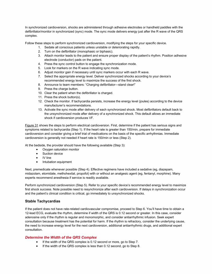
In synchronized cardioversion, shocks are administered through adhesive electrodes or handheld paddles with the defibrillator/monitor in synchronized (sync) mode. The sync mode delivers energy just after the R wave of the QRS complex.
Follow these steps to perform synchronized cardioversion, modifying the steps for your specific device. 1. Sedate all conscious patients unless unstable or deteriorating rapidly. 2. Turn on the defibrillator (monophasic or biphasic). 3. Attach monitor leads to the patient and ensure proper display of the patient’s rhythm. Position adhesive
electrode (conductor) pads on the patient. 4. Press the sync control button to engage the synchronization mode. 5. Look for markers on the R wave indicating sync mode. 6. Adjust monitor gain if necessary until sync markers occur with each R wave. 7. Select the appropriate energy level. Deliver synchronized shocks according to your device’s
recommended energy level to maximize the success of the first shock. 8. Announce to team members: “Charging defibrillator—stand clear!” 9. Press the charge button. 10. Clear the patient when the defibrillator is charged. 11. Press the shock button(s). 12. Check the monitor. If tachycardia persists, increase the energy level (joules) according to the device
manufacturer’s recommendations. 13. Activate the sync mode after delivery of each synchronized shock. Most defibrillators default back to
the unsynchronized mode after delivery of a synchronized shock. This default allows an immediate shock if cardioversion produces VF.
Figure 31 shows the steps to perform electrical cardioversion. First, determine if the patient has serious signs and symptoms related to tachycardia (Step 1). If the heart rate is greater than 150/min, prepare for immediate cardioversion and consider giving a brief trial of medications on the basis of the specific arrhythmias. Immediate cardioversion is generally not needed if heart rate is 150/min or less (Step 2).
At the bedside, the provider should have the following available (Step 3): • Oxygen saturation monitor • Suction device • IV line • Intubation equipment
Next, premedicate whenever possible (Step 4). Effective regimens have included a sedative (eg, diazepam, midazolam, etomidate, methohexital, propofol) with or without an analgesic agent (eg, fentanyl, morphine). Many experts recommend anesthesia if service is readily available.
Perform synchronized cardioversion (Step 5). Refer to your specific device’s recommended energy level to maximize first shock success. Note possible need to resynchronize after each cardioversion. If delays in synchronization occur and the patient’s clinical condition is critical, go immediately to unsynchronized shocks.
Stable Tachycardias
If the patient does not have rate-related cardiovascular compromise, proceed to Step 6. You’ll have time to obtain a 12-lead ECG, evaluate the rhythm, determine if width of the QRS is 0.12 second or greater. In this case, consider adenosine only if the rhythm is regular and monomorphic, and consider antiarrhythmic infusion. Seek expert consultation because treatment has the potential for harm. If the rhythm is refractory, consider the underlying cause, the need to increase energy level for the next cardioversion, additional antiarrhythmic drugs, and additional expert consultation.
Determine the Width of the QRS Complex • If the width of the QRS complex is 0.12 second or more, go to Step 7. • If the width of the QRS complex is less than 0.12 second, go to Step 8.
In some cases, a “stable” tachycardia is actually an early sign that the patient is becoming unstable, and you should initiate a search for underlying causes early to avoid further deterioration.
You must be able to classify the type of tachycardia (wide or narrow; regular or irregular) and intervene appropriately as outlined in the Adult Tachycardia With a Pulse Algorithm. During this case, you will perform initial assessment and management of regular narrow-complex rhythms (except sinus tachycardia), and you’ll treat them with vagal maneuvers, adenosine, β-blocker or calcium channel blocker.
If the rhythm does not convert, consider expert consultation. If the patient becomes clinically unstable, prepare for immediate unsynchronized shock or synchronized cardioversion.
Understanding Sinus Tachycardia
Sinus tachycardia is a heart rate that is greater than 100/min, has P waves, and is generated by sinus node discharge. The heart rate in tachycardia typically does not exceed 220/min and is age-related. Sinus tachycardia usually does not exceed 120 to 130/min, and it has a gradual onset and gradual termination. Reentry SVT has an abrupt onset and termination.
Note that sinus tachycardia is excluded from the Adult Tachycardia With a Pulse Algorithm. Sinus tachycardia is caused by external influences on the heart, such as fever, anemia, hypotension, blood loss, or exercise—systemic, not cardiac, conditions. Sinus tachycardia is a regular rhythm, although the rate may be slowed by vagal maneuvers. In sinus tachycardia, the goal is to identify and correct the underlying systemic cause, and cardioversion is contraindicated.
β-Blockers may cause clinical deterioration if the cardiac output falls when a compensatory tachycardia is blocked. This is because cardiac output is determined by the volume of blood ejected by the ventricles with each contraction (stroke volume) and the heart rate.
Cardiac output (CO) = Stroke volume (SV) × Heart rate
If a condition such as a large AMI limits ventricular function (severe heart failure or cardiogenic shock), the heart compensates by increasing the heart rate. If you attempt to reduce the heart rate in patients with a compensatory tachycardia, cardiac output will fall, and the patient’s condition will likely deteriorate.
Rhythms for Stable Tachycardia
Tachycardia classifications include the appearance of the QRS complex, heart rate, and whether they are regular or irregular:
• Narrow–QRS complex (SVT) tachycardias (QRS less than 0.12 second) in order of frequency o –Sinus tachycardia o –Atrial fibrillation o –Atrial flutter o –AV nodal reentry
• Wide–QRS complex tachycardias (QRS 0.12 second or more) o –Monomorphic VT o –Polymorphic VT o –SVT with aberrancy
• Regular or irregular tachycardias o –Irregular narrow-complex tachycardias are probably atrial fibrillation
Drugs for Stable Tachycardia
Drugs for tachycardia include • Adenosine 6 mg IV (follow with saline flush); second dose (if required) 12 mg IV (follow with saline flush) • Several analgesic and sedative agents are also used during electrical cardioversion, but those agents
are not covered in this course.
Approach to Stable Tachycardia
A stable tachycardia refers to a condition in which the patient has • A heart rate greater than 100/min • No significant signs or symptoms caused by the increased rate • A potential underlying cardiac electrical abnormality that generates the rhythm
Questions to Determine Classification
Classification of the tachycardia requires the careful clinical evaluation of these questions: • Are symptoms present or absent? • Are symptoms due to the tachycardia? • Is the patient stable or unstable? • Is the QRS complex narrow or wide? • Is the rhythm regular or irregular? • Is the QRS monomorphic or polymorphic? • Is the rhythm sinus tachycardia?
The answers guide subsequent diagnosis and treatment.
Managing Stable Tachycardia: The Adult Tachycardia With a Pulse Algorithm
As noted in the Unstable Tachycardia Case, the keys to managing a patient with any tachycardia are assessing the appropriateness for the clinical condition, identifying and treating underlying causes (Step 1), and determining whether pulses are present and, if so, whether the patient is stable or unstable and then providing treatment based on the patient’s condition and rhythm. If the patient is pulseless, manage the patient according to the Adult Cardiac Arrest Algorithm (Figure 41). If the patient has pulses, manage the patient according to the Adult Tachycardia With a Pulse Algorithm (Figure 30).
If a tachycardia and a pulse are present, perform the steps of the BLS, Primary, and Secondary Assessments. Determine if serious signs or symptoms are present and due to the tachycardia. This will direct you to either the stable or unstable section of the algorithm.
• If significant signs or symptoms are due to the tachycardia, immediate cardioversion is indicated (see the Unstable Tachycardia Case).
• If the patient develops pVT or VF, deliver unsynchronized high-energy shocks (defibrillation energy) and follow the Adult Cardiac Arrest Algorithm.
• If the patient has polymorphic VT, treat the rhythm as VF and deliver high-energy unsynchronized shocks (ie, defibrillation energy).
In this case, the patient is stable, so you will manage according to the stable section of the Adult Tachycardia With a Pulse Algorithm (Figure 30). A precise identification of the rhythm (eg, reentry SVT, atrial flutter) may not be possible at this time.
Applying the Adult Tachycardia With a Pulse Algorithm to Stable Patients
In this case, a patient has stable tachycardia with a pulse. Conduct the steps in the Adult Tachycardia With a Pulse Algorithm to evaluate and manage the patient.
Patient Assessment
Step 1 directs you to assess the appropriateness for the patient’s clinical condition. Typically, a heart rate greater than 150/min at rest is due to tachyarrhythmias other than sinus tachycardia.
BLS and ACLS Assessments

Using the BLS, Primary, and Secondary Assessments to guide your approach, identify and treat underlying causes (Step 2):
• Maintain patent airway; assist breathing as necessary. • Give oxygen (if hypoxemic). • Use a cardiac monitor to identify rhythm; monitor blood pressure and oximetry. • Obtain IV access. • Obtain 12-lead ECG if available.
If symptoms persist, proceed to Step 3. If the patient is stable, go to Step 8.
IV Access and 12-Lead ECG
If the patient with tachycardia is stable (ie, no serious signs or symptoms related to the tachycardia), you have time to evaluate the rhythm and decide on treatment options. Establish IV access if not already obtained. Obtain a 12-lead ECG (if available) or rhythm strip to determine if the QRS is narrow (less than 0.12 second) or wide (0.12 second or more).
Decision Point: Wide or Narrow
The path of treatment is now determined by whether the QRS is wide or narrow and whether the rhythm is regular or irregular. If a monomorphic wide-complex rhythm is present and the patient is stable, consider adenosine (only if regular and monomorphic), consider antiarrhythmic infusion, and seek expert consultation. Treat polymorphic wide-complex tachycardia with immediate unsynchronized shock.
Wide-Complex Tachycardias
Wide-complex tachycardias are defined as a QRS of 0.12 second or more, but consider seeking expert consultation for help identifying the rhythm. The most common forms of life-threatening wide-complex tachycardias likely to deteriorate to VF are
• Monomorphic VT • Polymorphic VT
Determine if the rhythm is regular or irregular. • A regular wide-complex tachycardia is presumed to be VT or SVT with aberrancy. • An irregular wide-complex tachycardia may be atrial fibrillation with aberrancy, pre-excited atrial
fibrillation (atrial fibrillation using an accessory pathway for antegrade conduction), or polymorphic VT/torsades de pointes. These advanced rhythms require additional expertise or expert consultation. In addition, consider adenosine (only if regular and monomorphic) and antiarrhythmic infusion.
If the rhythm is likely VT or SVT in a stable patient, treat based on the algorithm for that rhythm.
Recent evidence suggests that if the rhythm etiology cannot be determined and is regular in its rate and monomorphic, IV adenosine is relatively safe for both treatment and diagnosis. IV antiarrhythmic drugs may be effective. We recommend:
• Procainamide 20 to 50 mg/min IV until arrhythmia suppressed, hypotension ensues, QRS duration increases more than 50%, or maximum dose 17 mg/kg IV is given. Maintenance infusion: 1 to 4 mg/min IV. Avoid if prolonged QT or congestive heart failure.
• Amiodarone (first dose) 150 mg IV over 10 minutes. Repeat as needed if VT recurs. Follow by maintenance infusion of 1 mg/min IV for first 6 hours.
• Sotalol 100 mg (1.5 mg/kg) IV over 5 minutes. Avoid if prolonged QT.
In the case of irregular wide-complex tachycardia, management focuses on control of the rapid ventricular rate (rate control), conversion of hemodynamically unstable atrial fibrillation to sinus rhythm (rhythm control), or both. Seek expert consultation.
Treating Tachycardia

You may not always be able to distinguish between supraventricular (aberrant) and ventricular wide-complex rhythms, so be aware that most wide-complex (broad-complex) tachycardias are ventricular in origin.
If a patient is pulseless, follow the Adult Cardiac Arrest Algorithm.
If a patient becomes unstable, do not delay treatment for further rhythm analysis. For stable patients with wide-complex tachycardias, consider expert consultation because treatment has the potential for harm.
Critical Concepts: Drugs to Avoid in Patients With Irregular Wide-Complex Tachycardia Avoid AV nodal blocking agents such as adenosine, calcium channel blockers, digoxin, and possibly β-blockers in patients with pre-excitation atrial fibrillation, because these drugs may cause a paradoxical increase in the ventricular response.
Narrow QRS, Regular Rhythm
The therapy for narrow QRS with regular rhythm is to attempt vagal maneuvers, give adenosine, give a β-blocker or calcium channel blocker, and consider expert consultation. Vagal maneuvers, adenosine, and β-blocker or calcium channel blockers are the preferred initial interventions for terminating narrow-complex tachycardias that are symptomatic (but stable) and supraventricular in origin. Valsalva maneuvers or carotid sinus massage alone will terminate about 25% of SVT, and adenosine is required for the remainder.
• If SVT does not respond to vagal maneuvers, give adenosine 6 mg IV (follow with saline flush) in a large (eg, antecubital) vein over 1 second, and elevate the arm immediately.
• If SVT does not convert within 1 to 2 minutes, give a second dose of adenosine 12 mg IV (follow with saline flush) following the same procedure above.
Adenosine increases AV block and will terminate approximately 90% of reentry arrhythmias within 2 minutes. Adenosine will not terminate atrial flutter or atrial fibrillation but will slow AV conduction, allowing you to identify flutter or fibrillation waves.
Adenosine is safe and effective in pregnancy, but it has several important drug interactions. Patients with significant blood levels of theophylline, caffeine, or theobromine may require larger doses, and you should reduce the initial dose to 3 mg IV for patients taking dipyridamole or carbamazepine. Due to recent case reports of prolonged asystole after adenosine administration to patients with transplanted hearts or after central venous administration, you may consider lower doses such as 3 mg IV in these situations.
Adenosine may cause bronchospasm, so generally, you should not give adenosine to patients with asthma or chronic obstructive pulmonary disease, particularly if patients are actively bronchospastic.
If the rhythm converts with adenosine, it is probable reentry SVT. Observe patients for recurrence, and treat any recurrence with adenosine or longer-acting AV nodal blocking agents, such as the non-dihydropyridine calcium channel blockers (verapamil and diltiazem) or β-blockers. Typically, you should obtain expert consultation if the tachycardia recurs.
If the rhythm does not convert with adenosine, it is possible atrial flutter, ectopic atrial tachycardia, sinus tachycardia, or junctional tachycardia, and you should obtain expert consultation about diagnosis and treatment.
Critical Concepts: What to Avoid With AV Nodal Blocking Agents Do not use AV nodal blocking drugs for pre-excited atrial fibrillation or flutter because these drugs are unlikely to slow the ventricular rate and may even accelerate the ventricular response. Also, be careful when combining AV nodal

blocking agents of varying duration, such as calcium channel blockers or β-blockers, because their actions may overlap if given serially and provoke profound bradycardia.
Tachycardia Algorithm: Advanced Management Steps
As an ACLS provider, you should be able to recognize a stable narrow-complex or wide-complex tachycardia, classify the rhythm as regular or irregular, and provide initial management. You may treat regular narrow-complex tachycardias initially with vagal maneuvers, adenosine, and β-blocker or calcium channel blocker, but if these are unsuccessful, you’ll need to consider expert consultation.
If you have experience with the differential diagnosis and therapy of stable tachycardias that do not respond to initial treatment, you can review the Adult Tachycardia With a Pulse Algorithm for additional steps and pharmacologic agents used in the treatment of these arrhythmias, both for rate control and for termination of the arrhythmia.
If at any point you become uncertain or uncomfortable while treating a stable patient, seek expert consultation because treatment has the potential for harm.