takotsubo cardiomyopathy (tcm)
TRANSCRIPT
CHEST PAIN & HYPOTENSION AFTER ELECTRIC CARDIOVERSION HANA AKSELROD, PGY-3 || MAINE MEDICAL CENTER || BAR HARBOR, SEPT 2015
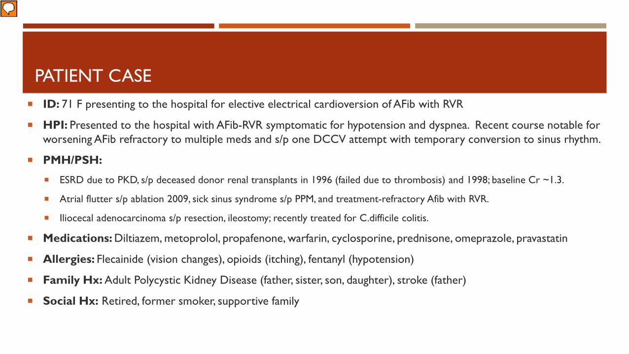
PATIENT CASE ID: 71 F presenting to the hospital for elective electrical cardioversion of AFib with RVR
HPI: Presented to the hospital with AFib-RVR symptomatic for hypotension and dyspnea. Recent course notable for worsening AFib refractory to multiple meds and s/p one DCCV attempt with temporary conversion to sinus rhythm.
PMH/PSH:
ESRD due to PKD, s/p deceased donor renal transplants in 1996 (failed due to thrombosis) and 1998; baseline Cr ~1.3.
Atrial flutter s/p ablation 2009, sick sinus syndrome s/p PPM, and treatment-refractory Afib with RVR.
Iliocecal adenocarcinoma s/p resection, ileostomy; recently treated for C.difficile colitis.
Medications: Diltiazem, metoprolol, propafenone, warfarin, cyclosporine, prednisone, omeprazole, pravastatin
Allergies: Flecainide (vision changes), opioids (itching), fentanyl (hypotension)
Family Hx: Adult Polycystic Kidney Disease (father, sister, son, daughter), stroke (father)
Social Hx: Retired, former smoker, supportive family

PATIENT CASE
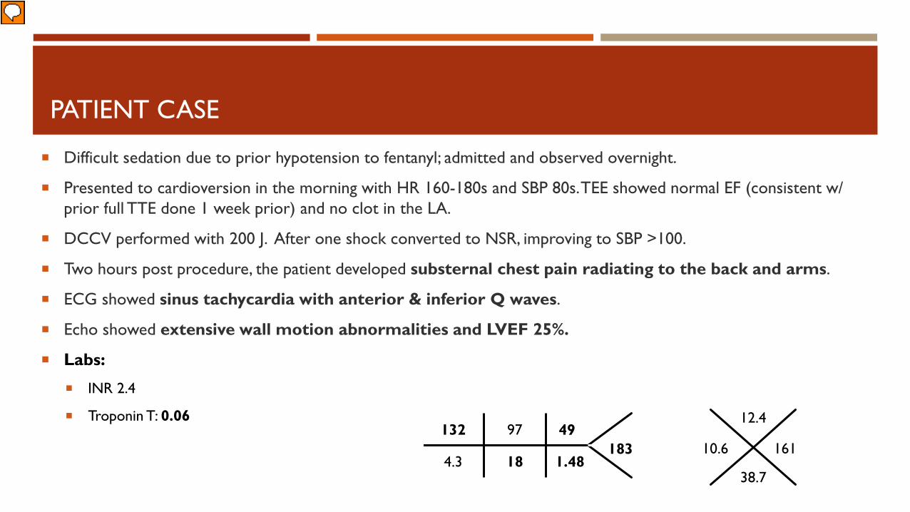
Difficult sedation due to prior hypotension to fentanyl; admitted and observed overnight.
Presented to cardioversion in the morning with HR 160-180s and SBP 80s. TEE showed normal EF (consistent w/ prior full TTE done 1 week prior) and no clot in the LA.
DCCV performed with 200 J. After one shock converted to NSR, improving to SBP >100.
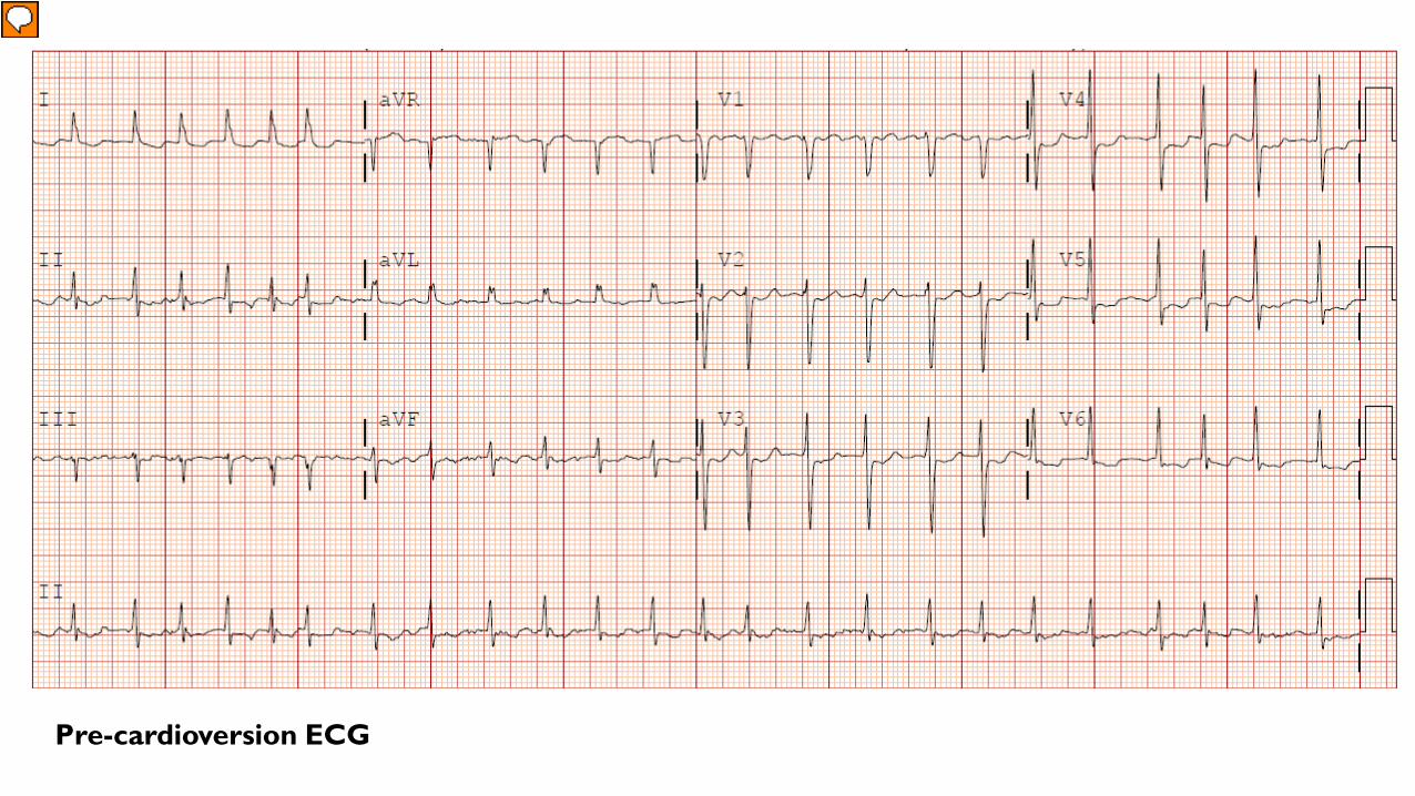
Pre-cardioversion ECG
PATIENT CASE
Difficult sedation due to prior hypotension to fentanyl; admitted and observed overnight.
Presented to cardioversion in the morning with HR 160-180s and SBP 80s. TEE showed normal EF (consistent w/ prior full TTE done 1 week prior) and no clot in the LA.
DCCV performed with 200 J. After one shock converted to NSR, improving to SBP >100.
Two hours post procedure, the patient developed substernal chest pain radiating to the back and arms.
ECG showed sinus tachycardia with anterior & inferior Q waves.
Echo showed extensive wall motion abnormalities and LVEF 25%.
Labs:
INR 2.4
Troponin T: 0.06
1.48 18 4.3
49 97 132 183 10.6
161
12.4
38.7
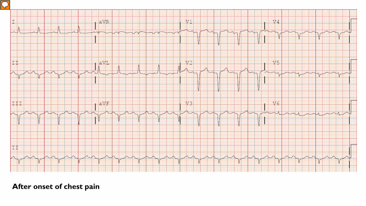
After onset of chest pain
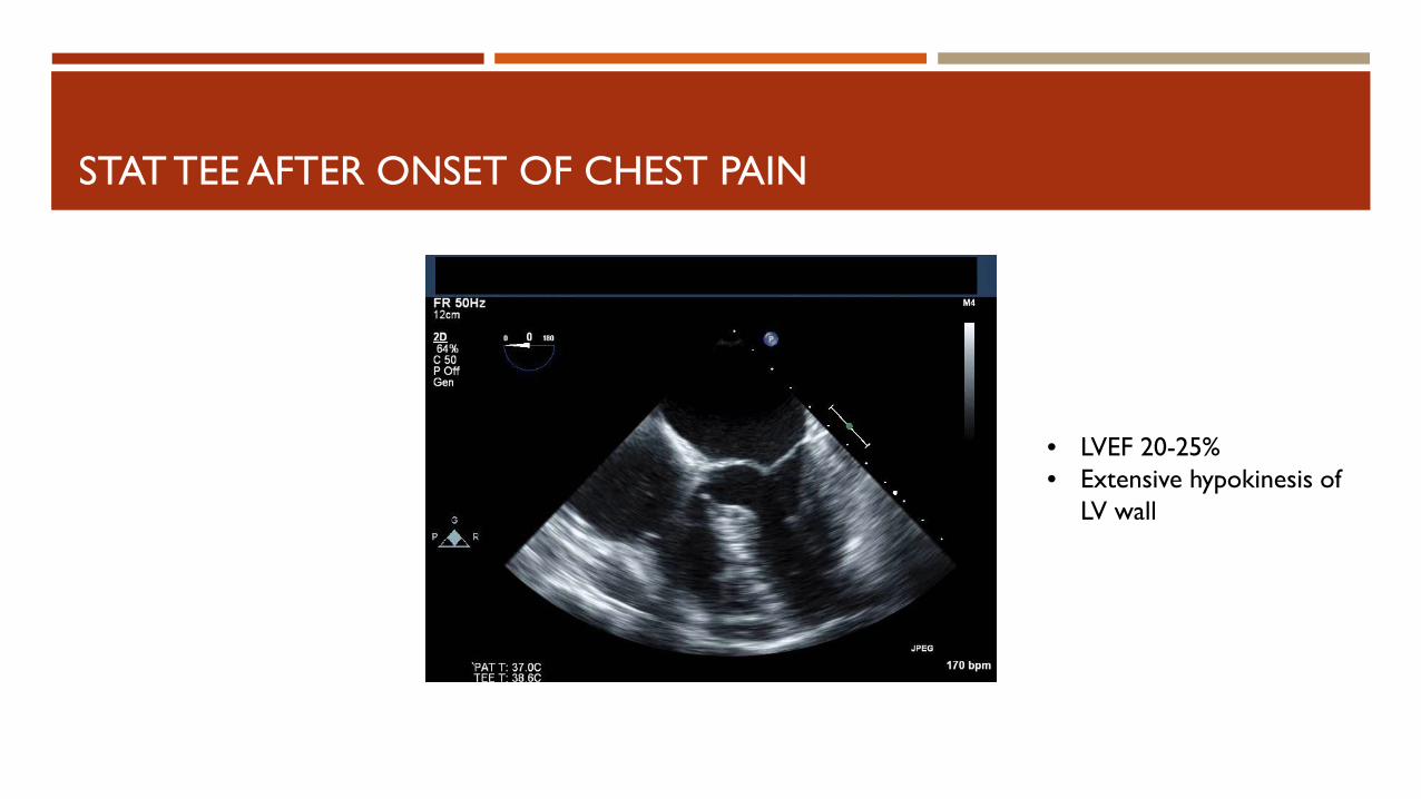
STAT TEE AFTER ONSET OF CHEST PAIN
• LVEF 20-25% • Extensive hypokinesis of
LV wall
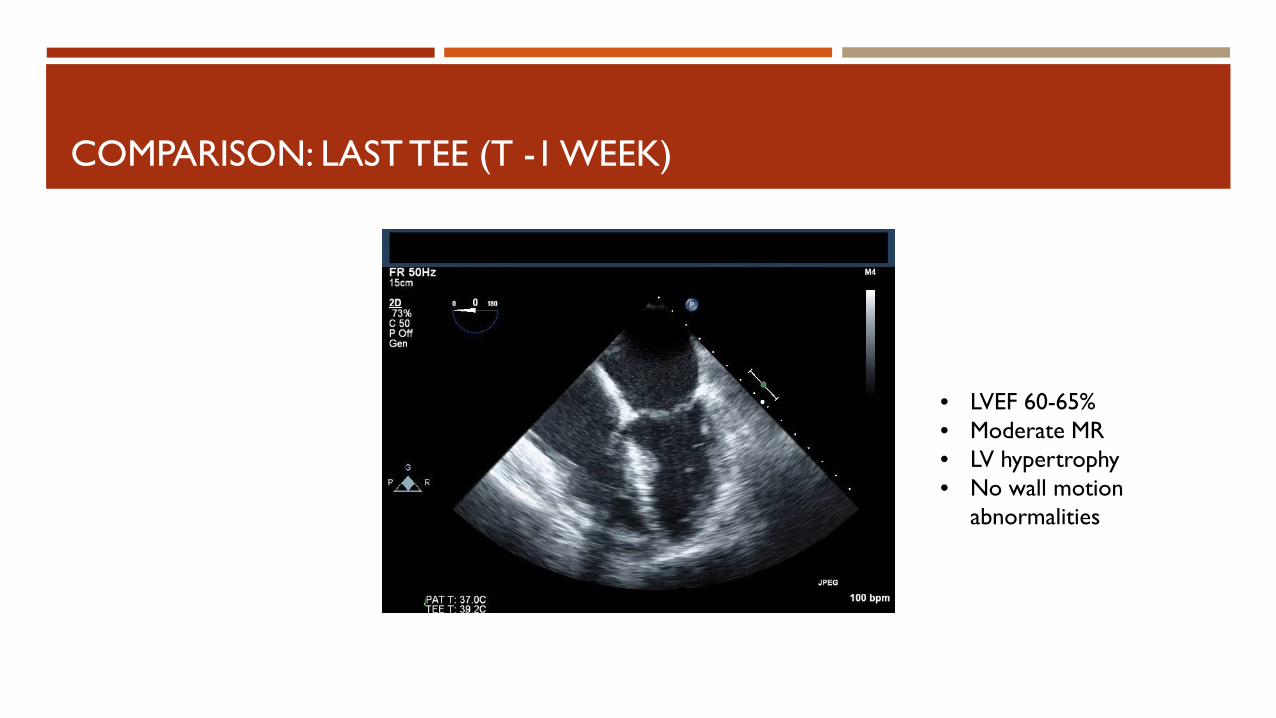
COMPARISON: LAST TEE (T -1 WEEK)
• LVEF 60-65% • Moderate MR • LV hypertrophy • No wall motion
abnormalities
NEW-ONSET ACUTE LV DYSFUNCTION AFTER DCCV
Differential diagnosis
NEW-ONSET ACUTE LV DYSFUNCTION AFTER DCCV
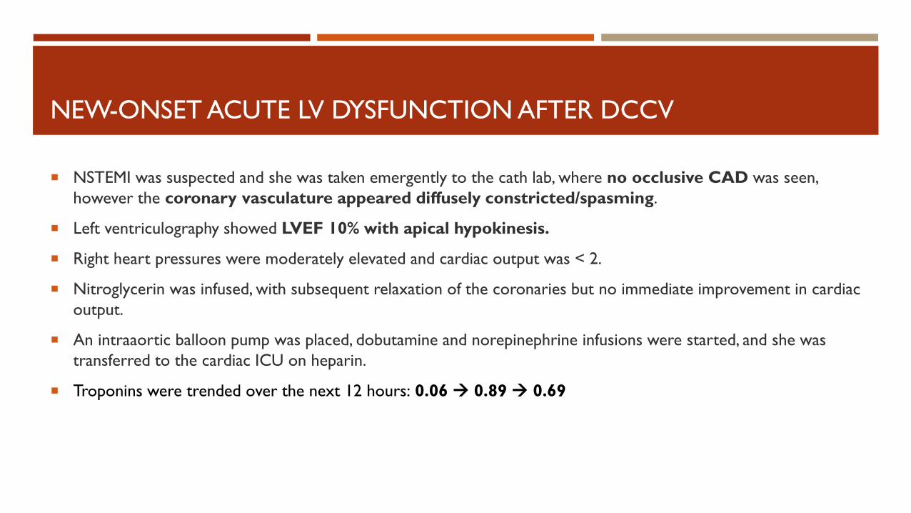
NSTEMI was suspected and she was taken emergently to the cath lab, where no occlusive CAD was seen, however the coronary vasculature appeared diffusely constricted/spasming.
Left ventriculography showed LVEF 10% with apical hypokinesis.
Right heart pressures were moderately elevated and cardiac output was < 2.
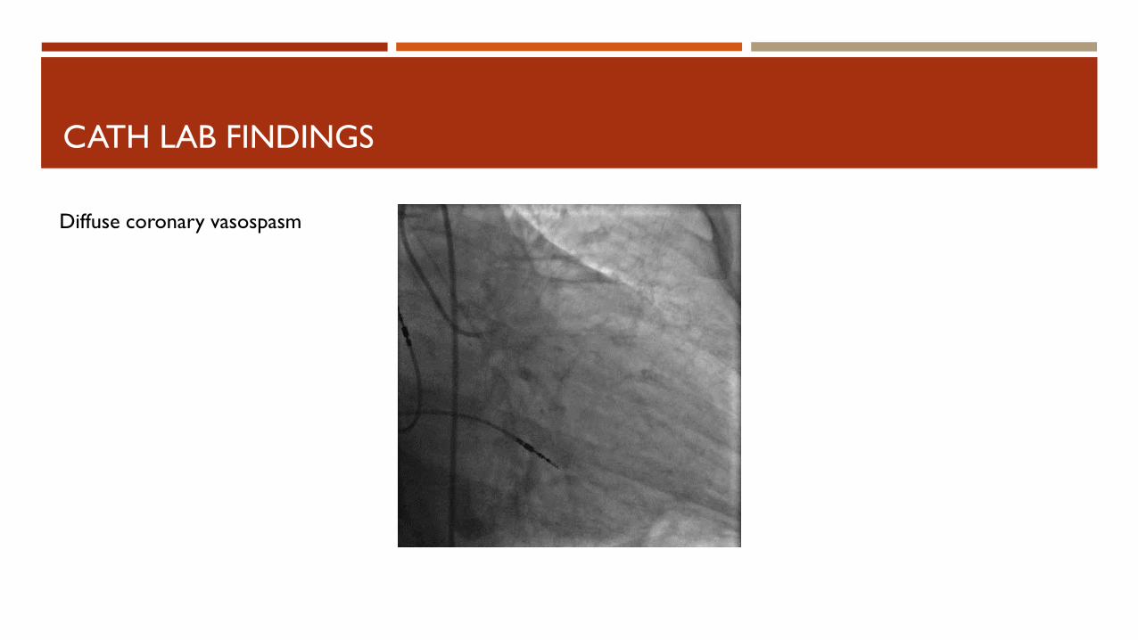
Nitroglycerin was infused, with subsequent relaxation of the coronaries but no immediate improvement in cardiac output.
An intraaortic balloon pump was placed, dobutamine and norepinephrine infusions were started, and she was transferred to the cardiac ICU on heparin.
Troponins were trended over the next 12 hours: 0.06 0.89 0.69
CATH LAB FINDINGS
Diffuse coronary vasospasm
CATH LAB FINDINGS
Post nitroglycerin infusion
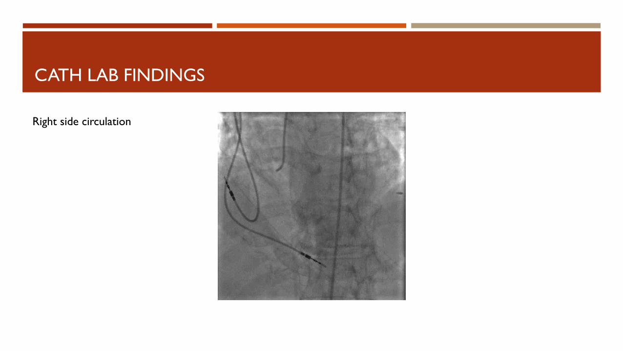
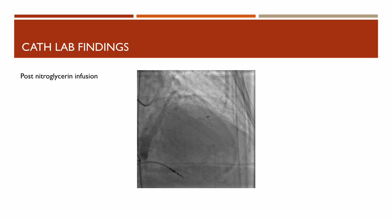
CATH LAB FINDINGS
Right side circulation
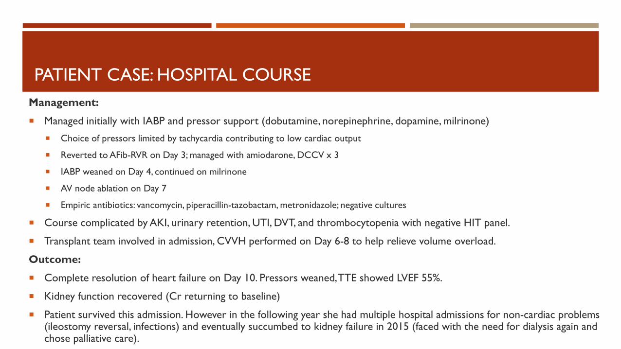
PATIENT CASE: HOSPITAL COURSE Management:
Managed initially with IABP and pressor support (dobutamine, norepinephrine, dopamine, milrinone)
Choice of pressors limited by tachycardia contributing to low cardiac output
Reverted to AFib-RVR on Day 3; managed with amiodarone, DCCV x 3
IABP weaned on Day 4, continued on milrinone
AV node ablation on Day 7
Empiric antibiotics: vancomycin, piperacillin-tazobactam, metronidazole; negative cultures
Course complicated by AKI, urinary retention, UTI, DVT, and thrombocytopenia with negative HIT panel.
Transplant team involved in admission, CVVH performed on Day 6-8 to help relieve volume overload.
Outcome:
Complete resolution of heart failure on Day 10. Pressors weaned, TTE showed LVEF 55%.
Kidney function recovered (Cr returning to baseline)
Patient survived this admission. However in the following year she had multiple hospital admissions for non-cardiac problems (ileostomy reversal, infections) and eventually succumbed to kidney failure in 2015 (faced with the need for dialysis again and chose palliative care).
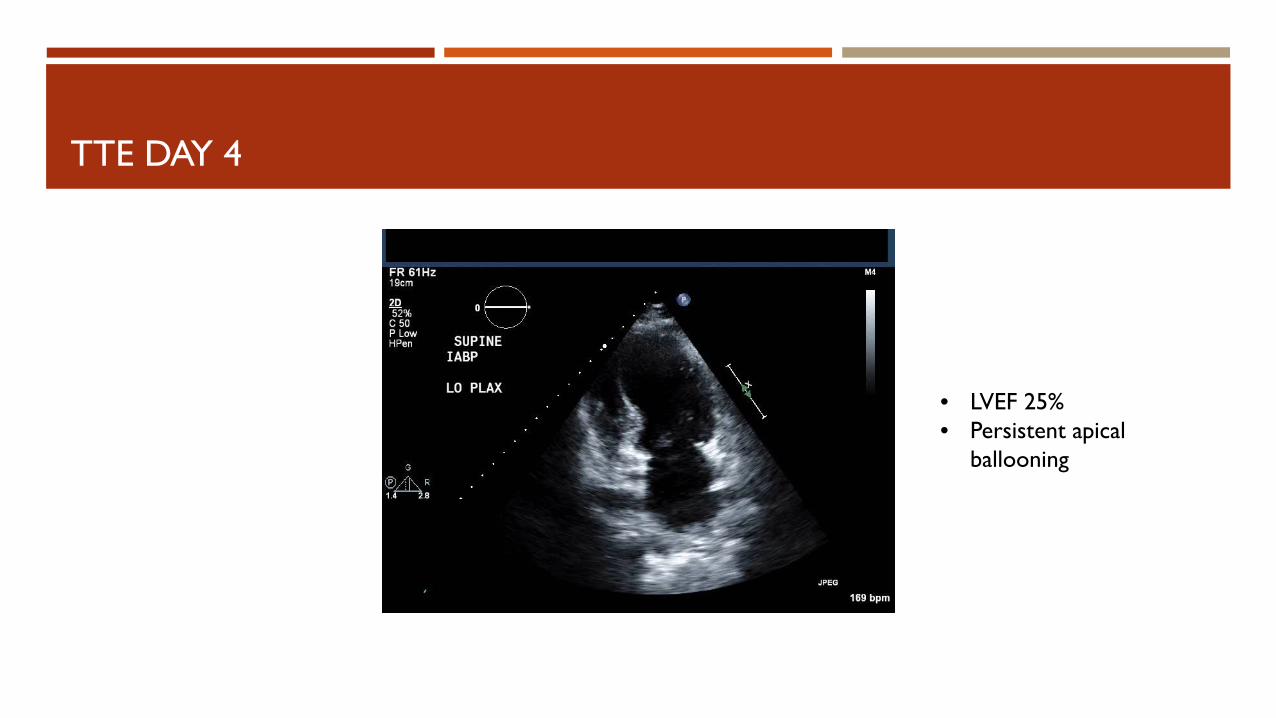
TTE DAY 4
• LVEF 25% • Persistent apical
ballooning
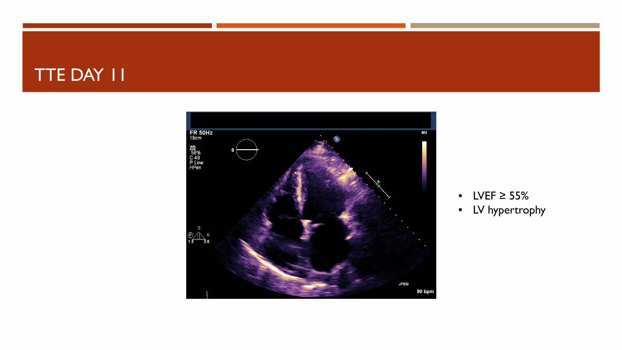
TTE DAY 11
• LVEF ≥ 55% • LV hypertrophy
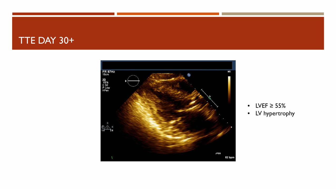
TTE DAY 30+
• LVEF ≥ 55% • LV hypertrophy

PATIENT CASE: HOSPITAL COURSE Management:
Managed initially with IABP and pressor support (dobutamine, norepinephrine, dopamine, milrinone)
Choice of pressors limited by tachycardia contributing to low cardiac output
Reverted to AFib-RVR on Day 3; managed with amiodarone, DCCV x 3
IABP weaned on Day 4, continued on milrinone
AV node ablation on Day 7
Empiric antibiotics: vancomycin, piperacillin-tazobactam, metronidazole; negative cultures
Course complicated by AKI, urinary retention, UTI, DVT, and thrombocytopenia with negative HIT panel.
Transplant team involved in admission, CVVH performed on Day 6-8 to help relieve volume overload.
Outcome:
Complete resolution of heart failure on Day 10. Pressors weaned, TTE showed LVEF 55%.
Kidney function recovered (Cr returning to baseline)
Patient survived this admission. However in the following year she had multiple hospital admissions for non-cardiac problems (ileostomy reversal, infections) and eventually succumbed to kidney failure in 2015 (faced with the need for dialysis again and chose palliative care).
TAKOTSUBO CARDIOMYOPATHY EPIDEMIOLOGY, PATHOPHYSIOLOGY, AND MANAGEMENT
EPIDEMIOLOGY
Incidence: consensus opinion, ~2% of patients presenting with MI to hospital, and up to 10% in select groups of post-menopausal women
Large hospital in California: of 1,297 post-menopausal women presenting with ACS-like syndrome and positive troponins, 5.9% met criteria for Takotsubo Cardiomyopathy (TCM)
In the National Inpatient Sample study (2008-2009), 24701 patients with TCM were compared to controls with MI and Ortho presentations:
More likely to be female, Caucasian, and wealthier
Less likely to have “traditional” cardiovascular risk factors
More likely to have h/o CVA, mood disorders, drug abuse, malignancy, chronic liver disease, or sepsis
No difference in age vs. controls
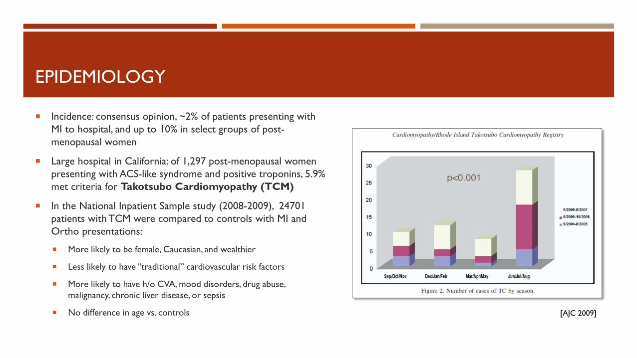
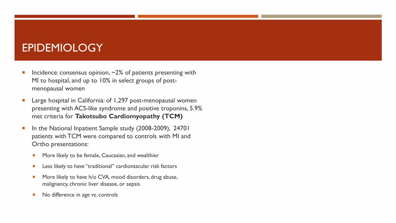
EPIDEMIOLOGY
Incidence: consensus opinion, ~2% of patients presenting with MI to hospital, and up to 10% in select groups of post-menopausal women
Large hospital in California: of 1,297 post-menopausal women presenting with ACS-like syndrome and positive troponins, 5.9% met criteria for Takotsubo Cardiomyopathy (TCM)
In the National Inpatient Sample study (2008-2009), 24701 patients with TCM were compared to controls with MI and Ortho presentations:
More likely to be female, Caucasian, and wealthier
Less likely to have “traditional” cardiovascular risk factors
More likely to have h/o CVA, mood disorders, drug abuse, malignancy, chronic liver disease, or sepsis
No difference in age vs. controls [AJC 2009]
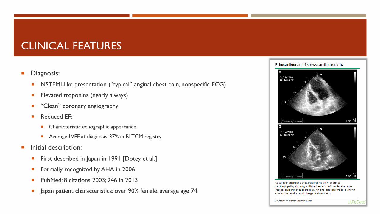
CLINICAL FEATURES
Diagnosis:
NSTEMI-like presentation (“typical” anginal chest pain, nonspecific ECG)
Elevated troponins (nearly always)
“Clean” coronary angiography
Reduced EF:
Characteristic echographic appearance
Average LVEF at diagnosis: 37% in RI TCM registry
Initial description:
First described in Japan in 1991 [Dotey et al.]
Formally recognized by AHA in 2006
PubMed: 8 citations 2003; 246 in 2013
Japan patient characteristics: over 90% female, average age 74
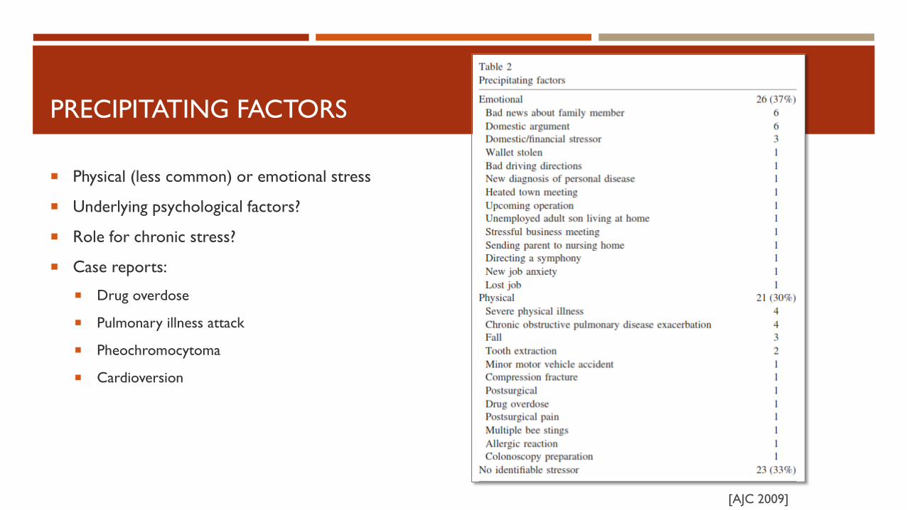
PRECIPITATING FACTORS
Physical (less common) or emotional stress
Underlying psychological factors?
Role for chronic stress?
Case reports:
Drug overdose
Pulmonary illness attack
Pheochromocytoma
Cardioversion
[AJC 2009]
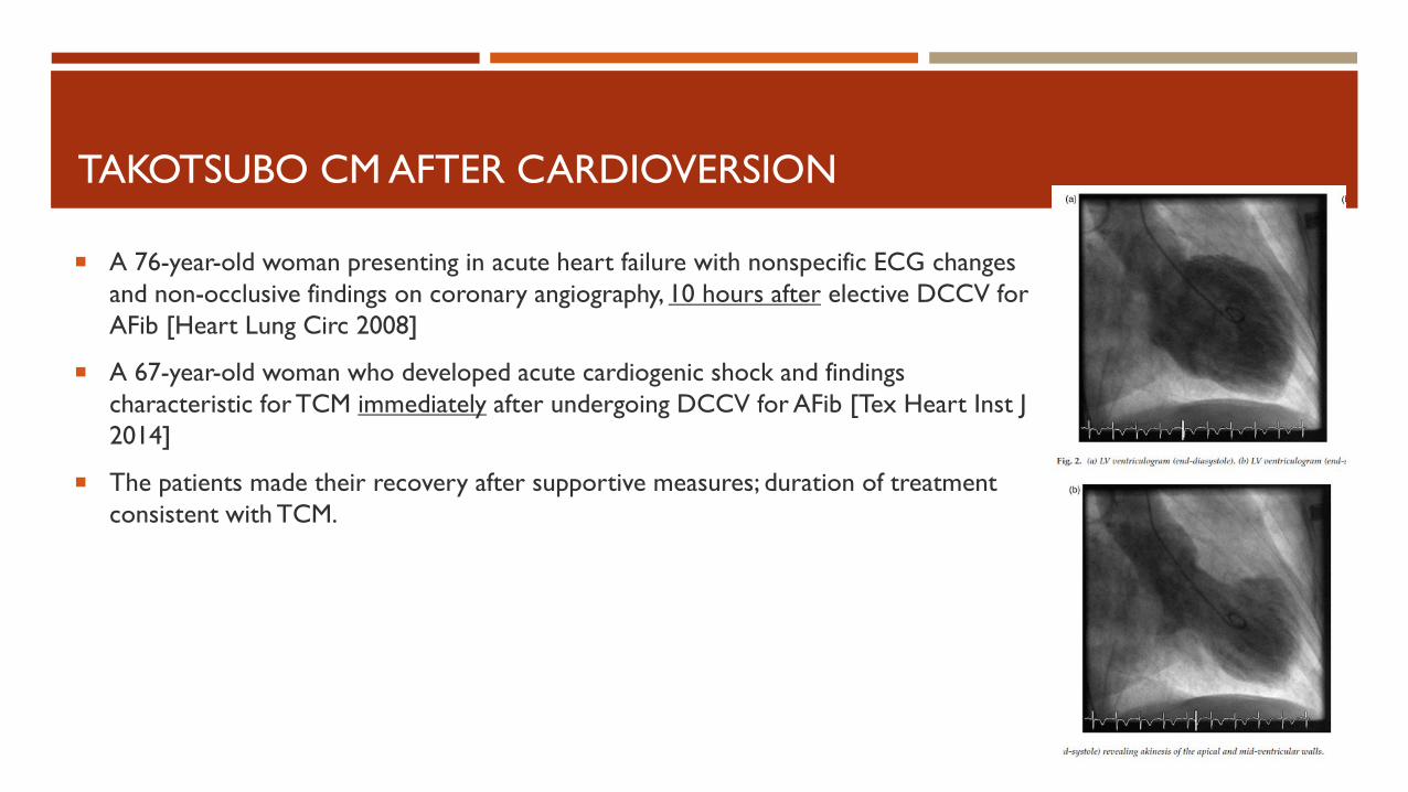
A 76-year-old woman presenting in acute heart failure with nonspecific ECG changes and non-occlusive findings on coronary angiography, 10 hours after elective DCCV for AFib [Heart Lung Circ 2008]
A 67-year-old woman who developed acute cardiogenic shock and findings characteristic for TCM immediately after undergoing DCCV for AFib [Tex Heart Inst J 2014]
The patients made their recovery after supportive measures; duration of treatment consistent with TCM.
TAKOTSUBO CM AFTER CARDIOVERSION
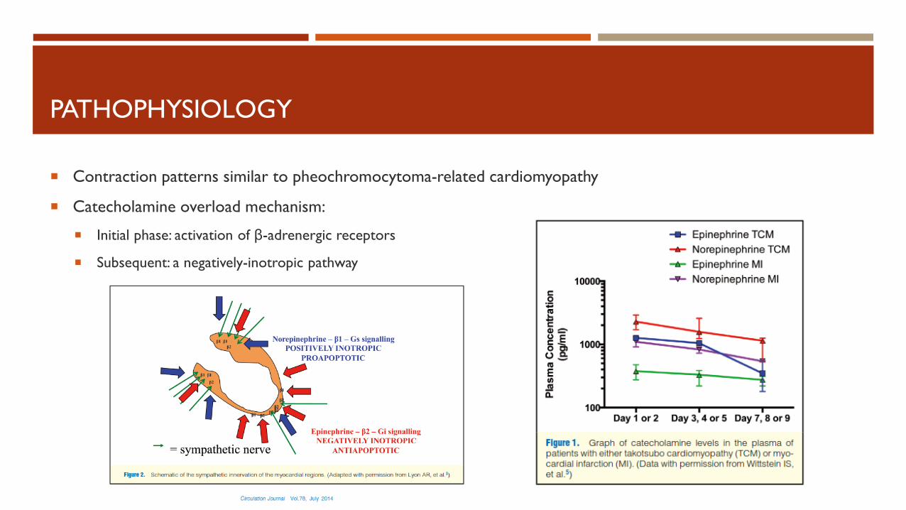
PATHOPHYSIOLOGY
Contraction patterns similar to pheochromocytoma-related cardiomyopathy
Catecholamine overload mechanism:
Initial phase: activation of β-adrenergic receptors
Subsequent: a negatively-inotropic pathway
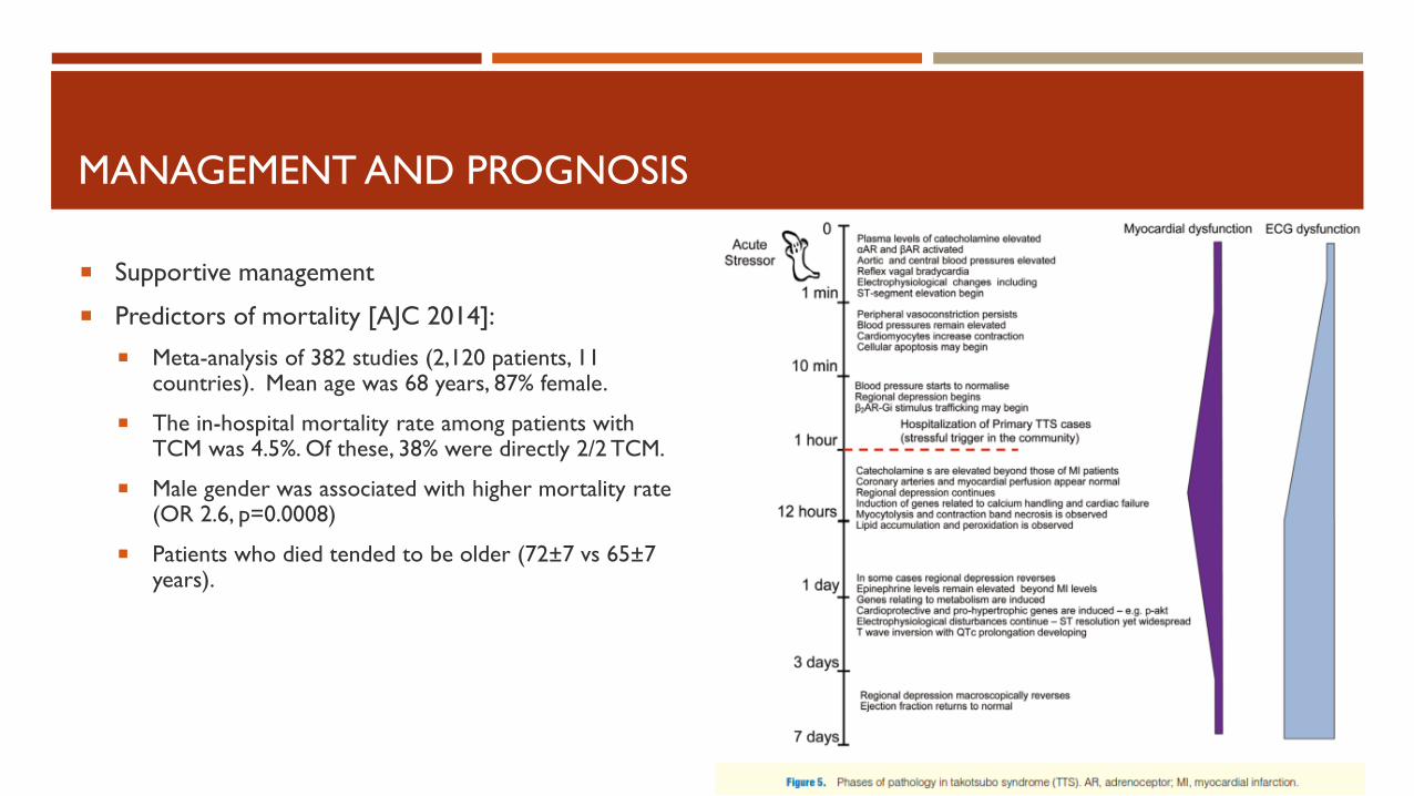
MANAGEMENT AND PROGNOSIS
Supportive management
Predictors of mortality [AJC 2014]:
Meta-analysis of 382 studies (2,120 patients, 11 countries). Mean age was 68 years, 87% female.
The in-hospital mortality rate among patients with TCM was 4.5%. Of these, 38% were directly 2/2 TCM.
Male gender was associated with higher mortality rate (OR 2.6, p=0.0008)
Patients who died tended to be older (72±7 vs 65±7 years).
OTHER COMPLICATIONS OF DCCV
Thromboembolic events
Transient hypotension associated with sedation (fentanyl)
Induction of arrhythmias (bradycardia, AV block, asystole, VT/VF)
Electric burns to skin / soft tissues
Injuries to health care workers
No direct myotoxicity from electricity in studies
SUMMARY POINTS
Considering Takotsubo cardiomyopathy in relation to iatrogenic events
Risks and benefits of direct-current cardioversion
Medical complexity, stress, and risk of adverse events
Challenges in communication and expectations
REFERENCES
El-Sayed AM, Brinjikji W, Salka S. Demographic and co-morbid predictors of stress (takotsubo) cardiomyopathy. Am J Cardiol. 2012 Nov 1;110(9):1368-72.
Sharkey SW, Maron BJ. Epidemiology and clinical profile of Takotsubo cardiomyopathy. Circ J. 2014;78(9):2119-28.
Sy F, Basraon J, Zheng H, Singh M, Richina J, Ambrose JA. Frequency of Takotsubo cardiomyopathy in postmenopausal women presenting with an acute coronary syndrome. Am J Cardiol. 2013 Aug 15;112(4):479-82.
Sanchez-Recalde A, Costero O, Oliver JM, Iborra C, Ruiz E, Sobrino JA. Images in cardiovascular medicine. Pheochromocytoma-related cardiomyopathy: inverted Takotsubo contractile pattern. Circulation. 2006 May 2;113(17):e738-9.
Friedman PL, Montgomery S, Matas N. Sotalol and a broken heart. J Cardiovasc Electrophysiol. 2010 Feb;21(2):207-10.
Singh K, Carson K, Shah R, Sawhney G, Singh B, Parsaik A, Gilutz H, Usmani Z, Horowitz J. Meta-analysis of clinical correlates of acute mortality in takotsubo cardiomyopathy. Am J Cardiol. 2014 Apr 15;113(8):1420-8.
Wright PT, Tranter MH, Morley-Smith AC, Lyon AR. Pathophysiology of takotsubo syndrome: temporal phases of cardiovascular responses to extreme stress. Circ J. 2014;78(7):1550-8.
Regnante RA, Zuzek RW, Weinsier SB, Latif SR, Linsky RA, Ahmed HN, Sadiq I. Clinical characteristics and four-year outcomes of patients in the Rhode Island Takotsubo Cardiomyopathy Registry. Am J Cardiol. 2009 Apr 1;103(7):1015-9
Siegfried JS, Bhusri S, Guttenplan N, Coplan NL. Takotsubo cardiomyopathy as a sequela of elective direct-current cardioversion for atrial fibrillation. Tex Heart Inst J. 2014 Apr 1;41(2):184-7.
THANK YOU
Dr. John Love
Drs. Mehdi Gheshlaghi and Deirdre Mooney
Dr. Stephen Hayes
Christopher Barnaby
“Med C” team, July 2014