tamilnadu dr mgr university mbbs prefinal ent august 2006 question paper with solution
DESCRIPTION
The Tamilnadu Dr MGR Medical University MBBS Prefinal ENT August 2006 question paper with solutionTRANSCRIPT
The Tamilnadu Dr MGR Medical University
Third (Final) M.B.B.S. Degree Examination (August 2006) question paper with solutions
Dr T Balasubramanian
Drtbalu's otolaryngology online

Tamilnadu Dr. MGR Medical University
Third (Final) M.B.B.S. Degree Examination (August 2006)
Time: Three hours
Answer all questions
I. Write Essay:
1. Describe the clinical features, diagnosis and management of cancer oesophagus. (15 marks)
Patients with cancer oesophagus manifest with:
a.Difficulty in swallowing
b. Painful swallowing.
Usually dysphagia is the most common symptom. Initially dysphagia is pronounced for solid food. Fluids and semisolid diet are better tolerated.
c. These patients manifest with excessive loss of weight which could be due to:
Difficulty in swallowing
Reduced appetite due to malignancy.
d. Heart burns – These patients characteristically complain of burning pain in the midline of the chest. This type of pain progressively gets worse and is made still worse by the act of swallowing.
e. Voice change – This is usually due to involvement of recurrent laryngeal nerves causing paralysis of vocal folds. Right recurrent laryngeal nerve is commonly involved in these patients. During early stages these patients may have varying degrees of aspiration. Sometimes aspiration may be severe enough to cause aspiration pneumonitis.
f. Disruption of peristalsis – The sheer bulk of tumor present intraluminally within the oesophagus may cause disruption of normal peristalsis. This could cause nausea, vomiting and food regurgitation.
g. Haematemesis – If the tumor is friable it can cause intraluminal bleeding leading on to hematemesis.
h. Compression symptoms – Increasing bulk of oesophageal mass can cause compression of local structures. It can cause compression to trachea leading on to upper airway obstruction. Another area of compression is at the level of superior vena cava causing superior vena caval syndrome. Erosion of trachea may lead to troublesome tracheo oesophageal fistula.
Drtbalu's otolaryngology online
Symptoms caused by superior vena cava syndrome include:
1. Dyspnoea
2. Swelling of the face / upper extremities
3. Headache
4. Orthopnoea
5. Nasal stuffiness
6. Light headedness
Symptoms caused by metastatic lesions:
Hepatic metastasis can lead to jaundice, ascitis.
Lung metastasis could cause shortness of breath, pleural effusion.
Diagnosis:
Radiology:
Barium swallow / meal can reveal occlusal mass in the oesophagus.
CT scan imaging will reveal the extent of the lesion, compression to adjacent structures if any.
Metastatic lesions can also be identified by performing CT scan.
PET scans can be used to identify and ascertain whether the lesion is active and metabolically active.
Upper GI endoscopy:
This is the standard in the diagnosis of oesophageal cancer. It helps in identifying the exact location of the lesion, biopsying the lesion. The location of the tumor is generally measured from the incisor.
Histopathology:
This offers the final diagnosis. Adenocarcinoma is common in the lower oesophagus while squamous cell carcinoma is prevalent in the upper third of oesophagus.
Management:
The ideal management modality is determined by:
Drtbalu's otolaryngology online
1. Cellular type of cancer
2. Stage of the disease
3. General condition of the patient
Priority should be given to the nutritional needs of the patient.
Manitenance of good oral hygiene.
As a first step in ensuring adequate nutrition to the patient naso gastric feeding can be resorted to.
To alleviate oesophageal obstruction stenting could be resorted to. Stents are usually used to keep the lumen of oesophagus patent. They also play a vital role in occluding tracheo oesophageal fistulas if present.
Surgical management:
This can be classified into therapeutic and palliative procedures.
If the tumor is resectable then segemental resection of the involved segment of the oesophagus can be performed. The shortened oesophagus can be corrected by interposing stomach / jejunal flaps.
Types of oesophagectomy:
Types of esophagectomy:
• Thoracoabdominal approach- which opens the abdominal and thoracic cavities together.• Two stage Ivor Lewis (also called Lewis-Tanner) approach- with an initial laparotomy and
construction of a gastric tube, followed by a right thoracotomy to excise the tumor and create an esophagogastric anastomosis.
• Three stage McKeown approach- where a third incision in the neck is made to complete the cervical anastomosis.Describe the clinical features, diagnosis and management of cancer oesophagus.
•
Endoscopic resection of oesophageal tumors:
1. This procedure is safe
2. Less invasive
3. Useful to treat early lesions
Patients who can undergo endoscopic resection are those with early lesions which does not involve the muscularis mucosa. Lasers can be used to assist in these resections.
Chemotherapy is reserved for advanced lesions. It also depends on the histological type of the tumor. The drugs used include:
Cisplatin
5-flurouracil.
Drtbalu's otolaryngology online
2. Describe the etiology, pathology, clinical features and management of cholesteatoma. (2+1+4+3=10)
Definition of cholesteatoma: Cholesteatoma is defined as a cystic bag like structure lined by stratified squamous epithelium on a fibrous matrix. This sac contains desquamated squamous epithelium. This sac is present in the attic region. Cholesteatoma is also defined as 'skin in wrong place'. Cholesteatoma is known to contain all the layers of skin epithelium. The basal layer (germinating layer) is present on the outer surface of cholesteatoma sac in contact with the walls of the middle ear cleft.
Theories of bone invasion by cholesteatoma:
1. Pressure theory - states that increase in the pressure caused by enlarging cholesteatoma cause bone erosion. Ischemia has been attributed as the cause in this theory.
2. Enzymatic theory: Inside the cholesteatoma are present multinucleated osteoclasts and histiocytes. These cells release acid phosphatase, collagenase and other proteolytic enzymes. These enzymes are known to cause bone erosion.
3. Pyogenic osteitis: Pyogneic bacteria may release enzymes which could cause bone resorption.
Types of cholesteatoma:
1. Congenital cholesteatoma
2. Primary acquired cholesteatoma
3. Secondary acquired cholesteatoma
Congenital cholesteatoma: is known to arise from embryonic cell rests present in the middle ear cavity and temporal bone. These cell rests are known to commonly occur in cerebello pontine angle and petrous apex. Infact congenital cholesteatoma is seen as a whitish mass behind an intact tympanic membrane.
Derlacki and Clemis laid down the following as criteria to diagnose congenital cholesteatoma:
1. The patient should not have previous episodes of middle ear disease
2. Ear drum must be intact and normal
3. It is purely an incidental finding
4. If discharge and ear drum perforation is present then it should be contrued that congential cholesteatoma has managed to erode the tympanic membrane.
Clinical features: The disorder is an incidental finding. The common location of congenital cholesteatoma is the antero superior quadrant of tympanic membrane, postero superior quadrant
Drtbalu's otolaryngology online
being the next common site of involvement. Anteriorly situated congenital cholesteatomas are known to affect the eustachean tube function causing conductive deafness due to middle ear effusion, where as posterior congenital cholesteatoma is known to cause conductive deafness due to impairment of ossicular chain mobility.
Staging of congenital cholesteatoma:
Staging as suggested by Derlacki and Clemis: They were the first to stage congenital cholesteatoma. They classified congenital cholesteatoma into
1. Petrous pyramid cholesteatoma
2. Cholesteatoma involving the mastoid cavity
3. Cholesteatoma involving the middle ear cavity.
Potsic suggested the following staging mechanism:
Stage I : Single quadrant involvement with no ossicular / mastoid involvement.
Stage II : Multiple quadrant involvement with no ossicular / mastoid involvement
Stage III : Ossicular involvement without mastoid involvement
Stage IV : Mastoid extension
Nelson's staging:
Type I : Involvement of mesotympanum without involvement of incus / stapes
Type II : Involvement of mesotympanum / attic along with erosion of ossicles without extension into the mastoid cavity
Type III : Involvement of mesotympanum with mastoid extension
Staging this disease will help in deciding the modality of treatment and in predicting the long term prognosis.
Acquired Cholesteatoma: can be divided into two types, primary acquired and secondary acquired cholesteatomas.
Primay acquired cholesteatoma: In this condition there is no history of preexisting or previous episodes of otitis media or perforation. Lesions just arise from the attic region of the middle ear.
Secondary acquired cholesteatoma: always follows active middle ear infection which manages to destroy the ear drum along with the annulus. This type of destruction is common in acute necrotising otitis media following exanthematous fevers like measles etc.
Theories to explain pathogenesis of cholesteatoma:
Various theories have been postulated to explain the pathogenesis of cholesteatoma. They are:
1. Cawthrone theory: This theory suggested by cawthrone in 1963 suggested that cholesteatoma always originated from congential embryonic cell rests present in various areas of the temporal bone.
2. Theory of immigration: This theory was suggested by Tumarkin. He was of the view that cholesteatoma was derived by immigration of squamous epithelium from the deep portion of the
Drtbalu's otolaryngology online

external auditory canal into the middle ear cleft through a marginal perforation or a total perforation of the ear drum as seen in acute necrotising otitis media.
3. Theory of invagination: This theory was suggested by Toss. He theorised that persistent negative pressure in the attic region causes invagination of pars flaccida causing a retraction pocket. This retraction pocket becomes later filled with desquamted epithelial debris which forms a nidus for the infection to occur later. Common organisms known to infect this keratin debris are Psuedomonas, E. coli, B. Proteus etc.
Toss also classified attic retraction pockets into 4 grades:
1. Grade I: The retracted pars flaccida is not in contact with the neck of the malleus.
2. Grade II: The retracted pars flaccida is in contact with the neck of the malleus to such an extent that it seems to clothe the neck of the malleus.
3. Grade III: Here inaddition to the retracted pars flaccida being in contact with the neck of the malleus there is also a limited erosion of the outer attic wall or scutum.
4. Grade IV: In this grade in addition to all the above said changes there is severe erosion of the outer attic wall or scutum.
4. Metaplastic theory: This theory was first suggested by Wendt in 1873. He took into consideration the histological changes seen in various portions of the middle ear cavity. The attic area of the middle ear cavity is lined by pavement type of epithelium. This epithelium undergoes metaplastic changes in response to subclinical infection. This metaplastic mucosa is squamous in nature there by forming a nidus for cholesteatoma formation in the attic region.
Of all the above mentioned theories, the theory of invagination appears to be the most plausible one currently explaining the various pathologic features of cholesteatoma.
Clinical features of acquired cholesteatoma:
Ear discharge: is scanty and foul smelling. Infact the odur is best described as musty in nature. This is due to the presence of saprophytic infection and osteitis.
Hearing loss: is commonly conductive in nature. Some patients may even surprisingly have a normal hearing despite the presence of a huge cholesteatoma. This normal hearing could be attributed to the bridging effects of cholesteatomatous mass.
Sensorineural hearing loss if present could be attributed to the absorption of toxins through the round window membrane, or may be due to use of ototoxic antibiotics topically on a long term basis.
Ear ache: if present could be attributed to the presence of co existing otitis externa, or presence of extradural abscess.
Tinnitus if present may indicate imminent sensorineural hearing loss.
Vertigo may be present if there is erosion of lateral semicircular canal by the cholesteatomatous matrix. Fistula test if performed is positive in these patient.
Fistula test: This test is positive if there is a third window is present in the laryrinth due to the erosion of the labyrinthine bone. This commonly occurs in the lateral semicircular canal area. This test is performed using a snugly fitting siegles pneumatic speculum and slowly applying pressure by
Drtbalu's otolaryngology online

compressing the pneumatic bulb. If labyrinthine fistula is present the patient will feel giddy and will have nystagmus.
Facial palsy may indicate erosion of facial nerve canal with involvement of facial nerve.
On examiantion:
There is destruction of the outer attic wall, with presence of attic perforation. Cholesteatomatous flakes may be seen through the perforation like cotton wool.
There is associated sagging of the posterior superior meatal wall.
Hearing tests indicate conductive deafness commonly if labyrinth is uninvolved. It may turn out to be sensorineural hearing loss if there is associated erosion of the labyrinth.
X ray mastoids may show slcerosis with presence of cavity.
Management:
Since this is a surgical problem modified radical mastoidectomy is advocated in almost all of these patients.
The aims of the surgical procedure is as follows:
1. To exteriorise the disease
2. To create adequate ventilation to the middle ear cavity
3. To create a permanent skin lined cavity exposed to the exterior.
3. Discuss the etiology, clinical features, management and complications of acute frontal sinusitis. (2+3+3+2)
Introduction:
Acute frontal sinusitis is defined as inflammation of mucosal lining of frontal sinus and it’s out flow tract of less than 3 weeks duration. The incidence of acute frontal sinusitis is considerably lower when compared with that of maxillary sinusitis in adults and ethmoidal sinusitis in children. Early diagnosis and management of acute frontal sinusitis will go a long way in preventing development of complications.
Incidence:
Acute sinusitis commonly affects 20% of population. Acute frontal sinusitis affects about 4% of these individuals. Acute frontal sinusitis commonly affects adolescent males and young men. The age predilection is due to the fact that frontal sinuses become vascular and enlarge rapidly during 7 – 15 years of life. Male predilection largely remains unexplained.
Etiopathogenesis:
Acute frontal sinusitis is commonly preceded by viral infections of upper respiratory tract. Rhino virus has been commonly implicated. Other viruses like corona virus, respiratory syncitial virus and Para influenza viruses have been implicated. Viral infections up regulate inflammatory cytokines like IL6, IL8, Tumor necrosis factor ά, histamine and bradykinin. These
Drtbalu's otolaryngology online

viruses are also known to suppress neutrophils, macrophages and lymphocytic functions inhibiting immune response. The induction of inflammatory cascade causes mucosal oedema, occlusion of sinus Ostia, impairing mucociliary clearance mechanism. This causes stasis of secretions within the frontal sinus. Mucus stasis forms a nidus within the sinus for super added bacterial infections.
Host risk factors involved in the pathogenesis of acute frontal sinusitis:
These factors which predispose to acute frontal sinusitis include
1. Deviated nasal septum
2. Nasal polyposis
3. Immune deficiency states
4. Since frontal sinus is derived from the anterior ethmoidal cells these cells can cause obstruction to the outflow tract causing sinusitis. These structures include: agger nasi cells anteriorly, bulla posteriorly, supraorbital cells laterally and type I – type IV frontal cells.
A series of accessory ethmoidal cells line the frontal sinus outflow tract. These cells receive various names according to their position in relation to the sinus outflow tract. These cells include:
1. The agger nasi cell
2. Frontal intersinus septal cells
3. Suprabullar cells
4. Frontal / infundibular cells
Bent and Khun classified frontal infundibular cells based on their proximity to agger nasi cell.
A – Agger nasi cell
I – Type I frontal cell (a single air cell above agger nasi)
II – Type II frontal cell (a series of air cells above agger nasi but below the orbital roof)
III – Type III frontal cell (this cell extends into the frontal sinus but is contiguous with agger nasi cell)
IV – Type IV frontal cell lies completely within the frontal sinus
Diagnosis:
Acute frontal sinusitis is a clinical diagnosis depending on the duration of symptoms, i.e. lasting for less than 4 weeks. CT scans may show false positive results.
Drtbalu's otolaryngology online

Major diagnostic criteria include:
1. Pain / tenderness over frontal sinus (tenderness can be elicited by applying pressure in the floor of the frontal sinus.
2. Head ache showing classic periodicity (More during early morning hours and gets better as the day progresses). This is due to the gravitational effects of frontal sinus drainage.
3. Nasal obstruction
4. Purulent rhinorrhoea
5. Fever
6. Hyposmia / anosmia
Unless complications are suspected imaging is not a must in the diagnosis of acute frontal sinusitis.
Microbiology: Organisms causing acute infections of frontal sinus include S. Pneumoniae, H. Influenza, and Moraxella Catarrhalis.
Aims of treatment:
1. To control infections using antibiotics
2. To reduce oedema and remove obstruction to sinus ostium facilitating drainage
3. Medical treatment will suffice. Surgery is not needed.
Antibiotics chosen should be able to manage the infecting spectrum of organism.
Complications of acute frontal sinusitis:
Should be suspected in patients with:
1. Protracted symptoms with increasing severity
2. Periorbital oedema due to preseptal cellulitis
3. Painful and restricted eye movements (orbital cellulitis)
4. Neurological signs and symptoms indicate intracranial complications
Drtbalu's otolaryngology online

Meningitis is one of the important intracranial complications of acute frontal sinusitis. Signs and symptoms of meningitis include:
1. High fever
2. Photophobia
3. Neck pain
4. Neck stiffness
5. Severe headache
6. Altered mental status
Osteomyelitis is one of the complications of acute frontal sinusitis. This is caused by direct extension of infection or by thrombophlebitis involving the diploic veins. In patients with osteomyelitis of anterior table of frontal bone may lead to formation of subperiosteal abscess which present as swelling over the forehead. This is also known as the “Pott’s puffy tumor”.
Cavernous sinus thrombosis is a complication of acute frontal sinusitis. Thrombophlebitis involves the diploic veins which are valveless. The infection spreads to cavernous sinus causing thrombophlebitis. Patients with cavernous sinus thrombosis present with ophthalmoplegia, proptosis, visual loss, trigeminal nerve deficits.
Surgery is indicated in recalcitrant cases. It includes frontal sinus trephening and endoscopic decompression.
A – Agger nasi cell
I – Type I frontal cell (a single air cell above agger nasi)
II – Type II frontal cell (a series of air cells above agger nasi but below the orbital roof)
III – Type III frontal cell (this cell extends into the frontal sinus but is contiguous with agger nasi cell)
IV – Type IV frontal cell lies completely within the frontal sinus
Diagnosis:
Acute frontal sinusitis is a clinical diagnosis depending on the duration of symptoms, i.e. lasting
Drtbalu's otolaryngology online

for less than 4 weeks. CT scans may show false positive results.
Major diagnostic criteria include:
Pain / tenderness over frontal sinus (tenderness can be elicited by applying pressure in the floor of the frontal sinus. Head ache showing classic periodicity (More during early morning hours and gets better as the day progresses). This is due to the gravitational effects of frontal sinus drainage. Nasal obstruction Purulent rhinorrhoea Fever Hyposmia / anosmia Unless complications are suspected imaging is not a must in the diagnosis of acute frontal sinusitis.
Microbiology: Organisms causing acute infections of frontal sinus include S. Pneumoniae, H. Influenza, and Moraxella Catarrhalis.
Aims of treatment:
To control infections using antibiotics To reduce oedema and remove obstruction to sinus ostium facilitating drainage Medical treatment will suffice. Surgery is not needed. Antibiotics chosen should be able to manage the infecting spectrum of organism.
Complications of acute frontal sinusitis:
Should be suspected in patients with:
Protracted symptoms with increasing severity Periorbital oedema due to preseptal cellulitis Painful and restricted eye movements (orbital cellulitis) Neurological signs and symptoms indicate intracranial complications Meningitis is one of the important intracranial complications of acute frontal sinusitis. Signs and symptoms of meningitis include:
High fever Photophobia Neck pain
Drtbalu's otolaryngology online

Neck stiffness Severe headache Altered mental status Osteomyelitis is one of the complications of acute frontal sinusitis. This is caused by direct extension of infection or by thrombophlebitis involving the diploic veins. In patients with osteomyelitis of anterior table of frontal bone may lead to formation of subperiosteal abscess which present as swelling over the forehead. This is also known as the “Pott’s puffy tumor”.
Cavernous sinus thrombosis is a complication of acute frontal sinusitis. Thrombophlebitis involves the diploic veins which are valveless. The infection spreads to cavernous sinus causing thrombophlebitis. Patients with cavernous sinus thrombosis present with ophthalmoplegia, proptosis, visual loss, trigeminal nerve deficits.
Surgery is indicated in recalcitrant cases. It includes frontal sinus trephening and endoscopic decompression.
II. Write briefly on:
a. Malignant otitis externa:
Definition: Malignant otitis externa is a inflammatory disorder involving the external auditory canal caused by pseudomonas organism. Majority of these patients are elderly diabetics. This condition is termed as malignant otitis externa because of its propensity to cause complications. Hence the term malignant must not be construed in a histological sense.
History:
1838 - Toulmousch reported the first case of otitis externa1959 - Meltzer reported a case of pseudomonas osteomyelitis of temporal bone1968 - Chandler discussed the various clinical features and described it as a distinct clinical entity
The effectiveness of present day antibiotics in the management of this condition should provoke the physicians to abandon the term malignant while describing this condition.
Epidemiology:The typical patient with malignant otitis externa is an elderly diabetic, with males outnumbering females by twice the number. This could be due to the possibility of males being more prone to secrete wax which are more acidic in nature. Malignant otitis externa is very rare in children; if present it will be associated with malnutrition or HIV infection.
Pathophysiology:
Drtbalu's otolaryngology online
Malignant otitis externa is known to affect the external auditory canal and temporal bone. The causative organism being pseudomonas aeruginosa. These patients are invariably elderly diabetics. This disorder usually begins as otitis externa and progresses to involve the temporal bone. Spread of this disease occurs through the fissures of Santorini and osteo cartilagenous junction. This disorder could be caused by a combination of poor immune response and peculiar characteristics of the offending microbe.Immunity is reduced in patients with :1. Diabetes mellitus2. Blood cancer3. HIV infections4. Patients on anticancer drugs
It should also be remembered that diabetic patients have impaired phagocytosis, poor leukocytic response, and impaired intracellular digestion of bacteria. Diabetic patients secrete wax which has less lysozyme content than normal thereby reducing the effectiveness of wax as an antimicrobial agent.
Pseudomonas aeruginosa is a gram negative aerobe with polar flagella. It is found on the skin. It invariably behaves like an opportunistic pathogen. The pathogenicity of this organism is due to ability to secrete exotoxin and various enzymes like lecithinase, lipase, esterase, protease etc. Since this organism is clothed by a mucoid layer it is resistant to digestion by macrophages.
Clinical features:The patient gives history of trivial trauma to the ear often by ear buds, followed by pain and swelling involving the external auditory canal. Pain is often the common initial presentation. It is often severe, throbbing and worse during nights. It needs increasing doses of analgesics. On examination granulation tissue may be seen occupying the external canal. It often begins at the bony cartilaginous junction of the external canal. Discharge emanating from the external canal is scanty and foul smelling in nature. When the discharge is foul smelling it indicates the onset of osteomyelitis. Ironically the patient does not have fever or other constitutional symptoms.
Otoscopy: Reveals granulation tissue at the bony cartilaginous junction. The ear drum is usually normal. The external auditory canal skin is soggy and edematous.Cranial nerve palsies are common when the disease affects the skull base. The facial nerve is the most common nerve affected. As the disease progresses the lower three cranial nerves are affected close to the jugular foramen. Intracranial complications like meningitis and brain abscess are also known to occur.
Spread of infection:1. Inferiorly through the stylomastoid foramen to involve the facial nerve.2. Anteriorly to the parotid3. Posteriorly to the mastoid and sigmoid sinus4. Superiorly to the meninges and brain5. Medially to the sphenoid6. Spread through vascular channels are also common
Role of imaging:
* Conventional radiology is of no use. * * CT scan is useful in assessing bone destruction. * * MRI is useful in assessing soft tissue involvement.
Drtbalu's otolaryngology online
* * Radionucleotide scans with Technetium 99 helps in assessing bone involvement
Imaging algorithm in these patients are:1. TC99 scan to seek evidence of bone involvement2. If this is positive CT scan and MRI scan is a must to rule out bone and soft tissue involvement3. Serial Ga 67 scans to assess the efficacy of treatment modality.
Levenson's criteria for diagnosis of malignant otitis externa:
* Refractory otitis externa * * Severe nocturnal otalgia * * Purulent otorrhoea * * Granulation tissue in the external canal * * Growth of Pseudomonas aeruginosa from external canal * * Presence of diabetes and and other immunocompromised state
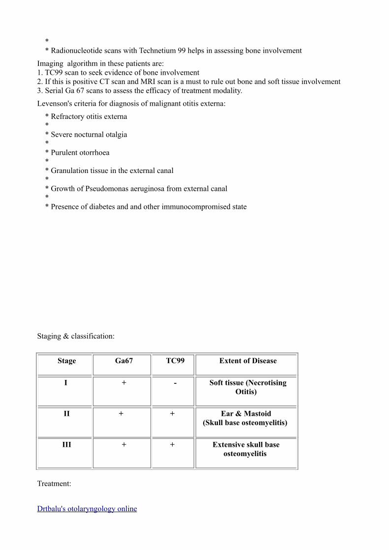
Staging & classification:
Stage Ga67 TC99 Extent of Disease
I + - Soft tissue (Necrotising Otitis)
II + + Ear & Mastoid(Skull base osteomyelitis)
III + + Extensive skull base osteomyelitis
Treatment:
Drtbalu's otolaryngology online
Extensive surgical procedures have failed miserably to cure this condition. The role of surgery is confined to only exclusion of malignancy by biopsy. Wound debridement is a possibility in advanced cases.
Medical management:Carbenicillin, Pipercillin, Ticarcillin can be used. Third and forth generation cephalosporins can be used.Ciprofloxacillin in doses of 1.5 g - 2.5 g /day in divided doses can be administered for a period of 2 weeks.Gentamycin can also be administered parenterally in doses of 80 mg iv two times a day in adults.
b. Cerumen:
This is also known as ear wax. This is a yellowish waxy substance secreted in the external auditory canal of humans. It protects the skin lining of the external auditory canal from excessive moisture. It also protects the external canal from bacteria, fungi and insects.
Humans are known to secrete two types of cerumen:
1. Soft and moist
2. Firm and dry
Persons secreting soft and moist type of ear wax have no problem due to its accumulation. It can easily be extruded by the normal cleansing mechanism of the external auditory canal. This difference in wax secretion has been traced to alterations in C11 gene. Persons secreting frim and dry wax are more prone for impaction of cerumen. Impaction of cerumen causes conductive hearing loss.
Cerumen is usually produced in the outer third of the cartilagenous portion of the external auditory canal. It is composed of:
1. Viscous secretions from sebaceous glands
2. Less viscous secretions from modified apocrine sweat glands
3. Shed layers of skin
Cerumen has been found to have bacterostatic effect. Excessive occlusion of the external canal due to accumulation of cerumen and desquamated epithelial cells associated with migration defect of the lining epithelium can cause keratosis obturans. This is a painful condition which needs to be treated by removing the mass under anesthesia.
Removal of cerumen can be performed using probes / curettes if the consistency is soft. If cerumen is excessively soft then cotton buds can be used for removal.
Firm cerumen should be lubricated by using ceruminolytics / liquid paraffin to soften it up before attempted removal.
Aural syringing is one of the painless way of removing accumulated cerumen.
c. Septoplasty:
Drtbalu's otolaryngology online
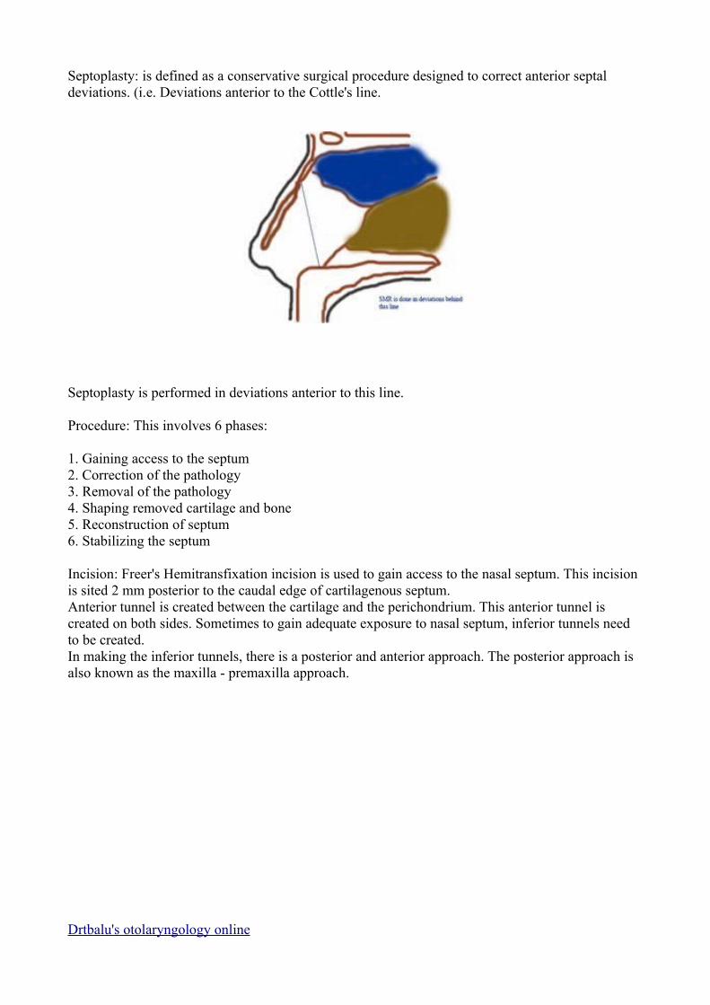
Septoplasty: is defined as a conservative surgical procedure designed to correct anterior septal deviations. (i.e. Deviations anterior to the Cottle's line.
Septoplasty is performed in deviations anterior to this line.
Procedure: This involves 6 phases:
1. Gaining access to the septum2. Correction of the pathology3. Removal of the pathology4. Shaping removed cartilage and bone5. Reconstruction of septum6. Stabilizing the septum
Incision: Freer's Hemitransfixation incision is used to gain access to the nasal septum. This incision is sited 2 mm posterior to the caudal edge of cartilagenous septum. Anterior tunnel is created between the cartilage and the perichondrium. This anterior tunnel is created on both sides. Sometimes to gain adequate exposure to nasal septum, inferior tunnels need to be created.In making the inferior tunnels, there is a posterior and anterior approach. The posterior approach is also known as the maxilla - premaxilla approach.
Drtbalu's otolaryngology online
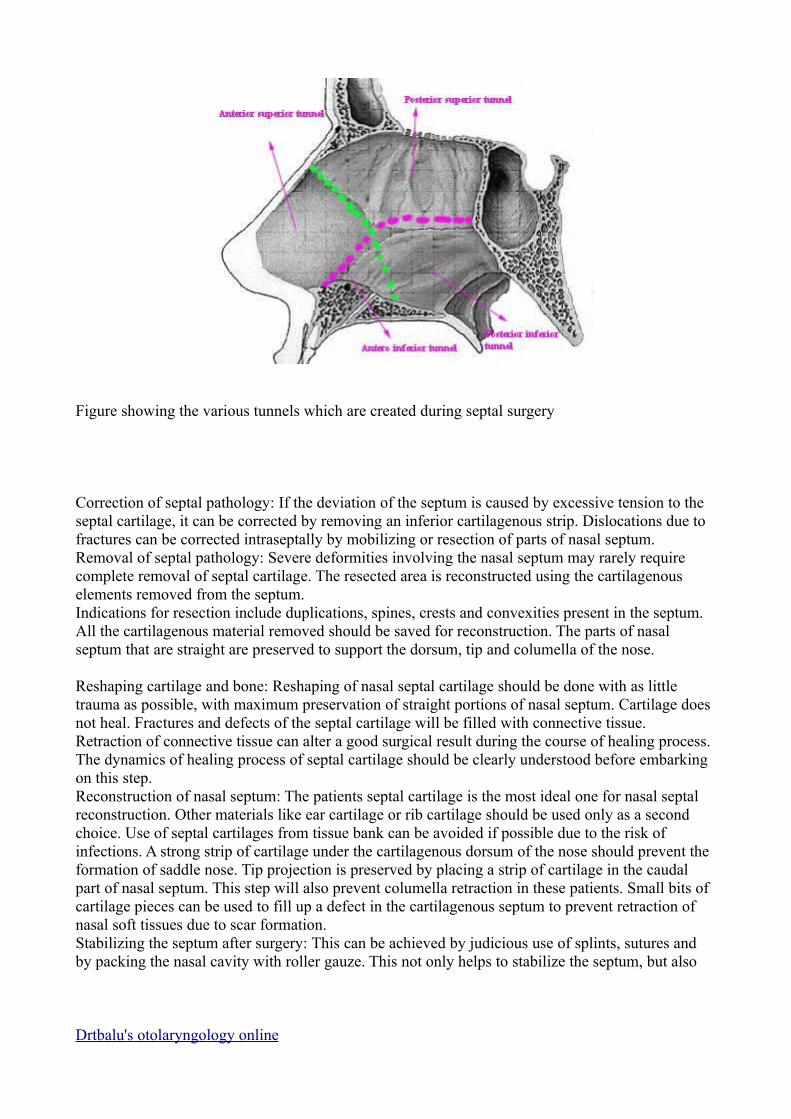
Figure showing the various tunnels which are created during septal surgery
Correction of septal pathology: If the deviation of the septum is caused by excessive tension to the septal cartilage, it can be corrected by removing an inferior cartilagenous strip. Dislocations due to fractures can be corrected intraseptally by mobilizing or resection of parts of nasal septum.Removal of septal pathology: Severe deformities involving the nasal septum may rarely require complete removal of septal cartilage. The resected area is reconstructed using the cartilagenous elements removed from the septum.Indications for resection include duplications, spines, crests and convexities present in the septum. All the cartilagenous material removed should be saved for reconstruction. The parts of nasal septum that are straight are preserved to support the dorsum, tip and columella of the nose.
Reshaping cartilage and bone: Reshaping of nasal septal cartilage should be done with as little trauma as possible, with maximum preservation of straight portions of nasal septum. Cartilage does not heal. Fractures and defects of the septal cartilage will be filled with connective tissue. Retraction of connective tissue can alter a good surgical result during the course of healing process. The dynamics of healing process of septal cartilage should be clearly understood before embarking on this step.Reconstruction of nasal septum: The patients septal cartilage is the most ideal one for nasal septal reconstruction. Other materials like ear cartilage or rib cartilage should be used only as a second choice. Use of septal cartilages from tissue bank can be avoided if possible due to the risk of infections. A strong strip of cartilage under the cartilagenous dorsum of the nose should prevent the formation of saddle nose. Tip projection is preserved by placing a strip of cartilage in the caudal part of nasal septum. This step will also prevent columella retraction in these patients. Small bits of cartilage pieces can be used to fill up a defect in the cartilagenous septum to prevent retraction of nasal soft tissues due to scar formation. Stabilizing the septum after surgery: This can be achieved by judicious use of splints, sutures and by packing the nasal cavity with roller gauze. This not only helps to stabilize the septum, but also
Drtbalu's otolaryngology online
keeps the mucoperichondrium in contact withe the nasal septum. This is important for the viability of nasal septum.
d. Absolute bone conduction test:
Absolute bone conduction test:
This test is performed to identify sensorineural hearing loss. In this test the hearing level of the patient is compared to that of the examiner. The examiner's hearing is assumed to be normal. In this test the vibrating fork is placed over the mastoid process of the patient after occluding the external auditory canal. As soon as the patient indicates that he is unable to hear the sound anymore, the fork is transferred to the mastoid process of the examiner after occluding the external canal. In cases of normal hearing the examiner must not be able to hear the fork, but in cases of sensori neural hearing loss the examiner will be able to hear the sound, then the test is interpreted as ABC reduced. It is not reduced in cases with normal hearing.
e. Otogenic brain abscess:
Otogenic brain abscess always develop in the temporal lobe or the cerebellum of the same side of the infected ear. Temporal lobe abscess is twice as common as cerebellar abscess. In children nearly 25% of brain abscesses are otogenic in nature, whereas in adults who are more prone to chronic ear infections the percentage rises to 50%. The routes of spread of infection has already been discussed above, the commonest being the direct extension through the eroded tegment plate. Although dura is highly resistant to infection, local pachymeningitis may be followed by thrombophlebitis penetrating the cerebral cortex, sometimes the infection could extent via the Virchow - Robin spaces in to the cerebral white matter. Cerebellar abscess is usually preceded by thrombosis of lateral sinus. Abscess in the cerebellum may involve the lateral lobe of the cerebellum, and it may be adherent to the lateral sinus or to a patch of dura underneath the Trautmann's triangle.
Drtbalu's otolaryngology online
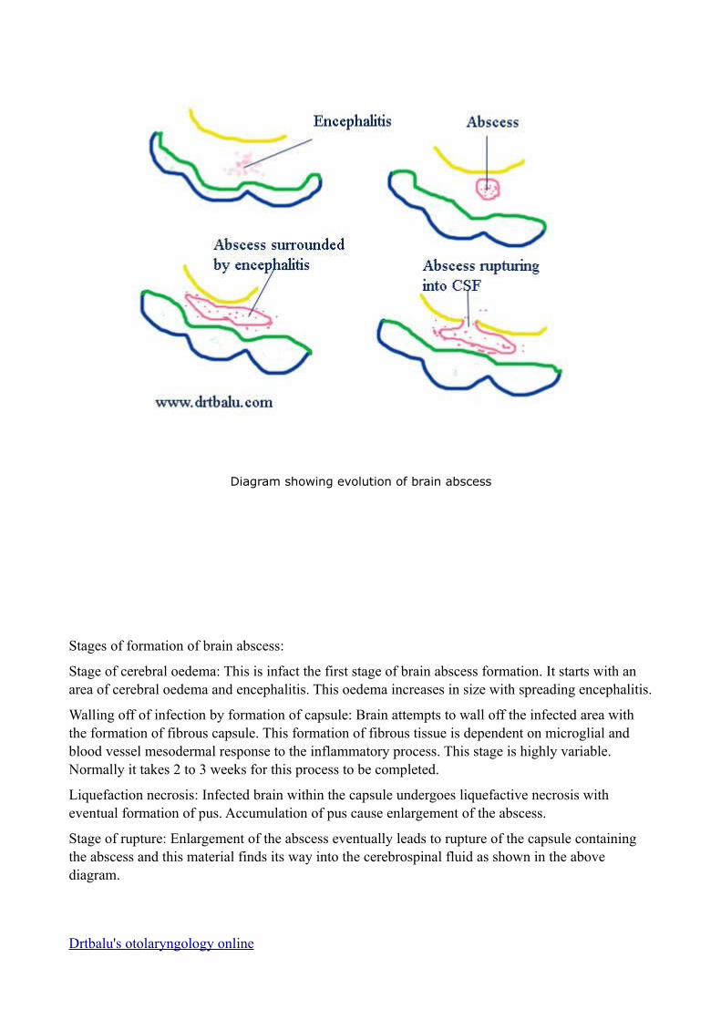
Diagram showing evolution of brain abscess
Stages of formation of brain abscess:
Stage of cerebral oedema: This is infact the first stage of brain abscess formation. It starts with an area of cerebral oedema and encephalitis. This oedema increases in size with spreading encephalitis.
Walling off of infection by formation of capsule: Brain attempts to wall off the infected area with the formation of fibrous capsule. This formation of fibrous tissue is dependent on microglial and blood vessel mesodermal response to the inflammatory process. This stage is highly variable. Normally it takes 2 to 3 weeks for this process to be completed.
Liquefaction necrosis: Infected brain within the capsule undergoes liquefactive necrosis with eventual formation of pus. Accumulation of pus cause enlargement of the abscess.
Stage of rupture: Enlargement of the abscess eventually leads to rupture of the capsule containing the abscess and this material finds its way into the cerebrospinal fluid as shown in the above diagram.
Drtbalu's otolaryngology online
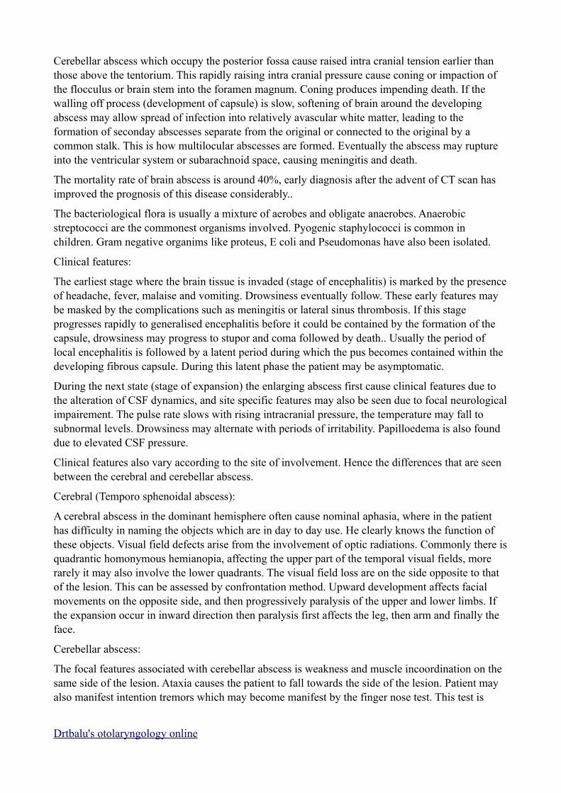
Cerebellar abscess which occupy the posterior fossa cause raised intra cranial tension earlier than those above the tentorium. This rapidly raising intra cranial pressure cause coning or impaction of the flocculus or brain stem into the foramen magnum. Coning produces impending death. If the walling off process (development of capsule) is slow, softening of brain around the developing abscess may allow spread of infection into relatively avascular white matter, leading to the formation of seconday abscesses separate from the original or connected to the original by a common stalk. This is how multilocular abscesses are formed. Eventually the abscess may rupture into the ventricular system or subarachnoid space, causing meningitis and death.
The mortality rate of brain abscess is around 40%, early diagnosis after the advent of CT scan has improved the prognosis of this disease considerably..
The bacteriological flora is usually a mixture of aerobes and obligate anaerobes. Anaerobic streptococci are the commonest organisms involved. Pyogenic staphylococci is common in children. Gram negative organims like proteus, E coli and Pseudomonas have also been isolated.
Clinical features:
The earliest stage where the brain tissue is invaded (stage of encephalitis) is marked by the presence of headache, fever, malaise and vomiting. Drowsiness eventually follow. These early features may be masked by the complications such as meningitis or lateral sinus thrombosis. If this stage progresses rapidly to generalised encephalitis before it could be contained by the formation of the capsule, drowsiness may progress to stupor and coma followed by death.. Usually the period of local encephalitis is followed by a latent period during which the pus becomes contained within the developing fibrous capsule. During this latent phase the patient may be asymptomatic.
During the next state (stage of expansion) the enlarging abscess first cause clinical features due to the alteration of CSF dynamics, and site specific features may also be seen due to focal neurological impairement. The pulse rate slows with rising intracranial pressure, the temperature may fall to subnormal levels. Drowsiness may alternate with periods of irritability. Papilloedema is also found due to elevated CSF pressure.
Clinical features also vary according to the site of involvement. Hence the differences that are seen between the cerebral and cerebellar abscess.
Cerebral (Temporo sphenoidal abscess):
A cerebral abscess in the dominant hemisphere often cause nominal aphasia, where in the patient has difficulty in naming the objects which are in day to day use. He clearly knows the function of these objects. Visual field defects arise from the involvement of optic radiations. Commonly there is quadrantic homonymous hemianopia, affecting the upper part of the temporal visual fields, more rarely it may also involve the lower quadrants. The visual field loss are on the side opposite to that of the lesion. This can be assessed by confrontation method. Upward development affects facial movements on the opposite side, and then progressively paralysis of the upper and lower limbs. If the expansion occur in inward direction then paralysis first affects the leg, then arm and finally the face.
Cerebellar abscess:
The focal features associated with cerebellar abscess is weakness and muscle incoordination on the same side of the lesion. Ataxia causes the patient to fall towards the side of the lesion. Patient may also manifest intention tremors which may become manifest by the finger nose test. This test is
Drtbalu's otolaryngology online

performed by asking the patient to touch the tip of the nose with the index finger first with the eyes open and then with the eyes closed. The patient may often overshoot the mark when attempted with the eyes closed in case of cerebellar abscess. The patient may also have spontaneous nystagmus. Dysdiadokinesis is also positive in these patients.
Investigations:
CT scan and MRI scans are the present modes of investigation. Scan is ideally performed using contrast media. These scans not only reveal the position and size of the abscess, the presence of localised encephalitis can be distinguished from that of an encapsulated abscess. Associated conditions such as subdural abscess, and lateral sinus thrombosis can also be seen.
Management:
Surgical drainage of the abscess, followed by mastoidectomy to clear the ear disorder.
Drtbalu's otolaryngology online