taxonomy, biology, and occurrence of some marine leeches in
TRANSCRIPT

Proc. Helminthol. Soc. Wash.46(2), 1979, pp. 254-264
Taxonomy, Biology, and Occurrence of Some MarineLeeches in Newfoundland Waters
M. C. MEYER AND R. A. KHA NDepartment of Zoology, University of Maine, Orono, Maine 04469, andDepartment of Biology and Marine Sciences Research Laboratory,Memorial University of Newfoundland, St. John's, Newfoundland A1C 5S7
ABSTRACT: Taxonomy, biology, and occurrence of four species of marine leeches, from the NWAtlantic, in the vicinity of Newfoundland were studied. Species include Johanssonia arctica (Jo-hansson), Platybdella olriki Malm, P. anarrhichae (Diesing), and Calliobdella nodulifera (Malm).
This paper is the second in our studies on marine leeches from the northwesternNorth Atlantic, in the vicinity of Newfoundland. Previously we (Khan and Meyer,1976) discussed the taxonomy and biology of four species of the Malmiana-Oceanobdella complex.
Taxonomy of marine leeches is poorly known, and information concerningNorth American representatives is particularly meager. The principal limitationon availability of material is because it is related to the degree of difficult y ofobtaining the host, which is usually fish. A few species infest Reptilia and Ar-thropoda. The arthropod hosts are usually decapod Crustacea, but in a few in-stances Pycnogonida are utilized. Some of the host-parasite data have been sum-marized by Knight-Jones (1962).
The best faunistic records are provided by the excellent knowledge of theiroccurrence on the coasts of Scandinavia and Greenland (Malm, 1863, 1865; Jo-hansson, 1896, 1899), the Arctic Ocean and northern regions of the Atlantic andPacific Oceans (Selensky, 1914b, 1915, 1923; Vasileyev, 1939). Occurrence rec-ords have been extracted and brought together by Epshtein (1967). Soos (1965)and Epshtein (1961, 1962, 1967, 1968) are particularly useful in coordinating earlyand recent nomenclature.
Materials and Methods
Animals were studied alive and fixed, in serial sections, dissections, and wholemounts. Before fixation leeches were anesthetized with 5 to 15% ethanol in sea-water. Following anesthetization, specimens were transferred to a slide andstraightened, and fixative (80% ethanol) added slowly. This resulted in straight,normally relaxed, and unflattened specimens. In mature and unengorged wormsproperly anesthetized prior to fixation there is uniformity of characters, with onlyslight variability.
Descriptive measurements include the means, followed by the range in paren-theses. Length measurements are overall, i.e., both suckers are included. Mea-surements are in mm, unless otherwise indicated, and are based upon 10 or morereadings. No attempt has been made to compile every host and all occurrencerecords for each locality as this would be redundant; however, all original hostspecies and locality records are included. Vernacular and scientific names off ishare from Leim and Scott (1966).
A stock population of Johanssonia arctica was established on spider crabs,Chionoecetes opilio (Fabricius), in May 1973 and maintained throughout the
Copyright © 2011, The Helminthological Society of Washington
OF WASHINGTON, VOLUME 46, NUMBER 2, JULY 1979 • 255
study in holding tanks (100 x 80 x 65 cm), with a water flow of approximately3 liters/min. Longhorn sculpins (Myoxocephalus octodecemspinosus) were alsoplaced in the same tank. Fasted leeches fed on the sculpins and subsequentlyattached to the crabs. Water temperature varied between -1 and 3°C. Chionoe-cetes opilio and Hyas coarctatus Leach were collected in crab pots (160 to 360m deep) off coastal Newfoundland and maintained alive in tanks at the MarineSciences Research Laboratory, Logy Bay.
Wolffish, Atlantic and spotted (Anarhichas lupus and A. minor, respectively),Atlantic cod (Gadus morhua), eelpouts: Laval's, Vahl's, and Arctic (Lycodeslavalaei, L. vahlii, L. reticulatus, respectively) were collected by trawls (80 to210 m) on the Grand Banks, maintained alive in tanks aboard the A. T. Cameron,and examined for leeches at the laboratory. Other species offish, which includedwinter flounders (Pseudopleuronectes americanus), and Atlantic sea raven(Hemitripterus americanus}, collected by divers, were negative for leeches. Fishexamined for leeches were obtained in April through June.
Johanssonia arctica (Johansson 1899)
A brief history of our knowledge of the species involved is necessary. Selensky(1914b:209) proposed the generic name Johanssonia for leeches infesting Atlanticwolffish (Anarhichas lupus}, from the Gulf of Kola, near the Biological Stationat Murmansk, which he described as Johanssonia kolaensis. In the same paperSelensky assigned to Johanssonia a species infesting a Pycnogonida (Nymphonstromi), from the same general locality, which he had earlier described (1914a)as Ichthyobdella pantopodium. In using Ichthyobdella Selensky followed Jo-hansson (1899:687), who used Ichthyobdella as a provisional category for speciesof Piscicolidae of uncertain generic assignment.
Based upon specimens from the Karischen Sea and Greenland, Johansson(1899:671) described Oxytonostoma arctica. In a critical review of the species ofOxytonostoma and Johanssonia, in the Zoological Institute of the Academy ofSciences, USSR, which included many O. arctica identified by Johansson andVasileyev, Epshtein (1961, 1968 [in the second paper, based on dissections, theauthor authenticated his provisional decisions in the earlier paper]) made twotaxonomic changes. He synonymized Johanssonia pantopodium with Oxytonos-toma arctica and, because O. arctica has structural characters that are not com-patible with those of Oxytonostoma, transferred the species to Johanssonia.
Anatomy
EXTERNAL FEATURES: Body rounded, becoming narrower toward the ends;clitellum indistinct; trachelosome only slightly narrower than urosome. Bodyshape varies only slightly. Anterior sucker discoidal, eccentrically attached, an-terior exposed portion nearly 3 times as great as posterior ventral surface, andsharply separated from trachelosome by a prominent peduncle (Fig. 1). Posteriorsucker cupuliform, weakly developed, and not sharply separated from urosome(Fig. 3). Lacking eyes, metameric ocelli and ocelli on anal sucker, and colormarkings on body surface.
MEASUREMENTS: length 21.5 (18.2-29.3); width of trachelosome and uro-some, 0.9 (0.7-1.1) and 1.2 (0.9-2.1) respectively; transverse diameter of oral andanal suckers 1.2 (0.7-1.4) and 1.3 (1.1-1.4), respectively.
Copyright © 2011, The Helminthological Society of Washington

256 PROCEEDINGS OF THE HELMINTHOLOGICAL SOCIETY
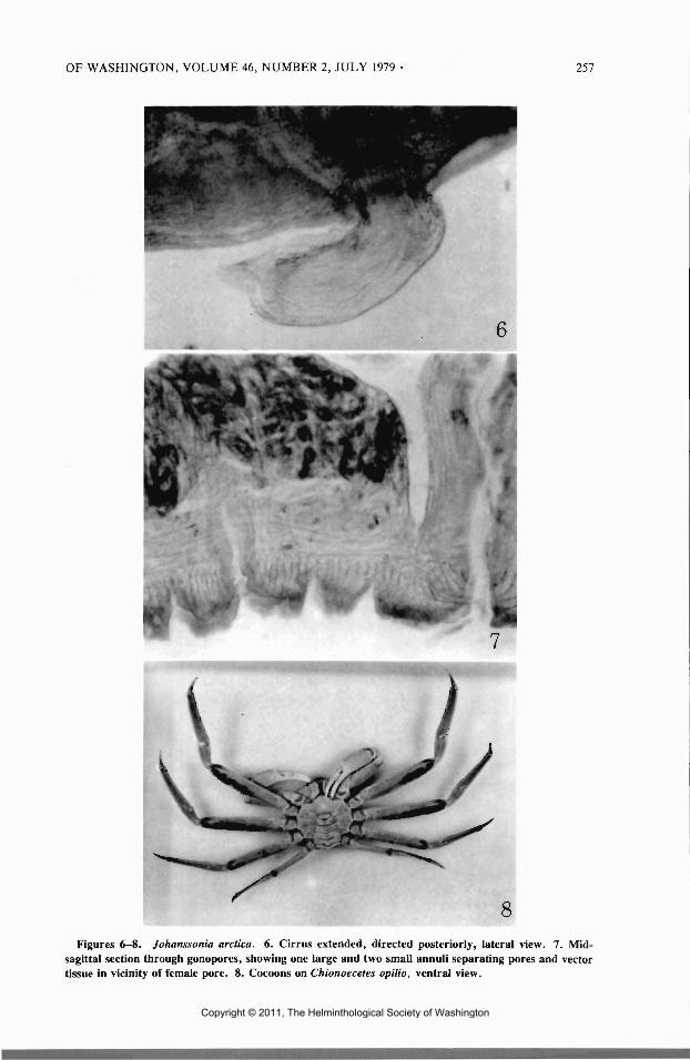
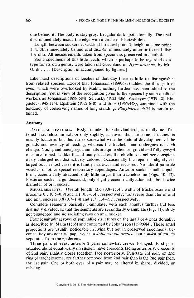
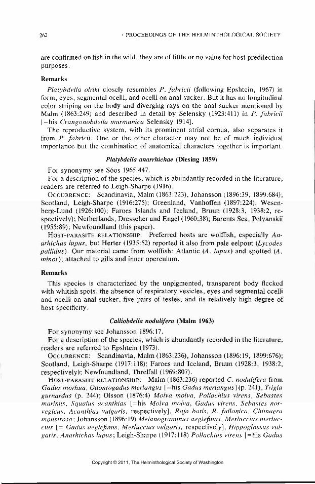
10Figures 1-5. Johanssonia arctica. 1. Anterior sucker and adjacent trachelosomal rings, ventral
view. 2. Complete segment, showing annulation, respiratory vesicles, and papillae, dorsal view. 3.Posterior sucker and adjacent urosomal rings, dorsal view. 4. Posterior region of digestive system, withintestine displaced to the right to show postcaeca and fenestrae, dorsal view. 5. Reproductive system,dorsal view.
Figures 9-13. 9. Johanssonia arctica, cocoon, lateral view. 10-13. Platybdella olriki. 10. Anteriorsucker and adjacent trachelosomal rings, dorsal view. 11. Complete segments, showing annulation,nerve ganglia, segmental ocelli, and testes. 12. Posterior sucker and adjacent urosomal rings, dorsalview. 13. Reproductive system, dorsal view.
Copyright © 2011, The Helminthological Society of Washington
OF WASHINGTON, VOLUME 46, NUMBER 2, JULY 1979 257
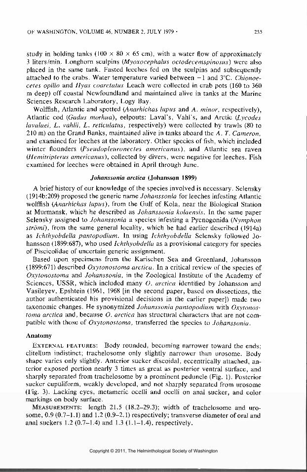
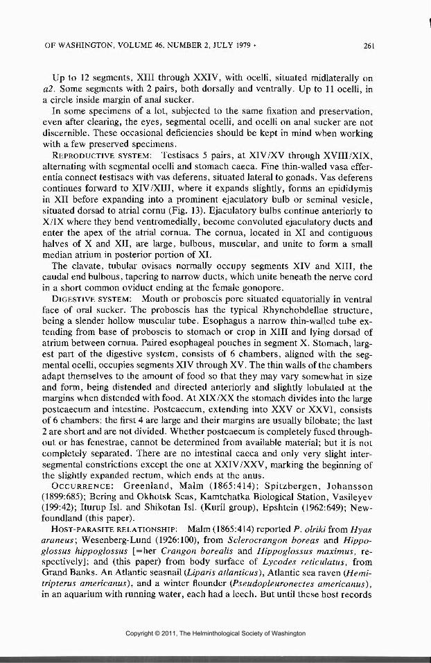
8Figures 6-8. Johanssonia arctica. 6. Cirrus extended, directed posteriorly, lateral view. 7. Mid-
sagittal section through gonopores, showing one large and two small annul! separating pores and vectortissue in vicinity of female pore. 8. Cocoons on Chionoecetes opilio, ventral view.
Copyright © 2011, The Helminthological Society of Washington
258 • PROCEEDINGS OF THE HELMINTHOLOGICAL SOCIETY
Respiratory vesicles 11 pairs, beginning in segment XIII , not always apparentexternally but visible in serial sections. In complete segments, primary annuli areeach divided twice, resulting in a 12-annulate segment (Fig. 2) expressed byformula (cl - c4 + c5 - c8 + c9 - clT). If greater recognition is given smallerannuli and depth of furrows, the number of annuli per segment increases. Ac-cording to Epshtein (1968:1012 and Fig. 2) c9 and ell were each divided intodiscrete annuli, resulting in the 14-annulate segment. Thus, mapping of annulationis fraught with difficulty because of subtle variations among specimens and thevagaries of personal interpretation.
Papillae in 12 rows on body surface, the dorsal 6 are slightly larger, apparentin living and most preserved specimens. Largest and most regular papillae onneural annulus (c6) of complete segments. Papillae usually in straight lines, a pairof paramedians, supramarginals, and marginals, but they may be somewhat scat-tered and some may be missing. Position and relative size of the most constantpapillae shown in Figure 2.
REPRODUCTIVE SYSTEM: The reproductive system, especially that of themale, is rather complicated in structure (Fig. 5). Testisacs 6 pairs, intrasegmen-tally at XII I through XVIII , anterior margins reaching to nerve ganglia, and al-ternating with gastric chambers.
Vas deferens, a capillary tube in testicular region, expands into a convolutedand loosely folded epididymis in segments XII I and XII , then reduced in diameterand continues cephalad in a relatively straight course, lateral to atrium, beforeincreasing again in diameter to form ejaculatory duct which bends ventrad to joinatrium. Ejaculatory ducts and adjacent atrial cornua, which form a conspicuouspart of vas deferens, are remarkable in that they extend far forward of atrium,to 4th free ganglion (of segment X) or beyond; but their exact position and formdiffer somewhat among specimens.
Beginning with epididymis all parts of sperm duct contain sperm. Terminallythere is a large cirrus pouch, or bursa, containing an extensible cirrus, occupyinga space between 5th and 6th free ganglia (of XI and XII) , which extends hori-zontally backwards, before bending ventrad and ending with male gonopore.Cirrus is a cylindrical, muscular troughlike structure, fused ventrally and opendorsally. Cirrus when extended in preserved specimens, directed posteriorly(Fig. 6).
Ovisacs elongated thin-walled sacs, between ganglia of XII and XIV . Eachconsists of 2 parts, a posterior globoid or ellipsoidal, thin-walled sac filled withdeveloping ova, and an anterior tubular portion containing mostly mature ova.Passing beneath esophagus the ducts become thickened with connective tissueand muscle fibers, representing vector tissue (Fig. 7), and converge below nervecord to unite just before the female gonopore, so that there is practically nocommon oviduct. Gonopores separated by 3 annuli, 1 large and 2 small.
DIGESTIVE SYSTEM: Mouth or proboscis pore on small elevation, situatedslightly postequatorially in sucker cavity; proboscis at rest very long, extendingto 4th free ganglion (of segment X), where it joins the esophagus. Esophagealcaeca small, in anterior l/2 of XI. Stomach, beginning at XII/XIII , with 7 lateralcaeca, which tend to alternate with the testisacs. Size and shape of these caecasomewhat variable, depending on recency of feeding, but even if relatively empty,they tend to assume the shape and degree of expansion when animal is gorged.
Copyright © 2011, The Helminthological Society of Washington
OF WASHINGTON, VOLUM E 46, NUMBER 2, JULY 1979 • 259
At XIX/XX , off the last stomach caecum, alimentary tract branches into apostcaecum and an intestine (Fig. 4). The ventrally situated postcaecum has 5small fenestrae, aligned with the ganglia; the dorsal intestine has 4 paired, an-terolaterally directed caeca, decreasing in size from fore to aft. Posterior to thelast caecum, intestine continues as a convoluted tube, ending in a spacious rectumwhich tapers to a minute anus.
OCCURRENCE: Greenland, and Karischen Sea, Cape Middendorff, Johansson(1899:672); Kola Bay, Murmansk, Selensky (1914a:273); Kara Sea, Wesenberg-Lund (1926:98); Alaska Peninsula, Stepovak Bay, Moore and Meyer (1951:55);Barents Sea, Eastern Siberian Sea, Laptev Sea, Epshtein (1961:1121); Newfound-land (this paper).
HOST-PARASITE RELATIONSHIP: Selensky (1914a:273) reported A. arctica[ = his Ichthyobdella pantopodium] from a Pycnogonida (Nymphon stromi); Ep-shtein (1961:1121) [=his Oxytonostoma arctica] from Pycnogonida (Colossendeissp.); and (this paper) decapod Crustacea, Chionoecetes opilio and Hyas coarc-tatus, and Atlantic cod, Gadus morhua.
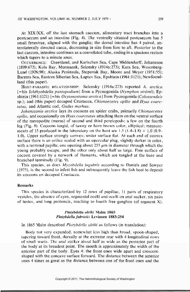
Johanssonia arctica lays its cocoons on spider crabs, primarily Chionoecetesopilio, and occasionally on Hyas coarctatus attaching them on the ventral surfaceof the meropodite (merus) of second and third pereiopods; a few on the fourthleg (Fig. 8). Cocoons turgid, of tawny or horn-brown color, elliptical; measure-ments of 15 produced in the laboratory on the host are 1.5 (1.4-1.8) x 1.0 (0.9-1.0). Upper surface strongly convex; under surface flat. At each end of convexsurface there is an orifice sealed with an opercular plug, slightly darker in color,with a terminal papilla; one opening about 255 /u,m in diameter through which theyoung probably escape, and the other only about half as large. Free surface ofcocoon covered by a network of filaments, which are tangled at the base andbranched terminally (Fig. 9).
This species, as does Myzobdella lugubris according to Daniels and Sawyer(1975), is the second to infest fish and subsequently leave the fish host to depositits cocoons on decapod Crustacea.
Remarks
This species is characterized by 12 rows of papillae, 11 pairs of respiratoryvesicles, the absence of eyes, segmental ocelli and ocelli on anal sucker, six pairsof testes, and long proboscis, reaching to fourth free ganglion (of segment X).
Platybdella olriki Malm 1865Platybdella fabricii: Levinsen 1883:254
In 1865 Malm described Platybdella olriki as follows (in translation):
Body not very expanded, somewhat less high than broad, spoon-shaped,tapering toward front, dorsally at the extreme rear with 4 longitudinal rowsof small warts. The anal sucker about half as wide as the posterior part ofthe body at its broadest point. The mouth is approximately the width of theanterior part of the body. Eyes 4: the front ones wide apart and crescent-shaped with the concave surface forward. The distance between the anteriorones 4 times as great as the distance between one of the front ones and the
Copyright © 2011, The Helminthological Society of Washington
260 • PROCEEDINGS OF THE HELMINTHOLOGICAL SOCIETY
one behind it. The body is clay-grey. Irregular dark spots dorsally. The analdisc immediately inside the edge with a circle of blackish dots.
Length between suckers 9; width at broadest point 3; height at same point2; width immediately behind oral disc 34; immediately anterior to anal discIV e mm. Al l measurements taken from specimens preserved in alcohol.
Some specimens of this littl e leech, which is perhaps to be regarded as atype for its own genus, were taken off Greenland on Hyas araneus, by Mr.Olrik . . . . [Description unaccompanied by figures.]
Like most descriptions of leeches of that day there is littl e to distinguish itfrom related species. Except that Johansson (1899:685) added the third pair ofeyes, which were overlooked by Malm, nothing further has been added to thedescription. Yet in view of the recognition given to the species by such qualifiedworkers as Johansson (1899:684), Selensky (1923:406), Vasileyev (1939:42), Rin-guelet (1945:114), Epshtein (1962:648), and Soos (1965:448), combined with thetendency of conserving names of long standing, Platybdella olriki is herein re-tained.
Anatomy
EXTERNAL FEATURES: Body rounded to subcylindrical, normally not flat-tened; trachelosome not, or only slightly, narrower than urosome. Urosome isusually fusiform, but this varies somewhat with the state of development of thegonads and recency of feeding, whereas the trachelosome undergoes no suchchange. Young and unengorged animals are quite slender; gravid and full y gorgedones are robust. Unlike that of many leeches, the clitellum is neither conspicu-ously enlarged nor distinctively colored. Occasionally the region is slightly en-larged but in most cases it is faintly narrower and recessed. No lateral pulsatilevesicles or other special respiratory appendages. Anterior sucker small, cupuli-form, eccentrically attached, only littl e larger than trachelosome (Figs. 10, 12).Posterior sucker large, discoidal, only slightly eccentrically attached, about twicediameter of oral sucker.
MEASUREMENTS: Overall length 12.6 (9.8-15.4); width of trachelosome andurosome 0.7 (0.5-0.9) and 1.1 (0.7-1.4), respectively; transverse diameter of oraland anal suckers 0.8 (0.7-1.4) and 1.7 (1.4-2.1), respectively.
Complete segments basically 3-annulate, with each annulus further but lessdistinctly divided, so that the segments are secondarily 6-annulate (Fig. 11). Bodynot pigmented and no radiating rays on anal sucker.
Four longitudinal rows of papillalike structures on the last 3 or 4 rings dorsally,as described by Malm (1865) and confirmed by Johansson (1899:684). These smallprojections are usually noticeable in living but not in preserved specimens, be-cause they are not true papillae, as in Johanssonia arctica, but consist of cuticleseparated from the epidermis.
Three pairs of eyes, anterior 2 pairs somewhat crescent-shaped. First pair,situated about equatorially on sucker, have crescents facing anteriorly; crescentsof 2nd pair, slightly closer together, face posteriorly. Punctate 3rd pair, on 2ndring of trachelosome, are farther removed from 2nd pair than is the 2nd pair fromthe 1st pair. One or both eyes of a pair may be altered in shape, divided, ormissing.
Copyright © 2011, The Helminthological Society of Washington
OF WASHINGTON, VOLUME 46, NUMBER 2, JULY 1979 • 261
Up to 12 segments, XII I through XXIV , with ocelli, situated midlaterally ona2. Some segments with 2 pairs, both dorsally and ventrally. Up to 11 ocelli, ina circle inside margin of anal sucker.
In some specimens of a lot, subjected to the same fixation and preservation,even after clearing, the eyes, segmental ocelli, and ocelli on anal sucker are notdiscernible. These occasional deficiencies should be kept in mind when workingwith a few preserved specimens.
REPRODUCTIVE SYSTEM: Testisacs 5 pairs, at XIV/X V through XVIII/XIX ,alternating with segmental ocelli and stomach caeca. Fine thin-walled vasa effer-entia connect testisacs with vas deferens, situated lateral to gonads. Vas deferenscontinues forward to XIV/XIII , where it expands slightly, forms an epididymisin XII before expanding into a prominent ejaculatory bulb or seminal vesicle,situated dorsad to atrial cornu (Fig. 13). Ejaculatory bulbs continue anteriorly toX/I X where they bend ventromedially, become convoluted ejaculatory ducts andenter the apex of the atrial cornua. The cornua, located in XI and contiguoushalves of X and XII , are large, bulbous, muscular, and unite to form a smallmedian atrium in posterior portion of XL
The clavate, tubular ovisacs normally occupy segments XIV and XIII , thecaudal end bulbous, tapering to narrow ducts, which unite beneath the nerve cordin a short common oviduct ending at the female gonopore.
DIGESTIVE SYSTEM: Mouth or proboscis pore situated equatorially in ventralface of oral sucker. The proboscis has the typical Rhynchobdellae structure,being a slender hollow muscular tube. Esophagus a narrow thin-walled tube ex-tending from base of proboscis to stomach or crop in XII I and lying dorsad ofatrium between cornua. Paired esophageal pouches in segment X. Stomach, larg-est part of the digestive system, consists of 6 chambers, aligned with the seg-mental ocelli, occupies segments XIV through XV. The thin walls of the chambersadapt themselves to the amount of food so that they may vary somewhat in sizeand form, being distended and directed anteriorly and slightly lobulated at themargins when distended with food. At XIX/X X the stomach divides into the largepostcaecum and intestine. Postcaecum, extending into XXV or XXVI , consistsof 6 chambers: the first 4 are large and their margins are usually bilobate; the last2 are short and are not divided. Whether postcaecum is completely fused through-out or has fenestrae, cannot be determined from available material; but it is notcompletely separated. There are no intestinal caeca and only very slight inter-segmental constrictions except the one at XXIV/XXV , marking the beginning ofthe slightly expanded rectum, which ends at the anus.
OCCURRENCE: Greenland, Malm (1865:414); Spitzbergen, Johansson(1899:685); Bering and Okhotsk Seas, Kamtchatka Biological Station, Vasileyev(199:42); Iturup Isl. and Shikotan Isl. (Kuril group), Epshtein (1962:649); New-foundland (this paper).
HOST-PARASITE RELATIONSHIP: Malm (1865:414) reported P. olriki from Hyasaraneus; Wesenberg-Lund (1926:100), from Sclerocrangon boreas and Hippo-gloss us hippoglossus [=her Crangon borealis and Hippoglossus maximus, re-spectively]; and (this paper) from body surface of Lycodes reticulatus, fromGrand Banks. An Atlantic seasnail (Liparis atlanticus), Atlantic sea raven (Hemi-tripterus americanus}, and a winter flounder (Pseudopleuronectes americanus},in an aquarium with running water, each had a leech. But until these host records
Copyright © 2011, The Helminthological Society of Washington
262 • PROCEEDINGS OF THE HELMINTHOLOGICAL SOCIETY
are confirmed on fish in the wild, they are of littl e or no value for host predilectionpurposes.
Remarks
Platybdella olriki closely resembles P. fabricii (following Epshtein, 1967) inform, eyes, segmental ocelli, and ocelli on anal sucker. But it has no longitudinalcolor striping on the body and diverging rays on the anal sucker mentioned byMalm (1863:249) and described in detail by Selensky (1923:411) in P. fabricii[=his Crangonobdella murmanica Selensky 1914].
The reproductive system, with its prominent atrial cornua, also separates itfrom P. fabricii. One or the other character may not be of much individualimportance but the combination of anatomical characters together is important.
Platybdella anarrhichae (Diesing 1859)
For synonymy see Soos 1965:447.For a description of the species, which is abundantly recorded In the literature,
readers are referred to Leigh-Sharpe (1916).OCCURRENCE: Scandinavia, Malm (1863:223), Johansson (1896:39, 1899:684);
Scotland, Leigh-Sharpe (1916:275); Greenland, Vanhoffen (1897:224), Wesen-berg-Lund (1926:100); Faroes Islands and Iceland, Bruun (1928:3, 1938:2, re-spectively); Netherlands, Dresscher and Engel (1960:38); Barents Sea, Polyanskii(1955:89); Newfoundland (this paper).
HOST-PARASITE RELATIONSHIP: Preferred hosts are wolffish, especially An-arhichas lupus, but Herter (1935:52) reported it also from pale eelpout (Lycodespallidus). Our material came from wolffish: Atlantic (A. lupus) and spotted (A.minor); attached to gills and inner operculum.
Remarks
This species is characterized by the unpigmented, transparent body fleckedwith whitish spots, the absence of respiratory vesicles, eyes and segmental ocelliand ocelli on anal sucker, five pairs of testes, and its relatively high degree ofhost specificity.
Calliobdella nodulifera (Malm 1963)
For synonymy see Johansson 1896:17.For a description of the species, which is abundantly recorded in the literature,
readers are referred to Epshtein (1973).OCCURRENCE: Scandinavia, Malm (1863:236), Johansson (1896:19, 1899:676);
Scotland, Leigh-Sharpe (1917:118); Faroes and Iceland, Bruun (1928:3, 1938:2,respectively); Newfoundland, Threlfall (1969:807).
HOST-PARASITE RELATIONSHIP: Malm (1863:236) reported C. nodulifera fromGadus morhua, Odontogadus merlangus [ = his Gadus merlangus} (p. 241), Triglagurnardus (p. 244); Olsson (1876:4) Molva molva, Pollachius virens, Sebastesmarinus, Squalus acanthias [ = his Molva molva, Gadus virens, Sebastes nor-vegicus, Acanthias vulgaris, respectively], Raja batis, R. fullonica, Chimaeramonstrosa; Johansson (1896:19) Melanogrammus aeglefinus, Merluccius merluc-cius [= Gadus aeglefinus, Merluccius vulgaris, respectively], Hippoglossus vul-garis, Anarhichas lupus; Leigh-Sharpe (1917:118) Pollachius virens [ = his Gadus
Copyright © 2011, The Helminthological Society of Washington
OF WASHINGTON, VOLUME 46, NUMBER 2, JULY 1979 • 263
carbonarius]', Bruun (1928:3) Anarhichas minor; Threlfall (1969:807) Sqiialusacanthias. Our material came from eelpouts: Laval's (Lycodes lavalaei) andVahl's (L. vahlii).
Remarks
This species is characterized by the yellowish-brown pigment spots coveringthe body, 11 pairs of respiratory vesicles (discernible in living animals, and usuallyin preserved specimens), the absence of eyes, segmental ocelli and ocelli on analsucker, six pairs of testes, and its broad host tolerance.
Acknowledgments
Grateful acknowledgment is due the National Research Council of Canada, andW. D. Machin, former Dean of Sciences, Memorial University of Newfoundland,for financial support to the junior author; G. Hillier , Marine Sciences ResearchLaboratory, for photographs; Environmental Canada, Fisheries and Marine Ser-vices, St. John's, for providing J. Murphy with working space and fish holdingfacilities aboard the A. T. Cameron; E.G. Hoyle, Maine State Museum, Augusta,for assisting with the drawings; and D. J. Klemm, US EPA, National Environ-mental Research Laboratory, Cincinnati, Ohio, and E. M. Burreson, VirginiaInstitute of Marine Science, Gloucester Point, Virginia, for the English transla-tion of Epshtein 1967 and 1973, respectively. Special thanks are due A. A.Barden Jr., University of Maine, Orono, whose critical reading of the manu-script contributed to its improvement.
Literature Cited
Bruun, A. F. 1928. XVIII . Marine Hirudinea. Zool. of the Faroes. Copenhagen. 5 pp.Bruun, A. F. 1938. Marine Hirudinea. The Zoology of Iceland. Copenhagen 2(21): 1-5.Daniels, B. A., and R. T. Sawyer. 1975. The biology of the leech Myzobdella lugubris infesting blue
crabs and catfish. Biol. Bull. 148:193-198.Dresscher, G. N., and H. Engel. 1960. De Nederlandse bloedzuigers (Hirudinea). Konink. Neder.
Natuur. Ver. No. 39, 60 pp.Epshtein, V. M. 1961. A review of the fish leeches (Hirudinea: Piscicolidae) from the northern seas
of SSSR. Dokl. Akad. Nauk SSSR 141:1508-1511. (Trans, from Russian. 1961. Proc. Acad. Sci.USSR 141:1121-1124.)
Epshtein, V. M. 1962. A survey off ish leeches (Hirudinea: Piscicolidae) from the Bering and Okhtskand from the Sea of Japan. Dokl. Akad. Nauk SSSR 144:1181-1184. (Trans, from Russian. 1962.Proc. Acad. Sci. USSR 144:648-651.)
Epshtein, V. M. 1967. Regularities of the geographical distribution of the marine leeches (Hirudinea:Piscicolidae). Zool. Zh. 46:680-691. (Trans, from Russian. 1975. Scitran, Santa Barbara, Calif.)
Epshtein, V. M. 1968. Revision of the genera Oxytonostoma and Johanssonia. Zool. Zh. 47:1011-1021. (Trans, from Russian. 1969. National Lending Library for Science and Technology, York-shire, England.)
Epshtein, V. M. 1973. Diagnosis of the genera Calliobdella, Trachelobdella, Limnotrachelobdellaand Baicolbdella (Hirudinea, Piscicolidae) and determination of the taxonomic value of the di-agnostic characteristics. Zool. Zh. 52:332-341. (Trans from Russian. 1974. P. C. Rossbacher)
Herter, K. 1935. Hirudinea. Die Tierwelt der Nord-und Ostsee. Akad. Verlag., Leipzig Teil 6:45-106.
Johansson, L. 1896. Bidrag til l kannedomen om Sveriges Ichthyobdellider. Akad. Afhandl. UpsalaUniv., 122 pp.
Johansson, L. 1899. Die Ichthyobdelliden im Zoologischen Reichsmuseum in Stockholm. Ofvers.Kongl. Vetensk.-Akad. Forh. 1898. 55:665-687.
Copyright © 2011, The Helminthological Society of Washington
264 • PROCEEDINGS OF THE HELMINTHOLOGICAL SOCIETY
Khan, R. A., and M. C. Meyer. 1976. Taxonomy and biology of some Newfoundland marine leeches(Rhynchobdellae: Piscicolidae). J. Fish. Res. Bd. Can. 33:1699-1714.
Knight-Jones, E. W. 1962. Systematics of marine leeches. Pages 169-186 in K. H. Mann, Leeches(Hirudinea): their structure, physiology, ecology and embryology. Pergamon Press, Inc., NewYork. 201 pp.
Leigh-Sharpe, W. H. 1916. Platybdella anarrhichae. Parasitology 8:274-293.Leigh-Sharpe, W. H. 1917. Calliobdella nodulifera (Malm 1863). Proc. R. Phys. Soc. Edinburgh
20:118-122.Leim, A. H., and W. B. Scott. 1966. Fishes of the Atlantic Coast of Canada. Bull. Fish. Res. Bd.
Can. 155, 485 pp.Malm, A. W. 1863. Svenska Iglar, Disciferae. Kongl. Vetensk. Vitt . Samh. Handl. 8:153-263.Malm, A. W. 1865. Ichthyologiska Bidrag til l Skandinaviens Fauna. Forhand. Skand. Naturf. 9:405-
414. (Hirudinea pp. 413-414.)Moore, J. P., and M. C. Meyer. 1951. Leeches (Hirudinea) from Alaskan and adjacent waters.
Wasmann J. Biol. 9:11-77.Olsson, P. 1876. Bidrag til l Skandinaviens helminthfauna I. Svenska Vetenskap Akademiens Hand-
linger, Stockholm 14(1):35 pp. (Hirudinea: pp. 2-6.)Polyanskii, Y. I. 1955. Parasites of the fish of the Barents Sea. Tr. Zool. Inst. Akad. Nauk SSSR
19:158 pp. (Israel Program for Scientific Translations, 1966.)Ringuelet, R. 1945. Hirudineos del Museo de la Plata. Rev. Mus. La Plata 4:95-137.Selensky, W. D. 1914a. Uber einige auf Arthropoden schmarotzende Ichthyobdelliden. Zool. Anz.
44:270-282.Selensky, W. D. 1914b. Notes on the fauna of the Hirudinea of the coast of Murman. Trav. Soc.
Imp. Sci. Nat. St. Petersbourg 45(7&8): 197-210. (Trans, from Russian. 1955. M. C. Meyer, ed.;211-214. French resume.)
Selensky, W. D. 1915. Etudes Morphologiques et Systematiques sur les Hirudinees. 1. L'organisationdes Ichthyobdellides. Petrograd, 256 pp. (Trans, from Russian. 1955. M. C. Meyer & J. P. Moore,eds.)
Selensky, W. D. 1923. Crangonobdella murmanica n.g., n.sp., eine auf Sderocrangon schmarotz-ende Ichthyobdellidae. Zool. Jahrb. 46:397-488.
Soos, A. 1965. Identification key to the leech (Hirudinoidea) genera of the world, with a catalogueof the species. 1. Family: Piscicolidae. Acta Zool. Acad. Sci. Hung. 11:417-463.
Threlfall, W. 1969. Some parasites from elasmobranchs in Newfoundland. J. Fish. Res. Bd. Can.26:805-811.
Vanhoffen, E. 1897. Die fauna und flora Gronlands. Gronland-Expedition der gesellschaft fur Erd-kunde zu Berlin 1891-1893. Zweiter Bd., Hirudinea pp. 221-224.
Vasileyev, E. A. 1939. The Ichthyobdellidae of the Far East. Trudy Karelskogo GosudarstvennogoPedagogicheskogo Instituta. Biol. Ser. 1:25-68. (Trans, from Russian. 1967. Antonia Glasse.)
Wesenberg-Lund, Elise. 1926. Igler og Oligochaeter. Medd. Gr0nland. K0benhavn 23 (Suppl.) pp.93-115.
Copyright © 2011, The Helminthological Society of Washington