tb path & pathogenesis
TRANSCRIPT

Dr. Aswini Kumar MohapatraDr. Aswini Kumar Mohapatra
Professor and HeadProfessor and Head
Dept. of Pulmonary MedicineDept. of Pulmonary Medicine

TUBERCULOSISTUBERCULOSIS
PATHOLOGYPATHOLOGY
&&
PATHOGENESISPATHOGENESIS
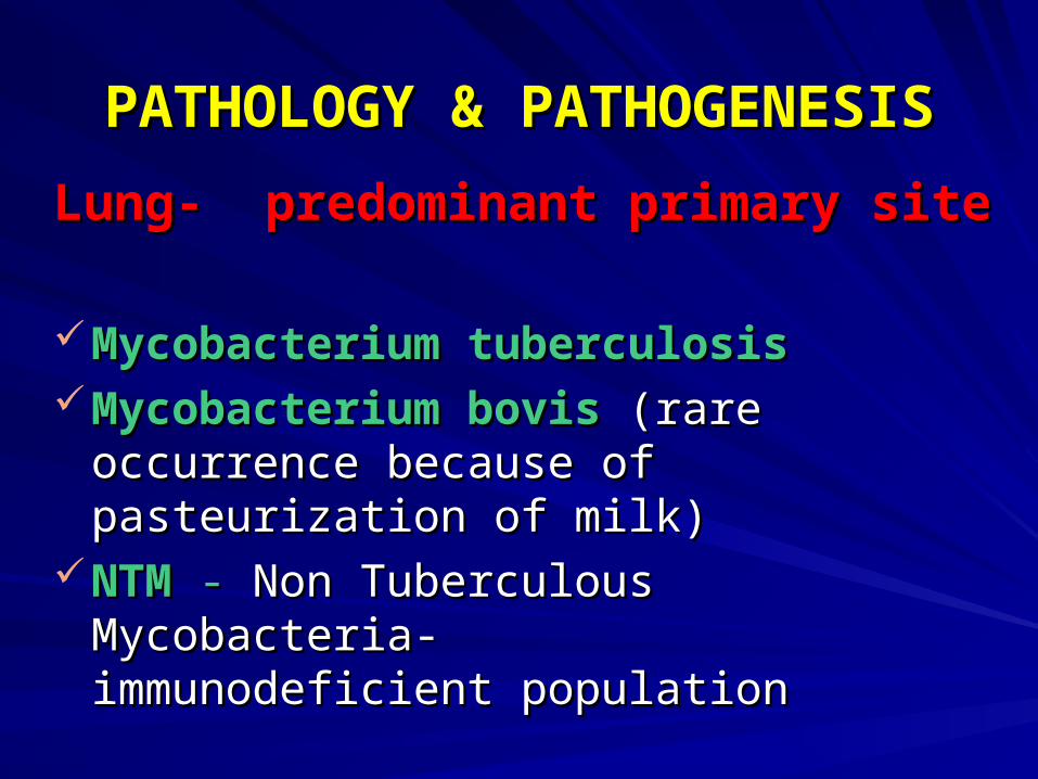
PATHOLOGY & PATHOGENESISPATHOLOGY & PATHOGENESIS
Lung- predominant primary siteLung- predominant primary site
Mycobacterium tuberculosisMycobacterium tuberculosisMycobacterium bovisMycobacterium bovis (rare occurrence (rare occurrence
because of pasteurization of milk)because of pasteurization of milk)NTMNTM - - Non Tuberculous Mycobacteria- Non Tuberculous Mycobacteria-
immunodeficient populationimmunodeficient population

Mycobacterium tuberculosisMycobacterium tuberculosis-- non motilenon motile
aerobic aerobic
catalase producingcatalase producing
acid fast bacillus acid fast bacillus (AFB)(AFB)
Tuberculosis is transmitted by aerosolTuberculosis is transmitted by aerosol. . PTB is the predominant form of diseasePTB is the predominant form of disease
Extra pulmonary TB is commonly Extra pulmonary TB is commonly accompanied by pulmonary diseaseaccompanied by pulmonary disease

PATHOLOGYPATHOLOGY
PTB -PTB - Primary TB Primary TB
Post- primary TBPost- primary TB
Primary TBPrimary TB--Infection occurring for the first timeInfection occurring for the first time
Most characteristic feature- Most characteristic feature-
GHON’S COMPLEX GHON’S COMPLEX
(PRIMARY COMPLEX)(PRIMARY COMPLEX)

GHON’S COMPLEXGHON’S COMPLEX--
1. Pulmonary focus 1. Pulmonary focus (Ghon’s focus)(Ghon’s focus)
a sub pleural focusa sub pleural focus
usually < 1cm in dia.usually < 1cm in dia.
can occur anywhere in the lungcan occur anywhere in the lung
2. Draining lymphatics2. Draining lymphatics
3. Hilar nodes3. Hilar nodes
Lesions present in the primary focus as Lesions present in the primary focus as well as lymphnodes -well as lymphnodes - TuberclesTubercles

TUBERCLETUBERCLE--
1.Epitheloid cells1.Epitheloid cells
2.langhan’s giant cells2.langhan’s giant cells
3.Lymphocytes3.Lymphocytes
Centre of the tubercle may undergo necrosis- Centre of the tubercle may undergo necrosis- caseation necrosiscaseation necrosis

Fate of primary complexFate of primary complex--Most heal with or without calcificationMost heal with or without calcificationBacilli escape from the draining nodes to Bacilli escape from the draining nodes to the circulation and then to other parts of the circulation and then to other parts of lung and other organslung and other organsMay fail to heal May fail to heal → → progressive primary progressive primary TBTBEnlarged lymhnodes may compress Enlarged lymhnodes may compress airways airways → → collapse of lobes / segmentcollapse of lobes / segment
EPITUBERCULOSISEPITUBERCULOSIS

Nodes-may erode the airwaysNodes-may erode the airways →→ discharge caseous material into the discharge caseous material into the airways airways → → acute tuberculous acute tuberculous pneumoniapneumonia
Infection from nodes-Infection from nodes-
pleura (pleurisy)pleura (pleurisy)
pericardium (pericarditis)pericardium (pericarditis)
Infection from sub pleural focusInfection from sub pleural focus → → spread spread to pleura to pleura → → pleural effusionpleural effusion
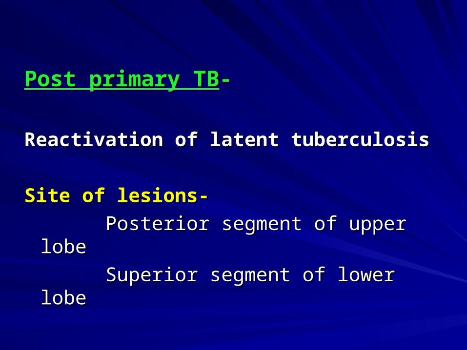
Post primary TBPost primary TB--
Reactivation of latent tuberculosisReactivation of latent tuberculosis
Site of lesions-Site of lesions-
Posterior segment of upper lobePosterior segment of upper lobe
Superior segment of lower lobeSuperior segment of lower lobe
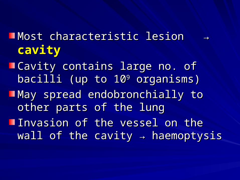
Most characteristic lesion Most characteristic lesion → → cavitycavityCavity contains large no. of bacilli (up to 10Cavity contains large no. of bacilli (up to 1099 organisms)organisms)
May spread endobronchially to other parts of May spread endobronchially to other parts of the lungthe lung
Invasion of the vessel on the wall of the Invasion of the vessel on the wall of the cavity cavity → → haemoptysishaemoptysis

Rupture of the cavity into the pleural spaceRupture of the cavity into the pleural space
TB empyemaTB empyema
pneumothoraxpneumothorax
Prolonged and repeated exposure is Prolonged and repeated exposure is essential for a full blown disease processessential for a full blown disease process
Smear positive PTB Smear positive PTB →→ “OPEN CASE”“OPEN CASE” →→ readily transmit infectionreadily transmit infection

PATHOGENETIC STAGESPATHOGENETIC STAGES--
4 stages4 stages
Stage-I Stage-I stage of onset / stage of no stage of onset / stage of no bacillary growthbacillary growth
Stage-IIStage-II stage of symbiosis stage of symbiosis
Stage-IIIStage-III stage of immunological control stage of immunological control
Stage-IVStage-IV stage of liquefaction stage of liquefaction

CLINICAL FEATURESCLINICAL FEATURES
SymptomsSymptoms--disease of protean manifestations and mimic disease of protean manifestations and mimic many diseases.many diseases.Constitutional & Respiratory symptoms.Constitutional & Respiratory symptoms.ConstitutionalConstitutional--Tiredness, headache, weight loss, fever, night Tiredness, headache, weight loss, fever, night sweats and loss of appetitesweats and loss of appetite Fever- late afternoon or eveningFever- late afternoon or evening Associated laryngeal TB- hoarseness of Associated laryngeal TB- hoarseness of
voice voice AmenorrhoeaAmenorrhoea

RespiratoryRespiratory--
CoughCough- -
most common most common → → min. 3 weeks duration, min. 3 weeks duration, dry or productive dry or productive
Sputum may be mucoid, mucopurulent, purulent or Sputum may be mucoid, mucopurulent, purulent or blood tingedblood tinged
Haemoptysis-Haemoptysis-
blood stain sputum, massive haemoptysis (rupture of blood stain sputum, massive haemoptysis (rupture of bronchial artery)bronchial artery)
Chest pain-Chest pain-
dull aching dull aching acute chest pain- TB pleurisyacute chest pain- TB pleurisy pneumothoraxpneumothorax

Breathlessness-Breathlessness-
extensive tuberculosisextensive tuberculosis
bronchial obstructionbronchial obstruction
pneumothoraxpneumothorax
pleural effusionpleural effusion
Localized wheeze-Localized wheeze-
endobronchial TBendobronchial TB
( pressure of enlarged lymph ( pressure of enlarged lymph nodes on the bronchus ) nodes on the bronchus )
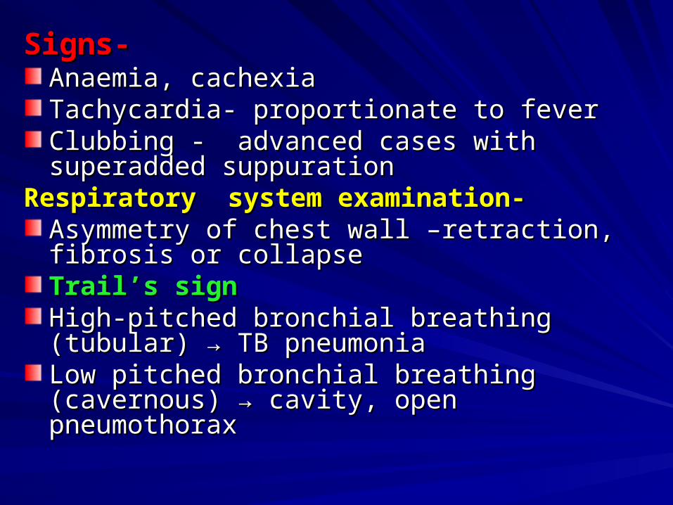
Signs-Signs-Anaemia, cachexiaAnaemia, cachexiaTachycardia- proportionate to feverTachycardia- proportionate to feverClubbing - advanced cases with superadded Clubbing - advanced cases with superadded suppurationsuppuration
Respiratory system examination-Respiratory system examination-Asymmetry of chest wall –retraction, fibrosis or Asymmetry of chest wall –retraction, fibrosis or collapsecollapseTrail’s signTrail’s signHigh-pitched bronchial breathing (tubular) High-pitched bronchial breathing (tubular) →→ TB TB pneumoniapneumoniaLow pitched bronchial breathing (cavernous) Low pitched bronchial breathing (cavernous) →→ cavity, open pneumothoraxcavity, open pneumothorax

Fine crepitations Fine crepitations →→ post-tussive post-tussive crepitationcrepitation → → sign of tuberculosis sign of tuberculosis infiltrationinfiltration
Post-tussive suctionPost-tussive suction →→ over a cavity over a cavity after coughing when its walls are not too after coughing when its walls are not too rigidrigid→→ occurs due to re-entry of air occurs due to re-entry of air

DIAGNOSISDIAGNOSIS1.Laboratory1.Laboratory2.Roentgenography(chest x-ray)2.Roentgenography(chest x-ray)Laboratory- Laboratory- sputumsputum-- Gold standard in the diagnosisGold standard in the diagnosis Detection of AFB in stained smear to be examined Detection of AFB in stained smear to be examined
microscopically microscopically 3 sputum samples to be examined 3 sputum samples to be examined (SPOT-(SPOT-
MORNING- SPOT)MORNING- SPOT) Method Method →→ Z-N staining (Ziehl- Neelson carbol Z-N staining (Ziehl- Neelson carbol
fuchsin stain)fuchsin stain) Sample should contain 10,000 AFB/ml for the AFB Sample should contain 10,000 AFB/ml for the AFB
to be detected by smear examinationto be detected by smear examination
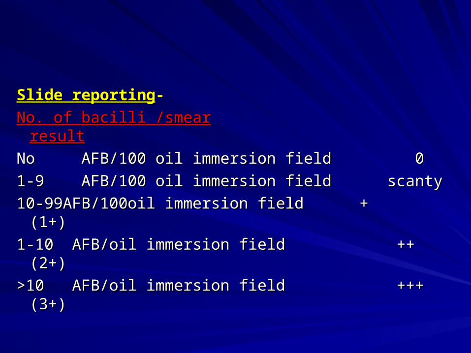
Slide reportingSlide reporting--
No. of bacilli /smearNo. of bacilli /smear resultresult
No AFB/100 oil immersion field 0No AFB/100 oil immersion field 0
1-9 AFB/100 oil immersion field scanty1-9 AFB/100 oil immersion field scanty
10-99AFB/100oil immersion field + (1+)10-99AFB/100oil immersion field + (1+)
1-10 AFB/oil immersion field ++ (2+)1-10 AFB/oil immersion field ++ (2+)
>10 AFB/oil immersion field +++ (3+)>10 AFB/oil immersion field +++ (3+)

Gastric lavageGastric lavage diagnosis of PTB in young children instead of diagnosis of PTB in young children instead of
sputumsputum lavage reveal organism in 30to40% of the cases lavage reveal organism in 30to40% of the cases performed early in the morning performed early in the morning →→ concentration is concentration is
highesthighest Nasogastric tube- stomach contents are aspiratedNasogastric tube- stomach contents are aspirated Fibreoptic bronchoscopyFibreoptic bronchoscopy Bronchoscopic aspirate, bronchial washing, Bronchoscopic aspirate, bronchial washing,
bronchoalveolar lavage(BAL)fluid, Trans bronchial bronchoalveolar lavage(BAL)fluid, Trans bronchial lung biopsylung biopsy
UrineUrineCSFCSFTissueTissuebiopsy specimens of lymph node, liverbiopsy specimens of lymph node, liver

Culture methodsCulture methods--Yield from culture is on average twice as that of Yield from culture is on average twice as that of
microscopymicroscopyTwo types of media commonly usedTwo types of media commonly used 1.egg based 1.egg based →→ L-J media, Pentragnani media, L-J media, Pentragnani media,
ATS media ATS media 2.agar based 2.agar based →→ Middle brook 7H10,7H11 media Middle brook 7H10,7H11 mediaAdvantage-Advantage- (a) more sensitive and can be positive when the (a) more sensitive and can be positive when the
bacterial load in the sample is about 10AFB / mlbacterial load in the sample is about 10AFB / ml (b) for precise identification of the causative (b) for precise identification of the causative
organismorganismDisadvantage- Disadvantage- result is delayed, min.6 wks., whereas in smear it result is delayed, min.6 wks., whereas in smear it
is easy to perform and is cheapis easy to perform and is cheap
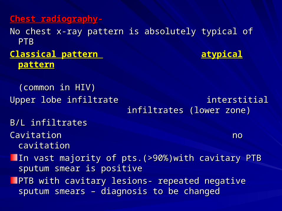
Chest radiographyChest radiography--
No chest x-ray pattern is absolutely typical of PTBNo chest x-ray pattern is absolutely typical of PTB
Classical pattern Classical pattern atypical patternatypical pattern
(common in HIV)(common in HIV)
Upper lobe infiltrate interstitial Upper lobe infiltrate interstitial infiltrates infiltrates
(lower zone)(lower zone)
B/L infiltratesB/L infiltrates
Cavitation no cavitationCavitation no cavitation
In vast majority of pts.(>90%)with cavitary PTB In vast majority of pts.(>90%)with cavitary PTB sputum smear is positivesputum smear is positive
PTB with cavitary lesions- repeated negative PTB with cavitary lesions- repeated negative sputum smears – diagnosis to be changedsputum smears – diagnosis to be changed

D/D of CXR finding associated with PTB-D/D of CXR finding associated with PTB-1.Cavitation-1.Cavitation- Infection- pneumoniaInfection- pneumonia lung abscesslung abscess fungal infectionfungal infection Non-infectious- bronchogenic carcinomaNon-infectious- bronchogenic carcinoma connective tissue diseaseconnective tissue disease occupational lung diseaseoccupational lung disease2.Unilateral infiltration-2.Unilateral infiltration- pneumonia pneumonia bronchogenic cabronchogenic ca3.Bilateral infiltration-3.Bilateral infiltration- pneumonia pneumonia connective tissue diseaseconnective tissue disease sarcoidosissarcoidosis4.Mediastinal lymphadenopathy-4.Mediastinal lymphadenopathy- lymphomalymphoma bronchogenic cabronchogenic ca sarcoidosissarcoidosis
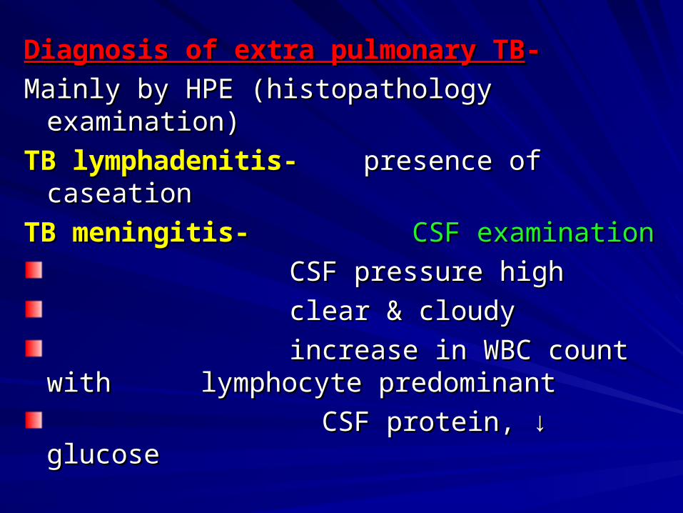
Diagnosis of extra pulmonary TBDiagnosis of extra pulmonary TB--
Mainly by HPE (histopathology examination)Mainly by HPE (histopathology examination)
TB lymphadenitis- TB lymphadenitis- presence of caseation presence of caseation
TB meningitis-TB meningitis- CSF examinationCSF examination
CSF pressure highCSF pressure high
clear & cloudyclear & cloudy
increase in WBC count with increase in WBC count with lymphocyte predominant lymphocyte predominant
CSF protein, CSF protein, ↓↓ glucose glucose

Tuberculin skin testTuberculin skin test--Tuberculin-purified protein derived from tubercle Tuberculin-purified protein derived from tubercle bacillibacilli
Purified protein derivative (PPD)Purified protein derivative (PPD)
Injected intradermallyInjected intradermally
Induration read after 48 hrs.Induration read after 48 hrs.
Hypersensitivity reactionHypersensitivity reaction
Positive Tuberculin test- indicates infectionPositive Tuberculin test- indicates infection
Positive tuberculin test- induration > 10mmPositive tuberculin test- induration > 10mm
Negative tuberculin test- induration < 10mmNegative tuberculin test- induration < 10mm
A positive tuberculin test is only one piece A positive tuberculin test is only one piece evidence in favour of dignosis of Tuberculosisevidence in favour of dignosis of Tuberculosis