tc-99mgalactosyl …jnm.snmjournals.org/content/25/7/779.full.pdfbasicchemistry radiochemistry...
TRANSCRIPT
BASIC CHEMISTRY
RADIOCHEMISTRY AND RADIOPHARMACEUTICALS
Tc-99m Galactosyl-Neoglycoalbumin: In Vitro Characterization of
Receptor-Mediated Binding
David R. Vera, Kenneth A. Krohn, Robert C. Stadalnik, and Paul O. Scheibe
University of California, Davis, Medical Center, Sacramento, California
Hepatic binding protein (HBP) is a membrane receptor that binds and transportsplasma glycoproteins from hepatic blood to hepatocellular lysosomes. We havecharacterized the in vitro binding of Tc-99m galactosyl-neoglycoalbumin (Tc-
NGA), a synthetic HBP ligand, to liver membrane. Structural modifications of NGAresulted in the alteration of the equilibrium constant, KA, and the forward-bindingrate constant, kb. Binding was second-order; the relative amount of membrane-
bound NGA depended on the initial concentrations of ligand and membrane. Membrane displacement studies, using carrier ligands in contrast to previously boundTc-NGA or I-NGA, correlated with the binding characteristics of a native HBP ligand, asialo-orosomucoid. We used computer simulation to study the detectability
of the changes in HBP concentration at different values of kb. The simulations indicated that radiopharmacokinetic sensitivity to alterations in [HBP] should be possible using a neoglycoalbumin preparation with a carbohydrate density within therange of 15 to 25 galactose units per albumin molecule.
J NucÃMed 25: 779-787, 1984
The process by which receptor molecules bind ligand substrates exhibits two unique properties; molecular specificity and saturability. Each relates a significant chemical parameter to the kinetics of the bindingreaction. The first property, molecular specificity,identifies the contribution of chemical structure to thereaction rate, and is quantified by the forward and reverse rate constants (kb and k_b). The saturability of areceptor-ligand interaction is the result of a finitenumber of ligand binding sites provided by the receptormolecule.
Because measurements of equilibrium affinity constant and kinetic rate constants relate chemical structureand stoichiometry to chemical kinetics, the developmentof receptor-binding radiopharmaceuticals has benefited
Received Aug. 1, 1983; revision accepted Mar. 28, 1984.For reprints contact: Robert C. Stadalnik, MD, Divisionof Nuclear
Medicine, University of California, Davis, Medical Center, 2315Stockton Blvd., Sacramento, CA 95817.
from assays of in vitro binding. The approach has beento screen various ligand derivatives for the appropriateparameters that will permit adequate accumulation ata target with a given receptor concentration. For themost part, these studies have emphasized thermody-namic (equilibrium) measurements. For example,Eckelman developed a biphasic model by which themaximum theoretical target-to-background ratio couldbe calculated (/). Data for the model were supplied byin vitro measurements of equilibrium binding constants.After screening several labeled muscarinic derivatives,he selected 3-quinuclidinyl-4-iodo-benzilate (4-lodoQNB) as the most promising candidate for the mappingof muscarinic receptors in heart and brain (2). As another example, design of a neuroleptic agent utilized bothequilibrium and transient-state binding data (3) tocompare the binding rate constants of haloperidol andspiroperidol. The higher affinity of the latter compoundwas a prominent reason for choosing C-11 spiroperidol(4) as the optimal tracer.
Volume 25, Number 7 779
by on June 16, 2018. For personal use only. jnm.snmjournals.org Downloaded from

VERA, KROHN, STADALNIK, AND SCHEIBE
A second use of in vitro experiments has been themeasurement of nonspecific binding exhibited by a labeled derivative. Katzenellenbogen used in vitro techniques to search for an estrogenic analog with low nonspecific binding. After formulating an index of bindingselectivity (5) based on the relationship of ligand lipo-philicity to nonspecific binding, 16a-[Br-77]bromoes-tradiol-17(S (6) was selected for the imaging of estrogen-dependent mammary tumors.
Our laboratory has been interested in a class of receptors called lectins (7), which bind specific carbohydrate residues of glycoproteins. Hepatic binding protein( H BP) resides on the cell surface of mammalian hepa-tocytes (8), where it binds and transports galactose-terminated glycoproteins to the hepatic lysosomes. Extensive studies (9) have implicated HBP as the normalmechanism for the homeostasis of plasma glycoproteins.Based on the significance of HBP in hepatic physiology,we proposed the use of this receptor as the basis for adiagnostic agent capable of routine measurement of liverfunction (10).
This paper presents results that characterize a synthetic HBP radioligand, Tc-99m galactosyl-neogly-coalbumin (Tc-NGA), as a receptor-binding radio-pharmaceutical, in terms of saturability, nonspecificbinding, tissue specificity, and molecular specificity.Lastly, we present the concept of kinetic sensitivity,which measures the ability of a physiochemical parameter to alter the in vivo uptake of a radiopharmaceutical.To illustrate this concept, we simulated the biokineticsof the Tc-NG A-HBP system.
MATERIALS AND METHODS
Radiopharmaceutical synthesis and quality control.Synthesis of the analog ligand, galactosyl-neoglycoal-bumin (NGA), was accomplished by the covalent coupling of a bifunctional reagent, 2-imino-2-methoxy-ethyl-l-thio-/3-D-galactopyranoside (IME-thiogalactose)to the primary amino groups of human serum albumin(HSA). The IME-thiogalactose was synthesized in foursteps (11-13) and repeatedly recrystallized (methanol)and decolorized (activated charcoal in warm methanol).The coupling reaction was carried out in fresh boratebuffer (Clarke's, pH 8.6, 0.2M). With a 20 mg/ml solution of HSA and an incubation temperature of 25°C,
the reaction reached its maximum yield within 1 hr.Unreacted IME-thiogalactose was removed from theproduct by diafiltration* with five exchange volumes of
isotonic saline. The NGA solution was then concentratedto 125 mg/ml. A colorimetrie analysis (14) was used tomeasure the number of galactose groups attached to eachalbumin molecule.
The albumin derivative was labeled with either io-dine-125 or technetium-99m. Chloramine-T (75) produced I-NGA yields in excess of 95%. The electrolytic
method of Benjamin (16) produced technetium-NGAlabels with yields also in excess of 95%. The labeledproducts were passed through a polydextran column( 120 X 1.5 cm). Quality control, which included polya-cetate electrophoresis (250 V, 20 min) and polyacryl-amide gel-filtration chromatography (20 X 1.0cm) (10),showed the chromatographically purified NGA to befree of multimeric forms of albumin and of free tech-netium or iodine. The technetium label of all neogly-coalbumin preparations was stable for at least 4 hr.Concentration of the pooled eluate was measured by uvabsorbance (280 nm, am = 2.49 X 10~5 I/mole).
Membrane receptor preparation and binding assay.Rabbit hepatocyte membrane was isolated using themethod described by Ray (17). The binding assays werecarried out under the conditions outlined by Van Lenten(18). For the equilibrium studies, four initial concentrations and eight neoglycoalbumin preparations of labeled NGA were used: 4, 20, 100, and 200 pA/; and 4,6, 7, 13, 21, 25, 37, and 44 average galactose groups peralbumin molecule. Studies that followed the time courseof the binding reaction showed that equilibrium wasreached within 30 min (79). As a result, the filtrationstep was carried out after a 1-hr incubation. The filtersbearing the membrane-bound NGA, and standardscontaining the initial amount of ligand, were counted inan automatic gamma counter using a 10-80 keV windowfor 1-125 NGA and a 100-200 keV window for Tc-99m
NGA. Binding displacement experiments were performed using NGA-10, NGA-25, and NGA-44. Afterincubation with labeled NGA, a 1000-fold molar excessof either cold NGA, N-acetylgalactosamine, or D-glu-cose was added to the reaction tube. The latter carbohydrate is not capable of inhibiting HBP binding or ofdisplacing previously bound HBP ligands (20). Displacement of the previously bound NGA was followedby filtering sets of reaction tubes at various times afterthe addition of the displacement reagent. Unless noted,all assays used 250 ¿igof membrane per assay tube. Dataanalysis used standard regression techniques (27).
In vivo simulations. Using the results of the in vitromeasurements, we simulated the NGA kinetics to determine whether physiologic changes in receptor concentration or blood flow would alter the liver's time-
activity curves. Stimulation of NGA kinetics was performed by a FORTRAN-coded program (79) that utilized a numeric integrator (22). The program was executed in a standard nuclear medicine computer using thevideo terminal for graphic output. The kinetic model(Fig. 1) (23) used three compartments: extrahepaticblood, hepatic blood, and hepatocyte-bound NGA. Thefirst two compartments communicate by hepatic bloodflow. The formation of the receptor-ligand complex depends on the number of receptors and the forwardbinding rate constant, kb.
The simulation required values for each of the above
780 THE JOURNAL OF NUCLEAR MEDICINE
by on June 16, 2018. For personal use only. jnm.snmjournals.org Downloaded from
BASIC SCIENCESRADIOCHEMISTRY AND RAD1OPHARMACEUTICALS
CONTROL OF GALACTOSE COUPLING
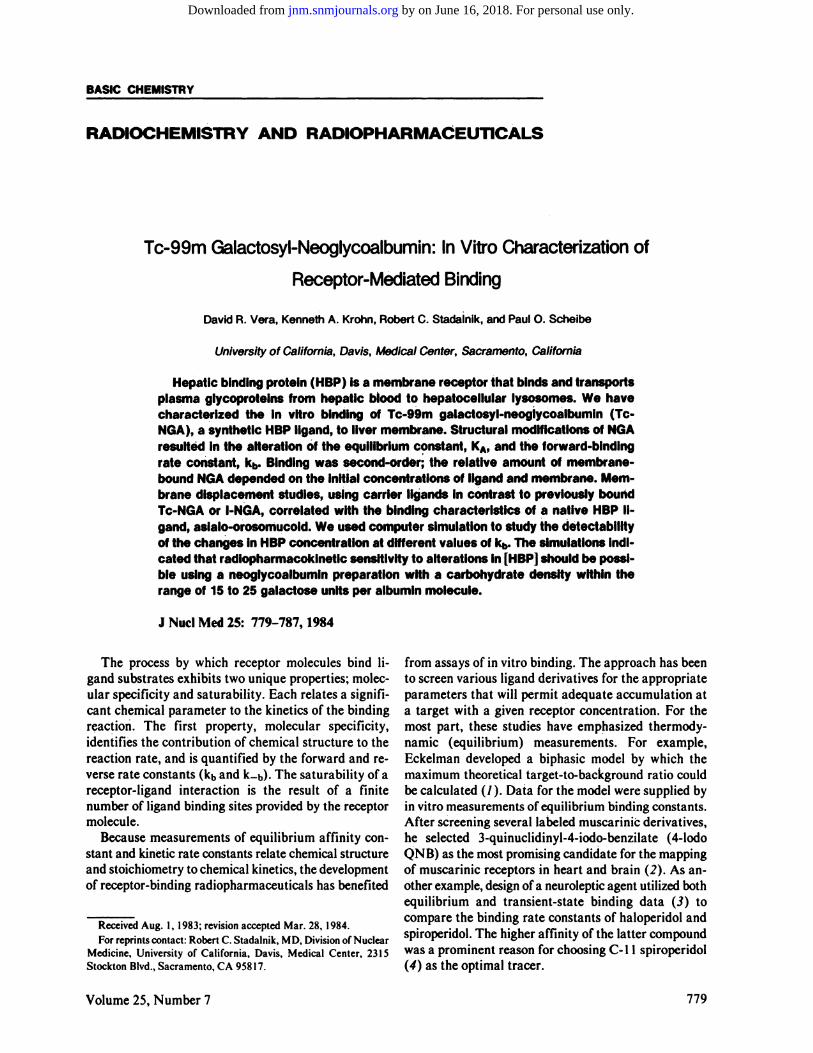
FIG. 1. Model for Tc-NGA kinetics incorporates three processes:hemodynamic delivery of ligand, receptor binding of ligand to hep-
atocyte membrane, and catabolism of ligand by hepatic lysosomes.
parameters and the volumes of each compartment. Thevolumes and the hepatic blood flow for a 70-kg humanwere taken from a standard textbook (24). The numberof receptors within the liver was estimated from in vitrobinding data using isolated rat hepatocytes (25). The kbfor NGAs with various galactose-coupling ratios werethose calculated from our in vitro data. The model alsocontains a metabolic rate constant, a, whose value wastaken from rabbit in vivodata (¡0).The simulations useda molar dose of ligand that would occupy 1% of the receptor sites. Table 1 lists the symbols for the model andthe values of each parameter.
RESULTS
The affinity of labeled NGA could be syntheticallycontrolled. The molar ratio of the reactants used in thecoupling reaction (IME-thiogalactose and HSA) controlled the number of galactose groups coupled to eachalbumin molecule (Fig. 2). The carbohydrate density ofthe neoglycoalbumin determined the equilibrium constant for binding of the NGA preparation to hepaticmembrane. Casting the equilibrium binding data ofNGAs with different carbohydrate densities into the
so
4C
30C
E
1 25
sT)
Ou
150.2 M Borala Buffar2O mg/ml HSA3 hours. 25 "C. pH 8.5
so no ISO 200IME-Thiogalactosa /HSA
250 300
(mol/mol)ISO
FIG. 2. Number of galactose groups per albumin molecule couldbe controlled by molar ratio of reactants, IME-thiogalactose, and
human serum albumin (HSA).
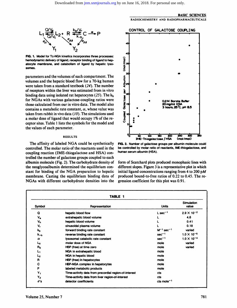
form of Scatchard plots produced monophasic lines withdifferent slopes. Figure 3 is a representative plot in whichinitial ligand concentrations ranging from 4 to 200 pA/produced bound-to-free ratios of 0.22 to 0.45. The regression coefficient for this plot was 0.91.
TABLE1SymbolQV.vhv,kbk-boUROLiL2RCPY,Y2CT'SReprésentationhepatic
bloodflowextrahepatic
bloodvolumehepatic
bloodvolumesinusoidal
plasmavolumeforward
binding rateconstantreverse
binding rateconstantlysosomal
catabolic rateconstantmolardose ofNGAHBP
(free) at timezeroNGA
in extrahepaticbloodNGA
in hepaticbloodHBP
(free) inhepatocytesHBP-NGAcomplex inhepatocyteslabeled
metabolicproductsTime-activitydata from precordialregion-of-interestTime-activity
data from liverregion-of-interestdetector
coefficientsUnitsL
sec"1LLLM"1
sec"1sec-1sec"1molemolemolemolemolemolemolectsctscts
mole" 1Simulation
value2.9
X10~24.60.410.18varied1.0
X10~51.0
X10-4variedvaried
Volume 25, Number 7 781
by on June 16, 2018. For personal use only. jnm.snmjournals.org Downloaded from
VERA, KROHN, STADALNIK, AND SCHEIBE
TABLE2Galactose
density(M/mole)4437252113764*
250 ¿iglyophilizedmembrane.T
Range of LO= 4 to 200 pM..
IN VITRO l-125-NGA MEMBRANE* BINDINGDATAEquilibrium
constant, KA(AT1)7.0
±1.4 X1096.7±0.7 X1098.8±2.6 X1095.3±0.8 X1097.7±3.1 X1095.3±1.5 X1095.4±1.3 X1095.2±1.0 X1092.5±1.7X1094.1±1.4 X1095.3±0.9 X1091.2±0.7 X1092.3±0.5 X1091.3±0.8 X10⁹No
bindingNobindingNo
bindingNo
bindingB/P'
ränge0.09-0.230.21-0.520.13-0.490.2-0.41.0-2.82.3-3.71.0-1.42.2-3.51.0-1.42.2-3.52.2-3.00.08-0.140.13-0.280.11-0.300.02-0.030.03-0.050.04-0.010.01-0.01Regressioncoefficient0.940.960.770.920.850.850.840.690.840.690.910.840.970.63
Equilibrium constants (KA), the range of the bound-to-free values, and the coefficient of the regression linefor each plot are listed in Table 2 for neoglycoalbuminshaving different galactose densities. The Scatchard plotsof all NGA preparations produced sloped lines that werecomposed of clusters of data points (as in Fig. 3), eachcluster representing different initial amounts of ligand.Neoglycoalbumins with less than seven sugars per albumin exhibited low binding ratios (B/F < 0.05) andproduced Scatchard plots with scattered data points andno slope. Increasing the amount of membrane (50, 100,
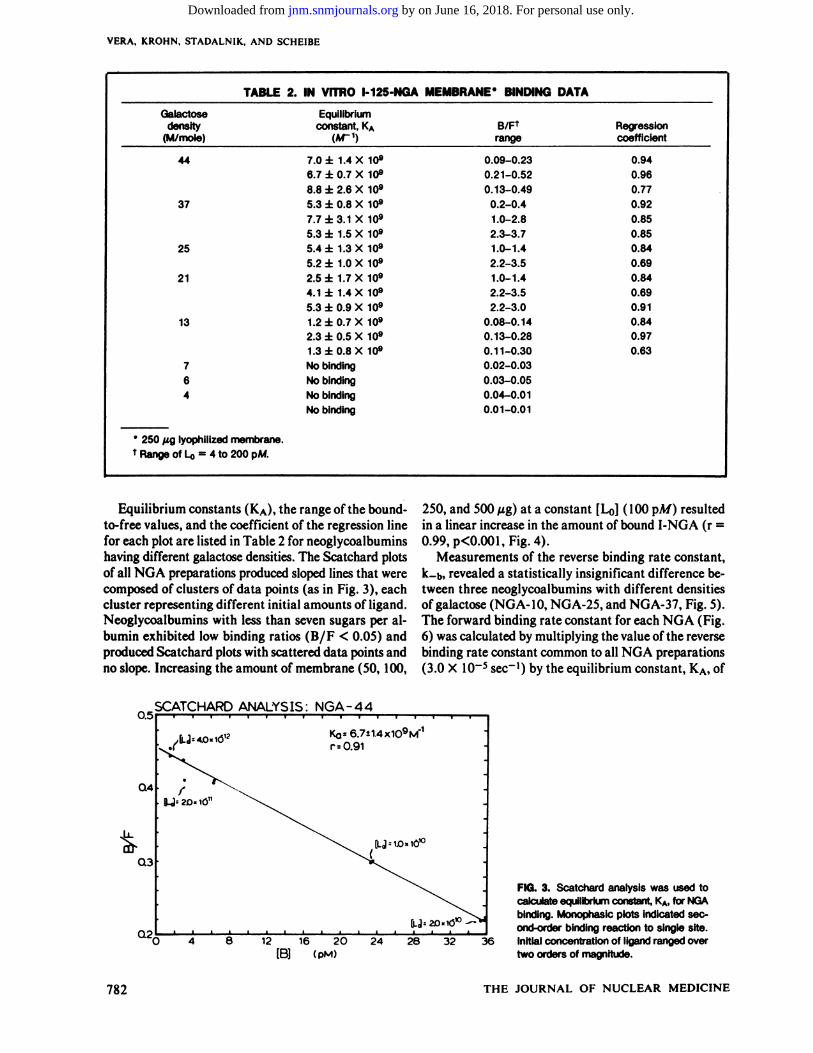
250, and 500 ng) at a constant [L0] (100 pA/) resultedin a linear increase in the amount of bound I-NGA (r =0.99, p<0.001,Fig. 4).
Measurements of the reverse binding rate constant,k_b, revealed a statistically insignificant difference between three neoglycoalbumins with different densitiesof galactose (NG A-10,NGA-25, and NGA-37, Fig. 5).The forward binding rate constant for each NGA (Fig.6) was calculated by multiplying the value of the reversebinding rate constant common to all NGA preparations(3.0 X 10~5 sec"1) by the equilibrium constant, KA,of
O.5
Q4
0.3
SCATCHARD ANALYSIS: NGA-44
0.2
KQ=6.7i1.4x1O9M"1
r =0.91
[LJ OJO«IO10
O 4 12 16 20 24 28 32[B] (pM)
36
FIG. 3. Scatchard analysis was used tocalculate equilibrium constant, KA, for NGAbinding. Monophasic plots indicated second-order binding reaction to single site.
Initial concentration of ligand ranged overtwo orders of magnitude.
782 THE JOURNAL OF NUCLEAR MEDICINE
by on June 16, 2018. For personal use only. jnm.snmjournals.org Downloaded from
BASIC SCIENCESRADIOCHEMISTRY AND RADIOPHARMACEUTICALS
14
12
10
RECEPTOR-DEPENDENT BINDING
2o..
r .099
0 100 200 3OO 400Membrane (ug)
500 600
FIG. 4. Amount of membrane-bound 1-125 NGA varied linearly withamount of membrane within each reaction tube (r = 0.99, p
<0.001).
each NGA. Both N-acetylgalactosamine and cold NGA
displaced labeled NGA that was previously bound to thereceptor (Fig. 7), but D-glucose did not displace the
bound ligand. The rate of displacement, when challengedby N-acetylgalactosamine, was the same as for the dis
placement of unlabeled NGA. The radioactive label(iodine or technetium) did not affect the displacementrates.
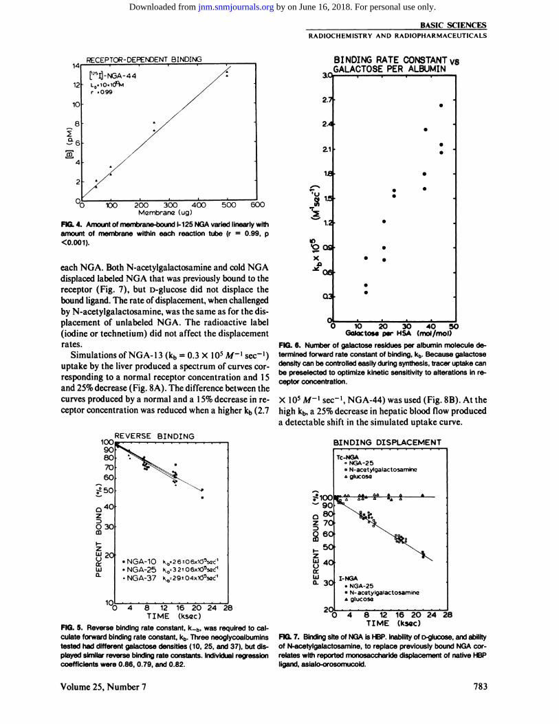
Simulations of NGA-13 (kb = 0.3 X IO5 M~{ sec~')
uptake by the liver produced a spectrum of curves corresponding to a normal receptor concentration and 15and 25% decrease (Fig. 8A). The difference between thecurves produced by a normal and a 15% decrease in receptor concentration was reduced when a higher kb (2.7
1OO90607060
50
O3OO
y 20ceLUCL
REVERSE BINDING
•NGA-1O•NGA-25•NGA-37
KD=3 2 •-0 6x10⁵sec 'k.D=29«04xl05soc'
04 8 12 16 20 24 28TIME (ksGc)
FIG. 5. Reverse binding rate constant, k_b, was required to calculate forward binding rate constant, kb. Three neoglycoalbuminstested had different galactose densities (10, 25, and 37), but displayed similar reverse binding rate constants. Individual regressioncoefficients were 0.86, 0.79, and 0.82.
3.0,
BINDING RATE CONSTANT vsGALACTOSE PER ALBUMIN
2.7
2.4
21
te
1.2
inO OS
Xp
0.3
IO 2O 30Galactose par MSA
40 50(mol/mol)
FIG. 6. Number of galactose residues per albumin molecule determined forward rate constant of binding, kb. Because galactosedensity can be controlled easily during synthesis, tracer uptake canbe preselected to optimize kinetic sensitivity to alterations in receptor concentration.
X IO5 M-{ sec"1, NGA-44) was used (Fig. 8B). At the
high kb, a 25% decrease in hepatic blood flow produceda detectable shift in the simulated uptake curve.
BINDING DISPLACEMENT
1OO908O70
6O
5O
40
30
2OJ
Tc-NGA°NGA-25" N-acetylgalactosamino¿glucosa
I-NGA. NGA-25•N-acetylgalactosammaA glucose
'04 8 12 16 2O 24 28
TIME (kscc)
FIG. 7. Binding site of NGA is HBP. Inability of D-glucose, and abilityof N-acetylgalactosamine, to replace previously bound NGA cor
relates with reported monosaccharide displacement of native HBPligand, asialo-orosomucoid.
Volume 25, Number 7 783
by on June 16, 2018. For personal use only. jnm.snmjournals.org Downloaded from

VERA, KROHN, STADALNIK, AND SCHEIBE
XJOi
90
80
. 70i
i 60
\I
: 30
20
10
SIMULAT ION OF Te-NGA KINETICS
W 2O 3O 40 5O 60MINUTES AFTER INJECTION
FIG. 8. Top: Hepatic accumulation of NGA was simulated to investigate ability of different receptor concentrations to change liveruptake curves. Neoglycoalbumin having 13 galactose groups peralbumin exhibited uptake curves that were altered by different hepatic receptor concentrations.
100
6pZ
SIMULATION OF Tc-NGA KINETICS
O IO 2O 3O 40 5OMINUTES AFTER INJECTION
FIG. 8. Bottom: Simulations using forward binding rate constantof 2.7 X 10⁵ M~1 see"1 indicated that changes in receptor con
centration would have little effect on hepatic accumulation of anNGA having 44 galactose groups per albumin. 15% decrease inreceptor produced curve similar to normal curve. A 25 % decreaseproduced only slight variation. Uptake curve was sensitive, however, to 25% decrease in hepatic blood flow.
DISCUSSION
Receptor-binding agents provide new perspectives tothe design of radiopharmaceuticals. Because it is possibleto characterize both chemically and physically the process responsible for tissue binding, quantifiable criteriafor radiopharmaceutical performance can be established.We will describe the receptor binding of NGA in termsof saturability, nonspecific binding, tissue specificity, andmolecular specificity. A fifth criterion, kinetic sensitivity,forms a useful predictor of the in vivo performance of aradiopharmaceutical.
Saturability. A principal characteristic of receptor-ligand binding is the dependence of the reaction rate on
the amount of initial ligand and receptor. Although theterm saturability is commonly used to describe this dependence, the saturation of an entire receptor populationcan occur only under the extreme condition of vastly highkb and ligand-receptor ratio. This is especially the casein vivo when receptor recycling or turnover occurs.
Second-order reactions, if allowed to reach equilibrium and operated under conditions that prevent ca-tabolism of the ligand-receptor complex, will fit theScatchard model. Thus, the relative amount of ligandbound to the membrane preparation (B/F) when thereaction has reached equilibrium will depend on theinitial amounts of ligand and receptor. In vitro assaysusing plasma-membrane preparations assume a directrelationship between the weight of membrane (which isusually lyophilized) and the number of receptors. Because the hepatic membrane preparation is devoid oflysosomal enzymes, the NGA-HBP complex is not ca-tabolized. As a result, the increased B/F values producedby increasing the initial NGA concentration, ([Lo], Fig.3), and the linear dependence of bound NGA with differing amounts of membrane (Fig. 4), confirm the second-order nature of NGA binding. This second-orderbehavior is also exhibited by HBP binding to native li-gands where the reaction rate depends upon the concentration of ligand (26) and receptor (27). This alsosuggests that in vivo hepatic accumulation of Tc-NGAwill be controlled by the number of HBP molecules thatcommunicate with hepatic blood.
Specificity: nonspecific binding. The monophasicScatchard plots from our equilibrium binding studiesindicate a lack of nonspecific binding (NSB) of NGAto the membrane preparation. The plots, which covereda range of two orders of magnitude in initial NGA concentration, were similar to those obtained from thebinding of asialo-orosomucoid (25,28) and asialofetuin(28). Nonspecific binding, which is usually attributedto lipophilic interaction with membrane structures, superimposes an additional line on a Scatchard plot. Although the slope of the NSB line, and hence the bindingaffinity, is lower in magnitude than that for binding tothe specific receptor, the number of nonspecific sites canbe extremely high. As a result, these high-concentration,low-affinity binding sites can compete effectively againstmuch lower concentrations of high-affinity receptor sites,and artificially elevate the binding capacity for the ligand. The low NSB exhibited by NGA was not unexpected; unlike the neuroleptic and estrogenic agents—which are moderately lipophilic and thus exhibit NSB(4,5)—Tc-NGA is a carbohydrate derivative of albuminand hence is highly hydrophilic.
Specificity: tissue. An important factor governingtheperformance of a receptor-binding radiopharmaceuticalis the tissue distribution of the receptor. Optimal tissuespecificity exists when the receptor resides in a single celltype within a single organ. Eliminating competition from
784 THE JOURNAL OF NUCLEAR MEDICINE
by on June 16, 2018. For personal use only. jnm.snmjournals.org Downloaded from

BASIC SCIENCESRADIOCHEMISTRY AND RAD1OPHARMACEUTICALS
other organs maximizes accumulation of ligand by thetarget organ. Such competition may be extremely detrimental if the competing tissue receives a higher percentage of cardiac output. Hepatic binding protein ispresent only in a single cell type, the hepatocytes (29,30),of a single organ, the liver (31).
Other receptor-binding radiopharmaceuticals aremuch less tissue-specific. For example, estrogen receptors are present not only in mammary tumors, but alsoin the uterus (32) and liver (33). Muscarinic receptorshave been found in the brain (34), ileum (35), myocardium (36), and pancreas (37). Dopamine receptors notonly provide synaptic transfer of information in thecentral nerve system (38), but also mediate nervouscontrol of vascular smooth muscle (39). Dopaminergicreceptors have been characterized in coronary (40), renal(41), and mesenteric (41) arteries. The high tissuespecificity of HBP ligands was a major factor in our selection of the HBP-NGA system asa model for the development of receptor-binding radiopharmaceuticals.
Specificity: molecular. The unique feature of receptorsand their ligands is the complementarity of their molecular structures. One measure of receptor-ligandspecificity is the rate at which the reaction proceeds. Theforward binding rate constant, kb, of a given ligandprovides a quantitative link between chemical structureand molecular specificity. An increase in the galac-tose-to-albumin ratio of NG A is an alteration of ligand
structure in the sense that an increase of carbohydratedensity in a ligand increases the probability of substrate-receptor interaction based on a simple geometricmodel.
The displacement studies established HBP as the receptor responsible for the binding of NGA to livermembrane. Native HBP ligands (asialo-orosomucoid,for example) can be displaced from membrane (20,18)or isolated receptor (42) by a molar excess of N-acety-galactosamine, but not by D-glucose (Fig. 1).
The rate of displacement, and hence the k_b, were thesame for NGA preparations with different carbohydratedensities (Fig. 5). Using isolated rat hepatocytes, similarvalues of k_b have also been measured for asialo-orosomucoid and asialofetuin (43) [(5.4 ±1.5) X 10~6and(6.3 ±1.5) X 10~6sec-', respectively, at 10°C],these
being two native H BP ligands that have different bloodclearance half-times (44) and different forward bindingrate constants (26).
Kinetic sensitivity. Another, and more critical, question regarding radiopharmaceutical performance is thesensitivity of target-organ pharmacokinetics to alterations in specific physiological parameters that are di-agnostically significant. The most important parametersare blood flow and receptor concentration. If a changein receptor concentration is not capable of producing anobservable alteration in the time-activity data, onecannot estimate the value of that parameter.
Using computer simulation of our kinetic model, wewere able to investigate the influence of kb upon the kinetic sensitivity of the receptor concentration and hepaticblood flow. The dependence of kbon the number of galactose groups per albumin allows us to modify kb synthetically while keeping all other radiopharmaceuticalproperties constant. By quantifying binding in terms ofthe fundamental units of receptor-ligand function, kbandRO,we were able to simulate the performance of receptor-ligand reaction within the context of two other processes: hemodynamic delivery of the ligand to the liver,and lysosomal metabolism. The equilibrium constant,KA, is not a kinetic parameter and thus cannot providea measure by which the hemodynamic delivery, receptorbinding, and metabolic fluxes may be compared.
The relative magnitudes of these three rate processesdetermine the ability of the radiopharmaceutical uptakemeasurements to reflect changes in receptor concentration. This was illustrated by the simulations (Fig. 8).Using an injection of NGA having 13 galactose groupsper albumin (NGA-13), and a carbohydrate density thatexhibited a kb of 0.3 X IO5 A/~" sec"1, we found the
hepatic uptake sensitive to alterations in receptor concentration: decreases in HBP concentration shifted thecurves downward and to the right (Fig. 8A). Ata highercarbohydrate density (44 galactose groups per albumin,NGA-44), and forward binding rate constant kb = 2.7X IO5 A/-' sec"1, the kinetic sensitivity to HBP con
centration was severely diminished: a 25% decrease inconcentration was required to produce a minimally different curve (Fig. 8B). However, a 25% decrease in hepatic blood flow significantly altered the uptake curve.Under real conditions the ability to distinguish betweeneach curve will be limited by the amount of noise in thetime-activity data. Thus, if time-activity data are toenable us to observe a change in kinetic parametersproduced by a ligand with a high forward binding rateconstant, we will need high levelsof activity and minimalorgan motion.
A high forward binding rate constant is similar to ahigh extraction coefficient, and indicates a radiopharmaceutical that will accumulate within a target organat a rate limited by the flow of blood into the organ.Prevention of this condition can be achieved if the first-order parameter of the binding reaction, kb[R]0, is of thesame magnitude as the parameter for target-organperfusion, Q/ V, where Q is blood flow in I/sec and V isthe distribution volume of the tracer (19).
Our simulations indicate a kinetic insensitivity to HBPconcentration when kb is greater than 2.7 X IO5A/~'sec"1. The ability to control the carbohydrate density
synthetically has enabled us to prepare various HBP ligands with a range of forward binding rate constants.Using healthy rabbits, we then injected and followed thetime-course of Tc-NGAs of differing kband molar doses(45). The observation of affinity-dependent uptake and
Volume 25, Number 7 785
by on June 16, 2018. For personal use only. jnm.snmjournals.org Downloaded from
VERA, KROHN, STADALNIK, AND SCHEIBE
dose-dependent uptake of the radiopharmaceuticalconfirmed our simulations and the concept of phar-macokinetic sensitivity.
Although all receptor-ligand systems differ in theirbiochemistry, each will require as a fundamental kineticstructure: (a) delivery of the ligand to the target tissue,and (b) binding of the ligand to the receptor. As a result,these tracers may display delivery-limited kinetics. Dueto the fenestrated structure of the hepatic sinusoids,macromolecules such as Tc-NGA can communicatefreely with the cell surface. Thus, liver blood flow, ratherthan interstitial diffusion, governs the delivery of Tc-NGA to the receptor. As a result, the Tc-NGA-HBPsystem provides an excellent opportunity to study theinfluence of radiopharmaceutical delivery (hepatic bloodflow) upon the ability to observe changes in receptorbiochemistry (HBP affinity and concentration).
FOOTNOTE
* Amicon; PM30; Danvers, MA.
ACKNOWLEDGMENTS
We are grateful for the many discussions with Drs. Gilbert Ashwelland Allan Green, and the technical assistance of Scott Steffen, KarenFcldman, and Marsha Gregor. This work was supported by AmericanCancer Society grants #RD-6I and #PDT-180, and the Fred andJulia Rusch Foundation.
This work presented in part at the 30th Annual Meeting of the Society of Nuclear Medicine, St. Louis, MO, June 7-10, 1983.
Supported by the American Cancer Society grants #RD-61 and#PDT-180, and the Fred and Julia Rusch Foundation.
REFERENCES
/. ECKELMAN WC, REBA RC, GIBSON RE, et al: Receptor-binding radiotracers: A class of potential radiopharmaceuti-cals. J NucÃMed 20:350-357, 1979
2. ECKELMAN WC: Receptor-specific radiopharmaceuticals.In Computed Emission Tomography. Ell PJ, Holman BI, eds.Oxford, Oxford University Press.
3. LEYSEN JE, GOMMEREN W, LADURON PM: Spiperone:A ligand of choice for neuroleptic receptors. I. Kinetics andcharacteristics of in vitro binding. Biochem Pharmacol 27:307-316, 1978
4. FOWLER JS, ARNETT CD, WOLF AP, et al: ["CjSpirop-eridol: Synthesis, specific activity determination, and bio-distribution in mice. J NucÃMed 23:437-445, 1982
5. KATZENELLENBOGEN JA, HEIMAN DF, CARLSON KE,et al: In vivo and in vitro steroid receptor assays in the designof estrogen radiopharmaceuticals. In Receptor-Binding Radiotracers. Eckelman WC, ed. Boca Raton, Florida, CRCPress, 1982, pp 93-126
6. KATZENELLENBOGEN JA, MCELVANY KD, SENDEROFFSG, et al: 16-[77Br]Bromo-l 10-methoxyestradiol-17/3: agamma-emitting estrogen imaging agent with high uptake andretention by target organs. J NucÃMed 23:411-419, 1982
7. NEUFELD EF, ASHWELL G: Carbohydrate recognitionsystems for receptor-mediated pinocytosis. In The Biochemistry ofGlycoproleins and Proteoglycans. Lennarz, WJ,ed. New York, Plenum Press, 1980, pp 241-266
8. STUCKERT RJ, MORELL AG: Hepatic binding protein: The
galactose-specific receptor of mammalian hepatocytes.Hepatology 3:750-757, 1983
9. ASHWELL G, STEER CJ: Hepatic recognition and catabo-lism of serum glycoproteins. JAMA 246:2358-2364, 1981
10. KROHN KA, VERA DR, STADALNIK RC: A complementary radiopharmaceutical and mathematical model forquantitating hepatic-binding protein receptors. In Receptor-Binding Radiotracers. Vol. II, Eckelman WC, ed. BocaRaton, Florida, CRC Press, 1982, pp 41-59
//. BARCZAI-MARTOS M, KORÖSYF: Preparation of aceto-brome-sugars: Nature 165:369, 1950
12. CHIPOWSKY S, LEE YC: Synthesis of 1-thioaldosides havingan amino group at the aglycon terminal. Carbohydrate Research 31:339-346,1973
13. LEEYC,STOWELLCP, KRANTzMJ: 2-Imino-2-methox-yethyl 1-thioglycosides: New reagents for attaching sugarsto proteins. Biochem 15:3956-3963, 1976
14. DUBOIS M, GILLES KA, HAMILTON JK, et al: Colorimetriemethod for determination of sugars and related substances.AnalChem 28:350-356, 1956
15. GREENWOOD FC, HUNTER WM, GLOVER JS: The preparation of 131I-labelled human growth hormone of high specific radioactivity. Biochem J 89:114-123, 1963
16. BENJAMIN PP: A rapid and efficient method of preparing99mTc-human serum albumin: Its clinical applications. IntJ Appi Radiai 20:187-194, 1969
17. RAY TK: A modified method for the isolation of the plasmamembrane from rat liver. Biochim Biophys Acta 196:1-9,1970
18. VAN LENTEN L, ASHWELL G: The binding of desialylatedglycoproteins by plasma membranes of rat liver: Developmentof a quantitative inhibition assay. J Biol Chem 247:4633-4640,1972
19. VERA DR: The Design and Development of 'a Hepatic Ra-
diopharmacokinetic System. Ph.D. dissertation, Universityof California, Davis, 1982, Univ. Microfilms #82-27899
20. STOCKERT RJ, MORELL AG, SCHEINBERG IH: Hepaticbinding protein: The protective role of its sialic acid residues.Science 197:667-668, 1977
21. BEVINGTON PR: Data Reduction and Error Analysis forthe Physical Sciences. 1st Edition, New York, McGraw Hill,1969
22. GEAR CW: The automatic integration of ordinary differential equations. CommACM 14:176-179, 1971
23. VERA DR, THOMAS AJ, SCHEIBE PO, et al: Identifiabilityof kinetic models for in vivo receptor systems. J Label CompRadiopharm 18:88-90, 1980
24. GUYTON AC: Textbook of Medical Physiology, 4th Edition,Chap. 74, Philadelphia, W. B. Saunders, 1971
25. STEER CJ, ASHWELL G: Studies on a mammalian hepaticbinding protein specific for asialoglyproteins: Evidence forreceptor recycling in isolated rat hepatocytes. J Biol Chem255:3008-3013,1980
26. TOLLESHAUG H: Binding and internalizaron of asialogly-coproteins by isolated rat hepatocytes. Int J Biochem 13:45-51,1981
27. TOLLESHAUG H, BERG T: Chloroquine reduces the numberof asialoglycoprotein receptors in the hepatocyte plasmamembrane. Biochem Pharmacol 28:2919-2922, 1979
28. KOLSET SO, TOLLESHAUG H, BERG T: The effects ofcolchicine and cytochalasin B on uptake and degradation ofasialo-glycoproteins in isolated rat hepatocytes, ExperimentalCell Research 122:159-167, 1979
29. HUBBARD AL, WILSON G, ASHWELL G, et al: An electronmicroscope autoradiographic study of the carbohydrate recognition systems in rat liver: I. Distribution of 125I-ligandsamong the liver cell types. J Cell Biol 83:47-64, 1979
786 THE JOURNAL OF NUCLEAR MEDICINE
by on June 16, 2018. For personal use only. jnm.snmjournals.org Downloaded from

BASIC SCIENCESRADIOCHEMISTRY AND RAD1OPHARMACEUTICALS
30. STEER CJ, CLARENBURG R: Unique distribution of gly-
coprotein receptors on parenchymal and sinusoidal cells of ratliver. J Biol Chem 254:4457-4461, 1979
31. WALL DA, WILSON G, HUBBARD AL: The galactose-specific recognition system of mammalian liver: The route ofligand internalization in rat hepatocytes. Cell 21:79-93,1980
32. JENSENEV.JACOBSONHI: Basic guides to the mechanismof estrogen action. Recent Prog Harm Res 18:387-414,
196233. PORTER LE, ELM MS, VAN THIEL DH, et al: Character
ization and quantitation of human hepatic estrogen receptor.Gastroenterology 84:704-712, 1983
34. YAMAMURA HI, SNYDER SH: Muscarinic cholinergicbinding in rat brain. Proc Nati AcadSci USA 71:1725-1729,1974
35. YAMAMURA HI, SNYDER SH: Muscarinic cholinergicreceptor binding in the longitudinal muscle of the guinea pigileum with [3H]quinuclidinyl benzilate. Mol Pharmacol10:861-867,1974
36. FIELDS JZ, ROESKE WR, MORKIN E, et al: Cardiac mus-carinic cholinergic receptors: Biochemical identification andcharacterization. J Biol Chem 253:3251-3258, 1978
37. No KH, MORISSETJ, POIRIER GG: Muscarinic receptorsof the pancreas: A correlation between displacement of(3H)-quinuclidinyl benzilate binding and amylase secretion.Pharmacol 18:263-270, 1979
38. WOODRUFF GN: Plenary lecture on dopamine receptors.In Advances in Dopamine Research. Kohsaka M, Shohmori
T, Tsukada Y, Woodruff GN, eds. Oxford, Pergamon Press,1982, pp 1-24
39. GOLDBERG LI: The dopamine vascular receptor: Agonistsand antagonists. In Peripheral Dopaminergic Receptors. ImbsJ-L, Schwartz J, eds. Oxford, Pergamon Press, 1979, pp1-12
40. TAKENAKA F, SAKANASHI M, ISHIHARA T, MORISHITAH: Dopaminergic receptors in the coronary arteries. In Peripheral Dopaminergic Receptors. Imbs J-L, Schwartz J, eds.Oxford, Pergamon Press, 1979, pp 167-172
41. YEH BK, McNAY JL, GOLDBERG LI: Attenuation of dopamine renal and mesenteric vasodilation by haloperidol.Evidence for a specific dopamine receptor. J Pharmacol ExpT'Aeri 68:303-309, 1969
42. HUDGIN RL, PRICER WE, JR, ASHWELL G, et al: Theisolation and properties of a rabbit liver binding protein specific for asialoglycoproteins. J Biol Chem 249:5536-5543,1974
43. BAENZIGER JU, MAYNARD Y: Human hepatic lectin:Physiochemical properties and specificity. J Biol Chem255:4607-4613, 1980
44. MORELL AG, GREGORIADIS G, SCHEINBERG IH, et al:The role of sialic acid in determining the survival of glyco-proteins in the circulation. J Biol Chem 246:1461-1467,1971
45. VERA DR, KROHN KA, STADALNIK RC, et al: Tc-99m-Galactosyl-neoglycoalbumin: In vivo characterization of receptor-mediated binding to hepatocytes. Radiology 151:191-196, 1984
Volume 25, Number 7 787
by on June 16, 2018. For personal use only. jnm.snmjournals.org Downloaded from
1984;25:779-787.J Nucl Med. David R. Vera, Kenneth A. Krohn, Robert C. Stadalnik and Paul O. Scheibe BindingTc-99m Galactosyl-Neoglycoalbumin: In Vitro Characterization of Receptor-Mediated
http://jnm.snmjournals.org/content/25/7/779This article and updated information are available at:
http://jnm.snmjournals.org/site/subscriptions/online.xhtml
Information about subscriptions to JNM can be found at:
http://jnm.snmjournals.org/site/misc/permission.xhtmlInformation about reproducing figures, tables, or other portions of this article can be found online at:
(Print ISSN: 0161-5505, Online ISSN: 2159-662X)1850 Samuel Morse Drive, Reston, VA 20190.SNMMI | Society of Nuclear Medicine and Molecular Imaging
is published monthly.The Journal of Nuclear Medicine
© Copyright 1984 SNMMI; all rights reserved.
by on June 16, 2018. For personal use only. jnm.snmjournals.org Downloaded from