tdrd1 mtr-1,atudor-related gene, is essential for male ... · tdrd1 mtr-1,atudor-related gene, is...
TRANSCRIPT
Tdrd1�Mtr-1, a tudor-related gene, is essential formale germ-cell differentiation and nuage�germinalgranule formation in miceShinichiro Chuma*†, Mihoko Hosokawa*, Kouichi Kitamura*, Shinya Kasai*‡, Makio Fujioka§, Masateru Hiyoshi¶�,Kazufumi Takamune¶, Toshiaki Noce**, and Norio Nakatsuji*
*Department of Development and Differentiation, Institute for Frontier Medical Sciences, and §Graduate School of Medicine, Kyoto University,Kyoto 606-8507, Japan; ¶Department of Biological Science, Faculty of Science, Kumamoto University, Kumamoto 860-8555, Japan; and**Mitsubishi Kagaku Institute of Life Sciences, Tokyo 194-8511, Japan
Edited by Kathryn V. Anderson, Sloan–Kettering Institute, New York, NY, and approved August 31, 2006 (received for review March 8, 2006)
Embryonic patterning and germ-cell specification in mice are reg-ulative and depend on zygotic gene activities. However, there aremouse homologues of Drosophila maternal effect genes, includingvasa and tudor, that function in posterior and germ-cell determi-nation. We report here that a targeted mutation in Tudor domaincontaining 1�mouse tudor repeat 1 (Tdrd1�Mtr-1), a tudor-relatedgene in mice, leads to male sterility because of postnatal spermat-ogenic defects. TDRD1�MTR-1 predominantly localizes to nuage�germinal granules, an evolutionarily conserved structure in thegerm line, and its intracellular localization is downstream of mousevasa homologue�DEAD box polypeptide 4 (Mvh�Ddx4), similar toDrosophila vasa-tudor. Tdrd1�Mtr-1 mutants lack, and Mvh�Ddx4mutants show, strong reduction of intermitochondrial cement, aform of nuage in both male and female germ cells, whereaschromatoid bodies, another specialized form of nuage in spermat-ogenic cells, are observed in Tdrd1�Mtr-1 mutants. Hence, inter-mitochondrial cement is not a direct prerequisite for oocyte de-velopment and fertility in mice, indicating differing requirementsfor nuage and�or its components between male and female germcells. The result also proposes that chromatoid bodies likely havean origin independent of or additional to intermitochondrial ce-ment. The analogy between Mvh-Tdrd1 in mouse spermatogeniccells and vasa-tudor in Drosophila oocytes suggests that thismolecular pathway retains an essential role(s) that functions indivergent species and in different stages�sexes of the germ line.
Germ-line specification in many animals depends on theasymmetric partitioning of maternal determinants in oo-
cytes. In Drosophila, females with homozygous loss-of-functionmutations in posterior-group genes produce embryos that aredefective in pole cells and abdominal formation, and severalproducts of such genes, including oskar, vasa, and tudor, arelocalized to the pole plasm (1, 2). oskar triggers germ-lineformation upstream of vasa and tudor (3, 4), and vasa regulatesthe intracellular distribution of Tudor protein (5). Interestingly,discrete granulofibrous structures, termed polar granules, arepresent in the pole plasm (6), and the products of oskar, vasa, andtudor accumulate in the polar granules (1, 2). Similar cytoplasmicstructures are also observed in the germ line of diverse animals,such as P granules in Caenorhabditis elegans (7) and germinalgranules in Xenopus (8). Based on morphological similaritiesincluding amorphous shape, the absence of surrounding mem-branes, and close association with mitochondria or nuclei, theseribonucleoprotein-rich cytoplasmic structures are collectivelycalled nuage (9) or germinal granules. Nuage in oocytes andearly embryos is thought to participate in the accumulation�partitioning of germ-line determinants, although the evidence iscircumstantial, resting on the properties of mutants lackingnuage components and nuage (2).
In mice, germ-cell specification occurs among pluripotentepiblast cells depending on intercellular induction from somaticcells (10–13), and the maternal contribution to this specification
has not been identified. Correspondingly, the presence of nuageduring this determination process remains obscure. On the otherhand, nuage in mice is discernible in differentiating germ cells atlater stages, such as in spermatogonia and developing oocytes,and is most prominently observed in meiotic spermatocytes andhaploid spermatids (14–17). In this study, we use the traditionalterm ‘‘intermitochodrial cement’’ for nuage observed amongmitochondrial clusters in spermatogonia, spermatocytes, and inoocytes, and ‘‘chromatoid body’’ for larger solitary aggregatesthat are characteristic in spermatocytes and spermatids. Nuagein differentiating germ cells is not generally involved in asym-metric partitioning, and thus its developmental function maydiffer from that present during germ-line specification.
Interestingly, a homologue of Drosophila vasa, which encodesa DEAD box RNA helicase (18, 19), exists in mice (20).However, the mouse vasa homologue�DEAD box polypeptide 4(Mvh�Ddx4) is expressed in differentiating germ cells rather thanduring germ-cell specification, and MVH protein has beenshown to localize to chromatoid bodies in spermatids (21). Thetargeted disruption of Mvh leads to male-specific sterility be-cause of postnatal defects in early spermatocytes (22), althoughMvh is expressed in both male and female germ cells.
Tudor domain containing 1�mouse tudor repeat 1 (Tdrd1�Mtr-1;herein referred to as Tdrd1 following mouse genome informaticsnomenclature) is a tudor-related gene in mice (23, 24). TDRD1protein contains four Tudor domains and a zinc-finger MYNDdomain. The repeated copies of the Tudor domain were origi-nally identified in Drosophila tudor protein, which is geneticallydownstream of vasa in regard to its intracellular localization (5,25–28). We previously showed that Tdrd1 is expressed in differ-entiating germ cells, and that the protein predominantly localizesto intermitochondrial cement in spermatocytes and chromatoidbodies in spermatids (24). The germ-line expression, domainarchitecture, and intracellular localization to nuage are commonproperties shared by mouse TDRD1 and D. tudor.
In this study, we report that a targeted mutation of Tdrd1, apotential hypomorphic allele, leads to complete male sterility
Author contributions: S.C. and N.N. designed research; S.C., M. Hosokawa, K.K., S.K., andM.F. performed research; S.C., M. Hosokawa, K.K., M. Hiyoshi, K.T., and T.N. contributednew reagents�analytic tools; S.C., M. Hosokawa, and K.K. analyzed data; and S.C. wrote thepaper.
The authors declare no conflict of interest.
This article is a PNAS direct submission.
Data deposition: The sequences reported in this paper have been deposited in the GenBankdatabase (accession nos. AB183526–AB183529).
†To whom correspondence should be addressed. E-mail: [email protected].
‡Present address: Department of Molecular Psychiatry, Tokyo Institute of Psychiatry, Tokyo156-8585, Japan.
�Present address: Division of Hematopoiesis, Center for AIDS Research, Kumamoto Univer-sity, Kumamoto 860-0811, Japan.
© 2006 by The National Academy of Sciences of the USA
15894–15899 � PNAS � October 24, 2006 � vol. 103 � no. 43 www.pnas.org�cgi�doi�10.1073�pnas.0601878103

because of postnatal spermatogenic defects. Comparative anal-yses of the Tdrd1 and Mvh mutants revealed that the two geneshave conserved properties and relationships analogous to thoseof tudor and vasa in Drosophila oocytes. Notably, Tdrd1 mutantslack, and Mvh mutants showed great reduction of, intermito-chondrial cement in both spermatogenic cells and oocytes,although both mutants are male-specific sterile. This findingprovides evidence that nuage assembly is not a critical prereq-uisite for oocyte development and female fertility in mice andsuggests differing requirements for nuage and�or its componentsbetween male and female germ cells in mice.
ResultsTDRD1 Is a Component of Mouse Nuage�Germinal Granules in bothMale and Female Germ Cells. We previously reported that TDRD1protein is present in fetal prospermatogonia, postnatal sper-matocytes, and round spermatids, and that it localizes to inter-mitochondrial cement in spermatocytes and chromatoid bodiesin round spermatids (24). In this study, we produced an anti-TDRD1 antibody, which was more sensitive than the previousone, and refined the immunostaining conditions by minimizingthe tissue fixation strength to better retain the antigenicity. Bythese modifications, we found that TDRD1 was also detectablein postnatal spermatogonia (Fig. 1A, arrowheads) and in oocytes(Fig. 1E), with a fine granular appearance in the cytoplasm. Byimmunoelectron microscopy, TDRD1 in these cells were clearlylocalized to nuage, i.e., intermitochondrial cement (Fig. 1 B andF), like that in spermatocytes (Fig. 1C) and chromatoid bodies
in spermatids (Fig. 1D). TDRD1 is the only molecule reportedso far that localizes to nuage in spermatogonia and especially inoocytes in mice.
Targeted Mutagenesis of Tdrd1. As a basis for designing a targetingvector of Tdrd1, we carried out 5�-RACE and obtained fourtranscript variants of Tdrd1 as summarized in Fig. 2A (for details,see Supporting Text, which is published as supporting informa-tion on the PNAS web site). The Tdrd1 gene consisted of 27exons that spanned �50 kb on chromosome 19. Because themultiple domains of TDRD1 protein were encoded in separatedexons, a Tdrd1 gene-targeting vector was designed to disrupt thefirst coding ATG of transcript variants 1–3. The drug selectioncassette also replaced the promoter region of transcript variant4 (Fig. 2B). Chimeric mice produced from homologous recom-binant ES cells (29) were mated with wild-type females, andheterozygous mice were intercrossed to derive homozygous
Fig. 1. TDRD1 is a component of nuage in both male and female germ cells.(A) Immunostaining of a section of an adult testis with anti-TDRD1 antibody(green) counterstained with Hoechst dye (blue). Spermatogonia, pachytene-spermatocytes, and round spermatids are indicated by arrowheads, an arrow,and open arrowheads. (B–D) Immunoelectron microscopy of testis sectionswith anti-TDRD1 antibody. Intermitochondrial cement in a spermatogonium(B) and a spermatocyte (C) and a chromatoid body in a round spermatid (D) areindicated by arrowheads. (E) An adult ovary section immunostained forTDRD1 (green) and counterstained with Hoechst dye (blue). The arrowheadindicates a primary ooctye. (F) Immunoelectron microscopy of a primaryoocyte for TDRD1. The arrowheads mark TDRD1 signals on intermitochondrialcement. (Scale bars: A and E, 10 �m; B–D and F, 1 �m.)
Fig. 2. Targeted mutagenesis of Tdrd1. (A) The 5� regions of four transcriptvariants of Tdrd1 are depicted under the exon structure. The arrowheadsindicate transcription initiation sites, the open and filled boxes representuntranslated and translated regions, and the arrow marks the first coding ATGof transcript variants 1–3. (B) The targeted allele of Tdrd1. The neomycin-resistant cassette replaced the first coding ATG of transcript variants 1–3 andthe promoter region of transcript variant 4. (C) Southern blot analysis of tailgenomic DNAs digested with SpeI and hybridized with the probe shown in B.(D) Western blotting of testis lysates probed with anti-TDRD1 (Upper) andanti-�-actin (Lower) antibodies. The arrowhead indicates wild-type TDRD1.The two faint bands seen in the Tdrd1tm1/tm1 lane are nonspecific signals,because they are not detected in the Tdrd1tm1/� lane. (E and F) Immunostain-ing of sections of wild-type (E) and Tdrd1tm1/tm1 (F) testes with anti-TDRD1antibody. Spermatocytes and round spermatids are indicated by arrowheadsand arrows. The dotted lines demarcate seminiferous tubules, and autofluo-rescence is labeled with an asterisk. (G) The domain composition of wild-typeTDRD1. A potential first ATG in the truncated Tdrd1tm1 mRNAs (see text)corresponds to methionine 245 of the wild-type TDRD1 (arrow). (Scale bar:100 �m.)
Chuma et al. PNAS � October 24, 2006 � vol. 103 � no. 43 � 15895
DEV
ELO
PMEN
TAL
BIO
LOG
Y
mice. Genotypes of the offspring were confirmed by Southernblotting (Fig. 2C) and by sequencing PCR products around therecombination junction (data not shown).
TDRD1 protein was undetectable in homozygous mutanttestes by Western blotting probed with anti-TDRD1 C-terminalantibody (Fig. 2D). However, immunostaining of sections ofhomozygous testes with the antibody exhibited weak signals (Fig.2 E and F). RT-PCR analyses of Tdrd1 transcripts revealed thathomozygous testes produced truncated forms of transcript vari-ants 1 and 2 in which exons 1 and 2 were directly joined to exon4, skipping the 5�-UTR of exon 3. The truncated transcripts hada potential ORF of 928 aa (Fig. 2G) with the first ATG in exon8, which was the same ORF in the wild-type transcript variant 4.The 5�-UTRs were �400 bp long, and two ORFs preceded thefirst ATG in exon 8. The translation efficiency from this poten-tial ORF was quite low if present at all, because no truncatedproduct was detected by Western blotting of heterozygous testesthat contained the normal composition of spermatogenic cells(Fig. 2D). However, low levels of leaky scanning or reinitiationof translation (30) could account for the weak signals detectedby immunostaining of homozygous testes (Fig. 2 E and F) and inovaries (data not shown). This potential hypomorphic allele,targeted mutation 1 (Tdrd1tm1), caused clear phenotypes asfollows.
Spermatogenic Defects in Tdrd1tm1/tm1 Mice. Tdrd1tm1/tm1 mice wereviable and showed no abnormalities in gross appearance. How-ever, Tdrd1tm1/tm1 males were sterile, whereas females werefertile. Testes of Tdrd1tm1/tm1 adult mice were much smaller(�30% by weight) than wild-type testes (Fig. 6A, which ispublished as supporting information on the PNAS web site), andspermatogenesis was severely disorganized (Fig. 6 B–D). Incontrast, Tdrd1tm1/tm1 ovaries showed no histological abnormal-ities (Fig. 6 E and F).
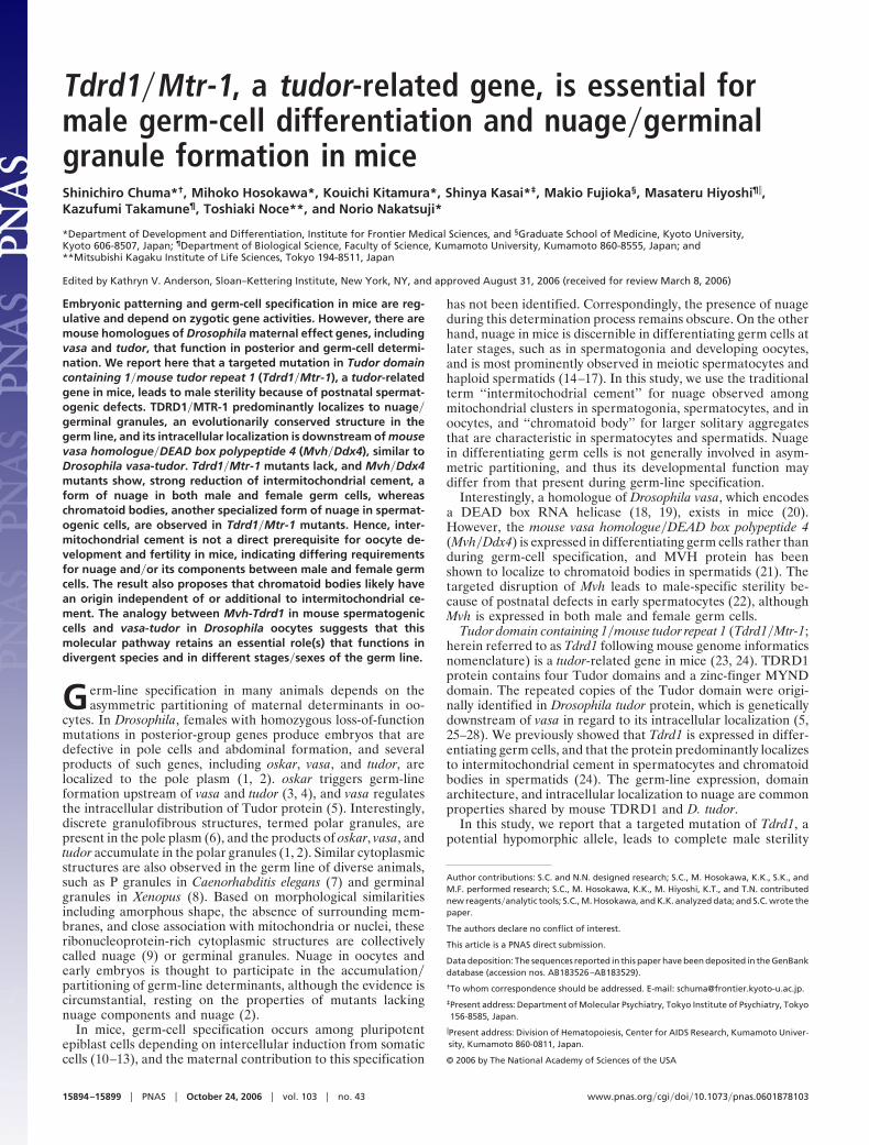
Immunostaining for germ-cell-specific nuclear antigen (31)and for meiotic synaptonemal complex protein 3 (SYCP3�SCP3;ref. 32) showed that spermatogonia and early spermatocytesat the leptotene-zygotene stages were normally present inTdrd1tm1/tm1 testes (Fig. 3 A and B). However, the differentiationof spermatocytes later than the pachytene-diplotene stages wasblocked in a subset of seminiferous tubules (Fig. 3B, arrowhead),whereas in others, meiosis progressed further (Fig. 3B, arrow,and C and D), and haploid round spermatids up to step 7 weredifferentiated (Fig. 3 E and F). At 8 weeks after birth, �20% ofTdrd1tm1/tm1 seminiferous tubules contained only spermatogonia
and leptotene-zygotene spermatocytes, 65% contained up topachytene-diplotene spermatocytes, and the remaining 15%harbored round spermatids. Cellular elongation or nuclearcondensation of spermatids did not occur in Tdrd1tm1/tm1 testes,and mature spermatozoa were lacking, consistent with malesterility.
RT-PCR analyses confirmed the above histological results(Fig. 3G). Sycp3, Sycp1, and Acr, which are expressed in meioticspermatocytes (32–34), were detected in Tdrd1tm1/tm1 testes atapproximately the same levels as in wild type. Odf1�RT7 andTnp2, whose expression begins in round spermatids (35, 36),were also detected but at reduced levels, and Prm1, which isexpressed at the latest stage of round spermatid differentiation(37), was undetectable in Tdrd1tm1/tm1 testes.
The defects among Tdrd1tm1/tm1 spermatocytes could arisethrough an intrinsic requirement of Tdrd1 function or through anindirect effect after the lack of elongated spermatids and sper-matozoa, which could cause the detachment of spermatocytesfrom the seminiferous epithelium. To distinguish between thesepossibilities, we examined the first occurrence of spermatocytedefects in prepubertal testes (for details, see Supporting Text andFig. 7, which is published as supporting information on the PNASweb site). At 14 days postpartum (dpp), when the first populationof spermatocytes reaches the pachytene-diplotene stages butbefore the appearance of round spermatids, apoptosis wasalready increased among Tdrd1tm1/tm1 spermatocytes comparedwith wild type. Then, elevated apoptosis was seen among bothspermatocytes and round spermatids at 20 dpp and continuallyin adult Tdrd1tm1/tm1 testes. Thus, spermatogenic defects inTdrd1tm1/tm1 testes first occur in a subset of spermatocytes, andthe remaining spermatocytes further develop up to round sper-matids, whose differentiation is then completely blocked, fol-lowed by apoptotic cell death.
Mvh Regulates the Intracellular Localization of TDRD1. MVH�DDX4is another component of mouse nuage that functions in postnatalspermatogenesis (21, 22). To reveal a possible relationshipbetween Tdrd1 and Mvh, we compared the localization patternof these two proteins (data are presented in Fig. 8, which ispublished as supporting information on the PNAS web site, andSupporting Text). In brief, TDRD1 showed a fine granularappearance that corresponds to intermitochondrial cement inspermatogonia and developing oocytes, whereas MVH wasdiffusely observed in the cytoplasm in these cells. Duringspermatocyte differentiation, MVH showed granular distribu-
Fig. 3. Differentiation defects among meiotic spermatocytes and in haploid round spermatids in Tdrd1tm1/tm1 mutants. (A and B) Sections of wild-type (A) andTdrd1tm1/tm1 (B) testes stained for germ-cell-specific nuclear antigen (GENA; green), SYCP3�SCP3 (red) and with Hoechst 33258 (blue). In a Tdrd1tm1/tm1
seminiferous tubule (B, arrowhead), only GENA-positive but SCP3-negative spermatogonia are observed, whereas in another (B, arrow), pachytene-zygotenespermatocytes are differentiated. (C and D) Higher-magnification views of the sections of wild-type (C) and Tdrd1tm1/tm1 (D) testes stained for SYCP3 (red) andwith Hoechst 33258 (blue). Meiotic synaptonemal complexes are normally seen in Tdrd1tm1/tm1 spermatocytes (arrowheads). (E and F) Sections of wild-type (E)and Tdrd1tm1/tm1 (F) spermatids stained with Rhodamine-PNA (red), anti-Golgi-58k antibody (green), and Hoechst 33258 (blue). Round spermatids with acrosomesare present in Tdrd1tm1/tm1 testis (F, and arrowhead), but further elongation of spermatids was not observed. (G) RT-PCR analyses of wild-type and Tdrd1tm1/tm1
testes for spermatocyte and spermatid-marker genes. RT, reverse transcriptase; ���, wild-type; tm1�tm1, Tdrd1tm1/tm1. (Scale bars: A and B, 50 �m; C–F, 10 �m.)
15896 � www.pnas.org�cgi�doi�10.1073�pnas.0601878103 Chuma et al.
tion that merged with TDRD1, but such localization of MVHwas gradual and occurred later than TDRD1. In round sperma-tids, the two proteins precisely colocalized to larger solitaryaggregates that correspond to chromatoid bodies (21, 24). In all,TDRD1 more consistently localized to nuage than MVH duringgerm cell differentiation, whereas MVH translocated from thecytoplasm to nuage in spermatocytes.
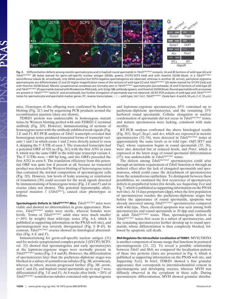
The expression and localization of TDRD1 and MVH werenext examined in each reciprocal mutant. In Tdrd1tm1/tm1 testes,MVH protein was detected, and cytoplasmic granules in sper-matocytes and spermatids were similarly seen as in wild type(Fig. 4 A and B). On the other hand, in Mvh1098/1098 testes whosespermatogenesis ceases during the meiotic prophase of sper-matocytes (22), TDRD1 protein in spermatogonia and survivingspermatocytes did not exhibit characteristic granular localiza-tion. Instead, TDRD1 was diffusely distributed in the cytoplasmor more notably showed a crescent-like accumulation around thenucleus in spermatocytes (Fig. 4 C and D).
In Tdrd1tm1/tm1 oocytes, MVH was diffusely present in thecytoplasm as in wild type (Fig. 4 E and F). In contrast, thelocalization of TDRD1 was altered in Mvh1098/1098 oocytes, andthe characteristic granular distribution, seen in wild type (Fig. 4G and H), was disrupted. Therefore, the intracellular localizationof TDRD1 in both spermatogenic cells and oocytesis is down-stream of Mvh, whereas Tdrd1tm1/tm1 mutation does not affectMVH distribution. Considering Mvh1098/1098 mutants are male-specific sterile, this result revealed that TDRD1 localization tonuage is not directly associated with female fertility.
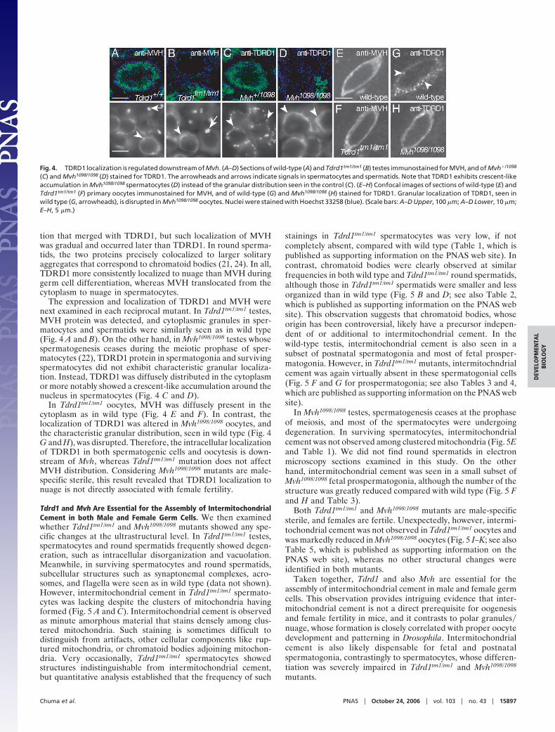
Tdrd1 and Mvh Are Essential for the Assembly of IntermitochondrialCement in both Male and Female Germ Cells. We then examinedwhether Tdrd1tm1/tm1 and Mvh1098/1098 mutants showed any spe-cific changes at the ultrastructural level. In Tdrd1tm1/tm1 testes,spermatocytes and round spermatids frequently showed degen-eration, such as intracellular disorganization and vacuolation.Meanwhile, in surviving spermatocytes and round spermatids,subcellular structures such as synaptonemal complexes, acro-somes, and flagella were seen as in wild type (data not shown).However, intermitochondrial cement in Tdrd1tm1/tm1 spermato-cytes was lacking despite the clusters of mitochondria havingformed (Fig. 5 A and C). Intermitochondrial cement is observedas minute amorphous material that stains densely among clus-tered mitochondria. Such staining is sometimes difficult todistinguish from artifacts, other cellular components like rup-tured mitochondria, or chromatoid bodies adjoining mitochon-dria. Very occasionally, Tdrd1tm1/tm1 spermatocytes showedstructures indistinguishable from intermitochondrial cement,but quantitative analysis established that the frequency of such
stainings in Tdrd1tm1/tm1 spermatocytes was very low, if notcompletely absent, compared with wild type (Table 1, which ispublished as supporting information on the PNAS web site). Incontrast, chromatoid bodies were clearly observed at similarfrequencies in both wild type and Tdrd1tm1/tm1 round spermatids,although those in Tdrd1tm1/tm1 spermatids were smaller and lessorganized than in wild type (Fig. 5 B and D; see also Table 2,which is published as supporting information on the PNAS website). This observation suggests that chromatoid bodies, whoseorigin has been controversial, likely have a precursor indepen-dent of or additional to intermitochondrial cement. In thewild-type testis, intermitochondrial cement is also seen in asubset of postnatal spermatogonia and most of fetal prosper-matogonia. However, in Tdrd1tm1/tm1 mutants, intermitochndrialcement was again virtually absent in these spermatogonial cells(Fig. 5 F and G for prospermatogonia; see also Tables 3 and 4,which are published as supporting information on the PNAS website).
In Mvh1098/1098 testes, spermatogenesis ceases at the prophaseof meiosis, and most of the spermatocytes were undergoingdegeneration. In surviving spermatocytes, intermitochondrialcement was not observed among clustered mitochondria (Fig. 5Eand Table 1). We did not find round spermatids in electronmicroscopy sections examined in this study. On the otherhand, intermitochondrial cement was seen in a small subset ofMvh1098/1098 fetal prospermatogonia, although the number of thestructure was greatly reduced compared with wild type (Fig. 5 Fand H and Table 3).
Both Tdrd1tm1/tm1 and Mvh1098/1098 mutants are male-specificsterile, and females are fertile. Unexpectedly, however, intermi-tochondrial cement was not observed in Tdrd1tm1/tm1 oocytes andwas markedly reduced in Mvh1098/1098 oocytes (Fig. 5 I–K; see alsoTable 5, which is published as supporting information on thePNAS web site), whereas no other structural changes wereidentified in both mutants.
Taken together, Tdrd1 and also Mvh are essential for theassembly of intermitochondrial cement in male and female germcells. This observation provides intriguing evidence that inter-mitochondrial cement is not a direct prerequisite for oogenesisand female fertility in mice, and it contrasts to polar granules�nuage, whose formation is closely correlated with proper oocytedevelopment and patterning in Drosophila. Intermitochondrialcement is also likely dispensable for fetal and postnatalspermatogonia, contrastingly to spermatocytes, whose differen-tiation was severely impaired in Tdrd1tm1/tm1 and Mvh1098/1098
mutants.
Fig. 4. TDRD1 localization is regulated downstream of Mvh. (A–D) Sections of wild-type (A) and Tdrd1tm1/tm1 (B) testes immunostained for MVH, and of Mvh�/1098
(C) and Mvh1098/1098 (D) stained for TDRD1. The arrowheads and arrows indicate signals in spermatocytes and spermatids. Note that TDRD1 exhibits crescent-likeaccumulation in Mvh1098/1098 spermatocytes (D) instead of the granular distribution seen in the control (C). (E–H) Confocal images of sections of wild-type (E) andTdrd1tm1/tm1 (F) primary oocytes immunostained for MVH, and of wild-type (G) and Mvh1098/1098 (H) stained for TDRD1. Granular localization of TDRD1, seen inwild type (G, arrowheads), is disrupted in Mvh1098/1098 oocytes. Nuclei were stained with Hoechst 33258 (blue). (Scale bars: A–D Upper, 100 �m; A–D Lower, 10 �m;E–H, 5 �m.)
Chuma et al. PNAS � October 24, 2006 � vol. 103 � no. 43 � 15897
DEV
ELO
PMEN
TAL
BIO
LOG
Y
DiscussionThe properties and functions of mammalian nuage remainelusive. The chromatoid body in spermatids has been relativelywell described because of its prominence by light and electronmicroscopy. It is implicated in RNA storage and regulationbased on the abundance of RNA and RNA-binding proteins suchas transition protein 2 mRNA (38) and p48�52 (39). Recently,Kotaja et al. (40) reported that the chromatoid body in sperma-tids contains Dicer and microRNAs, proposing that the structureis involved in a microRNA pathway. Intracellular and intercel-lular movement of chromatoid bodies also suggests that thestructure functions in RNA trafficking and gene dosage com-pensation (17). Intermitochondrial cement, on the other hand, ismuch less studied, although it is more consistently present duringgerm-cell differentiation. We showed that TDRD1 specificallylocalizes to both intermitochondrial cement and chromatoidbodies. It is the only identified protein that constitutes nuage inboth male and female germ cells and thus would provide avaluable clue to mark and study properties of mammalian nuage.
Tdrd1tm1/tm1 mutants showed male sterility with postnatalspermatogenic defects. The observed phenotypes may reflect ahypomorph or antimorph of this gene. However, heterogenousphenotypes among spermatocytes and spermatids, similar tothose observed in Tdrd1tm1/tm1 mutants, were also reported forthe null mutant of Grth�Ddx25, which encodes an RNA helicasethat localizes to chromatoid bodies (41). We also found thatMvh1098/1098 testes harbor a small population of round sperma-tids, although most spermatocytes are degenerated during themeiotic prophase (data not shown). Thus, heterogenous defectsduring postnatal spermatogenesis may represent a commonloss-of-function phenotype of a group of nuage components,including TDRD1. Although further entire deletion of the Tdrd1gene would help examine whether additional phenotypes exist,the Tdrd1tm1 allele revealed that the tudor-related gene playsessential roles in postnatal spermatogenesis in mice. ThisTdrd1tm1 allele also provides a precious mutant that lacksintermitochondrial cement in both male and female germ cells.
The molecular function of the repeated copies of the Tudordomain remains elusive. The single Tudor domain in the SurvivalMotor Neuron protein binds to Sm proteins of small nuclearribonucleoproteins and functions in the assembly of the spliceo-somal complex (42, 43). Consistently, TDRD1 and Sm proteinsform complexes and colocalize in chromatoid bodies (24).Recently, 53BP1 containing two neighboring Tudor folds thatcomprise a single globular domain was shown to bind methylatedhistone H3 (44, 45). Multiple Tudor domains may function toassemble proteins that associate with each Tudor domain intomacromolecular complexes. In addition, D. tudor is required forthe localization of mitochondrial ribosomal RNAs to polargranules (46), implying that it also acts in RNA assembly. Inagreement with the presumed scaffold function of multipleTudor domains, hypomorphic and null mutants of D. tudorgreatly reduce the number and size of polar granules in oocytes(25, 28), and Tdrd1tm1/tm1 mutants lacked intermitochondrialcement in both male and female germ cells. In contrast, chro-matoid bodies in spermatids were clearly observed in Tdrd1tm1/
tm1 mutants, although they were smaller and less organizedcompared with wild type. The origin of chromatoid bodies hasbeen obscure and controversial. The structure has been sug-gested to arise from intermitochondrial cement or nuclei basedon morphological similarities and topographical relations (14).We propose that the chromatoid body incorporates componentsfrom intermitochondrial cement, as exemplified by TDRD1 thatlocalizes to both structures, but it should also have anothersource of precursors such as from the nucleus. In mice, there areother Tudor-related proteins, TDRD6 and TDRD7�TRAP,which localize to chromatoid bodies (M.H., unpublished work).It is conceivable that these proteins compensate TDRD1 for itsspermatogenic function and the assembly of chromatoid bodiesin Tdrd1tm1/tm1 mutants. Recently, RNF17 was also shown tocontain multiple Tudor domains in addition to a RING fingerdomain. Interestingly, Rnf17 gene-targeted mice exhibit malesterility, but RNF17 localizes to cytoplasmic structures that aredistinct from intermitochondrial cement and chromatoid bodies(47). The RNF17 granules may represent a novel type of nuagethat has an essential function in spermatogenesis in mice.
We showed that Mvh is upstream of TDRD1 with respect tointracellular localization. The perinuclear accumulation ofTDRD1 in Mvh1098/1098 spermatocytes suggests a possibility thatTDRD1 takes part in trafficking molecules from the nucleus tothe intermitochondrial regions, and that this TDRD1 activitydepends on Mvh. The results for Tdrd1 and Mvh are comparableto those reported for D. tudor and vasa in several aspects: (i) bothgenes in Drosophila and mice are expressed and function in thegerm line (25, 48, 49), (ii) the protein products of these geneslocalize to nuage in common (5, 50, 51), (iii) polar granules arenot formed or greatly reduced in vasa and tudor mutants similarly
Fig. 5. Tdrd1 and Mvh are essential for the assembly of intermitochondrialcement in both male and female germ cells. Electron microscopy of adulttestes (A–E), fetal testes (F–H), and adult ovaries (I–K) from mice with theindicated genotypes. (A and B) A wild-type spermatocyte (A) and roundspermatid (B). The arrowheads mark intermitochondrial cement (A) and achromatoid body (B). (C) A Tdrd1tm1/tm1 spermatocyte in which intermitochon-drial cement is not discernible or quite diminutive, although a cluster ofmitochondria is observable. (D) A Tdrd1tm1/tm1 round spermatid. A chromatoidbody is clearly seen (arrowhead), whereas it appears sparse compared withwild type (B). (E) A morphologically unimpaired Mvh1098/1098 spermatocyte.Intermitochondrial cement is not detected. (F–H) Mitochondrial clusters inwild-type (F), Tdrd1tm1/tm1 (G), and Mvh1098/1098 (H) fetal prospermatogonia at17.5 days postcoitum. Intermitochondrial cement, seen in wild type (F, arrow-heads), is not discernible in the two mutant cells (G and H). (I–K) Primaryoocytes of wild-type (I), Tdrd1tm1/tm1 (J), and Mvh1098/1098 (K) mutants. Inter-mitochondrial cement, present in wild type (I, arrowhead), is again notobserved in the two mutant oocytes (J and K). Note that a small subset ofMvh1098/1098 prospermatogonium and oocyte sections show intermitochon-drial cement as summarized in Tables 3 and 5. (Scale bars: 1 �m.)
15898 � www.pnas.org�cgi�doi�10.1073�pnas.0601878103 Chuma et al.

to intermitochondrial cement in Mvh and Tdrd1 mutants (25, 28,48, 49), and (iv) the intracellular distribution of Tudor�TDRD1is regulated downstream of vasa�Mvh (5). One clear difference,on the other hand, is that Drosophila vasa-tudor maternallyfunction in oocytes, whereas mouse Mvh-Tdrd1 zygotically playroles in spermatogenic cells. Thus, a group of genes and theirmolecular pathway that are essential for germ-cell specificationand early embryonic patterning in Drosophila are likely con-served and analogously function in the germ line in mice butduring the postnatal differentiation of male germ cells. Nuage isa site of assembly for these evolutionarily conserved moleculesand probably retains a function(s) that is essential and indepen-dent of the differentiation stage, sex, and species of the germline.
One particular and intriguing question raised in this study iswhether mouse oocytes lacking intermitochondrial cement, asthose in Tdrd1tm1/tm1 and Mvh1098/1098 mutants, are not substan-tially affected at the molecular level, or whether certain aber-rations actually occur, but overall catastrophe does not follow.The latter possibility is supported by the notion that oocytes areless susceptible to meiotic checkpoint controls than spermato-genic cells in mice (52). This may also explain the sexualdimorphisms in Tdrd1tm1/tm1 and Mvh1098/1098 phenotypes. Similarquestions would also apply to fetal and postnatal spermatogonia.
The identification of molecular changes underlying these twomutant oocytes and spermatogonial cells would help to furtherreveal cellular and developmental features associated with nu-age, an enigmatic ribonucleoprotein structure specifically con-served in the germ line.
Materials and MethodsTargeted Mutagenesis. A 1.5-kb Tdrd1 genomic fragment up-stream of the first coding ATG and a 6-kb fragment downstreamof exon 3 were ligated to the neomycin resistance gene cassetteof pKO SelectNeo (Lexicon Genetics, The Woodlands, TX) andto the diphtheria toxin A fragment gene cassette from pND3. TheTdrd1 targeting vector was used to derive homologous recom-binant ES cells, and chimeric mice were produced from tworecombinant ES clones. Male chimeras of one line were matedwith wild-type females to obtain heterozygous mice.
For further details, see Supporting Text.
We thank A. Nagy (University of Toronto, Toronto, ON, Canada) for R1ES cells; Y. Nishimune (Osaka University, Osaka, Japan) for antigerm-cell-specific nuclear antigen (GENA) antibody TRA98�104; T. Tada, H.Suemori, M. Shoji, T. Tanaka, E. Moribe, and M. Tanaka for experi-mental advice and assistance; and A. B. Cowan for comments on themanuscript. This study was supported by Grants-in-Aid from the Min-istry of Education, Culture, Sports, Science, and Technology of Japan.
1. Lehmann R, Ephrussi A (1994) Ciba Found Symp 182:282–96; discussion296–300.
2. Saffman EE, Lasko P (1999) Cell Mol Life Sci 55:1141–1163.3. Ephrussi A, Lehmann R (1992) Nature 358:387–392.4. Smith JL, Wilson JE, Macdonald PM (1992) Cell 70:849–859.5. Bardsley A, McDonald K, Boswell RE (1993) Development (Cambridge, UK)
119:207–219.6. Mahowald AP (1962) J Exp Zool 151:201–216.7. Strome S, Wood WB (1982) Proc Natl Acad Sci USA 79:1558–1562.8. Czolowska R (1969) J Embryol Exp Morphol 22:229–251.9. Eddy EM (1975) Int Rev Cytol 43:229–280.
10. Lawson KA, Hage WJ (1994) Ciba Found Symp 182:68–84; discussion 84–91.11. Tam PP, Zhou SX (1996) Dev Biol 178:124–132.12. Lawson KA, Dunn NR, Roelen BA, Zeinstra LM, Davis AM, Wright CV,
Korving JP, Hogan BL (1999) Genes Dev 13:424–436.13. McLaren A (2003) Dev Biol 262:1–15.14. Fawcett DW, Eddy EM, Phillips DM (1970) Biol Reprod 2:129–153.15. Eddy EM (1974) Anat Rec 178:731–757.16. Russell L, Frank B (1978) Anat Rec 190:79–97.17. Parvinen M (2005) Int J Androl 28:189–201.18. Hay B, Jan LY, Jan YN (1988) Cell 55:577–587.19. Lasko PF, Ashburner M (1988) Nature 335:611–617.20. Fujiwara Y, Komiya T, Kawabata H, Sato M, Fujimoto H, Furusawa M, Noce
T (1994) Proc Natl Acad Sci USA 91:12258–12262.21. Toyooka Y, Tsunekawa N, Takahashi Y, Matsui Y, Satoh M, Noce T (2000)
Mech Dev 93:139–149.22. Tanaka SS, Toyooka Y, Akasu R, Katoh-Fukui Y, Nakahara Y, Suzuki R,
Yokoyama M, Noce T (2000) Genes Dev 14:841–853.23. Wang PJ, McCarrey JR, Yang F, Page DC (2001) Nat Genet 27:422–426.24. Chuma S, Hiyoshi M, Yamamoto A, Hosokawa M, Takamune K, Nakatsuji N
(2003) Mech Dev 120:979–990.25. Boswell RE, Mahowald AP (1985) Cell 43:97–104.26. Golumbeski GS, Bardsley A, Tax F, Boswell RE (1991) Genes Dev 5:2060–
2070.27. Ponting CP (1997) Trends Biochem Sci 22:51–52.28. Thomson T, Lasko P (2004) Genesis 40:164–170.29. Nagy A, Rossant J, Nagy R, Abramow-Newerly W, Roder JC (1993) Proc Natl
Acad Sci USA 90:8424–8428.30. Kozak M (1999) Gene 234:187–208.31. Tanaka H, Pereira LA, Nozaki M, Tsuchida J, Sawada K, Mori H, Nishimune
Y (1997) Int J Androl 20:361–366.
32. Lammers JH, Offenberg HH, van Aalderen M, Vink AC, Dietrich AJ, HeytingC (1994) Mol Cell Biol 14:1137–1146.
33. Meuwissen RL, Offenberg HH, Dietrich AJ, Riesewijk A, van Iersel M,Heyting C (1992) EMBO J 11:5091–5100.
34. Kashiwabara S, Arai Y, Kodaira K, Baba T (1990) Biochem Biophys ResCommun 173:240–245.
35. Shih DM, Kleene KC (1992) Mol Reprod Dev 33:222–227.36. van der Hoorn FA, Tarnasky HA, Nordeen SK (1990) Dev Biol
142:147–154.37. Mali P, Kaipia A, Kangasniemi M, Toppari J, Sandberg M, Hecht NB, Parvinen
M (1989) Reprod Fertil Dev 1:369–382.38. Saunders PT, Millar MR, Maguire SM, Sharpe RM (1992) Mol Reprod Dev
33:385–391.39. Oko R, Korley R, Murray MT, Hecht NB, Hermo L (1996) Mol Reprod Dev
44:1–13.40. Kotaja N, Bhattacharyya SN, Jaskiewicz L, Kimmins S, Parvinen M, Filipowicz
W, Sassone-Corsi P (2006) Proc Natl Acad Sci USA 103:2647–2652.41. Tsai-Morris CH, Sheng Y, Lee E, Lei KJ, Dufau ML (2004) Proc Natl Acad Sci
USA 101:6373–6378.42. Buhler D, Raker V, Luhrmann R, Fischer U (1999) Hum Mol Genet 8:2351–
2357.43. Selenko P, Sprangers R, Stier G, Buhler D, Fischer U, Sattler M (2001) Nat
Struct Biol 8:27–31.44. Charier G, Couprie J, Alpha-Bazin B, Meyer V, Quemeneur E, Guerois R,
Callebaut I, Gilquin B, Zinn-Justin S (2004) Structure (London) 12:1551–1562.45. Huyen Y, Zgheib O, Ditullio RA, Jr, Gorgoulis VG, Zacharatos P, Petty TJ,
Sheston EA, Mellert HS, Stavridi ES, Halazonetis TD (2004) Nature 432:406–411.
46. Amikura R, Hanyu K, Kashikawa M, Kobayashi S (2001) Mech Dev 107:97–104.47. Pan J, Goodheart M, Chuma S, Nakatsuji N, Page DC, Wang PJ (2005)
Development (Cambridge, UK) 132:4029–4039.48. Schupbach T, Wieschaus E (1986) Roux’s Arch Dev Biol 195:302–317.49. Nusslein-Volhard C, Frohnhofer HG, Lehmann R (1987) Science 238:1675–
1681.50. Hay B, Ackerman L, Barbel S, Jan LY, Jan YN (1988) Development (Cam-
bridge, UK) 103:625–640.51. Liang L, Diehl-Jones W, Lasko P (1994) Development (Cambridge, UK)
120:1201–1211.52. Morelli MA, Cohen PE (2005) Reproduction 130:761–781.
Chuma et al. PNAS � October 24, 2006 � vol. 103 � no. 43 � 15899
DEV
ELO
PMEN
TAL
BIO
LOG
Y