team mimesis · 2018-02-15 · activity report 2017 team mimesis computational anatomy and...
TRANSCRIPT

Activity Report 2017
Team MIMESIS
Computational Anatomy and Simulation forMedicineInria teams are typically groups of researchers working on the definition of a common project,and objectives, with the goal to arrive at the creation of a project-team. Such project-teams mayinclude other partners (universities or research institutions).
RESEARCH CENTERNancy - Grand Est
THEMEComputational Neuroscience andMedicine


Table of contents
1. Personnel . . . . . . . . . . . . . . . . . . . . . . . . . . . . . . . . . . . . . . . . . . . . . . . . . . . . . . . . . . . . . . . . . . . . . . . . . . . . . . . . 12. Overall Objectives . . . . . . . . . . . . . . . . . . . . . . . . . . . . . . . . . . . . . . . . . . . . . . . . . . . . . . . . . . . . . . . . . . . . . . . . 2
2.1. Team Overview 22.2. Challenges 2
3. Research Program . . . . . . . . . . . . . . . . . . . . . . . . . . . . . . . . . . . . . . . . . . . . . . . . . . . . . . . . . . . . . . . . . . . . . . . . 33.1. Real Time Patient-Specific Computational Models 33.2. Adaptive Meshing and Advanced Simulation Techniques 43.3. Data-driven Simulation 5
4. Application Domains . . . . . . . . . . . . . . . . . . . . . . . . . . . . . . . . . . . . . . . . . . . . . . . . . . . . . . . . . . . . . . . . . . . . . .64.1. Surgical Training 64.2. Pre-operative Planning 64.3. Intra-operative Navigation 7
5. Highlights of the Year . . . . . . . . . . . . . . . . . . . . . . . . . . . . . . . . . . . . . . . . . . . . . . . . . . . . . . . . . . . . . . . . . . . . . 76. New Software and Platforms . . . . . . . . . . . . . . . . . . . . . . . . . . . . . . . . . . . . . . . . . . . . . . . . . . . . . . . . . . . . . . 7
6.1. SOFA 76.2. SofaPardisoSolver 86.3. SOFA Xray rendering 8
7. New Results . . . . . . . . . . . . . . . . . . . . . . . . . . . . . . . . . . . . . . . . . . . . . . . . . . . . . . . . . . . . . . . . . . . . . . . . . . . . . . 97.1. Augmented Reality in Surgical Navigation 9
7.1.1. Organ Pose Estimation for Augmented Reality in Hepatic Surgery 97.1.2. Image-driven Stochastic Estimation of Boundary Conditions 9
7.2. Advanced Numerical Modeling and Simulation 107.2.1. Face-based Smoothed Finite Element Method for Real-time Simulation of Soft Tissue 107.2.2. Immersed Boundary Method for Real-time 107.2.3. Error Control in Surgical Simulations 11
7.3. Model-based Image Registration 117.3.1. Intraoperative Biomechanical Registration of the Liver 117.3.2. Registration of Cell Nuclei in Cell Microscopy 12
7.4. Reconstruction of Geometries from Images 137.4.1. Automatic Skeletonization of Vascular Trees in Pre-operative CT Images 137.4.2. Template-based Recovery of Elastic Shapes from Monocular Video 13
7.5. Simulation for Intra-operative Rehearsal 148. Bilateral Contracts and Grants with Industry . . . . . . . . . . . . . . . . . . . . . . . . . . . . . . . . . . . . . . . . . . . . . 149. Partnerships and Cooperations . . . . . . . . . . . . . . . . . . . . . . . . . . . . . . . . . . . . . . . . . . . . . . . . . . . . . . . . . . . 15
9.1. Regional Initiatives 159.1.1. Institute of Image-Guided Surgery (IHU) Strasbourg 159.1.2. Research and Clinical Partners 15
9.2. National Initiatives 169.2.1. ADT (Action de Développement Technologique) 169.2.2. ANR (Agence Nationale de la Recherche) 169.2.3. Inria Collaborations 179.2.4. National Collaborations 17
9.3. European Initiatives 179.3.1. FP7 & H2020 Projects 179.3.2. Informal Collaborations 18
9.4. International Initiatives 189.5. International Research Visitors 18
10. Dissemination . . . . . . . . . . . . . . . . . . . . . . . . . . . . . . . . . . . . . . . . . . . . . . . . . . . . . . . . . . . . . . . . . . . . . . . . . . 1910.1. Promoting Scientific Activities 19
2 Activity Report INRIA 2017
10.1.1. Scientific Events Organisation 1910.1.1.1. Member of the Organizing Committees 1910.1.1.2. Reviewing Activities 19
10.1.2. Journal 1910.1.3. Invited Talks 1910.1.4. Scientific Expertise 1910.1.5. Research Administration 19
10.2. Teaching - Supervision - Juries 2010.2.1. Teaching 2010.2.2. Supervision 2010.2.3. Juries 20
10.3. Popularization 2011. Bibliography . . . . . . . . . . . . . . . . . . . . . . . . . . . . . . . . . . . . . . . . . . . . . . . . . . . . . . . . . . . . . . . . . . . . . . . . . . .21
Team MIMESIS
Creation of the Team: 2015 July 01
Keywords:
Computer Science and Digital Science:A2.5. - Software engineeringA3.1.1. - Modeling, representationA3.1.4. - Uncertain dataA3.2.2. - Knowledge extraction, cleaningA5.1. - Human-Computer InteractionA5.3.4. - RegistrationA5.4.4. - 3D and spatio-temporal reconstructionA5.4.5. - Object tracking and motion analysisA5.6. - Virtual reality, augmented realityA6.1.1. - Continuous Modeling (PDE, ODE)A6.1.5. - Multiphysics modelingA6.2.8. - Computational geometry and meshes
Other Research Topics and Application Domains:B2.4. - TherapiesB2.4.3. - SurgeryB2.6. - Biological and medical imagingB2.7. - Medical devicesB2.7.1. - Surgical devices
1. PersonnelResearch Scientists
Stéphane Cotin [Team leader, Senior Researcher, HDR]David Cazier [Univ de Strasbourg, Senior Researcher, HDR]Igor Peterlik [Inria, Researcher]Hadrien Courtecuisse [CNRS, Researcher]Lionel Untereiner [Univ de Strasbourg, Researcher, from Oct 2017]
Post-Doctoral FellowsChristoph Paulus [Inria, from Apr 2017]Nazim Haouchine [Inria, until Jan 2017]Antoine Petit [Inria, from Jun 2017]
PhD StudentsNicolas Golse [Hôpital Paul-Brousse]Raffaella Trivisonne [Inria]Yinoussa Adagolodjo [Univ de Strasbourg]Jaime Garcia Guevara [Inria]Andréa Mendizabal [Univ de Strasbourg, from Sep 2017]Sergei Nikolaev [Inria, from May 2017]Jean-Nicolas Brunet [Inria, from Jun 2017]
2 Activity Report INRIA 2017
Technical staffRémi Bessard Duparc [Inria]Bruno Marques [Inria]Frederick Roy [Inria]Etienne Schmitt [Inria, until Feb 2017]
InternsKatell Berdellou [Univ de Strasbourg, from Feb 2017 until Jul 2017]Martina de Landro [Inria, from Oct 2017]Guillaume Grosshenny [Inria, from Jun 2017 until Aug 2017]Alexandre Dolle [Inria, until Jan 2017]Qi Cheng Hua [Inria, from Jun 2017 until Aug 2017]Vincent Magnoux [Inria, from Jul 2017]Roland Maier [Inria, until Jan 2017]Adriana Vrablova [Inria, from Jul 2017 until Sep 2017]
Administrative AssistantsIsabelle Blanchard [Inria]Ouiza Herbi [Inria]
2. Overall Objectives2.1. Team Overview
At the end of 2011, a part of Inria team SHACRA moved from Lille to Strasbourg to join the newly createdInstitute of Image-guided Surgery (IHU) whose main objective is to develop novel clinical technologies at thecrossroads of laparoscopic surgery, flexible endoscopy, and interventional radiology. Similar institutes havebeen created in the past decade around the world with the same global objective: to create a synergy betweenclinicians and scientists to develop new technologies that can redefine healthcare with a strong emphasis onclinical translation.
The global objective of research team MIMESIS is to create a synergy between clinicians and scientists todevelop new technologies that can redefine healthcare, with a strong emphasis on clinical translation. Toachieve this goal, we have joined IHU and we collaborate with numerous partners in both academic andprivate domain.
The scientific objectives of the team MIMESIS are related to this ambitious objective. Over the past yearswe have developed new approaches supporting advanced simulations in the context of simulation for training.We now propose to focus our research on the use of real-time simulation for per-operative guidance. Theunderlying objectives include numerical techniques for real-time computation and data-driven simulationdedicated to patient-specific modeling. This last topic is a transversal research theme and raises several openproblems, ranging from non-rigid registration to augmented reality.
2.2. ChallengesThe core research topics of the MIMESIS project-team essentially aim at improving the realism and fidelityof interactive simulations of medical procedures. This increase in realism makes it possible to envisage newclinical applications, in particular per-operative guidance, that currently rely on imaging techniques, but couldgreatly benefit from our expertise in real-time numerical simulation.
To reach these objectives we have identified several challenges that lie at the intersection of several scientificdomains. Our research projects are currently organized around three main axes:
• Real-time Patient-Specific Computational Models• Adaptive Meshing and Advanced Simulation Techniques• Image-driven Simulation
Team MIMESIS 3
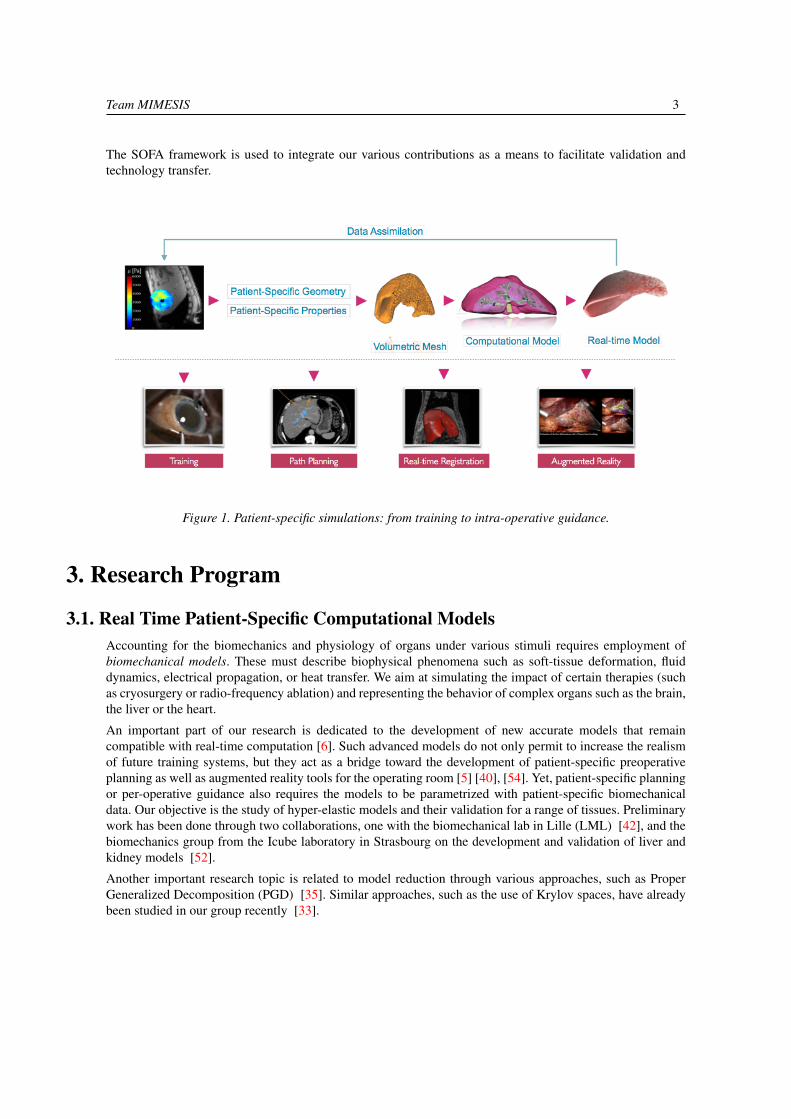
The SOFA framework is used to integrate our various contributions as a means to facilitate validation andtechnology transfer.
Figure 1. Patient-specific simulations: from training to intra-operative guidance.
3. Research Program
3.1. Real Time Patient-Specific Computational ModelsAccounting for the biomechanics and physiology of organs under various stimuli requires employment ofbiomechanical models. These must describe biophysical phenomena such as soft-tissue deformation, fluiddynamics, electrical propagation, or heat transfer. We aim at simulating the impact of certain therapies (suchas cryosurgery or radio-frequency ablation) and representing the behavior of complex organs such as the brain,the liver or the heart.
An important part of our research is dedicated to the development of new accurate models that remaincompatible with real-time computation [6]. Such advanced models do not only permit to increase the realismof future training systems, but they act as a bridge toward the development of patient-specific preoperativeplanning as well as augmented reality tools for the operating room [5] [40], [54]. Yet, patient-specific planningor per-operative guidance also requires the models to be parametrized with patient-specific biomechanicaldata. Our objective is the study of hyper-elastic models and their validation for a range of tissues. Preliminarywork has been done through two collaborations, one with the biomechanical lab in Lille (LML) [42], and thebiomechanics group from the Icube laboratory in Strasbourg on the development and validation of liver andkidney models [52].
Another important research topic is related to model reduction through various approaches, such as ProperGeneralized Decomposition (PGD) [35]. Similar approaches, such as the use of Krylov spaces, have alreadybeen studied in our group recently [33].

4 Activity Report INRIA 2017
We continue our work on cardiac electro-physiology simulation [53], with a focus on patient-specificadaptation of the model. We also study a similar problem, related to the modeling of the electrical conductionin the brain, in the context of Deep Brain Stimulation (DBS) [34], [38]. In this neurosurgical procedure,electrodes are implanted deep into the brain and, connected to a brain pacemaker, send electrical impulses tospecific regions. A final objective is to solve optimization problems in the context of heat diffusion. This is akey element of the development of a planning system that can estimate the locations of the electrodes leadingto an optimal therapeutic effect [54].
Figure 2. Left: patient-specific liver model with its vascular system. Middle: patient specific depolarization times.Right: cryoablation in the kidney.
3.2. Adaptive Meshing and Advanced Simulation TechniquesMost simulations in the field of biomechanics, physiological modeling, or even computer graphics, areperformed using finite element approaches. Such simulations require a discretization of the domain of interest,and this discretization is traditionally made of tetrahedral or hexahedral elements. The topology defined bythese elements is usually considered as being invariant. However, this is not a realistic assumption if the modelis to be employed during a real surgical intervention.
The first objective of this work is to jointly develop advanced topological operations and new finite elementapproaches that can leverage the use of dynamic topologies. In particular we focus our research on multi-resolution meshes where elements are subdivided in areas where numerical errors need to be kept small [51],[55].
Our second objective is to improve, at the numerical level, the efficiency, robustness, and quality of thesimulations. To reach these goals, we essentially rely on two main directions: adaptive meshing to allowmesh transformations during a simulation and support cuts, local remeshing or dynamic refinement in areasof interest; and numerical techniques, such as asynchronous solvers, domain decomposition and model orderreduction [35], [36], [46], [47].
We also work on mixed Finite Element Modeling where both tetrahedra and hexahedra can be used at the sametime, allowing an ideal compromise between numerical efficiency and mesh adaptation to complex geometries.This research also includes the study of domain decomposition techniques and other coupling techniques formulti-domain multi-physics simulations.
Once the problem, as defined in the previous challenge, has been discretized, we need to solve a large systemof linear or nonlinear equations. In both cases, it is necessary to employ numerical solvers repeatedly toconstruct the solution representing the state of the simulated system. In the past years, we have contributedto this topic through our work on asynchronous preconditioning [36]. We would like to pursue this area ofresearch exploiting the relevant advances in hierarchy-based topologies (e.g. the multi-grid methods). We will
Team MIMESIS 5
also consider advanced non-linear solvers which are necessary for correct resolution of hyper-elastic modelsand composite models.
Finally, to improve computational times from a programming stand-point, we have started a collaboration withthe CAMUS team at Inria. This collaboration aims at using smart code analysis and on-the-fly parallelism toautomatically speed-up computation times. In a typical scenario, the modeled organ or tissue is surroundedby its environment represented by other organs, connective tissues or fat. Further, during the intervention, thetissues are manipulated with instruments. Therefore, the interaction will also be an important aspect of ourresearch. We have already developed methods for modeling of advanced interactions between organs, tissuesand tools [50], [37]. We will continue exploiting novel methods such as partial factorization [56] and integrateour approach with other techniques such as augmented Lagrangian.
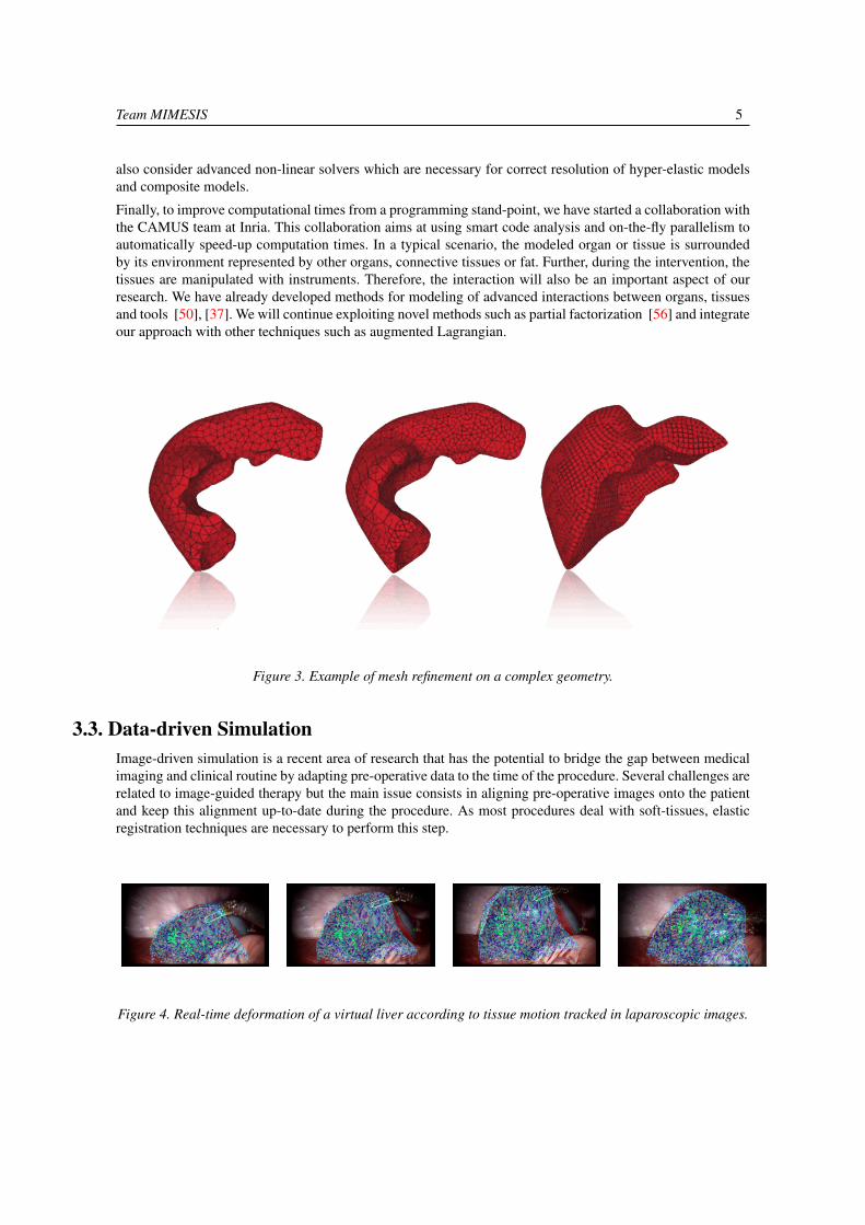
Figure 3. Example of mesh refinement on a complex geometry.
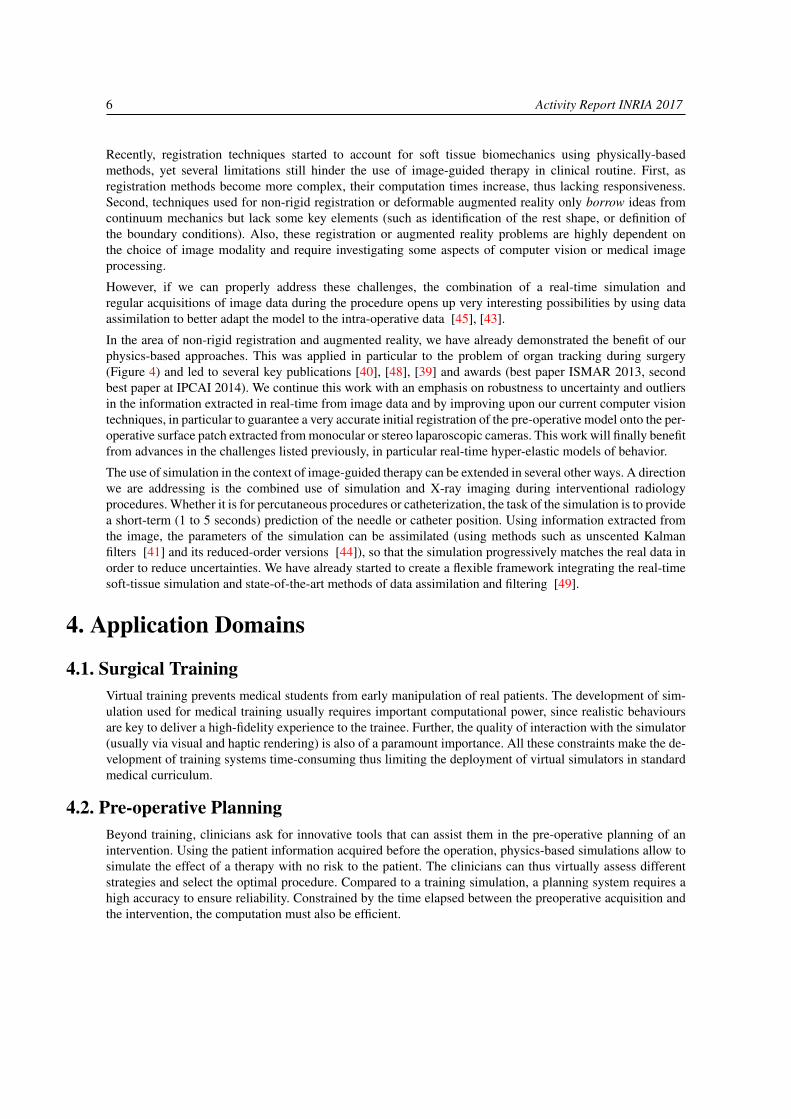
3.3. Data-driven SimulationImage-driven simulation is a recent area of research that has the potential to bridge the gap between medicalimaging and clinical routine by adapting pre-operative data to the time of the procedure. Several challenges arerelated to image-guided therapy but the main issue consists in aligning pre-operative images onto the patientand keep this alignment up-to-date during the procedure. As most procedures deal with soft-tissues, elasticregistration techniques are necessary to perform this step.
Figure 4. Real-time deformation of a virtual liver according to tissue motion tracked in laparoscopic images.
6 Activity Report INRIA 2017
Recently, registration techniques started to account for soft tissue biomechanics using physically-basedmethods, yet several limitations still hinder the use of image-guided therapy in clinical routine. First, asregistration methods become more complex, their computation times increase, thus lacking responsiveness.Second, techniques used for non-rigid registration or deformable augmented reality only borrow ideas fromcontinuum mechanics but lack some key elements (such as identification of the rest shape, or definition ofthe boundary conditions). Also, these registration or augmented reality problems are highly dependent onthe choice of image modality and require investigating some aspects of computer vision or medical imageprocessing.
However, if we can properly address these challenges, the combination of a real-time simulation andregular acquisitions of image data during the procedure opens up very interesting possibilities by using dataassimilation to better adapt the model to the intra-operative data [45], [43].
In the area of non-rigid registration and augmented reality, we have already demonstrated the benefit of ourphysics-based approaches. This was applied in particular to the problem of organ tracking during surgery(Figure 4) and led to several key publications [40], [48], [39] and awards (best paper ISMAR 2013, secondbest paper at IPCAI 2014). We continue this work with an emphasis on robustness to uncertainty and outliersin the information extracted in real-time from image data and by improving upon our current computer visiontechniques, in particular to guarantee a very accurate initial registration of the pre-operative model onto the per-operative surface patch extracted from monocular or stereo laparoscopic cameras. This work will finally benefitfrom advances in the challenges listed previously, in particular real-time hyper-elastic models of behavior.
The use of simulation in the context of image-guided therapy can be extended in several other ways. A directionwe are addressing is the combined use of simulation and X-ray imaging during interventional radiologyprocedures. Whether it is for percutaneous procedures or catheterization, the task of the simulation is to providea short-term (1 to 5 seconds) prediction of the needle or catheter position. Using information extracted fromthe image, the parameters of the simulation can be assimilated (using methods such as unscented Kalmanfilters [41] and its reduced-order versions [44]), so that the simulation progressively matches the real data inorder to reduce uncertainties. We have already started to create a flexible framework integrating the real-timesoft-tissue simulation and state-of-the-art methods of data assimilation and filtering [49].
4. Application Domains
4.1. Surgical TrainingVirtual training prevents medical students from early manipulation of real patients. The development of sim-ulation used for medical training usually requires important computational power, since realistic behavioursare key to deliver a high-fidelity experience to the trainee. Further, the quality of interaction with the simulator(usually via visual and haptic rendering) is also of a paramount importance. All these constraints make the de-velopment of training systems time-consuming thus limiting the deployment of virtual simulators in standardmedical curriculum.
4.2. Pre-operative PlanningBeyond training, clinicians ask for innovative tools that can assist them in the pre-operative planning of anintervention. Using the patient information acquired before the operation, physics-based simulations allow tosimulate the effect of a therapy with no risk to the patient. The clinicians can thus virtually assess differentstrategies and select the optimal procedure. Compared to a training simulation, a planning system requires ahigh accuracy to ensure reliability. Constrained by the time elapsed between the preoperative acquisition andthe intervention, the computation must also be efficient.
Team MIMESIS 7
4.3. Intra-operative NavigationBesides the surgery training and planning, another major need from clinicians is surgical guidance. While thepractician is performing the operation, a guidance system provides enriched visual feedback. This is especiallyuseful with the emergence of minimally invasive surgery (MIS) where the visual information is often stronglylimited. It can be used for example to avoid critical area such as vessels or to highlight the position of atumour during its resection. In the MIS technique, the clinician does not interact with organs directly as in theopen surgery, but manipulates instruments inserted through trocars placed in small incisions in the wall of theabdominal cavity. The surgeon can observe these instruments on a display showing a video stream captured byan endoscopic camera inserted through the navel. The main advantage of the method resides in reducing painand time recovery, in addition to reducing bleeding and risks of infection. However, from a surgical standpoint,the procedure is quite complex since the field of view is considerably reduced and the direct manipulation oforgans is not possible.
5. Highlights of the Year
5.1. Highlights of the YearPrix de thèse 2016 en Génie Biologique et Médical attributed to Rosalie Plantefève for herthesis Augmented Reality and Numerical Simulation for Resection of Hepatic Tumor. The awardis attributed by three scientific bodies: IEEE EMBS, Société Française de Génie Biologique etMédical, and Alliance pour le Génie Biologique et Médical. In this context, R. Plantefève was invitedto submite a paper to the Journal on Innovation and Research in BioMedical Engineering and themanuscript was accepted for publication [17].
Runner up for the best poster award at the IEEE International Symposium on Mixed andAugmented Reality 2017 with the poster Deformed Reality: Proof of concept and PreliminaryResults [32]. The poster introduced a new paradigm to interactively manipulate objects in a scenein a deformable manner. Using the core principle of augmented reality to estimate a rigid pose overtime, the method enables the user to deform the targeted object while it is being rendered with itsnatural texture, giving the sense of a real-time object editing in the user environment. The resultsshow that the method is capable of opening new ways of not only augmenting the scene but also tointeract with it in real by imposing possibly non-linear transformations to selected entities.
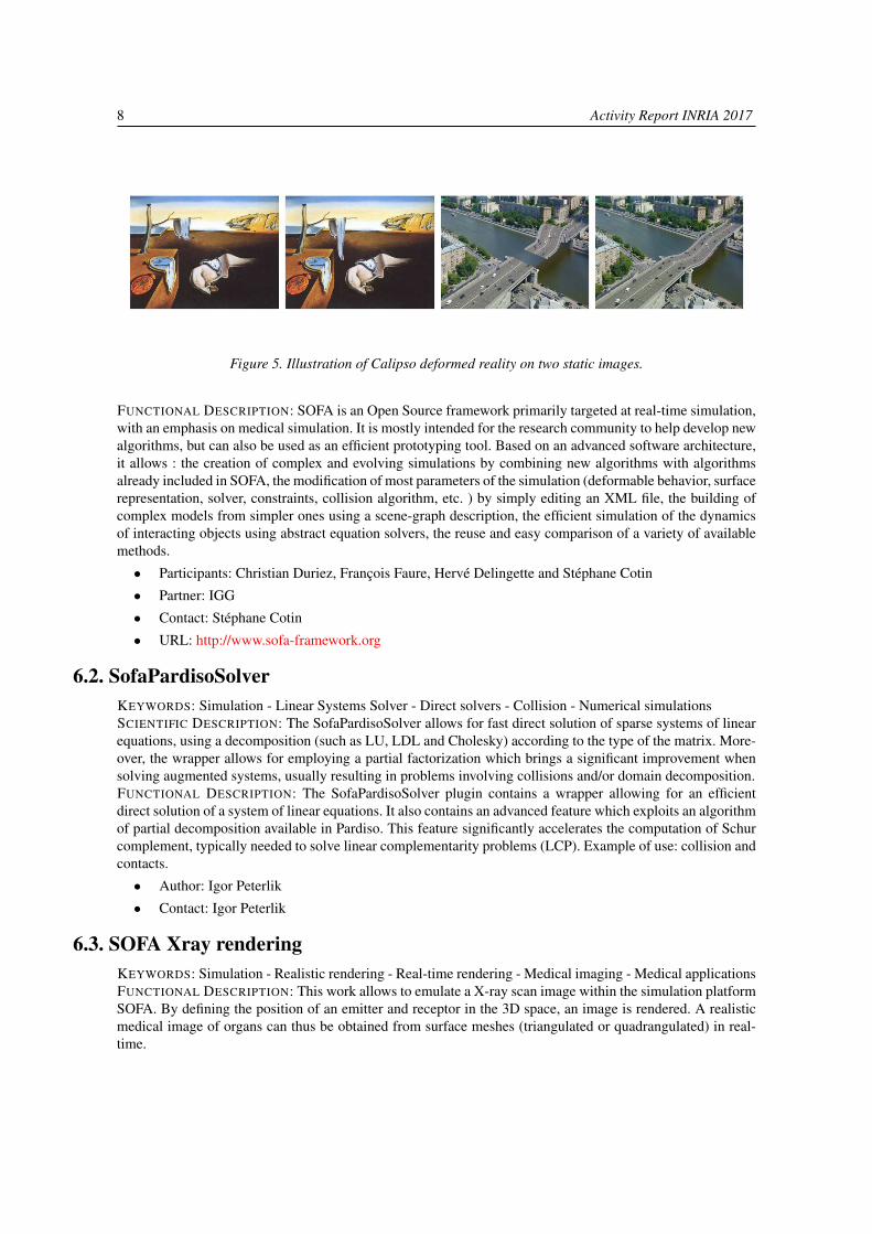
The physics-based image and video editing tool Calipso was resumed in Two-minutes papers onYouTube. At the end of 2017, the video has more that 35k views. Calipso is an interactive methodfor editing images and videos in a physically-coherent manner. The main idea is to perform physics-based manipulations by running a full physics simulation on proxy geometries given by non-rigidlyaligned CAD models. Running these simulations allows us to apply new, unseen forces to move ordeform selected objects, change physical parameters such as mass or elasticity, or even add entirenew objects that interact with the rest of the underlying scene.
6. New Software and Platforms
6.1. SOFASimulation Open Framework ArchitectureKEYWORDS: Real time - Multi-physics simulation - Medical applications
8 Activity Report INRIA 2017
Figure 5. Illustration of Calipso deformed reality on two static images.
FUNCTIONAL DESCRIPTION: SOFA is an Open Source framework primarily targeted at real-time simulation,with an emphasis on medical simulation. It is mostly intended for the research community to help develop newalgorithms, but can also be used as an efficient prototyping tool. Based on an advanced software architecture,it allows : the creation of complex and evolving simulations by combining new algorithms with algorithmsalready included in SOFA, the modification of most parameters of the simulation (deformable behavior, surfacerepresentation, solver, constraints, collision algorithm, etc. ) by simply editing an XML file, the building ofcomplex models from simpler ones using a scene-graph description, the efficient simulation of the dynamicsof interacting objects using abstract equation solvers, the reuse and easy comparison of a variety of availablemethods.
• Participants: Christian Duriez, François Faure, Hervé Delingette and Stéphane Cotin
• Partner: IGG
• Contact: Stéphane Cotin
• URL: http://www.sofa-framework.org
6.2. SofaPardisoSolverKEYWORDS: Simulation - Linear Systems Solver - Direct solvers - Collision - Numerical simulationsSCIENTIFIC DESCRIPTION: The SofaPardisoSolver allows for fast direct solution of sparse systems of linearequations, using a decomposition (such as LU, LDL and Cholesky) according to the type of the matrix. More-over, the wrapper allows for employing a partial factorization which brings a significant improvement whensolving augmented systems, usually resulting in problems involving collisions and/or domain decomposition.FUNCTIONAL DESCRIPTION: The SofaPardisoSolver plugin contains a wrapper allowing for an efficientdirect solution of a system of linear equations. It also contains an advanced feature which exploits an algorithmof partial decomposition available in Pardiso. This feature significantly accelerates the computation of Schurcomplement, typically needed to solve linear complementarity problems (LCP). Example of use: collision andcontacts.
• Author: Igor Peterlik
• Contact: Igor Peterlik
6.3. SOFA Xray renderingKEYWORDS: Simulation - Realistic rendering - Real-time rendering - Medical imaging - Medical applicationsFUNCTIONAL DESCRIPTION: This work allows to emulate a X-ray scan image within the simulation platformSOFA. By defining the position of an emitter and receptor in the 3D space, an image is rendered. A realisticmedical image of organs can thus be obtained from surface meshes (triangulated or quadrangulated) in real-time.
Team MIMESIS 9
Version compatible with SOFA v17.06
• Authors: Stéphane Cotin and Frédérick Roy
• Contact: Stéphane Cotin
7. New Results
7.1. Augmented Reality in Surgical Navigation7.1.1. Organ Pose Estimation for Augmented Reality in Hepatic Surgery
Participants: Y. Adagolodjo, R. Trivisonne, H. Courtecuisse, S. Cotin
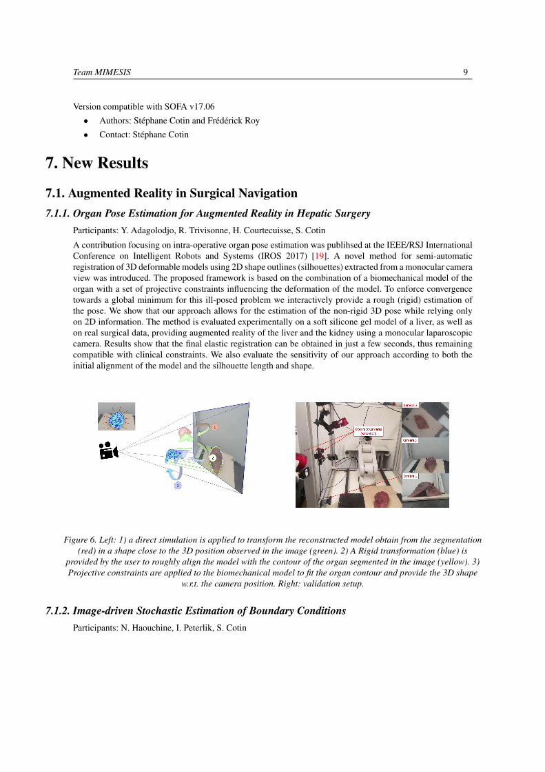
A contribution focusing on intra-operative organ pose estimation was publihsed at the IEEE/RSJ InternationalConference on Intelligent Robots and Systems (IROS 2017) [19]. A novel method for semi-automaticregistration of 3D deformable models using 2D shape outlines (silhouettes) extracted from a monocular cameraview was introduced. The proposed framework is based on the combination of a biomechanical model of theorgan with a set of projective constraints influencing the deformation of the model. To enforce convergencetowards a global minimum for this ill-posed problem we interactively provide a rough (rigid) estimation ofthe pose. We show that our approach allows for the estimation of the non-rigid 3D pose while relying onlyon 2D information. The method is evaluated experimentally on a soft silicone gel model of a liver, as well ason real surgical data, providing augmented reality of the liver and the kidney using a monocular laparoscopiccamera. Results show that the final elastic registration can be obtained in just a few seconds, thus remainingcompatible with clinical constraints. We also evaluate the sensitivity of our approach according to both theinitial alignment of the model and the silhouette length and shape.
Figure 6. Left: 1) a direct simulation is applied to transform the reconstructed model obtain from the segmentation(red) in a shape close to the 3D position observed in the image (green). 2) A Rigid transformation (blue) is
provided by the user to roughly align the model with the contour of the organ segmented in the image (yellow). 3)Projective constraints are applied to the biomechanical model to fit the organ contour and provide the 3D shape
w.r.t. the camera position. Right: validation setup.
7.1.2. Image-driven Stochastic Estimation of Boundary ConditionsParticipants: N. Haouchine, I. Peterlik, S. Cotin
10 Activity Report INRIA 2017
A novel method was proposed in the context of image-driven stochastic simulation employed in the intra-operative navigation [25]. In the proposed approach, the boundary conditions are modeled as stochasticparameters. The method employs the reduced-order unscented Kalman filter to transform in real-time theprobability distributions of the parameters, given observations extracted from intra-operative images. Themethod is evaluated using synthetic, phantom and real data acquired in vivo on a porcine liver. A quantitativeassessment is presented and it is shown that the method significantly increases the predictive power of thebiomechanical model employed by a framework implemented the augmented reality for surgical navigation.
7.2. Advanced Numerical Modeling and Simulation7.2.1. Face-based Smoothed Finite Element Method for Real-time Simulation of Soft Tissue
Participants: A. Mendizabal, C. Paulus, R. Bessard-Duparc, I. Peterlik, S. Cotin
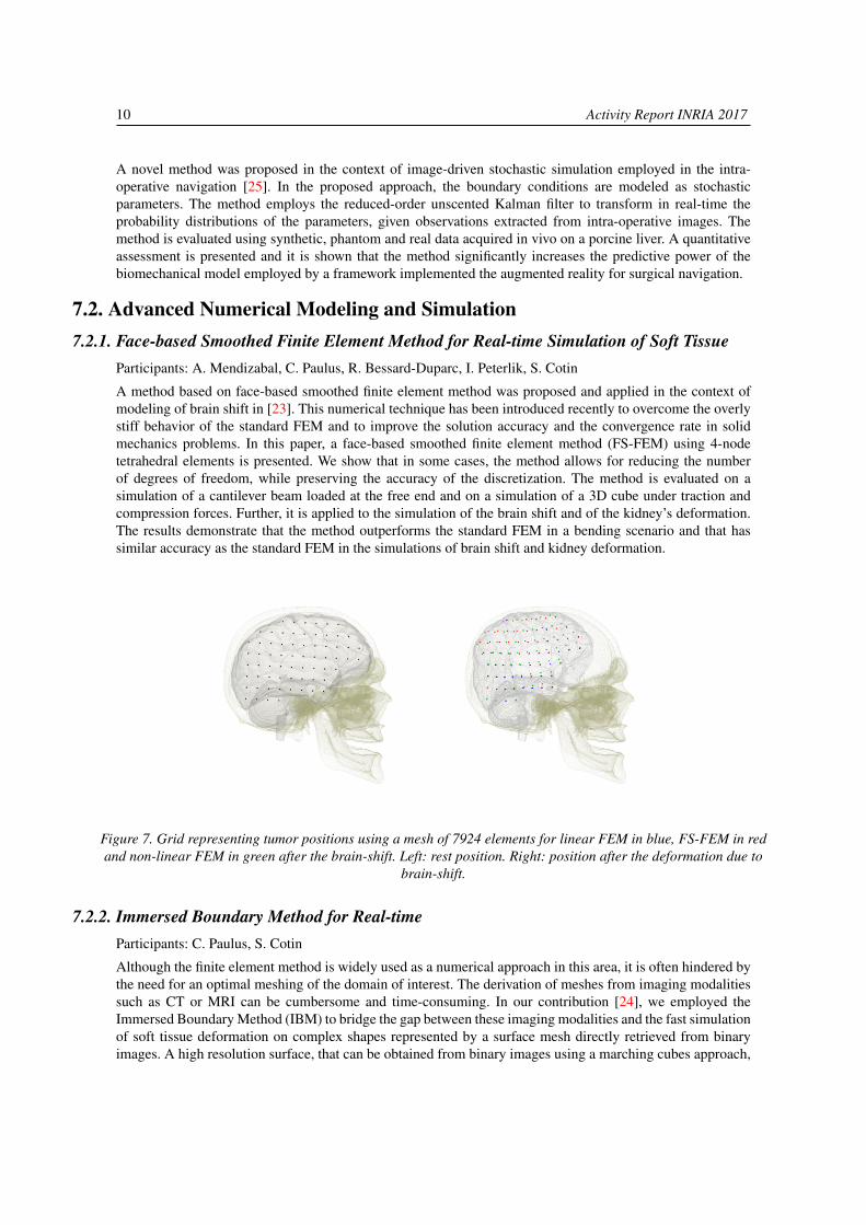
A method based on face-based smoothed finite element method was proposed and applied in the context ofmodeling of brain shift in [23]. This numerical technique has been introduced recently to overcome the overlystiff behavior of the standard FEM and to improve the solution accuracy and the convergence rate in solidmechanics problems. In this paper, a face-based smoothed finite element method (FS-FEM) using 4-nodetetrahedral elements is presented. We show that in some cases, the method allows for reducing the numberof degrees of freedom, while preserving the accuracy of the discretization. The method is evaluated on asimulation of a cantilever beam loaded at the free end and on a simulation of a 3D cube under traction andcompression forces. Further, it is applied to the simulation of the brain shift and of the kidney’s deformation.The results demonstrate that the method outperforms the standard FEM in a bending scenario and that hassimilar accuracy as the standard FEM in the simulations of brain shift and kidney deformation.
Figure 7. Grid representing tumor positions using a mesh of 7924 elements for linear FEM in blue, FS-FEM in redand non-linear FEM in green after the brain-shift. Left: rest position. Right: position after the deformation due to
brain-shift.
7.2.2. Immersed Boundary Method for Real-timeParticipants: C. Paulus, S. Cotin
Although the finite element method is widely used as a numerical approach in this area, it is often hindered bythe need for an optimal meshing of the domain of interest. The derivation of meshes from imaging modalitiessuch as CT or MRI can be cumbersome and time-consuming. In our contribution [24], we employed theImmersed Boundary Method (IBM) to bridge the gap between these imaging modalities and the fast simulationof soft tissue deformation on complex shapes represented by a surface mesh directly retrieved from binaryimages. A high resolution surface, that can be obtained from binary images using a marching cubes approach,

Team MIMESIS 11
is embedded into a hexahedral simulation grid. The details of the surface mesh are properly taken into accountin the hexahedral mesh by adapting the Mirtich integration method. In addition to not requiring a dedicatedmeshing approach, our method results in higher accuracy for less degrees of freedom when compared to otherelement types. Examples on brain deformation demonstrate the potential of our method.
Figure 8. Simulation of brain shift using a detailed surface mesh embedded into an hexahedral grid. Boundaryconditions are applied onto the exact surface, not the grid (left).
7.2.3. Error Control in Surgical SimulationsParticipants: H. Courtecuisse, S. Cotin
A contribution [16] presents the first real-time a posteriori error-driven adaptive finite element approach forreal-time simulation and demonstrates the method on a needle insertion problem.
We use corotational elasticity and a frictional needle–tissue interaction model. The problem is solved usingfinite elements and the refinement strategy relies upon a hexahedron-based finite element method, combinedwith a posteriori error estimation driven local h-refinement, for simulating soft tissue deformation. Weproposed to control the local and global error level in the mechanical fields (e.g. displacement or stresses)during the simulation. We show the convergence of the algorithm on academic examples, and demonstrateits practical usability on a percutaneous procedure involving needle insertion in a liver. For the latter case, wecompare the force displacement curves obtained from the proposed adaptive algorithm with that obtained froma uniform refinement approach. Error control guarantees that a tolerable error level is not exceeded during thesimulations. Local mesh refinement accelerates simulations. The work provides a first step to discriminatebetween discretization error and modeling error by providing a robust quantification of discretization errorduring simulations.
7.3. Model-based Image Registration7.3.1. Intraoperative Biomechanical Registration of the Liver
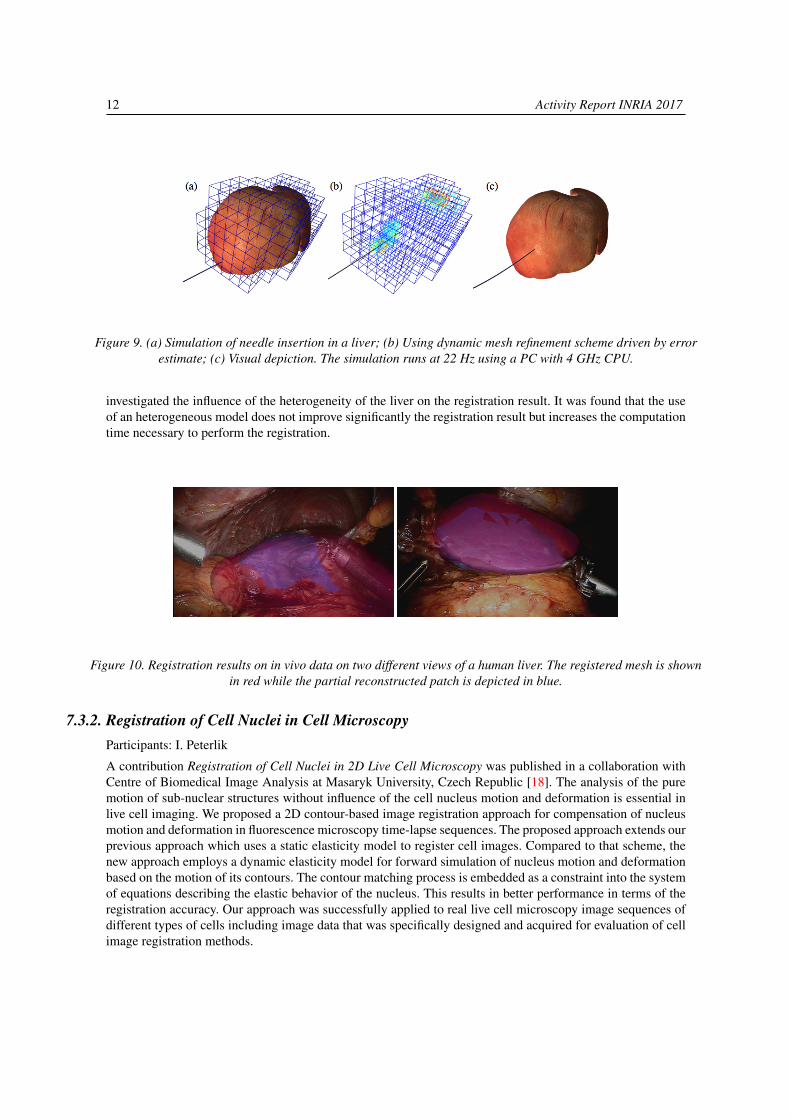
Participants: R. Plantefève, I. Peterlik, S. Cotin
Different aspects of model-based registration in the context of surgical navigation employing the augmentedreality were analyzed in an invited contribution [17] published in the context of the attributed Prix de thèsede former Ph.D. student Rosalie Plantefève. Preoperative images such as computed tomography scans ormagnetic resonance imaging contain lots of valuable information that are not easily available for surgeonsduring an operation. To help the clinicians better target the structures of interest during an intervention,many registration methods that align preoperative images onto the intra-operative view of the organs havebeen developed. For important organ deformation, biomechanical model-based registration has proven to bea method of choice. Using an existing model-based registration algorithm for laparoscopic liver surgery we
12 Activity Report INRIA 2017
Figure 9. (a) Simulation of needle insertion in a liver; (b) Using dynamic mesh refinement scheme driven by errorestimate; (c) Visual depiction. The simulation runs at 22 Hz using a PC with 4 GHz CPU.
investigated the influence of the heterogeneity of the liver on the registration result. It was found that the useof an heterogeneous model does not improve significantly the registration result but increases the computationtime necessary to perform the registration.
Figure 10. Registration results on in vivo data on two different views of a human liver. The registered mesh is shownin red while the partial reconstructed patch is depicted in blue.
7.3.2. Registration of Cell Nuclei in Cell MicroscopyParticipants: I. Peterlik
A contribution Registration of Cell Nuclei in 2D Live Cell Microscopy was published in a collaboration withCentre of Biomedical Image Analysis at Masaryk University, Czech Republic [18]. The analysis of the puremotion of sub-nuclear structures without influence of the cell nucleus motion and deformation is essential inlive cell imaging. We proposed a 2D contour-based image registration approach for compensation of nucleusmotion and deformation in fluorescence microscopy time-lapse sequences. The proposed approach extends ourprevious approach which uses a static elasticity model to register cell images. Compared to that scheme, thenew approach employs a dynamic elasticity model for forward simulation of nucleus motion and deformationbased on the motion of its contours. The contour matching process is embedded as a constraint into the systemof equations describing the elastic behavior of the nucleus. This results in better performance in terms of theregistration accuracy. Our approach was successfully applied to real live cell microscopy image sequences ofdifferent types of cells including image data that was specifically designed and acquired for evaluation of cellimage registration methods.

Team MIMESIS 13
Figure 11. Tracks of line features overlaid with the first image of the sequence. The tracks represent the motion ofthe points of the line features sampled with 30 pixel interval for better visibility. The tracks are shown for (a)unregistered data, (b) after registration with the contour-based approach [19], (c) after registration with the
intensity-based approach [9], (d) after registration with the static version of our approach, and (e) afterregistration with the proposed dynamic approach. White arrows indicate tracks with the most visible difference
between (d) and (e).
7.4. Reconstruction of Geometries from Images7.4.1. Automatic Skeletonization of Vascular Trees in Pre-operative CT Images
Participants: R. Plantefève, I. Peterlik
An algorithm of an automatic skeletonization of vascularization based on Dijkstra minimum-cost spanningtree was published in [27]. The result is an extension of an existing graph-based method where the vasculartopology is constructed by computation of shortest paths in a minimum-cost spanning tree obtained frombinary mask of the vascularization. We suppose that the binary mask is extracted from a 3D CT imageusing automatic segmentation and thus suffers from important artifacts and noise. When compared to theoriginal algorithm, the proposed method (i) employs a new weighting measure which results in smoothing ofextracted topology and (ii) introduces a set of tests based on various geometric criteria which are executedin order to detect and remove spurious branches. The method is evaluated on vascular trees extracted fromabdominal contrast-enhanced CT scans and MR images. The method is quantitatively compared to the originalversion of the algorithm showing the importance of proposed modifications. Since the branch testing dependson parameters, the para-metric study of the proposed method is presented in order to identify the optimalparametrization.
7.4.2. Template-based Recovery of Elastic Shapes from Monocular VideoParticipants: N. Haouchine, S. Cotin
A method of template-based 3D recovery of elastic shapes using Lagrange multiplied was presented at a topcomputed-vision conference [21]. By exploiting the object’s elasticity, in contrast to isometric methods thatuse inextensibility constraints, a large range of deformations can be handled. Our method is expressed as asaddle point problem using Lagrangian multipliers resulting in a linear system which unifies both mechanicaland optical constraints and integrates Dirichlet boundary conditions, whether they are fixed or free. Weexperimentally show that no prior knowledge on material properties is needed, which exhibit the genericusability of our method with elastic and inelastic objects with different kinds of materials. Comparison withexisting techniques are conducted on synthetic and real elastic objects with strains ranging from 25% to 130%resulting to low errors.
14 Activity Report INRIA 2017
Figure 12. The proposed method illustrated on an example with a soft ball colliding the ground in slow motion. Noprior knowledge of material properties in considered. The spherical volume model is composed of 512 linear P1
tetrahedral elements. The recovery and augmentation is performed in real-time at 25 FPS.
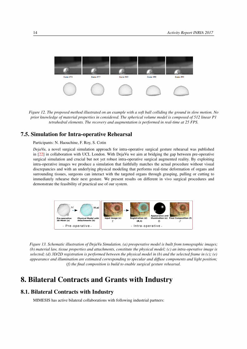
7.5. Simulation for Intra-operative RehearsalParticipants: N. Haouchine, F. Roy, S. Cotin
DejaVu, a novel surgical simulation approach for intra-operative surgical gesture rehearsal was publishedin [22] in collaboration with UCL London. With DejaVu we aim at bridging the gap between pre-operativesurgical simulation and crucial but not yet robust intra-operative surgical augmented reality. By exploitingintra-operative images we produce a simulation that faithfully matches the actual procedure without visualdiscrepancies and with an underlying physical modeling that performs real-time deformation of organs andsurrounding tissues, surgeons can interact with the targeted organs through grasping, pulling or cutting toimmediately rehearse their next gesture. We present results on different in vivo surgical procedures anddemonstrate the feasibility of practical use of our system.
Figure 13. Schematic illustration of DejaVu Simulation. (a) preoperative model is built from tomographic images;(b) material law, tissue properties and attachments, constitute the physical model; (c) an intra-operative image isselected; (d) 3D/2D registration is performed between the physical model in (b) and the selected frame in (c); (e)appearance and illumination are estimated corresponding to specular and diffuse components and light position;
(f) the final composition is build to enable surgical gesture rehearsal.
8. Bilateral Contracts and Grants with Industry8.1. Bilateral Contracts with Industry
MIMESIS has active bilateral collaborations with following industrial partners:
Team MIMESIS 15
InSimo: A startup providing biomedical simulation software which are able to reproduce thebehavior of organs, tissues and surgical procedures in a realistic and interactive way. Createdin January 2013 as a spin-off forces by former members of team SHACRA (the predecessor ofMIMESIS). Currently, we collaborate on simulations of eye surgery as well as on preparation ofprojects aiming at validation of algorithms and codes of simulation framework SOFA.
Altran: A global leader in innovation and high-tech engineering consulting, Altran accompaniessupports its clients in the creation and development of their new products and services. We have acommon history of successful collaboration via CIFRE Ph.D. thesis of Rosalie Plantefève. A newCIFRE Ph.D. will start on 01/01/2018 focusing on fusion of multisensor data in the context of intra-operative navigation of catheters.
Siemens: A global leader in healthcare industry. Via IHU, we collaborate with Siemens in thecontext of the IHU project CIOS Alpha Fusion dealing with augmentation of the intra-operativeimage provided by a fluoroscopic imaging modality with pre-operative data.
Renumics: A German startup focusing on automation of computer aided engineering (CAE) usingartificial intelligence in general and machine learning techniques in particular. In close collaborationwith SOFA Consortium, MIMESIS is involved in preparation of projects aiming at validation ofSOFA.
Naviworks: A South Korean company specialized in ICT convergence simulation/IoT smart con-trolling. We collaborate on simulation and visualization in the context of interventional radiology.
9. Partnerships and Cooperations
9.1. Regional Initiatives9.1.1. Institute of Image-Guided Surgery (IHU) Strasbourg
The Institute of Image-Guided Surgery of Strasbourg develops innovative surgery to deliver personalizedpatient care, combining the most advanced minimally invasive techniques and the latest medical imagingmethods.
Project CIOS Alpha Fusion funded by IHU Strasbourg has started at the beginning of 2017. The goal of theproject is to develop a solution for real-time, accurate, image fusion between 3D anatomical data and 2D X-rayimages. This requires to spatially align these two imaging datasets with each other, knowing that a deformationhas occurred between the 2 acquisitions. We consider two different cases, of increasing scientific complexity:static image fusion using 2 fluoroscopic images taken at 2 different angles, and dynamic image fusion usinga single fluoroscopic image. We also consider two additional scenarios: in the first one, a 3D image or a 3Dmodel has been obtained from a preoperative CTA or MRA while in the second scenario it has been acquiredusing an intra-operative contrast-enhanced CBCT. In the second case, tissue deformation between the 2D and3D data is significantly reduced.
The project team involves scientists from the MIMESIS team at Inria, engineers from Siemens as industrialpartner, and clinicians from the NHC hospital and IHU.
9.1.2. Research and Clinical PartnersAt the regional level, the MIMESIS team collaborates with
ICube Automatique Vision et Robotique (AVR): We have been collaborating with the medi-cal robotics team on percutaneous procedures, in particular robotized needle insertion (with Prof.Bernard Bayle), and needle tracking in medical images (with Elodie Breton). We are also collabo-rating with Jonathan Vappou on elastography.
16 Activity Report INRIA 2017
ICube Informatique Géométrique et Graphqiue (IGG): MIMESIS joined the IGG team anddevelops collaboration in the domain of dynamic topologies, mainly through the use of the CGoGNframework. CGoGN is a C++ library for the manipulation of meshes. It implements combinatorialmaps and their multiresolution extensions and has been used in various high level application likethe simulation of crowds of autonomous agents and the simulation of cuts, tears and fractures in thecontext of surgical simulations.Nouvel Hôpital Civil, Strasbourg: since 2014 we have been working with Prof. David Gaucher, anophthalmologist and expert in retina surgery. This led to the submission of the ANR project RESETwhich started in March 2015. We also collaborate with Prof. Patrick Pessaux, a surgeon who helpsus in the context of the SOFA-OR project.
9.2. National Initiatives9.2.1. ADT (Action de Développement Technologique)
Team MIMESIS received a support for the development of the SOFA framework through two ADTs:DynMesh (Sep 2015 – Aug 2017): The objectives of the ADT was the coupling of SOFA,the physical simulation platform supported by Inria, and CGoGN, the mesh management librarydeveloped within the ICube lab at Strasbourg. The goal is to extend the physical engine SOFA withthe topological kernel of CGoGN that supports a wide variety of mesh and many local remeshingoperations. The coupling of both software libraries will provide users of physical engines withnew tools for the development of simulations involving topological changes like cutting, fracturing,adaptation of the resolution or improving contact management or collision detection. The impactsare numerous and will be operated directly within the MIMESIS Team, with our partners or throughthe establishment of new collaborations.ASNAP (Accélération des Simulations Numériques pour l’Assistance Peropératoire, Jan 2017 – Dec2018). We are partners of ADT ASNAP with principal investigator being Inria team CAMUS. Thegoal of the project is a significant acceleration of physics-based simulations developed by MIMESIS.The technologies such as Apollo, XFOR, ORWL, developed by team CAMUS are used to optimizethe execution of different components of framework SOFA, taking into account the possibilitiesprovided by modern CPUs and GPGPUs. Since team CAMUS is also located in Strasbourg, theproject benefits from the geographical location: an engineer Maxim Mogé was recruited, startingfrom 01/01/2017 and he shares his time between the two teams.
9.2.2. ANR (Agence Nationale de la Recherche)MIMESIS participates in the following ANR projects:
RESET: This project started in March 2015 and will end in May 2017. Its objective is to developa high-fidelity training system for retinal surgery. Retinal surgery is an increasingly performedprocedure for the treatment of a wide spectrum of retinal pathologies. Yet, as most micro-surgicaltechniques, it requires long training periods before being mastered. This simulator is built uponour scientific expertise in the field of real-time simulation, and our success story for technologytransfer in the field of cataract surgery simulation (MSICS simulation developed for the HelpMeSeefoundation).Coordinator: MIMESISPartners: the InSimo company, the AVR team of the ICube lab.EVEREST: The overall objective of the EVEREST project is thus to bring a leap forward in factor-ization of large sparse tensors in order to improve the accessibility, completeness and reliability ofreal-world KBs. This line of research could have a huge impact in industry (Semantic Web, biomed-ical applications, etc.). For that reason, Xerox Research Center Europe is supporting this project andwill supply data, provide expertise and ease industrial transfer. This proposal is also consistent withthe long-term research direction of its principal partner, Heudiasyc, since it contributes in severalaspects of the 10 years LabEx program on Technological Systems of Systems started in 2011.

Team MIMESIS 17
Coordinator: IHU Strasbourg
Partners: Inria, IRCAD, University of Strasbourg, Siemens Healthcare, Karl Storz GmbH., Univer-sity of Twente
9.2.3. Inria CollaborationsMIMESIS is closely connected to the SOFA Consortium, created by Inria in November 2015 with the objectiveto support the SOFA community and encourage contributions from new SOFA users. The Consortium shouldalso be a way to better answer to the needs of academic or industrial partners. MIMESIS actively participatesat the development of SOFA and contributed to the evolution of the framework. Moreover, MIMESIS alsoparticipates in an initiative aiming at verification and validation of codes and algorithms of SOFA.
Further, MIMESIS actively collaborates with the following Inria teams:
MAGRIT: The team at Inria Grand Est focuses on research in computer vision and is also activelyinvolved in computer-based solutions for the planning or the simulation of interventional radiologyprocedures, with a strong collaboration with the CHU in Nancy. We collaborate with MAGRIT in thearea of interventional radiology and augmented reality. Currently, two PhD thesis are co-supervisedby researcher from Magrit: the PhD thesis of Jaime Garcia Guevara and Raffaella Trivisonne.
CAMUS: The team focuses on developing, adapting and extending automatic parallelizing andoptimizing techniques, as well as proof and certification methods, for the efficient use of currentand future multi-core processors. Currently, we collaborate with team CAMUS on parallelization offramework SOFA in ADT project ASNAP.
DEFROST: The team conducts research in soft robotics. We continue mutual interaction withDEFROST mainly in the context of contact modeling.
9.2.4. National CollaborationsAt the national level, the MIMESIS team collaborates with:
The TIMC laboratory(Techniques de l’Ingénierie Médicale et de la Complexité) in Grenoble:this large research group has a strong background in computer-aided surgery, medical imaging,registration, statistical and bio-mechanical modeling. We have regular interactions with variousmembers of this group. We are collaborating with Yohan Payan (DR CNRS) on the modeling andsimulation of the brain shift. A common PhD thesis started on that topic in late 2014. Other areas ofinterest are in the field of advanced soft tissue modeling and computer aided surgery.
The LML laboratory(Laboratoire de Mécanique de Lille): a French research laboratory (UMRCNRS 8107) part of the Carnot institute ARTS. With more than two hundred researchers, LMLfocuses on the following research areas: mechanical reliability and Tribology, fluid mechanics, civilengineering and soil mechanics.
Hôpital Paul-Brousse: a hospital in South Paris. We collaborate with Centre Hépato-Biliaire viathe co-supervision of the Ph.D. thesis of Nicolas Golse, MD, who is a surgeon at the center.
9.3. European Initiatives9.3.1. FP7 & H2020 Projects
Program: H2020, Innovative Training Network, MSCA
Project acronym: HiPerNav
Project title: High performance soft tissue navigation
Coordinator: Oslo University Hospital
Other partners: SINTEF Trondheim, University of Bern
18 Activity Report INRIA 2017
Abstract: HiPerNav is an Innovative Training Network (ITN) funded through a Marie Skłodowska-Curie grant. There will be 14 fully funded and 2 partially funded PhD’s working on the project. Theproject aims to improve soft tissue navigation through research and development, to improve severalbottleneck areas:
• Creating effective pre-operative model(s) and planning
• Faster and more accurate intra-operative model updates
• Faster and more accurate model-to-patient registration
• More intuitive user-interaction and effective work flow
• Usage of high performance computing (e.g. GPU)
9.3.2. Informal CollaborationsUniversity of Twente: Thanks to our clinical partner IHU, we collaborate with Prof. StefanoStramigioli, head of a group at Robotics and Mechatronics laboratory.
Faculty of Informatics, Masaryk University, Czech Republic: We collaborate on simulation ofliving cells in fluorescent microscopy. The collaboration resulted in a presentation at an internationalconference [29] and a journal paper [18].
Team Legato, University of Luxembourg: we have an active collaboration with Prof. StéphaneBordas on error estimation in real-time simulations of deformable objects. The collaboration resultedin a common publication [16].
9.4. International InitiativesThe MIMESIS team actively collaborates with following international partners:
CIMIT & Harvard Medical School, Boston, USA: We collaborate on a project REBOASim inthe contect of interventional radiology, , in particular the design and development of a hardwareinterface for tracking catheters and guidewires. The common DoD project REBOASim focuseson development of the physics-based models for catheter and guidewire motion, blood flow andgraphical rendering towards a novel simulator for REBOA that will include physical vascular access,simulated passage of the IR instruments into the aorta with accompanying training/educationalcontent, device withdrawal and closure: Duration of the project: Feb 2017 – Feb 2019.
9.5. International Research Visitors9.5.1. Visits of International Scientists
From Feb 2017 to July 2017, Prof. Adam Wittek joined team MIMESIS as a visiting scientist. Prof. Wittekis with Intelligent Systems for Medicine Laboratory, School of Mechanical and Chemical Engineering at theUniversity of Western Australia, Perth. His research focuses on patient-specific biomechanical modeling andhe has published an important number of high-quality publications on this topic with more than 2,000 citations.
During his stay, Prof. Wittek provided his highly valuable expertise in various domains of patient-specificsimulations and advanced techniques of modeling of deformations in soft tissues such as meshless methods.He was also involved in projects related to insertions of flexible needles into soft tissues.
9.5.1.1. Internships
From Jul 2017 to Dec 2017, Vincent Magnoux, a Canadian PhD student from École polytechnique deMontréal, joined MIMESIS as an international intern. During his stay, he has worked on implementing andvalidating a meshless method for computing organ deformation. This work also involved exploring methodsto accelerate these computations on multi-core systems for an interactive simulation.
Team MIMESIS 19
10. Dissemination
10.1. Promoting Scientific Activities10.1.1. Scientific Events Organisation10.1.1.1. Member of the Organizing Committees
David Cazier contributed to the organization of the Annual workshop of the Animation & Simulation group ofthe GDR IGRV of the CNRS in Strasbourg.
10.1.1.2. Reviewing ActivitiesStephane Cotin provided reviews for: Int. Conf. on Information Processing in Computer-AssistedInterventions, Workshop on Virtual Reality Interaction and Physical SimulationIgor Peterlik provided reviews for: Int. Conf. of Medical Image Computing and Computer AssistedIntervention SocietyAntoine Petit provided reviews for: International Conference on Robotics and Automation
10.1.2. Journal10.1.2.1. Reviewing Activities
Stephane Cotin provided reviews for: International Journal of Computer Assisted Radiology andSurgeryDavid Cazier provided reviews for: Computer-Aided Design, Visual Computer, Computer & Graph-ics, Int. Journal on Virtual RealityIgor Peterlik provided reviews for: Computer and GraphicsAntoine Petit provided reviews for: International Journal Of Robotics Research, Robotics andAutomation LettersChristoph Paulus provided reviews for: MDPI Journal SymmetryLionel Untereiner provided reviews for: Special Issue on Parallel and Distributed Algorithms ofConcurrency and Computation
10.1.3. Invited TalksKeynote lecture by S. Cotin at 10th Medical Korea conference (Seoul, South Korea)Invited lecture by S. Cotin FMTS conference (Strasbourg, France)Invited lecture by S. Cotin B.E.S.T. symposium (Strasbourg, France)Invited talk by S. Cotin at Fraunhofer MEVIS lab (Bremen, Germany)Invited lecture by S. Cotin at European Computer-Assisted Liver Surgery Society (Mainz, Germany)Invited lecture by S. Cotin at 127th annual meeting of the French Ophthalmology Association (Paris,France)Invited lecture by S. Cotin at the French Academy of Surgery (Paris, France)
10.1.4. Scientific ExpertiseIgor Peterlik has been providing a scientific expertise at Masaryk University, Czech Republicas a consultant and co-investigator of a project unded by Grant Agency of the Czech Republic:Development of Reliable Methods for Automated Quantitative Characterization of Cell Motility inFluorescence Microscopy.
10.1.5. Research AdministrationDavid Cazier is a member and local coordinator for a CITEPH project Paleo GTM: A Paleo Geologicaland Topological Modeler. Subject and expected contributions: multiresolution meshing and visualization forhandling of massive geological data.

20 Activity Report INRIA 2017
The project started in Sep 2017 (duration 2 years) and involves following partners:• GEOSIRIS SAS (StartUp)• Laboratoire ICUBE, UMR 7357 (Université de Strasbourg)• Laboratoire XLIM, UMR 7352 (Université de Poitiers)• Laboratoire LSIS, UMR 7296 (Université d’Aix-Marseille)
10.2. Teaching - Supervision - Juries10.2.1. Teaching
Master: Igor Peterlik, Modélisation des systèmes vivants, 17h, M2, University of StrasbourgMaster: Igor Peterlik, Visualisation des données et simulation, 10h, M1, University of StrasbourgMaster: Hadrien Courtecuisse, Real time simulation, 30h, M2, University of StrasbourgMaster: Hadrien Courtecuisse, Visualisation des données et simulation, 10h, M2, University ofStrasbourgMaster: Hadrien Courtecuisse, Visualisation des données et simulation, 10h, M1, University ofStrasbourgLicence: David Cazier, Web technologies and programming, 96h, L3, University of Strasbourg
10.2.2. SupervisionPhD: Christoph Paulus, Modeling and real-time simulation of topological changes in soft tissue,University of Strasbourg, 03/04/2017 [11]PhD: Fanny Morin, Non linear simulation for intra-operative guidance for neurosurgery, UniversitéGrenoble Alpes, 05/10/2017PhD in progress: Jaime Garcia Guevara, Augmented ultrasound imaging for hepatic surgery,01/09/2015, supervised by Stéphane Cotin, Marie-Odile BergerPhD in progress: Raffaella Trivisonne, Computer-aided vascular interventions, 01/09/2015,Stéphane Cotin, Erwan KerrienPhD in progress: Yinoussa Adagolodjo, Coupling between robotics and medical simulation forautomated procedures, 01/02/2015, supervised by Hadrien CourtecuissePhD in progress: Nicolas Golse, Navigation using the augmented reality during hepatic surgery,01/09/2016, supervised by Stéphane CotinPhD in progress: Lukáš Rucka, Validation and verification of soft tissue models, 01/09/2016, co-supervised by Igor Peterlik, supervised by Prof. Ludek Matyska at Masaryk University, CzechRepublicPhD in progress: Sergei Nikolaev, Characterization of boundary conditions for biomechanicalmodeling of liver, 01/05/2017, supervised by Stéphane Cotin, co-supervised by Igor Peterlik andHadrien CourtecuissePhD in progress: Jean-Nicolas Brunet, Characterization of boundary conditions for biomechanicalmodeling of liver, 01/09/2017, supervised by Stéphane CotinPhD in progress: Andrea Mendizabal, Numerical simulation of soft tissues and machine learning,01/09/2017, supervised by Stéphane Cotin
10.2.3. JuriesStéphane Cotin was a member of jury of HDR of Christian Herlin (MD): Imagerie et simulationpour la chirurgie plastique et reconstructrice. Université de Montpellier. Nov 2017
10.3. Popularization
Team MIMESIS 21
Demonstration at 50 Years Inria on Nov 10 in Paris, attended by Mr. Mounir Mahjoubi, secrétaired’état auprès du premier ministre, chargé du numérique.
11. BibliographyMajor publications by the team in recent years
[1] H. COURTECUISSE, J. ALLARD, K. PIERRE, S. P.-A. BORDAS, S. COTIN, C. DURIEZ. Real-time simulationof contact and cutting of heterogeneous soft-tissues, in "Medical Image Analysis", February 2014, 20 p. ,https://hal.inria.fr/hal-01097108
[2] F. FAURE, C. DURIEZ, H. DELINGETTE, J. ALLARD, B. GILLES, S. MARCHESSEAU, H. TALBOT, H.COURTECUISSE, G. BOUSQUET, I. PETERLIK, S. COTIN. SOFA: A Multi-Model Framework for InteractivePhysical Simulation, in "Soft Tissue Biomechanical Modeling for Computer Assisted Surgery", Y. PAYAN(editor), Studies in Mechanobiology, Tissue Engineering and Biomaterials, Springer, June 2012, vol. 11, pp.283-321 [DOI : 10.1007/8415_2012_125], https://hal.inria.fr/hal-00681539
[3] N. HAOUCHINE, S. COTIN, I. PETERLIK, J. DEQUIDT, M. SANZ-LOPEZ, E. KERRIEN, M.-O. BERGER.Impact of Soft Tissue Heterogeneity on Augmented Reality for Liver Surgery, in "IEEE Transactions on Visual-ization and Computer Graphics", 2015, vol. 21, no 5, pp. 584 - 597 [DOI : 10.1109/TVCG.2014.2377772],https://hal.inria.fr/hal-01136728
[4] N. HAOUCHINE, J. DEQUIDT, M.-O. BERGER, S. COTIN. Monocular 3D Reconstruction and Augmentationof Elastic Surfaces with Self-occlusion Handling, in "IEEE Transactions on Visualization and ComputerGraphics", 2015, 14 p. [DOI : 10.1109/TVCG.2015.2452905], https://hal.inria.fr/hal-01186011
[5] N. HAOUCHINE, J. DEQUIDT, I. PETERLIK, E. KERRIEN, M.-O. BERGER, S. COTIN. Image-guidedSimulation of Heterogeneous Tissue Deformation For Augmented Reality during Hepatic Surgery, in "ISMAR- IEEE International Symposium on Mixed and Augmented Reality 2013", Adelaide, Australia, October 2013,https://hal.inria.fr/hal-00842855
[6] I. PETERLÍK, C. DURIEZ, S. COTIN. Modeling and Real-Time Simulation of a Vascularized Liver Tissue, in"Medical Image Computing and Computer-Assisted Intervention–MICCAI 2012", 2012, pp. 50–57, https://hal.archives-ouvertes.fr/hal-00800546
[7] H. TALBOT, N. HAOUCHINE, I. PETERLIK, J. DEQUIDT, C. DURIEZ, H. DELINGETTE, S. COTIN. SurgeryTraining, Planning and Guidance Using the SOFA Framework, in "Eurographics", Zurich, Switzerland, May2015, https://hal.inria.fr/hal-01160297
[8] H. TALBOT, F. ROY, S. COTIN. Augmented Reality for Cryoablation Procedures, in "SIGGRAPH 2015", LosAngeles, United States, August 2015, https://hal.inria.fr/hal-01180848
[9] H. TALBOT, F. SPADONI, C. DURIEZ, M. SERMESANT, M. O’NEILL, P. JAÏS, S. COTIN, H. DELINGETTE.Interactive training system for interventional electrocardiology procedures, in "Medical image analysis",2017, vol. 35, pp. 225–237
[10] L. UNTEREINER, P. KRAEMER, D. CAZIER, D. BECHMANN. CPH: a compact representation for hierarchi-cal meshes generated by primal refinement, in "Computer Graphics Forum", 2015, vol. 34, no 8, pp. 155-166,5-Year Impact Factor : 1,920 [DOI : 10.1111/CGF.12667], https://hal.archives-ouvertes.fr/hal-01162098
22 Activity Report INRIA 2017
Publications of the yearDoctoral Dissertations and Habilitation Theses
[11] C. J. PAULUS. Topological Changes in Simulations of Deformable Objects, University of Strasbourg, April2017, https://hal.inria.fr/tel-01516170
Articles in International Peer-Reviewed Journals
[12] Z. JIANG, J.-F. WITZ, P. LECOMTE-GROSBRAS, J. DEQUIDT, S. COTIN, C. RUBOD, C. DURIEZ, M.BRIEU. Multiorgan Motion Tracking in Dynamic Magnetic Resonance Imaging for Evaluation of PelvicSystem Mobility and Shear Strain, in "Strain", February 2017, vol. 53, no 2 [DOI : 10.1111/STR.12224],https://hal.inria.fr/hal-01496177
[13] E. KERRIEN, A. YUREIDINI, J. DEQUIDT, C. DURIEZ, R. ANXIONNAT, S. COTIN. Blood vessel modelingfor interactive simulation of interventional neuroradiology procedures, in "Medical Image Analysis", January2017, vol. 35, pp. 685 - 698 [DOI : 10.1016/J.MEDIA.2016.10.003], https://hal.inria.fr/hal-01390923
[14] F. MORIN, H. COURTECUISSE, I. REINERTSEN, F. LE LANN, O. PALOMBI, Y. PAYAN, M. CHABANAS.Brain-shift compensation using intraoperative ultrasound and constraint-based biomechanical simulation, in"Medical Image Analysis", 2017, vol. 40, pp. 133 - 153 [DOI : 10.1016/J.MEDIA.2017.06.003], https://hal.archives-ouvertes.fr/hal-01560157
[15] I. PETERLIK, H. COURTECUISSE, R. ROHLING, P. ABOLMAESUMI, C. NGUAN, S. COTIN, S. E. SALCUD-EAN. Fast Elastic Registration of Soft Tissues under Large Deformations, in "Medical Image Analysis", 2017,forthcoming, https://hal.inria.fr/hal-01613757
[16] H. PHUOC BUI, S. TOMAR, H. COURTECUISSE, S. COTIN, S. BORDAS. Real-time Error Control forSurgical Simulation, in "IEEE Transactions on Biomedical Engineering", June 2017, 12 p. , https://hal.inria.fr/hal-01514621
[17] R. PLANTEFÈVE, I. PETERLIK, S. COTIN. Intraoperative Biomechanical Registration of the Liver: Does theHeterogeneity of the Liver Matter?, in "IRBM", 2017, forthcoming, https://hal.inria.fr/hal-01625573
[18] D. V. SOROKIN, I. PETERLIK, M. TEKTONIDIS, K. ROHR, P. MATULA. Non-rigid Contour-Based Reg-istration of Cell Nuclei in 2D Live Cell Microscopy Images Using a Dynamic Elasticity Model, in "IEEETransactions on Medical Imaging", 2017, 1 p. , forthcoming [DOI : 10.1109/TMI.2017.2734169], https://hal.inria.fr/hal-01613732
International Conferences with Proceedings
[19] Y. ADAGOLODJO, R. TRIVISONNE, N. HAOUCHINE, S. COTIN, H. COURTECUISSE. Silhouette-based PoseEstimation for Deformable Organs Application to Surgical Augmented Reality, in "IROS 2017 - IEEE/RSJInternational Conference on Intelligent Robots and Systems", Vancouver, Canada, September 2017, https://hal.archives-ouvertes.fr/hal-01578815
[20] J. G. GARCIA GUEVARA, I. PETERLIK, M.-O. BERGER, S. COTIN. Automatic biomechanical graphmatching CT-CBCT fusion, in "Surgetica 2017", Strasbourg, France, November 2017, https://hal.archives-ouvertes.fr/hal-01587952
Team MIMESIS 23
[21] N. HAOUCHINE, S. COTIN. Template-based Monocular 3D Recovery of Elastic Shapes using LagrangianMultipliers, in "Computer Vision and Pattern Recognition (CVPR)", Honolulu, Hawai, United States, July2017, https://hal.inria.fr/hal-01524609
[22] N. HAOUCHINE, D. STOYANOV, F. ROY, S. COTIN. DejaVu: Intra-operative Simulation for Surgical GestureRehearsal, in "Medical Image Computing and Computer Assisted Interventions Conference MICCAI 2017",Quebec City, Canada, October 2017, https://hal.archives-ouvertes.fr/hal-01542395
[23] A. MENDIZABAL, R. B. DUPARC, H. PHUOC BUI, C. J. PAULUS, I. PETERLIK, S. COTIN. Face-basedSmoothed Finite Element Method for Real-time Simulation of soft tissue, in "SPIE Medical Imaging", Orlando,United States, February 2017, https://hal.inria.fr/hal-01444595
[24] C. J. PAULUS, R. MAIER, D. PETERSEIM, S. COTIN. An Immersed Boundary Method for Detail-PreservingSoft Tissue Simulation from Medical Images, in "Computational Biomechanics for Medicine", Quebec,Canada, September 2017, https://hal.inria.fr/hal-01578447
[25] I. PETERLIK, N. HAOUCHINE, L. RUCKA, S. COTIN. Image-driven Stochastic Identification of BoundaryConditions for Predictive Simulation, in "20th International Conference on Medical Image Computing andComputer Assisted Intervention", Québec, Canada, September 2017, https://hal.inria.fr/hal-01570811
[26] A. PETIT, F. FICUCIELLO, G. A. FONTANELLI, L. VILLANI, B. SICILIANO. Using Physical Mod-eling and RGB-D Registration for Contact Force Sensing on Deformable Objects, in "ICINCO 2017- 14th International Conference on Informatics in Control, Automation and Robotics", Madrid, Spain,
SCITEPRESS, SPRINGER (editors), Proceedings of the 14th International Conference on Informaticsin Control, Automation and Robotics, ScitePress, July 2017, vol. 2, no 978-989-758-263-9, pp. 24-33[DOI : 10.5220/0006415900240033], https://hal.archives-ouvertes.fr/hal-01617243
[27] R. PLANTEFÈVE, S. KADOURY, A. TANG, I. PETERLIK. Robust Automatic Graph-based Skeletonization ofHepatic Vascular Trees, in "CIIV STENT 2017", Québec, Canada, Proceedings CIIV STENT 2017, September2017, https://hal.inria.fr/hal-01576771
[28] L. RUCKA, I. PETERLÍK. Fast reconstruction of image deformation field using radial basis function, in"ISBI2017 - International Symposium on Biomedical Imaging", Melbourne, Australia, April 2017, https://hal.inria.fr/hal-01445483
[29] D. V. SOROKIN, I. PETERLIK, V. V. ULMAN, D. SVOBODA, M. MAŠKA. Model-Based Generation ofSynthetic 3D Time-Lapse Sequences of Motile Cells with Growing Filopodia, in "International Symposium onBiomedical Imaging", Melbourne, Australia, April 2017, https://hal.inria.fr/hal-01445488
[30] R. TRIVISONNE, E. KERRIEN, S. COTIN. Augmented 3D Catheter Navigation using Constrained Shape fromTemplate, in "Hamlyn Symposium", London, United Kingdom, June 2017, https://hal.inria.fr/hal-01545693
Conferences without Proceedings
[31] H. P. BUI, S. TOMAR, H. COURTECUISSE, S. COTIN, S. P. A. BORDAS. Real-time Error Control forSurgical Simulation, in "BIOMECHANICS AND COMPUTER ASSISTED SURGERY MEETS MEDICALREALITY", Lille, France, August 2017, https://hal.archives-ouvertes.fr/hal-01571194
Other Publications
24 Activity Report INRIA 2017
[32] N. HAOUCHINE, A. PETIT, F. ROY, S. COTIN. Deformed Reality: Proof of concept and preliminary results,October 2017, ISMAR 2017 - 16th IEEE International Symposium on Mixed and Augmented Reality, Poster,https://hal.inria.fr/hal-01636772
References in notes
[33] J. ALLARD, H. COURTECUISSE, F. FAURE. Implicit FEM solver on GPU for interactive deformationsimulation, in "GPU computing gems Jade Edition", 2011, pp. 281–294
[34] A. BILGER, J. DEQUIDT, C. DURIEZ, S. COTIN. Biomechanical simulation of electrode migration for deepbrain stimulation, in "Medical Image Computing and Computer-Assisted Intervention–MICCAI 2011", 2011,pp. 339–346
[35] F. CHINESTA, P. LADEVEZE, E. CUETO. A short review on model order reduction based on propergeneralized decomposition, in "Archives of Computational Methods in Engineering", 2011, vol. 18, no 4,395 p.
[36] H. COURTECUISSE, J. ALLARD, K. PIERRE, S. P.-A. BORDAS, S. COTIN, C. DURIEZ. Real-time simulationof contact and cutting of heterogeneous soft-tissues, in "Medical Image Analysis", February 2014, 20 p. ,https://hal.inria.fr/hal-01097108
[37] F. DERVAUX, I. PETERLIK, J. DEQUIDT, S. COTIN, C. DURIEZ. Haptic Rendering of Interacting DynamicDeformable Objects Simulated in Real-Time at Different Frequencies, in "IROS - IEEE/RSJ InternationalConference on Intelligent Robots and Systems", Tokyo, Japan, IEEE, November 2013, https://hal.inria.fr/hal-00842866
[38] N. HAMZÉ, A. BILGER, C. DURIEZ, S. COTIN, C. ESSERT. Anticipation of brain shift in deep brainstimulation automatic planning, in "Engineering in Medicine and Biology Society (EMBC), 2015 37th AnnualInternational Conference of the IEEE", IEEE, 2015, pp. 3635–3638
[39] N. HAOUCHINE, J. DEQUIDT, M.-O. BERGER, S. COTIN. Single View Augmentation of 3D Elastic Objects,in "International Symposium on Mixed and Augmented Reality - ISMAR", Munich, Germany, September2014, https://hal.inria.fr/hal-01056323
[40] N. HAOUCHINE, J. DEQUIDT, E. KERRIEN, M.-O. BERGER, S. COTIN. Physics-based Augmented Realityfor 3D Deformable Object, in "Eurographics Workshop on Virtual Reality Interaction and Physical Simula-tion", Darmstadt, Germany, December 2012, https://hal.inria.fr/hal-00768362
[41] S. J. JULIER, J. K. UHLMANN. A new extension of the Kalman filter to nonlinear systems, in "Int. symp.aerospace/defense sensing, simul. and controls", Orlando, FL, 1997, vol. 3, no 26, pp. 182–193
[42] P. LECOMTE-GROSBRAS, M. NASSIROU - DIALLO, J.-F. WITZ, D. MARCHAL, J. DEQUIDT, S. COTIN, M.COSSON, C. DURIEZ, M. BRIEU. Towards a better understanding of pelvic system disorders using numericalsimulation, in "Medical Image Computing and Computer Assisted Intervention", Nagoya, Japan, September2013, https://hal.inria.fr/hal-00831245
[43] P. MOIREAU, D. CHAPELLE, P. LE TALLEC. Joint state and parameter estimation for distributed mechanicalsystems, in "Computer Methods in Applied Mechanics and Engineering", 2008, vol. 197, no 6-8, pp. 659-677[DOI : 10.1016/J.CMA.2007.08.021], https://hal.archives-ouvertes.fr/hal-00175623
Team MIMESIS 25
[44] P. MOIREAU, D. CHAPELLE. Reduced-order Unscented Kalman Filtering with application to parameter iden-tification in large-dimensional systems, in "ESAIM: Control, Optimisation and Calculus of Variations", 2011,vol. 17, no 2, pp. 380-405, See also erratum DOI:10.1051/cocv/2011001 [DOI : 10.1051/COCV/2010006],https://hal.inria.fr/inria-00550104
[45] P. MOIREAU, D. CHAPELLE, M. YVINEC. Cardiac motion extraction from images by filtering estimationbased on a biomechanical model, in "FIMH’09 - 5th International Conference Functional Imaging andModeling of the Heart", Nice, France, N. AYACHE, H. DELINGETTE, M. SERMESANT (editors), LectureNotes in Computer Science, Springer, June 2009, vol. 5528, pp. 220-228 [DOI : 10.1007/978-3-642-01932-6_24], https://hal.inria.fr/hal-00772059
[46] C. J. PAULUS, N. HAOUCHINE, D. CAZIER, S. COTIN. Augmented Reality during Cutting and Tearingof Deformable Objects, in "The 14th IEEE International Symposium on Mixed and Augmented Reality",Fukuoka, Japan, September 2015, 6 p. , https://hal.inria.fr/hal-01184495
[47] C. J. PAULUS, N. HAOUCHINE, D. CAZIER, S. COTIN. Surgical Augmented Reality with TopologicalChanges, in "Medical Image Computing and Computer Assisted Interventions", München, Germany, October2015, https://hal.inria.fr/hal-01184498
[48] I. PETERLIK, H. COURTECUISSE, C. DURIEZ, S. COTIN. Model-Based Identification of AnatomicalBoundary Conditions in Living Tissues, in "Information Processing in Computer Assisted Interventions",Fukuoka, Japan, June 2014 [DOI : 10.1007/978-3-319-07521-1_21], https://hal.inria.fr/hal-01264434
[49] I. PETERLIK, A. KLIMA. Towards an efficient data assimilation in physically-based medical simulations,in "Bioinformatics and Biomedicine (BIBM), 2015 IEEE International Conference on", IEEE, 2015, pp.1412–1419
[50] I. PETERLIK, M. NOUICER, C. DURIEZ, S. COTIN, A. KHEDDAR. Constraint-based haptic rendering ofmultirate compliant mechanisms, in "IEEE Transactions on Haptics (ToH)", June 2011, vol. 4, no 3, pp. 175-187 [DOI : 10.1109/TOH.2011.41], https://hal-lirmm.ccsd.cnrs.fr/lirmm-00784081
[51] T. PITIOT, D. CAZIER, T. JUND, A. HABIBI, P. KRAEMER. Deformable polygonal agents in crowdsimulation, in "Computer Animation and Virtual Worlds", 2014, vol. 25, no 3-4, pp. 341–350
[52] R. PLANTEFÈVE, I. PETERLIK, N. HAOUCHINE, S. COTIN. Patient-specific Biomechanical Modeling forGuidance during Minimally-invasive Hepatic Surgery, in "Annals of Biomedical Engineering", August 2015,https://hal.inria.fr/hal-01205194
[53] H. TALBOT, S. MARCHESSEAU, C. DURIEZ, M. SERMESANT, S. COTIN, H. DELINGETTE. Towards aninteractive electromechanical model of the heart, in "Interface focus", 2013, vol. 3, no 2, 20120091 p.
[54] H. TALBOT, F. ROY, S. COTIN. Augmented Reality for Cryoablation Procedures, in "SIGGRAPH 2015", LosAngeles, United States, August 2015, https://hal.inria.fr/hal-01180848
[55] L. UNTEREINER, D. CAZIER, D. BECHMANN. n-Dimensional multiresolution representation of subdivisionmeshes with arbitrary topology, in "Graphical Models", 2013, vol. 75, no 5, pp. 231–246

26 Activity Report INRIA 2017
[56] G. ZAVARISE, P. WRIGGERS. Trends in Computational Contact Mechanics, Lecture Notes inApplied and Computational Mechanics, Springer Berlin Heidelberg, 2011, https://books.google.fr/books?id=faSoa3k6NCYC